Embed Size (px)
Citation preview
Ultrasound for Pregnancy Page 1 of 23
http://qawww.aetna.com/cpb/medical/data/100_199/0199_draft.html 05/06/2015
Clinical Policy Bulletin: Ultrasound for Pregnancy
Revised February 2015
Number: 0199
Policy
I. Aetna considers ultrasounds not medically necessary if done solely to
determine the fetal sex or to provide parents with a view and photograph of
the fetus
II. Aetna considers a fetal ultrasound with detailed anatomic examination
medically necessary for the following indications:
A. To evaluate the fetus for amniotic band syndrome (also known as
amniotic constriction band syndrome); or
B. To evaluate fetuses with soft sonographic markers of aneuploidy:
1. Absent or hypoplastic nasal bone; or
2. Choroid plexus cyst; or
3. Echogenic bowel; or
4. Echogenic intracardiac focus; or
5. Fetal pyelectasis; or
6. Increased nuchal translucency (fetal nuchal translucency
measurement of 3.5 mm or greater in the first trimester); or
7. Shortened long bones (femur or humerus); or
C. If there are known or suspected fetal anatomic abnormalities,
including:
1. Anatomic abnormalities due to genetic conditions (see
attached ICD-9 coding); or
Ultrasound for Pregnancy Page 2 of 23
http://qawww.aetna.com/cpb/medical/data/100_199/0199_draft.html 05/06/2015
2. Pregnancies resulting from advanced reproductive
technology (ART)*; or
3. Severe obesity (body mass index [BMI] of 35 or
more) complicating pregnancy.
III. More than 1 detailed ultrasound fetal anatomic examination per pregnancy
per practice is considered experimental and investigational, as there is
inadequate evidence of the clinical utility of multiple serial detailed fetal
anatomic ultrasound examinations during pregnancy.
IV. Aetna considers detailed ultrasound fetal anatomic examination
experimental and investigational for all other indications including routine
evaluation of pregnant women who are on bupropion (Wellbutrin) or
levetiracetam (Keppra), pregnant women with low pregnancy-associated
plasma protein A, and pregnant women who smoke or abuse cannabis.
There is inadequate evidence of the clinical utility of detailed ultrasound
fetal anatomic examination for indications other than evaluation of
suspected fetal anatomic abnormalities. Detailed ultrasound fetal anatomic
examination is not considered medically necessary for routine screening of
normal pregnancy, or in the setting of maternal idiopathic pulmonary
hemosiderosis.
V. Aetna considers three-dimensional (3D) and four-dimensional (4D) fetal
ultrasounds experimental and investigational because of a lack of evidence
that 3D and 4D ultrasounds alter management over standard two-
dimensional (2D) ultrasounds such that clinical outcomes are improved.
* Assisted Reproductive Technology (ART) is a form of complex infertility treatment
where the egg and sperm are fertilized outside the body and the resulting embryo
is transferred back into the uterus. The most well-recognized forms of ART include
in-vitro fertilization (IVF), frozen embryo transfers (FET), and intra-cytoplasmic
sperm injection (ICSI).
For Aetna’s policy on first trimester ultrasonographic assessment of fetal nuchal
skinfold thickness, see CPB 0282 - Noninvasive Down Syndrome Screening.
See also: CPB 0106 - Fetal Echocardiograms.
Background
This policy is based in part on The American College of Obstetricians and
Gynecologists (ACOG) Practice Bulletin on Ultrasonography in Pregnancy and
guidelines from the Society for Maternal-Fetal Medicine (SMFM).
Ultrasonography in pregnancy should be performed only when there is a valid
medical indication. ACOG (2009) stated, "The use of either two-dimensional or
three-dimensional ultrasonography only to view the fetus, obtain a picture of the
fetus, or determine the fetal sex without a medical indication is inappropriate and
contrary to responsible medical practice."
Ultrasound for Pregnancy Page 3 of 23
http://qawww.aetna.com/cpb/medical/data/100_199/0199_draft.html 05/06/2015
Indications for a first-trimester ultrasound (performed before 13 weeks and 6 days
of gestation) include:
As adjunct to chorionic villus sampling, embryo transfer, or localization and
removal of an intra-uterine device
To assess for certain fetal anomalies, such as anencephaly, in patients at
high risk
To confirm cardiac activity
To confirm the presence of an intra-uterine pregnancy
To diagnosis or evaluate multiple gestations
To estimate gestational age
To evaluate a suspected ectopic pregnancy
To evaluate maternal pelvic or adnexal masses or uterine abnormalities
To evaluate pelvic pain
To evaluate suspected hydatidiform mole
To evaluate vaginal bleeding
To screen for fetal aneuploidy.
ACOG recommended that in the absence of specific indications, the optimal time
for an obstetric ultrasound examination is between 18 to 20 weeks of gestation
because anatomically complex organs, such as the fetal heart and brain, can be
imaged with sufficient clarity to allow detection of many major malformations. This
recommendation is based primarily on consensus and expert opinion (Level C).
ACOG stated that it may be possible to document normal structures before 18
weeks of gestation but some structures can be difficult to visualize at that time
because of fetal size, position, and movement; maternal abdominal scars; and
increased maternal abdominal wall thickness. A 2nd or 3rd trimester ultrasound
examination, however, may pose technical limitations for an anatomic evaluation
due to suboptimal imaging, and when this occurs, ACOG recommended
documentation of the technical limitation and that a follow-up examination may be
helpful.
ACOG uses the terms "standard" (also called basic), "limited," and
"specialized" (also called detailed) to describe various types of ultrasound
examinations performed during the 2nd or 3rd trimesters.
Standard Examination
A standard ultrasound includes an evaluation of fetal presentation, amniotic fluid
volume, cardiac activity, placental position, fetal biometry, and fetal number, plus
an anatomic survey. A standard examination of fetal anatomy includes the
following essential elements:
Abdomen (stomach, kidneys, bladder, umbilical cord insertion site into the
fetal abdomen, umbilical cord vessel number)
Chest (heart)
Extremities (presence or absence of legs and arms)
Head, face and neck (cerebellum, choroid plexus, cisterna magna, lateral
cerebral ventricles, midline falx, cavum septi pellucidi, upper lip)
Sex (medically indicated in low-risk pregnancies only for the evaluation of
multiple gestations).
Ultrasound for Pregnancy Page 4 of 23
http://qawww.aetna.com/cpb/medical/data/100_199/0199_draft.html 05/06/2015
Spine (cervical, thoracic, lumbar, and sacral spine).
Limited Examination
A limited examination does not replace a standard examination and is performed
when a specific question requires investigation (e.g., to confirm fetal heart activity
in a patient experiencing vaginal bleeding or to establish fetal presentation during
labor). A limited examination may be performed during the 1st trimester to
evaluate interval growth, estimate amniotic fluid volume, evaluate the cervix, and
assess the presence of cardiac activity.
Specialized Examination
A detailed or targeted anatomic examination is performed when an anomaly is
suspected on the basis of history, laboratory abnormalities, or the results of either
the limited or standard examination. Other specialized examinations might include
fetal Doppler ultrasonography, biophysical profile, amniotic fluid assessment, fetal
echocardiography, or additional biometric measurements. Specialized
examinations are performed by an operator with experience and expertise in such
ultrasonography who determines that components of the examination on a case-
by-case basis.
Indications for a 2nd and 3rd trimester ultrasound include the following:
Adjunct to amniocentesis or other procedure
Adjunct to cervical cerclage placement
Adjunct to external cephalic version
Determination of fetal presentation
Estimation of gestational age
Evaluation for abnormal biochemical markers
Evaluation for fetal well-being
Evaluation for premature rupture of membranes of premature labor
Evaluation in those with a history of previous congenital anomaly
Evaluation of abdominal and pelvic pain
Evaluation of cervical insufficiency
Evaluation of fetal condition in late registrants for prenatal care
Evaluation of fetal growth
Evaluation of pelvic mass
Evaluation of suspected amniotic fluid abnormalities
Evaluation of suspected ectopic pregnancy
Evaluation of suspected fetal death
Evaluation of suspected multiple gestation
Evaluation of suspected placental abruption
Evaluation of suspected uterine abnormality
Evaluation of vaginal bleeding
Examination of suspected hydatidiform mole
Follow-up evaluation of a fetal anomaly
Follow-up evaluation of placental location for suspected placenta previa
Significant discrepancy between uterine size and clinical dates
To assess for findings that may increase the risk of aneuploidy
To screen for fetal anomalies.
Ultrasound for Pregnancy Page 5 of 23
http://qawww.aetna.com/cpb/medical/data/100_199/0199_draft.html 05/06/2015
The Society for Maternal-Fetal Medicine (SMFM) has stated that a fetal ultrasound
with detailed anatomic examination (CPT 76811) is not necessary as a routine
scan for all pregnancies (SMFM, 2004). Rather, this scan is necessary for a
known or suspected fetal anatomic or genetic abnormality (i.e., previous
anomalous fetus, abnormal scan this pregnancy, etc.), or increased risk for fetal
abnormality (e.g. AMA, diabetic, fetus at risk due to teratogen or genetics,
abnormal prenatal screen). Thus, the SMFM has stated that the performance of
this scan is expected to be rare outside of referral practices with special expertise
in the identification of, and counseling about, fetal abnormalities (SMFM, 2004;
SMFM, 2012).
SMFM has also determined that no more than 1 fetal ultrasound with detailed
anatomic examination is necessary per pregnancy, per practice, when medically
necessary (SMFM, 2004; SMFM, 2012). Once this detailed fetal anatomical
examination is done, a second one should not be performed unless there are
extenuating circumstances with a new diagnosis. The SMFM has stated that it is
appropriate to repeat the detailed fetal anatomical ultrasound examination when a
patient is seen by another maternal-fetal medicine specialist practice, for example,
for a second opinion on a fetal anomaly, or if the patient is referred to a tertiary
center in anticipation of delivering an anomalous fetus at a hospital with
specialized neonatal capabilities.
A focused ultrasound assessment is sufficient for follow-up to provide a re-
examination of a specific organ or system known or suspected to be abnormal, or
when doing a focused assessment of fetal size by measuring the bi-parietal
diameter, abdominal circumference, femur length, or other appropriate
measurements (SMFM, 2004).
An ultrasound without detailed anatomic examination is appropriate for a fetal
maternal evaluation of the number of fetuses, amniotic/chorionic sacs, survey of
intra-cranial, spinal and abdominal anatomy, evaluation of a 4-chamber heart view,
assessment of the umbilical cord insertion site, assessment of amniotic fluid
volume, and evaluation of maternal adenexa when visible and appropriate (SMFM,
2004).
Amniotic band sequence refers to a highly variable spectrum of congenital
anomalies that occur in association with amniotic bands. Amniotic banding affects
approximately 1 in 1,200 live births. It is also believed to be the cause of 178 in
10,000 miscarriages. Up to 50 % of cases have other congenital anomalies
including cleft lip, cleft palate, and clubfoot deformity. Hand and finger anomalies
occur in up to 80 %. The diagnosis is based upon the presence of characteristic
structural findings on prenatal ultrasound or postnatal physical examination. The
diagnosis should be suspected when limb amputations or atypical body wall or
craniofacial defects are present, or when bands of amnion are seen crossing the
gestational sac and adherent to the fetus.
In a practice bulletin on screening for fetal chromosomal anomalies, ACOG (2007)
has stated that patients who have a fetal nuchal translucency measurement of 3.5
mm or greater in the 1st trimester, despite a negative result on an aneuploidy
screen, normal fetal chromosomes, or both, should be offered a targeted
ultrasound examination, fetal echocardiogram, or both, because such fetuses are
Ultrasound for Pregnancy Page 6 of 23
http://qawww.aetna.com/cpb/medical/data/100_199/0199_draft.html 05/06/2015
at a significant risk for non-chromosomal anomalies, including congenital heart
defects, abdominal wall defects, diaphragmatic hernias, and genetic syndromes.
The ACOG practice bulletin on the use of psychiatric medications during
pregnancy and lactation (2008) stated that atypical anti-depressants are non-
tricyclic anti-depressants and non-selective serotonin reuptake
inhibitors antidepressants that work by distinct pharmacodynamic mechanisms.
The atypical anti-depressants include bupropion, duloxetine, mirtazapine,
nefazodone, and venlafaxine. The limited data of fetal exposure to these anti-
depressants do not suggest an increased risk of fetal anomalies or adverse
pregnancy events. In the one published study of bupropion exposure in 136
patients, a significantly increased risk of spontaneous abortion, but not an
increased risk of major malformations, was identified. In contrast, the bupropion
registry maintained at GlaxoSmithKline has not identified any increased risk of
spontaneous abortion, although these data have not undergone peer review.
In a Cochrane review, Stampalija and colleagues (2010) evaluated the effects on
pregnancy outcome, and obstetric practice, of routine utero-placental Doppler
ultrasound in 1st and 2nd trimester of pregnancy in pregnant women at high- and
low-risk of hypertensive complications. These investigators searched the
Cochrane Pregnancy and Childbirth Group's Trials Register (June 2010) and the
reference lists of identified studies. Randomized and quasi-randomized controlled
trials of Doppler ultrasound for the investigation of utero-placental vessel
waveforms in 1st and 2nd trimesters compared with no Doppler ultrasound were
included in this review. These researchers excluded studies where uterine
vessels have been assessed together with fetal and umbilical vessels. Two
authors independently assessed the studies for inclusion, assessed risk of bias
and carried out data extraction. They found 2 studies involving 4,993 participants.
The methodological quality of the trials was good. Both studies included women at
low-risk for hypertensive disorders, with Doppler ultrasound of the uterine arteries
performed in the 2nd trimester of pregnancy. In both studies, pathological finding
of uterine arteries was followed by low-dose aspirin administration. They identified
no difference in short-term maternal and fetal clinical outcomes; identified no
randomized studies assessing the utero-placental vessels in the 1st trimester or in
women at high-risk for hypertensive disorders. The authors concluded that
present evidence failed to show any benefit to either the baby or the mother when
utero-placental Doppler ultrasound was used in the 2nd trimester of pregnancy in
women at low-risk for hypertensive disorders. However, this evidence can not be
considered conclusive with only 2 studies included. There were no randomized
studies in the 1st trimester, or in women at high-risk. They stated that more
research is needed to examine if the use of utero-placental Doppler ultrasound
may improve pregnancy outcome.
Three-Dimensional and Four-Dimensional Ultrasound in Obstetrics
Three-dimensional (3D) ultrasound can furnish a 3D image of the fetus. To create
a 3D image, a transducer takes a series of thin slices of the subject, and a
computer translates these images and presents them in 3 dimensions.
Proponents of 3D ultrasound scanning have argued that volumetric measurements
from 3D ultrasound scan are more accurate and that both clinicians and parents
can better appreciate a certain abnormality with a 3D scan than a standard 2-
Ultrasound for Pregnancy Page 7 of 23
http://qawww.aetna.com/cpb/medical/data/100_199/0199_draft.html 05/06/2015
dimensional (2D) scan. In addition, there is the possibility of increasing
psychological bonding between the parents and the baby (Ji et al, 2005).
In the diagnosis of congenital anomalies, there is evidence to suggest that smaller
defects such as spina bifida, cleft lip and palate, and polydactyly may be more
lucidly demonstrated with 3D ultrasound (Gonçalves et al, 2005; Kurjak et al,
2007). Other more subtle features such as low-set ears, facial dysmorphia or
clubbling of feet may be better assessed, which has the potential to lead to more
effective diagnoses of chromosomal abnormalities.
In addition, the use of 3D technology can reduce scanning time while maintaining
adequate visualization of the fetus in obstetrical ultrasound (Benacerraf et al,
2005; Benacerraf et al, 2006).
Jones et al (2010) examined the intra- and inter-observer reproducibility of
3D power Doppler (3DPD) data acquisition from women at 12 weeks gestation,
which were then subsequently measured by a single observer. Women with an
uncomplicated, viable singleton pregnancy were scanned between 12 + 0 and 13
+ 6 weeks gestations with a Voluson 730 Expert. 3DPD data were acquired of the
whole placenta by 2 observers: the first observer captured 2 data sets and the
second a single dataset. Each data set was analysed using VOCAL in the A plane
with 9 degree rotation steps. A total of 18 low-risk women were recruited with a
total of 54 data sets analyzed. The intra-class correlation coefficient (ICC) was
highest for the vascular indices vascularization index (VI) and vascularization-flow
index (VFI), greater than 0.75. Intra-class correlation coefficient for flow index (FI)
showed moderate correlation at 0.47 to 0.65. Bland Altman plots showed the most
precise vascular index to be the FI (-15 % to 10 % for inter-observer agreement).
There was no bias between datasets. Prospective studies are now required to
identify if this analysis tool and method is sensitive enough to recognise patients
with early-onset placental dysfunction.
More recently, 4-dimensional (4D) or dynamic 3D scanners have come on the
market, with the attraction of being able to look at fetal movements. These have
also been referred to as "reassurance scans" or "entertainment scans."
Proponents argue that 4D scans may have an important catalytic effect for
mothers to bond to their babies before birth. However, the impact of 4D scans on
diagnosis and management of fetal abnormalities is unknown.
Three-dimensional ultrasound appears to have been useful in research on fetal
embryology. However, there is no evidence that the results of 3D ultrasound
alters clinical management over standard 2D ultrasound such that clinical
outcomes are improved. Whether 3D ultrasound will provide unique, clinically
relevant information remains to be seen.
Current guidelines on ultrasonography in pregnancy from ACOG (2009) state:
"The technical advantages of 3-dimensional ultrasonography include its ability to
acquire and manipulate an infinite number of planes and to display ultrasound
planes traditionally inaccessible by 2-dimensional ultrasonography. Despite these
technical advantages, proof of a clinical advantage of 3-dimensional
ultrasonography in prenatal diagnosis in general is still lacking. Potential areas of
promise include fetal facial anomalies, neural tube defects, and skeletal
malformations where 3-dimensional ultrasonography may be helpful in diagnosis
Ultrasound for Pregnancy Page 8 of 23
http://qawww.aetna.com/cpb/medical/data/100_199/0199_draft.html 05/06/2015
as an adjunct to, but not a replacement for, 2-dimensional ultrasonography. Until
clinical evidence shows a clear advantage to conventional 2-dimensional
ultrasonography, 3-dimensional ultrasonography is not considered a required
modality at this time."
Yagel et al (2009) described the state of the science of 3D/4D ultrasound (3D/4D
US) applications in fetal medicine. They noted that 3D/4D US applications are
many and varied. Their use in fetal medicine varies with the nature of the tissue to
be imaged and the challenges each organ system presents, versus the
advantages of each ultrasound application. The investigators stated that 3D/4D
US has been extensively applied to the study of the fetus. Fetal applications
include all types of anatomical assessment, morphometry and volumetry, as well
as functional assessment. The authors concluded that 3D/4D US provides many
advantages in fetal imaging; however, its contribution to improving the accuracy of
fetal scanning over rates achieved with 2D US, remains to be established.
In a prospective study, Chen et al (2009) examined the feasibility and
reproducibility of measurements of nasal bone length using a 3D US in the 1st
trimester. A total of 118 consecutive pregnant women attending for Down
syndrome screening at 11- to 13(+6)-week were recruited. They had successful
fetal nasal bone measurement by 2D US by 4 operators. Three-dimensional
volumes were recorded in the mid-sagittal plane of fetal profile by the 5th operator
and examined using multi-planar techniques. Another independent investigator
randomly compared his measurements with 1 of the 4 operators. In the
subsequent 3D examination, the nasal bone length could be examined in 94 cases
(79.7 %). The mean difference between the 2D and 3D measurements was 0.19
mm [95 % confidence interval (CI): 0.08 to 0.31] (p < 0.05). Limits of agreement
were -0.73 to 1.11. The mean differences between these 2 observers were 0.66
mm (95 % CI: -0.47 to 0.86) (p < 0.05). The authors concluded that there was
significant inter-method difference between the results obtained by 2D and 3D, as
well as substantial inter-observer variation in 3D measurement of fetal nasal bone
length in the 1st trimester. They stated that independent 3D measurement of
nasal bone offers no additional advantages over 2D US.
Kurjak and colleagues (2010) stated that an evolving challenge for obstetricians is
to better define normal and abnormal fetal neurological function in utero in order to
better predict ante-natally which fetuses are at risk for adverse neurological
outcome. In a multi-center study, these investigators examined the use of 4D US
in the assessment of fetal neurobehavior in high-risk pregnancies. Pre-natal
neurological assessment was carried out in high-risk fetuses using 4D US applying
the recently developed Kurjak ante-natal neurodevelopmental test (KANET). Post-
natal neurological assessment was performed using Amiel Tison's neurological
assessment at term (ATNAT) for all live-borns and general movement (GM)
assessment for those with borderline and abnormal ATNAT. Inclusion criteria
were met by 288 pregnant women in 4 centers of whom 266 gave birth to a live-
born baby. It was revealed that 234 fetuses were neurologically normal, 7
abnormal and 25 borderline. Out of 7 abnormal fetuses ATNAT was borderline in
5 and abnormal in 2, whereas GM assessment was abnormal in 5 and definitely
abnormal in 2. Out of 25 KANET borderline fetuses, ATNAT was normal in 7,
borderline in 17 and abnormal in 1, whereas the GM assessment was as follows:
normal optimal in 4, normal suboptimal in 20, and abnormal in 1. In summary, out
Ultrasound for Pregnancy Page 9 of 23
http://qawww.aetna.com/cpb/medical/data/100_199/0199_draft.html 05/06/2015
of 32 borderline and abnormal fetuses, ATNAT was normal in 7, borderline in 22
and abnormal in 3; GM assessment was normal optimal in 4, normal suboptimal in
20, abnormal in 6 and definitely abnormal in 2. The authors concluded that 4D US
requires further studies before being recommended for wider clinical practice.
Hata et al (2011) presented 2 cases of amniotic band syndrome diagnosed using
2D ultrasound with 3D/4D ultrasound in early pregnancy. In case 1, at 13 weeks'
gestation, multiple amniotic bands, acrania, the absence of fingers and amputation
of the toes bilaterally were clearly shown using trans-vaginal 3D/4D ultrasound. In
case 2, at 15 weeks' gestation, several amniotic bands, acrania and a cleft lip were
depicted with trans-abdominal 3D/4D ultrasound. The spatial relationship between
the amniotic bands and the fetus was clearly visualized and easily discernible by
3D/4D ultrasound. The parents and families could readily understand the fetal
conditions and undergo counseling; they then choose the option of termination of
pregnancy. The authors concluded that 3D/4D ultrasound has the potential to be
a supplement to conventional 2D ultrasound in evaluating amniotic band
syndrome.
In a pilot study, Antsaklis et al (2011) evaluated the use of 3D ultrasonography as
an alternative for examining fetal anatomy and nuchal translucency (NT) in the first
trimester of pregnancy. A total of 199 low-risk pregnant women undergoing 1st
trimester ultrasound scan for fetal anomalies were included in this study. The NT
and fetal anatomy were evaluated by 3D ultrasonography after the standard 2D
examination. The gold standard in this study was the 2D ultrasonography. In
some of the evaluated parameters, the 3D method approaches the conventional
2D results. These parameters are the crown-rump length (CRL), the skull-brain
anatomy (93.5 %), the spine (85.4 %), the upper limbs (88.4 %) and the lower
limbs (87.9 %) and the examination of the fetal abdomen (98.5 %). Some of the
anatomic parameters under evaluation revealed a statistically significant difference
in favor of the 2D examination. During the 3D examination the nasal bone was
identified in 62.1 % of the cases, the stomach in 85.9 %, and the urinary bladder in
57.3 % of the cases. The NT was assessed accurately in 50 % of the cases
compared to 2D examination. The authors concluded that the 3D ultrasound is
insufficient for the detailed fetal anatomy examination during the 1st trimester of
pregnancy.
An UpToDate review on "Idiopathic pulmonary hemosiderosis" (Milman, 2012)
does not mention the use of detailed ultrasound fetal anatomic examination.
According to the Product Insert of Keppra (Pregnancy Category C), there are no
adequate and well-controlled studies in pregnant women. In animal studies,
levetiracetam produced evidence of developmental toxicity, including teratogenic
effects, at doses similar to or greater than human therapeutic doses. Keppra
should be used during pregnancy only if the potential benefit justifies the potential
risk to the fetus. As with other anti-epileptic drugs, physiological changes during
pregnancy may affect levetiracetam concentration. There have been reports of
decreased levetiracetam concentration during pregnancy. Discontinuation of anti-
epileptic treatments may result in disease worsening, which can be harmful to the
mother and the fetus.
In a Cochrane review, Grivell et al (2012) noted that policies and protocols for fetal
surveillance in the pregnancy where impaired fetal growth is suspected vary
Ultrasound for Pregnancy Page 10 of 23
http://qawww.aetna.com/cpb/medical/data/100_199/0199_draft.html 05/06/2015
widely, with numerous combinations of different surveillance methods. These
researchers evaluated the effects of ante-natal fetal surveillance regimens on
important peri-natal and maternal outcomes. These investigators searched the
Cochrane Pregnancy and Childbirth Group's Trials Register (February 29, 2012).
Randomized and quasi-randomized trials comparing the effects of described ante-
natal fetal surveillance regimens were selected for analysis. Review authors
independently assessed trial eligibility and quality and extracted data. They
included 1 trial of 167 women and their babies. This trial was a pilot study
recruiting alongside another study, therefore, a separate sample size was not
calculated. The trial compared a twice-weekly surveillance regimen (biophysical
profile, non-stress tests, umbilical artery and middle cerebral artery Doppler and
uterine artery Doppler) with the same regimen applied fortnightly (both groups had
growth assessed fortnightly). There were insufficient data to assess this review's
primary infant outcome of composite peri-natal mortality and serious morbidity
(although there were no peri-natal deaths) and no difference was seen in the
primary maternal outcome of emergency caesarean section for fetal distress (risk
ratio (RR) 0.96; 95 % CI: 0.35 to 2.63). In keeping with the more frequent
monitoring, mean gestational age at birth was 4 days less for the twice-weekly
surveillance group compared with the fortnightly surveillance group (mean
difference (MD) -4.00; 95 % CI: -7.79 to -0.21). Women in the twice-weekly
surveillance group were 25 % more likely to have induction of labor than those in
the fortnightly surveillance group (RR 1.25; 95 % CI: 1.04 to 1.50). The authors
concluded that there is limited evidence from randomized controlled trials to inform
best practice for fetal surveillance regimens when caring for women with
pregnancies affected by impaired fetal growth. They stated that more studies are
needed to evaluate the effects of currently used fetal surveillance regimens in
impaired fetal growth.
A choroid plexus cyst is a small fluid-filled structure within the choroid of the lateral
ventricles of the fetal brain. Choroid plexus cysts are identified in approximately
1% to 2% of fetuses in the second trimester and they occur equally in male and
female fetuses. According to the Society for Maternal-Fetal Medicine (SMFM,
2013), when a choroid plexus cyst is identified, the presence of structural
malformations and other sonographic markers of aneuploidy should be assessed
with a detailed fetal anatomic survey performed by an experienced provider.
Detailed examination of the fetal heart (4-chamber view and outflow tracts view)
and hands (for “clenching” or other abnormal positioning) should be included, as
well as fetal biometry for assessment of intrauterine growth restriction. If no other
sonographic abnormalities are present, the choroid plexus cyst is considered
isolated.
Gindes et al (2013) evaluated the ability of 3D ultrasound for demonstrating the
palate of fetuses at high-risk for cleft palate. A total of 57 fetuses at high-risk for
cleft palate were referred to specialist for ultrasonography at 12 to 40 weeks'
gestation. A detailed assessment of palate was made using both 2D and 3D
ultrasounds on the axial plane. Antenatal diagnoses were compared with post-
natal findings. Cleft palate was suspected in 13 (22.8 %); a normal palate was
demonstrated in 38 (67 %), and in 6 (10.2 %), the palate view could not be
obtained. Mean gestational age at the first visit was 27 weeks 6 days (range of 12
to 40 weeks 3 days). Examination after delivery revealed that 1 of the 38 fetuses
with presumed normal palate had a cleft hard palate, and 1 had a cleft soft palate
Ultrasound for Pregnancy Page 11 of 23
http://qawww.aetna.com/cpb/medical/data/100_199/0199_draft.html 05/06/2015
(false negative = 5 %). Among the 13 fetuses with suspected cleft palate, 3 had
an intact palate (false-positive = 23 %). Sensitivity, specificity, positive-predictive
value, and negative-predictive value of detection of palatal clefts were 71.4 %,
91.9 %, 62.5 %, and 94.4 %, respectively. The authors concluded that using 3D
ultrasounds, they diagnosed a cleft palate in 83 % of high-risk cases, with 5 %
false negative. They stated that 3D technology might produce some technical
artifacts resulting in a 23 % false-positive rate.
Kanenishi et al (2013) evaluated the frequency of fetal facial expressions at 25 to
27 weeks of gestation using 4D ultrasound. A total of 24 normal fetuses were
examined using 4D ultrasound. The face of each fetus was recorded continuously
for 15 mins. The frequencies of tongue expulsion, yawning, sucking, mouthing,
blinking, scowling, and smiling were assessed and compared with those observed
at 28 to 34 weeks of gestation in a previous study. Mouthing was the most
common facial expression at 25 to 27 weeks of gestation; the frequency of
mouthing was significantly higher than that of the other 6 facial expressions (p <
0.05). Yawning was significantly more frequent than the other facial expressions,
apart from mouthing (p < 0.05). The frequencies of yawning, smiling, tongue
expulsion, sucking, and blinking differed significantly between 25 to 27 and 28 to
34 weeks (p < 0.05). The authors concluded that the results indicated that facial
expressions can be used as an indicator of normal fetal neurologic development
from the 2nd to the 3rd trimester. They stated that 4D ultrasound may be a
valuable tool for assessing fetal neurobehavioral development during gestation.
These preliminary findings need to be validated by well-designed studies.
Votino et al (2013) evaluated prospectively the use of 4D spatio-temporal image
correlation (STIC) in the evaluation of the fetal heart at 11 to 14 weeks' gestation.
The study involved off-line analysis of 4D-STIC volumes of the fetal heart acquired
at 11 to 14 weeks' gestation in a population at high-risk for congenital heart
disease (CHD). Regression analysis was used to investigate the effect of
gestational age, maternal body mass index, quality of the 4D-STIC volume, use of
a trans-vaginal versus trans-abdominal probe and use of color Doppler
ultrasonography on the ability to visualize separately different heart structures.
The accuracy in diagnosing CHD based on early fetal echocardiography (EFE)
using 4D-STIC versus conventional 2D ultrasound was also evaluated. A total of
139 fetuses with a total of 243 STIC volumes were included in this study.
Regression analysis showed that the ability to visualize different heart structures
was correlated with the quality of the acquired 4D-STIC volumes. Independently,
the use of a trans-vaginal approach improved visualization of the 4-chamber view,
and the use of Doppler improved visualization of the outflow tracts, aortic arch and
inter-ventricular septum. Follow-up was available in 121 of the 139 fetuses, of
which 27 had a confirmed CHD. A diagnosis based on EFE using 4D-STIC was
possible in 130 (93.5 %) of the 139 fetuses. Accuracy in diagnosing CHD using
4D-STIC was 88.7 %, and the results of 45 % of the cases were fully concordant
with those of 2D ultrasound or the final follow-up diagnosis. Early fetal
echocardiography using 2D ultrasound was possible in all fetuses, and accuracy in
diagnosing CHD was 94.2 %; 5 of the 7 false-positive or false-negative cases were
minor CHD. The authors concluded that in fetuses at 11 to 14 weeks' gestation,
the heart can be evaluated offline using 4D-STIC in a large number of cases, and
this evaluation is more successful the higher the quality of the acquired volume.
Ultrasound for Pregnancy Page 12 of 23
http://qawww.aetna.com/cpb/medical/data/100_199/0199_draft.html 05/06/2015
Moreover, they stated that 2D ultrasound remains superior to 4D-STIC at 11 to 14
weeks, unless volumes of good to high quality can be obtained.
Ahmed (2014) stated that CHD is the commonest congenital anomaly. It is much
more common than chromosomal malformations and spinal defects. Its' estimated
incidence is about 4 to 13 per 1,000 live births. Congenital heart disease is a
significant cause of fetal mortality and morbidity. Antenatal diagnosis of CHD is
extremely difficult and requires extensive training and expertise. The detection
rate of CHD is very variable and it ranged from 35 to 86 % in most studies. In the
light of the above, the introduction of the new 3D/4D based STIC is highly
welcomed to improve antenatal detection of CHD. Spatio-temporal image
correlation is an automated device incorporated into the ultrasound probe and has
the capacity to perform slow sweep to acquire a single 3D volume. This acquired
volume is composed of a great number of 2D frames. This volume can be
analyzed and re-analyzed as required to demonstrate all the required cardiac
views. It also provides the examiner with the ability to review all images in a
looped cine sequence. The author concluded that this technology has the ability
to improve the ability to examine the fetal heart in the acquired volume and
decrease examination time; it is a promising tool for the future.
Tonni et al (2014) described the application of a novel 3D ultrasound
reconstructing technique (OMNIVIEW) that may facilitate the evaluation of cerebral
midline structures at the 2nd trimester anatomy scan. Fetal cerebral midline
structures from 300 consecutive normal low-risk pregnant women were studied
prospectively by 2D and 3D ultrasound between 19 to 23 weeks of gestation. All
the newborn infants underwent pediatric follow-up and were considered normal up
to 2 years of life. In addition, 5 confirmed pathologic cases were evaluated and the
abnormal features using this technique were described in this clinical series. Off-
line volume data sets displaying the corpus callosum and the cerebellar vermis
anatomy were accurately reconstructed in 98.5 % and 96 % of cases from sagittal
and axial planes, respectively. For pathological cases, an agreement rate of 0.96
and 0.91 for mid-sagittal and axial planes, respectively, was observed. The
authors concluded that this study demonstrated the feasibility of including 3D
ultrasound as an adjunct technique for the evaluation of cerebral midline structures
in the 2nd trimester fetus. Moreover, they stated that future prospective studies
are needed to evaluate if the application of this novel 3D reconstructing technique
as a step forward following 2D second trimester screening scan will improve the
prenatal detection of cerebral midline anomalies in the low-risk pregnant
population.
Sharp et al (2014) noted that fetal assessment following PPROM may result in
earlier delivery due to earlier detection of fetal compromise. However, early
delivery may not always be in the fetal or maternal interest, and the effectiveness
of different fetal assessment methods in improving neonatal and maternal
outcomes is uncertain. In a Cochrane review, these researchers examined the
effectiveness of fetal assessment methods for improving neonatal and maternal
outcomes in PPROM. Examples of fetal assessment methods that would be
eligible for inclusion in this review include fetal cardiotocography, fetal movement
counting and Doppler ultrasound. They searched the Cochrane Pregnancy and
Childbirth Group's Trials Register (June 30, 2014) and reference lists of retrieved
studies. Randomized controlled trials (RCTs) comparing any fetal assessment
Ultrasound for Pregnancy Page 13 of 23
http://qawww.aetna.com/cpb/medical/data/100_199/0199_draft.html 05/06/2015
methods, or comparing one fetal assessment method to no assessment were
selected for analysis. Two review authors independently assessed trials for
inclusion into the review. The same 2 review authors independently assessed trial
quality and independently extracted data. Data were checked for accuracy.
These researchers included 3 studies involving 275 women (data reported for 271)
with PPROM at up to 34 weeks' gestation. All 3 studies were conducted in the
United States. Each study investigated different methods of fetal assessment. One
study compared weekly endovaginal ultrasound scans with no assessment (n
= 93), one compared amniocentesis with no assessment (n = 47), and one
compared daily non-stress testing with daily modified biophysical profiling (n =
135). These investigators were unable to perform a meta-analysis, but were able
to report data from individual studies. There was no convincing evidence of
increased risk of neonatal death in the group receiving endovaginal ultrasound
scans compared with the group receiving no assessment (risk ratio (RR) 7.30, 95
% CI: 0.39 to 137.54; 1 study, 92 women), or in the group receiving amniocentesis
compared with the group receiving no amniocentesis (RR 1.00, 95 % CI: 0.07 to
15.00; 1 study, 44 women). For both these interventions, these researchers
inferred that there were no fetal deaths in the intervention or control groups. The
study comparing daily non-stress testing with daily modified biophysical profiling
did not report fetal or neonatal death. Primary outcomes of maternal death and
serious maternal morbidity were not reported in any study. Overall, there were few
statistically significant differences in outcomes between the comparisons. The
overall quality of evidence was poor, because participant blinding was not possible
for any study. The authors concluded that there is insufficient evidence on the
benefits and harms of fetal assessment methods for improving neonatal and
maternal outcomes in women with PPROM to draw firm conclusions. The overall
quality of evidence that does exist is poor. They stated that further high-quality
RCTs are needed to guide clinical practice.
Appendix
According to the Society for Maternal Fetal Medicine (SMFM, 2012), a detailed
fetal anatomic ultrasound (CPT code 76811) includes all of the components of the
routine fetal ultrasound (CPT code 76805), plus a detailed fetal anatomical survey.
The SMFM (2012) has stated that the following are fetal and maternal anatomical
components for the detailed fetal anatomic ultrasound (CPT code 76811). Not all
components will be required. Components considered integral to the code are
marked (*).
Evaluation of intracranial, facial and spinal anatomy:
Lateral ventricles*, third and fourth ventricles
Cerebellum*, integrity of lobes*, vermis*
Cavum septum pellucidum
Cisterna magna measurement*
Nuchal thickness measurement (15-20 weeks)*
Integrity of cranial vault
Examination of brain parenchyma, (e.g. for calcifications)
Ear position, size
Face
Upper lip integrity*
Ultrasound for Pregnancy Page 14 of 23
http://qawww.aetna.com/cpb/medical/data/100_199/0199_draft.html 05/06/2015
Palate*
Facial profile*
Evaluation of the neck (e.g. for masses)
Evaluation of the chest:
Presence of masses*
Pleural effusion*
Integrity of both sides of the diaphragm*
Appearance of ribs
Evaluation of the heart:
Cardiac location and axis*
Outflow tracts*
Evaluation of the abdomen:
Bowel *
Adrenal glands
Gallbladder
Liver
Spleen
Ascites*
Masses
Evaluation of genitalia:
Gender (whether or not parents wish to know sex of child)
Evaluation of limbs:
Number, size, and architecture*
Anatomy and position of hands*
Anatomy and position of feet*
Evaluation of the placenta and cord:
Placental cord insertion site*
Placental masses*
Umbilical-cord (number of arteries)
Evaluation of amniotic fluid:
Amniotic Fluid Index*
Evaluation of the cervix (Not required)
Evaluation of the maternal adnexa when feasible*
Note: If any of the required fetal or maternal components are non-visualized due to
fetal position, late gestational age, maternal habitus, etc., it must be clearly noted
in the ultrasound report in order to meet the requirements to bill for the service
(SMFM, 2012).
Follow-up ultrasound performed after a detailed anatomic ultrasound (CPT code
76811), should be reported as CPT 76816 (Ultrasound, pregnant uterus, real time
Ultrasound for Pregnancy Page 15 of 23
http://qawww.aetna.com/cpb/medical/data/100_199/0199_draft.html 05/06/2015
with image documentation, follow-up) (SMFM, 2012). This includes performing a
focused assessment of fetal size by measuring the BPD, abdominal
circumference, femur length, or other appropriate measurements, or a detailed re-
examination of a specific organ or system known or suspected to be abnormal.
CPT code 76805 (Ultrasound, pregnant uterus, real time with image
documentation, fetal and maternal evaluation, after first trimester (greater than or
equal to 14 weeks 0 days), would be reported to determine the number of fetuses,
amniotic/chorionic sacs, survey of intracranial, spinal, and abdominal anatomy,
evaluation of a 4-chamber heart view, assessment of the umbilical cord insertion
site, assessment of amniotic fluid volume, and evaluation of maternal adnexa
when visible when appropriate (SMFM, 2012).
CPT code 76805 and ICD-9 code V28.3 are reported when performing a routine
screening ultrasound (no maternal or fetal indications or abnormal findings)
(SMFM, 2012).
CPT Codes / HCPCS Codes / ICD-9 Codes
Routine fetal ultrasounds:
CPT codes covered if selection criteria are met:
76801 Ultrasound, pregnant uterus, real time with image
documentation, fetal and maternal evaluation, first trimester
(<14 weeks 0 days), transabdominal approach; single or first
gestation
+ 76802 each additional gestation (List separately in addition to code
for primary procedure)
76805 Ultrasound, pregnant uterus, real time with image
documentation, fetal and maternal evaluation, after first
trimester (> or = 14 weeks 0 days), transabdominal approach;
single or first gestation [second and/or third trimester]
+ 76810 each additional gestation (List separately in addition to code
for primary procedure)
76815 Ultrasound, pregnant uterus, real time with image
documentation, limited (e.g., fetal heart beat, placental
location, fetal position, and/or qualitative amniotic fluid
volume), 1 or more fetuses
76816 Ultrasound, pregnant uterus, real time with image
documentation, follow-up (e.g., re-evaluation of fetal size by
measuring standard growth parameters and amniotic fluid
volume, re-evaluation of organ system(s) suspected or
confirmed to be abnormal on a previous scan), transabdominal
approach, per fetus
Ultrasound for Pregnancy Page 16 of 23
http://qawww.aetna.com/cpb/medical/data/100_199/0199_draft.html 05/06/2015
ICD-9 codes covered (for routine fetal ultrasounds) if selection criteria are
met:
640.00 -
676.94
Complications of pregnancy and childbirth
V22.0 - V23.9 Supervision of pregnancy
V28.3 Encounter for routine screening for malformation using
ultrasonics
V28.4 Screening for fetal growth retardation using ultrasonics
V28.81 Encounter for fetal anatomic survey
Detailed fetal ultrasounds:
CPT codes covered if selection criteria are met:
76811 Ultrasound, pregnant uterus, real time with image
documentation, fetal and maternal evaluation plus detailed
fetal anatomic examination, transabdominal approach; single
or first gestation [second and/or third trimester]
+ 76812 each additional gestation (List separately in addition to code
for primary procedure)
Other specified HCPCS codes related to the CPB:
J1953 Other specified HCPCS codes related to the CPB:
ICD-9 codes covered (for detailed fetal ultrasounds) if selection criteria
are met:
278.01 Morbid obesity [severe obesity with a BMI of 35 or>]
647.43 Malaria complicating pregnancy, antepartum condition or
complication
647.53 Maternal rubella, antepartum
647.63 Other viral diseases complicating pregnancy, antepartum
condition or complication
647.83 Other specified infectious and parasitic diseases complicating
pregnancy, antepartum condition or complication
648.03 Diabetes mellitus complicating pregnancy, antepartum
condition or complication
648.33 Drug dependence complicating pregnancy, antepartum
condition or complication
648.53 Congenital cardiovascular disorders complicating pregnancy,
antepartum condition or complication
Ultrasound for Pregnancy Page 17 of 23
http://qawww.aetna.com/cpb/medical/data/100_199/0199_draft.html 05/06/2015
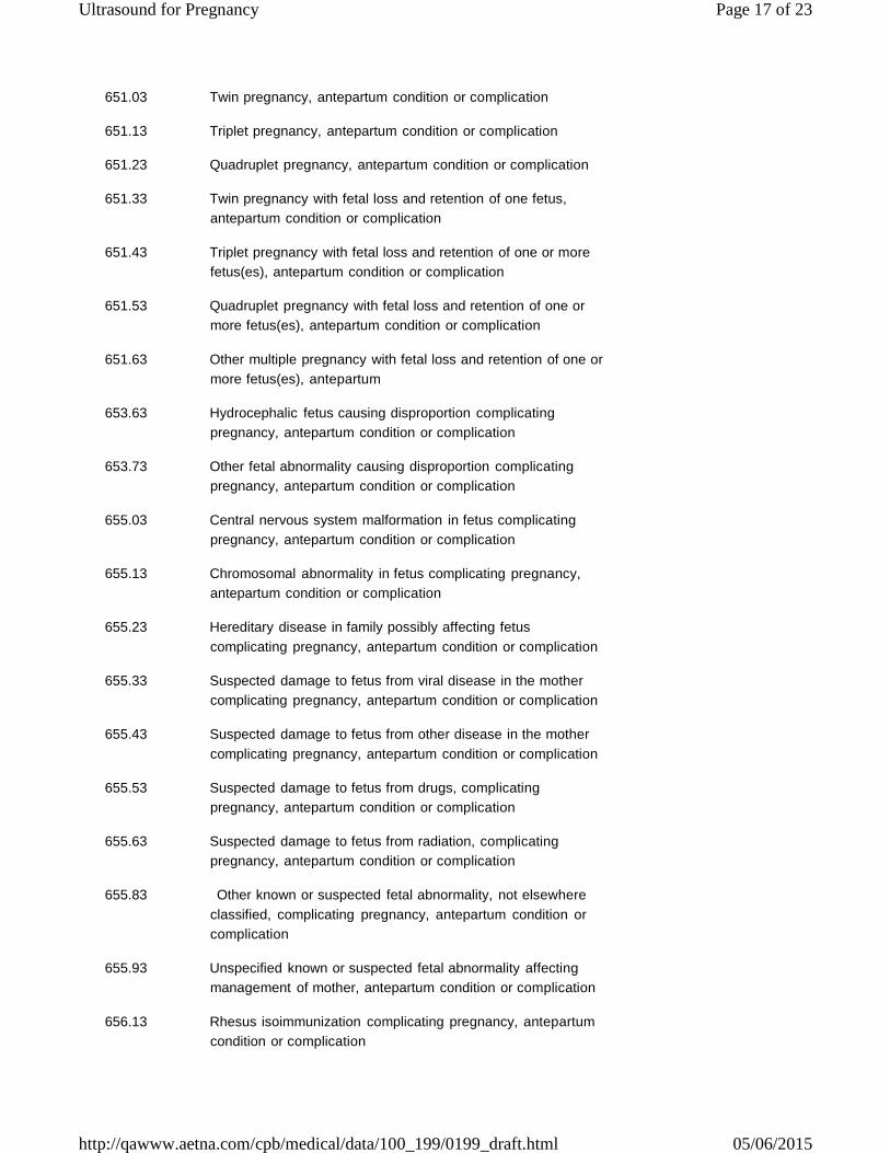
651.03 Twin pregnancy, antepartum condition or complication
651.13 Triplet pregnancy, antepartum condition or complication
651.23 Quadruplet pregnancy, antepartum condition or complication
651.33 Twin pregnancy with fetal loss and retention of one fetus,
antepartum condition or complication
651.43 Triplet pregnancy with fetal loss and retention of one or more
fetus(es), antepartum condition or complication
651.53 Quadruplet pregnancy with fetal loss and retention of one or
more fetus(es), antepartum condition or complication
651.63 Other multiple pregnancy with fetal loss and retention of one or
more fetus(es), antepartum
653.63 Hydrocephalic fetus causing disproportion complicating
pregnancy, antepartum condition or complication
653.73 Other fetal abnormality causing disproportion complicating
pregnancy, antepartum condition or complication
655.03 Central nervous system malformation in fetus complicating
pregnancy, antepartum condition or complication
655.13 Chromosomal abnormality in fetus complicating pregnancy,
antepartum condition or complication
655.23 Hereditary disease in family possibly affecting fetus
complicating pregnancy, antepartum condition or complication
655.33 Suspected damage to fetus from viral disease in the mother
complicating pregnancy, antepartum condition or complication
655.43 Suspected damage to fetus from other disease in the mother
complicating pregnancy, antepartum condition or complication
655.53 Suspected damage to fetus from drugs, complicating
pregnancy, antepartum condition or complication
655.63 Suspected damage to fetus from radiation, complicating
pregnancy, antepartum condition or complication
655.83 Other known or suspected fetal abnormality, not elsewhere
classified, complicating pregnancy, antepartum condition or
complication
655.93 Unspecified known or suspected fetal abnormality affecting
management of mother, antepartum condition or complication
656.13 Rhesus isoimmunization complicating pregnancy, antepartum
condition or complication
Ultrasound for Pregnancy Page 18 of 23
http://qawww.aetna.com/cpb/medical/data/100_199/0199_draft.html 05/06/2015
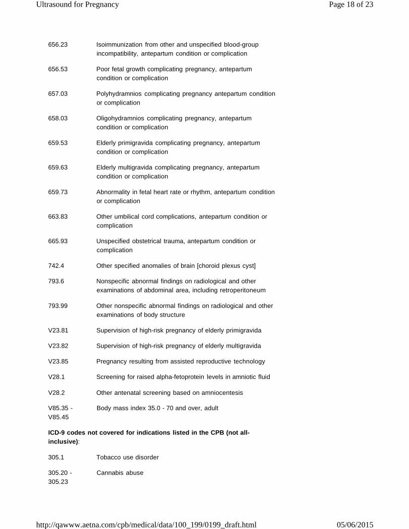
656.23 Isoimmunization from other and unspecified blood-group
incompatibility, antepartum condition or complication
656.53 Poor fetal growth complicating pregnancy, antepartum
condition or complication
657.03 Polyhydramnios complicating pregnancy antepartum condition
or complication
658.03 Oligohydramnios complicating pregnancy, antepartum
condition or complication
659.53 Elderly primigravida complicating pregnancy, antepartum
condition or complication
659.63 Elderly multigravida complicating pregnancy, antepartum
condition or complication
659.73 Abnormality in fetal heart rate or rhythm, antepartum condition
or complication
663.83 Other umbilical cord complications, antepartum condition or
complication
665.93 Unspecified obstetrical trauma, antepartum condition or
complication
742.4 Other specified anomalies of brain [choroid plexus cyst]
793.6 Nonspecific abnormal findings on radiological and other
examinations of abdominal area, including retroperitoneum
793.99 Other nonspecific abnormal findings on radiological and other
examinations of body structure
V23.81 Supervision of high-risk pregnancy of elderly primigravida
V23.82 Supervision of high-risk pregnancy of elderly multigravida
V23.85 Pregnancy resulting from assisted reproductive technology
V28.1 Screening for raised alpha-fetoprotein levels in amniotic fluid
V28.2 Other antenatal screening based on amniocentesis
V85.35 -
V85.45
Body mass index 35.0 - 70 and over, adult
ICD-9 codes not covered for indications listed in the CPB (not all-
inclusive):
305.1 Tobacco use disorder
305.20 -
305.23
Cannabis abuse
Ultrasound for Pregnancy Page 19 of 23
http://qawww.aetna.com/cpb/medical/data/100_199/0199_draft.html 05/06/2015
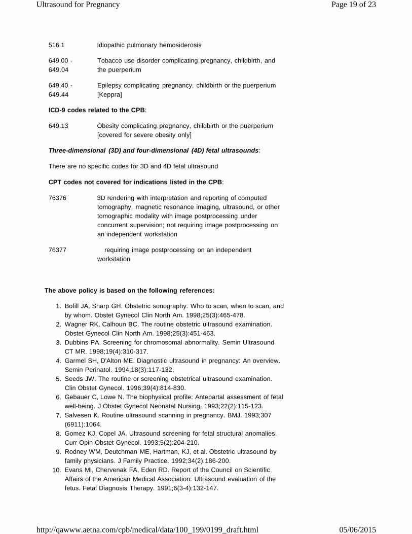
516.1 Idiopathic pulmonary hemosiderosis
649.00 -
649.04
Tobacco use disorder complicating pregnancy, childbirth, and
the puerperium
649.40 -
649.44
Epilepsy complicating pregnancy, childbirth or the puerperium
[Keppra]
ICD-9 codes related to the CPB:
649.13 Obesity complicating pregnancy, childbirth or the puerperium
[covered for severe obesity only]
Three-dimensional (3D) and four-dimensional (4D) fetal ultrasounds:
There are no specific codes for 3D and 4D fetal ultrasound
CPT codes not covered for indications listed in the CPB:
76376 3D rendering with interpretation and reporting of computed
tomography, magnetic resonance imaging, ultrasound, or other
tomographic modality with image postprocessing under
concurrent supervision; not requiring image postprocessing on
an independent workstation
76377 requiring image postprocessing on an independent
workstation
The above policy is based on the following references:
1.
2.
3.
4.
5.
6.
7.
8.
9.
10.
Bofill JA, Sharp GH. Obstetric sonography. Who to scan, when to scan, and
by whom. Obstet Gynecol Clin North Am. 1998;25(3):465-478.
Wagner RK, Calhoun BC. The routine obstetric ultrasound examination.
Obstet Gynecol Clin North Am. 1998;25(3):451-463.
Dubbins PA. Screening for chromosomal abnormality. Semin Ultrasound
CT MR. 1998;19(4):310-317.
Garmel SH, D'Alton ME. Diagnostic ultrasound in pregnancy: An overview.
Semin Perinatol. 1994;18(3):117-132.
Seeds JW. The routine or screening obstetrical ultrasound examination.
Clin Obstet Gynecol. 1996;39(4):814-830.
Gebauer C, Lowe N. The biophysical profile: Antepartal assessment of fetal
well-being. J Obstet Gynecol Neonatal Nursing. 1993;22(2):115-123.
Salvesen K. Routine ultrasound scanning in pregnancy. BMJ. 1993;307
(6911):1064.
Gomez KJ, Copel JA. Ultrasound screening for fetal structural anomalies.
Curr Opin Obstet Gynecol. 1993;5(2):204-210.
Rodney WM, Deutchman ME, Hartman, KJ, et al. Obstetric ultrasound by
family physicians. J Family Practice. 1992;34(2):186-200.
Evans MI, Chervenak FA, Eden RD. Report of the Council on Scientific
Affairs of the American Medical Association: Ultrasound evaluation of the
fetus. Fetal Diagnosis Therapy. 1991;6(3-4):132-147.
Ultrasound for Pregnancy Page 20 of 23
http://qawww.aetna.com/cpb/medical/data/100_199/0199_draft.html 05/06/2015
11.
12.
13.
14.
15.
16.
17.
18.
19.
20.
21.
22.
23.
24.
25.
26.
27.
American College of Obstetricians and Gynecologists (ACOG). Multiple
gestation. ACOG Technical Bulletin No.131. Washington, DC: ACOG;
August 1989.
American Academy of Pediatrics (AAP) and American College of
Obstetricians and Gynecologists (ACOG). Guidelines for Perinatal Care.
4th ed. Elk Grove Village, IL: AAP; August 1997.
American College of Obstetricians and Gynecologists (ACOG), Committee
on Obstetric Practice. Guidelines for diagnostic imaging during pregnancy.
ACOG Committee Opinion No. 158. Washington, DC: ACOG; September
1995.
American College of Obstetricians and Gynecologists (ACOG), Committee
on Practice Bulletins -- Obstetrics. ACOG Practice Bulletin. Clinical
Management Guidelines for Obstetrician-Gynecologists. Prenatal diagnosis
of fetal chromosomal abnormalities. Obstet Gynecol. 2001;97(5 Pt 1):suppl
1-12.
Barnett SB, Maulik D; International Perinatal Doppler Society. Guidelines
and recommendations for safe use of Doppler ultrasound in perinatal
applications. J Matern Fetal Med. 2001;10(2):75-84.
Lakhani K, Seifalian AM, Atiomo WU, Hardiman P. Polycystic ovaries. Br J
Radiol. 2002;75(889):9-16.
Kurjak A, Kupesic S, Simunic V. Ultrasonic assessment of the peri- and
postmenopausal ovary. Maturitas. 2002;41(4):245-254.
Kupesic S, Kurjak A, Hajder E. Ultrasonic assessment of the
postmenopausal uterus. Maturitas. 2002;41(4):255-267.
Timor-Tritsch IE, Platt LD. Three-dimensional ultrasound experience in
obstetrics. Curr Opin Obstet Gynecol. 2002;14(6):569-575.
Jurkovic D. Three-dimensional ultrasound in gynecology: A critical
evaluation. Ultrasound Obstet Gynecol. 2002;19(2):109-117.
Davies G, Wilson RD, Desilets V, et al. Amniocentesis and women with
hepatitis B, hepatitis C, or human immunodeficiency virus. J Obstet
Gynaecol Can. 2003;25(2):145-148, 149-152.
Bricker L, Garcia J, Henderson J, et al. Ultrasound screening in pregnancy:
A systematic review of the clinical effectiveness, cost-effectiveness and
women's views. Health Technol Assess. 2000;4(16):i-vi, 1-193.
Demianczuk NN, Van Den Hof MC, Farquharson D, et al. The use of first
trimester ultrasound. Obstet Gynaecol Can. 2003;25(10):864-875.
Institute for Clinical Systems Improvement (ICSI). Prenatal ultrasound as a
screening test. ICSI Technology Assessment Report No. 16. Bloomington,
MN: ICSI; updated October 2002. Available at: http://www.icsi.org.
Accessed March 31, 2004.
Hata T, Kanenishi K, Inubashiri E, et al. Three-dimensional sonographic
features of placental abnormalities. Gynecol Obstet Invest. 2004;57(2):61-
65.
American College of Obstetricians and Gynecologists. ACOG Committee
Opinion #296: First-trimester screening for fetal aneuploidy. Obstet
Gynecol. 2004;104(1):215-217.
American College of Obstetricians and Gynecologists (ACOG Committee
on Ethics. ACOG Committee Opinion. Number 297, August 2004.
Nonmedical use of obstetric ultrasonography. Obstet Gynecol. 2004;104
(2):423-424.
Ultrasound for Pregnancy Page 21 of 23
http://qawww.aetna.com/cpb/medical/data/100_199/0199_draft.html 05/06/2015
28.
29.
30.
31.
32.
33.
34.
35.
36.
37.
38.
39.
40.
41.
42.
Society for Maternal-Fetal Medicine (SMFM), Coding Committee. White
paper on ultrasound code 76811. Announcements. Washington, DC:
SMFM; May 24, 2004. Available at: http://www.smfm.org/index.cfm?
zone=news&nav=viewnews&newsID=238&smfmon=yes. Accessed March
17, 2005.
American College of Obstetricians and Gynecologists (ACOG), Committee
on Practice Bulletins -- Obstetrics. Ultrasonography in pregnancy. ACOG
Practice Bulletin No. 58. Washington, DC: ACOG; December 2004.
Morin L, Van den Hof MC; Society of Obstetricians and Gynaecologists of
Canada. SOGC clinical practice guidelines. Ultrasound evaluation of first
trimester pregnancy complications. Number 161, June 2005. Int J Gynaecol
Obstet. 2006;93(1):77-81.
Goncalves LF, Lee W, Espinoza J, Romero R. Three- and 4-dimensional
ultrasound in obstetric practice: does it help? J Ultrasound Med. 2005;24
(12):1599-1624.
Kurjak A, Miskovic B, Andonotopo W, et al. How useful is 3D and 4D
ultrasound in perinatal medicine? J Perinat Med. 2007;35(1):10-27.
Ji EK, Pretorius DH, Newton R, et al. Effects of ultrasound on maternal-fetal
bonding: A comparison of two- and three-dimensional imaging. Ultrasound
Obstet Gynecol. 2005;25(5):473-477.
Benacerraf BR, Shipp TD, Bromley D. How sonographic tomography will
change the face of obstetric sonography: A pilot study. J Ultrasound Med.
2005;24(3):371-378.
Benacerraf BR, Shipp TD, Bromley B. Improving the efficiency of
gynecologic sonography with 3-dimensional volumes: A pilot study. J
Ultrasound Med. 2006;25(2):165-171.
Benacerraf BR, Shipp TD, Bromley B. Three-dimensional US of the fetus:
Volume imaging. Radiology. 2006;238(3):988-996.
American College of Obstetricians and Gynecologists (ACOG) Committee
on Health Care for Underdeserved Women; ACOG Committee on Obstetric
Practice. ACOG committee opinion. Number 316, October 2005. Smoking
cessation during pregnancy. Obstet Gynecol. 2005;106(4):883-888.
American College of Obstetricians and Gynecologists (ACOG), Committee
on Practice Bulletins -- Obstetrics. Ultrasonography in pregnancy. ACOG
Practice Bulletin No. 98. Washington, DC: ACOG; October 2008.
Clinical Practice Obstetrics Committee; Maternal Fetal Medicine Committee,
Delaney M, Roggensack A, Leduc DC, et al. Guidelines for the management
of pregnancy at 41+0 to 42+0 weeks. J Obstet Gynaecol Can.
2008;30(9):800-823.
Chen M, Lee CP, Lam YH, et al. Comparison of nuchal and detailed
morphology ultrasound examinations in early pregnancy for fetal structural
abnormality screening: A randomized controlled trial. Ultrasound Obstet
Gynecol. 2008;31(2):136-146; discussion 146.
American College of Obstetricians and Gynecologists (ACOG), Committee
on Practice Bulletins -- Obstetrics. Ultrasonography in pregnancy. ACOG
Practice Bulletin No. 101. Washington, DC: ACOG; February 2009.
ACOG Committee on Practice Bulletins -- Obstetrics. ACOG Practice
Bulletin: Clinical management guidelines for obstetrician-gynecologists
number 92, April 2008 (replaces practice bulletin number 87, November
Ultrasound for Pregnancy Page 22 of 23
http://qawww.aetna.com/cpb/medical/data/100_199/0199_draft.html 05/06/2015
43.
44.
45.
46.
47.
48.
49.
50.
51.
52.
53.
54.
55.
56.
57.
58.
2007). Use of psychiatric medications during pregnancy and lactation.
Obstet Gynecol. 2008;111(4):1001-1020.
Yagel S, Cohen SM, Messing B, Valsky DV. Three-dimensional and four-
dimensional ultrasound applications in fetal medicine. Curr Opin Obstet
Gynecol. 2009;21(2):167-174.
Chen M, Wang HF, Leung TY, et al. First trimester measurements of nasal
bone length using three-dimensional ultrasound. Prenat Diagn. 2009;29
(8):766-770.
Kurjak A, Abo-Yaqoub S, Stanojevic M, et al. The potential of 4D
sonography in the assessment of fetal neurobehavior -- multicentric study in
high-risk pregnancies. J Perinat Med. 2010;38(1):77-82.
Jones NW, Raine-Fenning N, Mousa H, et al. Evaluation of the
intraobserver and interobserver reliability of data acquisition for three-
dimensional power Doppler angiography of the whole placenta at 12 weeks
gestation. Ultrasound Med Biol. 2010;36(9):1405-1411.
Whitworth M, Bricker L, Neilson JP, Dowswell T. Ultrasound for fetal
assessment in early pregnancy. Cochrane Database Syst Rev. 2010;
(4):CD007058.
Alfirevic Z, Stampalija T, Gyte GM. Fetal and umbilical Doppler ultrasound
in normal pregnancy. Cochrane Database Syst Rev. 2010;(8):CD001450.
Alfirevic Z, Stampalija T, Gyte GM. Fetal and umbilical Doppler ultrasound
in high-risk pregnancies. Cochrane Database Syst Rev. 2010;
(1):CD007529.
Stampalija T, Gyte GM, Alfirevic Z. Utero-placental Doppler ultrasound for
improving pregnancy outcome. Cochrane Database Syst Rev. 2010;
(9):CD008363.
ACOG Committee on Practice Bulletins. ACOG Practice Bulletin No. 77:
Screening for fetal chromosomal abnormalities. Obstet Gynecol. 2007;109
(1):217-227.
Hata T, Tanaka H, Noguchi J. 3D/4D sonographic evaluation of amniotic
band syndrome in early pregnancy: A supplement to 2D ultrasound. J
Obstet Gynaecol Res. 2011;37(6):656-660.
Antsaklis A, Daskalakis G, Theodora M, et al. Assessment of nuchal
translucency thickness and the fetal anatomy in the first trimester of
pregnancy by two- and three-dimensional ultrasonography: A pilot study. J
Perinat Med. 2011;39(2):185-193.
Society for Maternal Fetal Medicine (SMFM), Coding Committee.
White Paper on Ultrasound Code 76811. Announcements. Washington,
DC: SMFM; revised May 26, 2012.
Milman N. Idiopathic pulmonary hemosiderosis. UpToDate [online serial].
Waltham, MA: UpToDate; reviewed October 2012.
Grivell RM, Wong L, Bhatia V. Regimens of fetal surveillance for impaired
fetal growth. Cochrane Database Syst Rev. 2012;6:CD007113.
Fuchs KM, Society for Maternal-Fetal Medicine (SMFM). Isolated fetal
choroid plexus cysts. Their implications and outcome. Contemp Obstet
Gynecol. April 1, 2013.
Gindes L, Weissmann-Brenner A, Zajicek M, et al. Three-dimensional
ultrasound demonstration of the fetal palate in high-risk patients: The
accuracy of prenatal visualization. Prenat Diagn. 2013;33(5):436-441.
Ultrasound for Pregnancy Page 23 of 23
http://qawww.aetna.com/cpb/medical/data/100_199/0199_draft.html 05/06/2015
59.
60.
61.
62.
63.
64.
Kanenishi K, Hanaoka U, Noguchi J, et al. 4D ultrasound evaluation of fetal
facial expressions during the latter stages of the second trimester. Int J
Gynaecol Obstet. 2013;121(3):257-260.
Votino C, Cos T, Abu-Rustum R, et al. Use of spatiotemporal image
correlation at 11-14 weeks' gestation. Ultrasound Obstet Gynecol. 2013;42
(6):669-678.
Ahmed BI. The new 3D/4D based spatio-temporal imaging correlation
(STIC) in fetal echocardiography: A promising tool for the future. J Matern
Fetal Neonatal Med. 2014;27(11):1163-1168.
Tonni G, Grisolia G, Sepulveda W. Second trimester fetal
neurosonography: Reconstructing cerebral midline anatomy and anomalies
using a novel three-dimensional ultrasound technique. Prenat Diagn.
2014;34(1):75-83.
Sharp GC, Stock SJ, Norman JE. Fetal assessment methods for improving
neonatal and maternal outcomes in preterm prelabour rupture of
membranes. Cochrane Database Syst Rev. 2014;10:CD010209.
Benacerraf CR. Sonographic findings associated with fetal aneuploidy.
UpToDate [online serial]. Waltham, MA: UpToDate; reviewed February
2015.
Copyright Aetna Inc. All rights reserved. Clinical Policy Bulletins are developed by Aetna to assist in
administering plan benefits and constitute neither offers of coverage nor medical advice. This Clinical Policy
Bulletin contains only a partial, general description of plan or program benefits and does not constitute a
contract. Aetna does not provide health care services and, therefore, cannot guarantee any results or
outcomes. Participating providers are independent contractors in private practice and are neither
employees nor agents of Aetna or its affiliates. Treating providers are solely responsible for medical advice
and treatment of members. This Clinical Policy Bulletin may be updated and therefore is subject to change.
CPT only copyright 2008 American Medical Association. All Rights Reserved.