Embed Size (px)
Citation preview
1
Clinical Laboratory Diagnosis of Intestinal Protozoa 1
Ian H. McHardy1#
, Max Wu1#
, Robyn Shimizu-Cohen1, Marc Roger Couturier
2,3 and Romney M. 2
Humphries1*
3
4
Authors’ Information: 5
1Pathology and Laboratory Medicine, UCLA, Los Angeles, CA, United States. 6
2Associated Regional and University Pathologists (ARUP) Laboratories, Institute for Clinical and 7
Experimental Pathology, Salt Lake City, UT; 8
9
3Department of Pathology, University of Utah School of Medicine, Salt Lake City, UT
# These 10
authors contributed equally to this article. 11
*Corresponding author 12
Mailing address: 10833 Le Conte Avenue. Brentwood Annex, Los Angeles, CA 90095. 13
JCM Accepts, published online ahead of print on 6 November 2013J. Clin. Microbiol. doi:10.1128/JCM.02877-13Copyright © 2013, American Society for Microbiology. All Rights Reserved.
on June 26, 2018 by guesthttp://jcm
.asm.org/
Dow
nloaded from
2
Abstract 14
Despite recent advances in diagnostic technology, microscopic examination of stool 15
specimens remains central to the diagnosis of most pathogenic intestinal protozoa. Microscopy 16
is, however, labor-intensive and requires a skilled technologist. New, highly sensitive diagnostic 17
methods have been developed for protozoa endemic to developed countries, including Giardia 18
lamblia (syn. G. intestinalis/ G. duodenalis) and Cryptosporidium spp., using technologies that, if 19
expanded, could effectively compliment or even replace microscopic approaches. To date, the 20
scope of such novel technology is limited and may not include common protozoa such as 21
Dientamoeba fragilis, Entamoeba histolytica or Cyclospora cayetanensis. This minireview 22
describes canonical approaches for the detection of pathogenic intestinal protozoa, while 23
highlighting recent developments and FDA-approved tools for clinical diagnosis of common 24
intestinal protozoa. 25
26
27
28
29
30
31
32
33
34
35
on June 26, 2018 by guesthttp://jcm
.asm.org/
Dow
nloaded from
3
Introduction 36
Protozoan infections significantly contribute to the burden of gastrointestinal illness 37
worldwide. While the prevalence of these infections is low in the United States, sporadic 38
outbreaks, including the 2013 outbreak of cyclosporiasis in the United States, underscore the 39
continued burden of disease these organisms present in developed countries. Giardia, 40
Cryptosporidium spp., Dientamoeba fragilis, Entamoeba spp. (including non-pathogenic 41
species), Blastocystis spp., and Cyclospora cayetanensis are the most common pathogenic 42
protozoa reported in developed settings (1). However, accurate determination of the incidence 43
of these infections is hampered by infrequent testing of stool for protozoa when patients 44
present with gastroenteritis (1), inappropriate test ordering by physicians (1,2,3), and the lack 45
of sensitive techniques by which to identify pathogenic protozoa in stool specimens. 46
The microscopic ova and parasite examination (O&P) is the traditional method for stool 47
parasite testing. Although the O&P is labor-intensive and requires a high level of skill for 48
optimal interpretation, this test remains the cornerstone of diagnostic testing for the intestinal 49
protozoa. At present, most clinical microbiology laboratories in the United States struggle with 50
the ability to provide quality O&P results within a clinically significant time frame (Table1). A 51
pressing concern for these laboratories is the shortage of skilled technologists capable of 52
reliably evaluating O&Ps. As the baby-boomer generation retires from the workforce, 53
inexperienced technologists, who in some instances are inadequately trained in parasitology, 54
are left to fill the void. Few laboratories in the United States encounter a sufficient number of 55
specimens that harbor intestinal protozoa to maintain technologist proficiency, let alone to 56
on June 26, 2018 by guesthttp://jcm
.asm.org/
Dow
nloaded from
4
allow for robust training of new technologists. As such, laboratories may be unable to 57
accurately identify pathogenic protozoa, differentiate these from nonpathogenic species, and 58
discriminate artifacts on O&P examinations. Further, in many understaffed laboratories, the 59
labor-intensive O&P is performed only once other laboratory tasks are completed, yielding long 60
turnaround times and limiting this test’s clinical utility. 61
To address competency issues, some laboratories have developed affiliations with 62
organizations that conduct parasitology surveillance in endemic regions of the world and have 63
unique access to clinical specimens for teaching and training purposes. Examples of such 64
organizations are the Walter Reed Army Institute of Research, the Naval Medical Research Unit, 65
the Joint Pathology Center (previously the Armed Forces Institute of Pathology, AFIP), and the 66
Centers for Disease Control and Prevention (CDC) DPDx laboratories. Laboratories may also 67
consider pooling resources on a local level, both for training purposes and to share specimens 68
for competency. In the authors’ laboratories, positive specimens are reviewed by all trained 69
technologists to maximize staff competency. 70
Long term solutions to these challenges include lessening laboratory reliance on the 71
O&P for the diagnosis of intestinal protozoa; indeed, some have already suggested limiting the 72
use of the O&P in routine clinical practice (4). Antigen detection tests for Giardia, 73
Cryptosporidium spp and Entamoeba histolytica have been cleared by the United States FDA 74
(Table 2), and are associated with significant improvements in the detection of these organisms 75
in stool. Unfortunately, no FDA-cleared antigen test detects D. fragilis, which is a pathogenic 76
protozoon frequently encountered in many US laboratories (RMH, MRC unpublished 77
on June 26, 2018 by guesthttp://jcm
.asm.org/
Dow
nloaded from
5
observations). Regardless, some have suggested the use of algorithmic testing that involves 78
front-line antigen testing for Giardia and Cryptosporidium which, when negative, can be 79
reflexed to traditional microscopic approaches (5). Successful implementation of such a system 80
would likely require developing a physician guidance tool to aid in appropriate ordering, as the 81
laboratory very rarely receives information required to determine if the test is requested in the 82
clinical context of gastrointestinal complaints or as part of the evaluation of a returning 83
traveler, immigrant or patient prior to transplantation. Furthermore, such algorithmic testing 84
delays diagnosis of pathogens not initially tested for. 85
There is a pressing need for newer diagnostic test options, to replace the O&P. Such 86
tests should broadly detect most, if not all, pathogens commonly identified microscopically. 87
Multiplexed PCR has the potential to meet this need. However, only one such assay has been 88
cleared by the United States Food and Drug Administration (FDA) to date, the highly 89
multiplexed Luminex xTAG® GPP, which detects Giardia and Cryptosporidium in addition to 90
numerous bacterial and viral targets. Such molecular assays, depending on their design, may 91
require a laboratory with proficiency in molecular testing, which would limit their use to major 92
academic hospitals and reference laboratories. Alternatively, sample-to-answer solutions, 93
which provide direct diagnosis from unprocessed samples, such as the BioFireTM
Diagnostics 94
FilmArray® platform, could be used in virtually any laboratory setting. 95
Despite the challenges outlined in Table1, detection of intestinal protozoa is still almost 96
exclusively based on O&P microscopic examination. This article will thus review optimal 97
diagnostic approaches and the microscopic morphology of key pathogenic protozoa. The 98
on June 26, 2018 by guesthttp://jcm
.asm.org/
Dow
nloaded from
6
pathogenesis of some protozoa discussed is controversial, including Blastocystis hominis and 99
Dientamoeba fragilis. Other common protozoa, such as Endolimax nana, are not discussed 100
herein, as less is known about their potential virulence. Antigen and molecular-based detection 101
methods are also summarized. 102
Specimen Collection 103
Optimal recovery and microscopic identification of protozoa from patients with 104
intestinal infections is dependent on proper collection and preservation of fecal specimens. 105
Well-recognized factors that influence the sensitivity of parasite examinations include patient 106
medications, specimen collection interval and the preservation of stool prior to testing (6). The 107
diagnostic yield of the O&P is also significantly impacted by the number of stool specimens 108
collected and submitted to the laboratory for testing. Many intestinal protozoa are irregularly 109
shed; and data suggest that a single stool specimen submitted for microscopic examination 110
will detect 58-72% of protozoa present (4, 7). Hiatt and colleagues found that evaluating 111
three specimens, as opposed to one, resulted in an increased yield of 22.7% for E. histolytica, 112
11.3% for Giardia, and 31.1% for D. fragilis (8). As such, many laboratories continue to 113
request 3 specimens be collected and submitted for testing; specimen collection is optimally 114
every other day, over up to 10 days (6). However, alternative approaches have been proposed 115
to help curtail unnecessary testing, including application of an algorithm that requires a 116
negative specimen and persistence of symptoms before a second or third specimen is 117
analyzed by the laboratory (4). Specimens may also be pooled prior to screening by 118
microscopy. In contrast, the enhanced sensitivity of molecular detection methods may 119
on June 26, 2018 by guesthttp://jcm
.asm.org/
Dow
nloaded from
7
require only 1 specimen for testing to achieve sensitivity equal to, if not greater than, 120
microscopy. One study demonstrated a 14% increase in yield for gastrointestinal protozoa 121
when a real-time PCR was performed on a single stool specimen, as compared to microscopy 122
on three (5). 123
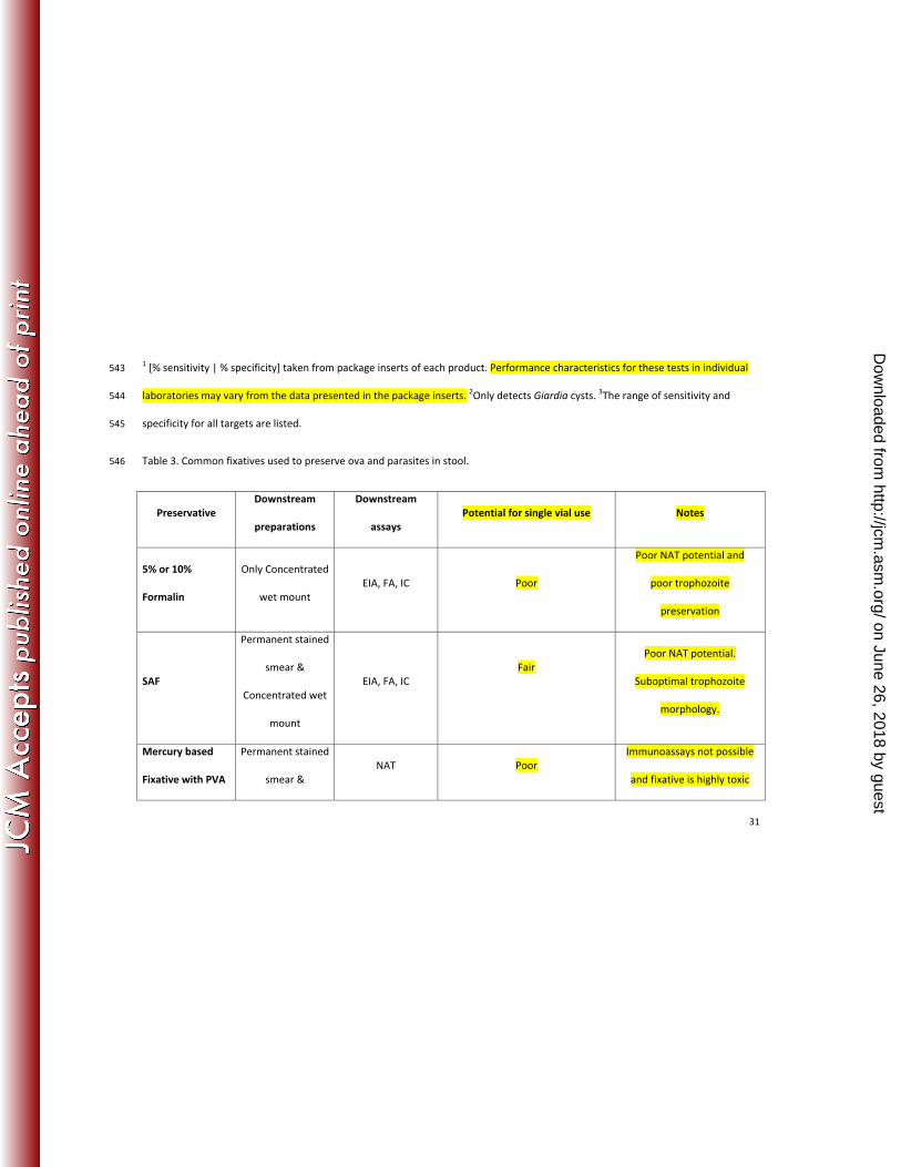
Stool Preservation 124
While visualizing motility in unpreserved specimens may facilitate diagnosis, this 125
technique is impractical for most laboratories as transport of fresh stool to the laboratory for 126
testing is rarely within the requisite time frame for examination (i.e. 30-60 minutes). A variety 127
of stool fixatives have been developed and modified in recent decades for use with traditional 128
microscopic examination. Those that remain widely used and commercially available include 129
the formalin, sodium acetate-acetic acid-formalin (SAF), Schaudinn’s Fluid, polyvinyl alcohol-130
containing fixatives (mercury, copper, or zinc-based), and mercury-free/formalin-free fixatives. 131
A two vial collection system, consisting of one vial containing 5-10% buffered formalin for use in 132
concentrated wet mounts and a second vial containing a polyvinyl alcohol-based preservative 133
for permanent stained smears, is considered the “gold standard”. However, concern over 134
working with toxic formalin in the laboratory and the environmental impact and disposal costs 135
associated with the use of mercury-based fixatives have led many to consider alternate 136
preservatives and single-tube collection systems (9). SAF may be used, to achieve this goal, if 137
coupled with iron hematoxylin for the permanent stained smear; however, for laboratories 138
desiring to maintain the trichrome stain, SAF is not a valid option, as poor quality results have 139
been documented with this combination. 140
on June 26, 2018 by guesthttp://jcm
.asm.org/
Dow
nloaded from

8
Alternative stool preservatives Zinc and copper-based PVA formulations have been 141
developed and are commercially available to replace the mercury-based fixatives (10, 11). 142
In a paired study that evaluated 106 specimens prepared by zinc sulfate-PVA versus 143
mercuric chloride-PVA with the trichrome stain, 92.5% overall agreement was reported in 144
the overall morphology and numbers of organisms detected between the two methods (11); 145
in contrast, a study by the same group noted poor preservation of protozoa morphology 146
when a copper-based PVA formulation was evaluated (10). Examples of commercial 147
specimen collection kits using modification to the mercuric chloride PVA include PROTOFIX® 148
(AlphaTec, Vancouver, WA) which contains no mercury and minimal formalin; ECOFIX® 149
(Meridian Bioscience, Cincinnati, OH) which contains neither mercury nor formalin; 150
PARASAFE® (Cruinn Diagnostic, Dublin, Ireland) which also contains neither mercury nor 151
formalin. A study conducted by the CDC evaluated the performance of these preservatives 152
head-to-head with the traditional two-vial set of formalin and mercuric chloride PVA. This 153
study found ECOFIX® and PROTOFIX
®, but not PARASAFE
®, yielded an acceptable 154
morphologic quality to the preserved parasites on concentrated wet mounts as compared 155
to formalin fixed specimens. ECOFIX® alone yielded satisfactory protozoan morphology on 156
the permanent stained smears, when compared with stool preserved in mercuric chloride 157
PVA (9). In contrast, a separate study found significantly (p<0.001) reduced recovery of B. 158
hominis and Endolimax nana in 261 ECOFIX® preserved concentrates when compared to 159
formalin-fixed stool concentrates (12). Although the manufacturer of ECOFIX® has 160
developed a proprietary stain, ECOSTAIN®, the conventional trichrome stain can be used 161
with ECOFIX®, and has been shown to produce comparable protozoan morphology (12). 162
on June 26, 2018 by guesthttp://jcm
.asm.org/
Dow
nloaded from
9
TOTAL-FIX® (Medical Chemical Corporation, Torrance, CA) is relatively new, FDA-approved 163
mercury-, formalin- and PVA-free fixative. Similar to the ECOFIX®, specimens prepared by 164
TOTAL-FIX® can be used for concentration, permanent stain, and a variety of immunoassays for 165
detecting protozoa, though there have been no published reports describing the performance 166
of this fixative as compared to others, to date. Table 3 summarizes many available fixatives 167
used by clinical laboratories and highlights possible preparations and downstream assays for 168
each. 169
A major impediment to replacing the traditional two-vial systems by laboratories in the 170
United States is the requirement for laboratories to perform a verification study to confirm the 171
performance specifications of these products. Few institutions encounter a sufficient number of 172
positive clinical specimens to allow robust evaluation of these preservatives. Furthermore, in 173
order to perform a method comparison study, specimens would need to be collected in both 174
fixatives, which may require pre-approval or exemption status by local Institutional Review 175
Boards. Laboratories may thus need to develop creative means by which to evaluate these 176
fixatives prior to clinical use. A combination of approaches have been used in our laboratories, 177
including a comparison of the morphology of white cells present in stool preserved in both 178
fixatives, seeding fresh stool specimens with cultured protozoa, obtaining veterinary specimens 179
for testing, and consulting with published literature (if available) on the performance of these 180
products. 181
Detection of specific intestinal protozoa 182
Giardia lamblia (syn. Giardia intestinalis and Giardia duodenalis) 183
on June 26, 2018 by guesthttp://jcm
.asm.org/
Dow
nloaded from
10
Giardiasis is a common gastrointestinal parasitic infection associated with diarrhea, 184
stomach cramps, upset stomach, and excessive gas. Annually, roughly 20,000 US cases of 185
giardiasis are reported to the CDC, but are estimated to comprise as little as 1-10% of the total 186
infection burden despite being a nationally notifiable disease (13). While numerous diagnostic 187
tests are available for Giardia, its highly distinctive morphology facilitates microscopic 188
diagnosis. Giardia cysts can be observed in fresh smears, on formalin-ethyl acetate 189
concentration or permanent stained smear, although the latter is associated with higher 190
sensitivity for identification. Trophozoites are not always found in stool as encystation begins 191
before passage through the colon. In cases where Giardia is suspected but not detected in 192
stool, duodenal specimens, such as those collected by a string test may be used for permanent 193
stains and concentrated wet mounts. Tear drop shaped trophozoites range from 10-20 ʅm in 194
length, 9-12 ʅm in width and contain two nuclei, a sucking disk, 4 pairs of flagella, 2 axonemes 195
and 2 median bodies. Cysts contain 4 nuclei, 4 axonemes, and 4 median bodies, and range from 196
11-14 ʅm in length and 7-10 ʅm in width (Figure 1E). 197
While Giardia cysts are easily recognizable on permanent stained smears, they are shed 198
sporadically and O&P examinations are often insufficient to demonstrate the presence of this 199
organism (14). Alles and colleagues demonstrated a sensitivity of 66.4% for the detection of 200
Giardia via a permanent stained smear, albeit chlorazol black stain was performed as opposed 201
to the more standard trichrome, and the number of specimens tested per patient was not 202
taken into account (15). Regardless, detection of Giardia is improved through the use of antigen 203
detection assays, several of which are commercially available and widely used in clinical 204
laboratories across the United States. For example, in the aforementioned study by Alleles and 205
on June 26, 2018 by guesthttp://jcm
.asm.org/
Dow
nloaded from
11
colleagues, a sensitivity of 99.2% for the detection of Giardia was observed with a commercial, 206
direct fluorescent antibody (DFA) test. Both the permanent stained smear and the DFA were 207
100% specific for Giardia in the 2,696 stool specimens examined by this study (15). In addition 208
to the DFA, which requires laboratory access to a fluorescent microscope, 209
immunochromatographic (IC) tests and enzyme immunoassays (EIAs) are commercially 210
available for the detection of Giardia (Table 2). IC tests are optimally suited for laboratories 211
with lower capacity for diagnostic complexity, while EIA-based tests may be more appropriate 212
for high-throughput screening in high prevalence areas. A study comparing four EIAs including 213
the FDA-approved ProSpecT® (Remel, Lenexa, KS) and CELISA (Cellabs, Brookvale, NSW, 214
Australia) assays, found sensitivities that ranged from 63-91%, and specificities of ш95% for all 215
assays (16). A second study demonstrated 94-100% sensitivity and 100% specificity when 5 216
Giardia EIAs were evaluated with 100 positive and 50 negative specimens (17). Table 2 provides 217
an overview of many of the available FDA-approved EIAs and their respective sensitivities and 218
specificities, as determined by the manufacturer, for detection of Giardia either alone or in 219
combination with other pathogenic protozoa. 220
Dientamoeba fragilis 221
Dientamoebiasis is an enteric infection caused by the flagellate D. fragilis. Symptoms 222
associated with infection vary dramatically, with some individuals suffering nausea, vomiting, 223
and diarrhea containing mucous and abdominal discomfort; while others are asymptomatic. 224
Accordingly, as with the case of B. hominis, described further below, there is some uncertainty 225
about the pathogenesis of D. fragilis. However, the morbidity associated with some infections 226
on June 26, 2018 by guesthttp://jcm
.asm.org/
Dow
nloaded from
12
justifies its inclusion as a definitive pathogen (18). The prevalence of D. fragilis has been 227
estimated in many studies, and ranges from 1.1 – 20% in patients in the developed world with 228
diarrhea, but may be higher in select populations or if molecular methods are used for 229
detection (19). 230
Despite this relatively high prevalence, no antigen-based, molecular or serologic 231
diagnostics have been commercially developed to aid with laboratory identification. As such, 232
detection of D. fragilis on the permanent stained smear is the current standard. Unfortunately, 233
D. fragilis is difficult to identify morphologically. No cyst stage has been observed in humans, 234
although a cyst stage has been recently observed in mice (20). Trophozoites range from 5-15 235
ʅm in length and 9-12 ʅm in width and contain 1-2 characteristically fragmented nuclei. While 236
well-preserved specimens might contain cells with the classically described tetrad nuclei (Figure 237
1D), in general practice nuclei will only have visible holes through the center of the nucleus. 238
Given its indistinct appearance, diagnosis is often only possible by experienced technologists, 239
leading to many potentially missed infections. Even under ideal conditions, with prompt 240
preservation of stool and evaluation by a skilled technologist, permanent stained smears are 241
only 34% sensitive as compared to molecular methods (21). 242
Cryptosporidium spp. 243
Cryptosporidiosis is a gastrointestinal infection caused by various species of 244
Cryptosporidium. Fecal-oral transmission via contaminated food, drinking water and public 245
swimming pools is responsible for most infections. Like all coccidian intestinal parasites, the 246
small and poorly staining Cryptosporidium oocysts can be easily missed in routine O&P exams. 247
on June 26, 2018 by guesthttp://jcm
.asm.org/
Dow
nloaded from
13
Sensitivity of light microscopy is improved by performing modified acid fast (MAF) stains, 248
though even this modification has been shown to be associated with a sensitivity of only 54.8% 249
(15). Furthermore, MAF staining is typically only performed upon physician request, or if the 250
technologist detects structures suspicious for Cryptosporidium on the permanent stained 251
smear. Unfortunately, many physicians assume that testing for Cryptosporidium is included 252
with the routine O&P, and infrequently order specialized stains or Cryptosporidium 253
immunoassays, even in outbreak situations (3). Upon MAF staining, Cryptosporidium spp. 254
oocysts appear as bright red spheres (4-6 ʅm) containing four crescent shaped sporozoites 255
(which may or may not be seen in all oocysts) (Figure 1H). Additionally, oocysts may also 256
occlude stain resulting in transparent “ghost” cells. 257
As is the case for Giardia, sensitivity of detection is improved when an EIA or DFA (Table 258
2) is used. Multiple studies have evaluated the sensitivity and specificity of the available kits 259
and found overall similar performance for EIA and DFA-based methods (sensitivity >90%, 260
specificity >95%; (17)). Rapid IC-based methods are significantly less sensitive, with one multi-261
institutional study reporting 50.1 - 86.7% sensitivity, dependent on manufacturer (22). Because 262
HIV-infected and immunocompromised individuals are particularly at risk for severe 263
complications due to infection with these coccidian parasites, physicians should consider 264
routinely ordering of DFA at minimum, and molecular-based assays, if available, for patients 265
with suspect cryptosporidiosis. 266
Giardia or Cryptosporidium spp. are two of the most common protozoan infections in 267
the United States and multiple combined tests have been developed to facilitate rapid 268
on June 26, 2018 by guesthttp://jcm
.asm.org/
Dow
nloaded from
14
screening for both organisms simultaneously. Such tests include EIAs, IC assays, DFA assays, and 269
multiplex PCR assays. A comparison between several DFA and EIA tests for Giardia and 270
Cryptosporidium revealed that: A) DFA tests tended to have slightly higher sensitivity for both 271
organisms; B) the Merifluor® Cryptosporidium/Giardia test had the highest sensitivity of the 272
DFAs; and C) the specificity of all tested EIA and DFA tests were 100% (17). However, these 273
assays do not detect D. fragilis and as such, these tests do not replace the O&P for routine 274
testing. 275
Cyclospora cayetanensis 276
Cyclosporiasis is usually a self-limiting gastroenteritis caused by the coccidian, C. 277
cayetanensis. Due to poor uptake of most conventional stains by C. cayetanensis oocysts, 278
microscopic detection can be challenging, but remains the recommended diagnostic method 279
(14). C. cayetanensis oocysts may stain irregularly by trichrome or the MAF stain. As is the case 280
with Cryptosporidium, not all oocysts will take up these stains in a single smear, which may lead 281
inexperienced technologists to overlook the organism. When observed, Cyclospora oocysts in 282
stool are easily identified as 8-10 ʅm refractile spheres with a central morula, resembling 283
wrinkled cellophane (Figure 1G). If Cyclospora infection is specifically suspected (e.g. during 284
established outbreaks), use of a modified safranin staining protocol provides consistent 285
reddish-orange staining of oocysts, and thus simplifies identification (23). It is important to 286
recognize that Cryptosporidium does not consistently stain by modified safranin, and as a result 287
this stain should not replace a MAF stain in general practice. In addition to the modified 288
safranin stain, oocysts of C. cayetanensis in a standard concentrated wet-mount intrinsically 289
on June 26, 2018 by guesthttp://jcm
.asm.org/
Dow
nloaded from
15
autofluoresce white-blue under UV light using a 330-365 nm excitation filter. Less intense, blue-290
green autofluorescence can be seen using a 450-490 nm excitation filter. This property aids in 291
the identification of Cyclospora, however all fluorescent structures should be visualized by light 292
microscopy to verify the morphology 293
(http://www.asm.org/images/PSAB/CyclosporaWhitePaper2013.pdf). 294
Relman et. al developed a nested PCR assay that targets the 18S rRNA gene that has 295
been used in outbreak situations to confirm Cyclospora (23). Many other molecular techniques 296
have been developed for the identification of Cyclospora (1), but there are no FDA-approved or 297
analyte specific reagents for Cyclospora available in the U.S. Biofire (Salt Lake City, UT) 298
FilmArray® GI Panel includes C. cayetanensis and is currently available in the U.S. with research 299
use only (RUO) status, but is in clinical trials with the FDA. 300
Cystoisospora belli 301
Cystoisosporiasis is a relatively uncommon gastroenteritis caused by the coccidian C. 302
belli that can result in cholera-like symptoms in up to 1% of HIV-infected or otherwise 303
immunocompromised individuals (25). Detection of oocysts from stool or duodenal samples is 304
simplified by their distinctive size and shape. However, C. belli oocysts are only easily 305
recognizable in concentrated wet mounts of O&P exams. Importantly, oocyst maturation 306
continues post-defecation and thus morphology depends upon the duration between specimen 307
collection and preservation. If placed immediately into preservative, long oval-shaped C. belli 308
oocysts (20-33 ʅm in length and 10-19 ʅm in width) will contain a single circular immature 309
sporoblast. If specimens are not quickly preserved, oocysts of roughly the same size and shape 310
on June 26, 2018 by guesthttp://jcm
.asm.org/
Dow
nloaded from
16
will contain 1-2 circular sporoblasts. While detection is relatively straight-forward from 311
concentrated wet-mounts, modified acid-fast, safranin or auramine rhodamine stains can be 312
used to increase contrast and simplify detection, though staining may interfere with sporoblast 313
visualization (Figure 1B) (26, 27). Similar to Cyclospora, the oocysts of Cystoisopora will 314
autofluoresce under the conditions described above. C. belli oocysts are not always found in 315
stool, and examination of duodenal specimens collected by biopsy or string test may be 316
necessary. 317
Entamoeba histolytica 318
Roughly 50 million worldwide cases of amoebic dysentery and 100,000 deaths are 319
associated with E. histolytica annually (28). Despite the extreme morbidity associated with 320
intestinal infections by E. histolytica, serological tests are not typically informative in 321
uncomplicated cases because seroconversion is rare outside the context of extra-intestinal 322
involvement. Despite their microscopic morphological similarity to Entamoeba dispar and 323
Entamoeba moshkovskii, intestinal infections with E. histolytica in non-endemic areas are still 324
primarily diagnosed via microscopy on the permanent stained smear. Organisms may be 325
accompanied by clubbed RBCs in cases of dysentery. On the permanent stained stool smear, E. 326
histolytica trophozoites are 12-60 ʅm in diameter and contain a single, well-defined nucleus 327
(Figure 1C). Spherical cysts measure 12-15 ʅm, contain 2-4 nuclei and occasionally have cigar-328
shaped, cytoplasmic chromatoidal bars. Nuclei of both forms are surrounded by an obvious 329
nuclear membrane, a compact, central karyosome, and evenly distributed peripheral 330
chromatin. Without evidence of erythrophagocytosis (which is seen most often in tissue 331
on June 26, 2018 by guesthttp://jcm
.asm.org/
Dow
nloaded from
17
specimens), E. histolytica is indistinguishable from E. dispar and should be annotated as E. 332
histolytica/dispar on the laboratory report. Ingested RBCs can only be definitively identified 333
when concomitant extracellular RBCs are visible. In cases of chronic amebic infection, ingested 334
RBCs are infrequently observed, making differentiation from E. dispar difficult. 335
In areas of the world where E. histolytica infection is endemic or if infection is 336
specifically suspected by a physician, antigen-based tests can be performed, though these 337
require unpreserved specimens. E. histolytica antigen tests that are specific for E. histolytica 338
employ monoclonal antibodies against the Gal/GalNAc-specific lectin expressed E. histolytica. 339
Not all commercially available antigen tests differentiate between E. histolyica and E. dispar 340
(Table 2). Sensitivity for the E. histolytica antigen detection tests has been shown in several 341
studies to range from 80-94%, as compared to PCR; but one study found the TechLab ELISA to 342
be less sensitive than microscopy (29). Examples of FDA-approved EIA tests for Entamoeba spp. 343
are included in Table 2 along with their sensitivity and specificity, as defined by their package 344
inserts. 345
Diagnosis of disseminated amebiasis caused by E. histolytica is challenging because the 346
stool O&P examinations are almost always negative for these patients. When such cases are 347
suspected, cecal or colonic endoscopy to look for hallmark lesions followed by endoscopic 348
biopsy to visualize the presence of E. histolytica trophozoites are quite helpful (30). This 349
algorithm has been shown to be effective in differentiating amebic colitis from colon cancer 350
and uncomplicated colitis (31, 32). Sigmoidoscopy material may also be submitted to the 351
laboratory for permanent stained smear evaluation. In patients with liver abscesses, serological 352
on June 26, 2018 by guesthttp://jcm
.asm.org/
Dow
nloaded from
18
assays are informative due to the concomitant systemic exposure to amoebic antigens (1); 95% 353
of patients with extraintestinal disease will be positive by serology. When evaluating patients 354
from endemic areas, it is important to be aware that modern serological assays, which employ 355
recombinant E. histolytica antigens, will turn negative following abscess treatment earlier than 356
did the traditional indirect hemagglutination based tests, which remained positive for at least 6 357
months following treatment. Serum and liver abscess aspirates from patients with 358
disseminated E. histolytica have been subjected to off-label antigenic testing with varying 359
sensitivity. 360
Blastocystis hominis 361
The pathogenicity of B. hominis is largely controversial given that it is commonly 362
identified in non-symptomatic individuals. Some experts hypothesize that B. hominis should be 363
split into multiple species, some being more pathogenic than others, though few studies have 364
been performed to confirm this hypothesis (33). The continuing uncertainty is primarily due to 365
the fact that all isolates of Blastocystis are morphologically similar and are occasionally found in 366
combination with other protozoan infections. However, in the absence of antigen detection or 367
molecular diagnostics, the standard method for detection is still microscopy. While B. hominis is 368
visible on wet mounts, definitive identification is easier with permanent stained smears. B. 369
hominis is typically 6-40 ʅm in diameter with a large central body surrounded by up to six small 370
nuclei (Figure 1F). The large central body often stains a characteristic red, green, or blue in 371
trichrome stained samples. Non-microscopic and molecular strategies for diagnosis will likely 372
hinge on whether studies can effectively differentiate pathogenic versus non-pathogenic strains 373
on June 26, 2018 by guesthttp://jcm
.asm.org/
Dow
nloaded from
19
(33). When observed on routine O&P, B. hominis should be reported, along with a semi-374
quantitative assessment. 375
Balantidium coli 376
Balantidiasis is an intestinal parasitic disease associated with ciliated B. coli trophozoites 377
that typically only affects immunocompromised or malnourished individuals and has a 378
worldwide distribution (34). Like many other intestinal protozoa, no established molecular or 379
serologic tests are available for B. coli. Instead, microscopic diagnosis is facilitated by its 380
distinctive size and morphology on concentrated wet mounts; diagnosis from permanent stains 381
is not recommended because trophozoites absorb large amounts of dye, masking its 382
characteristic features. 383
B. coli is the largest infectious intestinal protozoan at 50-100 microns in length and 40-384
70 microns in width. Trophozoites have fine, visible cilia and a large, kidney-bean shaped 385
macronucleus (Figure 1A). A single, polar cystosome, or oral groove, can also be detected on 386
some cells. The cyst form also has a visible macronucleus, but are smaller (50-70 µm long, 40-387
60 µm wide) and rounder than the trophozoites. Cysts have a thick cyst wall and often do not 388
have visible cilia. While molecular or serologic-based diagnostics might improve detection 389
sensitivity compared to microscopic diagnosis, development of such tests has been a low 390
priority due to the relative simplicity of microscopic detection and infrequency of infection in 391
the US. 392
393
on June 26, 2018 by guesthttp://jcm
.asm.org/
Dow
nloaded from
20
Implications for future diagnostics 394
As discussed above and documented in recent studies, multiplex PCR assays are both 395
more sensitive and specific than microscopy for the detection and identification of pathogenic 396
protozoa (35). However, despite a rapidly growing field of molecular and genetic technologies 397
for the clinical microbiology laboratory, diagnostic development for intestinal protozoan 398
parasites has remained relatively stagnant. Challenges associated with developing a 399
replacement test for the O&P includes coverage of all pathogenic species and the potential for 400
long-term, residual detection of previous infections. Furthermore, while analyte-specific 401
approaches may yield enhanced sensitivity for pathogenic protozoa, documentation of the 402
presence of human cells (white blood cells and erythrocytes), Charcot-Leyden crystals and non-403
pathogenic protozoa is lost. In particular, some physicians interpret the presence of non-404
pathogenic protozoa as indicative of patient exposure to contaminated food or water, although 405
there are no studies that have clearly demonstrated this to be fact. 406
The Luminex® xTAG
® Gastrointestinal Pathogen Panel has received FDA approval and can 407
simultaneously detect 14 enteropathogens, including Giardia and Cryptosporidium spp. This 408
assay is the first molecular method approved by the FDA for the detection of pathogenic 409
protozoa. The analyte specific reagents (ASRs) for the xTAG assay were recently evaluated; 410
while the overall number of positive specimens was low in this study (5-20 positives), the ASRs 411
were highly sensitive and specific for Cryptosporidium (95% sensitivity and 99% specificity), 412
Giardia (95% sensitivity and 99% specificity) and E. histolytica (100% sensitive and 89% specific) 413
on June 26, 2018 by guesthttp://jcm
.asm.org/
Dow
nloaded from
21
(36). The FDA-approved version of the assay does not include E. histolytica, but the reagents for 414
this analyte are available as research use only (RUO). 415
BioFire Diagnostics has in development a sample-to-answer gastrointestinal pathogen 416
panel that includes detection of Giardia, Cryptosporidium, E. histolytica and Cyclospora 417
cayentensis. Whether the company is able to collect sufficient number of specimens positive 418
for each target to garner FDA clearance, or if some will remain “RUO”, remains to be seen. Like 419
the Luminex panel, this platform does not include detection of D. fragilis, which is one of the 420
most commonly encountered protozoa in the United States. 421
One major critique for these multiplex panels is the cost per test, which is many times 422
higher than the reagents associated with performing the O&P. However, if an assay were to 423
replace the O&P examination, the savings in labor, from the perspective of these authors, 424
would far outweigh the cost associated with performing a multiplex commercial test. 425
426
Summary 427
In summary, adequate diagnosis of intestinal protozoa by the clinical laboratory is 428
limited by many factors (Table1). There is increasing demand for low complexity, high-429
throughput, and cost effective complements to (or replacements for) the labor-intensive 430
microscopy-based approaches to protozoan diagnosis. While efforts in this regard have been 431
slow to come, many diagnostic manufacturers are rising to the challenge, including Luminex® 432
and BioFireTM
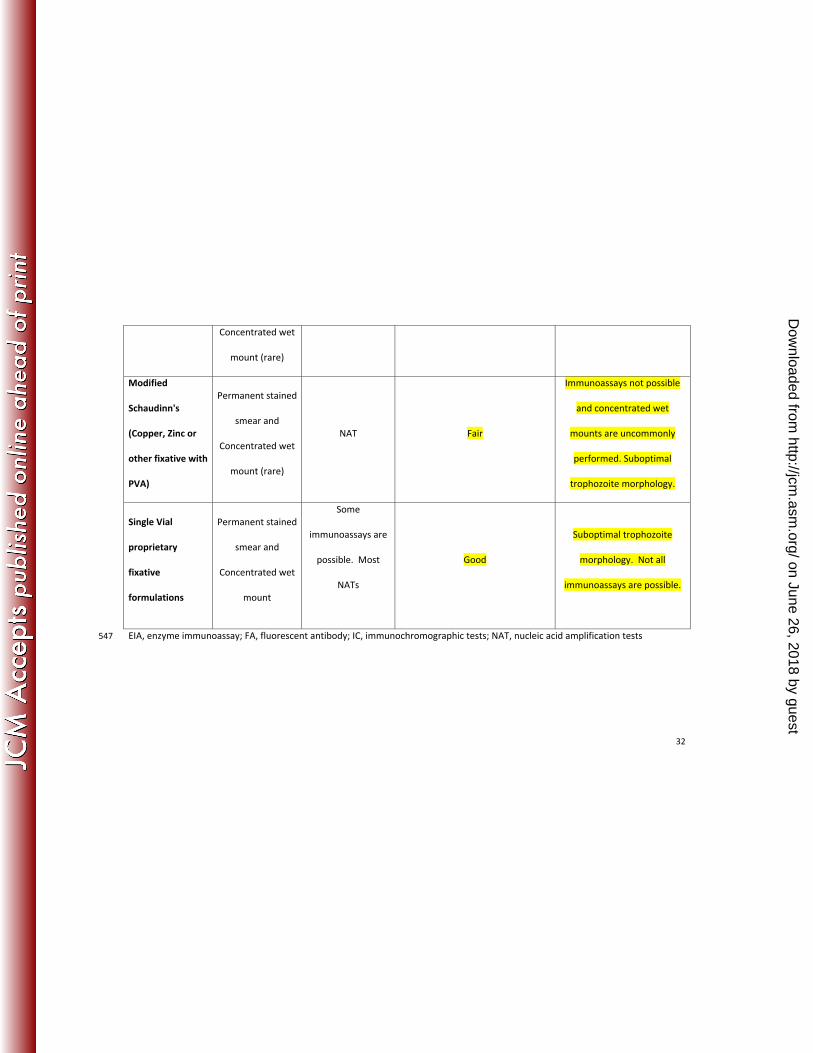
. These efforts may restore or enhance the laboratory’s ability to identify these 433
on June 26, 2018 by guesthttp://jcm
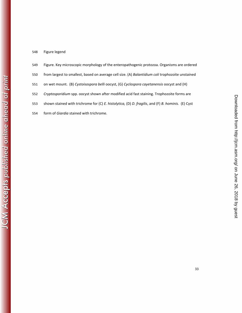
.asm.org/
Dow
nloaded from
22
pathogens, yielding increased knowledge on the present state of these diseases in the United 434
States and other countries. 435
436
Acknowledgments: We thank Dr. David A Bruckner for his insightful review of this manuscript.437
on June 26, 2018 by guesthttp://jcm
.asm.org/
Dow
nloaded from
23
References 438
1. Fletcher SM, Stark D, Harkness J, Ellis J. 2012. Enteric protozoa in the developed world: a 439
public health perspective. Clin. Microbiol. Rev. 25: 420-449. 440
2. Centers for Disease Control and Prevention. 2013. Notes from the Field: Use of Electronic 441
Messaging and the News Media to Increase Case Finding During a Cyclospora Outbreak - 442
Iowa, July 2013. MMWR 62: 613-614. 443
3. Polage CR, Stoddard GJ, Rolfs RT, Petti CA. 2011. Physician use of parasite tests in the United 444
States from 1997 to 2006 and in a Utah Cryptosporidium outbreak in 2007. J. Clin. 445
Microbiol. 49: 591-596. 446
4. Branda JA, Lin TY, Rosenberg ES, Halpern EF, Ferraro MJ. 2006. A rational approach to the 447
stool ova and parasite examination. Clin. Infect. Dis. 42: 972-978. 448
5. Bruijnesteijn van Coppenraet LE, Wallinga JA, Ruijs GJ, Bruins MJ, Verweij JJ. 2009. 449
Parasitological diagnosis combining an internally controlled real-time PCR assay for the 450
detection of four protozoa in stool samples with a testing algorithm for microscopy. Clin. 451
Microbiol. Infect. 15: 869-874. 452
6. Garcia LS (2009) Practical Guide to Diagnostic Parasitology. ASM Press. Washington, DC. 453
7. Nazer H, Greer W, Donnelly K, Mohamed AE, Yaish H, Kagalwalla A, Pavillard R. 1993. The 454
need for three stool specimens in routine laboratory examinations for intestinal 455
parasites. Br. J. Clin. Pract. 47: 76-78. 456
8. Hiatt RA, Markell EK, Ng E. 1995. How many stool examinations are necessary to detect 457
pathogenic intestinal protozoa? Am. J. Trop. Med. Hyg. 53: 36-39. 458
on June 26, 2018 by guesthttp://jcm
.asm.org/
Dow
nloaded from

24
9. Pietrzak-Johnston SM, Bishop H, Wahlquist S, Moura H, Da Silva ND, Nguyen-Dihn P. 2000. 459
Evaluation of commercially available preservatives for laboratory detection of helminths 460
and protozoa in human fecal specimens. J. Clin. Microbiol. 38: 1959-1964. 461
10. Garcia LS, Shimizu RY, Brewer TC, Bruckner DA. 1983. Evaluation of intestinal parasite 462
morphology in polyvinyl alcohol preservative: comparison of copper sulfate and 463
mercuric chloride bases for use in Schaudinn fixative. J. Clin. Microbiol. 17: 1092-1095. 464
11. Garcia LS, Shimizu RY, Shum A, Bruckner DA. 1993. Evaluation of intestinal protozoan 465
morphology in polyvinyl alcohol preservative: comparison of zinc sulfate- and mercuric 466
chloride-based compounds for use in Schaudinn's fixative. J. Clin. Microbiol. 31: 307-467
310. 468
12. Garcia LS, Shimizu RY. 1998. Evaluation of intestinal protozoan morphology in human fecal 469
specimens preserved in EcoFix: comparison of Wheatley's trichrome stain and EcoStain. 470
J. Clin. Microbiol. 36: 1974-1976. 471
13. Feng Y, Xiao L. 2011. Zoonotic potential and molecular epidemiology of Giardia species and 472
giardiasis. Clin. Microbiol. Rev. 24: 110-140. 473
14. Clinical and Laboratories Standards Institute (CLSI). 1997. Procedures for the Recovery and 474
Identification of Parasites from the Intestinal Tract; Approved Guideline, Second Edition. 475
CLSI document M28-A2. Clinical and Laboratories Standards Institute, Wayne PA. 476
15. Alles AJ, Waldron MA, Sierra LS, Mattia AR. 1995. Prospective comparison of direct 477
immunofluorescence and conventional staining methods for detection of Giardia and 478
Cryptosporidium spp. in human fecal specimens. J. Clin. Microbiol. 33: 1632-1634. 479
on June 26, 2018 by guesthttp://jcm
.asm.org/
Dow
nloaded from
25
16. Maraha B, Buiting AG. 2000. Evaluation of four enzyme immunoassays for the detection of 480
Giardia lamblia antigen in stool specimens. Eur. J. Clin. Microbiol. Infect. Dis. 19: 485-481
487. 482
17. Garcia LS, Shimizu RY. 1997. Evaluation of nine immunoassay kits (enzyme immunoassay 483
and direct fluorescence) for detection of Giardia lamblia and Cryptosporidium parvum in 484
human fecal specimens. J. Clin. Microbiol. 35: 1526-1529. 485
18. Johnson EH, Windsor JJ, Clark CG. 2004. Emerging from obscurity: biological, clinical, and 486
diagnostic aspects of Dientamoeba fragilis. Clin. Microbiol. Rev. 17: 553-570. 487
19. Barratt JL, Harkness J, Marriott D, Ellis JT and Stard D. 2011. A review of Dientamoeba 488
fragilis carriage in humasn: several reasons why this organism shoul be condired in the 489
diagnosis of gastrointestinal illness. Gut Microbes. 2: 3-12. 490
20. Munasinghe VS, Vella NG, Ellis JT, Windsor PA, Stark D. 2013. Cyst formation and faecal-491
oral transmission of Dientamoeba fragilis - the missing link in the life cycle of an 492
emerging pathogen. Int. J. Parasitol. 43: 879-883. 493
21. Stark D, Barratt J, Roberts T, Marriott D, Harkness J, Ellis J. 2010. Comparison of 494
microscopy, two xenic culture techniques, conventional and real-time PCR for the 495
detection of Dientamoeba fragilis in clinical stool samples. Eur. J. Clin. Microbiol. Infect. 496
Dis. 29: 411-416. 497
22. Agnamey P, Sarfati C, Pinel C, Rabodoniriina M, Kapel N, Dutoit E, Garnaud C, Diouf M, 498
Garin JF, Totet A, Derouin F, ANOFEL Cryptosporidium National Network. 2011. 499
Evaluation of four commercial rapid immunochromatographic assays for detection of 500
on June 26, 2018 by guesthttp://jcm
.asm.org/
Dow
nloaded from
26
Cryptosporidium antigens in stool samples: a blind multicenter trial. J. Clin. Microbiol. 501
49: 1605-1607. 502
23. Visvesvara GS, Moura H, Kovacs-Nace E, Wallace S, Eberhard ML. 1997. Uniform staining of 503
Cyclospora oocysts in fecal smears by a modified safranin technique with microwave 504
heating. J. Clin. Microbiol. 35: 730-733. 505
24. Relman DA, Schmidt TM, Gajadhar A, Sogin M, Cross J, Yoder K, Sethabutr O, Echeverria P. 506
1996. Molecular phylogenetic analysis of Cyclospora, the human intestinal pathogen, 507
suggests that it is closely related to Eimeria species. J. Infect. Dis. 173: 440-445. 508
25. Lindsay DS, Dubey JP, Blagburn BL. 1997. Biology of Isospora spp. from humans, nonhuman 509
primates, and domestic animals. Clin. Microbiol. Rev. 10: 19-34. 510
26. DPDx. 2009. Key points for laboratory diagnosis of cystoisosporiasis. 511
http://dpd.cdc.gov/dpdx/HTML/Cystoisosporiasis.htm 512
27. Cuomo MJN, Lawrence B, White, DB. 2012. DIAGNOSING MEDICAL PARASITES: A Public 513
Health Officers Guide to Assisting Laboratory and Medical Officers. 514
http://www.phsource.us/PH/PARA/. 515
28. WHO/PAHO/UNESCO. 1997. WHO/PAHO/UNESCO report. A consultation with experts on 516
amoebiasis. Mexico City, Mexico 28-29 January, 1997. Epidemiol. Bull. 18: 13-14. 517
29. Fotedar R, Stark D, Beebe N, Marriott D, Ellis J, Harkness J. 2007. Laboratory diagnostic 518
techniques for Entamoeba species. Clin. Microbiol. Rev. 20: 511-532. 519
30. Blumencranz H, Kasen L, Romeu J, Waye JD, LeLeiko NS. 1983. The role of endoscopy in 520
suspected amebiasis. Am. J. Gastroenterol. 78: 15-18. 521
on June 26, 2018 by guesthttp://jcm
.asm.org/
Dow
nloaded from
27
31. Abe T, Kawai N, Yasumaru M, Mizutani M, Akamatsu H, Fujita S, Nishida T, Iijima H, Tsujuii 522
M, Tsujimoto M. 2009. Ameboma mimicking colon cancer. Gastrointest. Endosc. 69: 523
757-758. 524
32. Upadhyay R, Gupta N, Gogia P, Chandra S. 2012. Poached egg appearance in intestinal 525
amebiasis. Gastrointest. Endosc. 76: 189-190. 526
33. Tan KS. 2008. New insights on classification, identification, and clinical relevance of 527
Blastocystis spp. Clin. Microbiol. Rev. 21: 639-665. 528
34. Schuster FL, Ramirez-Avila L. 2008. Current world status of Balantidium coli. Clin. Microbiol. 529
Rev. 21: 626-638. 530
35. Stark D, Al-Qassab SE, Barratt JL, Stanley K, Roberts T, Marriott D, Harkness J, Ellis JT. 531
2011. Evaluation of multiplex tandem real-time PCR for detection of Cryptosporidium 532
spp., Dientamoeba fragilis, Entamoeba histolytica, and Giardia intestinalis in clinical 533
stool samples. J. Clin. Microbiol. 49: 257-262. 534
36. Navidad JF, Griswold DJ, Gradus MS, Bhattacharyya S. 2013. Evaluation of Luminex xTAG 535
Gastrointestinal Pathogen Analyte-Specific Reagents for High-Throughput, Simultaneous 536
Detection of Bacteria, Viruses, and Parasites of Clinical and Public Health Importance. J. 537
Clin. Microbiol. 51: 3018-3024. 538
539
540
on June 26, 2018 by guesthttp://jcm
.asm.org/
Dow
nloaded from
28
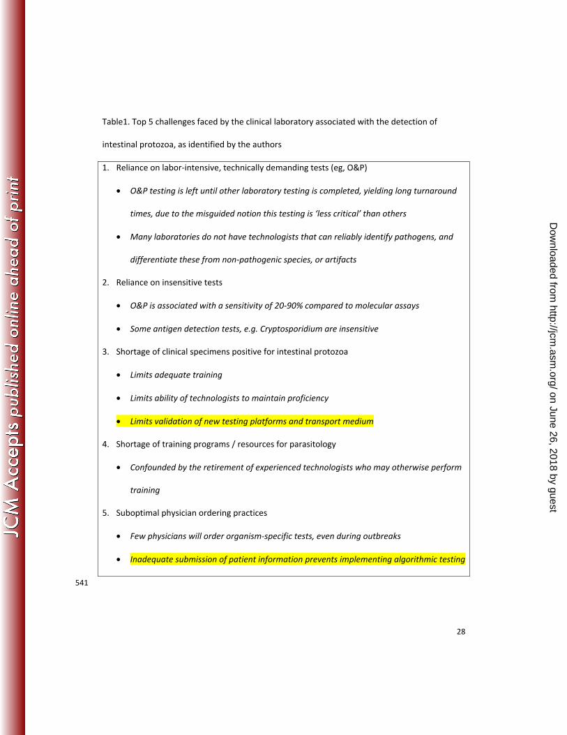
Table1. Top 5 challenges faced by the clinical laboratory associated with the detection of
intestinal protozoa, as identified by the authors
1. Reliance on labor-intensive, technically demanding tests (eg, O&P)
• O&P testing is left until other laboratory testing is completed, yielding long turnaround
times, due to the misguided notion this testing is ‘less critical’ than others
• Many laboratories do not have technologists that can reliably identify pathogens, and
differentiate these from non-pathogenic species, or artifacts
2. Reliance on insensitive tests
• O&P is associated with a sensitivity of 20-90% compared to molecular assays
• Some antigen detection tests, e.g. Cryptosporidium are insensitive
3. Shortage of clinical specimens positive for intestinal protozoa
• Limits adequate training
• Limits ability of technologists to maintain proficiency
• Limits validation of new testing platforms and transport medium
4. Shortage of training programs / resources for parasitology
• Confounded by the retirement of experienced technologists who may otherwise perform
training
5. Suboptimal physician ordering practices
• Few physicians will order organism-specific tests, even during outbreaks
• Inadequate submission of patient information prevents implementing algorithmic testing
541
on June 26, 2018 by guesthttp://jcm
.asm.org/
Dow
nloaded from
29
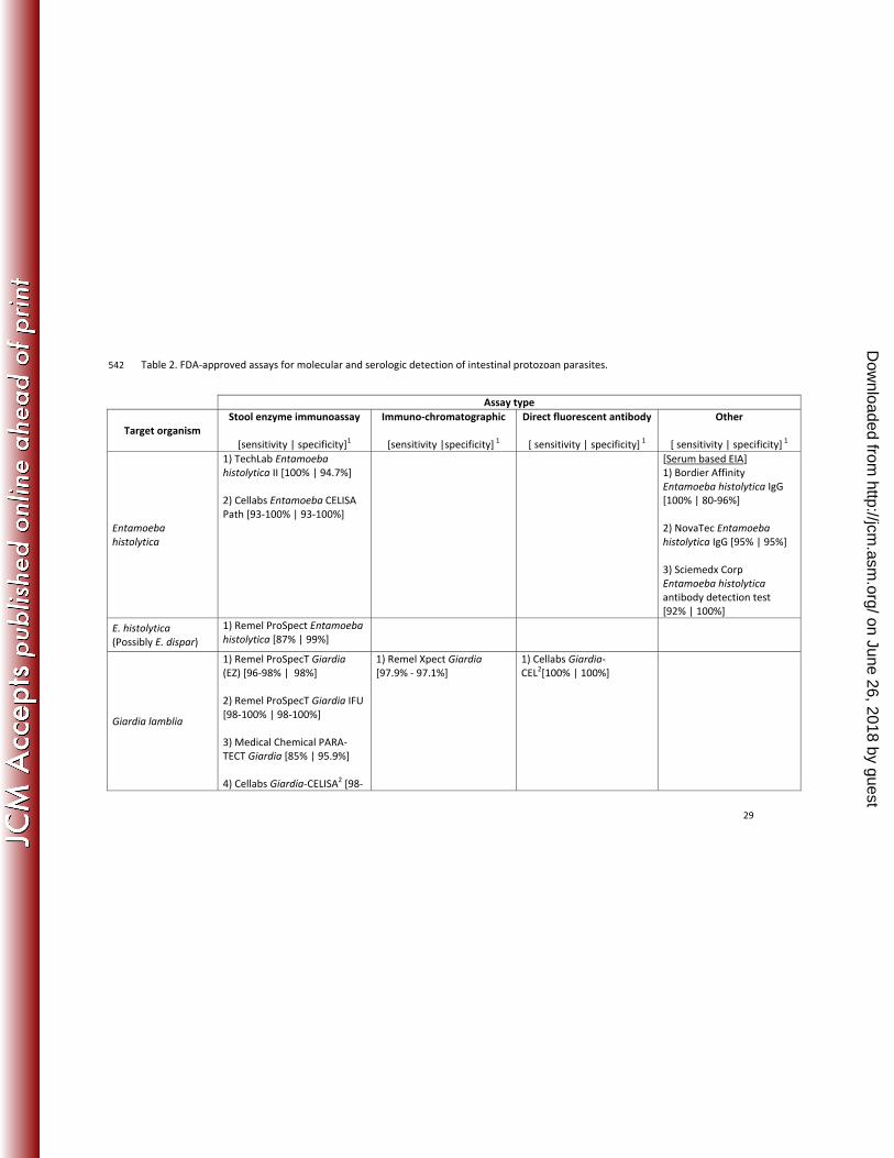
Table 2. FDA-approved assays for molecular and serologic detection of intestinal protozoan parasites. 542
Assay type
Target organism
Stool enzyme immunoassay
[sensitivity | specificity]1
Immuno-chromatographic
[sensitivity |specificity] 1
Direct fluorescent antibody
[ sensitivity | specificity] 1
Other
[ sensitivity | specificity] 1
Entamoeba
histolytica
1) TechLab Entamoeba
histolytica II [100% | 94.7%]
2) Cellabs Entamoeba CELISA
Path [93-100% | 93-100%]
[Serum based EIA]
1) Bordier Affinity
Entamoeba histolytica IgG
[100% | 80-96%]
2) NovaTec Entamoeba
histolytica IgG [95% | 95%]
3) Sciemedx Corp
Entamoeba histolytica
antibody detection test
[92% | 100%]
E. histolytica
(Possibly E. dispar)
1) Remel ProSpect Entamoeba
histolytica [87% | 99%]
Giardia lamblia
1) Remel ProSpecT Giardia
(EZ) [96-98% | 98%]
2) Remel ProSpecT Giardia IFU
[98-100% | 98-100%]
3) Medical Chemical PARA-
TECT Giardia [85% | 95.9%]
4) Cellabs Giardia-CELISA2 [98-
1) Remel Xpect Giardia
[97.9% - 97.1%]
1) Cellabs Giardia-
CEL2[100% | 100%]
on June 26, 2018 by guesthttp://jcm
.asm.org/
Dow
nloaded from
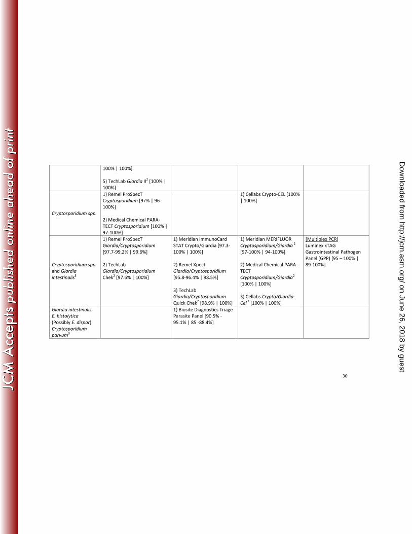
30
100% | 100%]
5) TechLab Giardia II2 [100% |
100%]
Cryptosporidium spp.
1) Remel ProSpecT
Cryptosporidium [97% | 96-
100%]
2) Medical Chemical PARA-
TECT Cryptosporidium [100% |
97-100%]
1) Cellabs Crypto-CEL [100%
| 100%]
Cryptosporidium spp.
and Giardia
intestinalis3
1) Remel ProSpecT
Giardia/Cryptosporidium
[97.7-99.2% | 99.6%]
2) TechLab
Giardia/Cryptosporidium
Chek2 [97.6% | 100%]
1) Meridian ImmunoCard
STAT Crypto/Giardia [97.3-
100% | 100%]
2) Remel Xpect
Giardia/Cryptosporidium
[95.8-96.4% | 98.5%]
3) TechLab
Giardia/Cryptosporidium
Quick Chek2 [98.9% | 100%]
1) Meridian MERIFLUOR
Cryptosporidium/Giardia 2
[97-100% | 94-100%]
2) Medical Chemical PARA-
TECT
Cryptosporidium/Giardia2
[100% | 100%]
3) Cellabs Crypto/Giardia-
Cel 2
[100% | 100%]
[Multiplex PCR]
Luminex xTAG
Gastrointestinal Pathogen
Panel (GPP) [95 – 100% |
89-100%]
Giardia intestinalis
E. histolytica
(Possibly E. dispar)
Cryptosporidium
parvum3
1) Biosite Diagnostics Triage
Parasite Panel [90.5% -
95.1% | 85 -88.4%]
on June 26, 2018 by guesthttp://jcm
.asm.org/
Dow
nloaded from
31
1 [% sensitivity | % specificity] taken from package inserts of each product. Performance characteristics for these tests in individual 543
laboratories may vary from the data presented in the package inserts. 2Only detects Giardia cysts.
3The range of sensitivity and 544
specificity for all targets are listed. 545
Table 3. Common fixatives used to preserve ova and parasites in stool. 546
Preservative
Downstream
preparations
Downstream
assays
Potential for single vial use Notes
5% or 10%
Formalin
Only Concentrated
wet mount
EIA, FA, IC Poor
Poor NAT potential and
poor trophozoite
preservation
SAF
Permanent stained
smear &
Concentrated wet
mount
EIA, FA, IC
Fair
Poor NAT potential.
Suboptimal trophozoite
morphology.
Mercury based
Fixative with PVA
Permanent stained
smear &
NAT Poor
Immunoassays not possible
and fixative is highly toxic
on June 26, 2018 by guesthttp://jcm
.asm.org/
Dow
nloaded from
32
Concentrated wet
mount (rare)
Modified
Schaudinn's
(Copper, Zinc or
other fixative with
PVA)
Permanent stained
smear and
Concentrated wet
mount (rare)
NAT Fair
Immunoassays not possible
and concentrated wet
mounts are uncommonly
performed. Suboptimal
trophozoite morphology.
Single Vial
proprietary
fixative
formulations
Permanent stained
smear and
Concentrated wet
mount
Some
immunoassays are
possible. Most
NATs
Good
Suboptimal trophozoite
morphology. Not all
immunoassays are possible.
EIA, enzyme immunoassay; FA, fluorescent antibody; IC, immunochromographic tests; NAT, nucleic acid amplification tests547
on June 26, 2018 by guesthttp://jcm
.asm.org/
Dow
nloaded from
33
Figure legend 548
Figure. Key microscopic morphology of the enteropathogenic protozoa. Organisms are ordered 549
from largest to smallest, based on average cell size. (A) Balantidium coli trophozoite unstained 550
on wet mount. (B) Cystoisospora belli oocyst, (G) Cyclospora cayetanensis oocyst and (H) 551
Cryptosporidium spp. oocyst shown after modified acid fast staining. Trophozoite forms are 552
shown stained with trichrome for (C) E. histolytica, (D) D. fragilis, and (F) B. hominis. (E) Cyst 553
form of Giardia stained with trichrome. 554
on June 26, 2018 by guesthttp://jcm
.asm.org/
Dow
nloaded from
Romney M. Humphries, Ph.D., D(ABMM), is an Assistant Professor in the Department of 1
Pathology and Laboratory Medicine in the David Geffen School of Medicine at the University of 2
California, Los Angeles. She is also the Section Chief for Clinical Microbiology at the UCLA Health 3
System, and program co-director for UCLA’s CPEP fellowship program. Romney received her 4
undergraduate degree in biochemistry from the University of Lethbridge, Canada. She 5
completed her PhD in bacterial pathogenesis in the laboratory of Dr. Glen Armstrong, at the 6
University of Calgary, Canada. Here, she investigated novel anti-infective strategies for the 7
prevention of bacterial gastroenteritis caused by Enteropathogenic Escherichia coli. Romney 8
subsequently completed a CPEP fellowship in Medical and Public Health Microbiology at the 9
University of California, Los Angeles under the direction of Dr. Michael Lewinski. One of 10
Romney’s research focuses is the detection and characterization of pathogens that cause 11
gastrointestinal diseases. 12
on June 26, 2018 by guesthttp://jcm
.asm.org/
Dow
nloaded from
![BMC Gastroenterology BioMed Central · the most sensitive imaging technique for intestinal endometriosis [13]. Yet, the gold standard for the diagno-sis is laparoscopy or laparotomy](https://img.pdfslide.us/doc/110x75/5f49e8e19d173238170d0077/bmc-gastroenterology-biomed-central-the-most-sensitive-imaging-technique-for-intestinal.jpg)

![fwei.he;Chao.Li;naoto.yokoya;qibin.Zhaog@riken.jp, qyaoaa ... · of application in remote sensing [35,36], medical diagno-sis [22], face recognition [30,36], quality control [19]](https://img.pdfslide.us/doc/110x75/5f0e8f397e708231d43fd3a8/fweihechaolinaotoyokoyaqibinzhaogrikenjp-qyaoaa-of-application-in.jpg)