Embed Size (px)
Citation preview
Tuberculous Ulcer of Oral Cavity
Oral and Maxillofacial Pathology Journal, July-December 2017;8(2):105-107 105
OMPJ
Tuberculous Ulcer of Oral Cavity1Neethu Kadar, 2Anila Namboodiripad, 3Mohamed Haris, 4R Divya
ABSTRACT
Introduction: Tuberculosis (TB) is a chronic granulomatous infection caused primarily by Mycobacterium tuberculosis. Although the pulmonary system is most commonly affected, TB can be manifested in other regions (extrapulmonary) including oral cavity. Oral TB can be either a primary lesion; or secondary to the underlying pulmonary TB.
Case report: The present case describes secondary TB mani-fested as a large nonhealing ulcerative lesion in the posterior palatal region of a 46-year-old male patient. Clinical examination revealed palpable submandibular and cervical lymph nodes along with a history of smoking habit, thereby leading clinicians to suspect malignancy. Biopsy and histopathological examina-tion of the oral lesion showed caseating granulomas. On referral for further investigations, pulmonary TB was diagnosed. The patient underwent antitubercular drug therapy for 6 months and the oral lesion subsided within 1 month of treatment.
Conclusion: Presentation of tuberculous lesions in the oral cavity is usually nonspecific and hence, they are often mistaken for other differential diagnoses. A detailed clinical examination followed by routine laboratory investigations has a prime role in early diagnosis.
Keywords: Extrapulmonary tuberculosis, Oral ulcer, Secondary tuberculosis.
How to cite this article: Kadar N, Namboodiripad A, Haris M, Divya R. Tuberculous Ulcer of Oral Cavity. Oral Maxillofac Pathol J 2017;8(2):105-107.
Source of support: Nil
Conflict of interest: None
INTRODUCTION
Tuberculosis (TB) is a chronic granulomatous infection caused primarily by Mycobacterium tuberculosis. Tubercle bacillus is an aerobic, acid-fast organism usually trans-mitted via droplet inhalation. Although the pulmonary system is most commonly affected, TB can be manifested
OMPJ
Case RePORt10.5005/jp-journals-10037-1111
1,3Senior Lecturer, 2Professor, 4Postgraduate Student1,2,4Department of Oral Pathology and Microbiology, Royal Dental College, Palakkad, Kerala, India3Department of Periodontics, Malabar Dental College & Research Centre, Malappuram, Kerala, India
Corresponding Author: Neethu Kadar, Senior Lecturer Department of Oral Pathology and Microbiology, Royal Dental College, Palakkad, Kerala, India, e-mail: [email protected]
in other regions (extrapulmonary) including the oral cavity.1 Based on the World Health Organization (WHO) 2016 reports, about 10.4 million new cases and 1.8 million deaths occur globally every year. India is among the six “high burden countries” that comprise 60% of total TB cases.2 Oral TB is a rare entity accounting for only 1.4% of total cases.3 But currently, tuberculous oral lesions are reemerging along with other extrapulmonary manifesta-tions as an outcome of drug-resistant TB and acquired immunodeficiency syndrome.4
This article portrays secondary TB manifested as a large nonhealing ulcerative lesion in the posterior palatal region of a 46-year-old male patient.
CASE REpORT
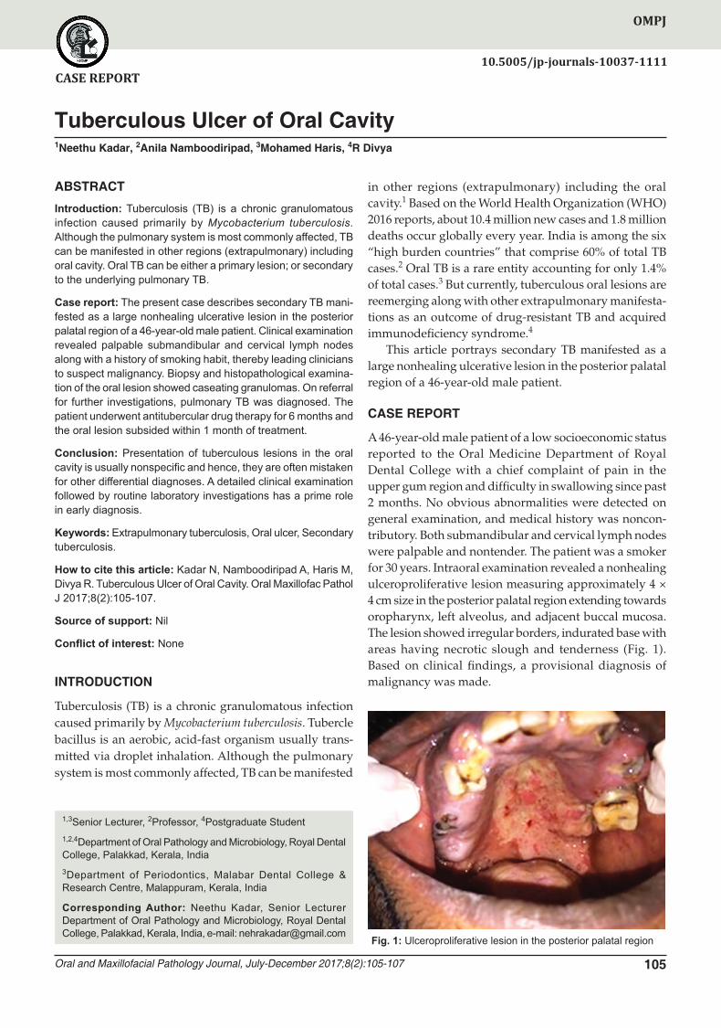
A 46-year-old male patient of a low socioeconomic status reported to the Oral Medicine Department of Royal Dental College with a chief complaint of pain in the upper gum region and difficulty in swallowing since past 2 months. No obvious abnormalities were detected on general examination, and medical history was noncon-tributory. Both submandibular and cervical lymph nodes were palpable and nontender. The patient was a smoker for 30 years. Intraoral examination revealed a nonhealing ulceroproliferative lesion measuring approximately 4 × 4 cm size in the posterior palatal region extending towards oropharynx, left alveolus, and adjacent buccal mucosa. The lesion showed irregular borders, indurated base with areas having necrotic slough and tenderness (Fig. 1). Based on clinical findings, a provisional diagnosis of malignancy was made.
Fig. 1: Ulceroproliferative lesion in the posterior palatal region
Neethu Kadar et al
106
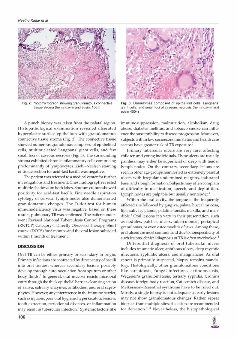
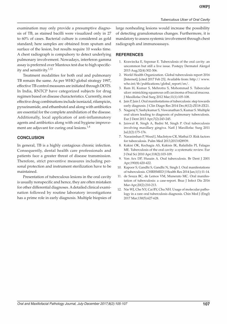
A punch biopsy was taken from the palatal region. Histopathological examination revealed ulcerated hyperplastic surface epithelium with granulomatous connective tissue stroma (Fig. 2). The connective tissue showed numerous granulomas composed of epithelioid cells, multinucleated Langhans’ giant cells, and few small foci of caseous necrosis (Fig. 3). The surrounding stroma exhibited chronic inflammatory cells comprising predominantly of lymphocytes. Ziehl–Neelsen staining of tissue section for acid-fast bacilli was negative.
The patient was referred to a medical center for further investigations and treatment. Chest radiograph revealed multiple shadows on both lobes. Sputum culture showed positivity for acid-fast bacilli. Fine needle aspiration cytology of cervical lymph nodes also demonstrated granulomatous changes. The Tridot test for human immunodeficiency virus was negative. Based on these results, pulmonary TB was confirmed. The patient under-went Revised National Tuberculosis Control Programs (RNTCP) Category-1 Directly Observed Therapy, Short course (DOTS) for 6 months and the oral lesion subsided within 1 month of treatment.
DISCUSSION
Oral TB can be either primary or secondary in origin. Primary infections are contracted by direct entry of bacilli into oral tissues, whereas secondary lesions possibly develop through autoinoculation from sputum or other body fluids.5 In general, oral mucosa resists microbial entry through the thick epithelial barrier, cleansing action of saliva, salivary enzymes, antibodies, and oral sapro-phytes. However, any interference in the immune barrier, such as injuries, poor oral hygiene, hyperkeratotic lesions, tooth extraction, periodontal diseases, or inflammation may result in tubercular infection.6 Systemic factors like
immunosuppression, malnutrition, alcoholism, drug abuse, diabetes mellitus, and tobacco smoke can influ-ence the susceptibility to disease progression. Moreover, subjects within low socioeconomic status and health care sectors have greater risk of TB exposure.7
Primary tubercular ulcers are very rare, affecting children and young individuals. These ulcers are usually painless, may either be superficial or deep with tender lymph nodes. On the contrary, secondary lesions are seen in older age groups manifested as extremely painful ulcers with irregular undermined margins, indurated base, and slough formation. Subjects may often complain of difficulty in mastication, speech, and deglutition. Lymph nodes are palpable but usually nontender.1
Within the oral cavity, the tongue is the frequently affected site followed by gingiva, palate, buccal mucosa, lips, salivary glands, palatine tonsils, maxilla, and man-dible.8 Oral lesions can vary in their presentation, such as nodules, patches, ulcers, tuberculomas, periapical granulomas, or even osteomyelitis of jaws. Among these, oral ulcers are most common and due to nonspecificity of such lesions, clinical diagnosis of TB is often overlooked.4
Differential diagnosis of oral tubercular ulcers includes traumatic ulcer, aphthous ulcers, deep mycotic infections, syphilitic ulcers, and malignancies. As oral cancer is primarily suspected, biopsy remains manda-tory. Histologically, other granulomatous conditions like sarcoidosis, fungal infections, actinomycosis, Wegener’s granulomatosis, tertiary syphilis, Crohn’s disease, foreign body reaction, Cat scratch disease, and Melkersson–Rosenthal syndrome have to be ruled out. Mostly, a single biopsy is not adequate as early lesions may not show granulomatous changes. Rather, repeat biopsies from multiple sites of a lesion are recommended for detection.9-11 Nevertheless, the histopathological
Fig. 2: Photomicrograph showing granulomatous connective tissue stroma (hematoxylin and eosin, 100×)
Fig. 3: Granulomas composed of epithelioid cells, Langhans’ giant cells, and small foci of caseous necrosis (hematoxylin and eosin 400×)
Tuberculous Ulcer of Oral Cavity
Oral and Maxillofacial Pathology Journal, July-December 2017;8(2):105-107 107
OMPJ
examination may only provide a presumptive diagno-sis of TB, as stained bacilli were visualized only in 27 to 60% of cases. Bacterial culture is considered as gold standard; here samples are obtained from sputum and surface of the lesion, but results require 10 weeks time. A chest radiograph is compulsory to detect underlying pulmonary involvement. Nowadays, interferon gamma assay is preferred over Mantoux test due to high specific-ity and sensitivity.1,12
Treatment modalities for both oral and pulmonary TB remain the same. As per WHO global strategy 1997, effective TB control measures are initiated through DOTS. In India, RNTCP have categorized subjects for drug regimen based on disease characteristics. Currently, most effective drug combinations include isoniazid, rifampicin, pyrazinamide, and ethambutol and along with antibiotics are essential for the complete annihilation of the disease. Additionally, local application of anti-inflammatory agents and antibiotics along with oral hygiene improve-ment are adjuvant for curing oral lesions.1,4
CONCLUSION
In general, TB is a highly contagious chronic infection. Consequently, dental health care professionals and patients face a greater threat of disease transmission. Therefore, strict preventive measures including per-sonal protection and instrument sterilization have to be maintained.
Presentation of tuberculous lesions in the oral cavity is usually nonspecific and hence, they are often mistaken for other differential diagnoses. A detailed clinical exami-nation followed by routine laboratory investigations has a prime role in early diagnosis. Multiple biopsies of
large nonhealing lesions would increase the possibility of detecting granulomatous changes. Furthermore, it is mandatory to assess systemic involvement through chest radiograph and immunoassays.
REFERENCES
1. Krawiecka E, Szponar E. Tuberculosis of the oral cavity: an uncommon but still a live issue. Postepy Dermatol Alergol 2015 Aug;32(4):302-306.
2. World Health Organization. Global tuberculosis report 2016 [Internet]; [cited 2017 Feb 23]. Available from: http:// www.who.int/tb/publications/global_report/en/.
3. Ram H, Kumar S, Mehrotra S, Mohommad S. Tubercular ulcer: mimicking squamous cell carcinoma of buccal mucosa. J Maxillofac Oral Surg 2012 Mar;11(1):105-108.
4. Jain P, Jain I. Oral manifestations of tuberculosis: step towards early diagnosis. J Clin Diagn Res 2014 Dec;8(12):ZE18-ZE21.
5. Nagaraj V, Sashykumar S, Viswanathan S, Kumar S. Multiple oral ulcers leading to diagnosis of pulmonary tuberculosis. Eur J Dent 2013 Apr;7(2):243-245.
6. Jaiswal R, Singh A, Badni M, Singh P. Oral tuberculosis involving maxillary gingiva. Natl J Maxillofac Surg 2011 Jul;2(2):175-176.
7. Narasimhan P, Wood J, MacIntyre CR, Mathai D. Risk factors for tuberculosis. Pulm Med 2013;2013:828939.
8. Kakisi OK, Kechagia AS, Kakisis IK, Rafailidis PI, Falagas ME. Tuberculosis of the oral cavity: a systematic review. Eur J Oral Sci 2010 Apr;118(2):103-109.
9. Von Arx DP, Husain A. Oral tuberculosis. Br Dent J 2001 Apr;190(8):420-422.
10. Kapoor S, Gandhi S, Gandhi N, Singh I. Oral manifestations of tuberculosis. CHRISMED J Health Res 2014 Jan;1(1):11-14.
11. de Souza BC, de Lemos VM, Munerato MC. Oral manifes-tation of tuberculosis: a case-report. Braz J Infect Dis 2016 Mar-Apr;20(2):210-213.
12. Nie WJ, Che NY, Cai BY, Chu NH. Usage of molecular patho-logy in a rare oral tuberculosis diagnosis. Chin Med J (Engl) 2017 Mar;130(5):627-628.


![Stem Cell 2017;8(1) · medical termination of pregnancy [1]. It is also used in the treatment of sarcoidosis, vasculitis, inflammatory bowel diseases, and severe refractory asthma](https://img.pdfslide.us/doc/110x75/5e1d3440b41cb87d0968a1e7/stem-cell-201781-medical-termination-of-pregnancy-1-it-is-also-used-in-the.jpg)