Embed Size (px)
Citation preview

8/21/2018
1
Joseph Sowka, OD, FAAO, DiplomateNova Southeastern University College of Optometry
Joseph Sowka, OD is/ has been a Consultant/ Speaker Bureau/Advisory Board member for Novartis, Allergan, Glaukos, andB&L. Dr. Sowka has no direct financial interest in any of thediseases, products or instrumentation mentioned in thispresentation. He is a co-owner of Optometric EducationConsultants (www.optometricedu.com)
The ideas, concepts, conclusions and perspectives presented
herein reflect the opinions of the speaker; he has not been
paid, coerced, extorted or otherwise influenced by any third
party individual or entity to present information that conflicts
with his professional viewpoints.
DISCLOSURE:
Conundrum: Is This Really
Glaucoma?
IS THIS REALLY GLAUCOMA? WHY
DOES IT MATTER?
Treating a disease that they don’t have
- Expense and adverse effects
Treating one disease when its really another
- Vision loss and potentially worse
Not treating a disease that they do have
- Vision loss
In reality, when encountering patients with
mimicking and confounding conditions, the
diagnosis is challenging
NOT ALL –’OMAS’ ARE
GLAUCOMA
Pituitary adenoma
Craniopharyngioma
Meningioma
Glioma
Ischemioma
- Anterior ischemic optic neuropathy (AAION- cup enlargement but
devastating vision loss and disc pallor)
Retinaloma
- Retinal infarcts
Congenitaloma
Coincidentaloma
Misdiagnosoma
RULE #1
Pallor in excess of cupping indicates
something other than glaucoma
When the rim tissue is pale, suspect some
cause other than, or in addition to, glaucoma.
Rim pallor in glaucoma is very rare and is the
exception, not the rule.
- Disc pallor can only be accepted as part of glaucoma
when other potential causes have been eliminated
8/21/2018
2
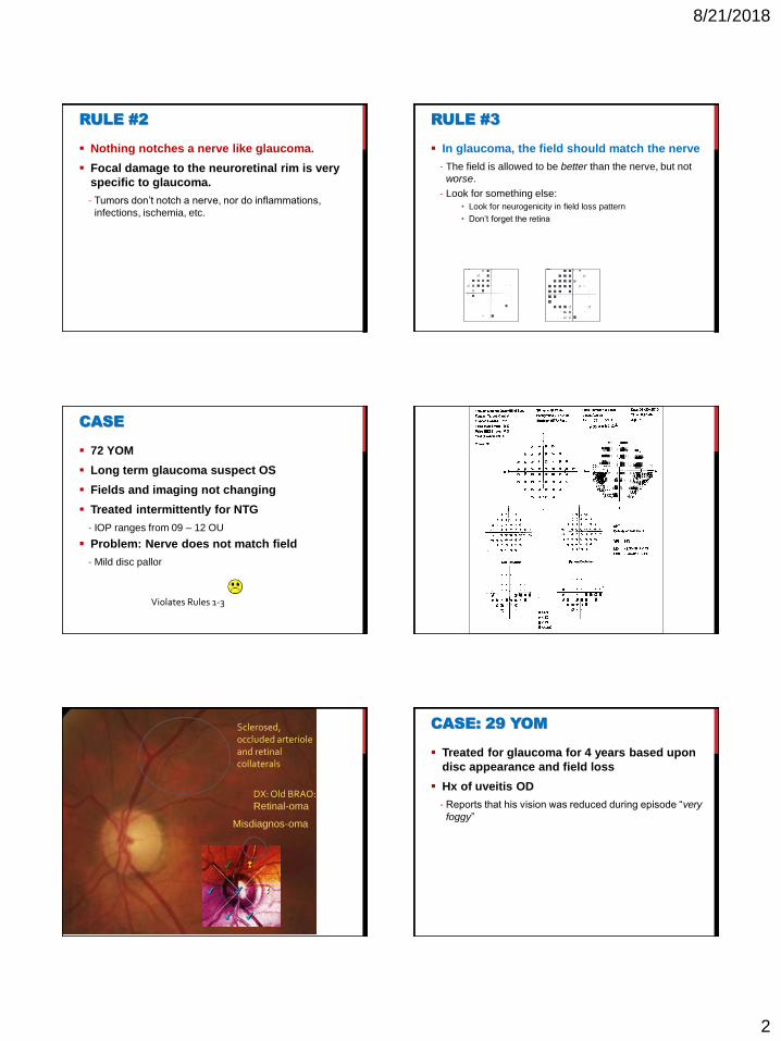
RULE #2
Nothing notches a nerve like glaucoma.
Focal damage to the neuroretinal rim is very
specific to glaucoma.
- Tumors don’t notch a nerve, nor do inflammations,
infections, ischemia, etc.
RULE #3
In glaucoma, the field should match the nerve
- The field is allowed to be better than the nerve, but not
worse.
- Look for something else:
• Look for neurogenicity in field loss pattern
• Don’t forget the retina
CASE
72 YOM
Long term glaucoma suspect OS
Fields and imaging not changing
Treated intermittently for NTG
- IOP ranges from 09 – 12 OU
Problem: Nerve does not match field
- Mild disc pallor
Violates Rules 1-3
Sclerosed, occluded arteriole and retinal collaterals
DX: Old BRAO: Retinal-oma
Misdiagnos-oma
CASE: 29 YOM
Treated for glaucoma for 4 years based upon
disc appearance and field loss
Hx of uveitis OD
- Reports that his vision was reduced during episode “very
foggy”
8/21/2018
3
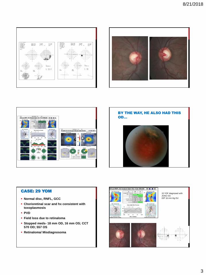
BY THE WAY, HE ALSO HAD THIS
OD…
CASE: 29 YOM
Normal disc, RNFL, GCC
Chorioretinal scar and hx consistent with
toxoplasmosis
PVD
Field loss due to retinaloma
Stopped meds- 18 mm OD, 16 mm OS; CCT
570 OD; 557 OS
Retinaloma/ Misdiagnosoma
15 YOF diagnosed with
JOAG OU
IOP 16 mm Hg OU
8/21/2018
4
Age younger than 50 years was 93% specific
for nonglaucomatous cupping.
Eyes with nonglaucomatous cupping
associated with intracranial masses had
significantly lower levels of visual acuity than
did patients with glaucoma.
- visual acuity less than 20/40 was 77% specific for
nonglaucomatous cupping.
Eyes with glaucoma had significantly less
neuroretinal rim pallor, larger CDR, greater
vertical elongation of the optic cup, and
greater frequency of peripapillary atrophy
and disc hemorrhage than eyes with
nonglaucomatous cupping.
- disc hemorrhage was 100% specific for glaucoma
Pallor of the optic nerve in excess of cupping
is a highly specific sign of nonglaucomatous
cupping (90.4%)
“THE CUPPED DISC: WHO NEEDS
NEUROIMAGING?”
Patients with mass lesions:
- Visual acuity less than 20/40
- Vertically aligned visual fields defects
- Optic disc pallor in excess of cupping
- Age younger than 50 years
Greenfield DS, Siatkowski RM, Glaser JS, et al. The cupped disc: Who needs neuroimaging? Ophthalmology 1998; 105:1866-74.
CASE: 56 YOF
Dx POAG OU 5 years ago
Slowly progressive vision loss
LP OD; 20/30 OS
Used combo med- ran out months ago
IOP: 19 mm OD, 18 mm OS
CCT: 560; 544
8/21/2018
5
DIAGNOSING GLAUCOMA IN
ANOMALOUS DISCS
Rules of disc analysis rarely work in
anomalous discs
Look at other clues and risk factors
Recognize that we will likely make diagnostic
errors
Ensure that errors have least detrimental
effect to patient
72 YOM
Amblyopia OD and bilateral dense
cataracts
Undergoes cataract surgery OS
20/50 PO OS- no complications
Pt very unhappy with outcome
Diagnosed with advanced NTG
- Peak IOP 22 mm; CCT: 536, 531
- Referred to glaucoma specialist- treated to low
teens IOP
72 YOM: CHALLENGES
Doesn’t believe he has NTG
Semi-retired attorney who has:
Time
Money
Excellent internet access
Will stop at nothing to discover what is
wrong.
Marked Tilted Disc syndrome OU
Congenital-oma
How much field loss is due to TDS?
Field loss OS doesn’t explain vision
CASE 7: 72 YOWM
Evaluation including neuroimaging
unremarkable
- Macular OCT normal
Travels country consulting
- Conflicting opinions
Significant field loss OU
Possibly glaucoma as well as congenitaloma
(Double-oma)
- Continue treatment- “Do No Harm”
8/21/2018
6
JP: 38 YOF
Referred for glaucoma eval in 2002 after
failing LASIK screening
Had been treated since mid 20s for glaucoma
IOP in mid-upper teens off meds
CCT: 459 OD; 469 OS
Anomalous nerves with mild field loss
JP: NOW 49 YOF
Congenitally anomalous nerves with field
loss
Monitored for 11+ years
Field changes late
Pt now treated with IOP 09 mm OD; 10 mm OS
Pt had/had congenitaloma and now has
glaucoma
- Doubloma
SIMILAR…YET DIFFERENT
45 YOF
Referred for glaucoma evaluation
IOP never exceeds mid-teens
CCT: 554 OU
Marginal effect of meds
CONUNDRUMS
Field loss due to anomaly, glaucoma, or
both?
Progressive or congenital?
Mid-teen IOP and poor medical response
Treatment or observation?

8/21/2018
7
CONCLUSIONS
Disc pallor indicates something other than
glaucoma
Rim obliteration/ notching indicates
glaucoma
Glaucoma without risk factors is suspicious
Differentiating glaucoma from non-glaucoma
is challenging when risk factors (IOP) are
present
Fields and nerve should match
If discs are anomalous, mistakes can be
made. Err on the side of caution
ODE TO A CUPPED DISC
Oh, to have a cupped disc pink.
That my friend hath a glaucomatous stink.
But to have a cupped disc pale,
Call this glaucoma and you shall fail.
Disc and field damage that is one-sided
Simply cannot be abided.
It might be trauma, infarct or meningioma.
But if the rim is cut always remember,
Nothing notches a nerve like glaucoma
Conundrum:
The diagnostic imaging
doesn’t agree with my
diagnosis? Now what?
ANSWER:
Things have to make sense. If the imaging
findings to not fit with the anatomic and
functional correlates of pathophysiologic
change, trust your own knowledge and
judgment.
When in doubt, repeat the imaging study and
the visual field or both.
OCT TO VERIFY GLAUCOMA –
THE OPTIC NERVE HEAD?
RED DISEASE –
A NEW CLINICAL NON-ENTITY
A supratentorial, non-glaucomatous masquerade disease
Afflicts the educated patient (especially with Internet access)
with good health care plans and/or wealth
Debilitating to the patient and painful for the visual care
provider to treat
2005. Journal of Irreproducible Results and Senseless Studies

8/21/2018
8
WHAT DO YOU MAKE OF THESE…?
Garbage in, Garbage out.
HELP! THE DIAGNOSTIC IMAGING
DOESN’T AGREE WITH MY DIAGNOSIS!
Low risk OHTN
Local OD wants imaging for baseline
OCT RNFL NORMAL…
…but markedly abnormal
GCC OSSame patient, same day, same
quality, GCC now normal
Signal strength: 10/10 OD, OS on
both images
Don’t make clinical decisions based upon
bad data
CASE: 62 YOHM
Asymptomatic; 20/20 OD; OS
TA 30 mm OD, 28 mm OS
- Isolated measurement
- 12-17 mm OD, 13-17 mm OS
• 11 visits
Gonio: open OU w/o abnormalities
CCT: 597 OU

8/21/2018
9
So, What are your
thoughts?
Debate: Treat or Observe?Debate: Why the disparate findings?
Debate: Why the isolated IOP elevation?
GREEN DISEASE– AN INSIDIOUS
CLINICAL ENTITY
A glaucomatous process masquerading as non-disease
Afflicts inexperienced, poorly-educated, and lazy doctors who
simply want a machine to make all clinical decisions for them
Debilitating to the patient and painful for the visual care
provider, but a boon for malpractice attorneys
2015. Journal of Irreproducible Results and Senseless Studies
HELP! THE DIAGNOSTIC IMAGING
DOESN’T AGREE WITH MY DIAGNOSIS!
56 YOM- Glaucoma suspect since 2012

8/21/2018
10
Is this person
really a
glaucoma
‘suspect’?
A example of
Green
Disease
Green Disease
GREEN DISEASE GREEN DISEASE
8/21/2018
11
RED + GREEN =
YELLOW DISEASE?
PANOMAP
VERSION 8.0
PANOMAP
VERSION 8.0

8/21/2018
12
OCT IMAGING TAKE HOME POINTS
Serial overlays/imaging to determine
baseline (intra-session) noise
Good signal strength
Good segmentation without errors
Optic nerve head exam for disc
hemorrhage, pallor, myopic, and tilted
nerve heads
Determine structure-function correlation
Follow all ancillary tests visual fields and
optic nerve head photos for progression
CAUTIONS ABOUT IMAGING
No current technology is better than the
human eye and common sense
Beware of “Red Disease”
Treat Real Disease and not Red Disease
Don’t miss green disease
Know the limitations of the technology:
normative database, reproducibility,
resolution, quality of imaging
Technologies come and go
Conundrum: WHEN IS SURGERY
WRONG FOR THE PATIENT?
ANSWER:
When the risk of surgery is greater than its
expected benefit.
When it is more dangerous to undergo a
surgical procedure than to continue on the
same medical treatment.
When you would not recommend the same
intervention to your family members
GLAUCOMA SURGICAL DECISION
MAKING
Establishing the course of treatment
- Is the disc or field status stable or worse?
- If progression has occurred, over what time period?
- What is the rate of change?
- What is the risk of visual disability in the patient’s
lifetime?
- Is the patient aware of either decreased central visual
acuity or peripheral visual field loss?
• Classic question: Is it the cataract or the glaucoma or the age
related macular degeneration?
IMPORTANT QUESTIONS ABOUT
VALUE OF SURGICAL INTERVENTION
– HOW FAR TO GO?
- Does the patient value the visual acuity of Hand Motions or
Light Perception or remaining visual field?
- What is the status of the fellow eye?
- Is glaucoma a primary condition or related to a cause
(proliferative diabetic retinopathy, central retinal vein
occlusion, trauma)?
- Has a family member become visually disabled from
glaucoma?
- Has a family member lost vision after glaucoma surgery?

8/21/2018
13
IS FILTERING SURGERY A
PANACEA?
Trabeculectomy will give low IOP
- Single digits
Long history of success
Technically straightforward process
Eye never looks/ feels the same
Potential complications
RISKS OF GLAUCOMA SURGERY
Trabeculectomy
- Immediate postoperative period
• Hypotony – flat anterior chamber, acute cataract, angle
closure, choroidal effusion
• “Wipe out” or “snuff out” syndrome – acute loss of central
acuity without obvious intraoperative complication
• Decreased visual acuity - Patient only knows that they
see much worse after surgery
Glaucoma drainage implant surgery
- Muscle imbalance – noncommitant diplopia
ADDITIONAL RISKS OF GLAUCOMA
SURGERY
Late postoperative period
- Posterior synechiae formation – poor dilation
- Cataract formation
- Bleb scarring and return of high IOP
Very late postoperative period
• Endophthalmitis and blebitis
• Remember “RSVP” • R – Redness
• S – Sensitivity to light
• V – Vision Change
• P – Pain
Edna
20/20 OD, OS
Age 37
10-2 SS OS 10-2 SS OD
JUNIOR: 20/60 OD; 20/400 OS
56 YO
8/21/2018
14
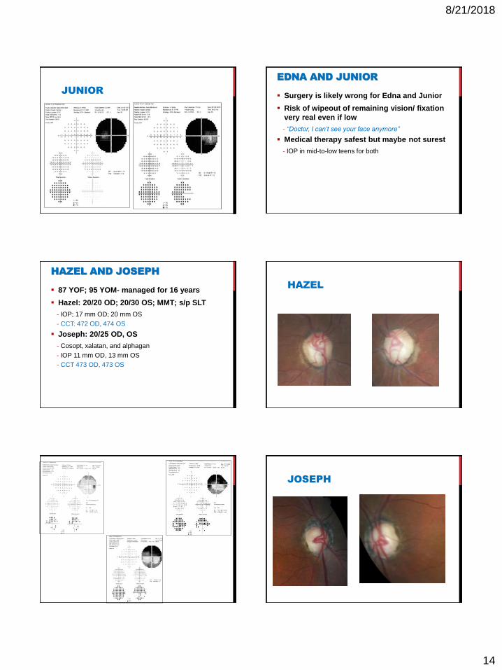
JUNIOR
EDNA AND JUNIOR
Surgery is likely wrong for Edna and Junior
Risk of wipeout of remaining vision/ fixation
very real even if low
- “Doctor, I can’t see your face anymore”
Medical therapy safest but maybe not surest
- IOP in mid-to-low teens for both
HAZEL AND JOSEPH
87 YOF; 95 YOM- managed for 16 years
Hazel: 20/20 OD; 20/30 OS; MMT; s/p SLT
- IOP; 17 mm OD; 20 mm OS
- CCT: 472 OD, 474 OS
Joseph: 20/25 OD, OS
- Cosopt, xalatan, and alphagan
- IOP 11 mm OD, 13 mm OS
- CCT 473 OD, 473 OS
HAZEL
JOSEPH

8/21/2018
15
JOSEPH
HAZEL AND JOSEPH
For whom is surgery right and for whom is
surgery wrong?
SUMMARY
Pts with bare fixation are at high risk of
surgical morbidity
- At some point even aggressive surgeons will decline
• “Better God than I take their vision”
Surgery is wrong for your patient when
someone you trust as your surgical
consultant would not recommend the same
procedure to their own family member.
Conundrum:
Help! My patient has a
disc hemorrhage and the
pressure is 12 mm. Now
what?
DISC HEMORRHAGES
Likely mechanical
Resolves within 6 weeks. This is the reason
that the incidence is difficult to determine.
Can be recurrent and, if it recurs, it typically
is in the same place on the disc each time
Disc hemorrhages do not constitute a
diagnosis of glaucoma nor a progression or
conversion to glaucoma or an endpoint for
any major glaucoma
Not all hemorrhages of the disc are disc hemorrhages.

8/21/2018
16
RISK FACTORS: DISC
HEMORRHAGES
Inferior, inferior temporal,
superior, and superior
temporal regions of the disc
are most susceptible and
account for virtually all true
glaucomatous disc
hemorrhages
Typically occurs where notches
and RNFL defects occur
Hemorrhages at other areas of the disc (nasal and temporal)
tend to not be associated with glaucoma.
OTHER CAUSES OF ‘DISC’HEMORRHAGES
PVD
HTN
Anemia
Diabetes
Vascular occlusion
Subarachnoid bleed
- Terson’s syndrome
• Subretinal and intraretinal
• May be juxtapapillary
BRVO PVD
Terson’s
55 YOF
Referred for NTG evaluation based upon disc
hemorrhage OS
IOP: 14 mm OD, 15 mm OS
0.3/0.3 OU without pallor, notching or RNFL
defect
OCT/ GDx- normal OU
55 YOF
Disc hemorrhages not characteristic of
glaucomatous hemorrhages
Structure normal
Plan: observation

8/21/2018
17
These photos are 18 months apart
Not all hemorrhages of the disc are disc hemorrhages.
Make sure that the glaucomatous characteristics
are there.
EARLY MANIFEST GLAUCOMA
TRIAL
Disc hemorrhages- predictive of progression
Treatment was unrelated to the presence or
frequency of disc hemorrhages.
- Disc hemorrhages were equally common in both the
treated and untreated groups of patients.
- Disc hemorrhages don’t occur in all glaucoma pts.
Disc hemorrhages cannot be considered an
indication of insufficient IOP-lowering
treatment,
- Glaucoma progression in eyes with disc hemorrhages
cannot be totally halted by IOP reduction.
OCULAR HYPERTENSION
TREATMENT STUDY
The occurrence of a disc hemorrhage
increased the risk of developing POAG 6-fold
in a univariate analysis and 3.7-fold in a
multivariate analysis that included baseline
factors predictive of POAG
Occurrence of an optic disc hemorrhage was
associated with an increased risk of
developing a POAG end point in participants
in the OHTS
- However, most eyes (86.7%) in which a disc
hemorrhage developed have not experienced a POAG
end point to date (5 years)
The incidence of ODH was 0.5% per year during
an average of 13 years before the development of
POAG and 1.2% per year during an average of 6
years after the development of POAG
ODH increased the risk of developing POAG 2.6-
fold in multivariate analysis
Randomization to the observation group, older
age, thinner central corneal thickness, larger
vertical cup-to-disc ratio, higher intraocular
pressure, and self-reported black race were
identified as risk factors for ODH.
Disc hemorrhages are a significant risk factor for progression, but do not
constitute a diagnosis or actual progression. Further, you cannot stop disc hemorrhages from occurring by
lowering IOP.
8/21/2018
18
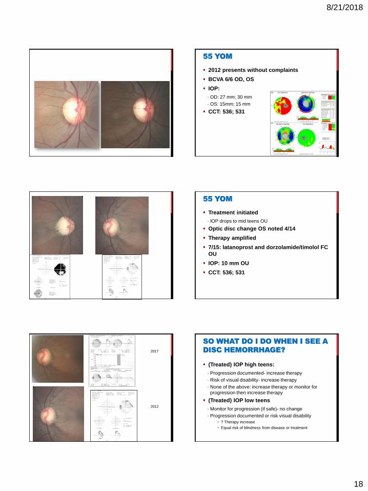
55 YOM
2012 presents without complaints
BCVA 6/6 OD, OS
IOP:
- OD: 27 mm; 30 mm
- OS: 15mm; 15 mm
CCT: 536; 531
55 YOM
Treatment initiated
- IOP drops to mid teens OU
Optic disc change OS noted 4/14
Therapy amplified
7/15: latanoprost and dorzolamide/timolol FC
OU
IOP: 10 mm OU
CCT: 536; 531
2017
2012
SO WHAT DO I DO WHEN I SEE A
DISC HEMORRHAGE?
(Treated) IOP high teens:
- Progression documented- increase therapy
- Risk of visual disability- increase therapy
- None of the above: increase therapy or monitor for
progression then increase therapy
(Treated) IOP low teens
- Monitor for progression (if safe)- no change
- Progression documented or risk visual disability
• ? Therapy increase
• Equal risk of blindness from disease or treatment

8/21/2018
19
THANK YOU FOR YOUR ATTENTION.
ALWAYS REMEMBER TO RECYCLE AND PROTECT
THE PLANET THAT WE WILL ULTIMATELY LEAVE
TO KEITH RICHARDS