Embed Size (px)
Citation preview
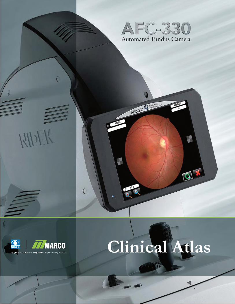
Automated Fundus Camera
Clinical Atlas

Myelinated-Nerves - A simple “birth mark.” This was the first time the patient could see what previous eye doctors told him concerning his eye.
Central Vein Occlusion - Photo was taken undilat-ed during routine pre-testing with an asymptomatic patient. The patient was dilated and the photos were re-taken after dilation. The photos were the same qual-ity dilated and undilated (the patient had cataracts so the photos were not sharp). The patient was referred to a retinal specialist for injections to restore her vision.
Commotio-Retinae - Bruising from blunt force trau-ma. This patient was kicked in the eye 10 years prior during a soccer event. Undilated photo.
Hollenhorst-Plaque - A small piece of cholesterol that got stuck in one of the small retinal vessels. This was a pre-test photo (undilated). The cholesterol could not be found easily on the eye though it is obvious in the photo. On a routine dilated exam, it would have been missed looking at the patient’s retina. The camera found it. The patient was advised to get a cholesterol and neck artery (carotid) exam. If this happens in the brain (cholesterol emboli), this would be a stroke.
Retinal-Hole - The retinal defect was out in the periphery (next to the ora serrata), out beyond the standard-7 shot mode. After switching to the manual mode, this shot was easily taken. This was the first time the patient could see the retinal hole after being told about it for 15-20 years.
Retina Disease

Pseudo Tumor Cerebri - Photos of a13-year-old white female presented for a routine eyeglass exam. Notice the optic nerve head swelling. Diagnosed with pseudo tumor. This is a very strong example of how important it is to perform retinal exams.
Symptoms of pseudo tumor cerebri:• headache,sometimesdaily,sometimessevere,not relieved by medication• hearingloss• impairedvisionoreventualblindness• memoryproblems• Migraineattackswithunexplainedtriggers• nausea• painbehindtheeyes• pulsatingintracranialnoises• shoulderand/orneckpain• tinnitus
Diabetic-Laser-Surgery - An eye with very poor vision(20/100)takenbyatechrecentlygraduatedfrom high school. This would have been an extremely difficult photo to do with other cameras. The AFC-330 easily takes photos that were previously difficult to take,orneedinghighlytrained/specializedstaff.
Coloboma - This is a Panoramic set acquired by the AFC-330 and auto-montaged with NAVIS-EX soft-ware: this displays a birth defect where a hole does not close.Effectstovisionisdependentuponlocationofthe hole. In this case the patient can fixate and it is located in the peripheral retina.
Retinal-Detachment - This patient was seen the day the detachment was in progress. The patient could see the detachment moving across, and her losing her vision as it happened. The patient was in surgery a couple hours after this photo. This photo was difficult to obtain because the patient had very little vision and could not focus on the camera target. The AFC was set to manual mode to get these shots.

Age-Related Macular Degeneration (AMD)
Dry-Macular-Degeneration - This was a pre-test photo on an undilated eye from a new patient referred from a retinal specialist for a low vision evaluation. The patient was recently dilated and the pre-test photo was clear to where the patient did not need to be dilat-ed again. Nothing more could be done with her glasses or magnifiers but her family with her got to see why she could no longer see. It was the first time they saw what was going on in her eye.
Wet AMD - Wet age-related macular degeneration. This is a pre-test photo on an undilated eye. The patient had seen the retinal specialist with dilation a week earlier.
Disciform-Scarring - The end stage of wet macular degeneration. These photos shown to the patient and family conveyed the reason to why you “just can’t make their glasses stronger” to see better. It is a good tool for patients to come to understand the reality of their vision and bring closure to it.
Drusen - This is a single 45 degree photo automatical-ly acquired with the AFC-330: Drusen are yellow or white build ups of extracellular material in the retinal layers. It is normal to have presence of drusen as we age, but large amounts can indicate signs of age-relat-edmaculardegeneration(AMD).
Stereo-Macula - Viewing with a stereo viewer. The AFC camera has the ability to take full 45 degree pho-tos of the retina twice (separated by 3 degrees). When doingthestereocompositionyoucanusethe“zoomout function” to get back to the full view of the retina (insteadofzoomedinontheONH)andvoila!Youcan get an entire retina in stereo. When you look at this photo in stereo full-screen, you can see the retina surrounding the macula is gone. There is just a central island of retina left. “Stereo macula” shots can also be used to see if there is any elevation (like a choroidal nevus).

Glaucoma - The ability to obtain so many stereo optic nervehead(ONH)shotsandseetheseonalarge22inch computer at full screen can change your diag-nosis and treatment plan for patients. To be able to setandfocusontheONHon-screen(insteadofwithan uncomfortable bright light slit lamp light) is in-valuable.Youcanreallyseewhatisgoingonwiththenerve - really a game changer. The stereo photos are also incredibly easy to get.
Glaucoma Disease Management
Congenital Cataract - Routine photos can be taken (internal nonmydriatic and external) on all patients during pre-testing since the AFC-330 is so quick and easy to use. Especially since it has the ability to in-terfacewithOfficeMatesoftware.Thereisnoneedtomanually re-input patient demographics into Navis or the AFC. The is just one of the everyday photos taken during a routine pre-test.
Intraocular-Lens - A patient returned after cataract surgery. She had extraordinarily large pupils (this photo was undilated) and she was shown her new lens put inside her eye. It was her “ah-ha” moment. She realizedwhatcataractsurgerywas.Shewasunabletounderstand the concept of cataract surgery from the explanations by the physician, the surgeon, or techs, but she was able to understand the photo.
Anterior Segment Images

Iris-Nevus - A routine pre-test photo. It is quick and easy with the AFC to document any changes in a pa-tient’s iris nevus (freckles) over time.
Corneal Foreign-Body - This photo was taken by a tech during the routine pre-test for a patient who had a foreign-body sensation. The physician saw the photo and was ready with an Algar Brush to remove the foreign object when it was time to see the patient. This is good legal-medical documentation of an external condition with a “retinal camera.”
Basal-Cell-Carcinoma - This eyelid cancer was confirmed via a biopsy by dermatology. This is one of those conditions you want documentation to compare to at the next exam and after excision.
Trichiasis – Anterior Photo acquired by the AFC-330: note the eye lashes growing abnormally back towards the eye. This can cause corneal scarring and potential vision loss if untreated.
Arcus Senilis – Anterior Photo acquired by the AFC-330: note the white ring visible in the peripheral cornea. The ring is related to high cholesterol.
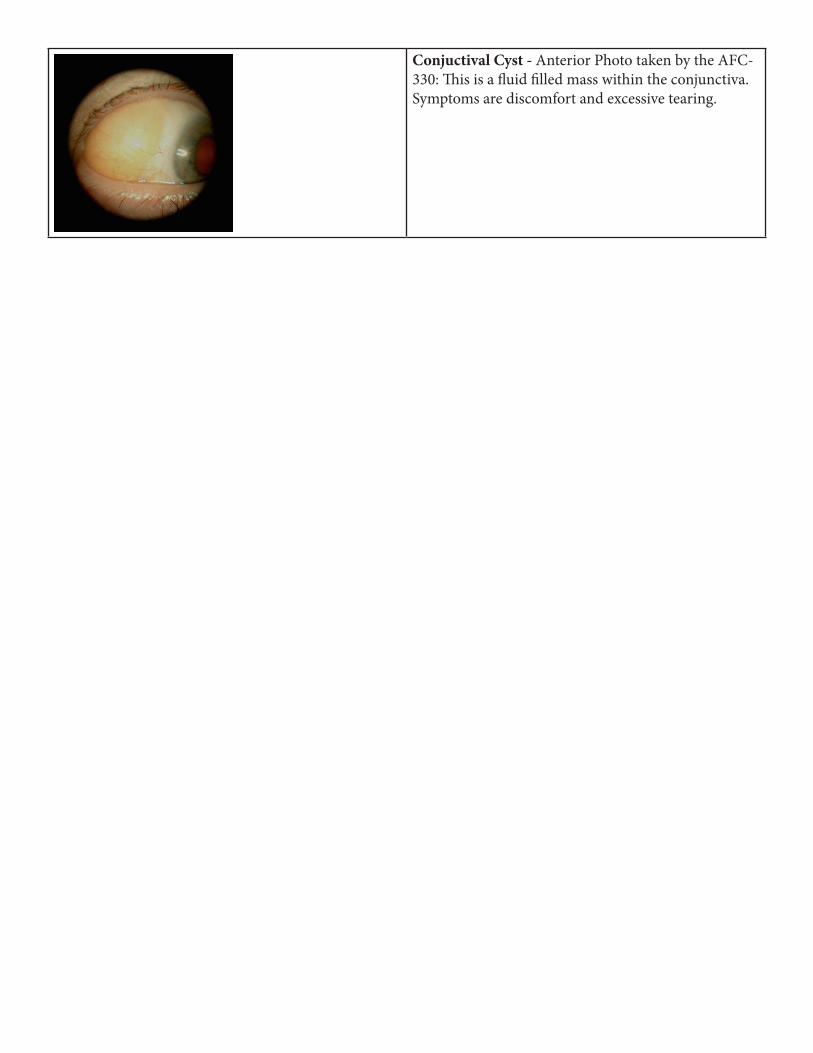
Conjuctival Cyst - Anterior Photo taken by the AFC-330: This is a fluid filled mass within the conjunctiva. Symptoms are discomfort and excessive tearing.

![[Text deleted] - California Digital Library · Clinical Nutrition Clinical Veterin Toxicolo Color Atlas of Canine and Feline O hthalmolo Color Atlas of Diseases and Disorders of the](https://img.pdfslide.us/doc/110x75/5b5b68727f8b9ac7498e02b7/text-deleted-california-digital-library-clinical-nutrition-clinical-veterin.jpg)


![[] Hill’s Atlas of Veterinary Clinical Anatomy(BookZa.org)](https://img.pdfslide.us/doc/110x75/55cf9294550346f57b97a2ce/-hills-atlas-of-veterinary-clinical-anatomybookzaorg.jpg)


![[] Hill’s Atlas of Veterinary Clinical Anatomy](https://img.pdfslide.us/doc/110x75/56d6bcd31a28ab30168ba0c3/-hills-atlas-of-veterinary-clinical-anatomy.jpg)