Embed Size (px)
Citation preview

10 Arthritides

CLINICAL IMAGAGINGAN ATLAS OF DIFFERENTIAL DAIGNOSIS
EISENBERG
DR. Muhammad Bin Zulfiqar PGR-FCPS III SIMS/SHL
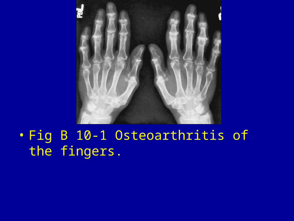
• Fig B 10-1 Osteoarthritis of the fingers.
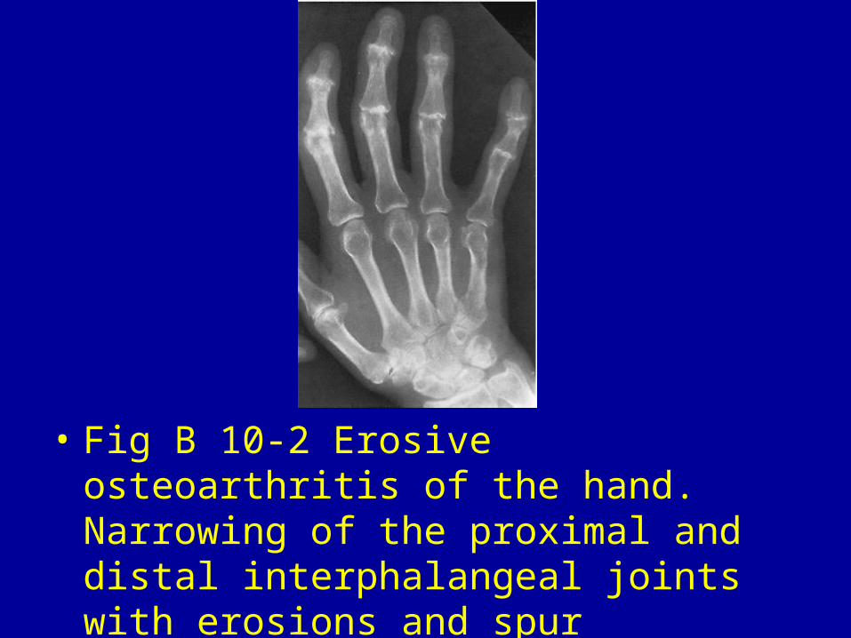
• Fig B 10-2 Erosive osteoarthritis of the hand. Narrowing of the proximal and distal interphalangeal joints with erosions and spur formation.
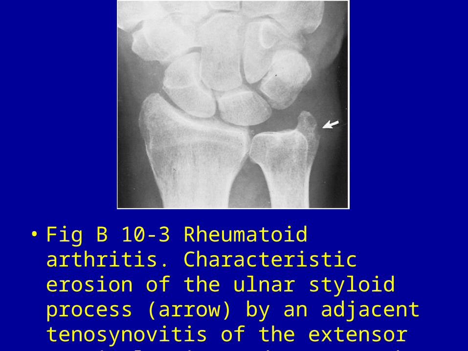
• Fig B 10-3 Rheumatoid arthritis. Characteristic erosion of the ulnar styloid process (arrow) by an adjacent tenosynovitis of the extensor carpi ulnaris tendon. Note the associated soft-tissue swelling.
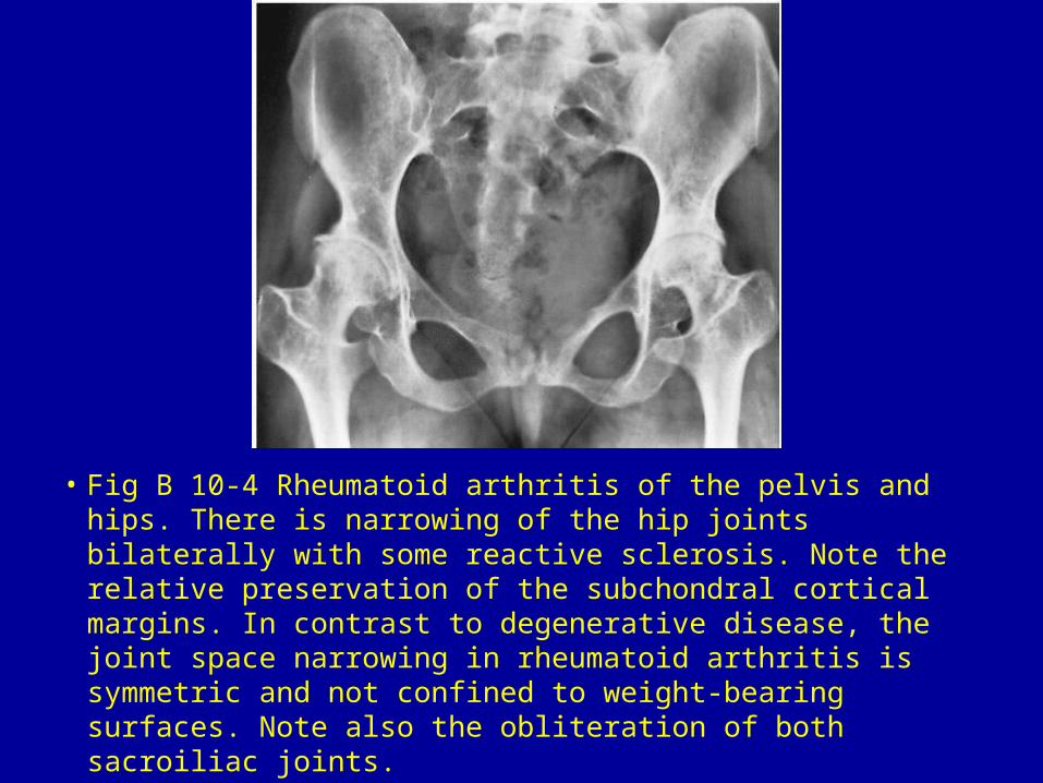
• Fig B 10-4 Rheumatoid arthritis of the pelvis and hips. There is narrowing of the hip joints bilaterally with some reactive sclerosis. Note the relative preservation of the subchondral cortical margins. In contrast to degenerative disease, the joint space narrowing in rheumatoid arthritis is symmetric and not confined to weight-bearing surfaces. Note also the obliteration of both sacroiliac joints.
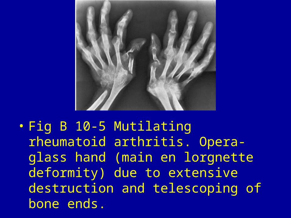
• Fig B 10-5 Mutilating rheumatoid arthritis. Opera-glass hand (main en lorgnette deformity) due to extensive destruction and telescoping of bone ends.
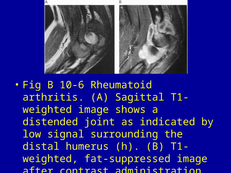
• Fig B 10-6 Rheumatoid arthritis. (A) Sagittal T1-weighted image shows a distended joint as indicated by low signal surrounding the distal humerus (h). (B) T1-weighted, fat-suppressed image after contrast administration shows diffuse enhancement of the pannus.22
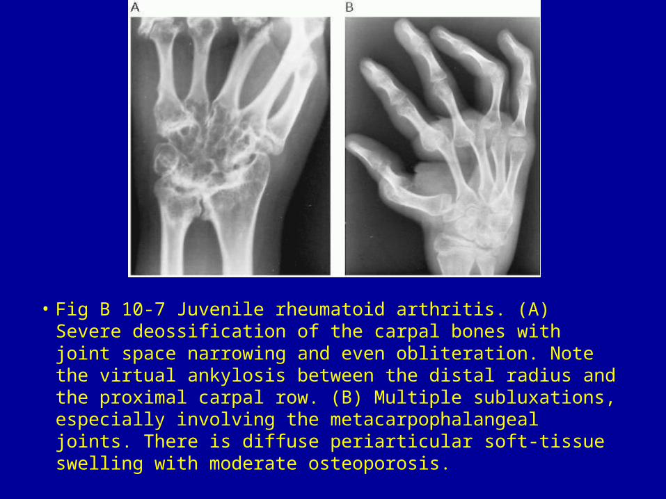
• Fig B 10-7 Juvenile rheumatoid arthritis. (A) Severe deossification of the carpal bones with joint space narrowing and even obliteration. Note the virtual ankylosis between the distal radius and the proximal carpal row. (B) Multiple subluxations, especially involving the metacarpophalangeal joints. There is diffuse periarticular soft-tissue swelling with moderate osteoporosis.
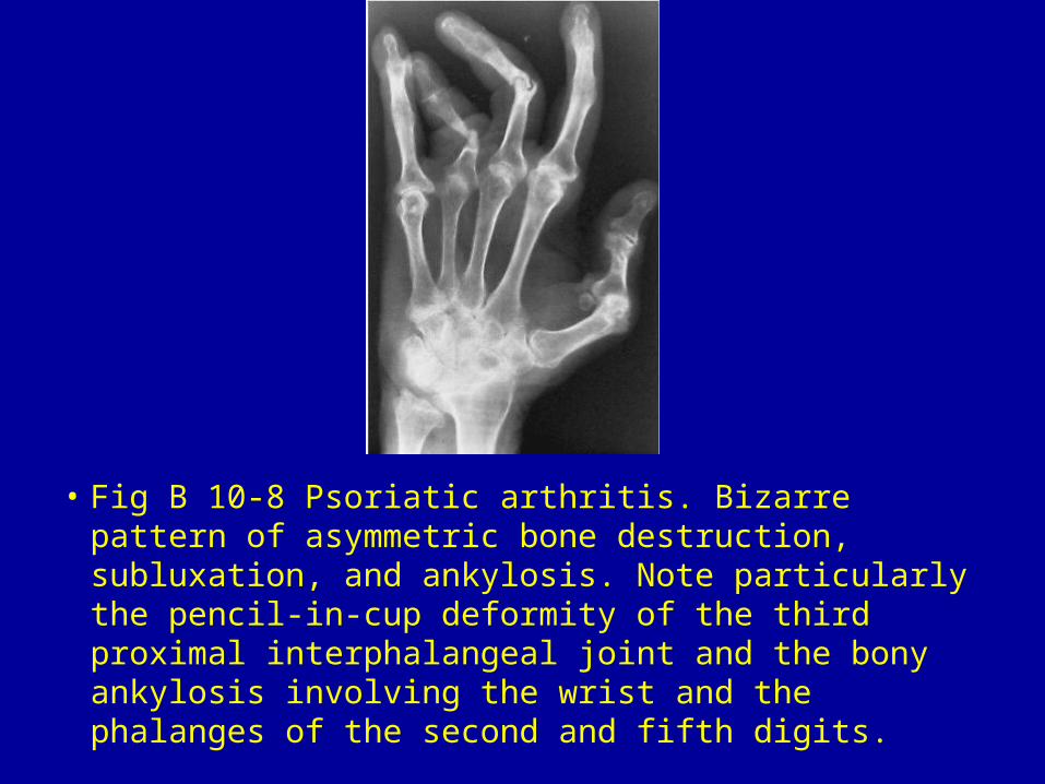
• Fig B 10-8 Psoriatic arthritis. Bizarre pattern of asymmetric bone destruction, subluxation, and ankylosis. Note particularly the pencil-in-cup deformity of the third proximal interphalangeal joint and the bony ankylosis involving the wrist and the phalanges of the second and fifth digits.
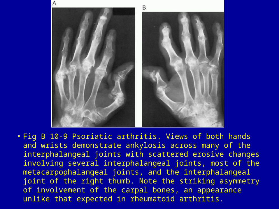
• Fig B 10-9 Psoriatic arthritis. Views of both hands and wrists demonstrate ankylosis across many of the interphalangeal joints with scattered erosive changes involving several interphalangeal joints, most of the metacarpophalangeal joints, and the interphalangeal joint of the right thumb. Note the striking asymmetry of involvement of the carpal bones, an appearance unlike that expected in rheumatoid arthritis.
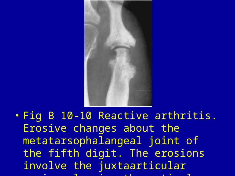
• Fig B 10-10 Reactive arthritis. Erosive changes about the metatarsophalangeal joint of the fifth digit. The erosions involve the juxtaarticular region, leaving the articular cortex intact.
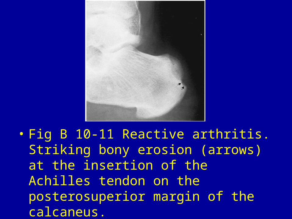
• Fig B 10-11 Reactive arthritis. Striking bony erosion (arrows) at the insertion of the Achilles tendon on the posterosuperior margin of the calcaneus.
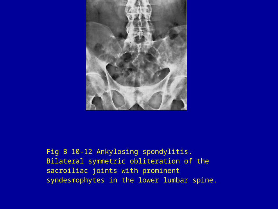
Fig B 10-12 Ankylosing spondylitis. Bilateral symmetric obliteration of the sacroiliac joints with prominent syndesmophytes in the lower lumbar spine.
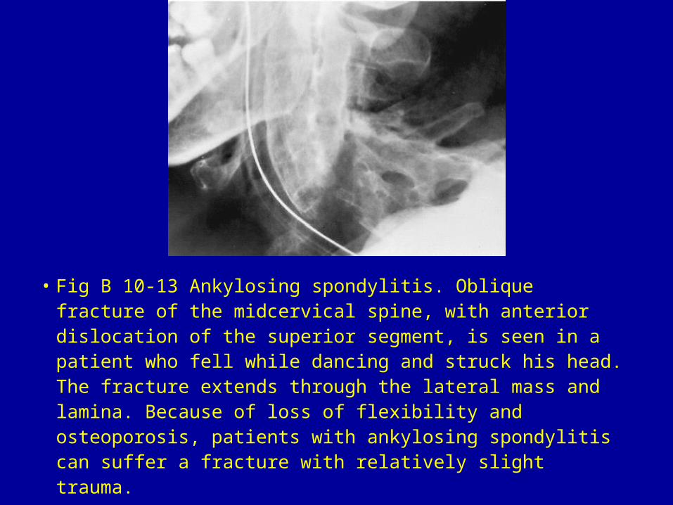
• Fig B 10-13 Ankylosing spondylitis. Oblique fracture of the midcervical spine, with anterior dislocation of the superior segment, is seen in a patient who fell while dancing and struck his head. The fracture extends through the lateral mass and lamina. Because of loss of flexibility and osteoporosis, patients with ankylosing spondylitis can suffer a fracture with relatively slight trauma.
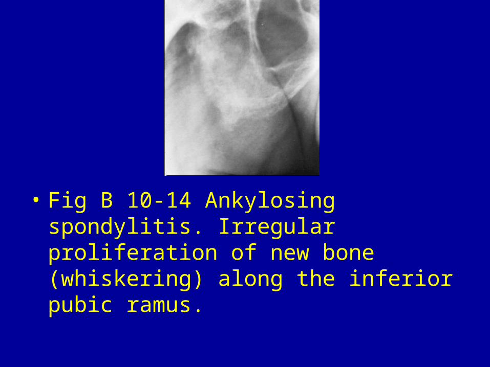
• Fig B 10-14 Ankylosing spondylitis. Irregular proliferation of new bone (whiskering) along the inferior pubic ramus.
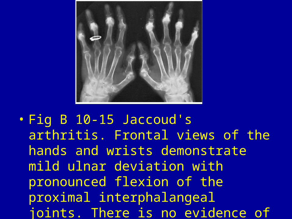
• Fig B 10-15 Jaccoud's arthritis. Frontal views of the hands and wrists demonstrate mild ulnar deviation with pronounced flexion of the proximal interphalangeal joints. There is no evidence of joint space narrowing or bone erosion.
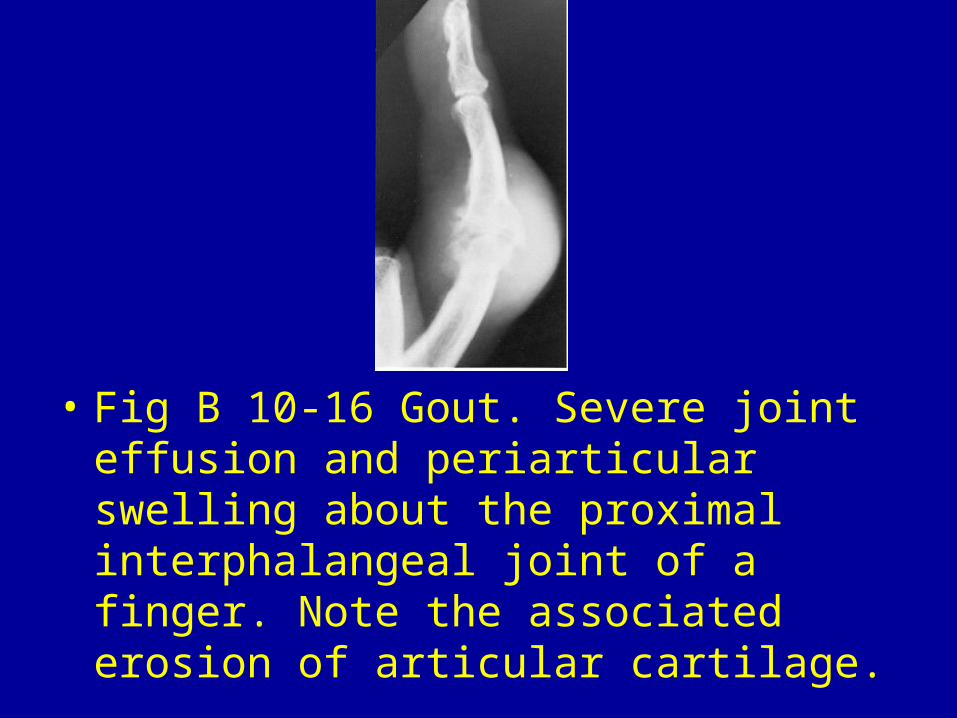
• Fig B 10-16 Gout. Severe joint effusion and periarticular swelling about the proximal interphalangeal joint of a finger. Note the associated erosion of articular cartilage.
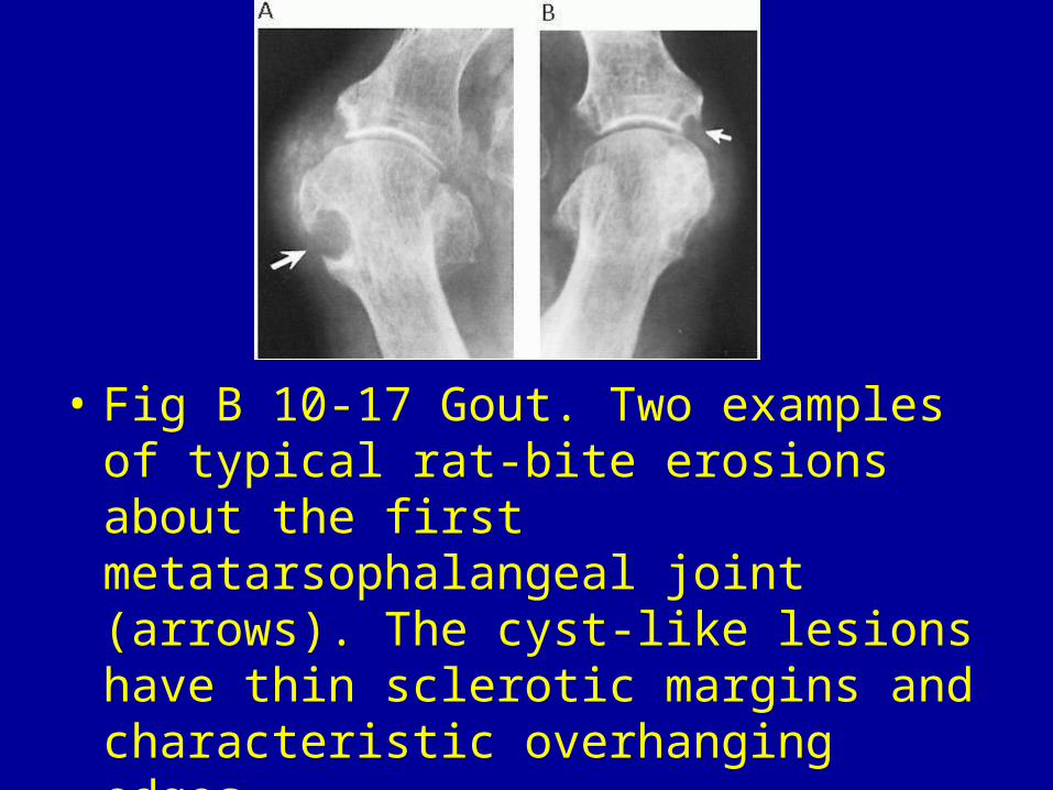
• Fig B 10-17 Gout. Two examples of typical rat-bite erosions about the first metatarsophalangeal joint (arrows). The cyst-like lesions have thin sclerotic margins and characteristic overhanging edges.
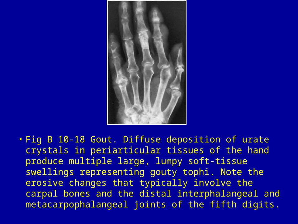
• Fig B 10-18 Gout. Diffuse deposition of urate crystals in periarticular tissues of the hand produce multiple large, lumpy soft-tissue swellings representing gouty tophi. Note the erosive changes that typically involve the carpal bones and the distal interphalangeal and metacarpophalangeal joints of the fifth digits.
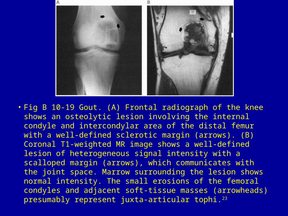
• Fig B 10-19 Gout. (A) Frontal radiograph of the knee shows an osteolytic lesion involving the internal condyle and intercondylar area of the distal femur with a well-defined sclerotic margin (arrows). (B) Coronal T1-weighted MR image shows a well-defined lesion of heterogeneous signal intensity with a scalloped margin (arrows), which communicates with the joint space. Marrow surrounding the lesion shows normal intensity. The small erosions of the femoral condyles and adjacent soft-tissue masses (arrowheads) presumably represent juxta-articular tophi.23
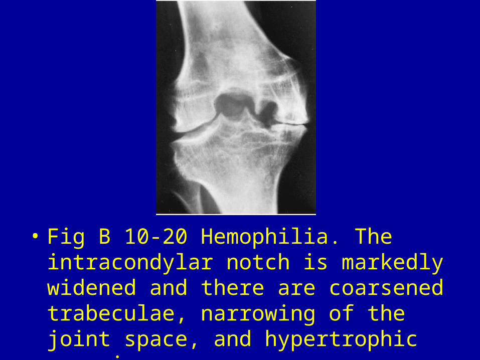
• Fig B 10-20 Hemophilia. The intracondylar notch is markedly widened and there are coarsened trabeculae, narrowing of the joint space, and hypertrophic spurring.
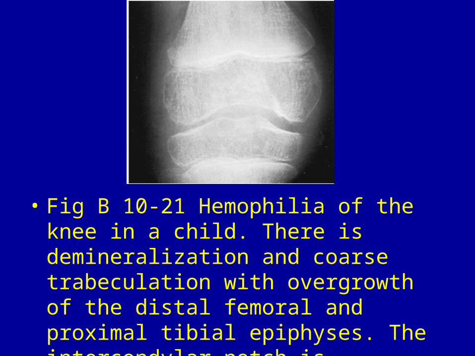
• Fig B 10-21 Hemophilia of the knee in a child. There is demineralization and coarse trabeculation with overgrowth of the distal femoral and proximal tibial epiphyses. The intercondylar notch is moderately widened.
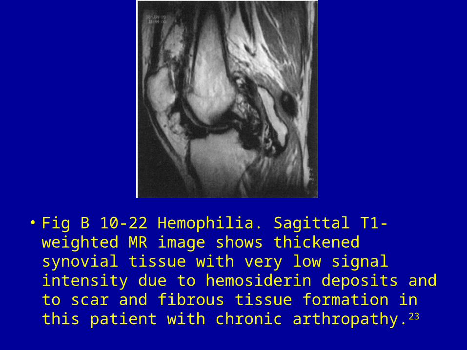
• Fig B 10-22 Hemophilia. Sagittal T1-weighted MR image shows thickened synovial tissue with very low signal intensity due to hemosiderin deposits and to scar and fibrous tissue formation in this patient with chronic arthropathy.23
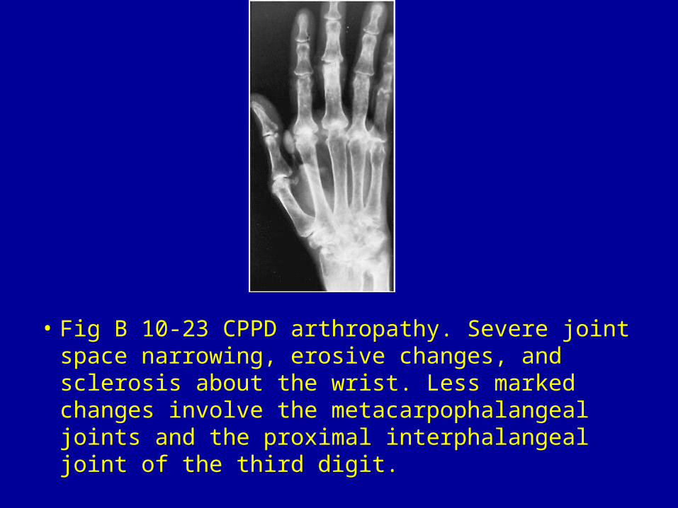
• Fig B 10-23 CPPD arthropathy. Severe joint space narrowing, erosive changes, and sclerosis about the wrist. Less marked changes involve the metacarpophalangeal joints and the proximal interphalangeal joint of the third digit.
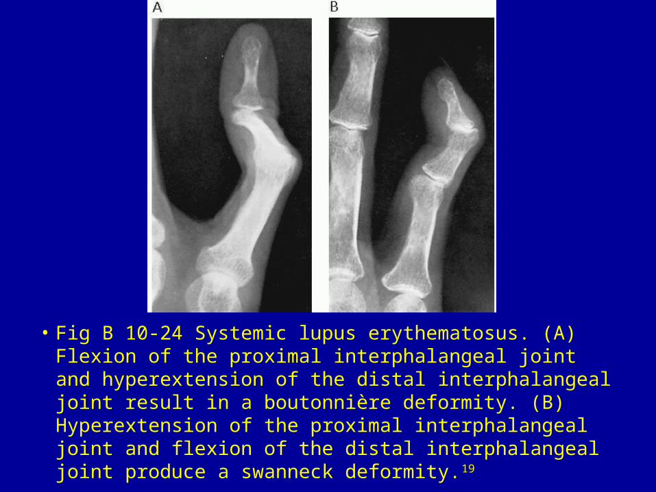
• Fig B 10-24 Systemic lupus erythematosus. (A) Flexion of the proximal interphalangeal joint and hyperextension of the distal interphalangeal joint result in a boutonnière deformity. (B) Hyperextension of the proximal interphalangeal joint and flexion of the distal interphalangeal joint produce a swanneck deformity.19
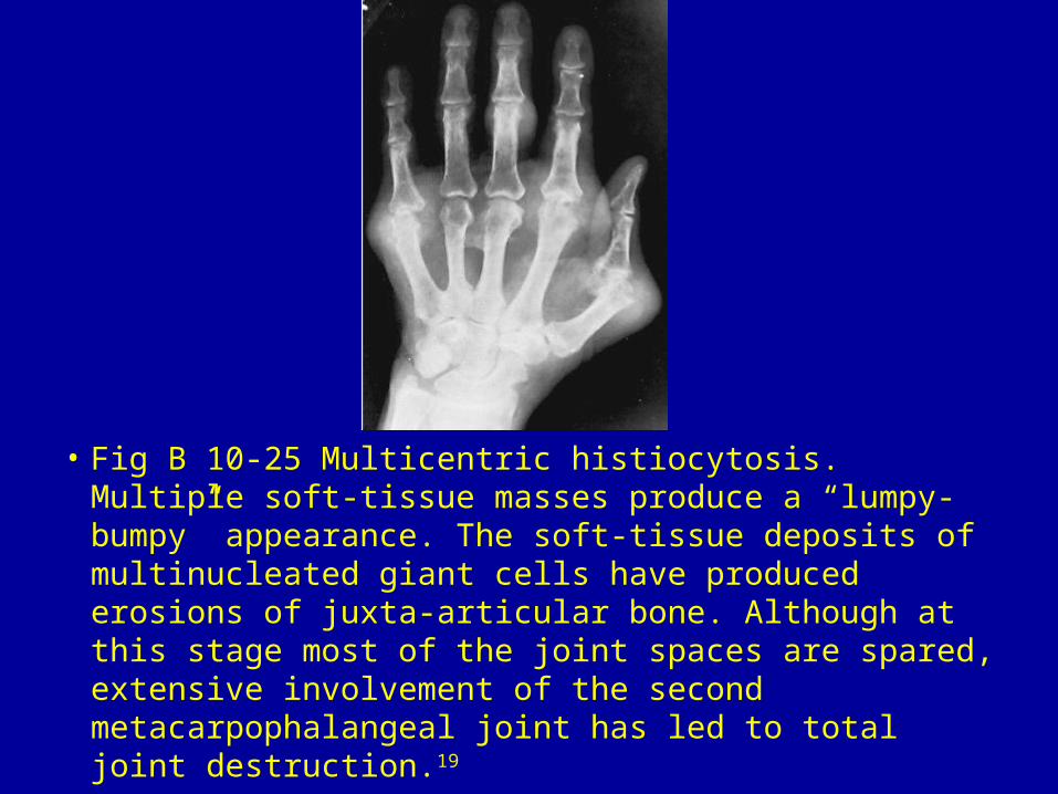
• Fig B 10-25 Multicentric histiocytosis. Multiple soft-tissue masses produce a “lumpy-bumpy” appearance. The soft-tissue deposits of multinucleated giant cells have produced erosions of juxta-articular bone. Although at this stage most of the joint spaces are spared, extensive involvement of the second metacarpophalangeal joint has led to total joint destruction.19
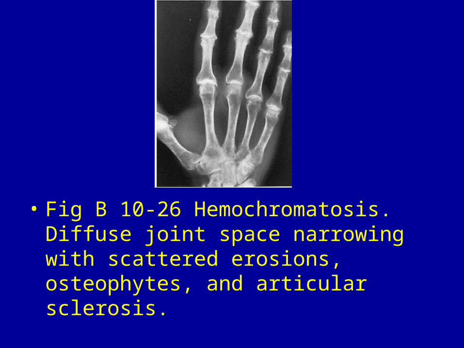
• Fig B 10-26 Hemochromatosis. Diffuse joint space narrowing with scattered erosions, osteophytes, and articular sclerosis.
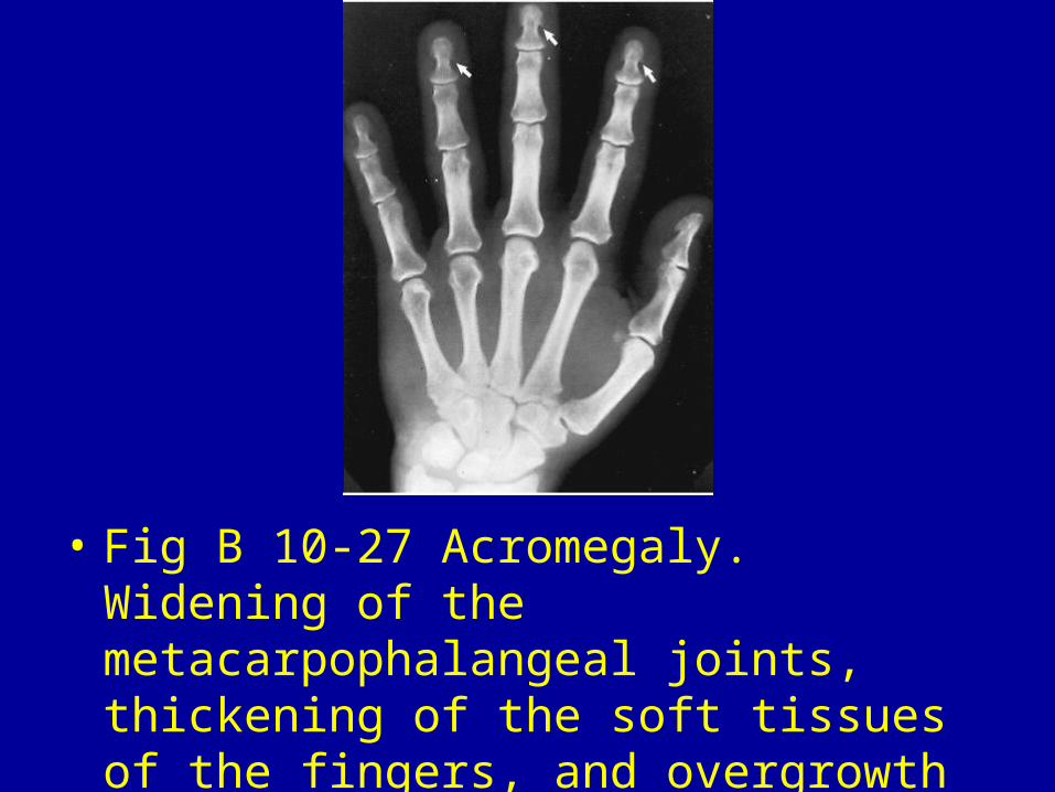
• Fig B 10-27 Acromegaly. Widening of the metacarpophalangeal joints, thickening of the soft tissues of the fingers, and overgrowth of the tufts of the distal phalanges (arrows).
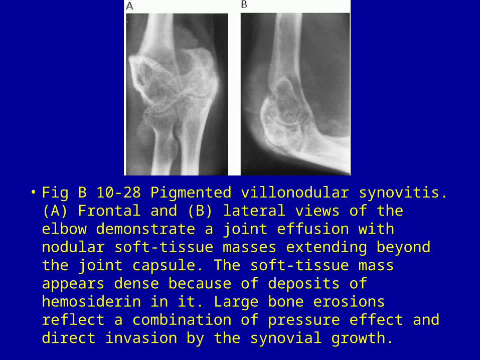
• Fig B 10-28 Pigmented villonodular synovitis. (A) Frontal and (B) lateral views of the elbow demonstrate a joint effusion with nodular soft-tissue masses extending beyond the joint capsule. The soft-tissue mass appears dense because of deposits of hemosiderin in it. Large bone erosions reflect a combination of pressure effect and direct invasion by the synovial growth.
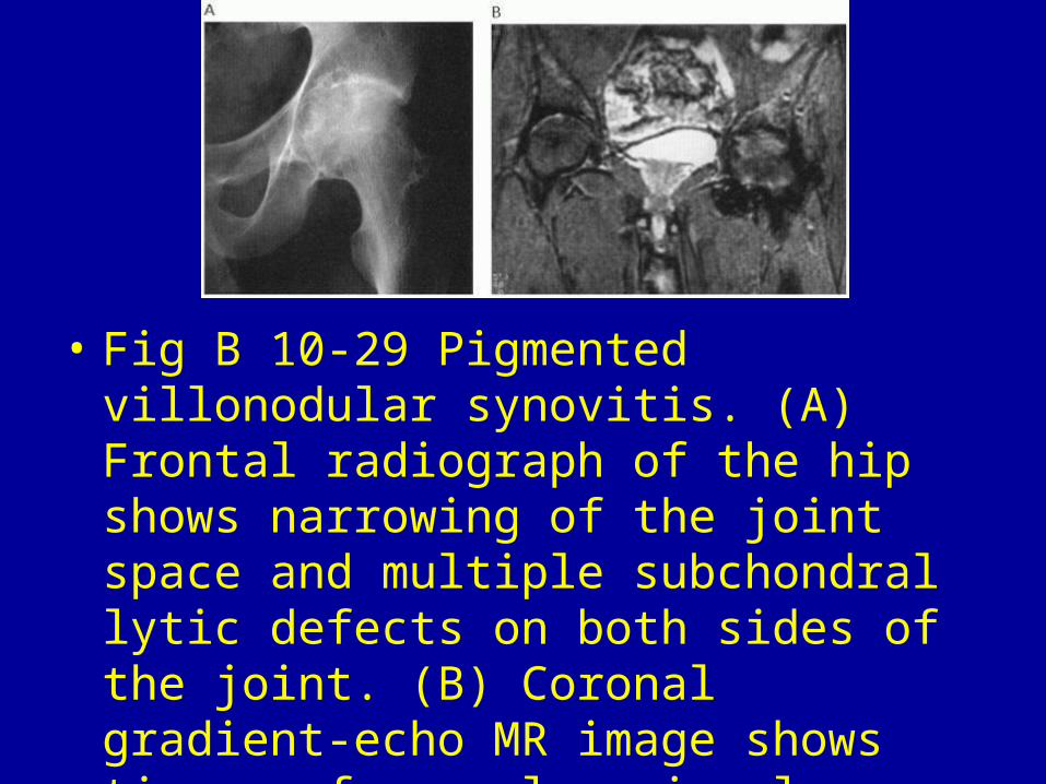
• Fig B 10-29 Pigmented villonodular synovitis. (A) Frontal radiograph of the hip shows narrowing of the joint space and multiple subchondral lytic defects on both sides of the joint. (B) Coronal gradient-echo MR image shows tissue of very low signal intensity outlining the joint capsule. Note the prominent deposition of hemosiderin.23
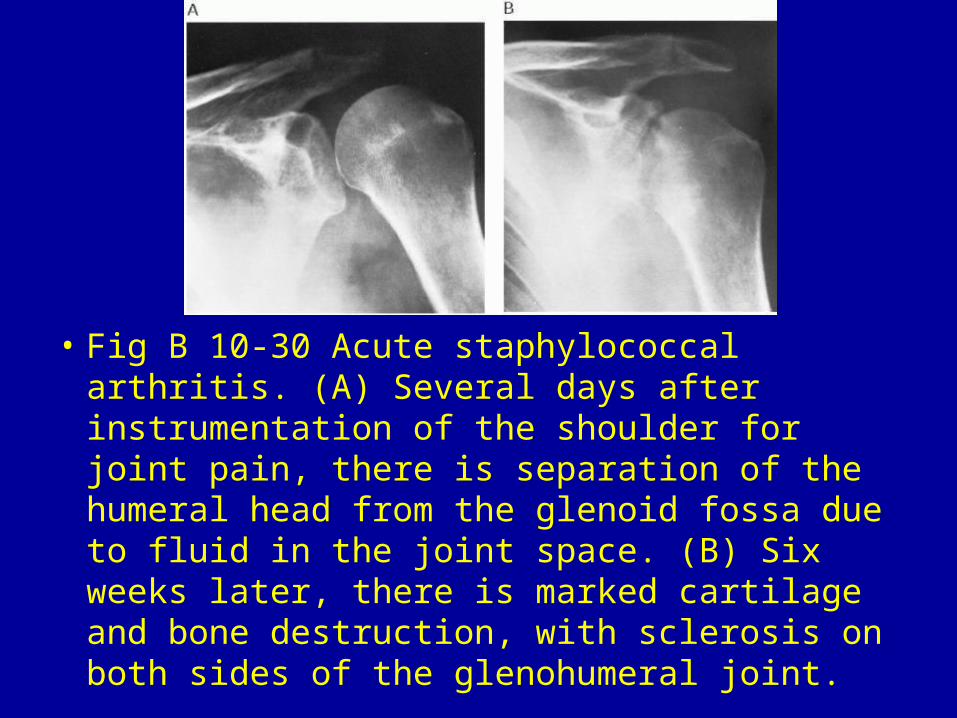
• Fig B 10-30 Acute staphylococcal arthritis. (A) Several days after instrumentation of the shoulder for joint pain, there is separation of the humeral head from the glenoid fossa due to fluid in the joint space. (B) Six weeks later, there is marked cartilage and bone destruction, with sclerosis on both sides of the glenohumeral joint.
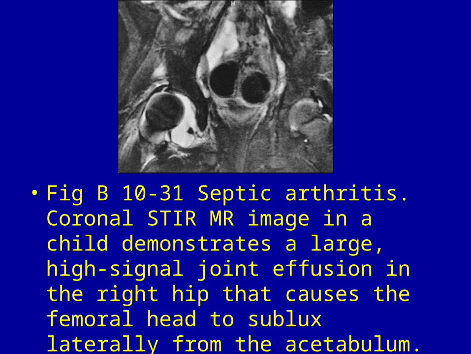
• Fig B 10-31 Septic arthritis. Coronal STIR MR image in a child demonstrates a large, high-signal joint effusion in the right hip that causes the femoral head to sublux laterally from the acetabulum. No bone erosion or marrow edema is evident.22
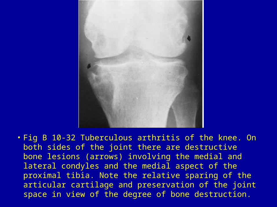
• Fig B 10-32 Tuberculous arthritis of the knee. On both sides of the joint there are destructive bone lesions (arrows) involving the medial and lateral condyles and the medial aspect of the proximal tibia. Note the relative sparing of the articular cartilage and preservation of the joint space in view of the degree of bone destruction.
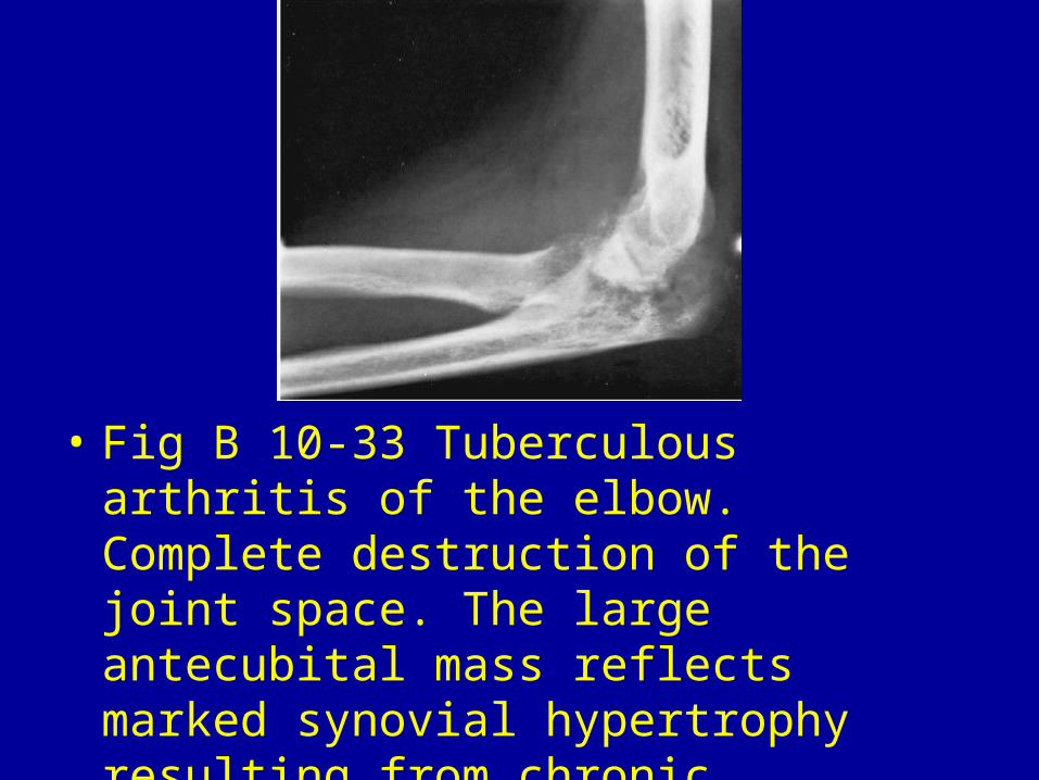
• Fig B 10-33 Tuberculous arthritis of the elbow. Complete destruction of the joint space. The large antecubital mass reflects marked synovial hypertrophy resulting from chronic granulomatous infection.19
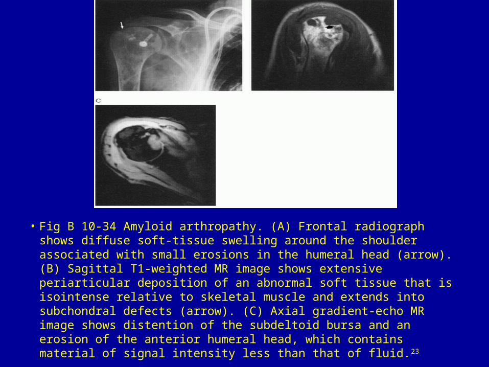
• Fig B 10-34 Amyloid arthropathy. (A) Frontal radiograph shows diffuse soft-tissue swelling around the shoulder associated with small erosions in the humeral head (arrow). (B) Sagittal T1-weighted MR image shows extensive periarticular deposition of an abnormal soft tissue that is isointense relative to skeletal muscle and extends into subchondral defects (arrow). (C) Axial gradient-echo MR image shows distention of the subdeltoid bursa and an erosion of the anterior humeral head, which contains material of signal intensity less than that of fluid.23
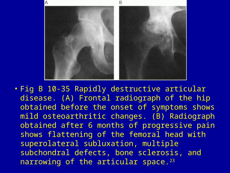
• Fig B 10-35 Rapidly destructive articular disease. (A) Frontal radiograph of the hip obtained before the onset of symptoms shows mild osteoarthritic changes. (B) Radiograph obtained after 6 months of progressive pain shows flattening of the femoral head with superolateral subluxation, multiple subchondral defects, bone sclerosis, and narrowing of the articular space.23
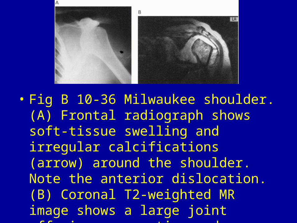
• Fig B 10-36 Milwaukee shoulder. (A) Frontal radiograph shows soft-tissue swelling and irregular calcifications (arrow) around the shoulder. Note the anterior dislocation. (B) Coronal T2-weighted MR image shows a large joint effusion, resorption and deformity of the humeral head, and complete rupture of the rotator cuff.23