Embed Size (px)
Citation preview
Clinical aspects of full-thickness wound healing
Albert E. Rivera, DO, James M. Spencer, MD*
Department of Dermatology, Mt Sinai School of Medicine, New York, NY, USA
Abstract Optimal management of full-thickness wounds requires a thorough knowledge of wound-
healing principles and practices. In the absence of underlying disease, almost every full-thickness
wound will heal with minimal intervention; however, the process can be enhanced by judicious wound
management. The first clinical decision to be made is whether to repair the wound or to allow it to heal
by second intention. This decision is guided by a host of objective and subjective factors.
Reconstruction options include primary closure, flaps, and grafts. Materials to aid reconstruction,
including the introduction of tissue adhesives, continue to evolve. Both primary and secondary intention
wounds are aided by occlusive dressings and adjutants. A plethora of wound-healing adjuncts have been
developed to aid wound healing in diseased states, and a working knowledge of their use is beneficial in
managing all full-thickness wounds.
D 2007 Elsevier Inc. All rights reserved.
Introduction
The management of full-thickness wounds is a com-
mon part of dermatologic practice. The healing of these
wounds is a complicated, coordinated series of events that
eventually lead to both a structurally and functionally
acceptable result. The initiation of this process is signaled
by an insult to the tissues, whether due to an unintentional
trauma, such as abrasions, excoriations, blisters, burns,
hypothermic injuries, ischemia, or numerous other etiolo-
gies, or a defect that has been planned, such as a surgical
incision or piercing. The sequence that follows leads to the
repair and restoration of the site in question. Numerous
growth factors and cell types are involved and are discussed
in more extensive detail in other articles contained within
this issue. Although the wounds may differ in appearance,
time to resolution, and other facets, they still all progress
through the same basic stages.1,2
0738-081X/$ – see front matter D 2007 Elsevier Inc. All rights reserved.
doi:10.1016/j.clindermatol.2006.10.001
* Corresponding author. St. Petersburg, FL 33761. Tel.: +1 727 572
1333; fax: +1 727 572 1331.
E-mail address: [email protected] (J.M. Spencer).
Downloaded from ClinicalKey.com at AlabamaFor personal use only. No other uses without permiss
Generally, there are 4 stages noted in the healing of any
wound: hemostasis, inflammation, proliferation or granula-
tion, and matrix formation or remodeling.3 -6 An under-
standing of this process allows the clinician to optimize
wound healing. These stages are not necessarily chronolog-
ically exclusive, but rather a dynamic and integrated
coordination of specific processes. These stages can be
allowed to proceed uninterrupted, as in healing by second-
ary intention, or influenced by primary closure with sutures,
staples, or adhesives; application of medications or topical
products; or placement of various traditional or synthetic
dressings. As healing progresses, tensile strength of the
wound improves. Wounds have minimal strength during the
first week or so of healing, approximately 30% to 50% in
4 to 6 weeks and 60% at around 6 months.7 Tensile strength
slowly increases after this point but usually only reaches a
maximum of 80% that of normal, undisrupted skin.
The duration and percentages noted are general guide-
lines for the time frame and performance expected based on
prior studies. Variation does occur due to the type, location,
and size of the defect. Clearly, the less invasive, more
superficial insults will conclude the entire process more
Clinics in Dermatology (2007) 25, 39–48
College of Osteopathic Medicine July 19, 2016.ion. Copyright ©2016. Elsevier Inc. All rights reserved.
A.E. Rivera, J.M. Spencer40
rapidly. As appreciated clinically, extremely vascular
structures such as the face, or sites closer to the heart, heal
at a faster rate. Those more distal, especially the lower
extremities, are more difficult to resolve. In addition, it has
been found that the healing time for full excisional wounds
was linearly related to the surface area, but was unrelated to
the depth.8 Many other factors, including comorbidities,
nutrition or prior injury are also contributing variables.
On a more macroscopic level, the physician has the
ability to directly influence the healing wound. An obvious
management strategy is to surgically repair the wound,
known as primary intention healing. Although numerous
other options are available, an often overlooked but valuable
choice is healing by secondary intention. Secondary
intention existed long before the surgical advancements
such as flap and graft utilization and ancillary tools that are
to be discussed. Another consideration includes a combina-
tion of both with partial closure. Numerous adjuncts for
primary closing and dressing the sites have been developed
and studied. Included are suture materials and adhesives that
allow wound edges to remain in proximity while the repair
process proceeds. Topical products, including antibiotic
ointments, are frequently applied to the sites. There is a
multitude of dressing types available for selection to protect
the site of repair and maintain an optimal environment
throughout the stages of healing. All of the above have their
own value depending on the specific wound in question and
the desired time frame or outcome.
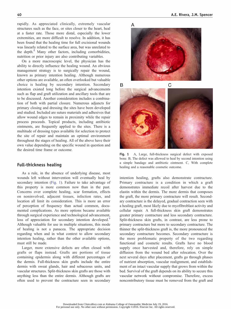
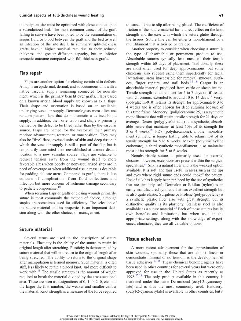
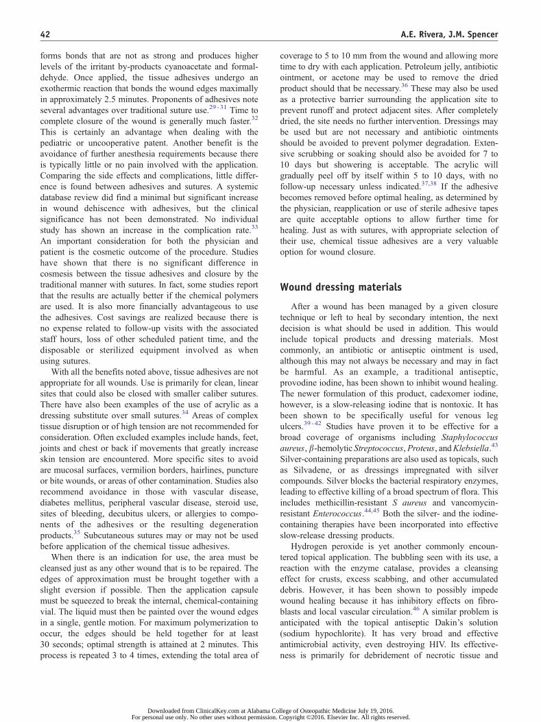
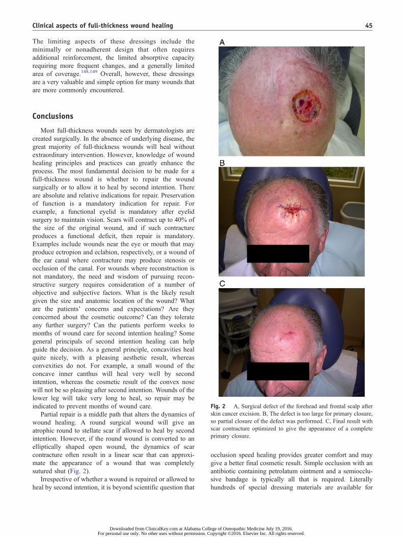
ig. 1 A, Large, full-thickness surgical defect with exposedone. B, The defect was allowed to heal by second intention using
simple bandage and antibiotic ointment. C, With complete
ealing and a reasonable cosmetic outcome.
Full-thickness healingAs a rule, in the absence of underlying disease, most
wounds left without intervention will eventually heal by
secondary intention (Fig. 1). Failure to take advantage of
this property is more common now than in the past.
Concerns over complete healing, scar formation, effects
on noninvolved, adjacent tissues, infection rates, and
location all limit its consideration. This is more an error
of perception of frequency than actual common, docu-
mented complications. As more options became available
through surgical experience and technological advancement,
loss of appreciation for secondary intention developed.9
Although valuable for use in multiple situations, this mode
of healing is not a panacea. The appropriate decision
regarding when and in what context to allow secondary
intention healing, rather than the other available options,
must still be made.
Larger, more extensive defects are often closed with
grafts or flaps instead. Grafts are portions of tissue
containing epidermis along with different percentages of
the dermis. Full-thickness skin grafts include the entire
dermis with sweat glands, hair and sebaceous units, and
vascular structures. Split-thickness skin grafts are those with
anything less than the entire dermis. Although grafts are
often used to prevent the contracture seen in secondary
Downloaded from ClinicalKey.com at Alabama CoFor personal use only. No other uses without permission.
Fb
a
h
intention healing, grafts also demonstrate contracture.
Primary contracture is a condition in which a graft
demonstrates immediate recoil after harvest due to the
elastin within the dermis. The more dermis that composes
the graft, the more primary contracture will result. Second-
ary contracture is the delayed, gradual contraction seen with
a healing graft, most likely due to myofibroblast activity and
cellular repair. A full-thickness skin graft demonstrates
greater primary contracture and less secondary contracture.
Split-thickness skin grafts, in contrast, are less prone to
primary contracture but more to secondary contracture. The
thinner the split-thickness graft is, the more pronounced the
secondary contracture becomes. Secondary contracture is
the more problematic property of the two regarding
functional and cosmetic results. Grafts have no blood
supply once harvested and, therefore, rely on simple
diffusion from the wound bed after relocation. Over the
next several days after placement, grafts go through phases
of nutrient absorption, vascular realignment, and establish-
ment of an intact vascular supply that grows from within the
bed. Survival of the graft depends on its ability to secure this
vascular network without compromise. Therefore, excess
noncontributory tissue must be removed from the graft and
llege of Osteopathic Medicine July 19, 2016. Copyright ©2016. Elsevier Inc. All rights reserved.
Clinical aspects of full-thickness wound healing 41
the recipient site must be optimized with close contact upon
a vascularized bed. The most common causes of the graft
failing to survive have been noted to be the accumulation of
serous fluid or blood between the graft and the bed as well
as infection of the site itself. In summary, split-thickness
grafts have a higher survival rate due to their reduced
thickness and greater diffusion capacity, but an inferior
cosmetic outcome compared with full-thickness grafts.
Flap repair
Flaps are another option for closing certain skin defects.
A flap is an epidermal, dermal, and subcutaneous unit with a
native vascular supply remaining connected for nourish-
ment, which is the primary benefit over grafts. Flaps based
on a known arterial blood supply are known as axial flaps.
Their shape and orientation is based on an available,
underlying vascular supply. Dermatologists most often use
random pattern flaps that do not contain a defined blood
supply. In addition, their orientation and shape is primarily
defined by the defect to be filled rather than by the vascular
source. Flaps are named for the vector of their primary
motion: advancement, rotation, or transposition. They may
also be bfreeQ flaps, excised units of skin and deep tissue in
which the vascular supply is still a part of the flap but is
temporarily transected then reestablished at a more distant
location to a new vascular source. Flaps are valuable to
redirect tension away from the wound itself to more
favorable sites when poorly or nonvascularized sites are in
need of coverage or when additional tissue mass is desirable
for padding delicate areas. Compared to grafts, there is less
concern of complications from fluid collections and
infection but more concern of ischemic damage secondary
to pedicle compromise.10
When securing flaps or grafts or closing wounds primarily,
suture is most commonly the method of choice, although
staples are sometimes used for efficiency. The selection of
the suture material and its properties is an important deci-
sion along with the other choices of management.
Suture material
Several terms are used in the description of suture
materials. Elasticity is the ability of the suture to retain its
original length after stretching. Plasticity is demonstrated by
suture material that will not return to the original length after
being stretched. The ability to return to the original shape
after manipulation is termed memory. Such material is often
stiff, less likely to retain a placed knot, and more difficult to
work with.11 The tensile strength is the amount of weight
required to break the material divided by the cross-sectional
area. These are seen as designations of 0, 1-0, 2-0, etc, and
the larger the first number, the weaker and smaller caliber
the material. Knot strength is a measure of the force required
Downloaded from ClinicalKey.com at Alabama ColFor personal use only. No other uses without permission. C
to cause a knot to slip after being placed. The coefficient of
friction of the suture material has a direct effect on the knot
strength and the ease with which the suture glides through
the tissues. Suture line can be either a monofilament or a
multifilament that is twisted or braided.
Another property to consider when choosing a suture is
the type of absorbable or permanent product to use.
Absorbable sutures typically lose most of their tensile
strength within 60 days of placement. Traditionally, these
are most often used for deep approximations, but some
clinicians also suggest using them superficially for facial
lacerations, areas inaccessible for removal, mucosal surfa-
ces, finger repairs, and nail beds.12-18 Catgut is an
absorbable material produced from cattle or sheep intima.
Tensile strength remains intact for 5 to 7 days or, if treated
with chromium, extended to around 10 to 14 days.19 Vicryl
(polyglactin-910) retains its strength for approximately 3 to
4 weeks and is often chosen for deep suturing because of
this time frame. Monocryl (poliglecaprone 25) is a synthetic
monofilament that will retain tensile strength for 21 days on
average. Dexon (polyglycolic acid) is a synthetic, absorb-
able suture that maintains at least 50% of its strength for
3 or 4 weeks.20 PDS (polydiaxanone), another monofila-
ment synthetic, is longer lasting, able to retain most of its
tensile strength for 5 to 6 weeks. Maxon (polytimethylene
carbonate), a third synthetic monofilament, also maintains
most of its strength for 5 to 6 weeks.
Nonabsorbable suture is primarily used for external
closures; however, exceptions are present within the surgical
specialties.21 Silk is a natural fiber and is the weakest option
available. It is soft, and thus useful in areas such as the lips
and eyes where rigid suture ends could bpokeQ the patient.
Use of silk has largely been replaced by the use of synthetics
that are similarly soft. Dermalon or Ethilon (nylon) is an
easily manufactured synthetic that has excellent strength but
is also quite elastic. Surgilene or Prolene (polypropylene) is
a synthetic plastic fiber also with great strength, but its
distinctive quality is its plasticity. Stainless steel is also
available as a suture material.22 Each of these sutures has its
own benefits and limitations but when used in the
appropriate settings, along with the knowledge of experi-
enced clinicians, they are all valuable options.
Tissue adhesives
A more recent advancement for the approximation of
skin wounds, optimally those that are almost linear or
demonstrate minimal or no tension, is the development of
tissue adhesives.23-26 These chemical bonding agents have
been used in other countries for several years but were only
approved for use in the United States as recently as
1998.27,28 The only product available in this country is
marketed under the name Dermabond (octyl-2-cyanoacry-
late) and is thus the most commonly used. Histoacryl
(butyl-2-cyanoacrylate) is available in other countries, but it
lege of Osteopathic Medicine July 19, 2016.opyright ©2016. Elsevier Inc. All rights reserved.
A.E. Rivera, J.M. Spencer42
forms bonds that are not as strong and produces higher
levels of the irritant by-products cyanoacetate and formal-
dehyde. Once applied, the tissue adhesives undergo an
exothermic reaction that bonds the wound edges maximally
in approximately 2.5 minutes. Proponents of adhesives note
several advantages over traditional suture use.29 - 31 Time to
complete closure of the wound is generally much faster.32
This is certainly an advantage when dealing with the
pediatric or uncooperative patent. Another benefit is the
avoidance of further anesthesia requirements because there
is typically little or no pain involved with the application.
Comparing the side effects and complications, little differ-
ence is found between adhesives and sutures. A systemic
database review did find a minimal but significant increase
in wound dehiscence with adhesives, but the clinical
significance has not been demonstrated. No individual
study has shown an increase in the complication rate.33
An important consideration for both the physician and
patient is the cosmetic outcome of the procedure. Studies
have shown that there is no significant difference in
cosmesis between the tissue adhesives and closure by the
traditional manner with sutures. In fact, some studies report
that the results are actually better if the chemical polymers
are used. It is also more financially advantageous to use
the adhesives. Cost savings are realized because there is
no expense related to follow-up visits with the associated
staff hours, loss of other scheduled patient time, and the
disposable or sterilized equipment involved as when
using sutures.
With all the benefits noted above, tissue adhesives are not
appropriate for all wounds. Use is primarily for clean, linear
sites that could also be closed with smaller caliber sutures.
There have also been examples of the use of acrylic as a
dressing substitute over small sutures.34 Areas of complex
tissue disruption or of high tension are not recommended for
consideration. Often excluded examples include hands, feet,
joints and chest or back if movements that greatly increase
skin tension are encountered. More specific sites to avoid
are mucosal surfaces, vermilion borders, hairlines, puncture
or bite wounds, or areas of other contamination. Studies also
recommend avoidance in those with vascular disease,
diabetes mellitus, peripheral vascular disease, steroid use,
sites of bleeding, decubitus ulcers, or allergies to compo-
nents of the adhesives or the resulting degeneration
products.35 Subcutaneous sutures may or may not be used
before application of the chemical tissue adhesives.
When there is an indication for use, the area must be
cleansed just as any other wound that is to be repaired. The
edges of approximation must be brought together with a
slight eversion if possible. Then the application capsule
must be squeezed to break the internal, chemical-containing
vial. The liquid must then be painted over the wound edges
in a single, gentle motion. For maximum polymerization to
occur, the edges should be held together for at least
30 seconds; optimal strength is attained at 2 minutes. This
process is repeated 3 to 4 times, extending the total area of
Downloaded from ClinicalKey.com at Alabama CoFor personal use only. No other uses without permission.
coverage to 5 to 10 mm from the wound and allowing more
time to dry with each application. Petroleum jelly, antibiotic
ointment, or acetone may be used to remove the dried
product should that be necessary.36 These may also be used
as a protective barrier surrounding the application site to
prevent runoff and protect adjacent sites. After completely
dried, the site needs no further intervention. Dressings may
be used but are not necessary and antibiotic ointments
should be avoided to prevent polymer degradation. Exten-
sive scrubbing or soaking should also be avoided for 7 to
10 days but showering is acceptable. The acrylic will
gradually peel off by itself within 5 to 10 days, with no
follow-up necessary unless indicated.37,38 If the adhesive
becomes removed before optimal healing, as determined by
the physician, reapplication or use of sterile adhesive tapes
are quite acceptable options to allow further time for
healing. Just as with sutures, with appropriate selection of
their use, chemical tissue adhesives are a very valuable
option for wound closure.
Wound dressing materials
After a wound has been managed by a given closure
technique or left to heal by secondary intention, the next
decision is what should be used in addition. This would
include topical products and dressing materials. Most
commonly, an antibiotic or antiseptic ointment is used,
although this may not always be necessary and may in fact
be harmful. As an example, a traditional antiseptic,
provodine iodine, has been shown to inhibit wound healing.
The newer formulation of this product, cadexomer iodine,
however, is a slow-releasing iodine that is nontoxic. It has
been shown to be specifically useful for venous leg
ulcers.39 -42 Studies have proven it to be effective for a
broad coverage of organisms including Staphylococcus
aureus, b-hemolytic Streptococcus,Proteus, andKlebsiella.43
Silver-containing preparations are also used as topicals, such
as Silvadene, or as dressings impregnated with silver
compounds. Silver blocks the bacterial respiratory enzymes,
leading to effective killing of a broad spectrum of flora. This
includes methicillin-resistant S aureus and vancomycin-
resistant Enterococcus.44,45 Both the silver- and the iodine-
containing therapies have been incorporated into effective
slow-release dressing products.
Hydrogen peroxide is yet another commonly encoun-
tered topical application. The bubbling seen with its use, a
reaction with the enzyme catalase, provides a cleansing
effect for crusts, excess scabbing, and other accumulated
debris. However, it has been shown to possibly impede
wound healing because it has inhibitory effects on fibro-
blasts and local vascular circulation.46 A similar problem is
anticipated with the topical antiseptic Dakin’s solution
(sodium hypochlorite). It has very broad and effective
antimicrobial activity, even destroying HIV. Its effective-
ness is primarily for debridement of necrotic tissue and
llege of Osteopathic Medicine July 19, 2016. Copyright ©2016. Elsevier Inc. All rights reserved.
Clinical aspects of full-thickness wound healing 43
occasional cleansing. This solution should not be used as a
primary agent because it also inhibits wound healing. It
has been demonstrated that wound strength is decreased
and there is impaired epithelialization, fibroblast activity,
and overall cellular survival. Therefore, use should be
minimized with routine wounds and tailored only to
specific uses.47-51
There are numerous other topical alternatives. Included
are polysporin, neomycin, bacitracin, neosporin, mupirocin,
erythromycin, clindamycin, and others.52-55 Most often, the
postoperative choice of topical antibiotic is either bacitracin
or polysporin. Neomycin, although widely available, has
often been avoided because of a suggested increased
incidence of contact dermatitis. In patients who tend to
develop contact dermatitis, neomycin is a common precip-
itating allergen. However, most patients encountered do not
develop such a reaction. There is a small incidence of cross-
reactivity with bacitracin and neomycin and although there
is potential, this is not commonly problematic.56 Alter-
natives for those allergic to bacitracin are erythromycin,
mupirocin, or others. Mupirocin (Bactroban) is often used in
the treatment of nasal Staphylococcus colonization, but the
effectiveness of that indication has been questioned. In an
article by Smack et al,57 a comparison was performed
between bacitracin and white petrolatum (Vaseline). It was
reported that in ambulatory patients, there were no
significant safety concerns, a low, comparable rate of
infection, no noted allergic reactions, and no difference in
rate or quality of wound healing when using the petroleum
jelly.57 It is becoming an equally accepted choice of topical
treatment because of these properties and the ability to keep
the healing wound moist. As noted, there are numerous
other antimicrobial options available but those discussed
above are some of the most frequently encountered.
Wound healing adjuncts
It has long been observed that the skin of older
individuals heals more slowly and less optimally than that
of their younger counterparts. One defining point at which
healing differences occur is that of pre- and postmenopause.
This is in part due to the lower concentration of estrogen
reaching one of its end organs, the skin. These low levels
lead to impaired signal transduction, unregulated inflamma-
tion, and protein balance alteration, causing excessive
leukocytosis and decreased matrix deposition. Some studies
note that local and systemic estrogen application increase
collagen content, dermal thickness, and elastic properties.
Estrogen has also been noted to be a systemic and local
anti-inflammatory, decreasing the multiple proinflamma-
tory cytokines including macrophage migration inhibitory
factor.58 - 61 Some studies argue the beneficial effects and
improved results with the use of systemic hormone
replacement or oral contraceptive pills rather than topical
preparations.62 The topical use of estrogen, however, has
Downloaded from ClinicalKey.com at Alabama ColFor personal use only. No other uses without permission. C
been shown to confer definite benefit and warrants more
extensive study and determination of optimal use.
As noted before, wounds heal better in a moist
environment, especially in the presence of its own serous
fluid. This led to the investigation of growth factors such as
platelet-derived growth factor (PDGF). PDGF was ap-
proved for clinical use in 1988 and studies have shown
benefit in wound healing, with some noted to be in a dose-
related manner. Most of these studies have been done on
diabetic wounds, often using vehicles containing PDGF-
encoded adenoviruses. The concentration of other growth
factors increases, neovascularization is more pronounced,
collagen production is more robust (one study notes as
much as 3.5 times higher than untreated wounds), and the
collagen deposited demonstrates more organization. Spe-
cifically, PDGF-D increases macrophages and overall cell
density and aids in vascular maturation. The safety of
growth factor therapy has also been studied. There were no
deleterious changes in chemical, hematologic, or histologic
presentation. Adenovirus particles were noted locally
within the wound and distally within nodes but not in the
blood or other organs. In addition, an IgG response is
reported but it is well tolerated with no immunologic or
autoimmune side effects.63 -69 PDGF is a safe, effective
consideration for promotion of venous wound and other
full-thickness wound healing.
Another option in the care of a healing wound is the use
of occlusive dressings. There are several advantages to the
use of such products. One major benefit is the creation and
maintenance of a moist environment.70 This has been
proven to aid in wound healing rates by decreasing
desiccation, eschar formation, and inflammation as well as
allowing accumulation of growth factor–rich exudate that
promotes epithelialization.71 -75 Some studies have shown a
4% to 5% increase in healing rate for venous stasis ulcers,
20% to 30% increase for skin grafts and Mohs procedure
sites, and up to a 50% increase for dermabrasion sites. It is
also a physical barrier that offers additional protection and
helps to exclude bacteria.76 The pressure introduced by
placement assists in hemostasis and the prevention of
seromas or hematomas. Wound debridement is more
effective when done on a moist, occluded site. There is
also a decrease in pain that has consistently been found.
This is likely due to several factors including those above.
The mental security provided by the dressing is also very
beneficial. Patients are more comfortable with the site itself;
in addition, the dressing is appropriate for exposure in social
settings. A further advantage to occlusive dressing use is the
cost-effectiveness. The dressing itself is more costly, but
when the time and multiple changes of individual dressings,
such as wet to dry, are considered, the cumulative cost is
less. Overall, it has been found that there is at least a 50%
saving from occlusive vs traditional dressings.75,77,78
The primary concern noted with using these products is
the increase in bacterial growth.79-81 This is thoroughly
documented and has been demonstrated by bacterial growth
lege of Osteopathic Medicine July 19, 2016.opyright ©2016. Elsevier Inc. All rights reserved.
A.E. Rivera, J.M. Spencer44
counts frequently above the defined level of 105 colonies
per gram tissue.55,81-85 This increase is accompanied by a
shift to more gram-negative flora.81,86 This finding, how-
ever, must be distinguished from that of a true infection.
Colonization is not the primary concern. The focus is on an
area that demonstrates the classical signs of infection such
as pain, swelling, erythema, or warmth. Multiple studies
have shown that although there is an increase in bacterial
count there is not a higher rate of clinical wound
infection.86 -90 Synthetic, occlusive dressings, therefore, are
an extremely beneficial adjunct to wound care.91-93 Specific
examples would include the polymer films, polymer foams,
hydrocolloids, hydrogels and alginates, among others.
Polymer film dressings are polyurethane or copolyester
biosynthetics with an adhesive backing. They are permeable
to oxygen and carbon dioxide as well as water vapor, but
occlusive enough to retain wound fluids and prevent
introduction of bacteria. Most often the types of wounds
to which they are applied are intravenous catheter sites,
partial-thickness wounds, burns, postoperative laser sites,
decubitus ulcers, skin graft donor sites, and Mohs surgical
defect sites.94 -99 Drawbacks to the adhesive backing are
poor adherence, creation of a portal for bacterial entry,
interference or stripping of the wound bed, and serous
accumulation or leakage.100 There have been advances and
alterations of these dressings recently to reduce the
incidence of these events.
Polymer foam dressings consist of an inner, absorbable
polyurethane layer and an outer, semiocclusive polyure-
thane, polyester, Gore-Tex, or silicone outer layer.101-106
These dressings are most commonly implemented in venous
ulcers, Mohs defects, dermabrasion or other cavitary
lesions.104,105,107 Concerns with this option are that of
adherence, limited absorptive capacity, use limited to moist
areas, and the requirement to change the dressing every
1 to 3 days.106
Hydrocolloids are composed of ethylene vinyl acetate,
styrene isoprene, or polyisobutylene (all for adhesion),
along with a hydrophilic colloid base of pectin, carbox-
ymethyl cellulose, karaya, or quar and an outer polyurethane
or similarly impermeable layer.76,106 Common uses for
hydrocolloids are for venous ulcers, pressure ulcers, bullous
disorders, burns, partial-thickness injuries, dermabrasion
sites, and cushioning to prevent pressure or friction
lesions.108-117 There are disadvantages to the use of this
dressing material. There is frequently an odor associated
with the gels, and leakage or maceration often occurs if
excess exudate accumulates. In addition, the gel left behind
when changing the dressing can be confused with a
purulent, infected wound. Irrigation with saline should clear
the material and allow determination of true site appear-
ance.82,100,118 Although less common, it is possible that the
adhesive may cause disruption of epithelium with removal
of the dressing.119
Hydrogels are composed of water, usually greater than
90%, combined with polymers such as polyvinyl alcohol,
Downloaded from ClinicalKey.com at Alabama CoFor personal use only. No other uses without permission.
polyacrylimide ,or polyethylene oxide and a supportive
mesh or film. It is permeable to oxygen and can absorb a
significant amount of wound exudates. Documented uses of
hydrogels include split-thickness wounds, hair transplants,
dermabrasion, chemical peels, ulcers, thermal burns, post-
operative laser sites, and friction blisters.71,106,120,121 The
high specific heat affords a cool, soothing coverage to these
wounds.122-125 As with some of the other dressings, there is
an increase in bacterial count, specifically gram-negatives,
but no increase in infection rates.71,86
Alginates are dressings consisting of polysaccharides
derived from kelp such as Macrocystis pyrifera and
Laminaria digitata. It is obtained as a sodium salt, and
ion exchange with calcium or zinc is commonplace.
Although these materials are one of the least studied, there
are publications noting their value.75,126-130 A very valuable
property is the hemostatic ability of the dressing. This is
attributed to the calcium ion exchange with the sodium ions
of the wound bed, promoting the clotting cascade.131-134
Maintaining a moist environment is also a benefit because
the dressing absorbs exudates and transforms into a gel-like
consistency.135 Most often the alginate dressings are used on
chronic ulcerations, Mohs surgery defects, partial or full-
thickness burns, ingrown toenails, pressure sores, or other
exudative sites.75,113,136-139 Negative aspects of this choice
in dressing material include the following: no adhesive
backing requiring a secondary dressing, occasional macer-
ation of skin, possible drainage from highly exudative sites,
increased bacterial counts without true infection and, as with
hydrocolloids, a purulent appearing malodorous gel that
may lead to the mistaken assumption of infection.89,106,118
Use of a vacuum-assisted closure device has become a
popular adjunct to chronic wound management as well. The
basis of this device is that it establishes a subatmospheric,
negative pressure environment (optimally 125 mm Hg) for
the wound. This has been demonstrated to more quickly
reduce the surface area of wounds by increasing blood flow
and increasing cell division and, thus, the formation of
granulation tissue, as well as decreasing edema and exudate
collection.140 As with other dressings, the bacterial count is
altered. However, there is a decrease, rather than an
increase, in gram-negative organisms, along with an
increase in Staphylococcus growth.141 Neither of these
changes leads to clinical infection or negatively affects the
wound site. Because the device has been so beneficial,
multiple applications have been noted that include chronic
wounds, fasciitis, postsurgical sites, diabetic wounds, areas
of exposed hardware, peripheral vascular disease, spina
bifida, skin graft recipient sites, venous stasis ulcers,
decubitus ulcers, and others.142-146
Probably the most frequently encountered dressings
would be the Telfa pad or Band-Aid, which are absorptive
pads covered by a perforated plastic membrane.106,147 These
products are most commonly used for abrasions, biopsy
sites, small incisional wounds, Mohs surgery defects,
superficial burns, blisters, or mildly exudative wounds.148
llege of Osteopathic Medicine July 19, 2016. Copyright ©2016. Elsevier Inc. All rights reserved.
Clinical aspects of full-thickness wound healing 45
The limiting aspects of these dressings include the
minimally or nonadherent design that often requires
additional reinforcement, the limited absorptive capacity
requiring more frequent changes, and a generally limited
area of coverage.148,149 Overall, however, these dressings
are a very valuable and simple option for many wounds that
are more commonly encountered.
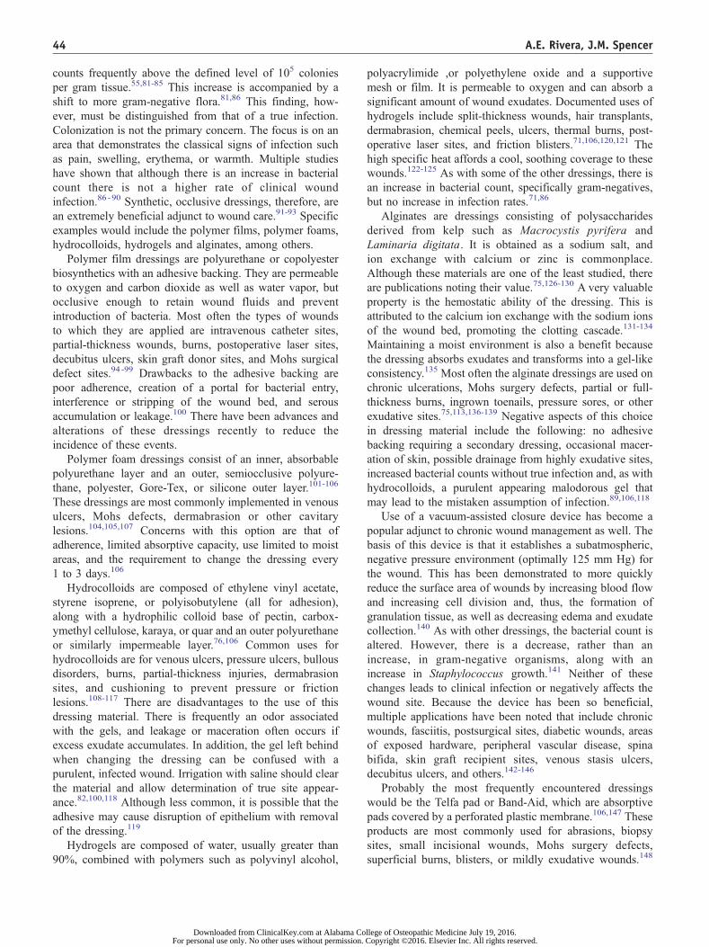
ig. 2 A, Surgical defect of the forehead and frontal scalp after
kin cancer excision. B, The defect is too large for primary closure,
o partial closure of the defect was performed. C, Final result with
car contracture optimized to give the appearance of a complete
rimary closure.
Conclusions
Most full-thickness wounds seen by dermatologists are
created surgically. In the absence of underlying disease, the
great majority of full-thickness wounds will heal without
extraordinary intervention. However, knowledge of wound
healing principles and practices can greatly enhance the
process. The most fundamental decision to be made for a
full-thickness wound is whether to repair the wound
surgically or to allow it to heal by second intention. There
are absolute and relative indications for repair. Preservation
of function is a mandatory indication for repair. For
example, a functional eyelid is mandatory after eyelid
surgery to maintain vision. Scars will contract up to 40% of
the size of the original wound, and if such contracture
produces a functional deficit, then repair is mandatory.
Examples include wounds near the eye or mouth that may
produce ectropion and eclabion, respectively, or a wound of
the ear canal where contracture may produce stenosis or
occlusion of the canal. For wounds where reconstruction is
not mandatory, the need and wisdom of pursuing recon-
structive surgery requires consideration of a number of
objective and subjective factors. What is the likely result
given the size and anatomic location of the wound? What
are the patients’ concerns and expectations? Are they
concerned about the cosmetic outcome? Can they tolerate
any further surgery? Can the patients perform weeks to
months of wound care for second intention healing? Some
general principals of second intention healing can help
guide the decision. As a general principle, concavities heal
quite nicely, with a pleasing aesthetic result, whereas
convexities do not. For example, a small wound of the
concave inner canthus will heal very well by second
intention, whereas the cosmetic result of the convex nose
will not be so pleasing after second intention. Wounds of the
lower leg will take very long to heal, so repair may be
indicated to prevent months of wound care.
Partial repair is a middle path that alters the dynamics of
wound healing. A round surgical wound will give an
atrophic round to stellate scar if allowed to heal by second
intention. However, if the round wound is converted to an
elliptically shaped open wound, the dynamics of scar
contracture often result in a linear scar that can approxi-
mate the appearance of a wound that was completely
sutured shut (Fig. 2).
Irrespective of whether a wound is repaired or allowed to
heal by second intention, it is beyond scientific question that
Downloaded from ClinicalKey.com at Alabama ColFor personal use only. No other uses without permission. C
Fs
s
s
p
occlusion speed healing provides greater comfort and may
give a better final cosmetic result. Simple occlusion with an
antibiotic containing petrolatum ointment and a semiocclu-
sive bandage is typically all that is required. Literally
hundreds of special dressing materials are available for
lege of Osteopathic Medicine July 19, 2016.opyright ©2016. Elsevier Inc. All rights reserved.
A.E. Rivera, J.M. Spencer46
problem wounds such as venous leg ulcers or diabetic foot
ulcers. The choice of which system to use for problem
wounds is based on the special characteristics of the
individual wound, the surrounding skin, and the patients
themselves. Thorough knowledge of wound healing and
management strategies allows the clinician to make the
optimal choice for each patient.
References
1. Ovington LG. Hanging wet-to-dry dressings out to dry. Home Healthc
Nurse 2001;19:477.
2. Orr JW, Taylor PT. Complications in gynecological surgery: preven-
tion, recognition, and management. In: Orr JW, Shingleton HM,
editors. Wound healing. Philadelphia7 JB Lippencott 1994. p. 167.
3. Kanegaye JT. A rational approach to the outpatient manage-
ment of lacerations in pediatric patients. Curr Probl Pediatr 1998;
28:205.
4. McNamara RN, Loiselle J. Laceration repair. In: Henretig F, King C,
editors. Textbook of pediatric emergency procedures. Baltimore7
Williams and Wilkins; 1997. p. 1141.
5. Piacquadio D. Synthetic surgical dressings. In: Wheeland RG, editor.
Cutaneous surgery. WB Saunders Company, 1994. p. 122 -36.
6. Glat PM, Longaker MT. Wound healing. In: Aston SJ, Beasley RW,
Thorne CH, editors. Grabb and Smith’s plastic surgery. 5th ed.
Philadelphia7 Lippencott; 1997. p. 3 -12.
7. Kane D. Wound healing and wound management. In: Krasner D,
editor. Chronic wound care. 2nd ed. Wayne (PA)7 Health Management
Publications, Inc; 1997. p. 1 -4.
8. Lawrence CM, Comaish S, Dahl MGC. Excision of skin tumors
without wound closure. Br J Dermatol 1986;115:563-71.
9. Spencer JM. Healing by second intention. In: Nouri K, Leal-Khouri S,
editors. Techniques in dermatologic surgery. Philadelphia7 Mosby;
2003. p. 117 -21.
10. Place MJ, Herber SC, Hardesty RA. Basic techniques and principles
in plastic surgery. In: Aston SJ, Beasley RW, Thorne CH, editors.
Grabb and Smith’s plastic surgery. 5th ed. Philadelphia7 Lippencott;
1997. p. 13 -25.
11. Moy RL, Waldman B, Hein DW. A review of sutures and suturing
technique. J Dermatol Surg Oncol 1992;18:785.
12. Karounis H, Gouin S, Eisman H, et al. A randomized, controlled trial
comparing long-term cosmetic outcomes of traumatic pediatric
lacerations repaired with absorbable plain gut versus nonabsorbable
nylon sutures. Acad Emerg Med 2004;11:730-5.
13. Hoffman MS, Villa A, Roberts WS, et al. Mass closure of the
abdominal wound with delayed absorbable suture in surgery for
gynecologic cancer. J Reprod Med 1991;36:356.
14. Webster RC, McCollough EG, Giandello PR, Smith RC. Skin wound
approximation wit new absorbable suture material. Arch Otolaryngol
1985;111:517.
15. Lober CW, Fenske NA. Suture materials for closing the skin and
subcutaneous tissues. Aesthetic Plast Surg 1986;10:245.
16. Start NJ, Armstrong AM, Robinson WJ. The use of chromic catgut in
the primary closure of scalp wounds in children. Arch Emerg Med
1989;6:216.
17. Guyuron B, Vaughan C. A comparison of absorbable and nonab-
sorbable suture materials for skin repair. Plast Reconstr Surg 1992;
89:234.
18. Shetty PC, Dicksheet S, Scalea TM. Emergency department repair of
hand lacerations using absorbable Vicryl sutures. J Emerg Med
1997;15:673.
19. Ethicon wound closure manual. St. Louis7 Ethicon, Inc; 1998-2000.
20. Trott AT. Wounds and lacerations. St. Louis7 Mosby-Year Book;
1991.
Downloaded from ClinicalKey.com at Alabama CoFor personal use only. No other uses without permission.
21. Morrow CP, Hernandez WL, Townsend DE, Disaia PJ. Pelvic
celiotomy in the obese patient. Obstet Gynecol 1977;127:335.
22. Menovsky T, Bartels RH, van Lindert EL, Grotenhuis JA. Skin
closure in carpal tunnel surgery: a prospective comparative study
between nylon, polyglactin 910 and stainless steel sutures. Hand Surg
2004;9:35-8.
23. Eaglstein WH, Sullivan T. Cyanoacrylates for skin closure. Dermatol
Clin 2005;23:193 -8.
24. Bernard L, Doyle J, Friedlander SF, et al. A prospective comparison
of octyl cyanoacrylate tissue adhesive (Dermabond) and suture for the
closure of excisional wounds in children and adolescents. Arch
Dermatol 2001;137:1177.
25. Rearson AS, Wolford RW. Management of skin trauma. Prim Care
2000;27:475.
26. Toriumi DM, Bagal AA. Cyanoacrylate tissue adhesives for skin
closure in the outpatient setting. Otolaryngol Clin North Am 2002;
35:103.
27. Bruns TB, Worthington JM. Using tissue adhesive for wound
repair: a practical guide to Dermabond. Am Fam Physician 2000;
61:1383.
28. Capellan O, Hollander JE. Management of lacerations in the
emergency department. Emerg Med Clin North Am 2003;21:205.
29. Yamamoto LG. Preventing adverse events and outcomes encountered
using Dermabond. Am J Emerg Med 2000;18:511.
30. Quinn JV, Drzewiecki A, Li MM, et al. A randomized, controlled trial
comparing a tissue adhesive with suturing in the repair of pediatric
facial lacerations. Ann Emerg Med 1993;22:1130.
31. Coulthard P, Worthington H, Esposito M, et al. Tissue adhesives for
closure of surgical incisions. Cochrane Database Syst Rev 2004;
2:CD004287.
32. Jallali N, Haji A, Watson CJ. A prospective randomized trial
comparing 2-octyl cyanoacrylate to conventional suturing in closure
of laparoscopic cholecystectomy incisions. J Laparoendosc Adv Surg
Tech A 2004;14:209 -11.
33. Barnett P, Jarman FC, Goodge J, et al. Randomised trial of histoacryl
blue tissue adhesive glue versus suturing in the repair of paediatric
lacerations. J Paediatr Child Health 1998;34:548.
34. Martin-Garcia RF, Janer AL, Rullan FV. Octyl-2-cyanoacrylate liquid
bandage as a wound dressing in facial excisional surgery: results of an
uncontrolled pilot study. Dermatol Surg 2005;31:670 -3.
35. Kronfol R. Tissue adhesives. UpToDate Online 13.2. Sept 9, 2004.
Accessed Aug 2005 http://www.utdol.com.
36. Singer AJ, Hollander JE, Valentine SM, et al. Prospective, random-
ized, controlled trial of tissue adhesive (2-octylcyanoacrylate) vs
standard wound closure techniques for laceration repair. Stony Brook
Octylcyanoacrylate Study Group. Acad Emerg Med 1998;5:94.
37. Quinn J, Wells G, Sutcliffe T, et al. A randomized trial comparing
octylcyanoacrylate tissue adhesive and sutures in the management of
lacerations. JAMA 1997;277:1527.
38. Holander J, Singer A. Laceration management. Ann Emerg Med
1999;34:356.
39. Holloway GA, Johansen KH, Barnes RW, et al. Multicenter trial of
cadexomer iodine to treat venous stasis ulcers. West J Med 1989;
151:35-8.
40. Zhou LH, Nahm WK, Badiavas E, Yulfit T, Falanga V. Slow release
iodine preparation and wound healing: in vitro effects consistent with
lack of in vivo toxicity in human chronic wounds. Br J Dermatol
2002;146:365-74.
41. Mober S, Hoffman L, Grennert ML, et al. A randomized trial of
cadexomer iodine in decubitus ulcers. J Am Geriatr Soc 1983;
31:462-5.
42. Ormiston MC, Seymour MT, Venn GE, et al. Controlled trial of
Iodosorb in chronic venous ulcers. BMJ 1985;291:308-10.
43. O’Toole EA, Goel M, Woodley DT. Hydrogen peroxide inhibits
human keratinocyte migration. Dermatol Surg 1996;22:525-9.
44. Demling RH, Desan TL. The role of silver in wound healing. Wounds
2001;13:5 -15.
llege of Osteopathic Medicine July 19, 2016. Copyright ©2016. Elsevier Inc. All rights reserved.
Clinical aspects of full-thickness wound healing 47
45. Wright JB, Lam K, Burrell RE. Silver coated wound dressings. Am J
Infect Control 1998;26:572-7.
46. Marolis DM, Berlin JA, Strom BL. Risk factors associated with the
failure of venous leg ulcer to heal. Arch Dermatol 1999;135:920 -6.
47. Olin BR, editor. Drugs: facts and comparisons—1991. Philadelphia7
Lippencott; 1991.
48. Kozol RA, Gilles C, Elgebaly SA. Effects of sodium hypochlorite
(Dakin’s solution) on cells of the wound module. Arch Surg 1998;
123:420-3.
49. Alvarez OM, Mertz PM, Eaglstein WH. The effect of occlusive
dressings on collagen synthesis and re-epithelialization in superficial
wounds. J Surg Res 1983;35:142-8.
50. Roupe G, Strannegard O. Anaphylactic shock elicited by topical
administration of bacitracin. Arch Dermatol 1969;100:450.
51. Cuzzell JZ. Wound care forum: artful solutions to chronic problems.
Am J Nurs 1985;85:162 -6.
52. Hood R, Shermock KM, Emerman C. A prospective, randomized
pilot evaluation of topical triple antibiotic versus mupirocin for the
prevention of uncomplicated soft tissue wound infections. Am J
Emerg Med 2004;22:1 -3.
53. Spann CT, Taylor SC, Weinberg JM. Topical antimicrobial agents in
dermatology. Dis Mon 2004;50:407 -21.
54. Lio PA, Kaye ET. Topical antibacterial agents. Infect Dis Clin North
Am 2004;18:717-33.
55. Reactions to topical antibiotics. In: Fisher AA, editor. Contact
dermatitis. 3rd ed. Philadelphia7 Lea & Febiger; 1986. p. 195 -210.
56. Fisher AA, Adams RM. Alternative for sensitizing neomycin topical
medicaments. Cutis 1981;28:491.
57. Smack DP, Harrington AC, Dunn C, et al. Infection and allergy
incidence in ambulatory surgery patients using white petrolatum vs
bacitracin ointment. A randomized controlled trial. JAMA 1996;276:
972 -7.
58. Ashcroft GS, Mills SJ, et al. Estrogen modulates cutaneous wound
healing by downregulating macrophage migration inhibitory factor.
J Clin Invest 2003;111:1309-18.
59. Ashcroft GS, Ashworth JJ. Potential role of estrogens in wound
healing. Am J Clin Dermatol 2003;4:737 -43.
60. Kovacs EJ. Aging, traumatic injury, and estrogen treatment. Exp
Gerontol 2005;40:549 -55.
61. Brincat MP, Baron YM, Galea R. Estrogens and the skin. Climacteric
2005;8:110 -23.
62. Batra RS, Dover JS, Hobbs L, Phillips TJ. Evaluation of the role of
exogenous estrogen in postoperative progress after laser skin
resurfacing. Dermatol Surg 2003;29:43-8.
63. Cross KJ, Mustoe TA. Growth factors in wound healing. Surg Clin
North Am 2003;83:531-45.
64. Vasquez R, Marien BJ, Gram C, et al. Proliferative capacity of venous
ulcer wound fibroblasts in the presence of platelet-derived growth
factor. Vasc Endovascular Surg 2004;38:355-60.
65. Eppley BL, Woodell JE, Higgins J. Platelet quantification and growth
factor analysis from platelet-rich plasma: implications for wound
healing. Plast Reconstr Surg 2004;114:1502-8.
66. Uutela M, Wirzenius M, Paavonen K, et al. PDGF-D induces
macrophage recruitment, increased interstitial pressure, and blood
vessel maturation during angiogenesis. Blood 2004;104:3198-204.
67. Man LX, Park JC, Terry MJ, et al. Lentiviral gene therapy with
platelet-derived growth factor B sustains accelerated healing of
diabetic wounds over time. Ann Plast Surg 2005;55:81 -6.
68. Lee JA, Conejero JA, Mason JM, et al. Lentiviral transfection with the
PDGF-B gene improves diabetic wound healing. Plast Reconstr Surg
2005;116:532-8.
69. Gu DL, Nguyen T, Gonzalez AM, et al. Adenovirus encoding human
platelet-derived growth factor-B delivered in collagen exhibits safety,
biodistribution, and immunogenicity profiles favorable for clinical
use. Mol Ther 2004;9:699 -711.
70. Telfer NR, Moy RL. Wound care after office procedures. J Dermatol
Surg Oncol 1993;19:722 -31.
Downloaded from ClinicalKey.com at Alabama ColFor personal use only. No other uses without permission. C
71. Mandy SH. A new primary wound dressing made of polyethylene
oxide gel. J Dermatol Surg Oncol 1983;9:153-5.
72. Barnett A, Berkowitz RL, Mills R, Vistnes LM. Comparison of
synthetic adhesive moisture vapor permeable and fine mesh gauze
dressings for split-thickness skin graft donor sites. Am J Surg 1975;
145:379-81.
73. Hein NT, Prawer SE, Katz HI. Facilitated wound healing using
transparent film dressing following Mohs micrographic surgery. Arch
Dermatol 1988;124:903-6.
74. Friedman S, Su DWP. Hydrocolloid occlusive dressing management
of leg ulcers. Arch Dermatol 1984;120:1329-36.
75. Attwood AI. Calcium alginate dressing accelerates split skin graft
donor site healing. Br J Plast Surg 1989;42:373-9.
76. Bolton LL, Johnson CL, Rijswijk LV. Occlusive dressings: thera-
peutic agents and effects on drug delivery. Dermatol Clin 1992;9:
573-83.
77. Alsbjorn BF, Ovesen H, Walther-Larsen S. Occlusive dressing versus
petrolatum gauze on drainage wounds. Acta Chir Scand 1990;156:
211 -3.
78. Gorse GJ, Messner RL. Improved pressure sore healing with
hydrocolloid dressings. Arch Dermatol 1987;123:765-71.
79. Marshall DA, Mertz PM, Eaglstein WH. Occlusive dressings: does
dressing type influence the growth of common bacterial pathogens?
Arch Surg 1990;125:1136-9.
80. Katz S, McGinley K, Leyden JJ. Semipermeable occlusive dressings.
Arch Dermatol 1986;122:58-62.
81. Mertz PM, Eaglstein WH. The effect of semiocclusive dressing on the
microbial population in superficial wounds. Arch Surg 1984;119:
287-9.
82. Gilchrist B, Reed C. The bacteriology of chronic venous stasis ulcers
treated with occlusive hydrocolloid dressings. Br J Dermatol 1989;
121:337-44.
83. Robson MS, Heggars JP. Quantitative bacteriology and inflammatory
mediators in soft tissues. In: Hunt TK, Heppenstall RB, Pines E,
Rovee D, editors. Soft and hard tissue repair: biological and clinical
aspects. New York7 Praeger; 1984. p. 483 -507.
84. Varghese MC, Balin AK, Carter MD, Caldwell D. Local environment
of chronic wounds under synthetic dressings. Arch Dermatol 1986;
122:5257.
85. Marples RR, Lingman AM. Growth of bacteria under adhesive tapes.
Arch Dermatol 1969;99:107.
86. Mertz PM, Marshall DA, Eaglstein WH. Occlusive wound dressings
to prevent invasion and wound infection. J Am Acad Dermatol
1985;12:662 -8.
87. Handfield-Jones SE, Grattan CEH, Simpson RA, et al. Comparison of
a hydrocolloid dressing and paraffin gauze in the treatment of venous
ulcers. Br J Dermatol 1988;121:337 -44.
88. Buchan IA, Andrews JK, Lang SM, et al. Clinical and laboratory
investigation of the composition and properties of human skin wound
exudates under semi-permeable dressings. Burns 1980;7:326-34.
89. Hutchinson JJ, McGuckin M. Occlusive dressings: a microbiologic
and clinical review. Am J Infect Control 1990;18:257-68.
90. Eaglstein WH. Experiences with biosynthetic dressings. J Am Acad
Dermatol 1985;12:434-40.
91. Lionelli GT, Lawrence WT. Wound dressings. Surg Clin North Am
2003;83:617 -38.
92. Menaker GM. Wound dressings for office-based surgery. Facial Plast
Surg 2004;20:91 -105.
93. Vermeulen H, Ubbink DT, Goossens A, et al. Systematic review of
dressings and topical agents for surgical wounds healing by secondary
intention. Br J Surg 2005;92:665 -72.
94. Lees V, Hyas S, Reid CD. A comparison of the use of polyethylene
sheet and Jelonet as temporary dressing for excised wounds. Br J Plast
Surg 1991;44:612 -4.
95. Hien NT, Prawer SE, Katz HI. Facilitated wound healing using
transparent film dressing following Mohs micrographic surgery. Arch
Dermatol 1988;124:903-6.
lege of Osteopathic Medicine July 19, 2016.opyright ©2016. Elsevier Inc. All rights reserved.
A.E. Rivera, J.M. Spencer48
96. Rubio PA. Use of semiocclusive, transparent film dressings for surgical
wound protection: experience in 3937 cases. Int Surg 1991;76:253-4.
97. Raab B. A new hydrophilic copolymer membrane for dermabrasion.
J Dermatol Surg Oncol 1991;17:323 -8.
98. Poulsen TD, Freund KG, Arendrup K, et al. Polyurethane film
(Opsite) vs impregnated gauze (Jelonet) in the treatment of outpatient
burns: a prospective, randomized study. Burns 1991;17:59 -61.
99. Vartak AM, Keswani MH, Patil AR, et al. Cellophane: a dressing for
split-thickness graft donor sites. Burns 1991;17:239-42.
100. Kannon GA, Garrett AB. Moist wound healing with occlusive
dressings. Dermatol Surg 1995;21:583-90.
101. Fletcher J. Understanding wound dressings: foam dressings. Nurs
Times 2005;101:50-1.
102. Falanga V. Occlusive wound dressings. Arch Dermatol 1988;124:872-7.
103. Acme United Corp. Lyofoam clinical experiences. Fairfield, CT7
Acme United Corporation; 1990.
104. Samson RH. Compression stockings and non-continuous use of
polyurethane foam dressings for the treatment of venous ulceration.
J Dermatol Surg Oncol 1990;19:68 -72.
105. Rubin JR, Alexander J, Plecha EJ, et al. Unna’s boot vs. polyurethane
foam dressings for the treatment of venous ulceration. Arch Surg
1990;125:489 -90.
106. Wheeland RG. Wound healing and the newer surgical dressings. In:
Moschella SL, Hurley HJ, editors. Dermatology. Philadelphia7
Saunders; 1992. p. 2305-11.
107. Ryan TJ. Wound dressing. Dermatol Clin 1993;11:207-13.
108. Afilalo M, Guttman A, Lloyd J. DuoDERM hydroactive dressing
versus silver sulphadiazine/Bactigras in the emergency treatment of
partial skin thickness burns. Burns 1992;18:313 -6.
109. Singh A, Halder S, Menon GR, et al. Meta-analysis of randomized
controlled trials on hydrocolloid occlusive dressing versus conven-
tional gauze dressing in the healing of chronic wounds. Asian J Surg
2004;27:326 -32.
110. Eriksson G. Comparison of two occlusive bandages in the treatment
of venous leg ulcers. Br J Dermatol 1986;114:227-30.
111. Eisenberg M. The effects of occlusive dressings on re-epithelialization
of wounds in children with epidermolysis bullosa. J Pediatr Surg
1986;21:892-4.
112. Nemeth AJ, Eaglstein WH, Taylor JR, et al. Faster healing and less
pain in skin biopsy sites treated with an occlusive dressing. Arch
Dermatol 1991;127:1679 -83.
113. Porter JM. A comparative investigation of re-epithelialization of split
skin graft donor areas after application of hydrocolloid and alginate
dressings. Br J Plast Surg 1991;44:333 -7.
114. Gorse GJ, Messner RL. Improved pressure sore healing with
hydrocolloid dressings. Arch Dermatol 1987;123:766 -71.
115. Phillips LG, Robson MC, Smith DJ, et al. Uses and abuses of a
biosynthetic dressing for partial skin thickness burns. Burns 1989;et
al15:254 -6.
116. Leipziger LS, Glushko V, DiBernardo B, et al. Dermal wound repair:
role of collagen matrix implants and synthetic polymer dressings.
J Am Acad Dermatol 1985;12:409-19.
117. Volder G. Successful treatment of chronic skin diseases with
clobetasol propionate and a hydrocolloid occlusive dressing. Acta
Dermatolvenereol 1992;72:69 -71.
118. Gilchrist B, Reed C. The bacteriology of chronic venous ulcers
treated with occlusive hydrocolloid dressings. Br J Dermatol 1989;
121:337 -44.
119. Zitelli JA. Delayed wound healing with adhesive wound dressings.
J Dermatol Surg Oncol 1984;10:709.
120. Geronemus RG, Robins P. The effect of two new dressings on
epidermal wound healing. J Dermatol Surg Oncol 1982;8:850 -2.
121. Kaya AZ, Turani N, Akyuz M. The effectiveness of a hydrogel
dressing compared with standard management of pressure ulcers.
J Wound Care 2005;14:42 -4.
122. Hampton S. A small study in healing rates and symptom control using
a new sheet hydrogel dressing. J Wound Care 2004;13:297 -300.
Downloaded from ClinicalKey.com at Alabama CoFor personal use only. No other uses without permission.
123. Alvares O. Moist environment of healing: matching the dressing to
the wound. Ostomy/Wound Management 1988;21:65 -83.
124. Helfman T, Ovington A, Falanga V. Occlusive dressings and wound
healing. Dermatol Clin 1994;12:121 -7.
125. Sawada Y, Suzuki T, Hatayama I, et al. Silicone gel including
antimicrobial agent. Br J Plast Surg 1990;43:48 -82.
126. Thomas S. Use of a calcium alginate dressing. Pharmaceut J 1985;
235:188-90.
127. Barnett SE, Varley SJ. The effects of calcium alginate on wound
healing. Ann R Coll Surg Engl 1987;69:153-5.
128. Agren MS. Percutaneous absorption of zinc from zinc oxide applied
topically to intact skin in man. Dermatologica 1990;180:36 -9.
129. Soderberg T, Agren M, Tengrup I, et al. The effects of an occlusive
zinc medicated dressing on the bacterial flora in excised wounds in
the rat. Infection 1989;17:27-31.
130. Fletcher J. Understanding wound dressings: alginates. Nurs Times
2005;101:53-4.
131. Groves AR, Lawrence JC. Alginate dressing as a donor site
haemostat. Ann R Coll Surg Engl 1986;68:27 -8.
132. Frazier R, Gilchrest T. Sorbsan calcium alginate fiber dressings in
footcare. Biomaterials 1983;4:222-4.
133. Jarvis PM, Galvin DAJ, Blair SD, McCollum CN. How does calcium
alginate achieve hemostasis in surgery? XIth International Congress
of Thrombosis and Hemostasis, Brussels, July.
134. Blair SD, Jarvis P, Salmon M, McCollum C. Clinical trial of calcium
alginate haemostatic swabs. Br J Surg 1990;77:568-70.
135. Placquadio D, Nelson DB. Alginates: a bnewQ dressing alternative.
J Dermatol Surg Oncol 1992;18:90 -8.
136. Fine BC, Sheckman PR, Bartlett JC. Incision and drainage of soft
tissue abscesses and bacteremia. Ann Intern Med 1985;103:645-7.
137. Lawrence JE, Blake GB. A comparison of calcium alginate and
scarlet red dressings in the healing of split thickness skin graft donor
sites. Br J Plast Surg 1991;44:247 -9.
138. Barnett AH, Odugbesan O. Seaweed-based dressings in the manage-
ment of leg ulcers and other wounds. Intern Ther Clin Mon
1998;9:70-6.
139. Burrows T, Welch MJ. The development and use of alginate fibres in
nonwovens for medial end-users. In: Cusick GE editor. Nonwoven
conference papers. Manchester, UK7 UMIST; 1983.
140. Venturi ML, Attinger CE, Mesbahi AN, et al. Mechanisms and
clinical applications of the vacuum-assisted closure (VAC) device: a
review. Am J Clin Dermatol 2005;6:185-94.
141. Moues CM, Vos MC, van den Bemd GJ, Stijnen T, Hovius SE.
Bacterial load in relation to vacuum-assisted closure wound therapy: a
prospective randomized trial. Wound Repair Regen 2004;12:11 -7.
142. Oczenski W, Waldenberger F, Nehrer G, et al. Vacuum-assisted
closure for the treatment of cervical and mediastinal necrotizing
fasciitis. J Cardiothorac Vasc Anesth 2004;18:336 -8.
143. Mendonca DA, Cosker T, Makwana NK. Vacuum-assisted closure to
aid wound healing in foot and ankle surgery. Foot Ankle Int
2005;26:761-6.
144. Schaffzin DM, Douglas JM, Stahl TJ, Smith LE. Vacuum-assisted clo-
sure of complex perineal wounds. Dis Colon Rectum 2004;47:1745-8.
145. Carson SN, Overall K, Lee-Jahshan S, Travis E. Vacuum-assisted
closure used for healing chronic wounds and skin grafts in the lower
extremities. Ostomy Wound Manage 2004;50:52 -8.
146. Lambert KV, Hayes P, McCarthy M. Vacuum-assisted closure: a
review of development and current applications. Eur J Vasc Endovasc
Surg 2005;29:219-26.
147. Phillips TJ, Kapoor V, Provan A, et al. A randomized prospective
study of hydroactive dressing vs conventional treatment after shave
biopsy excision. Arch Dermatol 1993;129:859-60.
148. Woodley DT, Kim YH. A double-blind comparison of adhesive
bandages with the use of uniform suction blister wounds. Arch
Dermatol 1992;128:1354.
149. Eaglstein WH. Occlusive dressings. J Dermatol Surg Oncol
1993;19:716-20.
llege of Osteopathic Medicine July 19, 2016. Copyright ©2016. Elsevier Inc. All rights reserved.