Embed Size (px)
Citation preview
CLINICAL AND RADIOLOGICAL BONE DENSITY STUDY OF IMMEDIATE PLACEMENT OF CORAL COATED DENTAL IMPLANT
By
All ABDUL KAWI All TAHER
Thesis submitted in fulfillment of the requirements for the degree of Master
August 2006
ACKNOWLEDGMENTS
In the name of Allah the most passionate and the most merciful
give my greatest appreciation and full respect to all my supervisors who had
contributed excellent advise, support and encouragement in this project.
The most special thanks due to Professor Dr. Ab Rani Samsudin, lecturer in the Oral
Maxillofacial Surgery Unit and Dean of School of Dental Sciences for his untiring
dedication, brilliant knowledge of research, support, understanding , exceptional advice
and leadership which without him none of this research could take place.
Special acknowledgment to Dr. Sam'an Malik Masudi, Co-supervisor, for his
assistance, advice and support through out the years that I have devoted to writing this
thesis.
Dr. Ramizu Bin Shaari and Dr. Shaifulizan Abdul-Rahman for their expertise in
implantology that gave me a great foundation in clinical implantology.
My respect and thanks are due to Dr. Nizam Abdullah for her time and contribution to
my research.
Dr. Mohd Ayub Sadiq for his expert analytical and mathematical contributions to this
study.
Dr. Abdullah Pohchi for his contribution, support and help.
My greatest appreciation to all the dental outpatients that volunteered to give their
time, effort and contribution to this thesis.
ii
I also extend my grateful appreciation and thanks to all the staff, nurses, dental officers
and research officers of Dental and Medical School, USM, especially
Oralmaxillofacial Surgery Unit for their help and support.
I also wish to express my gratitude to all my colleagues, classmates, fellow residents
and friends, for their friendship and support, especially Professor Dr. Abdul-Wahab
AI- Khawlani, Dr. Mohammed Radman, Dr. Ali AI-Sharabi, Dr. Ismail AI-Haj, Dr.
Hatem Hiba, Dr. Mohammed Noman AI-Hajibi, Dr. Najeeb Abu Rub, Dr. Hamed
Darzi Najafpour, Dr. Ahmed Almutawakel, Dr. Abdulaziz aludayni, Dr. Tawfeeq
Abdulla Ahmed, Dr. Nasralden Almery, Dr. Abo Abdulrazqy, Dr. Abdullah Mugeer,
Dr. Waheed, Dr. Tareq Alrahumi, Dr. Saeed Mohammed, Dr. Ayeman, Dr. Ali
Alashwel, Dr. Aaqil Malik, Dr. Osama Baha'a , Dr. Hazem Yosuf and Dr. Tariq
Abdussamad.
I would like to acknowledge with greatest thanks to my parents who always pray of
god for me and my brothers Khalid and Mohammed Abdui-Kawi whom have
supported me and always commune with me which gave me confidence to pursue my
research.
Last but not least to my beloved wife who support and understanding during the most
challenging period to whom I dedicated this dissertation.
To all named and unnamed friends and helpers especially our estimable Dean of
School of Dental Sciences Professor Dr. Ab Rani Samsudin, I again extend my
thanks.
iii
TABLE OF CONTENTS
ACKNOWLEDGEMENTS . . . . . . . . . . . . . . . . . . . . . . . . . . . . . . . . . . . . . . . . . . . . . . . . . . . . . . . . . . ii
TABLE OF CONTENTS . . . . . . . . . . . . . . . . . . . . . . . . . . . . . . . . . . . . . . . . . . . . . . . . . . . . . . . . . . iv
LIST OF TABLES . . . . . . . . . . . . . . . . . . . . . . . . . . . . . . . . . . . . . . . . . . . . . . . . . . . . . . . . . . . . . . . . . . . . . viii
LIST OF FIGURES................................................................. xi
LIST OF APPENDICES . . . . . . . . . . . . . . . . . . . . . . . . . . . . . . . . . . . . . . . . . . . . . . . . . . . . . . . . . . . . xii
ABSTRAK . . . . . . . . . . . . . . . . . . . . . . . . . . . . . . . . . . . . . . . . . . . . . . . . . . . . . . . . . . . . . . . . . . . . . . . . . . . . xiv
ABSTRACT . . . . . . . . . . . . . . . . . . . . . . . . . . . . . . . . . . . . . . . . . . . . . . . . . . . . . . . . . . . . . . . . . . . . . . . . .. xvi
CHAPTER ONE : INTRODUCTION
1.1 Background . . . . . . . . . . . . . . . . . . . . . . . . . . . . . . . . . . . . . . . . . . . . . . . . . . . . . . . . . . . . . . . . . . 1
1.2 Statement of the problem . . . . . . . . . . . . . . . . . . . . . . . . . . . . . . . . . . . . . . . . . . . . . . . . 4
1.3 Hypothesis . . . . . . . . . . . . . . . . . . . . . . . . . . . . . . . . . . . . . . . . . . . . . . . . . . . . . . . . . . . . . . . . . . . 4
1.4 Objectives . . . . . . . . . . . . . . . .. . . . . . . . . . . . . . . . . . . . . . . . . . . . . . . . . . . . . . . . . . . . . . . . . . 4
1.4.1 General objectives . . . . . . . . . . . . . . . . . . . . . . . . . . . . . . . . . . . . . . . . . . . . . . . . 4
1.4.2 Specific objectives . . . . . . . . . . . . . . . . . . . . . . . . . . . . . . . . . . . . . . . . . . . . . . . . 4
1.5 Significance of the study . . . . . . . . . . . . . . . . . . . . . . . . . . . . . . . . . . . . . . . . . . . . . . . . . . . 4
CHAPTER TWO: LITERATURE REVIEW
2.1 History of implant . . . . . . . . . . . . . . . . . . . . . . . . . . . . . . . . . . . . . . . . . . . . . . . . . . . . . . . . . . 6
2.2 Implant materials . . . . . . . . . . . . . . . . . . . . . . . . . . . . . . . . . . . . . . . . . . . . . . . . . . . . . . . . . . 9
2.3 Classification of materials................................................ 9
2.4 Types of implant.......................................................... 10
2.4.1 SUB-PERRIOSTEAL IMPLANTS . . . . . . . . . . . . . . . . . . . . . . . . . . . . . . 10
2.4.2 TRANS-OSTEAL IMPLANTS . . . . . . . . . . . . . . . . . . . . . . . . . . . . . . . . . 10
2.4.3 EN DO-OSTEAL IMPLANTS . . . . . . . . . . . . . . . . . . . . . . . . . . . . . . . . . . . . 1 0
2.5 Titanium .................................................................. . 12
iv
2.6 Osseointegration . . . . . . . . . . . . . . . . . . . . . . . . . . . . . . . . . . . . . .. . . . . . . . . . . . . . . . . . . . 12
2. 7 Bone remodeling . . . . . . . . . . . . . . . . . . . . . . . . . . . . . . . . . . . . . . . . . . . . . . . . . . . . . . . . . . 14
2.8 Bone healing ... ...... ... ... . .. . .. . .. ... ... ... ... ... ... ... ... ... ... ... ... ... 14
2.9 Bone density . . . . . . . . . . . . . . . . . . . . . . . . . . . . . . . . . . . . . . . . . . .. . . . . . . . . . . . . . . . . . . . . . . 15
2.1 0 Dental implant interface . . . . . . . . . . . . . . . . . . . . . . . . . . . . . . . . . . . . . . . . . . . . . . . . . . . 15
2.11 Hydroxyapatite-coated implant . . . . . . . . . . . . . . . . . . . . . . . . . . . . . . . . . . . . . . . . . . 16
2.12 Calcium carbonate natural coral . . . . . . . . . . . . . . . . . . . . . . . . . . . . . . . . . . . . . . . 18
2.13 Classification of bone quality . . . . . . . . . . . . . . . . . . . . . . . . . . . . . . . . . . . . . . . . . . . . . 26
2.14 Application of synthetic matrix gel . . . . . . . . . . . . . . . . . . . . . . . . . . . . . . . . . . . . . . . 33
CHAPTER THREE: MATERIALS AND METHODS
3.1 Study design ............................................................ .
3.2 Population ............................................................... .
35
35
3.2.1 Inclusion criteria ... ... . .. .. . ... ... ... ... ... ...... ... ... ... ... ... .. . 35
3.2.2 Exclusion criteria . . . . . . . . . . . . . . . . . . . . . . . . . . . . .. . . . . . . . . . . . . . . . . . . . . . 35
3.3 Research tools and materials . . . . . . . . . . . . . . . . . . . . . . . . . . . . . . . . . . . . . . . . . . . . . 37
3.3.1 Surgical instrument ... ... ... ... ... ... ... ... ... ... ... ... ... ... ... ... 37
3.3.2 Implant system- Frialit®-2 . . . ... . . . . . . ... ... . . . . . . . . . . . . . . . . .. . . . 38
3.3.3 Natural coral . . . . . . . . . . . . . . . . . . . . . . . . . . . . . . . . . .. . . . . . . . . . . . . . . . . . . . . . . 39
3.3.4 Synthetic matrix gel ... ... ... ...... ... ... ...... ... ... ... ... ...... ... 40
3.3.5 Radiographs .. . . . . . . . . . . ... . . . . . . . .. .. . . . . . . . ... . .. . . . . . . . . . . . . . . . . . . 42
3.4 Data collection ...... ... ... ... ... ... ... ... ... ... ... ... ...... ... ... ... .... 43
3.4.1 Patients data ... . .. ... .. . ... ... . .. ... ... . .. ... ... ... . .. ... . .. . .. ... .. . 43
3.4.2 Preoperative radiograph . . . . . . . . . . . . . . . . . . . . . . . . . . . . . . . . . . . . . . . . . . 43
3.4.3 Determination using the VIXWIN software.................. 45
3.5 Operative procedure...................................................... 47
3.5.1 Preoperative stage . . . . . . . . . . . . . . . . . . . . . . . . ... . . . . . . . . . . . . . . . ... . . . 47
v
3.5.2 Intra-operative stage . . . . . . . . . . . . . . . . . . . . . . . . . . . . . . . . . . . . . . . . . . . . . 48
3.5.2 (a) The first stage : 48
3.5.3 Postoperative care................................................. 51
3.5.4 First review at 1 week postoperative .. . .. . . . . ... .. . . .. ... . .. . 51
3.5.5 Second review at 2 weeks postoperative . . . . . . . . . . . . . . . . . . 52
3.5.6 Third review at 3 weeks postoperative..................... 52
3.5.2 (b) The second stage . . . . . . . . . . . . . . . . . . . . . ... . . . . . . .. . .. 53
3.6 Assessment of the radiographs . . . . . . . . . . . . . . . . . . . . . . . . . . . . . . . . . . . . . . . . 54
3.6.1 Measurement of implant length using VIXWIN software.. 56
3.6.1 (a) Mandible................................................. 56
3.6.1 (b) Maxilla................................................... 56
CHAPTER FOUR : RESULTS
4.1 Clinical results............................................................... 59
4.2 Radiological results....................................................... 63
CHAPTER FIVE : DISCUSSION 72
CHAPTER SIX : SUMMARY AND CONCLUSION
6.1 Summary... . . . . . . . . . . . . . . . . . . . . . . . . . . . . . . . . . . . . . . . . . . . . . . . . . . . . . . . . . . . . . . . . . . . 76
6.2 Limitation of the study..................................................... 76
6.3 Conclusion.................................................................. 76
6.4 Recommendations for future research................................ 76
vi

BIBLIOGRAPHY 78 ......................................................................
APPENDICES . ........................................................ ... ... ...... .. .
vii

Table 2.1
Table 2.2
Table 4.1
Table 4.2
Table 4.3
Table 4.4
LIST OF TABLES
Classification of Materials ..................................................... .
Flow chart showing the phases of Development and Evaluation of
Page
9
sea coral for bone grafting ...... ...... ...... ...... .. .. .. .... .. .. .. .. ...... ..... 21
Densitometric profile of all preoperative teeth .......................... . 61
The clinical evaluation of patients after placing immediate Implant .. 62
Densitometric profile of all immediate implants with coral bone graft
and without cor~ I bone graft . . . . . . . . . . . . . . . . . . . . . . . . . . . . . . . . . . . . . . . . . . . . . . . . . . . 63
The mean pre and postoperative densitometric comparing between
immediate coral coated implant and immediate non-coated implant. 65
viii
Figure 2.1
Figure 2.2
Figure 2.3
Figure 2.4
Figure 2.5
LIST OF FIGURES
Seashells hammered into the jaw to replace missing teeth ............ .
Types of Implant ................................................................. .
Dental implant osseointegration micrograph ............................... .
Natural processed coral. The arrows point at the pores that can be
Page
8
11
13
detected with the naked eyes.................................................. 19
Saggital diagrammatic representation from Maxillary and
Mandibular anterior regions showing bone resorption patterns from
cephalometric radiographs..................................................... 28
Figure 2.6 Classification of cortical and cancellous bone quality.................... 28
Figure 2.7 Versions of the Frialit-2 implant.............................................. 32
Figure 3.1 Flow chart of study............................................................... 36
Figure 3.2 The FRIALI-r®-2 surgical tray- Concise and well- organized...... 37
Figure 3.3 FRIALI-r®-2 colors provide................................................... 38
ix
Figure 3.4 Natural coral with size of 20- 251Jm... ... ...... ... ... ... ... ... .. . ... .. . . .. ... 39
Figure 3.5 Gengigel Hyaluronic acid ... ... ... ... ... ... ... ... ...... ... ... ... ... .. . ... ... .. . 41
Figure 3.6 Digitized OPG with the use of Gendex imaging system................. 42
Figure 3. 7 Positioning of the patient in the OPG machine............................ 43
Figure 3.8 Digitized OPG with magnification factor of 1.25........................... 44
Figure 3.9 Measurement bone of density.................................................. 46
Figure 3.10 Preoperative panoramic x-ray................................................. 47
Figure 3.11 Preoperative periapical x-ray.................................................. 47
Figure 3.12 Local anaesthesia being administered for tooth extraction . . . . . . . . . . . . 49
Figure 3.13 Removing the implant from the sterile packaging........................ 49
Figure 3.14 Implant coated with coral mixed with gengigel (coral gel).............. 49
Figure 3.15 Coral gel inserted into the extraction socket . . . . . . . . . . . . . . . . . . . . . . . . . . . . . . 49
Figure 3.16 Implant was inserted with a ratchet......................................... 50
X
=igure 3.17 Arrow (a) showing cover screw placed in the site to facilitate
healing and arrow (b) showing coral gel around implant . . . . . . . . . . . . . . . 50
Figure 3.18 The soft tissue was drawn over the implant and sutured to place..... 50
Figure 3.19 Wound Closure . . . . . . . . . . . . . . . . . . . . . . . . . . . . . . . . . . . . . . . . . . . . . . . . . . . . . . . . . . . . . . . . . . . . 50
Figure 3.20 Periapical x-ray at one week postoperative................................. 51
Figure 3.21 Periapical x-ray at two weeks postoperative................................ 52
Figure 3.22 Periapical x-ray at three weeks postoperative... . . . . . . . . . . . . . . . . . . . . . . . . . . 52
Figure 3.23 Orthopantomograph view at four months postoperative with an
implant at 36......... . . . . . . . . . . . . . . . . . . . . . . . . . . . . . . . . . . . . . . . . . . . . . . . . . . . . . . . . . . . . . . . 53
Figure 3.24 Abutment tooth 36... ... ... ...... ... ... ... ... ... ... ... ... ... ... ... ... ... ... ... ... . 53
Figure 3.25 Final crowning 36.................. .. . . . . . . . . . . . . . . . . . . . . . . . . . . . . . . . . . . . . . . . . . . . 53
Figure 3.26 Orthoralix 9200 digital OPG............ . . . . . . . . . . . . . . . . . . . . . . . . . . . . . . . . . . . . . . . . 55
Figure 3.27 Measurement of implant length using VIXWIN software................ 57
Figure 3.28 Yellow line passing through the inferior border of MF, red line
parallel to the inferior border of the mandible... . . . . . . . . . . . . . . . . . . . . . . . . . . . 58
xi
Figure 4.1 Points of measurement of bone density for obtaining preoperative
and postoperative densitometric value...................................... 60
Figure 4.2 Mean densitometric profile from the data showing normal
distribution of statistical analysis ............................................. . 66
Figure 4.3 Marginal means between coral coated and non-coated dental
mplant groups at coronal mesial points..................... . . . . . . . . . . . . . . . . 67
Figure 4.4 Marginal means between coral coated and non-coated dental
implant groups at coronal distal points...................................... 68
Figure 4.5 Marginal means between coral coated and non-coated dental
implant groups at midway mesial points... . . . . . . . . . . . . . . . . . . . . . . . . . . . . . . . . . 69
Figure 4.6 Marginal means between coral coated and non-coated dental
implant groups at midway distal points...................................... 70
Figure 4.7 Marginal means between coral coated and non-coated dental 71
implant groups at apex points ..................................... .
xii

PENDIX
PENDIX
PENDIX
LIST OF APPENDICES
(A) SCREENING FORM
(B) ACADEMIC ACTIVITIES
B (1) CLINICAL AND RADIOLOGICAL STUDY OF IMMEDIATE PLACEMENT
OF DENTAL IMPLANTS WITH CORAL BONE GRAFT. (ABSTRACT)
xiii
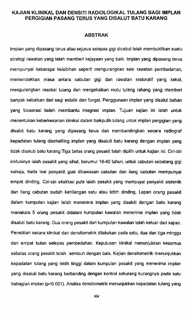
KAJIAN KLINIKAL DAN DENSITI RADIOLOGIKAL TULANG BAGI IMPLAN PERGIGIAN PASANG TERUS YANG DISALUT BATU KARANG
ABSTRAK
tmptan yang dipasang terus atau sejurus setepas gigi dicabut telah membuktikan suatu
strategi rawatan yang telah memberi kejayaan yang baik. tmptan yang dipasang terus
mempunyai beberapa kelebihan seperti mengurangkan sesi rawatan pembedahan,
memendekkan masa antara cabutan gigi dan rawatan restoratif yang kekat,
mengurangkan resobsi tutang dan mengekalkan mutu tutang rahang yang memberi
banyak kebaikan dari segi estetik dan fungsi. Penggunaan implan yang disalut bahan
yang bioserasi boleh membantu integrasi imptan. Tujuan kajian ini ialah untuk
menentukan keberkesanan klinikat datam baikpulih tulang untuk implan pergigian yang
disatut batu karang yang dipasang terus dan membandingkan secara radiograf
kepadatan tulang disekeliling implan yang disatuti batu karang dengan imptan yang
tidak disatuti batu karang.Tiga betas orang pesakit telah dipilih untuk kajian ini. Ciri-ciri
inklusinya ialah pesakit yang sihat, berumur 18-40 tahun, untuk cabutan sebatang gigi
sahaja, tiada tesi penyakit gusi dikawasan cabutan dan liang cabutan mempunyai
empat dinding. Ciri-ciri eksktusi pula ialah pesakit yang mempuyai penyakit sistemik
dan liang cabutan sudah kehilangan satu atau lebih dinding. Lapan orang pesakit
datam kumputan kajian telah menerima imptan yang disatuti dengan batu karang
manakala 5 orang pesakit didalam kumputan kawalan menerima imptan yang tidak
disatuti batu karang. Dua orang pesakit dari kumpulan kawatan telah keluar dari kajian.
Penelitian secara klinikal dan densitometrik dilakukan pada satu, dua dan tiga minggu
dan empat bulan setepas pembedahan. Keputusan klinikal menunjukkan kesemua
sebelas orang pesakit telah sembuh dengan baik. Kajian densitometrik menunjukkan
kepadatan tulang yang lebih tinggi dalam kumpulan pesakit yang menerima implan
yang disatuti batu karang berbanding dengan kontrol sekurang kurangnya pada satu
bahagian implan (p<0.001). Analisa densitometrik menunjukkan kepadatan tulang yang
xiv

lebih tinggi di semua lima bahagian implant dalam kumpulan implan yang disaluti batu
karang berbanding dengan kumpulan control. Walaubagaimana pun hanya bahagian
coronal mesial dan midway distal telah mempunyai kepadatan tulang yang lebih
signifikan, (p<0.002 dan p<0.024). Keputusan kajian ini membuktikan bahawa batu
karang buatan tempatan ialah suatu bahan bio yang sesuai untuk menyaluti implan
kerana kestabilan primer yang dihasilkan telah menyokong pertumbuhan tulang yang
mendorong kepada kestabilan sekunder. Graf batu karang yang bioserasi dan sifat
osteokonduktornya telah merangsangkan fenomena yang sangat bermakna dalam
implantologi.
XV
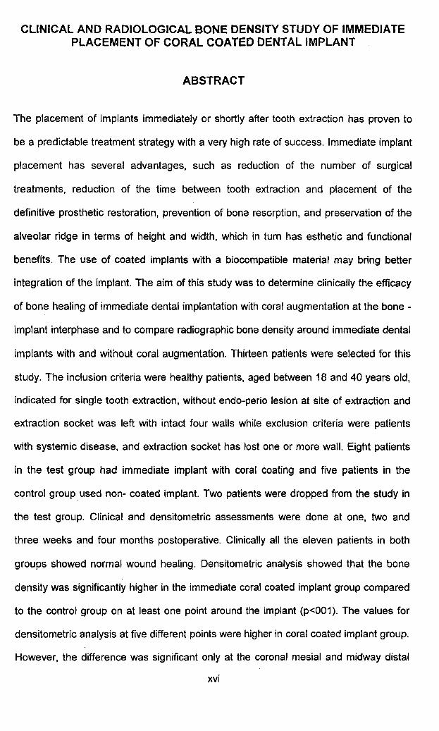
CLINICAL AND RADIOLOGICAL BONE DENSITY STUDY OF IMMEDIATE PLACEMENT OF CORAL COATED DENTAL IMPLANT
ABSTRACT
The placement of implants immediately or shortly after tooth extraction has proven to
be a predictable treatment strategy with a very high rate of success. Immediate implant
placement has several advantages, such as reduction of the number of surgical
treatments, reduction of the time between tooth extraction and placement of the
definitive prosthetic restoration, prevention of bone resorption, and preservation of the
alveolar ridge in terms of height and width, which in turn has esthetic and functional
benefits. The use of coated implants with a biocompatible material may bring better
integration of the implant. The aim of this study was to determine clinically the efficacy
of bone healing of immediate dental implantation with coral augmentation at the bone -
implant interphase and to compare radiographic bone density around immediate dental
implants with and without coral augmentation. Thirteen patients were selected for this
study. The inclusion criteria were healthy patients, aged between 18 and 40 years old,
indicated for single tooth extraction, without endo-perio lesion at site of extraction and
extraction socket was left with intact four walls while exclusion criteria were patients
with systemic disease, and extraction socket has lost one or more wall. Eight patients
in the test group had immediate implant with coral coating and five patients in the
control group used non- coated implant. Two patients were dropped from the study in
the test group. Clinical and densitometric assessments were done at one, two and
three weeks and four months postoperative. Clinically all the eleven patients in both
groups showed normal wound healing. Densitometric analysis showed that the bone
density was significantly higher in the immediate coral coated implant group compared
to the control group on at least one point around the implant (p<001 ). The values for
densitometric analysis at five different points were higher in coral coated implant group.
However, the difference was significant only at the coronal mesial and midway distal
xvi

points, (p<0.002 and p<0.024) respectively. Based upon the results of the present
study, it can be concluded that locally produced coral seemed to be a suitable material
for coating the surface of implants since it provided primary stability to the immediate
placement of the coated implants in the extraction sockets. This primary stability will
ensure new bone growth to provide the more stable secondary stability. The
biocompatibility of the coral graft and its role as an osteoconductor would have
encourage this very useful phenomena in implantology.
xvii
1.1 Background
CHAPTER ONE
INTRODUCTION
Immediate implants are defined as placement of implants in the course of surgical
extraction of the teeth to be replaced (Penarrocha, 2001 ). The insertion of implants
immediately after extraction is not new. In the eighties the University of Tubingen
advocated the procedure as the technique of choice for Tubingen and MOnchen
ceramic implants (Schulte, 1984). As a result of the success of the protocol designed
by Branemark and his team for their dental implant system, other procedures were
largely relegated for many years. Initially, a healing period of 9 -12 months was advised
between tooth extraction and implant placement (Bascones et a/., 2001 ). Nevertheless,
as a result of continued research, a number of the concepts contained in the
Branemark protocol and previously regarded as axiomatic - such as the submerged
technique concept, delayed loading, machined titanium surface, and others have since
been revised and improved upon even by the actual creators of the procedure.
Implantation immediately after tooth extraction offers several advantages for both
patients and clinicians, including shorter treatment time, less bone resorption, fewer
surgical sessions and easier definition of the implant position. It makes the use of
longer implants possible due to the preservation of ridge height and width. Moreover, it
provides better opportunities for osseointegration because of the healing potential of
the fresh extraction socket (Lazzara, 1989; Parel, 1990; Becker eta/., 1992; Werbitt,
1992; Fontana, 1994; Grunder eta/., 1999). Several human studies have been carried
out to compare the results of immediate and delayed implantation in extraction sockets,
(Yukna, 1991; Aughtun, 1995; Watzek, 1995; Van Steenberge, 2000), showing that
the immediate placement could provide a success rate for osseointegration similar to
1
that obtained from the placement of implants into ossified extraction sites (Tolman,
1991; Watzek, 1995; Rosenquist, 1996).
Placement of an implant immediately following loss or extraction of a tooth is
associated with the following advantages, particularly in the anterior region as follows: ·
• It is not necessary to wait approximately 12 months for complete bony healing and
reossification of the alveolus before implant placement.
• Placement of an implant will inhibit the alveolar ridge resorption that normally
occurs following tooth loss.
• The number of surgical procedures is reduced.
• The time during which the patient is partially edentulous is shortened, because
healing of the alveolus and healing-in of the implant occur simultaneously
(Rateitschak and Wolf, 1995).
Implants placed immediately post-extraction have proven to be a successful,
predictable treatment modality. The number of surgical appointments and length of
surgical restorative procedures are reduced, thereby preserving esthetics and
functional benefits. However, ther~ are some limitations to immediate implant
procedures. These limitations include a probable lack of soft tissue closure over the
extraction site (EI Charkawi, 2001).
The clinical efficacy of the Frialit-2 Implant has been well documented (Schulte et a/.,
1992; Gomez- Roman et a/., 2001; Vogel et a/., 1999; Wheeler 2000; Krennmair,
2002). The system, developed from the TObingen Implant, is based on over 25 years of
clinical experience with root-analog implants (Schulte and Heimke., 1976; D'Hoedt and
Schulte, 1989; Quayle eta/., 1989).
To achieve osseointegration, various authors have advocated a healing period under
mucosal cover-age, thereby avoiding premature loading, infection and apical migration
2

of the epithe1ial attachment. Some authorities do not regard this as a prerequisite for
osseointegration. The Tubingen Immediate Implant (Frialit-1) has been successfully
used in transmucosal applications since 1975 (Gomez-Roman eta/., 1997).
In recent years, the use of dental implants with a wider diameter than that of standard
implants has been increasingly common in clinical practice. Wide-diameter implants
were initially introduced as rescue implants and were predominantly used in the
posterior region upon failure of standard-width implants to allow adequate anchorage of
endosseous implants in cases ofreduced bone quantity and/or quality (Krennmair and
Waldenberger, 2004).
Traditional protocols for the extraction of teeth in preparation for root-form implant
placement advocate healing periods of 6 to 12 months before actual implant
placement. However, the alveolar ridge resorption that occurs during this healing
period may limit the treatment options. To avoid many !imitations, a number. of
immediate implant placement protocols have been suggested. However, their
predictability and long-term success have yet to be determined. Some of these
protocols advocate the use of alloplastic materials to aid in alveolar ridge preservation
and gap-filling around an implant placed immediately into and around an extraction
socket (Glickman eta/., 2001). In this study coral bone grafts was inserted into and
surface the immediate implant. The aim of the present study was to evaluate the
success rates of the immediate placement of implants with coral graft augmentation
within the extraction socket and compare to the immediate placement of implants
without coral graft augmentation.
3
1.2 Statement of the problem
Mobility of implant, delayed wound healing, unstable implant, poor healing of bone and
soft tissue around the implant may by complicated by large bony defects and alveolar
bone loss. Dental implant is today a routine form of oral rehabilitation option and
immediate placement implant technique is still a controversial issue.
1.3 Hypothesis
Immediate insertion of dental implants with coral graft augmentation into fresh
extraction socket in human provide better osseointegration than immediate insertion of
dental implants without coral graft.
1.4 Objectives
1.4.1 General objectives
To study the efficacy of dental implant coated with coral graft immediately placed into
dental post extraction socket of human._
1.4.2 Specific objectives
I. To determine clinically the efficacy of bone healing of immediate dental implantation
with coral augmentation at the bone - implant interphase.
II. To compare radiographic bone density around immediate dental implants with and
without coral augmentation.
1.5 Significance of the study
The results of this study will provide information on the bone healing and implants
stability after immediate placement of the coated implants using the locally (Tissue
Bank, Universiti Sains Malaysia (USM)) produced coral material which extracted from
marine invertebrates. This information will aid clinicians in selecting the appropriate
4
implants coating material for improve implants stability and biocompatibility. The
innovative aspect of this study is to propose a method to analyze the bone density,
reducing the need for histological analysis from human biopsy.
5
2.1 History of implant
CHAPTER TWO
LITERATURE REVIEW
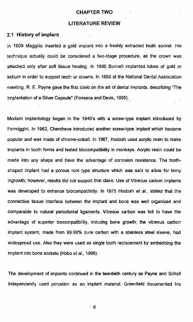
In 1809 Maggilio inserted a gold implant into a freshly extracted tooth socket. His
technique actually could be considered a two-stage procedure, as the crown was
attached only after soft tissue healing. In 1895 Bonnell implanted tubes of gold or
iridium in order to support teeth or crowns. In 1898 at the National Dental Association
meeting, R. E. Payne gave the first clinic on the art of dental implants, describing "The
Implantation of a Silver Capsule" (Fonseca and Davis, 1995).
Modern implantology began in the 1940's with a screw-type implant introduced by
Formiggini. In 1962, Chercheve introduced another screw-type implant which became
popular and was made of chrome-cobalt. In 1967, Hodosh used acrylic resin to make
implants in tooth forms and tested biocompatibility in monkeys. Acrylic resin could be
made into any shape and have the advantage of corrosion resistance. The tooth
shaped implant had a porous root type structure which was said to allow for bony
ingrowth; however, results did not support that claim. Use of Vitreous carbon implants
was developed to enhance biocompatibility. In 1975 Hodosh et a/., stated that the
connective tissue interface between the implant and bone was well organized and
comparable to natural periodontal ligaments. Vitreous carbon was felt to have the
advantage of superior biocompatibility, inducing bone growth; the vitreous carbon
implant system, made from 99.99% pure carbon with a stainless steel sleeve, had
widespread use. Also they were used as single tooth replacement by embedding the
implant into bone sockets (Hobo eta/., 1996).
The development of implants continued in the twentieth century as Payne and Scholl
independently used porcelain as an implant material. Greenfield documented ·his
. 6
implant technique with photographs and diagrams and called implant dentistry the
missing link of dentistry. He consistently described the phenomenon of oral tissue
healing around immobile implants made of 20 - gauge iridioplatinum wire soldered with
24-carat gold. He too used a two-stage procedure, allowing 6 to 8 weeks for bone to
"form through the root" before placing the· crown or bridge (Fonseca and Davis, 1995).
In 1951, Branemark began research leading to the development of an endosseous
implant system that popularized the concept of osseointegration. In 1981, when Adel
and his colleagues reported on a 15-year study of Branemark's ossseointegrated
implants in the treatment of the edentulous jaw, many dental practitioners believed that
dental implants could perhaps finally provide predictable high-level long-term success
rates. Evidence suggests that the earliest recorded use of artificial dental implants
dates back to ancient Egyptian and pre-Columbian eras. The first implant specimen
found appears to be from an excavated Mayan skull from A.D. 600, showing an
implanted tooth-shaped piece of shell to replace a missing lower incisor (Fonseca and
Davis, 1995) (Figure 2.1 ).
7
Figure 2.1: Seashells hammered into the jaw to replace missing teeth (Adapted from http://www.woodmandentistry.com).
8
2.2 Implant Materials
Implant materials are foreign materials that are brought into contact with a biological
system. Biomaterials are nonliving materials used for medical application (for example
as a dental implant) with the goal of achieving a reaction (interaction) with the biological
system (Rateitschak and Wolf, 1995).
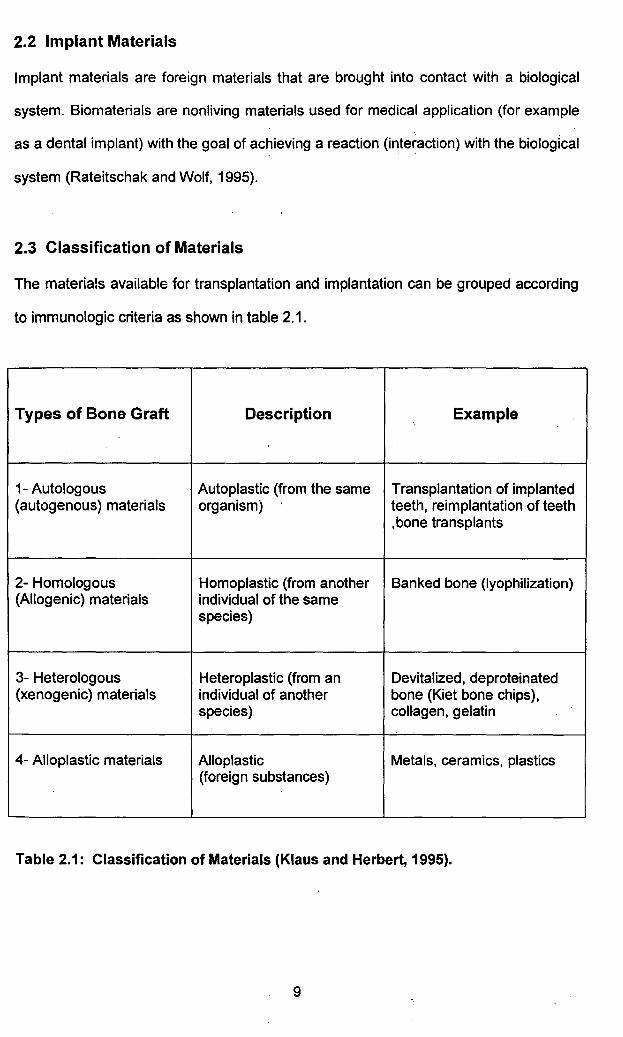
2.3 Classification of Materials
The materials available for transplantation and implantation can be grouped according
to immunologic criteria as shown in table 2.1.
Types of Bone Graft Description ·. Example
1- Autologous Autoplastic (from the same Transplantation of implanted (autogenous) materials organism) teeth, reimplantation of teeth
,bone transplants
2- Homologous Homoplastic (from another Banked bone (lyophilization) (Allogenic) materials individual of the same
species)
3- Heterologous Heteroplastic (from an Devitalized, deproteinated (xenogenic) materials individual of another bone (Kiet bone chips),
species) conagen, gelatin
4- Alloplastic materials Alto plastic Metals, ceramics, plastics (foreign substances)
Table 2.1: Classification of Materials (Klaus and Herbert, 1995).
9
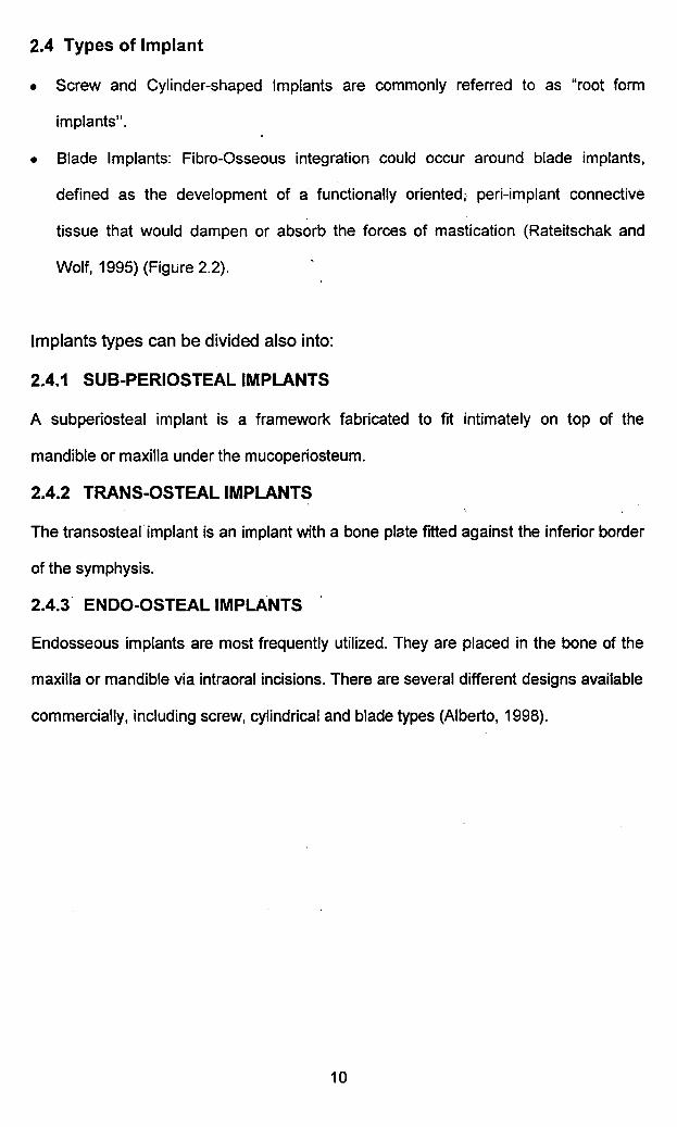
2.4 Types of Implant
• Screw and Cylinder-shaped Implants are commonly referred to as "root form
implants".
• Blade Implants: Fibre-Osseous integration could occur around blade implants,
defined as the development of a functionally oriented; peri-implant connective
tissue that would dampen or absorb the forces of mastication (Rateitschak and
Wolf, 1995) (Figure 2.2).
Implants types can be divided also into:
2.4.1 SUB-PERIOSTEAL IMPLANTS
A subperiosteal implant is a framework fabricated to fit intimately on top of the
mandible or maxilla under the mucoperiosteum.
2.4.2 TRANS-OSTEAL IMPLANTS
The transosteal implant is an implant with a bone plate frtted against the inferior border
of the symphysis.
2.4.3. EN DO-OSTEAL IMPLANTS
Endosseous implants are most frequently utilized. They are placed in the bone of the
maxilla or mandible via intraoral incisions. There are several different designs available
commercially, including screw, cylindrical and blade types (Alberto, 1998).
10
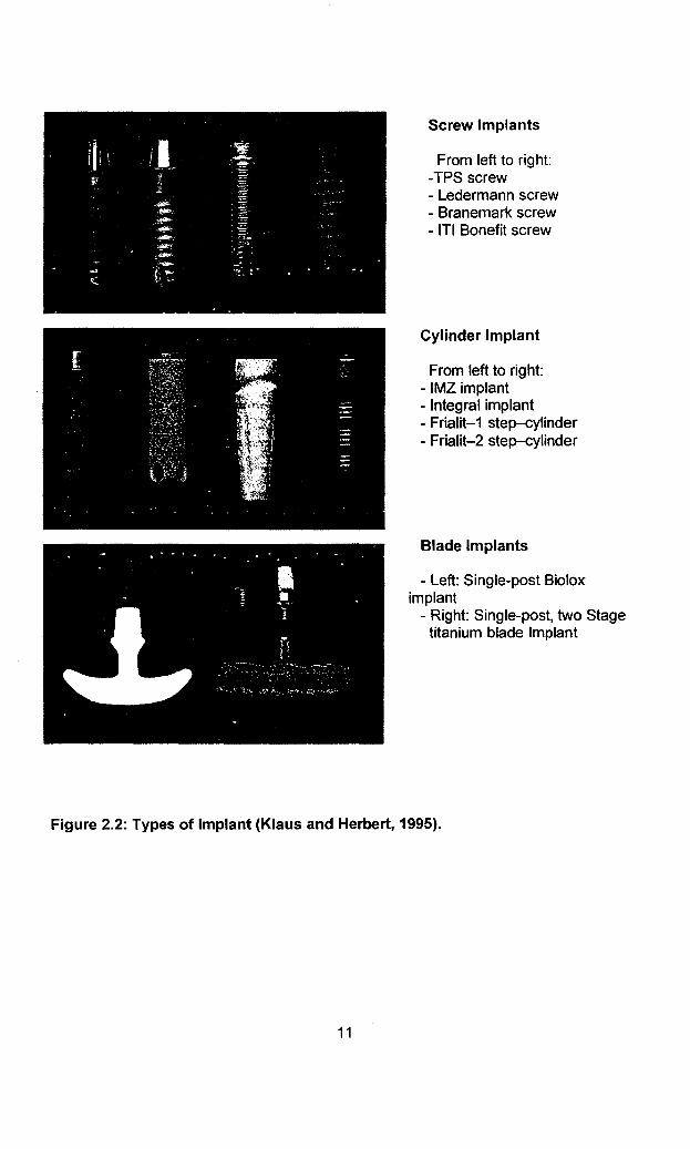
Screw Implants
From left to right: -TPS screw - Ledermann screw - Branemark screw - ITI Bonefit screw
Cylinder Implant
From left to right: - IMZ implant - Integral implant - Frialit-1 step-cylinder - Frialit-2 step-cylinder
Blade Implants
- Left: Single-post Biolox implant
- Right: Single-post, two Stage titanium blade Implant
Figure 2.2: Types of Implant (Klaus and Herbert, 1995).
11
2.5 Titanium
Titanium is widely used as dental implant material, because direct contact occurs
between bone and the implant surface (Knabe eta/., 2002).
It is the ideal metal for intra-osseous dental implants. It provokes a spontaneous oxide
layer formation on its surface protecting the metal from chemical attack, including
potentially aggressive body fluids (Sergio eta/., 2005).
Titanium alloy dental implants as an aid 'to prosthodontic rehabilitation are a relatively
new but important part of dentistry. The dental, biomaterials, and orthopedic literature
clearly show that titanium and other trace metals maybe found in the peri-implant
tissues, regional lymph, nodes, lungs, kidneys, livers, serum, and hair after implant
placement (Millennium, 2001).
Implants made of commercially pure titanium (cpTi) were the first to gain widespread
acceptance. Bone does not bond directly to either cpTi or titanium alloy (Ti-6A 1-4V)
implants. It attaches by means of a complex interaction between the extracellular
matrix tissues and the titanium - oxide layer formed when the metals are exposed to
air or tissue fluids (Kasemo and Lausmaa, 1985; Stanford and Keller, 1991 ).
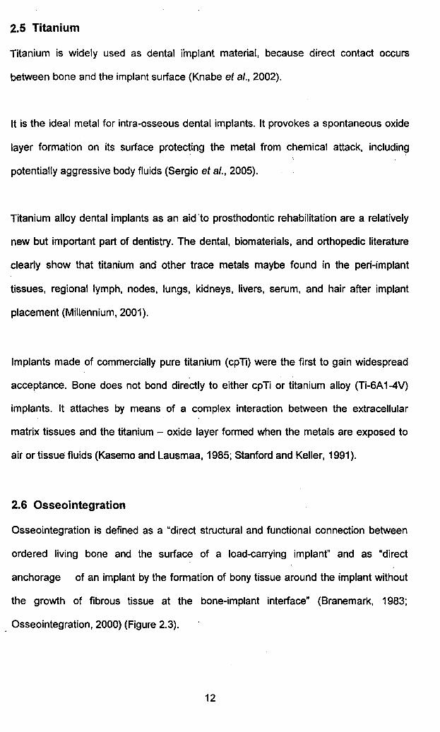
2.6 Osseointegration
Osseointegration is defined as a "direct structural and functional connection between
ordered living bone and the surface of a load-carrying implant" and as "direct
anchorage of an implant by the formation of bony tissue around the implant without
the growth of fibrous tissue at the bone-implant interface" (Branemark, 1983;
Osseointegration, 2000) (Figure 2.3).
12
It is now said that an implant is regarded as osseointegrated when there is no
progressive relative movement between the implant and the bone with which it has
direct contact (Branemark, 1983).
It also defined as the direct connection from implant to living remodeling bone without
any soft tissue component between implant and bone on the light microscopic the light
microscopic level (Gotz et at., 2004).
Figure 2.3: Dental implant osseointegration micrograph (Courtesy of Dr. Lyndon Cooper) A: dental implant, B: implant and alveolar bone interface, C: alveolar bone.
13
2. 7 Bone remodeling
Osseointegration requires new bone formation around the fixture, a process resulting
from remodeling within bone tissue. Remodeling, bone resorption and apposition, helps
maintain blood calcium levels and does not change the mass quantity of bone (Hobo,
1996}.
The development of a dynamic functioning attachment of implants to bone is imperative
for the long-term success of implant-supported dental prostheses. The most successful
material in long-term clinical studies of osseointegrated oral implants is commercially
pure titanium (Konig eta/., 1998; Sui et ~/., 2002}. Special surfaces have been studied
in order to be used in more complex surgical situations such as: immediate implant
placement, expansion of the residual ridge, or maxillary sinus floor elevation. The HA
coated implants should have the advantage of providing an osteoconductive surface for
enhanced bone growth (Kay, 1992; Reddy, 1995}. More recently, novel types of
implant systems have been developed with rough surfaces using different methods
such as: plasma spraying, blasting, etching, beading or sintering in order to increase
the bone implant contact surface.
Bone· has a unique capability of self-regeneration and remodeling to a certain extent
throughout life without leaving scar. If self-remodeling fails due to certain conditions
such as trauma, bone metabolic diseases, neoplasm and others, in used for bone
regeneration dental synthetic bone grafts and coated implant materials can be
applications (Lobato, 2006}.
2.8 Bone healing
Bone is a unique tissue. It can be injured and then can repair itself and return to full
function with or without scarring or deformity (Salter, 1983}. Embryonic bone
14

development is repeated in the healing of bone. The pattern of bony healing is dictated
by the host bed, vascular supply, oxygen tension and the stability of the bone
segments (Buckwalter eta/., 1995). Healing can occur either directly as primary bone
healing or secondarily, demonstrating an intermediate cartilaginous phase (Hollinger et
a/., 1994).
2.9 Bone density
Available bone is particularly important in implant density, and describes the external
architecture or volume of the edentulous area considered .for implants. In additi_on,
bone has an internal structure described in terms of quality of density, which reflects
the strength of the bone (Scortecci eta/., 2001).
2.1 0 Dental implant interface
The health or quality of the soft tissue surrounding an implant may be influenced by
many factors. The presence of keratinizing mucosa surrounding an implant is thought
to be a positive factor in maintaining soft-tissue health. In many implant systems, the
con-notion between the implant and the prosthesis creates a small microgram that has
been implicated in the ongoing health ~f soft tissue surrounding implants (Myshin and
Wiens, 2005).
Coating of implants with locally acting growth factors may influence the remodeling
process at the tissue-implant interface and therefore the integration of implants into
healing bone. Growth factors like plate-let-derived growth factor (PDGF), bone
morphogenetic proteins (BMPs), insulin-like growth factor (IGF) or TGF-13 facilitate the
osseointegration of different kinds of implants (Fischer eta/., 2003).
15
2.11 Hydroxyapatite-coated implant
Since the initial development of hydroxyapatite (HA)-coated dental implant in 1984,
numerous studies have demonstrated favorable or superior results for HA-coated
implants as compared with uncoated titanium implants. von Vlitterwijk demonstrated
65% of the 50flm thick HA coating was reabsorbed during unstable mechanical
condition. Despite numerous claims about one surface or another, there have been no
randomized clinical trials to compare efficacy of HA-~oated versus titanium-coated
endosseous implant in various types of the alveolar bones. The purpose of this study
was to compare the early success rate of HA-coated cylinder implants and TPS
cylinders in different regions of the mouth (Jones eta/., 1997).
Lekholm et a/. (1996) observed no differences between implants with and without
exposed threads after placement over ·a 5-year period of loading. Several studies
indicate low failure rates when placing implants in immediate extraction sockets
(Gomez-Roman et at., 1997; Tolman and Keller, 1991; Becker eta/., 1994; Schwartz
Arad and Chaushu, 1997; Fuga~otto, :1997). Yukna (1991) compared placement of
HA-coated implants in extraction sockets and healed sites in 14 patients and found no
differences.
The first clinical use of HA as a coating for endosseous dental implants appeared in
1984. HA is a naturally occurring calcium phosphate ceramic that is found in
abundance in tooth enamel, dentin, and bone. In its synthesized form, it is applied to a
Ti-6A 1-4V substrate (the usual method is plasma spraying) to form a nontoxic bioactive
coating that bonds chemically with adjacent bone. Block et at., (1987) and Meffert et
a/., (1987) when HA implants are compared with titanium, there is evidence for more
rapid osseointegration (Gerner et at., 1988). In animal studies, Block et at., (1987)
observed biointegration of HA implants as early as 4 weeks. After 10 months, 90% of
the coated implants had a continuous surface layer of lamellar bone connecting the
16
implant with the trabecular bone. In contrast, titanium implants exhibited
osseointegration only at 4 months, with 50% implant-bone contact at 10 months (Block,
1991).
In a second study Block eta/., (1989) found gingival fibers inserting directly into the
osseoid tissue covering the HA coating. The relative merits of HA and non-HA implants
remain controversial, and debate between their respective adherents continues to
enliven discussions in the field of implant dentistry. Reports based on anecdotal data
have suggested that HA coatings are unstable, tiave an increased susceptibility to
bacterial infection, and may be disposed to rapid bone loss or saucerization (Biesbrock
and Edgerton, 1995). In addition to being based on isolated case reports, these
arguments do not reflect the current state of implant technology. Improvements in the
crystallinity of HA coatings have eliminated a cause of failure in some early implant
designs (Kay, 1993; Lacefield, 1994). The incorporation of a machined metal collar in
most modern HA implants further enhances survival, because the machined surface
resists plaque formation and microbial colonization, both of which were common in
early implants when soft tissue changes exposed the porous HA coatings to the oral
cavity.
Calcium phosphate coated titanium and titanium alloy are widely used as dental
implant materials. These coatings have been found to accelerate initial stabilization of
implants by enhancing bony in growth and stimulating osseous apposition to the
implant surface, promoting a rapid fixation of the devices to the skeleton. Hence their
use as coatings of the endosteal portions implants. Of the various calcium phosphates
available, HA has been most commonly used as coating for titanium and its alloy
(Knabe eta/., 2004).
17

In the first phase, the postoperative stability is usually obtained using a proper surgical
technique and proper implant hardware. In the second, the long-term stability depends
on the bone adaptation to the stress pattern induced by the fixture. It follows that for the
proper evaluation of the long-term stability of the fixture it is fundamental to take into
consideration the mechanical properties of the bone surrounding the implant as a
remodeling tissue. It is well-known that the morphology of a bone is first established by
genetic factors and afterward the bon~ goes through dynamic shape and density
optimisation to adapt its mechanical properties and structural behavior to the local
stress (Soncini et a/ .• 2002).
2.12 Calcium Carbonate, Natural coral (NC)
Natural coral (NC) is considered as a xenograft. NC has been used as a biomaterial for
bone replacement because of several reasons such as t~_e material simplifies ~he
surgical procedure, harvesting of autologous bone is no longer necessary and no risk
of transmission of infection of Human Immunodeficiency Virus, Hepatitis B, Hepatitis C
and Creutzfeld Jacob-disease can be avoided with certainty (Volpi, 1999).
Coral is made by marine invertebrates that extract calcium and phosphates from the
sea to build a limestone exostructure in which they live in. This exostructure porous
and mimic the structure of natural bones. Therefore these limestone structures are
appropriate for bone grafting.
The NC used in this study is natural coral in the form of aragonite (more than 98%
CaC03) that is not altered by processing and it is a resorbable, porous, calcium
carbonate graft material produced by the National Tissue Bank, Universiti Sains
Malaysia. In recent studies dead sea coral of Porites species has been harvested from
Malaysian costal region for production of coral bone substitute (A license was provided
18
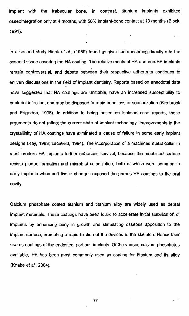
by the Department of Fisheries Malaysia to harvest dead coral for this purpose) (Figure
2.4).
Figure 2.4: Natural processed coral. The arrows point at the pores that can be detected with the naked eyes.
19
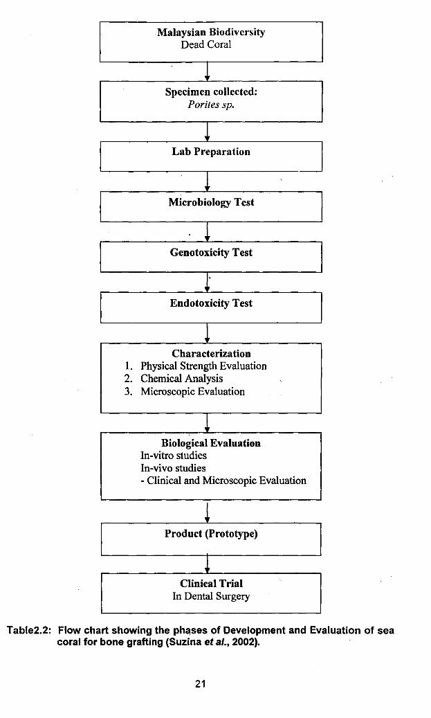
This prepared coral graft went through material characterization studies, biological
validation studies, in vitro and in vivo studies and finally followed by a controlled clinical
trial as shown in table 2.2 (Suzina eta/., 2002).
The Ames test results demonstrated that the prepared coral material did not exhibit
mutagenic activity under the chosen conditions. Thus, the m~terial can be considered
non-genotoxic (Suzina eta/., 2004).
20
Malaysian Biodiversity Dead Coral
! Specimen collected:
Porites sp.
J Lab Preparation
~ •.
Microbiology Test
~ Genotoxicity Test
~ Endotoxicity Test
! Characterization
1. Physical Strength Evaluation 2. Chemical Analysis ·.
3. Microscopic Evaluation
~ Biological Evaluation
In-vitro studies In-vivo studies - Clinical and Microscopic Evaluation
! Product (Prototype)
~ Clinical Trial •.
In Dental Surgery
Table2.2: Flow chart showing the ph~ses of Development and Evaluation of sea coral for bone grafting (Suzina eta/., 2002).
21
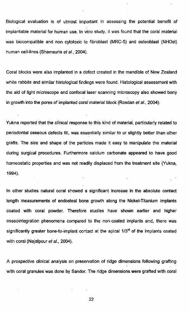
Biological evaluation is of utmost important in assessing the potential benefit of
implantable material for human use. In vitro study, it was found that the coral material
was biocompatible and non cytotoxic to fibroblast (MRC-5) and osteoblast (NHOst)
human cell-lines (Shamsuria eta/., 2004).
Coral blocks were also implanted in a defect created in the mandible of New Zealand
white rabbits and similar histological findings were found. Histological assessment with
the aid of light microscope and confocal laser scanning microscopy also showed bony
in growth into the pores of implanted coral material block (Rosdan eta/., 2004).
Yukna reported that the clinical response to this kind of material, particularly related to
periodontal osseous defects fill, was essentially similar to or slightly better than other
grafts. The size and shape of the particles made it easy to manipulate the material
during surgical procedures. Furthermore calcium carbonate appeared to have good
homeostatic properties and was not readily displaced from the treatment site (Yukna,
1994).
In other studies natural coral showed a significant increase in the absolute contact
length measurements of endosteal bon~ growth along the Nickel-Titanium implants
coated with coral powder. Therefore studies have shoWn earlier and higher
osseointegration phenomena compared to the non-coated implants and, there was
significantly greater bone-to-implant contact at the apical 1/3rd of the implants coated
with coral (Najafpour eta/., 2004).
A prospective clinical analysis on preservation of ridge dimensions following grafting
with coral granules was done by Sandor. The ridge dimensions were grafted with coral
22
and afterwards implant was placed. They claimed the grafting was successful and
dental implants were stable (Sandor et .a/., 2003).
NC exoskeleton is a bioactive material ·used as bone substitute in different surgical
specialties Maxillofacial and buccal surgery (Fricain et a/., 2002). On the contrary,
Lopez et a/., (1992) have recently shown that nacre, which associates calcium
carbonate and an organic matrix, might have osteogenic and osteoinductive properties
(Lopez et a/., 1995). All these results suggest that the organic matrix of coral
exoskeleton (COM) could be decisive in the integration or rejection of coral by bone.
Moreover, only a few studies have· been performed on. COM and all con~rn
biochemical analysis COM and all coricern the biochemical analysis of coral species
which are not used as bone substitutes (Allemand eta/., 1994). So the objective of this
study was to extract COM to carry out biochemical analysis and to study its specie
cytocompatibHity in vitro in contact with human bone marrow cells.
The ability of the human body to regenerate bony tissues that are lost or damaged is
limited. In the case of important bony defects, an autogenous bone graft is considered
as suitable transplant material because differences in biocompatibility and the risk of
transferring viruses from one individual to another are non.:existent. Removal of the
bone graft creates additional surgical trauma. Allogenic and xenogenic bone grafts
represent alternatives but several probleiJls are generally associated with them such as
in vivo resorption, virus transfer, considerable care, high cost and regular immune
defensive reaction. For all these reasons, bone substitutes are generating growing
interest and are frequently used in orthopedic surgery. They are alternatives to
autogenic, allogenic and xenogenic bone grafts. One hopes that they are replaced
gradually and completely by neoformed bone with the same bone characteristics at the
end of the restoration process. Natural coral, submitted to rigorous protocols of
preparation and purification, can be used as a replacement biomaterial for bone grafts
23
'
l
both in orthopaedic surgery and maxillo-craniofacial surgery. It can replace bony tissue
without inappropriate response from the. human body (biocompatibility); it develops a
chemical bond with the bone surface (bioactivity) and is able to form bony tissue when
it is in contact with bone (osteoconductivity) (Barbotteau eta/., 2003).
Schwartz-Arad and Chaushu (1998) reported a successful clinical outcome for 9 single
implants placed immediately after tooth extraction without incisions or primary flap
closure. Complete bone healing was achieved with papilla preservation and minimal
gingival recession. Clinical cases with extensive bone loss were excluded from the
study. The purposes of the present study were to evaluate implants placed immediately
after tooth extraction without incision or primary flap closure and to observe the peri-
implant soft tissue healing.
Brazilay et a/., (1991) used animal models to compare 48 immediately inserted
implants with conventionally placed implants. When both techniques were compared,
there were no significant changes in bone-to-implant interface 7 months following the
delivery of the prosthesis.
In animal and human studies, it has been shown that resorbable barriers can be
successfully used for bone augmentation purposes (Kostopoulos and Karring 1994)
and (Simien et a/., 1997). Furthermore, the combination of resorbable barriers and
immediately placed implants seems to be comparable with the combination of
nonresorbable barriers and immediately placed implants in terms of integration of the
implants.
Cordioli and colleagues (1994) reported the clinical experience of 47 patients
rehabilitated with a single-tooth restoration. The total implant survival rate was 94.4%.
Engquist and associates (1995) evaluated the outcome of single-tooth restorations
24