Embed Size (px)
Citation preview
Journalof
Periodontology&
Implant entistryD
Research Article
Clinical and Radiographic Evaluation of Immediate and
Delayed Single-tooth Implant Placement: An 18-month
Follow-up Study P.K. Sasi Kumar1 • Abinaya Ravikumar2 • Sugumari Elavarasu2 • Thanga kumaran3 • Bala Murugan4*
1Senior Lecturer, Department of Periodontics and Implantology 2Professor, Department of Periodontics and Implantology
3Associate Professor, Department of Periodontics and Implantology 4Senior Lecturer, Department of Prosthodontics, Crown and Bridge
*Corresponding Author; E-mail: [email protected]
Received: 10 February 2012; Accepted: 21 April 2013 J Periodontol Implant Dent 2013;5(2):41–54 | doi: 10.5681/jpid.2013.023
This article is available from: http://dentistry.tbzmed.ac.ir/jpid
© 2013 The Authors; Tabriz University of Medical Sciences This is an Open Access article distributed under the terms of the Creative Commons Attribution License
(http://creativecommons.org/licenses/by/3.0), which permits unrestricted use, distribution, and reproduction in any medium, provided the original work is properly cited.
Abstract Background and aims. Maintenance of interdental soft tissue and the need for aesthetics are increasingly recognized
as important criteria for implant success. The aim of this prospective clinical study was to compare the peri-implant and
prosthetic conditions for single-tooth implants, placed according to the immediate (Im) and the delayed (De) placement
protocols at 18-month follow-up examination. Materials and methods. After random allocation to the Immediate and Delayed groups, 14 patients were treated with a
single-tooth implant in the anterior or molar region of the maxilla or the mandible immediately (Im) or 6 months (De) after
tooth extraction, respectively. Patients attended a follow-up visit 18 months after implant placement corresponding to one
year of loading of the implant restorations. Peri-implant and prosthetic parameters were evaluated clinically and marginal
bone levels were measured on radiographs. Results. None of the implant restorations had failed after one year of function. Probing pocket depths decreased by up to
1.5 mm on average from the time of loading to the 18-month follow-up, no significant difference between the Im and De
groups was found (4.3 versus 4.2 mm). A statistically significant radiographic marginal bone loss had occurred in the Im
group (13.46%) as well as in the De group (15.62%) in the follow-up period.
Conclusion. Soft and hard tissue responses to single-tooth implants placed either in the anterior or molar region of the
maxilla or the mandible immediately (Im) or 6 months (De) after tooth extraction were similar in terms of the placement
protocols. Key words: Dental Implantation, extraction, maxilla, mandible.
42 Sasi Kumar et al.
Introduction
ental epidemiological studies demonstrate that missing teeth are commonly present in all age
groups.1 The loss of a single tooth is regarded a common cause of esthetic concerns, leading to psy-chological implications and non-physiologic occlu-sion, as a result of tipping of neighbouring teeth and super-eruption of opposing teeth.2 The clinical re-placement of lost natural teeth with osseointegrated implants has represented one of the most significant advances in restorative dentistry.
The primary reason for suggesting a “fixed partial denture” is its clinical ease and reduced treatment time.3 Patients have been advised to determine their desired level of replacing missing teeth and accept the limitations of a fixed partial denture and remov-able partial denture.6 Single-tooth implant survival reports have been most validated as predictable methods of tooth replacement.4,5 However, the most natural method to replace a missing tooth is with an implant, rather than preparing adjacent teeth.6
The first single tooth-crown restoration using a Branemark implant (Nobel Biocare) was placed in December 1982.7 Since Branemark introduced the “osseointegration” concept, advancement has fol-lowed three paths.8 This has been applied to replac-ing a single missing tooth or multiple missing teeth in various edentulous situations, new donor sites and techniques to transplant bone have given better ac-cess to patients for receiving the implants and fi-nally, efforts have been made to reduce the treatment period.
Single-tooth implants can be placed either in healed extraction sites (delayed) or fresh extraction sockets (immediate). Traditionally a single tooth im-plant was placed in a healed extraction site, allowing ossification to occur in 3-6 months.9 This delay dur-ing socket healing, coupled with the added surgical stage, was inconvenient as well as uncomfortable to the patient, who might be wearing conventional re-movable prosthesis.10
To achieve optimal esthetic appearance and re-duced treatment time, immediate implants have been studied in the literature.11-13 In this type, the implants can be placed either immediately after tooth extrac-tion (Immediate) or 15 days after tooth extraction (delayed-immediate).11 It is placed directly into fresh extraction sockets after preparation of the implant bed to achieve primary stability. Advantages of this technique include preservation of the alveolar bone, ideal axial positioning of the implant using the socket as a reference, eliminating the waiting period
of 3to 6 months, fewer surgical visits and shortened edentulous period. On the other hand, there is a po-tential risk factor as enhanced possibility of mis-match between the socket wall and implant, leading to fibrous tissue formation.9
A two-stage surgical technique was originally ad-vocated in order to optimize the process of new bone formation and remodeling, following implant place-ment.10 To minimize the risk of soft tissue encapsu-lation, it has been recommended to keep the implants submerged and load-free for 3�6 months.14 Follow-ing this period, a second-stage surgery is needed to connect the healing abutment to implant, holding the future prosthesis. After the second intervention, 4 to 6 weeks of healing period is needed for proper con-touring of the soft tissue around a healing abutment to allow for a predictable esthetic outcome.15
In one-stage surgical procedures, flaps are sutured around the polished neck of implants avoiding the need for second stage surgical intervention.14 Misch et al suggested a terminology for immediate restora-tion or occlusal loading.1 In general, when this pro-tocol was first implemented, only one-piece implants were used. However, later on, this procedure was performed with two-stage implants on which a heal-ing abutment was placed.16
In implants, the criteria for success should involve the establishment of a soft tissue contour with intact interproximal papilla and a predictable gingival out-come.17 The interdental bone and papilla height are correlated to the distance from contact point to cre-stal bone. If the measurement from the contact point to the crest of the bone is 5 mm, the papilla would present almost 100%. If the distance is greater than 6 mm, the papilla would present 50% or less.18 Based on this data, the clinician attempts to maintain 5 mm of distance from the contact point to the crestal bone, when placing the implant.
Adequate zone of keratinized mucosa measures as 2 mm of width, of which 1 mm is to be attached gin-giva. The attached gingiva is necessary for the main-tenance of gingival health and prevention of perio-dontal disease progression.19 Peri-implant and perio-dontal tissues may differ in their resistance to bacte-rial infection because supracrestal collagen fibers in implants are oriented in parallel, rather than a per-pendicular, configuration. This creates a much weaker mechanical attachment compared to natural teeth. Thus, adequate zone of keratinized mucosa adjacent to the implant has to be maintained.20
The influence of mucosal thickness on crestal bone loss around implant has been reported recently 21 and it is necessary that a minimum of 3 mm of peri-
D
Evaluation of immediate and delayed single-tooth implant 43
implant mucosa is required for the stable epithelial connective tissue attachment around implants. A thick mucosa is resilient and therefore prone to pocket formation, while a thin mucosa is friable and thus often prone to gingival recession.22
It must be emphasized that conclusions drawn on the soft tissue and hard tissue can influence the suc-cess rate of single-tooth implants, irrespective of immediate and delayed protocols. Moreover, clinical trials have most often focused on the success rates of implants. To the best of our knowledge, no random-ized controlled clinical studies on immediate and delayed implant placement have been conducted previously. The aim of this prospective clinical study was to evaluate and compare peri-implant and pros-thetic conditions for single-tooth implants, placed according to the immediate and the delayed place-ment protocols at 18-month follow-up examination.
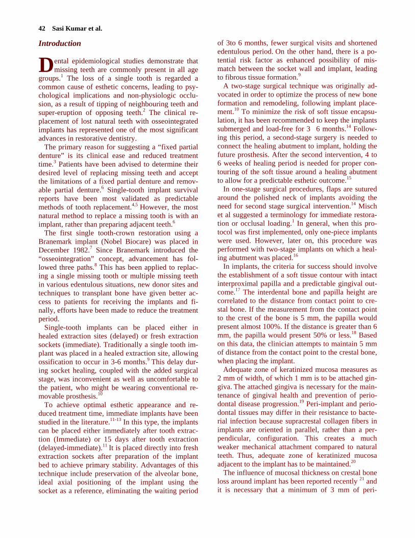
This study was designed and conducted by the De-partment of Periodontics, JKKN Dental College and Hospitals, Komarapalayam, Tamil Nadu, India, from November 2008 to October 2010, to evaluate the clinical and radiological peri-implant and prosthetic conditions of immediate and delayed single-tooth implants.
Materials and Methods
A Hi-Tec implant (Life Care implants) made up of titanium with self-threaded internal hex and selective integrated surface were used. Four diameters and two prosthetic platforms (standard and wide plat-form) of implants are available with variable diame-ters and lengths of 3.3, 3.75, 4.2, 5.0 mm and 8, 10, 11.5, 13, 16 mm. It has a round end that protects and prevents sinus membrane perforation.
Study design
A randomized prospective clinical trial was con-ducted to evaluate the clinical and radiological pa-rameters of immediate and delayed single-tooth im-plant placement. Fresh extraction sites with immedi-ate implant technique and healed site with delayed implant technique were followed. Ethical clearance was obtained from the Institutional Ethical Board prior to the study (Dr. MGR Medical University, Chennai, Tamilnadu, India). Fourteen (eight females, six males) patients of both sexes with an age range of 20 to 35 years were selected for the study from outpatient Department of Periodontics depending on the following selection criteria. Inclusion criteria
Single tooth space or space with adjacent natural
tooth2 1. Adjacent teeth: intact; restored with functionally
and esthetically good restorations; restored with prostheses precluding the addition of the missing tooth2
2. Patient reluctance to have adjacent teeth pre-pared2
3. Demonstrated maladaptive experience, or psy-chological reluctance to wear a removable partial denture2
Exclusion criteria
1. Inability to undergo a minor oral surgical proce-dure2
2. A history of substance abuse2 3. Psychoses2
4. Unrealistic esthetic expectations2 5. Presence of vital anatomic structures in close
proximity to a proposed implant site2 6. Insufficient bone quality or compromised health
of the local site as determined by radiographs and clinical inspection before implant placement (local cysts, soft tissue ulceration, persistent in-fections, insufficient healing of the previous ex-traction site)2
7. Insufficient bone quantity2 8. Inadequate mouth opening2 9. Insufficient vertical interarch space to accom-
modate the prostheses2 10. Incomplete facial growth and tooth eruption2
Study Design32 1st Stage surgery
(Implant placement)
2nd Stage surgery (Healing abutment)
1st Month
3rd-4th Month
Final abutment placement with
prosthetic procedure
9th Month
18th Month
4th– 5th Month Final restoration with baseline
Second Follow- Up
Third Follow- Up
(Implant placement)
2nd Stage surgery (Healing abutment)
1st Month
3rd-4th Month
Final abutment placement with
prosthetic procedure
9th Month
18th Month
1st Stage surgery
4th– 5th Month Final restoration with baseline
Second Follow- Up
Third Follow- Up
Criteria for grouping
The single-tooth implant sites were randomly se-
44 Sasi Kumar et al.
lected in either the upper or lower jaw, irrespective of whether it was an anterior or posterior region. The selected patients were categorized into two groups based on immediate and delayed implant placement protocols. Seven single-tooth implants were placed using immediate technique in the fresh extraction sockets. Seven single-tooth implants were placed using delayed technique in the healed bone sites.
Pre-surgical procedure
Intraoral and panoramic radiographs were taken for the preoperative evaluation of bone quality, implant position and orientation. A diagnostic template was made with a 5-mm ball bearing, incorporated around the curvature of the dental arch and worn by the pa-tient during the radiographic examination, which enabled the operator to determine the amount of magnification in the radiograph.1 Based on the ana-tomical site analysis, the appropriate implant diame-ter and platform size was selected to best fit the sin-gle-tooth edentulous area. After a preoperative workup, a diagnostic wax-up of the planned restora-tion and fabrication of a surgical stent was carried out before the implant surgery. This stent was made for proper positioning of implant shoulder and to provide an ideal emergence profile with long-term peri-implant hard and soft tissue support.76
Surgical procedure
All the 14 patients were surgically prepared with routine blood investigation and radiographic assess-ment. Local anesthesia was induced by infiltration with lignocaine (2%) and adrenaline (1:80,000) for the both groups.
Immediate group
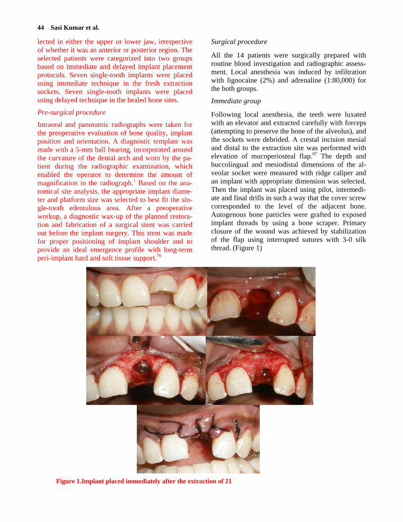
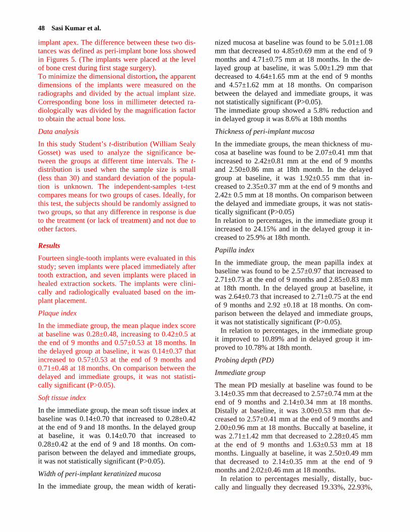
Following local anesthesia, the teeth were luxated with an elevator and extracted carefully with forceps (attempting to preserve the bone of the alveolus), and the sockets were debrided. A crestal incision mesial and distal to the extraction site was performed with elevation of mucoperiosteal flap.67 The depth and buccolingual and mesiodistal dimensions of the al-veolar socket were measured with ridge caliper and an implant with appropriate dimension was selected. Then the implant was placed using pilot, intermedi-ate and final drills in such a way that the cover screw corresponded to the level of the adjacent bone. Autogenous bone particles were grafted to exposed implant threads by using a bone scraper. Primary closure of the wound was achieved by stabilization of the flap using interrupted sutures with 3-0 silk thread. (Figure 1)
Figure 1.Implant placed immediately after the extraction of 21
Evaluation of immediate and delayed single-tooth implant 45
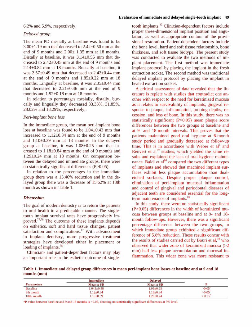
Delayed group
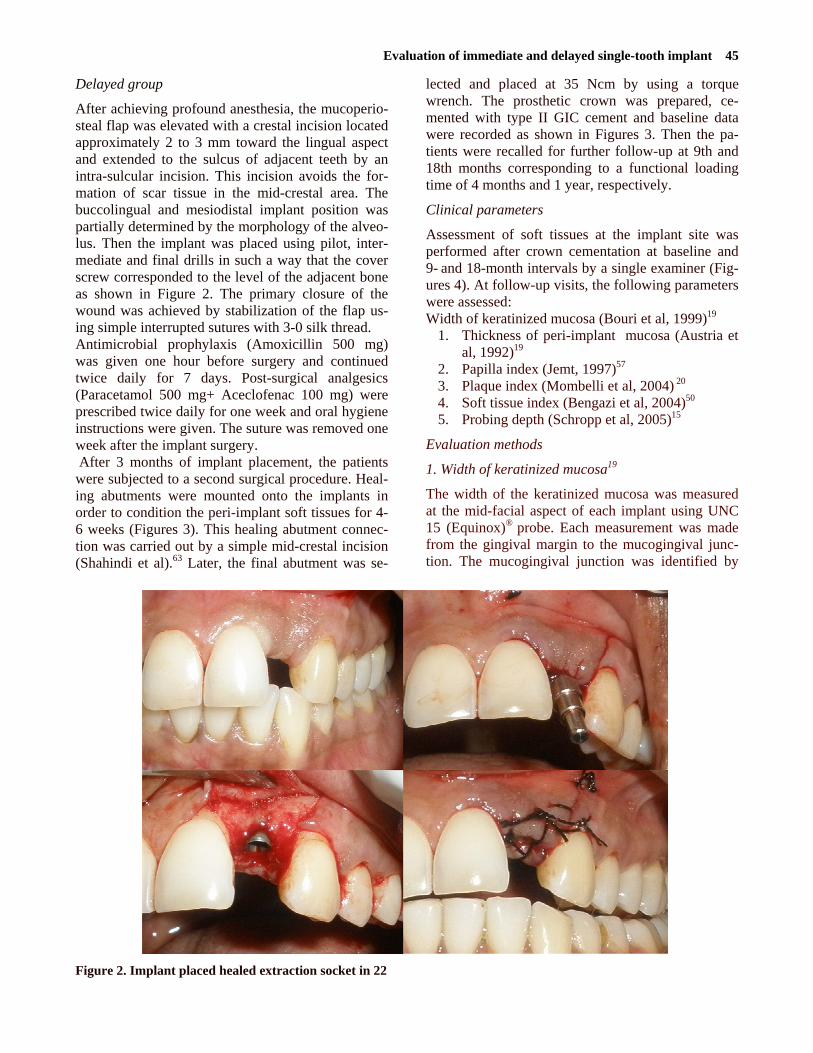
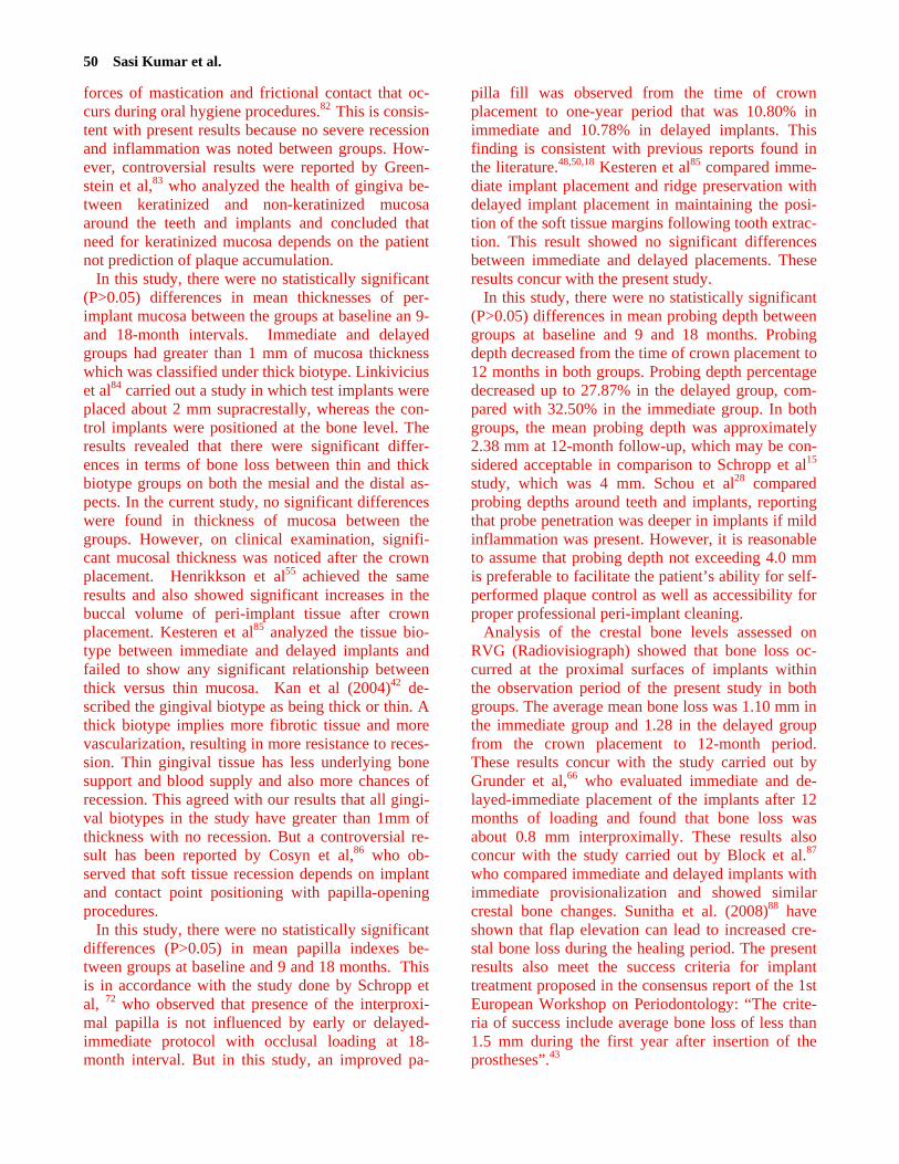
After achieving profound anesthesia, the mucoperio-steal flap was elevated with a crestal incision located approximately 2 to 3 mm toward the lingual aspect and extended to the sulcus of adjacent teeth by an intra-sulcular incision. This incision avoids the for-mation of scar tissue in the mid-crestal area. The buccolingual and mesiodistal implant position was partially determined by the morphology of the alveo-lus. Then the implant was placed using pilot, inter-mediate and final drills in such a way that the cover screw corresponded to the level of the adjacent bone as shown in Figure 2. The primary closure of the wound was achieved by stabilization of the flap us-ing simple interrupted sutures with 3-0 silk thread. Antimicrobial prophylaxis (Amoxicillin 500 mg) was given one hour before surgery and continued twice daily for 7 days. Post-surgical analgesics (Paracetamol 500 mg+ Aceclofenac 100 mg) were prescribed twice daily for one week and oral hygiene instructions were given. The suture was removed one week after the implant surgery. After 3 months of implant placement, the patients were subjected to a second surgical procedure. Heal-ing abutments were mounted onto the implants in order to condition the peri-implant soft tissues for 4-6 weeks (Figures 3). This healing abutment connec-tion was carried out by a simple mid-crestal incision (Shahindi et al).63 Later, the final abutment was se-
lected and placed at 35 Ncm by using a torque wrench. The prosthetic crown was prepared, ce-mented with type II GIC cement and baseline data were recorded as shown in Figures 3. Then the pa-tients were recalled for further follow-up at 9th and 18th months corresponding to a functional loading time of 4 months and 1 year, respectively.
Clinical parameters
Assessment of soft tissues at the implant site was performed after crown cementation at baseline and 9- and 18-month intervals by a single examiner (Fig-ures 4). At follow-up visits, the following parameters were assessed: Width of keratinized mucosa (Bouri et al, 1999)19
1. Thickness of peri-implant mucosa (Austria et al, 1992)19
2. Papilla index (Jemt, 1997)57
3. Plaque index (Mombelli et al, 2004) 20
4. Soft tissue index (Bengazi et al, 2004)50
5. Probing depth (Schropp et al, 2005)15
Evaluation methods
1. Width of keratinized mucosa19
The width of the keratinized mucosa was measured at the mid-facial aspect of each implant using UNC 15 (Equinox)® probe. Each measurement was made from the gingival margin to the mucogingival junc-tion. The mucogingival junction was identified by
Figure 2. Implant placed healed extraction socket in 22
46 Sasi Kumar et al.
the rolling technique, in which the mucosa was rolled until the non-movable portion of the attached keratinized tissue was identified.
2. Thickness of peri-implant mucosa19
The thickness of the gingiva around dental implant was measured approximately 2 mm apical to the gingival margin on the facial aspect of the implant. After topical anesthetic application, the thickness was measured gently inserting a sterile Endo reamer with a rubber stopper up to the contact of the under-
lying bone structure. The gingival biotype was con-sidered thin if the measurement was less than 1.0 mm and thick if it measured greater than 1.0 mm.42
3. Papilla index57
Clinical photographs were taken by a single exam-iner using the same magnification and illumination. These photographs were digitalized at a resolution of 1,000 dpi. Papilla was scored using a modified scale previously described by Jemt.57 The index was de-fined briefly as follows:
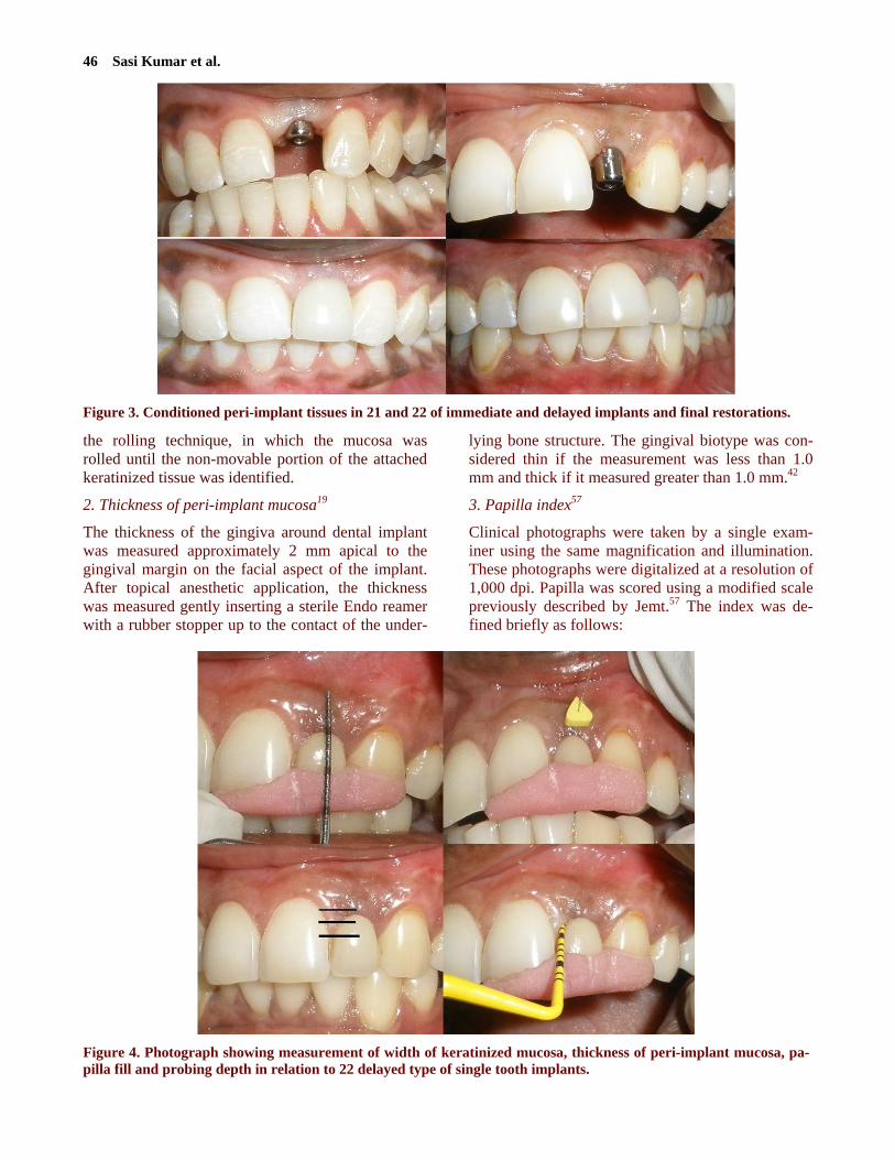
Figure 3. Conditioned peri-implant tissues in 21 and 22 of immediate and delayed implants and final restorations.
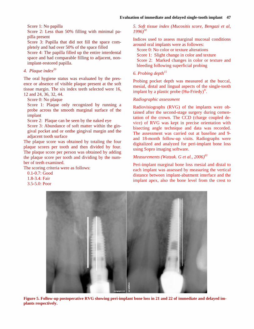
Figure 4. Photograph showing measurement of width of keratinized mucosa, thickness of peri-implant mucosa, pa-pilla fill and probing depth in relation to 22 delayed type of single tooth implants.
Evaluation of immediate and delayed single-tooth implant 47
Score 1: No papilla Score 2: Less than 50% filling with minimal pa-pilla present Score 3: Papilla that did not fill the space com-pletely and had over 50% of the space filled Score 4: The papilla filled up the entire interdental space and had comparable filling to adjacent, non-implant-restored papilla.
4. Plaque index20
The oral hygiene status was evaluated by the pres-ence or absence of visible plaque present at the soft tissue margin. The six index teeth selected were 16, 12 and 24, 36, 32, 44.
Score 0: No plaque Score 1: Plaque only recognized by running a probe across the smooth marginal surface of the implant Score 2: Plaque can be seen by the naked eye Score 3: Abundance of soft matter within the gin-gival pocket and or onthe gingival margin and the adjacent tooth surface
The plaque score was obtained by totaling the four plaque scores per tooth and then divided by four. The plaque score per person was obtained by adding the plaque score per tooth and dividing by the num-ber of teeth examined. The scoring criteria were as follows:
0.1-0.7: Good 1.8-3.4: Fair 3.5-5.0: Poor
5. Soft tissue index (Mucositis score, Bengazi et al, 1996)50
Indices used to assess marginal mucosal conditions around oral implants were as followes:
Score 0: No color or texture alterations Score 1: Slight change in color and texture Score 2: Marked changes in color or texture and bleeding following superficial probing
6. Probing depth15
Probing pocket depth was measured at the buccal, mesial, distal and lingual aspects of the single-tooth implant by a plastic probe (Hu-Friedy)®.
Radiographic assessment
Radiovisiographs (RVG) of the implants were ob-tained after the second-stage surgery during cemen-tation of the crown. The CCD (charge coupled de-vice) of RVG was kept in precise orientation with bisecting angle technique and data was recorded. The assessment was carried out at baseline and 9- and 18-month follow-up visits. Radiographs were digitalized and analyzed for peri-implant bone loss using Sopro imaging software.
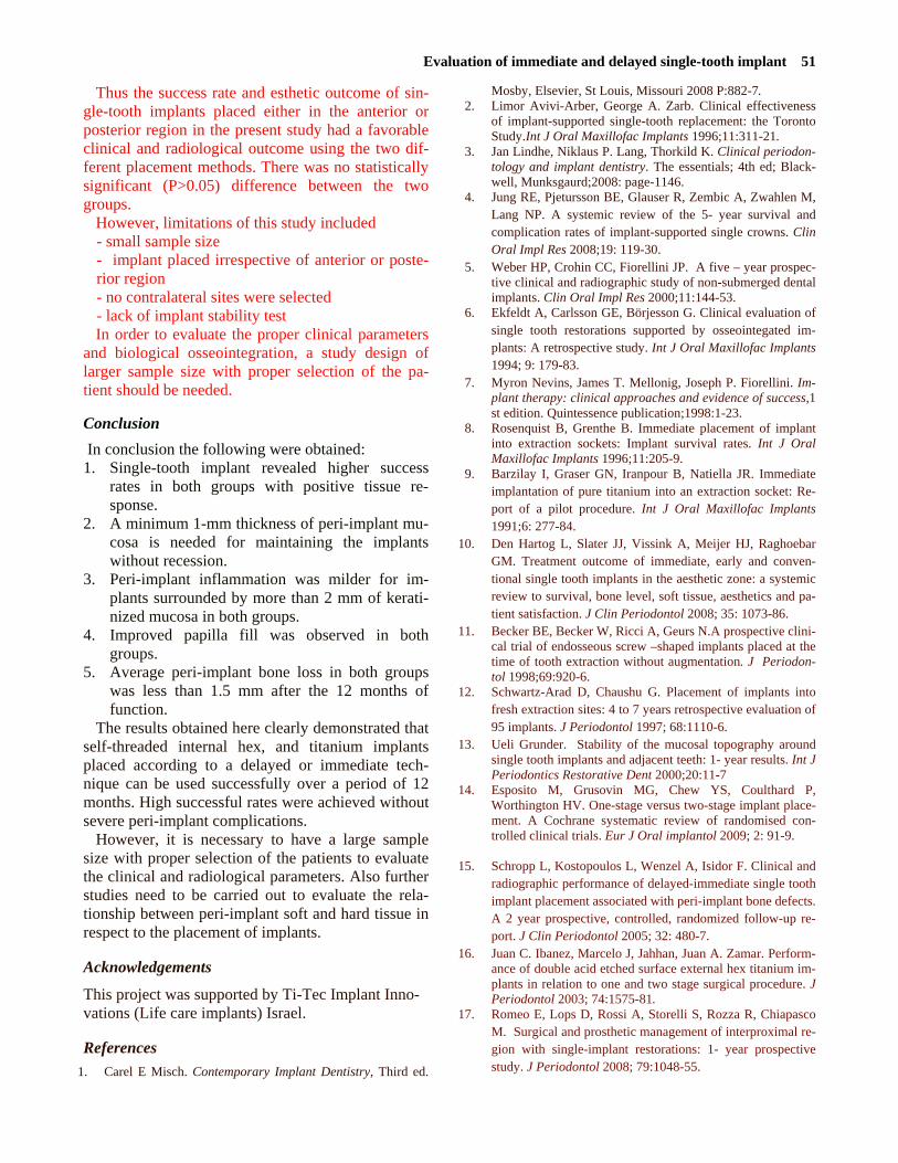
Measurements (Watzak. G et al., 2006)61
Peri-implant marginal bone loss mesial and distal to each implant was assessed by measuring the vertical distance between implant-abutment interface and the implant apex, also the bone level from the crest to
Figure 5. Follow-up postoperative RVG showing peri-implant bone loss in 21 and 22 of immediate and delayed im-plants respectively.
48 Sasi Kumar et al.
implant apex. The difference between these two dis-tances was defined as peri-implant bone loss showed in Figures 5. (The implants were placed at the level of bone crest during first stage surgery). To minimize the dimensional distortion, the apparent dimensions of the implants were measured on the radiographs and divided by the actual implant size. Corresponding bone loss in millimeter detected ra-diologically was divided by the magnification factor to obtain the actual bone loss.
Data analysis
In this study Student’s t-distribution (William Sealy Gosset) was used to analyze the significance be-tween the groups at different time intervals. The t-distribution is used when the sample size is small (less than 30) and standard deviation of the popula-tion is unknown. The independent-samples t-test compares means for two groups of cases. Ideally, for this test, the subjects should be randomly assigned to two groups, so that any difference in response is due to the treatment (or lack of treatment) and not due to other factors.
Results
Fourteen single-tooth implants were evaluated in this study; seven implants were placed immediately after tooth extraction, and seven implants were placed in healed extraction sockets. The implants were clini-cally and radiologically evaluated based on the im-plant placement.
Plaque index
In the immediate group, the mean plaque index score at baseline was 0.28±0.48, increasing to 0.42±0.5 at the end of 9 months and 0.57±0.53 at 18 months. In the delayed group at baseline, it was 0.14±0.37 that increased to 0.57±0.53 at the end of 9 months and 0.71±0.48 at 18 months. On comparison between the delayed and immediate groups, it was not statisti-cally significant (P>0.05).
Soft tissue index
In the immediate group, the mean soft tissue index at baseline was 0.14±0.70 that increased to 0.28±0.42 at the end of 9 and 18 months. In the delayed group at baseline, it was 0.14±0.70 that increased to 0.28±0.42 at the end of 9 and 18 months. On com-parison between the delayed and immediate groups, it was not statistically significant (P>0.05).
Width of peri-implant keratinized mucosa
In the immediate group, the mean width of kerati-
nized mucosa at baseline was found to be 5.01±1.08 mm that decreased to 4.85±0.69 mm at the end of 9 months and 4.71±0.75 mm at 18 months. In the de-layed group at baseline, it was 5.00±1.29 mm that decreased to 4.64±1.65 mm at the end of 9 months and 4.57±1.62 mm at 18 months. On comparison between the delayed and immediate groups, it was not statistically significant (P>0.05). The immediate group showed a 5.8% reduction and in delayed group it was 8.6% at 18th months
Thickness of peri-implant mucosa
In the immediate groups, the mean thickness of mu-cosa at baseline was found to be 2.07±0.41 mm that increased to 2.42±0.81 mm at the end of 9 months and 2.50±0.86 mm at 18th month. In the delayed group at baseline, it was 1.92±0.55 mm that in-creased to 2.35±0.37 mm at the end of 9 months and 2.42± 0.5 mm at 18 months. On comparison between the delayed and immediate groups, it was not statis-tically significant (P>0.05) In relation to percentages, in the immediate group it increased to 24.15% and in the delayed group it in-creased to 25.9% at 18th month.
Papilla index
In the immediate group, the mean papilla index at baseline was found to be 2.57±0.97 that increased to 2.71±0.73 at the end of 9 months and 2.85±0.83 mm at 18th month. In the delayed group at baseline, it was 2.64±0.73 that increased to 2.71±0.75 at the end of 9 months and 2.92 ±0.18 at 18 months. On com-parison between the delayed and immediate groups, it was not statistically significant (P>0.05).
In relation to percentages, in the immediate group it improved to 10.89% and in delayed group it im-proved to 10.78% at 18th month.
Probing depth (PD)
Immediate group
The mean PD mesially at baseline was found to be 3.14±0.35 mm that decreased to 2.57±0.74 mm at the end of 9 months and 2.14±0.34 mm at 18 months. Distally at baseline, it was 3.00±0.53 mm that de-creased to 2.57±0.41 mm at the end of 9 months and 2.00±0.96 mm at 18 months. Buccally at baseline, it was 2.71±1.42 mm that decreased to 2.28±0.45 mm at the end of 9 months and 1.63±0.53 mm at 18 months. Lingually at baseline, it was 2.50±0.49 mm that decreased to 2.14±0.35 mm at the end of 9 months and 2.02±0.46 mm at 18 months.
In relation to percentages mesially, distally, buc-cally and lingually they decreased 19.33%, 22.93%,
Evaluation of immediate and delayed single-tooth implant 49
6.2% and 5.9%, respectively.
Delayed group
The mean PD mesially at baseline was found to be 3.00±1.19 mm that decreased to 2.42±0.50 mm at the end of 9 months and 2.00± 1.35 mm at 18 months. Distally at baseline, it was 3.14±0.55 mm that de-creased to 2.42±0.45 mm at the end of 9 months and 2.14±0.84 mm at 18 months. Buccally at baseline, it was 2.57±0.49 mm that decreased to 2.42±0.44 mm at the end of 9 months and 1.85±0.22 mm at 18 months. Lingually at baseline, it was 2.35±0.44 mm that decreased to 2.21±0.46 mm at the end of 9 months and 1.92±0.18 mm at 18 months.
In relation to percentages mesially, distally, buc-cally and lingually they decreased 33.33%, 31.85%, 28.02% and 18.29%, respectively,.
Peri-implant bone loss
In the immediate group, the mean peri-implant bone loss at baseline was found to be 1.04±0.43 mm that increased to 1.12±0.34 mm at the end of 9 months and 1.10±0.39 mm at 18 months. In the delayed group at baseline, it was 1.08±0.25 mm that in-creased to 1.18±0.84 mm at the end of 9 months and 1.29±0.24 mm at 18 months. On comparison be-tween the delayed and immediate groups, there were no statistically significant differences (P>0.05).
In relation to the percentages in the immediate group there was a 13.46% reduction and in the de-layed group there was a decrease of 15.62% at 18th month as shown in Table 1.
Discussion
The goal of modern dentistry is to return the patients to oral health in a predictable manner. The single-tooth implant survival rates have progressively im-proved.77,78 The outcome of these implants depends on esthetics, soft and hard tissue changes, patient satisfaction and complications.17 With advancement in implant dentistry, more progressive treatment strategies have developed either in placement or loading of implants.78
Clinician- and patient-dependent factors may play an important role in the esthetic outcome of single-
tooth implants.32 Clinician-dependent factors include proper three-dimensional implant position and angu-lation, as well as appropriate contour of the provi-sional restoration. Patient-dependent factors include the bone level, hard and soft tissue relationship, bone thickness, and soft tissue biotype. The present study was conducted to evaluate the two methods of im-plant placement. The first method was immediate implant protocol by placing the implant in the fresh extraction socket. The second method was traditional delayed implant protocol by placing the implant in healed extraction socket.
A critical assessment of data revealed that the lit-erature is replete with studies that contradict one an-other with respect to the need for keratinized mucosa as it relates to survivability of implants, gingival re-sponse to plaque, inflammation, probing depths, re-cession, and loss of bone. In this study, there was no statistically significant (P>0.05) mean plaque score differences between the two groups at baseline and at 9- and 18-month intervals. This proves that the patients maintained good oral hygiene at 6-month study period and gradually decreased at follow-up time. This is in accordance with Weber et al5 and Renvert et al79 studies, which yielded the same re-sults and explained the lack of oral hygiene mainte-nance. Baldi et al80 compared the two different types of implants and showed that machined implant sur-faces exhibit less plaque accumulation than dual-etched surfaces. Despite proper plaque control, elimination of peri-implant mucosal inflammation and control of gingival and periodontal diseases of adjacent teeth are considered essential for the long-term maintenance of implants.81
In this study, there were no statistically significant (P>0.05) differences in the width of keratinized mu-cosa between groups at baseline and at 9- and 18-month follow-ups. However, there was a significant percentage difference between the two groups, in which immediate group exhibited a significant dif-ference of 5.8% reduction. These results concur with the results of studies carried out by Bouri et al,19 who observed that wider zone of keratinized mucosa (>2 mm) had less plaque accumulation and mucosal in-flammation. This wider zone was more resistant to
Table 1. Immediate and delayed group differences in mean peri-implant bone losses at baseline and at 9 and 18 months (mm)
Immediate Delayed Parameters Mean ± SD Mean ± SD
P
Baseline 1.043±0.48 1.08±0.25 >0.05*
9th month 1.12±0.34 1.18±0.84 >0.05*
18th month 1.10±0.39 1.28±0.24 > 0.05*
*P-value between baseline and 9 and 18 months is >0.05, denoting no statistically significant differences at 5% level.
50 Sasi Kumar et al.
forces of mastication and frictional contact that oc-curs during oral hygiene procedures.82 This is consis-tent with present results because no severe recession and inflammation was noted between groups. How-ever, controversial results were reported by Green-stein et al,83 who analyzed the health of gingiva be-tween keratinized and non-keratinized mucosa around the teeth and implants and concluded that need for keratinized mucosa depends on the patient not prediction of plaque accumulation.
In this study, there were no statistically significant (P>0.05) differences in mean thicknesses of per-implant mucosa between the groups at baseline an 9- and 18-month intervals. Immediate and delayed groups had greater than 1 mm of mucosa thickness which was classified under thick biotype. Linkivicius et al84 carried out a study in which test implants were placed about 2 mm supracrestally, whereas the con-trol implants were positioned at the bone level. The results revealed that there were significant differ-ences in terms of bone loss between thin and thick biotype groups on both the mesial and the distal as-pects. In the current study, no significant differences were found in thickness of mucosa between the groups. However, on clinical examination, signifi-cant mucosal thickness was noticed after the crown placement. Henrikkson et al55 achieved the same results and also showed significant increases in the buccal volume of peri-implant tissue after crown placement. Kesteren et al85 analyzed the tissue bio-type between immediate and delayed implants and failed to show any significant relationship between thick versus thin mucosa. Kan et al (2004)42 de-scribed the gingival biotype as being thick or thin. A thick biotype implies more fibrotic tissue and more vascularization, resulting in more resistance to reces-sion. Thin gingival tissue has less underlying bone support and blood supply and also more chances of recession. This agreed with our results that all gingi-val biotypes in the study have greater than 1mm of thickness with no recession. But a controversial re-sult has been reported by Cosyn et al,86 who ob-served that soft tissue recession depends on implant and contact point positioning with papilla-opening procedures.
In this study, there were no statistically significant differences (P>0.05) in mean papilla indexes be-tween groups at baseline and 9 and 18 months. This is in accordance with the study done by Schropp et al, 72 who observed that presence of the interproxi-mal papilla is not influenced by early or delayed-immediate protocol with occlusal loading at 18-month interval. But in this study, an improved pa-
pilla fill was observed from the time of crown placement to one-year period that was 10.80% in immediate and 10.78% in delayed implants. This finding is consistent with previous reports found in the literature.48,50,18 Kesteren et al85 compared imme-diate implant placement and ridge preservation with delayed implant placement in maintaining the posi-tion of the soft tissue margins following tooth extrac-tion. This result showed no significant differences between immediate and delayed placements. These results concur with the present study.
In this study, there were no statistically significant (P>0.05) differences in mean probing depth between groups at baseline and 9 and 18 months. Probing depth decreased from the time of crown placement to 12 months in both groups. Probing depth percentage decreased up to 27.87% in the delayed group, com-pared with 32.50% in the immediate group. In both groups, the mean probing depth was approximately 2.38 mm at 12-month follow-up, which may be con-sidered acceptable in comparison to Schropp et al15 study, which was 4 mm. Schou et al28 compared probing depths around teeth and implants, reporting that probe penetration was deeper in implants if mild inflammation was present. However, it is reasonable to assume that probing depth not exceeding 4.0 mm is preferable to facilitate the patient’s ability for self-performed plaque control as well as accessibility for proper professional peri-implant cleaning.
Analysis of the crestal bone levels assessed on RVG (Radiovisiograph) showed that bone loss oc-curred at the proximal surfaces of implants within the observation period of the present study in both groups. The average mean bone loss was 1.10 mm in the immediate group and 1.28 in the delayed group from the crown placement to 12-month period. These results concur with the study carried out by Grunder et al,66 who evaluated immediate and de-layed-immediate placement of the implants after 12 months of loading and found that bone loss was about 0.8 mm interproximally. These results also concur with the study carried out by Block et al.87 who compared immediate and delayed implants with immediate provisionalization and showed similar crestal bone changes. Sunitha et al. (2008)88 have shown that flap elevation can lead to increased cre-stal bone loss during the healing period. The present results also meet the success criteria for implant treatment proposed in the consensus report of the 1st European Workshop on Periodontology: “The crite-ria of success include average bone loss of less than 1.5 mm during the first year after insertion of the prostheses”.43
Evaluation of immediate and delayed single-tooth implant 51
Thus the success rate and esthetic outcome of sin-gle-tooth implants placed either in the anterior or posterior region in the present study had a favorable clinical and radiological outcome using the two dif-ferent placement methods. There was no statistically significant (P>0.05) difference between the two groups.
However, limitations of this study included - small sample size - implant placed irrespective of anterior or poste-rior region - no contralateral sites were selected - lack of implant stability test In order to evaluate the proper clinical parameters
and biological osseointegration, a study design of larger sample size with proper selection of the pa-tient should be needed. 7. Myron Nevins, James T. Mellonig, Joseph P. Fiorellini. Im-
plant therapy: clinical approaches and evidence of success,1 st edition. Quintessence publication;1998:1-23.
Conclusion In conclusion the following were obtained: 1. Single-tooth implant revealed higher success
rates in both groups with positive tissue re-sponse.
2. A minimum 1-mm thickness of peri-implant mu-cosa is needed for maintaining the implants without recession.
3. Peri-implant inflammation was milder for im-plants surrounded by more than 2 mm of kerati-nized mucosa in both groups.
4. Improved papilla fill was observed in both groups.
5. Average peri-implant bone loss in both groups was less than 1.5 mm after the 12 months of function.
The results obtained here clearly demonstrated that self-threaded internal hex, and titanium implants placed according to a delayed or immediate tech-nique can be used successfully over a period of 12 months. High successful rates were achieved without severe peri-implant complications.
However, it is necessary to have a large sample size with proper selection of the patients to evaluate the clinical and radiological parameters. Also further studies need to be carried out to evaluate the rela-tionship between peri-implant soft and hard tissue in respect to the placement of implants.
Acknowledgements This project was supported by Ti-Tec Implant Inno-vations (Life care implants) Israel.
References
1. Carel E Misch. Contemporary Implant Dentistry, Third ed.
Mosby, Elsevier, St Louis, Missouri 2008 P:882-7. 2. Limor Avivi-Arber, George A. Zarb. Clinical effectiveness
of implant-supported single-tooth replacement: the Toronto Study.Int J Oral Maxillofac Implants 1996;11:311-21.
3. Jan Lindhe, Niklaus P. Lang, Thorkild K. Clinical periodon-tology and implant dentistry. The essentials; 4th ed; Black-well, Munksgaurd;2008: page-1146.
4. Jung RE, Pjetursson BE, Glauser R, Zembic A, Zwahlen M, Lang NP. A systemic review of the 5- year survival and complication rates of implant-supported single crowns. Clin Oral Impl Res 2008;19: 119-30.
5. Weber HP, Crohin CC, Fiorellini JP. A five – year prospec-tive clinical and radiographic study of non-submerged dental implants. Clin Oral Impl Res 2000;11:144-53.
6. Ekfeldt A, Carlsson GE, Börjesson G. Clinical evaluation of single tooth restorations supported by osseointegated im-plants: A retrospective study. Int J Oral Maxillofac Implants 1994; 9: 179-83.
8. Rosenquist B, Grenthe B. Immediate placement of implant into extraction sockets: Implant survival rates. Int J Oral Maxillofac Implants 1996;11:205-9.
9. Barzilay I, Graser GN, Iranpour B, Natiella JR. Immediate implantation of pure titanium into an extraction socket: Re-port of a pilot procedure. Int J Oral Maxillofac Implants 1991;6: 277-84.
10. Den Hartog L, Slater JJ, Vissink A, Meijer HJ, Raghoebar GM. Treatment outcome of immediate, early and conven-tional single tooth implants in the aesthetic zone: a systemic review to survival, bone level, soft tissue, aesthetics and pa-tient satisfaction. J Clin Periodontol 2008; 35: 1073-86.
11. Becker BE, Becker W, Ricci A, Geurs N.A prospective clini-cal trial of endosseous screw –shaped implants placed at the time of tooth extraction without augmentation. J Periodon-tol 1998;69:920-6.
12. Schwartz-Arad D, Chaushu G. Placement of implants into fresh extraction sites: 4 to 7 years retrospective evaluation of 95 implants. J Periodontol 1997; 68:1110-6.
13. Ueli Grunder. Stability of the mucosal topography around single tooth implants and adjacent teeth: 1- year results. Int J Periodontics Restorative Dent 2000;20:11-7
14. Esposito M, Grusovin MG, Chew YS, Coulthard P, Worthington HV. One-stage versus two-stage implant place-ment. A Cochrane systematic review of randomised con-trolled clinical trials. Eur J Oral implantol 2009; 2: 91-9.
15. Schropp L, Kostopoulos L, Wenzel A, Isidor F. Clinical and
radiographic performance of delayed-immediate single tooth implant placement associated with peri-implant bone defects. A 2 year prospective, controlled, randomized follow-up re-port. J Clin Periodontol 2005; 32: 480-7.
16. Juan C. Ibanez, Marcelo J, Jahhan, Juan A. Zamar. Perform-ance of double acid etched surface external hex titanium im-plants in relation to one and two stage surgical procedure. J Periodontol 2003; 74:1575-81.
17. Romeo E, Lops D, Rossi A, Storelli S, Rozza R, Chiapasco M. Surgical and prosthetic management of interproximal re-gion with single-implant restorations: 1- year prospective study. J Periodontol 2008; 79:1048-55.
52 Sasi Kumar et al.
18. Choquet V, Hermans M, Adriaenssens P, Daelemans P, Tarnow DP, Malevez C. Clinical and radiological evaluation of the papilla level adjacent to single tooth dental implants. A retrospective study in the maxillary anterior region. J Pe-riodontol 2001; 72: 1364-71.
19. Bouri A Jr, Bissada N, Al-Zahrani MS, Faddoul F, Nouneh I. Width of keratinized gingiva and the Health status of the supporting Tissue around dental implants. Int J Oral Maxil-lofac Implants 2008; 23: 323-6.
20. Salvi GE, Lang NP. Diagnostic parameters for monitoring peri-implant conditions. Int J Oral Maxillofac Implants 2004; 19(supple): 116-27.
21. Linkevicius T, Apse P, Grybauskas S, Puisys A.The influ-ence of soft tissue thickness on crestal bone changes around implant: A 1-year prospective controlled clinical trial. Int J Oral Maxillofac Implants 2009; 24:712-9.
22. Kan JY, Rungcharassaeng K, Umezu K, Kois JC. Dimen-sions of peri-implant mucosa: An evaluation of maxillary anterior single implants in humans. J Periodontol 2003; 74:557-62.
23. Brägger U, Bürgin W, Lang NP, Buser D. Digital subtraction radiography for the assessment of changes in peri-implant bone density. Int J Oral Maxillofac Implants 1991; 6:160-6.
24. Becker W, Lynch SE, Lekholm U, Becker BE, Caffesse R, Donath K,et al. A comparison of ePTFE membranes alone or in combination with platelet-derived growth factors and in-sulin-like growth factor-I or demineralized freeze-dried bone in promoting bone formation around immediate extraction socket implants J Periodontol 1992; 63: 929-40.
25. Gher ME, Quintero G, Assad D, Monaco E, Richardson AC. Bone grafting and guided bone regeneration for immediate dental implants in humans. J Periodontol 1994:65;881-91.
26. Schropp L, Wenzel A, Kostopoulos L. Impact of conven-tional tomography on prediction of the appropriate implant size. Oral Surg Ral pathol, Oral Radiol Endod 2001; 92: 458-63.
27. De Smet E, van Steenberghe D, Quirynen M, Naert I. The influence of plaque and /or excessive loading on marginal soft and hard tissue reaction around implants; A review of literature and experience.Int J Periodontics Restorative Dent 2001:21;381-93.
28. Schou S, Holmstrup P, Stoltze K, Hjørting-Hansen E, Fiehn NE, Skovgaard LT. Probing around implants and teeth with healthy or inflamed peri-implant mucosa. A histological comparision in cynomolgus monkeys. Clin Oral Impl Res 2002:13;113-26.
29. Kan JY, Rungcharassaeng K, Lozada J. Immediate place-ment and provisionalization of maxillary anterior single im-plants: 1- year prospective study. Int J Oral Maxillofac Im-plants 2003; 18:31-9.
30. Schropp L, Wenzel A, Kostopoulos L, Karring T. Bone heal-ing and soft tissue contour changes following single tooth extraction: A clinical and radiographic 12-month prospective study. Int J Periodontics Restorative Dent 2003; 23:313-23.
31. Esposito M, Grusovin MG, Willings M, Coulthard P, Worthington HV. The effectiveness of immediate, early and conventional loading of dental implants: A Cochrane sys-tematic review of randomized controlled clinical trials. Int J
Oral Maxillofac Implants 2007; 22:893-904. 32. Marco Degidi, Arthur Belem Novaes, Diego Nardi. Outcome
analysis of immediately placed, immediately restored im-plants in the aesthetic area: The clinical relevance of differ-ent interimplant distances. Journal of periodontal: 2008; 79:1056-61.
33. Lops D, Mosca D, Müller A, Rossi A, Rozza R, Romeo E.Management of peri-implant soft tissues between tooth and adjacent immediate implant placed into fresh extraction single socket: a one-year prospective study on two different types of implant-abutment connection design. Minerva Stomatol 2011;60:403-15.
34. Luigi Guida, Giovannna lezzi, Marco Annunziata, Antonio Salierno. Immediate placement and loading of dental dental implants: A human histological case report. J Periodontol: 2008; 79:575-81.
35. Danial Buser, Stephen T, Chen, Hans Peter weler. Early im-plant placement following single tooth extraction in the es-thetic zone: biological rationale and surgical procedures. Int J Periodontics Restorative Dent 2008; 28:441-51.
36. Robertto crepsi, Palo Cappare, Enrico Gherlone, George E, Romanos. Immediate versus delayed loading of dental im-plants placed in fresh extraction sockets in maxillary esthetic zones. A clinical comparative study. Int J Oral Maxillofac Implants 2008;23:753-8.
37. William Becker. Immediate implant placement; Treatment planning and surgical steps for successful outcome.Br Dent J 2006;201:199-205.
38. Kahnberg KE.Immediate implant placement in fresh extrac-tion sockets. A Clinical report. Int J Oral Maxillofac Im-plants 2009; 24:282-8.
39. Linda Grutter, Urs Belser. Implant loading protocols for the partially edentulous esthetic zone. Int J Oral Maxillofac Im-plants 2009; 24(supple) :169-79.
40. Pedro Tortamano, Luiz otovio Alves Camargo, Marina stella Bello-Silva. Immediate placement and restoration in the es-thetic zone:A prospective study of 18 months follow-up. Int J Oral Maxillofac Implants 2010; 25:345-50.
41. Paseol Valentini, David A bensur, Jacques F. Albertini, Mar-cel Rocchessani. Immediate provisionalization of single ex-traction site implants in the esthetic zone: A clinical evalua-tion. . Int J Periodontics Restorative Dent 2010; 30:41-51.
42. Joseph Y. K. Kan, Taichiro Morimoto, Kitichai Rungcharas-saeng. Gingival biotype assessment in the esthetic zone: Vis-ual versus direct measurement. Int J Periodontics Restora-tive Dent 2010; 30:237-43.
43. Tomas albrektsson, Evert Dahl, Lars Enbom, Sigfrid Enge-vall, Bo Engquist. Osseointegrated oral implants. A Swedish multicenter study of 8139 consecutively inserted Nobel-pharma implants. J Periodontol 1988; 59:287-96.
44. Torsent Jemt, William R. Laney, David Harris, Patricck. Os-seointegrated implants for single tooth replacement: A 1-year report from a multicentre prospective study. Int J Oral Maxillofac Implants 1991; 6:29-36.
45. William R. Laney, Torsent Jemt, David Harris, Patrick J. Henry. Osseointegrated implants for single tooth replace-ment: Progress report from a multicentre prospective study after 3 years. Int J Oral Maxillofac Implants 1994; 9:49-54.
46. Bert Andersson, Per Odman, Ann-Marie Lindvall, Berit Lithner. Single tooth restorations supported by osseointe-grated implants: Results and experiences from a prospective study after 2 to 3 years. Int J Oral Maxillofac Implants 1995; 10:702-11.
47. Patric J. Henry, William R. Laney, Torsent Jemt, David Har-
Evaluation of immediate and delayed single-tooth implant 53
ris, Paul H. J. Krogh. Osseointegrated implants for single tooth replacement: A prospective 5 – year multicentre Study. Int J Oral Maxillofac Implants 1996; 11:450-5.
48. Torsent Jemt. Regeneration of gingival papilla after single-implant treatment. Int J Periodontics Restorative Dent 1997; 17: 27-333
49. Hebert Scheller, Pi Urgell, Christina Kultje, Iven Klineberg, Perry V Goldberg. A 5- year multicentre study on implant-supported single crown restorations. Int J Oral Maxillofac Implants 1998; 13:212-8.
50. Chang M , Wennstrom Jl, Odman, P. Andersson B. Implant supported single tooth replacements compared to contralat-eral teeth. Clin Oral Impl Res 1999; 10:185-94.
51. George E. Romanos,Georg H. Nentwig. Single tooth re-placements with a progressive thread design implant system: A retrospective clinical report. Int J Oral Maxillofac Im-plants 2000; 15:831-6.
52. D.P. Tarnow: S.C. Cho, S.S. Wallace. The effect of inter-implant distance on the height of inter-implant bone crest. J Periodontol 2000;71:546-9.
53. Lyndon Cooper, Felton, Carl F. Kugelberg, Stephen Ell-ner,Nancy Chaffee. A multicentre 12 month evaluation of single tooth implants restored 3 weeks after 1 stage surgery. Int J Oral Maxillofac Implants 2001; 16:182-92.
54. Jose Fabio Gastaldo, Patricia Romos Cury and Wilson Roberto Sendyk. Effect of the vertical and horizontal dis-tances between adjacent implants and between a tooth and an implant on the incidence of interproximal papilla. J Perio-dontol 2004;75:1242-6.
55. Henriksson K, Jemt T.Measurements of soft tissue volume in association with single-implant restorations: a 1-year com-parative study after abutment connection surgery. Clin Im-plant Dent Relat Res 2004;6:181-9.
56. Richard S. Appletion, Pirkka V. Nummikoski, Mark A. Pigno.A radiographic assessment of progressive loading on bone around single osseointegrated implants in the posterior maxilla. Clin Oral Impl Res 16; 2005; 161-7.
57. Mark r Ryser, Michael S, Block and Donald E. Mercante. Correlation of papilla to crestal bone levels around single tooth implants in immediate or delayed crown protocols. Int J Oral Maxillofac Implants 2005;63:1184-95.
58. Eugenio Romeo, Diego Lops, Leonardo Amorfini, Matteo Chiapasco. Clinical and radiological evaluation of small-diameter (3.3mm) implants followed for 1-7 years: a longi-tudinal study. Clin Oral Impl Res 2006; 17:139-48.
59. Leo Meijindert, Henny J. A. Meijer, Kees Stellinggsma, Gerry M. Raghoebar. Evaluation of esthetics of sigle tooth replacements using different bone augmentation procedures: a prospective randomized clinical study. Clin Oral Impl Res 2007; 18:715-9.
60. Giuseppe Cardaropoli, Ulf Lekholm, Jan L. Wennstrom. Tis-sue alteration at implant supported single tooth replacenents: a 1-year prospective clinical study. Clin Oral Impl Res 2006; 17:165-71.
61. Georg Watzak, Werner Zechner, Dieter Busenlechner, Chris-tof Amhart. Radiological and clinical follow-up of machined and anodized surface implants after mean functional loading for 33 months. Clin Oral Impl Res 2006; 17:651-7.
62. Carl E. Misch, Francine Misch-Dietsh, Jennifer Silc, Eliane Barboza. Posterior implant single-tooth replacement and status of adjacent teeth during a 10- year period. A retrospec-tive study. J Periodontol 2008;79:2378-82.
63. Peyman shahindi, Zhimon Jacobson, Serge Dibart, Jacob Pourati, Martha Nunn. Efficacy of a new technique in im-plant dentistry: A preliminary study. J Oral Maxillofac Im-
plants 2008; 23:926-34. 64. Young Kyun Kim, Su-Gwan Kim, Pil-young Yun. Prognosis
of single molar implants: A retrospective study. Int J Perio-dontics Restorative Dent 2010; 30:401-7.
65. Raymond A. Yukna. Clinical comparison of hydroxyapatite –coated titanium dental implants placed in fresh extraction sockets and healed sites. J Periodontol 1991;62 :468-72.
66. Ueli Grunder, Naoki Hatano, William J. Jackson, Steffen Kohler, Marvin Werbitt. A 3-year prospective multicentre follow-up report on the immediate and delayed-immediate placement of implants.J Oral Maxillofac Implants 1999; 14:210-6.
67. Lars Schropp, Lambros Kostopoulos, Ann Wenzel. Bone healing following immediate versus delayed placement of ti-tanium implants into extraction sockets. A prospective clini-cal study. Int J Oral Maxillofac Implants 2003; 18:189-99.
68. Ugo Covani, Claudia Bortolaia, Antonio Barone and Ludo-vico Sbordone. Bucco lingual crestal bone changes after immediate and delayed implant placement. J Periodontol 2004; 75:1605-12.
69. Lars schropp, Flemming Isidor, L abros Kostopoulos, Ann Wenzel. Patient experience of, and satisfaction with, de-layed-immediate versus delayed single tooth implant place-ment. Clin Oral Impl Res 2004;15:498-503.
70. Devorah Schwartz-Arad, Yael Yaniv, Liran Levin, and Israel Kaffe. A radiological evaluation of cervical bone loss asso-ciated with immediate and delayed implants placed for fixed restorations in edentulous jaws. J Periodontol 2004;75:652-7.
71. Zetu L, Wang HL. Management of inter-dental or inter im-plant papilla. J Clin Periodontol 2005;32:831-9.
72. Lars schropp, Flemming Isidor, Lambros Kostopoulos, Ann Wenzel. Inter proximal papilla levels early versus delayed placement of single –tooth implants. A controlled clinical trial. Int J Oral Maxillofac Implants 2005;20:753-761.
73. Robert. A. Jaffin, Mathew Kolesar, Akshay Kumar, Satoshi Ishikawa,Joseph Fiorellini. The radiographic bone loss pat-tern adjacent to immediately placed, immediately loaded im-plants. Int J Oral Maxillofac Implants 2007;22:187-94.
74. Marco Degidi, Diego Nardi and Adriano Piattelli. Peri-implant tissue and radiological bone levels in the immedi-ately restored single tooth implant: A retrospective analysis. J Periodontol 2008;79:252-9.
75. Momen A Atieh, Alan .G.T. Payne, Warwick.J. Duncan. Immediate placement or immediate restoration of single tooth implants for molar tooth replacement. A systemic re-view and meta-analysis. Int J Oral Maxillofac Implants 2010; 25:401-5.
76. Daniel Buser, William Martin, Belser. Optimizing esthetic for implant restoration in the anterior maxilla: Anatomic and surgical considerations. Int J Oral Maxillofac Implants 2004;19 (suppl):43-61
77. Ira H.Orenstein, Vincent Petrazzuolo, Harold f. Morris, and Shideru Ochi. Variables affecting survival of single tooth hydroxyapatite coated implants in anterior maxilla at 3 years. Ann Periodontol 2000; 68-78.
78. Rayan C. Taylor, Ddwin A. McGlumphy, Dimitris N. Ta-takis, F. Michael Beck. Radiographic and clinical evaluation of single tooth biolok implants: A 5 year study. Int J Oral Maxillofac Implants 2004;19:849-4.
79. Renvert S, Samuelsson E, Lidhal C, Persson GR. Mechani-cal non-surgical treatment of peri-implantitis: a double blind randomized clinical study J Clin Periodontol 2009;36:604-9.
80. Baldi D, Menini M, Pera F, Ravera G. Plaque accumulation on exposed titanium surfaces and peri-implant tissue behav-
54 Sasi Kumar et al.
iour. A preliminary 1-year clinical study. Int J Prosthodont 2009:22;447-55.
85. Van Kesteren CJ, Schoolfield J, West J, Oates T. A Prospec-tive randomized clinical study of changes in soft tissue posi-tion following immediate and delayed implant placement. Int J Oral Maxillofac Implants 2010;25:562-70.
81. Soft tissue enhancement around implants. Periodontol 2000 2008;47:113-32.
82. Abrahammsson I, Berglundh T, Wennstrom J, Lindhe J. Im-plant hard and soft tissues at different implant systems. A comparative study in dog. Clin Oral Implants Res 1996;7:212-9
86. Cosyn J, Sabzevar MM, De Bruyn H. Predictors of inter- proximal and mid-facial recession following single implant treatment in the anterior maxilla: a multivariate analysis. J Clin Periodontol 2012;39:895-903
83. Greenstein G, Cavallaro J. Clinical significance of kerati-nized gingiva around dental implants.Compend Contin E duc Dent 2011;32:24-31.
84. Linkevicius T, Apse P, Grybauskcas S, Puisys A. The influ-ence of soft tissue thickness on crestal bone changes around implants; a 1- year prospective controlled clinical trial. Int J Oral Maxillofac Implants 2009;24:712-9.
87. Block MS, Mercante DE, Lirette D, Mohamed W. Prospec-tive evaluation of immediate and delayed provisional single tooth restorations. Int J Oral Maxillofac Implants 2009;67:89-107.
88. Sunitha RV, Ramakrishnan T, Kumar S, E mmadi P. Soft tissue preservation and crestal bone loss around single tooth implants. J Oral Implantol 2008; 34:223-9.


![A tooth in time saves nine [poster]...A tooth in time saves nine REDESIGNING THE DENTURE JOURNEY ime Method Leeann Mullen SERVICE IMPROVEMENT & QUALITY MANAGER Issues Variation in](https://img.pdfslide.us/doc/110x75/5eccea2794aed2204942c29a/a-tooth-in-time-saves-nine-poster-a-tooth-in-time-saves-nine-redesigning-the.jpg)


![Telescopic Hybrid Denture Prosthesis with Anterior Metal ... · the tooth supported complete denture [1]. It is also considered as a removable periodontal prosthesis, as it provides](https://img.pdfslide.us/doc/110x75/5e730da7fe441f451260e15c/telescopic-hybrid-denture-prosthesis-with-anterior-metal-the-tooth-supported.jpg)