Embed Size (px)
Citation preview
International Journal of
Molecular Sciences
Article
Clinical and Histopathological Features of GelsolinAmyloidosis Associated with a Novel GSN Variant p.Glu580Lys
Maja Potrc 1, Marija Volk 2 , Matteo de Rosa 3,4 , Jože Pižem 5, Nataša Teran 2, Helena Jaklic 2, Aleš Maver 2,Brigita Drnovšek-Olup 1, Michela Bollati 3,4, Katarina Vogelnik 6, Alojzija Hocevar 7 , Ana Gornik 1,Vladimir Pfeifer 1, Borut Peterlin 2, Marko Hawlina 1 and Ana Fakin 1,*
�����������������
Citation: Potrc, M.; Volk, M.; de
Rosa, M.; Pižem, J.; Teran, N.; Jaklic,
H.; Maver, A.; Drnovšek-Olup, B.;
Bollati, M.; Vogelnik, K.; et al. Clinical
and Histopathological Features of
Gelsolin Amyloidosis Associated
with a Novel GSN Variant
p.Glu580Lys. Int. J. Mol. Sci. 2021, 22,
1084. https://doi.org/10.3390/
ijms22031084
Academic Editor: Tomasz Zarnowski
Received: 22 December 2020
Accepted: 14 January 2021
Published: 22 January 2021
Publisher’s Note: MDPI stays neutral
with regard to jurisdictional claims in
published maps and institutional affil-
iations.
Copyright: © 2021 by the authors.
Licensee MDPI, Basel, Switzerland.
This article is an open access article
distributed under the terms and
conditions of the Creative Commons
Attribution (CC BY) license (https://
creativecommons.org/licenses/by/
4.0/).
1 Eye Hospital, University Medical Centre Ljubljana, 1000 Ljubljana, Slovenia; [email protected] (M.P.);[email protected] (B.D.-O.); [email protected] (A.G.); [email protected] (V.P.);[email protected] (M.H.)
2 Clinical Institute of Genomic Medicine, University Medical Centre Ljubljana, 1000 Ljubljana, Slovenia;[email protected] (M.V.); [email protected] (N.T.); [email protected] (H.J.);[email protected] (A.M.); [email protected] (B.P.)
3 Institute of Biophysics, National Research Council, 20133 Milano, Italy; [email protected] (M.d.R.);[email protected] (M.B.)
4 Department of Biosciences, University of Milano, 20133 Milano, Italy5 Institute of Pathology, Faculty of Medicine, University of Ljubljana, 1000 Ljubljana, Slovenia;
[email protected] Department of Neurology, University of Ljubljana, 1000 Ljubljana, Slovenia; [email protected] Department of Rheumatology, University Medical Centre Ljubljana, 1000 Ljubljana, Slovenia;
[email protected]* Correspondence: [email protected]
Simple Summary: Gelsolin amyloidosis is a rare autosomal dominant genetic disease, which typi-cally affects the cornea, skin and sometimes other organ systems and is caused by mutations in agene coding for gelsolin protein (GSN). We describe a novel mutation of GSN gene, p.Glu580Lys,associated with gelsolin amyloidosis in six members of a two-generation family, who exhibited latticecorneal dystrophy, loose facial skin and irregular heart rhythm. In one patient we reported opticnerve impairment, which is possibly a novel feature associated with gelsolin amyloidosis.
Abstract: Gelsolin amyloidosis typically presents with corneal lattice dystrophy and is most fre-quently associated with pathogenic GSN variant p.Asp214Asn. Here we report clinical and histopatho-logical features of gelsolin amyloidosis associated with a novel GSN variant p.Glu580Lys. We studiedDNA samples of seven members of a two-generation family. Exome sequencing was performed in theproband, and targeted Sanger sequencing in the others. The heterozygous GSN variant p.Glu580Lyswas identified in six patients. The patients exhibited corneal dystrophy (5/6), loose skin (5/6) and/orheart arrhythmia (3/6) and one presented with bilateral optic neuropathy. The impact of the mutationon the protein structure was evaluated in silico. The substitution is located in the fifth domain ofgelsolin protein, homologous to the second domain harboring the most common pathogenic vari-ant p.Asp214Asn. Structural investigation revealed that the mutation might affect protein folding.Histopathological analysis showed amyloid deposits in the skin. The p.Glu580Lys is associated withcorneal dystrophy, strengthening the association of the fifth domain of gelsolin protein with thetypical amyloidosis phenotype. Furthermore, optic neuropathy may be related to the disease and isessential to identify before discussing corneal transplantation.
Keywords: gelsolin amyloidosis; Meretoja syndrome; lattice corneal dystrophy; optic neuropathy;GSN; cutis laxa; heart arrhythmia; optical coherence tomography
Int. J. Mol. Sci. 2021, 22, 1084. https://doi.org/10.3390/ijms22031084 https://www.mdpi.com/journal/ijms
Int. J. Mol. Sci. 2021, 22, 1084 2 of 15
1. Introduction
Gelsolin amyloidosis (also known as AGel, familial amyloidosis of Finnish type orMeretoja syndrome) is a form of autosomal dominant systemic amyloidosis associatedwith pathogenic mutations in the GSN gene, encoding the multidomain protein gelsolin [1].Gelsolin is an actin-binding protein involved in cell movement, cytokinesis, and apoptosis.It consist of six homologous domains, G1–G6. At resting intracellular Ca ion levels,the arrangement of the domains cover the actin-binding sites. With elevation of Ca(2+)concentration the structure of domains’ complex changes and allows actin binding [2].
Only a few hundred cases of patients with gelsolin amyloidosis have been describedin the literature, mostly of Finnish origin harboring the pathogenic variant p.Asp214Asnin the domain G2 [3]. For a long time, gelsolin amyloidosis has been associated with onlymutations of the residue 214, the aforementioned p.Asp214Asn and p.Asp214Tyr, alsoknown as the Danish variant [4,5]. Both mutations were shown to compromise calciumbinding [6,7].
Most frequently reported clinical features include lattice corneal dystrophy, loose skin(cutis laxa), and cranial nerve involvement, most frequently of the facial and trigeminalnerves [3,8]. Other reported features include carpal tunnel syndrome, proteinuria and renalfailure, heart arrhythmia and atrioventricular block [9]. Among the ocular signs, dry eye,Meibomian gland insufficiency, photophobia, early onset cataract, and secondary openangle glaucoma have been reported [10]. Autopsy examination of the eyes of patientswith gelsolin amyloidosis revealed ocular amyloid deposits in the conjunctiva, sclera,perineurium of the ciliary nerves, walls of the ciliary vessels, optic nerve sheaths, stromaof the ciliary body and along the choriocapillaris [11,12]. In recent years, cases harboringmutations of other residues of the GSN have also been reported. These substitutions residein other domains of the protein and, in some cases, their clinical presentation diverge fromthe classical one [13–18]. Here we report clinical and histopathological features of gelsolinamyloidosis associated with a novel GSN variant c.1738G>A (p.Glu580Lys) and reviewphenotypes associated with all known GSN variants to date.
2. Results2.1. Clinical Presentation
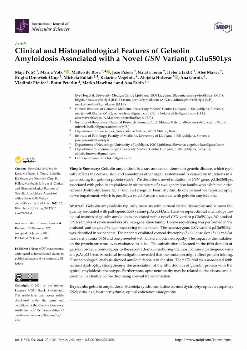
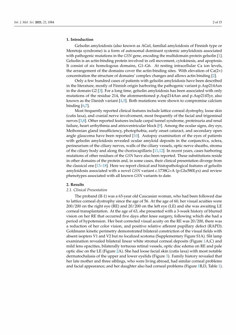
The proband (II-1) was a 63-year old Caucasian woman, who had been followed dueto lattice corneal dystrophy since the age of 56. At the age of 60, her visual acuities were200/200 on the right eye (RE) and 20/200 on the left eye (LE) and she was awaiting LEcorneal transplantation. At the age of 63, she presented with a 3-week history of blurredvision on her RE that occurred five days after knee surgery, following which she had aperiod of hypotension. Her best corrected visual acuity on the RE was 20/200, there wasa reduction of her color vision, and positive relative afferent pupillary defect (RAPD).Goldmann kinetic perimetry demonstrated bilateral constriction of the visual fields withabsent isopters V1 and V2 but no localized scotoma (Supplementary Figure S1A). Slit lampexamination revealed bilateral linear white stromal corneal deposits (Figure 1A,C) andmild lens opacities, bilaterally tortuous retinal vessels, optic disc edema on RE and paleoptic disc on the LE (Figure 2A). She had loose facial skin (cutis laxa) with most notabledermatochalasis of the upper and lower eyelids (Figure 3). Family history revealed thather late mother and three siblings, who were living abroad, had similar corneal problemsand facial appearance; and her daughter also had corneal problems (Figure 1B,D, Table 1).
Int. J. Mol. Sci. 2021, 22, 1084 3 of 15Int. J. Mol. Sci. 2021, 22, x FOR PEER REVIEW 3 of 17
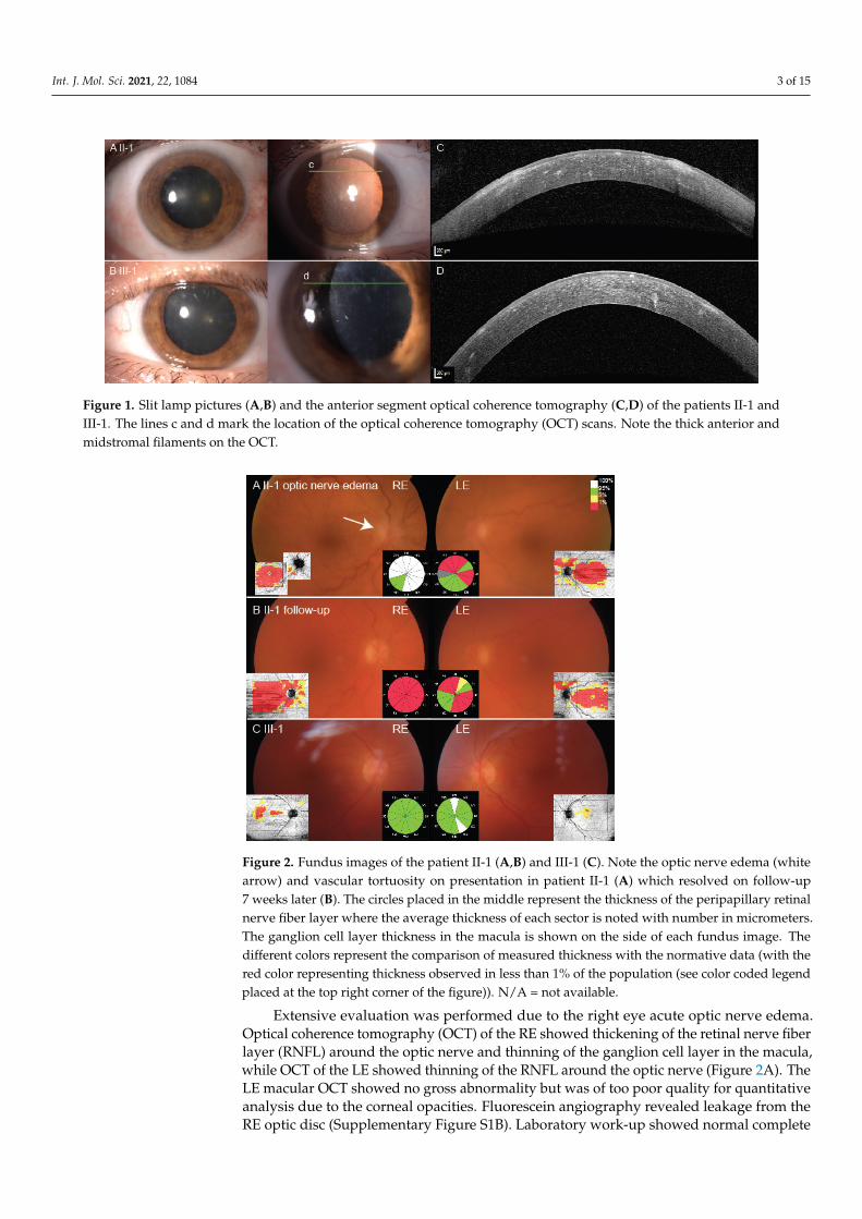
Figure 1. Slit lamp pictures (A,B) and the anterior segment optical coherence tomography (C,D) of the patients II-1 and III-1. The lines c and d mark the location of the optical coherence tomography (OCT) scans. Note the thick anterior and midstromal filaments on the OCT.
Figure 2. Fundus images of the patient II-1 (A,B) and III-1 (C). Note the optic nerve edema (white arrow) and vascular tortuosity on presentation in patient II-1 (A) which resolved on follow-up 7 weeks later (B). The circles placed in the middle represent the thickness of the peripapillary retinal nerve fiber layer where the average thickness of each sector is noted with number in micrometers. The ganglion cell layer thickness in the macula is shown on the side of each fundus image. The different colors represent the comparison of measured thickness with the normative data (with the red color representing thickness observed in less than 1% of the population (see color coded leg-end placed at the top right corner of the figure)). N/A = not available.
Figure 1. Slit lamp pictures (A,B) and the anterior segment optical coherence tomography (C,D) of the patients II-1 andIII-1. The lines c and d mark the location of the optical coherence tomography (OCT) scans. Note the thick anterior andmidstromal filaments on the OCT.
Int. J. Mol. Sci. 2021, 22, x FOR PEER REVIEW 3 of 17
Figure 1. Slit lamp pictures (A,B) and the anterior segment optical coherence tomography (C,D) of the patients II-1 and III-1. The lines c and d mark the location of the optical coherence tomography (OCT) scans. Note the thick anterior and midstromal filaments on the OCT.
Figure 2. Fundus images of the patient II-1 (A,B) and III-1 (C). Note the optic nerve edema (white arrow) and vascular tortuosity on presentation in patient II-1 (A) which resolved on follow-up 7 weeks later (B). The circles placed in the middle represent the thickness of the peripapillary retinal nerve fiber layer where the average thickness of each sector is noted with number in micrometers. The ganglion cell layer thickness in the macula is shown on the side of each fundus image. The different colors represent the comparison of measured thickness with the normative data (with the red color representing thickness observed in less than 1% of the population (see color coded leg-end placed at the top right corner of the figure)). N/A = not available.
Figure 2. Fundus images of the patient II-1 (A,B) and III-1 (C). Note the optic nerve edema (whitearrow) and vascular tortuosity on presentation in patient II-1 (A) which resolved on follow-up7 weeks later (B). The circles placed in the middle represent the thickness of the peripapillary retinalnerve fiber layer where the average thickness of each sector is noted with number in micrometers.The ganglion cell layer thickness in the macula is shown on the side of each fundus image. Thedifferent colors represent the comparison of measured thickness with the normative data (with thered color representing thickness observed in less than 1% of the population (see color coded legendplaced at the top right corner of the figure)). N/A = not available.
Extensive evaluation was performed due to the right eye acute optic nerve edema.Optical coherence tomography (OCT) of the RE showed thickening of the retinal nerve fiberlayer (RNFL) around the optic nerve and thinning of the ganglion cell layer in the macula,while OCT of the LE showed thinning of the RNFL around the optic nerve (Figure 2A). TheLE macular OCT showed no gross abnormality but was of too poor quality for quantitativeanalysis due to the corneal opacities. Fluorescein angiography revealed leakage from theRE optic disc (Supplementary Figure S1B). Laboratory work-up showed normal complete
Int. J. Mol. Sci. 2021, 22, 1084 4 of 15
blood count and moderately increased erythrocyte sedimentation rate (ESR) (64 mm/h) andC-reactive protein (CRP) (10 mg/L). Renal function tests displayed normal creatinine andglomerular filtration rate values. Giant cell arteritis was excluded with the ultrasound of thetemporal arteries (Supplementary Figure S1C) and the absence of other clinical symptomssuch as headache or jaw claudication. Computed tomography of the brain excludedcompressive lesions but showed multiple calcifications in the brain nuclei (SupplementaryFigure S1D), suggesting possible Morbus Fahr (primary familial brain calcification) orneurocysticercosis. The latter was excluded with negative serology for Taenia solium.Serology tests for Treponema pallidum, Lyme disease, and Toxoplasma gondii were alsonegative. Expanding intracranial lesions were excluded with the MRI of the brain, however,white matter punctate lesions were detected in the frontal lobes. On follow-up exam after7 weeks, the visual acuity was counting fingers at 1 m RE and 20/125 LE, and there wasbilateral atrophy of the optic nerve (Figure 2B). Neurological examination revealed signsof mild sensory ataxia and symptoms suggestive of carpal tunnel syndrome (CTS). Sheis awaiting neurophysiology studies to help localize the lesion responsible for sensoryataxia and to confirm the diagnosis of CTS. No cranial nerve involvement other than opticneuropathy was documented.
Int. J. Mol. Sci. 2021, 22, x FOR PEER REVIEW 4 of 17
Figure 3. The facial appearance of the patient II-1 before (A) and after (B) the upper eyelid blepharoplasty. Note the loose skin (cutis laxa) of the upper and lower eyelids, characteristic for gelsolin amyloidosis.
Extensive evaluation was performed due to the right eye acute optic nerve edema. Optical coherence tomography (OCT) of the RE showed thickening of the retinal nerve fiber layer (RNFL) around the optic nerve and thinning of the ganglion cell layer in the macula, while OCT of the LE showed thinning of the RNFL around the optic nerve (Figure 2A). The LE macular OCT showed no gross abnormality but was of too poor quality for quantitative analysis due to the corneal opacities. Fluorescein angiography revealed leak-age from the RE optic disc (Supplementary Figure S1B). Laboratory work-up showed nor-mal complete blood count and moderately increased erythrocyte sedimentation rate (ESR) (64 mm/h) and C-reactive protein (CRP) (10 mg/L). Renal function tests displayed normal creatinine and glomerular filtration rate values. Giant cell arteritis was excluded with the ultrasound of the temporal arteries (Supplementary Figure S1C) and the absence of other clinical symptoms such as headache or jaw claudication. Computed tomography of the brain excluded compressive lesions but showed multiple calcifications in the brain nuclei (Supplementary Figure S1D), suggesting possible Morbus Fahr (primary familial brain calcification) or neurocysticercosis. The latter was excluded with negative serology for Taenia solium. Serology tests for Treponema pallidum, Lyme disease, and Toxoplasma gondii were also negative. Expanding intracranial lesions were excluded with the MRI of the brain, however, white matter punctate lesions were detected in the frontal lobes. On follow-up exam after 7 weeks, the visual acuity was counting fingers at 1 m RE and 20/125 LE, and there was bilateral atrophy of the optic nerve (Figure 2B). Neurological examina-tion revealed signs of mild sensory ataxia and symptoms suggestive of carpal tunnel syn-drome (CTS). She is awaiting neurophysiology studies to help localize the lesion respon-sible for sensory ataxia and to confirm the diagnosis of CTS. No cranial nerve involvement other than optic neuropathy was documented.
The proband’s daughter (III-1) presented at the emergency ophthalmology depart-ment complaining of blurred vision and retrobulbar pain on her LE at the age of 40, when lattice corneal dystrophy was first observed. Six years later, after receiving her mother's genetic results, we invited her for an ophthalmological examination and genetic testing. At that time, she complained of glare and dry eye sensation as well as little, short lines in visual field of both eyes. Visual acuity was 20/25 on her RE and 20/20 on her LE. Octopus G2 top static perimetry revealed centrally decreased sensitivity bilaterally. There was no obvious dermatochalasis or other skin laxity, while bilateral corneal lattice dystrophy was seen on the slit lamp (Figure 1B,D). OCT of the optic nerve head was normal, however, the ganglion cell layer was thinned on the macular OCT (Figure 2C). Magnetic resonance imaging revealed left frontal focal cortical encephalomalacia due to a traumatic head in-jury she suffered 20 years prior. Neurological examination showed blepharospasm, oro-mandibular dystonia and torticollis to the right with 'no-no' type tremor of the head. In addition, increased muscle tone in the right leg, brisk tendon reflexes and bilateral exten-sor plantar response were noted and attributed to the old injury. No cranial nerve involve-ment was observed.
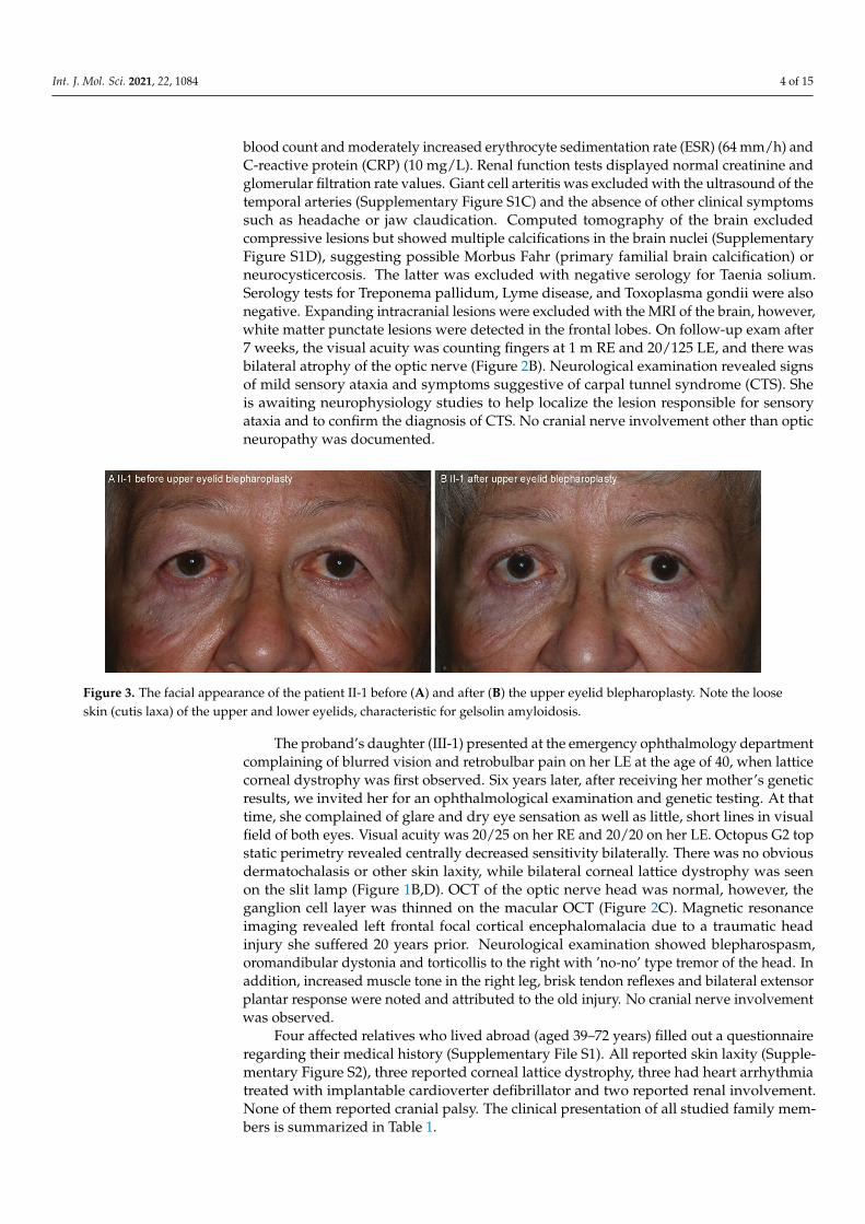
Figure 3. The facial appearance of the patient II-1 before (A) and after (B) the upper eyelid blepharoplasty. Note the looseskin (cutis laxa) of the upper and lower eyelids, characteristic for gelsolin amyloidosis.
The proband’s daughter (III-1) presented at the emergency ophthalmology departmentcomplaining of blurred vision and retrobulbar pain on her LE at the age of 40, when latticecorneal dystrophy was first observed. Six years later, after receiving her mother’s geneticresults, we invited her for an ophthalmological examination and genetic testing. At thattime, she complained of glare and dry eye sensation as well as little, short lines in visualfield of both eyes. Visual acuity was 20/25 on her RE and 20/20 on her LE. Octopus G2 topstatic perimetry revealed centrally decreased sensitivity bilaterally. There was no obviousdermatochalasis or other skin laxity, while bilateral corneal lattice dystrophy was seenon the slit lamp (Figure 1B,D). OCT of the optic nerve head was normal, however, theganglion cell layer was thinned on the macular OCT (Figure 2C). Magnetic resonanceimaging revealed left frontal focal cortical encephalomalacia due to a traumatic headinjury she suffered 20 years prior. Neurological examination showed blepharospasm,oromandibular dystonia and torticollis to the right with ’no-no’ type tremor of the head. Inaddition, increased muscle tone in the right leg, brisk tendon reflexes and bilateral extensorplantar response were noted and attributed to the old injury. No cranial nerve involvementwas observed.
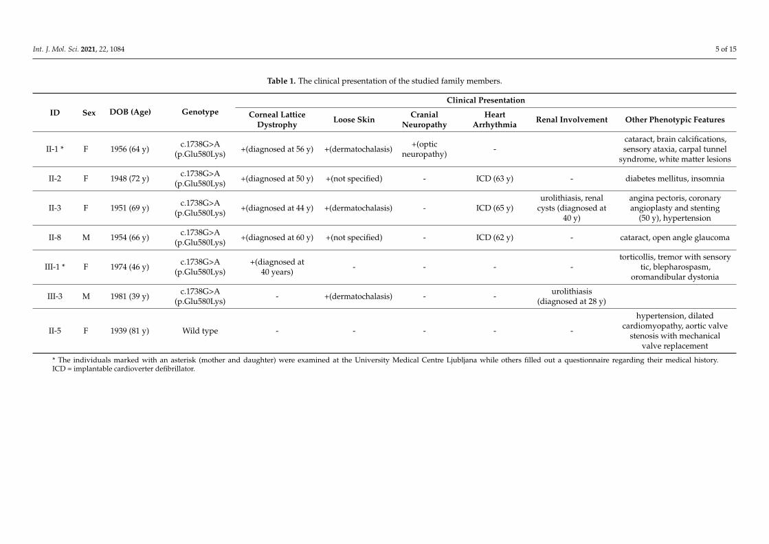
Four affected relatives who lived abroad (aged 39–72 years) filled out a questionnaireregarding their medical history (Supplementary File S1). All reported skin laxity (Supple-mentary Figure S2), three reported corneal lattice dystrophy, three had heart arrhythmiatreated with implantable cardioverter defibrillator and two reported renal involvement.None of them reported cranial palsy. The clinical presentation of all studied family mem-bers is summarized in Table 1.
Int. J. Mol. Sci. 2021, 22, 1084 5 of 15
Table 1. The clinical presentation of the studied family members.
ID Sex DOB (Age) GenotypeClinical Presentation
Corneal LatticeDystrophy Loose Skin Cranial
NeuropathyHeart
Arrhythmia Renal Involvement Other Phenotypic Features
II-1 * F 1956 (64 y) c.1738G>A(p.Glu580Lys) +(diagnosed at 56 y) +(dermatochalasis) +(optic
neuropathy) -cataract, brain calcifications,sensory ataxia, carpal tunnel
syndrome, white matter lesions
II-2 F 1948 (72 y) c.1738G>A(p.Glu580Lys) +(diagnosed at 50 y) +(not specified) - ICD (63 y) - diabetes mellitus, insomnia
II-3 F 1951 (69 y) c.1738G>A(p.Glu580Lys) +(diagnosed at 44 y) +(dermatochalasis) - ICD (65 y)
urolithiasis, renalcysts (diagnosed at
40 y)
angina pectoris, coronaryangioplasty and stenting
(50 y), hypertension
II-8 M 1954 (66 y) c.1738G>A(p.Glu580Lys) +(diagnosed at 60 y) +(not specified) - ICD (62 y) - cataract, open angle glaucoma
III-1 * F 1974 (46 y) c.1738G>A(p.Glu580Lys)
+(diagnosed at40 years) - - - -
torticollis, tremor with sensorytic, blepharospasm,
oromandibular dystonia
III-3 M 1981 (39 y) c.1738G>A(p.Glu580Lys) - +(dermatochalasis) - - urolithiasis
(diagnosed at 28 y)
II-5 F 1939 (81 y) Wild type - - - - -
hypertension, dilatedcardiomyopathy, aortic valve
stenosis with mechanicalvalve replacement
* The individuals marked with an asterisk (mother and daughter) were examined at the University Medical Centre Ljubljana while others filled out a questionnaire regarding their medical history.ICD = implantable cardioverter defibrillator.
Int. J. Mol. Sci. 2021, 22, 1084 6 of 15
2.2. Molecular Analysis
Exome sequencing in the proband revealed a novel heterozygous variant c.1738G>A(p.Glu580Lys) in the GSN gene (NM_000177.5, Supplementary Figure S3). The variantis absent from the controls (GnomAD. Available online: GnomAD.broadinstitute.org(accessed on 5 June 2020)) and the majority of in silico predictors indicate its damaging effect(including MetaSVM, REVEL and CADD meta-predictors). Family genetic studies revealedthe presence of the GSN variant in five relatives and confirmed the co-segregation of thevariant with the disease status. Based on the ACMG guidelines for variant classification [19],adapted in accordance with ACGS recommendations (Association for Clinical GenomicScience. Available online: https://www.acgs.uk.com/quality/best-practice-guidelines/(accessed on 16 December 2020)), we classified the variant as likely pathogenic. Theevidence for this classification was based on variant’s absence in the general population(PM2), strong segregation evidence (PP1_STR), theoretical predictions of pathogenicity(PP3), the results of a protein modeling study performed for this variant (PM1_SUP) andthe consistency of the finding with the clinical presentation observed in the patients withthis variant (PP4).
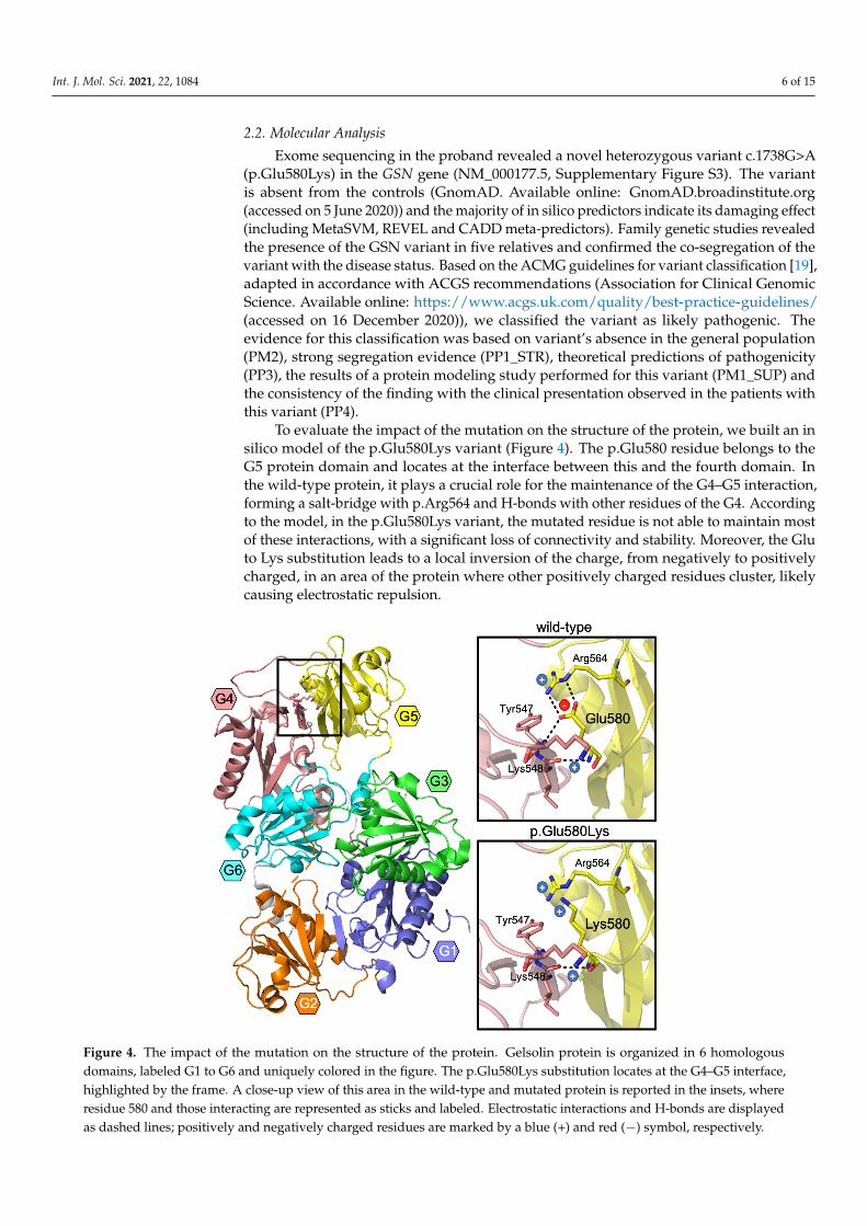
To evaluate the impact of the mutation on the structure of the protein, we built an insilico model of the p.Glu580Lys variant (Figure 4). The p.Glu580 residue belongs to theG5 protein domain and locates at the interface between this and the fourth domain. Inthe wild-type protein, it plays a crucial role for the maintenance of the G4–G5 interaction,forming a salt-bridge with p.Arg564 and H-bonds with other residues of the G4. Accordingto the model, in the p.Glu580Lys variant, the mutated residue is not able to maintain mostof these interactions, with a significant loss of connectivity and stability. Moreover, the Gluto Lys substitution leads to a local inversion of the charge, from negatively to positivelycharged, in an area of the protein where other positively charged residues cluster, likelycausing electrostatic repulsion.
Int. J. Mol. Sci. 2021, 22, x FOR PEER REVIEW 6 of 17
Genomic Science. Available online: https://www.acgs.uk.com/quality/best-practice-guidelines/ (accessed on 16 December 2020)), we classified the variant as likely patho-genic. The evidence for this classification was based on variant’s absence in the general population (PM2), strong segregation evidence (PP1_STR), theoretical predictions of path-ogenicity (PP3), the results of a protein modeling study performed for this variant (PM1_SUP) and the consistency of the finding with the clinical presentation observed in the patients with this variant (PP4).
To evaluate the impact of the mutation on the structure of the protein, we built an in silico model of the p.Glu580Lys variant (Figure 4). The p.Glu580 residue belongs to the G5 protein domain and locates at the interface between this and the fourth domain. In the wild-type protein, it plays a crucial role for the maintenance of the G4–G5 interaction, forming a salt-bridge with p.Arg564 and H-bonds with other residues of the G4. Accord-ing to the model, in the p.Glu580Lys variant, the mutated residue is not able to maintain most of these interactions, with a significant loss of connectivity and stability. Moreover, the Glu to Lys substitution leads to a local inversion of the charge, from negatively to positively charged, in an area of the protein where other positively charged residues clus-ter, likely causing electrostatic repulsion.
Figure 4. The impact of the mutation on the structure of the protein. Gelsolin protein is organized in 6 homologous do-mains, labeled G1 to G6 and uniquely colored in the figure. The p.Glu580Lys substitution locates at the G4–G5 interface, highlighted by the frame. A close-up view of this area in the wild-type and mutated protein is reported in the insets, where residue 580 and those interacting are represented as sticks and labeled. Electrostatic interactions and H-bonds are dis-played as dashed lines; positively and negatively charged residues are marked by a blue (+) and red (−) symbol, respec-tively.
Figure 4. The impact of the mutation on the structure of the protein. Gelsolin protein is organized in 6 homologousdomains, labeled G1 to G6 and uniquely colored in the figure. The p.Glu580Lys substitution locates at the G4–G5 interface,highlighted by the frame. A close-up view of this area in the wild-type and mutated protein is reported in the insets, whereresidue 580 and those interacting are represented as sticks and labeled. Electrostatic interactions and H-bonds are displayedas dashed lines; positively and negatively charged residues are marked by a blue (+) and red (−) symbol, respectively.
Int. J. Mol. Sci. 2021, 22, 1084 7 of 15
2.3. Pathological Findings
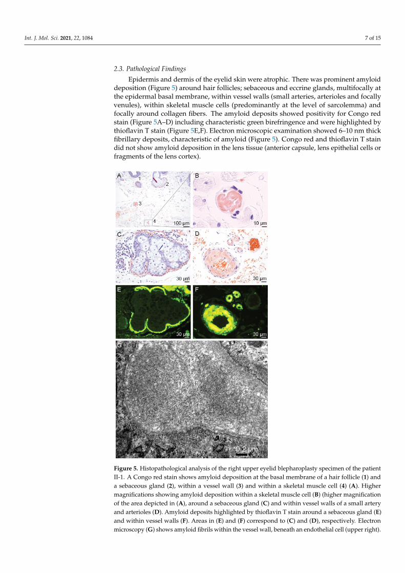
Epidermis and dermis of the eyelid skin were atrophic. There was prominent amyloiddeposition (Figure 5) around hair follicles; sebaceous and eccrine glands, multifocally atthe epidermal basal membrane, within vessel walls (small arteries, arterioles and focallyvenules), within skeletal muscle cells (predominantly at the level of sarcolemma) andfocally around collagen fibers. The amyloid deposits showed positivity for Congo redstain (Figure 5A–D) including characteristic green birefringence and were highlighted bythioflavin T stain (Figure 5E,F). Electron microscopic examination showed 6–10 nm thickfibrillary deposits, characteristic of amyloid (Figure 5). Congo red and thioflavin T staindid not show amyloid deposition in the lens tissue (anterior capsule, lens epithelial cells orfragments of the lens cortex).
Int. J. Mol. Sci. 2021, 22, x FOR PEER REVIEW 7 of 17
2.3. Pathological Findings Epidermis and dermis of the eyelid skin were atrophic. There was prominent amy-
loid deposition (Figure 5) around hair follicles; sebaceous and eccrine glands, multifocally at the epidermal basal membrane, within vessel walls (small arteries, arterioles and focally venules), within skeletal muscle cells (predominantly at the level of sarcolemma) and fo-cally around collagen fibers. The amyloid deposits showed positivity for Congo red stain (Figure 5A–D) including characteristic green birefringence and were highlighted by thio-flavin T stain (Figure 5E,F). Electron microscopic examination showed 6–10 nm thick fi-brillary deposits, characteristic of amyloid (Figure 5). Congo red and thioflavin T stain did not show amyloid deposition in the lens tissue (anterior capsule, lens epithelial cells or fragments of the lens cortex).
Figure 5. Histopathological analysis of the right upper eyelid blepharoplasty specimen of the patient II-1. A Congo red stain shows amyloid deposition at the basal membrane of a hair follicle (1) and a sebaceous gland (2), within a vessel wall (3) and within a skeletal muscle cell (4) (A). Higher magnifications showing amyloid deposition within a skeletal muscle cell (B) (higher magnification of the area depicted in (A), around a sebaceous gland (C) and within vessel walls of a small
Figure 5. Histopathological analysis of the right upper eyelid blepharoplasty specimen of the patientII-1. A Congo red stain shows amyloid deposition at the basal membrane of a hair follicle (1) anda sebaceous gland (2), within a vessel wall (3) and within a skeletal muscle cell (4) (A). Highermagnifications showing amyloid deposition within a skeletal muscle cell (B) (higher magnificationof the area depicted in (A), around a sebaceous gland (C) and within vessel walls of a small arteryand arterioles (D). Amyloid deposits highlighted by thioflavin T stain around a sebaceous gland (E)and within vessel walls (F). Areas in (E) and (F) correspond to (C) and (D), respectively. Electronmicroscopy (G) shows amyloid fibrils within the vessel wall, beneath an endothelial cell (upper right).

Int. J. Mol. Sci. 2021, 22, 1084 8 of 15
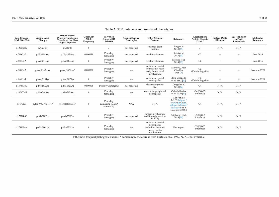
2.4. Review of the Phenotypes Associated with GSN Mutations
Ten GSN mutations, including ours, and their associated phenotypes are summarizedin Table 2 and their location in GSN protein is shown in Figure 6. The typical pheno-type was also originally described in families harboring the common pathogenic variantp.Asp214Asn, its hallmark being corneal lattice dystrophy. This symptom was observed inassociation with three other pathogenic variants, namely p.Asp214Tyr, p.Met544Arg andp.Glu580Lys (present report). All these substitutions reside either in the second domain ofthe protein or at the interface between the fourth and fifth protein domains. Five other GSNmutations were described in association with other phenotypes, such as renal amyloidosis(p.Gly194Arg and p.Asn211Lys), dermatomyositis (p.Pro459Arg) and seizures (p.Ala7fs).
Int. J. Mol. Sci. 2021, 22, x FOR PEER REVIEW 1 of 17
Figure 6. GSN variants with their location in the GSN protein and their pathogenicity designation according to the ClinVar database (Available online: ncbi.nlm.nih.gov/clinvar/ (accessed on 5 December 2020)). The phenotypes that were reported in association with these variants are listed in Table 2. Note the mutation p.Trp493GlyfsTer17 that was designated as likely pathogenic but not yet described in patients in the literature.
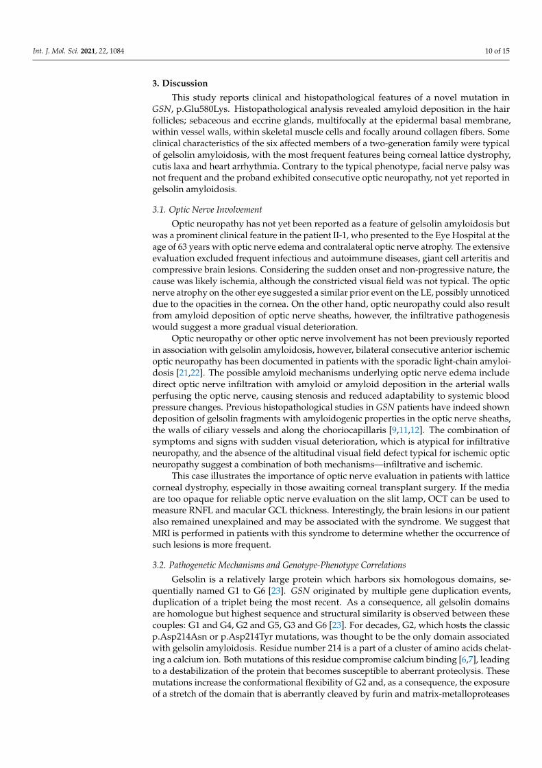
3. Discussion This study reports clinical and histopathological features of a novel mutation in GSN,
p.Glu580Lys. Histopathological analysis revealed amyloid deposition in the hair follicles; sebaceous and eccrine glands, multifocally at the epidermal basal membrane, within ves-sel walls, within skeletal muscle cells and focally around collagen fibers. Some clinical characteristics of the six affected members of a two-generation family were typical of gel-solin amyloidosis, with the most frequent features being corneal lattice dystrophy, cutis laxa and heart arrhythmia. Contrary to the typical phenotype, facial nerve palsy was not frequent and the proband exhibited consecutive optic neuropathy, not yet reported in gel-solin amyloidosis.
3.1. Optic Nerve Involvement Optic neuropathy has not yet been reported as a feature of gelsolin amyloidosis but
was a prominent clinical feature in the patient II-1, who presented to the Eye Hospital at the age of 63 years with optic nerve edema and contralateral optic nerve atrophy. The extensive evaluation excluded frequent infectious and autoimmune diseases, giant cell arteritis and compressive brain lesions. Considering the sudden onset and non-progres-sive nature, the cause was likely ischemia, although the constricted visual field was not typical. The optic nerve atrophy on the other eye suggested a similar prior event on the LE, possibly unnoticed due to the opacities in the cornea. On the other hand, optic neu-ropathy could also result from amyloid deposition of optic nerve sheaths, however, the infiltrative pathogenesis would suggest a more gradual visual deterioration.
Optic neuropathy or other optic nerve involvement has not been previously reported in association with gelsolin amyloidosis, however, bilateral consecutive anterior ischemic optic neuropathy has been documented in patients with the sporadic light-chain amyloi-dosis [21,22]. The possible amyloid mechanisms underlying optic nerve edema include direct optic nerve infiltration with amyloid or amyloid deposition in the arterial walls perfusing the optic nerve, causing stenosis and reduced adaptability to systemic blood pressure changes. Previous histopathological studies in GSN patients have indeed shown deposition of gelsolin fragments with amyloidogenic properties in the optic nerve sheaths, the walls of ciliary vessels and along the choriocapillaris [9,11,12]. The combination of symptoms and signs with sudden visual deterioration, which is atypical for infiltrative neuropathy, and the absence of the altitudinal visual field defect typical for ischemic optic neuropathy suggest a combination of both mechanisms—infiltrative and ischemic.
Figure 6. GSN variants with their location in the GSN protein and their pathogenicity designation according to the ClinVardatabase (Available online: ncbi.nlm.nih.gov/clinvar/ (accessed on 5 December 2020)). The phenotypes that were reportedin association with these variants are listed in Table 2. Note the mutation p.Trp493GlyfsTer17 that was designated as likelypathogenic but not yet described in patients in the literature.
Int. J. Mol. Sci. 2021, 22, 1084 9 of 15
Table 2. GSN mutations and associated phenotypes.
Base Change(NM_000177.4)
Amino AcidChange
Mature PlasmaProtein Numbering(Devoid of the 27-aa
Signal Peptide)
GnomADAllele
Frequency
Polyphen(Uniprot ID
P06396)Corneal Lattice
DystrophyOther Clinical
Features ReferenceLocalization:
Protein Domain(G1-6) *
Protein Desta-bilization
Susceptibilityto Furin
ProteolysisMolecularReference
c.100dupG p.Ala34fs p.Ala7fs 0 / not reported seizures, brainlesions
Feng et al.2018 [13] / N/A N/A
c.580G>A p.Gly194Arg p.Gly167Arg 0.000039 Probablydamaging not reported renal involvement Sethi et al.
2013 [14] G2 + + Bonì 2018
c.633C>A p.Asn211Lys p.Asn184Lys 0 Probablydamaging not reported renal involvement Efebera et al.
2014 [15] G2 + + Bonì 2016
c.640G>A p.Asp214Asn+ p.Asp187Asn# 0.000007 Probablydamaging yes
cutis laxa, cranialneuropathy, heartarrhythmia, renal
involvement
Meretoja, AnnClin Res.1969 [3]
G2(Ca-binding site) + + Isaacson 1999
c.640G>T p.Asp214Tyr p.Asp187Tyr 0 Probablydamaging yes cutis laxa, cranial
neuropathyde la Chapelleet al. 1992 [20]
G2(Ca-binding site) + + Isaacson 1999
c.1375C>G p.Pro459Arg p.Pro432Arg 0.000004 Possibly damaging not reported dermatomyositis-like
Oregel et al.2018 [16] G4 N/A N/A
c.1631T>G p.Met544Arg p.Met517Arg 0 Probablydamaging yes cutis laxa, peripheral
neuropathyCabral-Maciaset al. 2020 [17]
G4 (G4-G5interface) N/A N/A
c.1476del p.Trp493GlyfsTer17 p.Trp466fsTer17 0Probably
damaging (GERPscore 5.25)
N/A N/A
ClinVar ID:493491 https://www.ncbi.nlm.
nih.gov/clinvar/(accessed on 5
December 2020)
G4 N/A N/A
c.1732G>C p.Ala578Pro p.Ala551Pro 0 Probablydamaging not reported
cardiac involvement(additional mutation
in TTR)Sridharan et al.
2018 [18]G5 (G4:G5interface) N/A N/A
c.1738G>A p.Glu580Lys p.Glu553Lys 0 Probablydamaging yes
cutis laxa, cranialneuropathy
including the opticnerve, cardiacinvolvement
This report G5 (G4:G5interface) N/A N/A
# the most frequent pathogenic variant. * domain nomenclature is from Burtnick et al. 1997. N/A = not available.
Int. J. Mol. Sci. 2021, 22, 1084 10 of 15
3. Discussion
This study reports clinical and histopathological features of a novel mutation inGSN, p.Glu580Lys. Histopathological analysis revealed amyloid deposition in the hairfollicles; sebaceous and eccrine glands, multifocally at the epidermal basal membrane,within vessel walls, within skeletal muscle cells and focally around collagen fibers. Someclinical characteristics of the six affected members of a two-generation family were typicalof gelsolin amyloidosis, with the most frequent features being corneal lattice dystrophy,cutis laxa and heart arrhythmia. Contrary to the typical phenotype, facial nerve palsy wasnot frequent and the proband exhibited consecutive optic neuropathy, not yet reported ingelsolin amyloidosis.
3.1. Optic Nerve Involvement
Optic neuropathy has not yet been reported as a feature of gelsolin amyloidosis butwas a prominent clinical feature in the patient II-1, who presented to the Eye Hospital at theage of 63 years with optic nerve edema and contralateral optic nerve atrophy. The extensiveevaluation excluded frequent infectious and autoimmune diseases, giant cell arteritis andcompressive brain lesions. Considering the sudden onset and non-progressive nature, thecause was likely ischemia, although the constricted visual field was not typical. The opticnerve atrophy on the other eye suggested a similar prior event on the LE, possibly unnoticeddue to the opacities in the cornea. On the other hand, optic neuropathy could also resultfrom amyloid deposition of optic nerve sheaths, however, the infiltrative pathogenesiswould suggest a more gradual visual deterioration.
Optic neuropathy or other optic nerve involvement has not been previously reportedin association with gelsolin amyloidosis, however, bilateral consecutive anterior ischemicoptic neuropathy has been documented in patients with the sporadic light-chain amyloi-dosis [21,22]. The possible amyloid mechanisms underlying optic nerve edema includedirect optic nerve infiltration with amyloid or amyloid deposition in the arterial wallsperfusing the optic nerve, causing stenosis and reduced adaptability to systemic bloodpressure changes. Previous histopathological studies in GSN patients have indeed showndeposition of gelsolin fragments with amyloidogenic properties in the optic nerve sheaths,the walls of ciliary vessels and along the choriocapillaris [9,11,12]. The combination ofsymptoms and signs with sudden visual deterioration, which is atypical for infiltrativeneuropathy, and the absence of the altitudinal visual field defect typical for ischemic opticneuropathy suggest a combination of both mechanisms—infiltrative and ischemic.
This case illustrates the importance of optic nerve evaluation in patients with latticecorneal dystrophy, especially in those awaiting corneal transplant surgery. If the mediaare too opaque for reliable optic nerve evaluation on the slit lamp, OCT can be used tomeasure RNFL and macular GCL thickness. Interestingly, the brain lesions in our patientalso remained unexplained and may be associated with the syndrome. We suggest thatMRI is performed in patients with this syndrome to determine whether the occurrence ofsuch lesions is more frequent.
3.2. Pathogenetic Mechanisms and Genotype-Phenotype Correlations
Gelsolin is a relatively large protein which harbors six homologous domains, se-quentially named G1 to G6 [23]. GSN originated by multiple gene duplication events,duplication of a triplet being the most recent. As a consequence, all gelsolin domainsare homologue but highest sequence and structural similarity is observed between thesecouples: G1 and G4, G2 and G5, G3 and G6 [23]. For decades, G2, which hosts the classicp.Asp214Asn or p.Asp214Tyr mutations, was thought to be the only domain associatedwith gelsolin amyloidosis. Residue number 214 is a part of a cluster of amino acids chelat-ing a calcium ion. Both mutations of this residue compromise calcium binding [6,7], leadingto a destabilization of the protein that becomes susceptible to aberrant proteolysis. Thesemutations increase the conformational flexibility of G2 and, as a consequence, the exposureof a stretch of the domain that is aberrantly cleaved by furin and matrix-metalloproteases

Int. J. Mol. Sci. 2021, 22, 1084 11 of 15
with these proteolytic events leading to the production of amyloidogenic peptides [6,24,25].Recently described mutations in G4 [16,17] and G5 domains [18], however, suggest an alter-native and furin-independent pathway of gelsolin aggregation. The in silico model of thep.Glu580Lys variant predicts a net loss of connectivity and electrostatic repulsion causedby the Lys substitution, which might cause protein destabilization and misfolding, whichare features often associated to pathological aggregation and aberrant proteolysis [26].
In recent years, several GSN mutations have been discovered and reported in patientswith typical and atypical clinical presentations (Table 2). The classic phenotype that hasoriginally been described in the large families with p.Asp214Asn mutation [3] has pre-viously been observed only in patients harboring another mutation of the same residue(p.Asp214Tyr) [20] suggesting that the residue was phenotype-specific. This has beenchallenged by a recent publication of Cabral-Macias et al. in 2020, of a novel mutationp.Met544Arg, located in the G4:G5 interface, also causing a typical gelsolin phenotype,including the pathognomonic corneal lattice dystrophy [17]. The mutation p.Glu580Lysdescribed in the present study is also located in the G4:G5 interface and associated with thetypical phenotype and supports the observation that mutations at various locations are ableto produce classic phenotype of gelsolin amyloidosis and a common pathogenetic pathway.Reports of cases harboring other missense mutations in the domains G2 (p.Gly194Arg,p.Asn211Lys), G4 (p.Pro459Arg) and the G4:G5 interface (p.Ala578Pro) described morelocalized phenotypes including only renal, skin and/or heart disease without corneallattice dystrophy [13–18]. These clinical presentations were still within the classic pheno-typic spectrum and most reports included only a small number of cases, therefore, furtherstudies are needed to conclude whether they cause identical or a limited/overlappingphenotype. The disease pathogenesis may also differ. For example, two of the mutations inthe G2 domain, p.Gly194Arg and p.Asn211Lys, do not impair calcium binding [24,25] andpatients carrying these mutations only present with renal complications [14,15]. Neverthe-less, in comparison to missense mutations mentioned above, the frame-shifting mutationp.Ala34fs produced a notably different phenotype with none of the classic features butwith severe brain involvement including seizures and brain lesions [13], possibly related tohaploinsufficiency. These observations suggest that more basic research and clinical studiesare required to decipher and ultimately cure this rare yet disabling disease.
4. Materials and Methods4.1. Patients
Seven members of a two-generation family originating from Herzegovina region ofBosna and Herzegovina were ascertained for the study (pedigree shown in Figure 7). Theproband and her daughter (II-1 and III-1) underwent a detailed ophthalmological examwhile five relatives, who were unavailable for examination due to living abroad, filledout a clinical questionnaire regarding their medical history (Supplementary File S1) andprovided saliva samples for genetic analysis.
All participants signed a written informed consent prior to examination.
Int. J. Mol. Sci. 2021, 22, x FOR PEER REVIEW 3 of 17
4. Materials and Methods 4.1. Patients
Seven members of a two-generation family originating from Herzegovina region of Bosna and Herzegovina were ascertained for the study (pedigree shown in Figure 7). The proband and her daughter (II-1 and III-1) underwent a detailed ophthalmological exam while five relatives, who were unavailable for examination due to living abroad, filled out a clinical questionnaire regarding their medical history (Supplementary File S1) and pro-vided saliva samples for genetic analysis.
All participants signed a written informed consent prior to examination.
Figure 7. Three-generation family tree of seven members, six of them affected, is presented. Proband is indicated with an arrow. The genotypes of available family members are subscribed indicating the cosegregation of the identified GSN var-iant with the phenotypic features.
4.2. Clinical Examination Clinical examination in the patients II-1 and III-1 included visual acuity, visual field
(manual Goldmann kinetic perimetry (Haag Streit, Berne, Switzerland) or Octopus static perimetry (Octopus 101, program G2, Interzeag AG, Schlieren, Switzerland), slit lamp ex-amination, optical coherence imaging of the macula and the optic nerve (swept source optical coherence tomography; SS-OCT; Tritontm, Topcon, Tokyo, Japan), and neurologi-cal exam. The patient II-1 also underwent an additional work-up due to acute optic nerve edema (described in detail in Results).
4.3. Genetic and Bioinformatic Analysis Genetic analyses were performed in seven family members. Genomic DNA was ex-
tracted from blood or saliva samples according to the standard procedure. Sequencing of the defined clinical target was performed using next-generation sequencing on the iso-lated DNA sample of the proband, while in other family members, segregation analysis of identified variant was carried out using Sanger sequencing. Briefly, the fragmentation and enrichment of the isolated DNA sample were performed according to the Illumina Nextera Coding Exome capture protocol (Illumina, USA), with subsequent sequencing on Illumina NextSeq 550 in 2 × 100 cycles. After duplicates were removed, the alignment of reads to UCSC hg19 reference assembly was done using BWA algorithm (v0.6.3) and var-iant calling was done using GATK framework (v2.8). Only variants exceeding the quality score of 30.0 and depth of 5 were used for down-stream analyses. Variant annotation was performed using ANNOVAR and snpEff algorithms, with pathogenicity predictions in dbNSFPv2 database. Reference gene models and transcript sequences are based on RefSeq database. Structural variants were assessed using CONIFER v0.2.2 algorithm. Variants with population frequency exceeding 1% in gnomAD, synonymous variants, intronic var-iants and variants outside the clinical target were filtered out during analyses. An in-
Figure 7. Three-generation family tree of seven members, six of them affected, is presented. Proband is indicated with anarrow. The genotypes of available family members are subscribed indicating the cosegregation of the identified GSN variantwith the phenotypic features.

Int. J. Mol. Sci. 2021, 22, 1084 12 of 15
4.2. Clinical Examination
Clinical examination in the patients II-1 and III-1 included visual acuity, visual field(manual Goldmann kinetic perimetry (Haag Streit, Berne, Switzerland) or Octopus staticperimetry (Octopus 101, program G2, Interzeag AG, Schlieren, Switzerland), slit lampexamination, optical coherence imaging of the macula and the optic nerve (swept sourceoptical coherence tomography; SS-OCT; Tritontm, Topcon, Tokyo, Japan), and neurologicalexam. The patient II-1 also underwent an additional work-up due to acute optic nerveedema (described in detail in Results).
4.3. Genetic and Bioinformatic Analysis
Genetic analyses were performed in seven family members. Genomic DNA wasextracted from blood or saliva samples according to the standard procedure. Sequencing ofthe defined clinical target was performed using next-generation sequencing on the isolatedDNA sample of the proband, while in other family members, segregation analysis ofidentified variant was carried out using Sanger sequencing. Briefly, the fragmentation andenrichment of the isolated DNA sample were performed according to the Illumina NexteraCoding Exome capture protocol (Illumina, USA), with subsequent sequencing on IlluminaNextSeq 550 in 2 × 100 cycles. After duplicates were removed, the alignment of readsto UCSC hg19 reference assembly was done using BWA algorithm (v0.6.3) and variantcalling was done using GATK framework (v2.8). Only variants exceeding the qualityscore of 30.0 and depth of 5 were used for down-stream analyses. Variant annotation wasperformed using ANNOVAR and snpEff algorithms, with pathogenicity predictions indbNSFPv2 database. Reference gene models and transcript sequences are based on RefSeqdatabase. Structural variants were assessed using CONIFER v0.2.2 algorithm. Variants withpopulation frequency exceeding 1% in gnomAD, synonymous variants, intronic variantsand variants outside the clinical target were filtered out during analyses. An in-housepipeline was used for bioinformatic analyses of exome sequencing data, in accordance withGATK best practice recommendations [27]. The interpretation of sequence variants wasbased on ACMG/AMP standards and guidelines [19]. Sequencing the DNA sample, wereached median coverage of 67× and covered over 99.9% targeted regions with minimum10× depth of coverage [28]. Segregation analysis in the family members was performedusing targeted Sanger sequencing. Primer sequences are available upon request.
4.4. In Silico Analysis
The impact of the novel mutation on the structure of the gelsolin protein was evaluatedin silico. Structure of the p.Glu580Lys was obtained by in silico mutagenesis of the wild-type protein (pdb id 3FFN [29], followed by energy minimization to resolve unfavorableinteractions and clashes [30]. PyMOL (the PyMOL Molecular Graphics System, Version2.0 Schrödinger, LLC.) was used for the analysis of the interactions and preparation ofthe figure.
4.5. Pathological Analysis
Eyelid skin tissue was obtained during blepharoplasty for upper eyelid dermatochala-sis (performed by B.D.-O., Figure 3) and lens tissue was obtained during cataract surgery(performed by V.P.) of the patient II-1. The tissue samples were fixed in formalin andembedded in paraffin. Hematoxylin and eosin, Congo red and thioflavin T stains wereperformed. Electron microscopic examination of the skin sample was performed on theformalin-fixed tissue.
Congo red staining was performed automatically in Ventana Benchmark Special Stainsstainer with Congo Red Staining Kit (Ventana Medical Systems Inc., Tucson, AZ, USA). Forthioflavin T staining, slides were incubated in 1% working solution of thioflavin T for 7 min(Sigma Aldrich, Darmstadt, Germany), rinsed in deionized water and then kept in 1%CH3COOH for 20 min. Afterwards, the slides were rinsed and coverslipped directly fromdeionized water with Dako Fluorescence Mounting Medium (DAKO, Glostrup, Denmark).

Int. J. Mol. Sci. 2021, 22, 1084 13 of 15
4.6. Review of the Literature
Studies reporting GSN mutations accessible in the PubMed database in the periodfrom 1969–2020 were reviewed. The nomenclature of the reported mutations was anno-tated according to the originally transcribed and the cleaved plasma protein [31] to avoidconfusion in the nomenclature. The phenotypes were evaluated in order to determinewhether they included the typical corneal lattice dystrophy and/or other clinical features.
Research protocols adhered to the tenets of the Declaration of Helsinki. The informedconsent was acquired from the patients.
5. Conclusions
The novel GSN variant p.Glu580Lys is associated with the typical gelsolin amy-loidosis phenotype, including its hallmark, the corneal lattice dystrophy. Furthermore,optic neuropathy may be associated with the disease and is important to identify beforecorneal transplantation.
Although the novel pathogenic variant shares a similar clinical picture to FAF, theunderlying molecular mechanism might be different. Glu580Lys mutation, as many othersreviewed in this manuscript, localizes far from the second domain and unlikely leads toits destabilization and the exposure of the aberrant furin cleavage site. Recent molecularstudies [28] suggested an alternative, proteolysis-independent mechanism, but they awaitin vivo validation.
Supplementary Materials: The following are available online at https://www.mdpi.com/1422-0067/22/3/1084/s1, Figure S1: A. Goldmann visual field of the patient II-1 showing concentricbilateral constriction of the visual fields, with non-detectable isopters V1 and V2. B. The fluoresceinangiography of patient II-1. There were normal vessel filling times with leakage of contrast from theright optic disc in the late phase. C. The CT of the head of patient II-1 showing diffuse calcificationsof the nucleus pallidum and nucleus caudatus. D. Ultrasound of the temporal arteries: On themain, frontal and parietal branch of temporal artery no halo sign was seen and the compressionsign was negative. Thickness of intima-media complex on frontal branch of temporal artery was0.024 cm. Figure S2: Next generation sequencing revealing a novel heterozygous variant c.1738G>A(p.Glu580Lys) in the GSN gene in the proband. Figure S3: Loose skin (cutis laxa) in patients II-1 (A,F), II-2 (B), II-3 (C), II-8 (D) and II-5 (E). Note that all affected family members shown on the picture(II-1, II-2, II-3, II-8) exhibit lower lid dermatochalasis, whereas the family member II-5 carrying thewild type of GSN gene does not. File S1: Questionnaire.
Author Contributions: Conceptualization, A.F. and M.P.; methodology, A.F., M.P., M.V., M.d.R. andJ.P.; software, A.F., M.V., M.d.R. and M.B.; validation, A.F. and M.V.; formal analysis, A.F., M.P., M.V.,A.M., J.P., M.d.R. and M.B.; investigation, A.F, M.P., K.V., M.V. and A.G.; resources, A.F., M.P., A.G.,B.D.-O., V.P. and M.H.; data curation, A.F., M.P. and M.V.; writing—original draft preparation, M.P.,A.F., M.V., M.d.R. and J.P.; writing—review and editing, M.P., A.F., M.V., M.d.R., J.P., N.T., H.J., A.M.,B.D.-O., M.B., K.V., A.H., A.G., V.P., B.P. and M.H.; visualization, A.F. and M.P.; supervision, A.F.;project administration, A.F. and M.P.; funding acquisition, A.F. All authors have read and agreed tothe published version of the manuscript.
Funding: Slovenian Research Agency (ARRS), grant number J3-1750.
Institutional Review Board Statement: Medical board approval was not obtained as all examina-tions were done as a part of routine diagnostic procedures. The patients provided informed consentfor genetic analysis and publication of their medical information and images.
Informed Consent Statement: Informed consent was obtained from all subjects involved in the study.
Data Availability Statement: The data presented in this study are available on request from thecorresponding author. The data are not publicly available due to personal data protection.
Acknowledgments: We would like to thank the included family members for the collaboration.
Conflicts of Interest: The authors declare no conflict of interest.
Int. J. Mol. Sci. 2021, 22, 1084 14 of 15
Abbreviations
GSN Gelsolin geneRE Right eyeLE Left eyeOCT Optical coherence tomographyRNFL Retinal nerve fiber layerID Identity documentDOB Date of birthN/A Not available
References1. Solomon, J.P.; Page, L.J.; Balch, W.E.; Kelly, J.W. Gelsolin amyloidosis: Genetics, biochemistry, pathology and possible strategies
for therapeutic intervention. Crit. Rev. Biochem. Mol. Biol. 2012, 47, 282–296. [CrossRef] [PubMed]2. Choe, H.; Burtnick, L.D.; Mejillano, M.; Yin, H.L.; Robinson, R.C.; Choe, S. The calcium activation of gelsolin: Insights from the
3A structure of the G4-G6/actin complex. J. Mol. Biol. 2002, 324, 691–702. [CrossRef]3. Meretoja, J. Familial systemic paramyloidosis with lattice dystrophy of the cornea, progressive cranial neuropathy, skin changes
and various internal symptoms. A previously unrecognized heritable syndrome. Ann. Clin. Res. 1969, 1, 314–324. [PubMed]4. de la Chapelle, A.; Tolvanen, R.; Boysen, G.; Santavy, J.; Bleeker-Wagemakers, L.; Maury, C.P.; Kere, J. Gelsolin-derived familial
amyloidosis caused by asparagine or tyrosine substitution for aspartic acid at residue 187. Nat. Genet. 1992, 2, 157–160. [CrossRef]5. Maury, C.P.; Liljeström, M.; Boysen, G.; Törnroth, T.; de la Chapelle, A.; Nurmiaho-Lassila, E.L. Danish type gelsolin related
amyloidosis: 654G-T mutation is associated with a disease pathogenetically and clinically similar to that caused by the 654G-Amutation (familial amyloidosis of the Finnish type). J. Clin. Pathol. 2000, 53, 95–99. [CrossRef]
6. Giorgino, T.; Mattioni, D.; Hassan, A.; Milani, M.; Mastrangelo, E.; Barbiroli, A.; Verhelle, A.; Gettemans, J.; Barzago, M.M.;Diomede, L.; et al. Nanobody interaction unveils structure, dynamics and proteotoxicity of the Finnish-type amyloidogenicgelsolin variant. Biochim. Biophys. Acta Mol.r Basis Dis. 2019, 1865, 648–660. [CrossRef]
7. Zorgati, H.; Larsson, M.; Ren, W.; Sim, A.Y.L.; Gettemans, J.; Grimes, J.M.; Li, W.; Robinson, R.C. The role of gelsolin domain 3 infamilial amyloidosis (Finnish type). Proc. Natl. Acad. Sci. USA 2019, 116, 13958–13963. [CrossRef]
8. Kiuru, S. Familial amyloidosis of the Finnish type (FAF) A clinical study of 30 patients. Acta Neurol. Scand. 1992, 86, 346–353.[CrossRef]
9. Dammacco, R.; Merlini, G.; Lisch, W.; Kivela, T.T.; Giancipoli, E.; Vacca, A.; Dammacco, F. Amyloidosis and Ocular Involvement:An Overview. Semin. Ophthalmol. 2020, 35, 7–26. [CrossRef]
10. Casal, I.; Monteiro, S.; Abreu, C.; Neves, M.; Oliveira, L.; Beirao, M. Meretoja’s Syndrome: Lattice Corneal Dystrophy, GelsolinType. Case Rep. Med. 2017, 2017, 2843417. [CrossRef]
11. Kivela, T.; Tarkkanen, A.; Frangione, B.; Ghiso, J.; Haltia, M. Ocular amyloid deposition in familial amyloidosis, Finnish: Ananalysis of native and variant gelsolin in Meretoja’s syndrome. Investig. Ophthalmol. Vis. Sci. 1994, 35, 3759–3769.
12. Carrwik, C.; Stenevi, U. Lattice corneal dystrophy, gelsolin type (Meretoja’s syndrome). Acta Ophthalmol. 2009, 87, 813–819.[CrossRef]
13. Feng, X.; Zhu, H.; Zhao, T.; Hou, Y.; Liu, J. A new heterozygous G duplicate in exon1 (c.100dupG) of gelsolin gene causes Finnishgelsolin amyloidosis in a Chinese family. Brain Behav. 2018, 8, e01151. [CrossRef]
14. Sethi, S.; Theis, J.D.; Quint, P.; Maierhofer, W.; Kurtin, P.J.; Dogan, A.; Highsmith, E.W. Renal amyloidosis associated with a novelsequence variant of gelsolin. Am. J. Kidney Dis. 2013, 61, 161–166. [CrossRef]
15. Efebera, Y.A.; Sturm, A.; Baack, E.C.; Hofmeister, C.C.; Satoskar, A.; Nadasdy, T.; Nadasdy, G.; Benson, D.M.; Gillmore, J.D.;Hawkins, P.N.; et al. Novel gelsolin variant as the cause of nephrotic syndrome and renal amyloidosis in a large kindred. Amyloid2014, 21, 110–112. [CrossRef]
16. Oregel, K.Z.; Shouse, G.P.; Oster, C.; Martinez, F.; Wang, J.; Rosenzweig, M.; Deisch, J.K.; Chen, C.S.; Nagaraj, G. AtypicalPresentation of Gelsolin Amyloidosis in a Man of African Descent with a Novel Mutation in the Gelsolin Gene. Am. J. Case Rep.2018, 19, 374–381. [CrossRef]
17. Cabral-Macias, J.; Garcia-Montaño, L.A.; Pérezpeña-Díazconti, M.; Aguilar, M.C.; Garcia, G.; Vencedor-Meraz, C.I.; Graue-Hernandez, E.O.; Chacón-Camacho, O.F.; Zenteno, J.C. Clinical, histopathological, and in silico pathogenicity analyses in apedigree with familial amyloidosis of the Finnish type (Meretoja syndrome) caused by a novel gelsolin mutation. Mol. Vis. 2020,26, 345–354.
18. Sridharan, M.; Highsmith, W.E.; Kurtin, P.J.; Zimmermann, M.T.; Theis, J.D.; Dasari, S.; Dingli, D. A Patient with HereditaryATTR and a Novel AGel p.Ala578Pro Amyloidosis. Mayo Clin. Proc. 2018, 93, 1678–1682. [CrossRef]
19. Richards, S.; Aziz, N.; Bale, S.; Bick, D.; Das, S.; Gastier-Foster, J.; Grody, W.W.; Hegde, M.; Lyon, E.; Spector, E.; et al. Standardsand guidelines for the interpretation of sequence variants: A joint consensus recommendation of the American College of MedicalGenetics and Genomics and the Association for Molecular Pathology. Genet. Med. 2015, 17, 405–424. [CrossRef]
20. de la Chapelle, A.; Kere, J.; Sack, G.H.; Tolvanen, R.; Maury, C.P. Familial amyloidosis, Finnish type: G654—A mutation of thegelsolin gene in Finnish families and an unrelated American family. Genomics 1992, 13, 898–901. [CrossRef]

Int. J. Mol. Sci. 2021, 22, 1084 15 of 15
21. Neri, A.; Rubino, P.; Macaluso, C.; Gandolfi, S.A. Light-chain amyloidosis mimicking giant cell arteritis in a bilateral anteriorischemic optic neuropathy case. BMC Ophthalmol. 2013, 13, 82. [CrossRef]
22. Kanaan, M.Z.; Lorenzi, A.R.; Thampy, N.; Pandit, R.; Dayan, M. Bilateral Non-arteritic Anterior Ischaemic Optic Neuropathy asthe Presentation of Systemic Amyloidosis. Neuro Ophthalmol. 2017, 41, 330–334. [CrossRef]
23. Burtnick, L.D.; Koepf, E.K.; Grimes, J.; Jones, E.Y.; Stuart, D.I.; McLaughlin, P.J.; Robinson, R.C. The crystal structure of plasmagelsolin: Implications for actin severing, capping, and nucleation. Cell 1997, 90, 661–670. [CrossRef]
24. Boni, F.; Milani, M.; Porcari, R.; Barbiroli, A.; Ricagno, S.; de Rosa, M. Molecular basis of a novel renal amyloidosis due to N184Kgelsolin variant. Sci. Rep. 2016, 6, 33463. [CrossRef]
25. Boni, F.; Milani, M.; Barbiroli, A.; Diomede, L.; Mastrangelo, E.; de Rosa, M. Gelsolin pathogenic Gly167Arg mutation promotesdomain-swap dimerization of the protein. Hum. Mol. Genet. 2018, 27, 53–65. [CrossRef]
26. Stefl, S.; Nishi, H.; Petukh, M.; Panchenko, A.R.; Alexov, E. Molecular mechanisms of disease-causing missense mutations. J. Mol.Biol. 2013, 425, 3919–3936. [CrossRef]
27. DePristo, M.A.; Banks, E.; Poplin, R.; Garimella, K.V.; Maguire, J.R.; Hartl, C.; Philippakis, A.A.; del Angel, G.; Rivas, M.A.;Hanna, M.; et al. A framework for variation discovery and genotyping using next-generation DNA sequencing data. Nat. Genet.2011, 43, 491–498. [CrossRef]
28. Meynert, A.M.; Bicknell, L.S.; Hurles, M.E.; Jackson, A.P.; Taylor, M.S. Quantifying single nucleotide variant detection sensitivityin exome sequencing. BMC Bioinform. 2013, 14, 195. [CrossRef]
29. Nag, S.; Ma, Q.; Wang, H.; Chumnarnsilpa, S.; Lee, W.L.; Larsson, M.; Kannan, B.; Hernandez-Valladares, M.; Burtnick, L.D.;Robinson, R.C. Ca2+ binding by domain 2 plays a critical role in the activation and stabilization of gelsolin. Proc. Natl. Acad. Sci.USA 2009, 106, 13713–13718. [CrossRef]
30. Waterhouse, A.; Bertoni, M.; Bienert, S.; Studer, G.; Tauriello, G.; Gumienny, R.; Heer, F.T.; de Beer, T.A.P.; Rempfer, C.; Bordoli,L.; et al. SWISS-MODEL: Homology modelling of protein structures and complexes. Nucleic Acids Res. 2018, 46, W296–W303.[CrossRef]
31. Yin, H.L.; Kwiatkowski, D.J.; Mole, J.E.; Cole, F.S. Structure and biosynthesis of cytoplasmic and secreted variants of gelsolin. J.Biol. Chem. 1984, 259, 5271–5276. [CrossRef]