Embed Size (px)
Citation preview
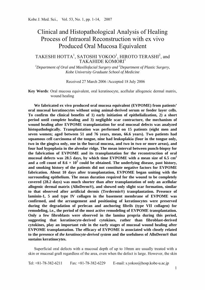
Kobe J. Med. Sci., Vol. 53, No. 1, pp. 1-14, 2007
Tel: +81-78-382-6211 Fax: +81-78-382-6229 E-mail: [email protected]
1
Clinical and Histopathological Analysis of Healing Process of Intraoral Reconstruction with ex vivo
Produced Oral Mucosa Equivalent TAKESHI HOTTA1, SATOSHI YOKOO1, HIROTO TERASHI2, and
TAKAHIDE KOMORI1 1Department of Oral and Maxillofacial Surgery and 2Department of Plastic Surgery,
Kobe University Graduate School of Medicine
Received 27 March 2006 /Accepted 18 July 2006
Key Words: Oral mucosa equivalent, oral keratinocyte, acellular allogeneic dermal matrix, wound healing
We fabricated ex vivo produced oral mucosa equivalent (EVPOME) from patients’
oral mucosal keratinocytes without using animal-derived serum or feeder layer cells. To confirm the clinical benefits of 1) early initiation of epithelialization, 2) a short period until complete healing and 3) negligible scar contracture, the mechanism of wound healing after EVPOME transplantation for oral mucosal defects was analyzed histopathologically. Transplantation was performed on 15 patients (eight men and seven women; aged between 51 and 76 years, mean, 66.6 years). Two patients had squamous cell carcinoma of the tongue, nine had leukoplakia (four in the tongue only, two in the gingiva only, one in the buccal mucosa, and two in two or more areas), and four had hypoplasia in the alveolar ridge. The mean interval between punch-biopsy for the fabrication of EVPOME and its transplantation for the reconstruction of oral mucosal defects was 28.5 days, by which time EVPOME with a mean size of 6.5 cm2 and a cell count of 8.6 × 10⁵ could be obtained. The underlying disease, past history, and smoking history of the patients did not constitute negative factors for EVPOME fabrication. About 10 days after transplantation, EVPOME began uniting with the surrounding epithelium. The mean duration required for the wound to be completely covered (28.2 days) was much shorter than after transplantation of only an acellular allogenic dermal matrix (AlloDerm®), and showed only slight scar formation, similar to that observed after artificial dermis (Terdermis®) transplantation. Presence of laminin-1, 5 and type IV collagen in the basement membrane of EVPOME was confirmed, and the arrangement and positioning of keratinocytes were preserved during the degradation of perlecan and anchoring fibrils (type VII collagen) for remodeling, i.e., the period of the most active remodeling of EVPOME transplantation. Only a few fibroblasts were observed in the lamina propria during this period, suggesting that keratinocyte-derived cytokines, rather than fibroblast-derived cytokines, play an important role in the early stages of mucosal wound healing after EVPOME transplantation. The efficacy of EVPOME is associated with closely related to the presence of the keratinocyte-derived system and the usefulness of AlloDerm® that sustains keratinocytes.
Superficial oral defects with a mucosal depth of up to 10mm are usually treated with a
skin or mucosal graft regardless of the area, even when the defect is large. However, the skin
T. HOTTA et al.
2
contains epidermal appendages, and its epidermal keratinization pattern also differs from that of oral mucosa, while the mucosa is quantitatively limited as a donor source. Moreover, both skin and mucosal grafting leads to tissue defects at the donor site. Advances in tissue engineering technology have made possible the development of artificial skin and mucosa to overcome these problems, the most crucial of which have been keratinocyte proliferation and stratification. While the methods attributed to Green et al. (9), Reinwald et al. (26), and Boyce et al. (5, 6) have been mainly used for such development, the principal drawback of their methods is the use of an irradiated xenogenic 3T3 mouse fibroblast feeder layer and of fetal calf serum (FCS). At present, the clinical application of these methods has not been approved by the ethical committees of almost all hospitals in Japan (7,27). Artificial dermis without keratinocytes has also been developed and widely applied in clinical practice. Its transplantation does not sacrifice donor site tissue, but its main healing mechanism is scar healing after the formation of granulation, primarily by fibroblasts, resulting in marked scar contracture at the recipient site (23). Moreover, another problem common to all types of artificial skin, mucosa and dermis is that they are difficult to manipulate.
We succeeded in fabricating ex vivo produced oral mucosa equivalent (EVPOME) from the oral mucosal epithelium of patients themselves, and previously reported the results of experimental studies (11, 12). For the study reported here, we upgraded our original method to a much cleaner keratinocyte culturing method by using an animal-product-free culture medium.
The aims of this study were: 1) a comparison of keratinocyte proliferation with the modified method using an animal-product-free culture medium and with our original method; 2) the clinical application and evaluation of EVPOME containing keratinocytes used for oral mucosal reconstruction; 3) a histopathological and electron-microscopic evaluation of the wound healing process after transplantation of EVPOME using the keratinocyte-derived system.
SUBJECTS AND METHODS
Fabrication of Ex Vivo Produced Oral Mucosa Equivalent (EVPOME) EVPOME is a composite human oral mucosa equivalent, which includes a dermal
component consisting of an acellular allogenic dermal matrix, AlloDerm® (LifeCell, Branchburg, NJ). AlloDerm® is a human dermis that has been decellularized to remove the risk of rejection and inflammation. It is made from pathogen-screened cadaveric skin and freeze-dried through a patented process that does not damage the crucial elements of the tissue structure, including the distribution and architecture of the collagen bundles. AlloDerm® is seeded with autogenous human oral keratinocytes to form an overlying stratified parakeratinized epithelial layer. What follows is a brief description of our protocol for the manufacture of EVPOME. First, at the outpatient clinic, under local anesthesia, a 5×5 mm punch-biopsy of parakeratinized oral mucosa is taken from the gingiva, with ample time before surgery to allow for fabrication of EVPOME of the dimensions required for the planned surgical reconstruction. Oral keratinocytes are dissociated from the biopsy specimen and expanded in an animal-product-free culture medium, EpiLife® (M-EPI-500-CA; Cascade Biologics Inc., Portland, OR) containing calcium at a concentration of 0.06 mM and Human Keratinocyte Growth Supplement-V2 (S-014-5; Cascade Biologics, Portland, OR). Once a sufficient number of oral keratinocytes has been harvested and seeded onto AlloDerm® to a thickness of 0.019 inches, the resultant composite of oral keratinocytes and AlloDerm® is cultured and submerged in EpiLife® for 4 days to form a continuous epithelial monolayer. The concentration of calcium in the culture medium is then raised to
ANALYSIS OF HEALING PROCESS AFTER EVPOME TRANSPLANTATION
3
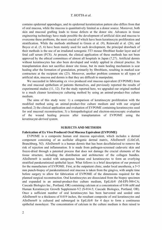
1.2 mM to enhance keratinocyte differentiation. The equivalents are raised to an air-liquid interface to induce stratification of the epithelial monolayer and cultured for an additional 7 days for generating a parakeratinized layer in preparation for the intraoral transplantation of EVPOME (Fig.1).
Fig.1: Production of Ex Vivo-Produced Oral Mucosa Equivalent (EVPOME) (A) Punch-biopsy of the oral mucosa (B) Keratinocyte in vitro: After culturing for 14 days (confluent) (C) Complete EVPOME (immediately before transplantation): Stratification of cultured keratinocytes on the acellular dermal matrix (AlloDerm®). (D) HE staining of EVPOME after culturing for 11 days (×100): Stratification of the culturing epithelium was observed, and cells in the superficial layer showed enhanced keratinization and were eosinophilic [(a): cultured epithelium, (b): AlloDerm®]
Transplantation of EVPOME into human oral mucosal defects EVPOME transplantation was for the treatment of superficial oral mucosal defects at the
Department of Oral and Maxillofacial Surgery, Kobe University Hospital with the approval of the ethics committee of Kobe University. Informed consent for the use of EVPOME and for enrolment in this study was obtained from all patients.
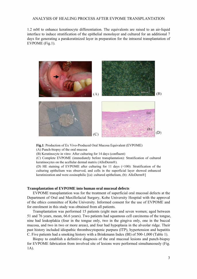
Transplantation was performed 15 patients (eight men and seven women; aged between 51 and 76 years, mean, 66.6 years). Two patients had squamous cell carcinoma of the tongue, nine had leukoplakia (four in the tongue only, two in the gingiva only, one in the buccal mucosa, and two in two or more areas), and four had hypoplasia in the alveolar ridge. Their past history included idiopathic thrombocytopenic purpura (ITP), hypertension and hepatitis C. Five patients had a smoking history with a Brinkmann Index (BI) of 500-1,000 (Table 1).
Biopsy to establish a definitive diagnosis of the oral mucosal lesions and punch-biopsy for EVPOME fabrication from involved site of lesions were performed simultaneously (Fig. 1A).
T. HOTTA et al.
4
For the surgical resection, a safety margin of 10 mm was used for cancer lesions and of 5 mm for leukoplakia. EVPOME fabricated before resection was sutured to the surgical wound (in muscle, periosteum or connective tissue) with 4-0 nylon, and tie-over fixation was accomplished with sterile cotton containing antibiotics (Fig.2). To maintain oral hygiene after transplantation, nasogastric feeding was administered for 7 days until the tie-over bolster was removed, and sutures were removed at the appropriate time after 7 or more days. Oral feeding was started with a soft diet and gradually changed to a normal diet. The patients were discharged after a mean period of 2 weeks and followed up in the outpatient clinic every week for 1 month after discharge, every other week for the subsequent 2 months and at 1-month intervals thereafter for 1 year. When necessary, a biopsy was carried out only once in each patient, and the specimens were used for this study.
The period until wound healing was defined as the number of days required for complete coverage of the surgical wound with epithelium after EVPOME transplantation.
Fig.2: Transplantation of EVPOME into human oral mucosal defect (A) Excision of oral leukoplakia with CO2 laser (B) Two circular pieces of EVPOME are applied onto the open wound and sutured into place. (C) Gauze bolster is placed on the EVPOME.
Histological and immunohistological studies
Biopsy samples were snap-frozen in liquid nitrogen or fixed with 10% formalin and embedded in paraffin for histologic examination. Specimens were cut into 5-μm sections and stained with haematoxylin-eosin (HE), Elastica van Gieson (EVG) and stained immunohistochemically with an avidin-biotin-peroxidase complex (ABC). The following specific antibodies were used: bFGF(C-19) (sc-79; Santa Cruz Biotechnology, Santa Cruz, CA), Type VII collagen (234192; Calbiochem, San Diego, CA), Laminin-1 A/B (MAb1904; Chemicon International Inc., Temecula, CA); Laminin γ2 (MAb19565; Chemicon International Inc., Temecula, CA), and Type IV collagen (52340-01; SBA, St. Louis, MO).
ANALYSIS OF HEALING PROCESS AFTER EVPOME TRANSPLANTATION
5
Electron-microscopic evaluation Specimens (AlloDerm®) for electron microscopy were fixed in 4% glutaraldehyde,
postfixed in 2% OSO4, and stained en bloc with 2% uranyl acetate. Ultra-thin sections were counterstained with uranyl acetate and lead citrate, and examined with a Hitachi H-300 electron microscope (Hitachi High-Tech Science Systems, Tokyo, Japan).
RESULTS Fabrication of EVPOME with animal-product-free culture medium and influence of patients’ background factors
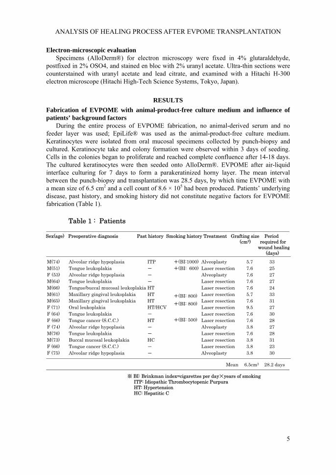
During the entire process of EVPOME fabrication, no animal-derived serum and no feeder layer was used; EpiLife® was used as the animal-product-free culture medium. Keratinocytes were isolated from oral mucosal specimens collected by punch-biopsy and cultured. Keratinocyte take and colony formation were observed within 3 days of seeding. Cells in the colonies began to proliferate and reached complete confluence after 14-18 days. The cultured keratinocytes were then seeded onto AlloDerm®. EVPOME after air-liquid interface culturing for 7 days to form a parakeratinized horny layer. The mean interval between the punch-biopsy and transplantation was 28.5 days, by which time EVPOME with a mean size of 6.5 cm2 and a cell count of 8.6 × 10⁵ had been produced. Patients’ underlying disease, past history, and smoking history did not constitute negative factors for EVPOME fabrication (Table 1).
Sex(age) Preoperative diagnosis Past history Smoking history Treatment Grafting size Period(cm2) required for
wound healing(days)
M(74)M(51)F (53)M(64)M(66)M(61)M(65)F (71)F (64)F (66)F (74)M(76)M(73)F (66)F (75)
Alveolar ridge hypoplasiaTongue leukoplakiaAlveolar ridge hypoplasiaTongue leukoplakiaTongue/buccal mucosal leukoplakiaMaxillary gingival leukoplakiaMaxillary gingival leukoplakiaOral leukoplakiaTongue leukoplakiaTongue cancer (S.C.C.)Alveolar ridge hypoplasiaTongue leukoplakiaBuccal mucosal leukoplakiaTongue cancer (S.C.C.)Alveolar ridge hypoplasia
ITP-
-
-
HTHTHTHT/HCV-
HT-
-
HC-
-
+(BI:1000)+(BI: 600)
+(BI: 800)+(BI: 800)
+(BI: 500)
AlveoplastyLaser resectionAlveoplastyLaser resectionLaser resectionLaser resectionLaser resectionLaser resectionLaser resectionLaser resectionAlveoplastyLaser resectionLaser resectionLaser resectionAlveoplasty
※ BI: Brinkman index=cigarettes per day×years of smokingITP: Idiopathic Thrombocytopenic PurpuraHT: HypertensionHC: Hepatitic C
5.77.67.67.67.65.77.69.57.67.63.87.63.83.83.8
Table 1 : Patients
332527272433312730282728312330
28.2 daysMean 6.5cm2
T. HOTTA et al.
6
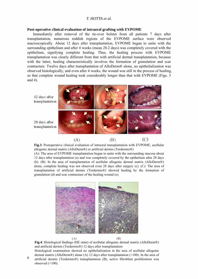
Post-operative clinical evaluation of intraoral grafting with EVPOME Immediately after removal of the tie-over bolster from all patients 7 days after
transplantation, numerous reddish regions of the EVPOME surface were observed macroscopically. About 12 days after transplantation, EVPOME began to unite with the surrounding epithelium and after 4 weeks (mean 28.2 days) was completely covered with the epithelium, signifying complete healing. Thus, the healing process with EVPOME transplantation was clearly different from that with artificial dermal transplantation, because with the latter, healing characteristically involves the formation of granulation and scar contracture. Twelve days after transplantation of AlloDerm® alone, no epithelialization was observed histologically, and even after 4 weeks, the wound was still in the process of healing, so that complete wound healing took considerably longer than that with EVPOME (Figs. 3 and 4).
Fig.3: Postoperative clinical evaluation of intraoral transplantation with EVPOME, acellular allogenic dermal matrix (AlloDerm®) or artificial dermis (Terdermis®) (A): The area of EVPOME transplantation began to unite with the surrounding mucosa about 12 days after transplantation (a) and was completely covered by the epithelium after 28 days (b). (B): In the area of transplantation of acellular allogenic dermal matrix (AlloDerm®) alone, complete healing was not observed even 28 days after surgery (c). (C): The area of transplantation of artificial dermis (Terdermis®) showed healing by the formation of granulation (d) and scar contracture of the healing wound (e).
Fig.4: Histological findings (HE stain) of acellular allogenic dermal matrix (AlloDerm®) and artificial dermis (Terdermis®) 12 days after transplantation Histological examination showed no epithelialization in the area of acellular allogenic dermal matrix (AlloDerm®) alone (A) 12 days after transplantation (×100). In the area of artificial dermis (Terdermis®) transplantation (B), active fibroblast proliferation was observed (×100).
ANALYSIS OF HEALING PROCESS AFTER EVPOME TRANSPLANTATION
7
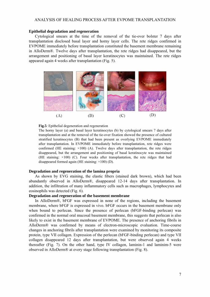
Epithelial degradation and regeneration Cytological smears at the time of the removal of the tie-over bolster 7 days after
transplantation disclosed basal layer and horny layer cells. The rete ridges confirmed in EVPOME immediately before transplantation constituted the basement membrane remaining in AlloDerm®. Twelve days after transplantation, the rete ridges had disappeared, but the arrangement and positioning of basal layer keratinocytes was maintained. The rete ridges appeared again 4 weeks after transplantation (Fig. 5).
Fig.5: Epithelial degeneration and regeneration The horny layer (a) and basal layer keratinocytes (b) by cytological smears 7 days after transplantation and at the removal of the tie-over fixation showed the presence of cultured stratified keratinocytes (B) that had been present as overlying EVPOME immediately after transplantation. In EVPOME immediately before transplantation, rete ridges were confirmed (HE staining: ×100) (A). Twelve days after transplantation, the rete ridges disappeared, but the arrangement and positioning of basal keratinocyte was maintained (HE staining: ×100) (C). Four weeks after transplantation, the rete ridges that had disappeared formed again (HE staining: ×100) (D).
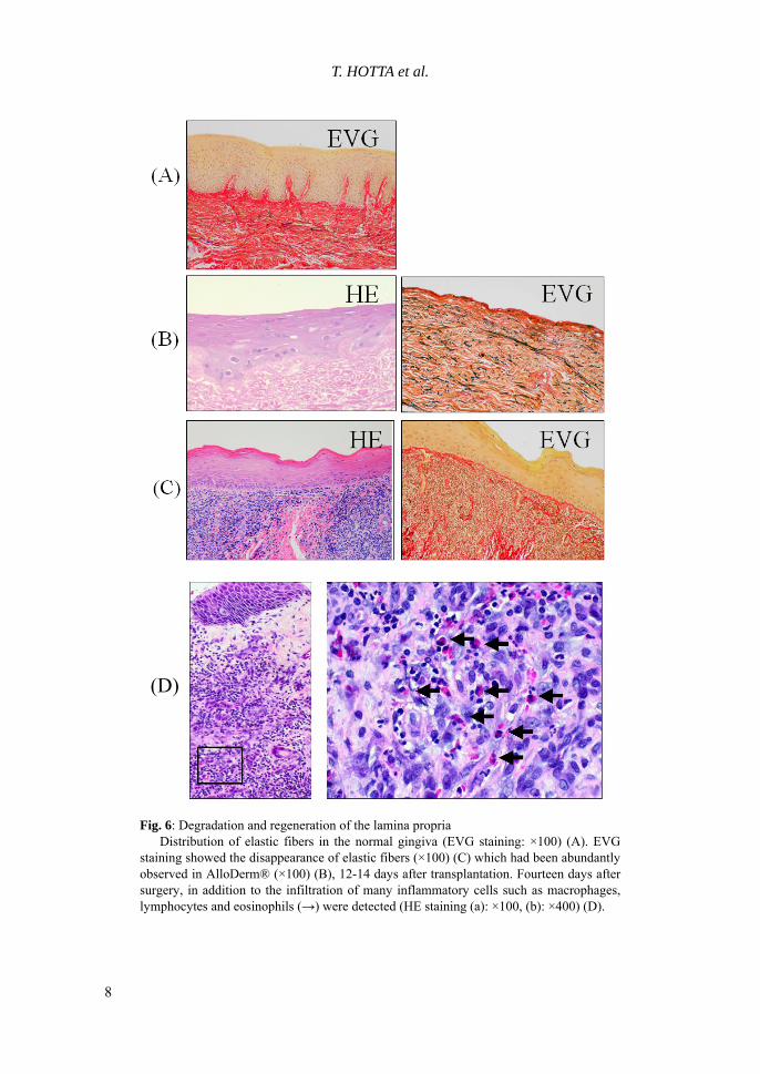
Degradation and regeneration of the lamina propria
As shown by EVG staining, the elastic fibers (stained dark brown), which had been abundantly observed in AlloDerm®, disappeared 12-14 days after transplantation. In addition, the infiltration of many inflammatory cells such as macrophages, lymphocytes and eosinophils was detected (Fig. 6). Degradation and regeneration of the basement membrane
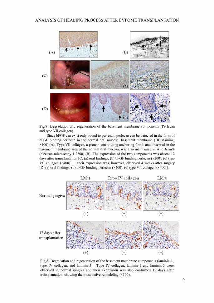
In AlloDerm®, bFGF was expressed in none of the regions, including the basement membrane, where bFGF is expressed in vivo. bFGF occurs in the basement membrane only when bound to perlecan. Since the presence of perlecan (bFGF-binding perlecan) was confirmed in the normal oral mucosal basement membrane, this suggests that perlecan is also likely to exist in the basement membrane of EVPOME. The presence of anchoring fibrils in AlloDerm® was confirmed by means of electron-microscopic evaluation. Time-course changes in anchoring fibrils after transplantation were examined by monitoring its composite protein, type VII collagen. Expression of the perlecan (bFGF-binding perlecan) and type VII collagen disappeared 12 days after transplantation, but were observed again 4 weeks thereafter (Fig. 7). On the other hand, type IV collagen, laminin-1 and laminim-5 were observed in AlloDerm® at every stage following transplantation (Fig. 8).
T. HOTTA et al.
8
Fig. 6: Degradation and regeneration of the lamina propria Distribution of elastic fibers in the normal gingiva (EVG staining: ×100) (A). EVG staining showed the disappearance of elastic fibers (×100) (C) which had been abundantly observed in AlloDerm® (×100) (B), 12-14 days after transplantation. Fourteen days after surgery, in addition to the infiltration of many inflammatory cells such as macrophages, lymphocytes and eosinophils (→) were detected (HE staining (a): ×100, (b): ×400) (D).
(D)
ANALYSIS OF HEALING PROCESS AFTER EVPOME TRANSPLANTATION
9
Fig.7: Degradation and regeneration of the basement membrane components (Perlecan and type VII collagen) Since bFGF can exist only bound to perlecan, perlecan can be detected in the form of bFGF binding perlecan in the normal oral mucosal basement membrane (HE staining: ×100) (A). Type VII collagen, a protein constituting anchoring fibrils and observed in the basement membrane area of the normal oral mucosa, was also maintained in AlloDerm® (electron-microscopy 1:2500) (B). The expression of the two components was absent 12 days after transplantation [C: (a) oral findings, (b) bFGF binding perlecan (×200), (c) type VII collagen (×400)]. Their expression was, however, observed 4 weeks after surgery [D: (a) oral findings, (b) bFGF binding perlecan (×200), (c) type VII collagen (×400)].
Fig.8: Degradation and regeneration of the basement membrane components (laminin-1, type IV collagen, and laminin-5) Type IV collagen, laminin-1 and laminin-5 were observed in normal gingiva and their expression was also confirmed 12 days after transplantation, showing the most active remodeling (×100).
T. HOTTA et al.
10
DISCUSSION In conventional keratinocyte culturing (4, 5, 6, 9, 20, 23, 26, 27, 32), the use of FCS
cannot be avoided; however, contact between keratinocytes and heterogenic proteins such as FCS during culturing involves the risk of prion disease as a transmissible infection such as Creutzfeldt-Jacob disease. Considering the risk of all types of potential infection attributable to contact with heterogenic proteins, we totally excluded animal-derived serum from the culture medium. In addition, the use of an irradiated 3T3 mouse fibroblast feeder layer has been regarded as xenotransplantation in the USA since 2002 (7), and in Japan also, the clinical application of such feeders is in principle not approved by the ethical committees of almost all institutes. By introducing EpiLife®, we succeeded in culturing keratinocytes without using any animal-derived serum or feeder layer. Our group was the first worldwide to develop and to clinically apply artificial mucosa produced with this method. This group comprises the Department of Oral and Maxillofacial Surgery, Niigata University Graduate School of Medical and Dental Science, the Department of Oral and Maxillofacial Surgery, Toyama University Faculty of Medicine, and the Department of Oral and Maxillofacial Surgery and Plastic Surgery, Kobe University Graduate School of Medicine.
During the period between the punch-biopsy at one site (about 0.25 cm2) and surgery, EVPOME with a mean size of 6.5 cm2 and a cell count of 8.6 × 10⁵ was produced. The number of days until the fabrication of EVPOME (mean, 28.5 days) and the properties of stratified keratinocytes were similar with both this new method and previously reported our conventional method (11, 12). Idiopathic thrombocytopenic purpura (ITP), hypertension, hepatitis C and a Brinkman Index of 500-1,000 in the past history of our subjects, did not prove to be negative factors for EVPOME fabrication. Of special interest was that, despite the inverse relation between differentiation and proliferation, oral mucosal keratinization (promotion of epithelial differentiation) attributed to nicotine in heavy smokers did not affect keratinocyte proliferation in the fabrication of EVPOME. This may be because the degree of keratinization was too slight to affect proliferation, or because EpiLife®, the medium we used, was more amenable to proliferation.
The mean period required for the wound to be completely covered by the epithelium (complete healing) after EVPOME transplantation was 28.2 days, which was similar to the period of 27.4 ± 1.2 days reported by Izumi et al. (13), but much shorter than after transplantation of AlloDerm® alone (46.0 ± 1.2 days). Our study also included three cases of superficial oral mucosal defect with a mean defective area of 5.0 cm2 treated with transplantation of AlloDerm® alone. The time required for complete healing in these cases exceeded 40 days, which is consistent with the time reported by Izumi et al. (13). Moreover, unlike the result of healing by the formation of granulation after artificial dermis transplantation, scar formation after EVPOME transplantation was negligible. The mean EVPOME transplantation area in the 15 patients in this study was about 6.5 cm2, and complete healing was attained without scar contracture about 1 month after surgery.
Clinical transplantation of EVPOME, compared with that of AlloDerm® alone or of artificial dermis, resulted in 1) earlier initiation of epithelialization, 2) a shorter period until complete healing, and 3) negligible scar contracture.
To confirm these clinical benefits, the histological mechanism of mucosal wound healing after EVPOME transplantation was analyzed.
First, changes in the lamina propria after transplantation, mainly remodeling of AlloDerm®, demonstrated that most of the numerous elastic fibers in AlloDerm® had disappeared 12 days after transplantation. In addition, the infiltration of macrophages, lymphocytes and eosinophils was detected. These findings suggest that the structure of the
ANALYSIS OF HEALING PROCESS AFTER EVPOME TRANSPLANTATION
11
lamina propria of EVPOME, that is, of AlloDerm®, was not maintained (unlike in the case of skin or mucous “grafts”), but that remodeling occurred as a result of degradation and regeneration.
Next, changes in the epithelial layer after transplantation of EVPOME were observed. Cytological smears at the time of the removal of the tie-over bolster 7 days after transplantation showed basal and horny layer cells, suggesting the presence of overlying stratified cultured keratinocytes in AlloDerm®. Immediately before transplantation of EVPOME, it was confirmed that rete ridges had remained in AlloDerm®. While rete ridges had become indistinct 12 days after transplantation, the arrangement and positioning of keratinocytes had been preserved. Four weeks after transplantation, the rete ridges had reformed, and the structure of the epithelium, similar to that in the normal oral mucosa, had been restored. Basement membrane regeneration including rete ridge formation is considered a parameter for the completion of wound healing (3, 21). During the wound-healing process after EVPOME transplantation, we took special note of the fact that the arrangement and positioning of basal layer cells had been maintained, even the rete ridges had disappeared. This prompted us to examine the mechanism of the early stage of healing as a clinically potentially useful characteristic of EVPOME.
Laminin-1 and type IV collagen form a lattice structure known as lamina densa in the epithelial basement membrane, which constitutes the main portion of the basement membrane (1, 33). Laminin-5 is involved, through α3β4 and α6β4 integrins, in the adhesion between the lamina densa and keratinocytes (2, 14, 19, 29). Perlecan is a proteoglycan that cross-links type IV collagen to laminin-1 (8, 18, 22), while anchoring fibrils (type VII collagen) are an important component connecting the lamina densa to the lamina propria (25). In our study, the presence of perlecan and anchoring fibrils (type VII collagen) was confirmed in the normal mucosa and AlloDerm® but was not expressed 12 days after transplantation, suggesting degradation in the remodeling; however, they appeared again 4 weeks after surgery. Yi et al. (33) have reported that laminin-1 induces keratinocyte infiltration into the dermal area, and that the ratio of laminin-1 to type IV collagen is important for the maintenance of the positioning of keratinocytes on the basement membrane. Of special interest is that the presence of laminin-1 and type IV collagen was confirmed, and the arrangement and positioning of keratinocytes was maintained during the degradation of perlecan and anchoring fibrils (type VII collagen) for remodeling, i.e., the period of the most active remodeling of EVPOME. Laminin-5 expression also increased, which may account for preservation of the positioning of keratinocyte (data not shown). This phenomenon was similar to basement membrane regeneration, mainly by laminin-5 in migrating keratinocytes, during the wound healing process (2, 14, 15, 28, 29).
In the early stage of wound healing after EVPOME transplantation, only a few fibroblasts in the lamina propria were observed, probably because the dense structure of EVPOME prevents fibroblast migration and infiltration from the surrounding tissue. Since fibroblasts secrete various cytokines for wound healing (24), it has been pointed out that, for improved graft take, fibroblast incorporation in one form or another into artificial skin and mucosa is a necessity. Nonetheless, in comparison with artificial dermis (Terdermis® Terumo, Tokyo, Japan), the healing process is achieved mainly by means of fibroblast-derived cytokines (24), EVPOME shows only slight scar formation, while the time needed until complete healing is similar. These findings suggest that keratinocyte-derived cytokines (13, 15) rather than fibroblast-derived cytokines play an important role in mucosal wound healing. The keratinocyte-derived system may also play an important role in the
T. HOTTA et al.
12
expression of a series of basement membrane components, and is thought to be an important factor for clinical efficacy.
In the keratinocyte-derived system, EVPOME can be regarded as biological wound dressing material, rather than a graft material. Mucosal keratinocytes show a lower grade of terminal differentiation and maintain their biological activity longer than do epidermal keratinocytes (10). As a result, the mucosal equivalent can be produced more quickly than the skin equivalent, thus making its emergency use for treatment of extensive burns possible. Furthermore, Ueda et al. (31) have reported that oral keratinocyte sheets grafted for skin repair become keratinized within 4 weeks. This phenomenon has been termed epitheliomesenchymal interaction indicating that epithelial specificity depends on the influence of the underlying mesenchymal tissue. We therefore suggest that the tissue-engineered human oral mucosa equivalent EVPOME could become a therapeutic option not only for oral mucosal defects but also for large skin defects such as extensive burns.
In conclusion, the efficacy of EVPOME is associated with the presence of the keratinocyte-derived system and the utility of AlloDerm® for the preservation of keratinocytes.
Although AlloDerm® has been commercially available in the USA, it is not approved by the Ministry of Health, Labour and Welfare of Japan because its safety has not been proved in Japan. The results of this study are expected to prove useful for the development of a new artificial dermis to replace AlloDerm® as a keratinocyte scaffold.
ACKNOWLEDGEMENTS
This study was supported by a Grant for the Development of Highly Advanced Medical Technology (B) from the Ministry of Education, Culture, Sports, Science and Technology of Japan for three consecutive years, from 2002 to 2004. We wish to thank Professors Chikara Saito and Ritsuo Takagi, Associate Professor Ichiro Suzuki, and Drs. Akihiko Iida and Michiko Yoshizawa (Department of Oral and Maxillofacial Surgery, Niigata University Graduate School of Medical and Dental Science) for their extensive and helpful advice.
REFERENCES
1. Alberts B, Johnson A, Lewis J, Raff M, Roberts K, Walter P, 2002, Molecular Biology of The Cell 4th ed.chapter19: 1106-1108.
2. Amano S, Akutsu N, Ogura Y, Nishiyama T, 2004, Increase of laminin 5 synthesis in human keratinocytes by acute wound fluid, inflammatory cytokines and growth factors, and lysophospholipids. Br J Dermatol. 151: 961-970.
3. Aoki S, Toda S, Anhdo T, Sugihara H, 2004, Bone marrow stromal cells, preadipocytes, and dermal fibroblasts promote epidermal regeneration in their distinctive fashions. Mol Biol Cell. 15: 4647-4657.
4. Bodner L, Grossman N, 2003, Autologous cultured mucosal graft to cover large intraoral mucosal defects: a clinical study. J Oral Maxillofac Surg 61: 169-173.
5. Boyce ST, Ham RG, 1983, Calcium regulated differentiation of normal epidermal keratinocytes in chemically defined clonal culture and serum-free serial culture. J Invest Dermatol 81: 33s-40s.
6. Boyce ST, Ham RG, 1985, Cultivation, frozen storage, and clonal growth of normal human epidermal keratinocytes in serum-free media. J Tissue Culture Methods 9: 83-93.
ANALYSIS OF HEALING PROCESS AFTER EVPOME TRANSPLANTATION
13
7. Crawford LM Jr, 2002, From the food and drug administration. JAMA 288: 688. 8. Evans MJ, Fanucchi MV, Van Winkle LS, Baker GL, Murphy AE, Nishio SJ,
Sannes PL, Plopper CG, 2002, Fibroblast growth factor-2 during postnatal development of the tracheal basement membrane zone. Am J Physiol Lung Cell Mol Physiol. 283: L1263-L1270.
9. Green H, Kehinde O, Thomas J, 1979, Growth of cultured human epidermal cells into multiple epithelia suitable for grafting. Proc Natl Acad Sci USA 76: 5665-5668.
10. Hata K, Kagami H, Ueda M, Torii S, Matsuyama M, 1995, The characteristics of cultured mucosal cell sheet as a material for grafting; comparison with cultured epidermal cell sheet. Ann Plast Surg 34: 530-538.
11. Izumi K, Takacs G, Terashi H, Feinberg SE, 1999, Ex vivo development of a composite human oral mucosal equivalent. J Oral Maxillofac Surg 57: 571-577.
12. Izumi K, Terashi H, Marcelo CL, Feinberg SE, 2000, Development and characterization of a tissue-engineered human oral mucosal equivalent produced in a serum-free culture system. J Dent Res 79: 798-805.
13. Izumi K, Feinberg SE, Iida A, Yoshizawa M, 2003, Intraoral grafting of an ex vivo produced oral mucosa equivalent: a preliminary report. Int .J.Oral Maxillofac. Surg. 32: 188-197.
14. Kainulainen T, Hakkinen L, Hamidi S, Larjava K, Kallioinen M, Peltonen J, Salo T, Larjava H, Oikarinen A, 1998, Laminin-5 expression is independent of the injury and the microenvironment during reepithelialization of wounds. J Histochem Cytochem. 46: 353-360.
15. Kondo S, Kooshesh F, Sauder DN, 1997, Penetration of keratinocyte-derived cytokines into basement membrane. J Cell Physiol. 171:190-195.
16. Marcelo CL, Kim YG, Kane JL, Voorhees JJ, 1978, Stratification, specialization and proliferation of primary keratinocyte cultures. J Cell Biol 79: 356-370.
17. Marcelo CL, Duell EA, Rhodes LM, Dunham WR, 1992, In vitro model of essential fatty acid deficiency. J Invest Dermatol 99: 703-708.
18. Mongiat M, Otto J, Oldershaw R, Ferrer F, Sato JD, Iozzo RV, 2001, Fibroblast growth factor-binding protein is a novel partner for perlecan protein core. J Biol Chem. 276: 10263-10271.
19. Nishiyama T, Amano S, Tsunenaga M, Kadoya K, Takeda A, Adachi E, Burgeson RE, 2000, The importance of laminin 5 in the dermal-epidermal basement membrane. J Dermatol Sci. 24 Suppl 1: S51-S59.
20. Peehl DM, Ham RG, 1980, Growth and differentiation of human keratinocytes without a feeder layer or conditioned medium .In Vitro. 16: 516-525.
21. Pellegrini G, Ranno R, Stracuzzi G, Bondanza S, Guerra L, Zambruno G, Micali G, De Luca M, 1999, The control of epidermal stem cells (holoclones) in the treatment of massive full-thickness burns with autologous keratinocytes cultured on fibrin. Transplantation. 68: 868-879.
22. Penc SF, Pomahac B, Winkler T, Dorschner RA, Eriksson E, Herndon M, Gallo RL, 1998, Dermatan sulfate released after injury is a potent promoter of fibroblast growth factor-2 function. J Biol Chem. 273: 28116-28121.
23. Phillips TJ, 1998, New skin for old. Arch Dermal 134: 344-349. 24. Ralston DR, Layton C, Dalley AJ, Boyce SG, Freedlander E, MacNeil S, 1997,
Keratinocytes contract human dermal extracellular matrix and reduce soluble fibronectin production by fibroblasts in a skin composite model. Br J Plast Surg. 50: 408-415.
T. HOTTA et al.
14
25. Regauer S, Seiler GR, Barrandon Y, Easley KW, Compton CC, 1990, Epithelial origin of cutaneous anchoring fibrils. J Cell Biol. 111: 2109-2115.
26. Rheinwald JG, Green H, 1975, Serial Cultivation of strains of human epidermal keratinocytes: the formation of keratinizing colonies from single cells. Cell 6: 331-343
27. Smola H, Reinke M, Shephard P, Krieg T, Hess S, 1999, Autologous patient serum for the culture of keratinocyte transplants reduces risk of transmittable disease. Lancet 353: 641-642.
28. Staiano-Coico L, Krueger JG, Rubin JS, D'limi S, Vallat VP, Valentino L, Fahey T 3rd, Hhawes A, Kingston G, Madden MR, 1993, Human keratinocyte growth factor effects in a porcine model of epidermal wound healing. J Exp Med. 178: 865-878.
29. Takeda A, Kadoya K, Shioya N, Uchinuma E, Tsunenaga M, Amano S, Nishiyama T, Burgeson RE, 1999, Pretreatment of human keratinocyte sheets with laminin 5 improves their grafting efficiency. J Invest Dermatol. 113: 38-42.
30. Terashi H, Izumi K, Rhodes LM, Marcelo CL, 2000, Human stratified squamous epithelia differ in cellular fatty acid composition. J Dermatol Sci 24: 14-24.
31. Ueda M, Hata K, Horie K, Torii S, 1995, The potential of oral mucosal cells for cultured epithelium: a preliminary report. Ann Plast Surg 35: 498-504.
32. Ueda M, Hata K, Sumi Y, Mizuno H, Niimi A, 1998, Peri-implant soft tissue management through use of cultured mucosal epithelium. Oral Surge Oral Med Oral Pathol Oral Radiol Endod 86: 393-400.
33. Yi JY, Yoon YH, Park hHS, Kim CH, Kim CH, Kang HJ, Lee E, Kim YY, Jin YJ, Kim TH, Son YS, 2001, Reconstruction of basement membrane in skin equivalent;role of laminin-1. Arch Dermatol Res 293: 356-362.