Embed Size (px)
Citation preview

CLINICAL AND GENETIC EXAMINATION OF LIMB
DEVELOPMENTAL DEFECTS
Ph.D. Thesis
Alexandra Tészás MD
Supervisor: György Kosztolányi MD, PhD, DSc
Program leader: Béla Melegh MD, PhD, DSc
Department of Medical Genetics
Medical Faculty, University of Pécs
2010

2
ABBREVIATIONS
bp – basepair
CGH – comparative genomic hybridisation
Chr – chromosome
DNA – deoxy-ribonucleic-acid
FISH – fluorescent in situ hybridisation
HOS – Holt Oram syndrome
Kb – kilo basepair
Mb – mega basepair
NIT DHM – National Institute of Traumatology, Department of Hand and
Microsurgery
pc - percentile
PCR – polymerase chain reaction
UP DMG – University of Pécs (Medical Faculty), Department of Medical
Genetics
SRPD – short rib polydactylia
STS – short tandem segments
TBX – T-box
TF – transcription factors

3
I. INTRODUCTION
The fundamental biological question is how the body plan is laid down
during embryonic development. In humans, the specialized cells, tissues
and organs arise from one cell, the fertilized oocyte. But how this complex
development can be controlled and what are the factors that explain
changes and rare abnormalities in this well conserved process?
Limb developmental defects are well-known, conspicuous
abnormalities. Their significance cannot be negligible, although they are
known to be rare conditions. Based upon international studies, limb
abnormalities occur in about 10 / 10 000 live births. The development of
scientific methods provides more and more opportunities which may help
to understand developmental processes and defects of this compound
system. Limbs are useful model systems to study the genetic control of
morphogenesis because, beside their complex anatomy and development,
their abnormalities are easy to recognize and verify.
Despite the latest techniques, genetic background of limb development
and its defects still belongs to an unrevealed part of science. Characteristic
limb malformations together with associated symptoms may help to find
the diagnosis and give opportunity for genetic examinations in
chromosomal or monogenic syndromes. In isolated limb defects the number
of target genes is around ten and can be detected only in low proportion of
the cases.
The role of transcription factors, among others, is well known during
normal embryogenesis, including limb development. They regulate protein
formation and function by binding to DNA and controlling gene expression.
Mutations are generally lethal, but contribute to endocrine disorders, tumor
genesis and developmental defects. They are necessary for cell growth,
proliferation, differentiation and are required in tissue and organ
development. Consequently transcription factors are essential in
developmental processes and homeostasis. Mutations of genes encoding
transcriptional regulators have been shown to be involved in various
genetic diseases, particularly in malformations of the skeletal system,
cranium, limbs (e.g.: hand-foot-genital syndrome), as well as in congenital
syndromes presenting anomalies of these organs combined with
somatic/mental retardation (e.g.: Feingold syndrome) . Evidences support
the need of the identification of transcription factor mutation in clinical
practice. Determination of transcription factors in congenital malformations
and genetic syndromes might be of clinical significance, even in the near
future, for exact causative diagnosis and genetic counseling in affected
families. In addition, studies of transcription factors may lead to a better
understanding of human embryogenesis.

4
The determination of the background responsible for the
developmental defect is very complex. Exact clinical diagnosis, based
upon detailed examination and classification is needed; patient database,
collection of similar cases may help to find the common origin. With the
classical and latest methods of genetics extended examinations have
become possible, revealing either bigger chromosomal abnormalities or
smaller mutations affecting the level of the base-pairs.
II. OBJECTIVES
The aim of understanding the development of the limb and their
abnormalities has leaded us to begin our comprehensive study which may
help to reveal the background of these abnormalities. The development of
diagnostic and prognostic methods can help prevention and hopefully, in
the near future, the substantive treatment as well.
The following aims were set in our study:
1. Patient collection, database
The first objective was to create a comprehensive database which may
help to handle and augment our patient files and give opportunity to
compare similar cases and follow examinations.
2. Clinical examination protocol
We aimed to establish a clinical examination protocol, based upon the
contemporary literature. A standard protocol was needed to ensure that
symptoms are described and patients are sorted on common aspects. It may
also help to recognize associated symptoms and provide enough data for
phenotype – genotype correlation analysis as well.
3. Sorting, classification
A standardized classification system was required to support exact
clinical diagnosis and genetic examination plans. Handling and sorting
cases can help to select patients for genetic examinations.
4. Genetic examination protocol
Our aim was to work out a genetic examination protocol that can help
to find the most effective way to reveal the background of limb
developmental defects. To this end we planned to apply conventional,
classical cytogenetic and molecular methods as well as the latest
technologies.
5. Examination of some specific genes (transcription factors)
In our study we wanted to focus on a group of genes playing an
important role in human embryogenesis. It seemed to be worth to focus on
the transcription factor gene family from the whole genomic system for
more detailed examination, due to their role in isolated limb defects as well
as in compound syndrome.

5
6. Phenotype – genotype correlation
We aimed to define and publish phenotype – genotype correlation data
gained in our studies.
III. METHODS
1. Creation of a patient database
Our study for collecting patients with limb developmental defects, and
referring them for detailed clinical and genetic examination, was initiated in
2005. In the beginning we sorted patients from our institute (University of
Pécs, Department of Medical Genetics). Cases with limb defects were
selected from patients who had visited our department in the last 14 years
(1993-2007).
In 2006 we got in touch with the National Institute of Traumatology,
Department of Hand and Microsurgery, where patients requiring
correctional operation due to inherited or acquired limb defects are treated.
Based upon their medical records and X-ray we chose cases from the last 4
year (2003-2007).
The information was summarized in a database.
2. Clinical examination, questionnaire
A questionnaire and a clinical examination protocol for patients with
inherited limb developmental abnormalities were constructed by the
candidate. Detailed anamnesis, physical examination (including
collaboration with other professionals if needed) seemed to be essential for
exact diagnosis.
3. Sorting, classification
Before genetic examinations are performed classification of the
abnormality is needed. Clinical (morphological, topological) and
developmental aspects were also considered during analysis of individual
cases.
All limbs were examined in available cases to determine the affected
ones. Abnormal number of bones, growth aberration or formal variances
may appear in different axis of the hands and feet. It is also important to
determine what kind of tissue is involved in the developmental defect.
Furthermore we examined some other organs, especially heart, kidneys and
central nervous system to reveal previously non-described additional
symptoms.
We found several references for sorting limb developmental defects
with different aspects. We tried to create our own classification system
which was based upon clinical signs, but considered the latest knowledge
about developmental processes and could help to plan genetic tests. As a
basic we chose the classification system of Swanson, accepted worldwide
6
in hand surgery. On the basis of our experiences we modified this
classification table, while also creating novel groups that can help to
categorize all the patients we have examined, on a simple way.
4. Genetic methods
Following detailed clinical inspection, genetic examinations were
performed in selected cases. Firstly, we usually indicated chromosomal
analysis that was followed by FISH in negative cases. Beside cytogenetic
methods we used state of the art molecular techniques as well, such as
arrayCGH that is a useful method to detect submicroscopic deletions or
duplications. Candidate attended a training course in Belgium to acquire the
technique of arrayCGH next applied in her study in Denmark. In some
cases we used other molecular techniques as well. We sequenced candidate
genes based upon the latest literature. STS marker analysis was performed
before sequencing in familial cases. We could indicate whole genome
linkage analysis and homozygote mapping altogether in two cases.
IV. RESULTS
1. Patient database
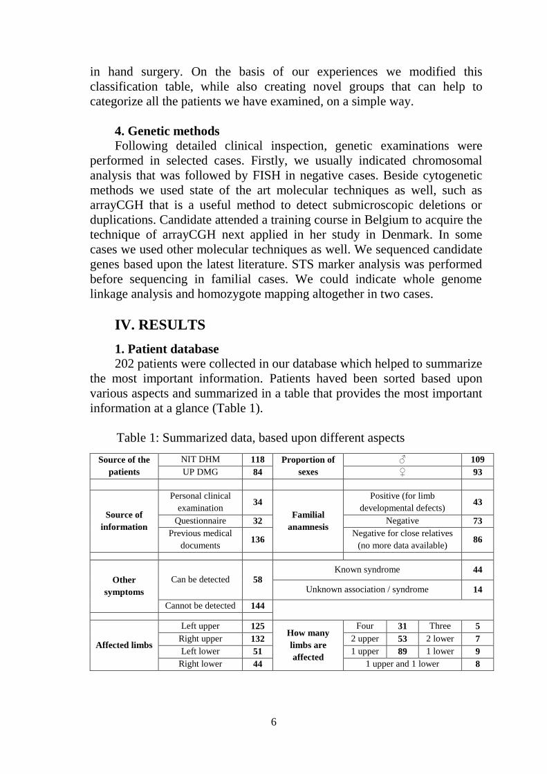
202 patients were collected in our database which helped to summarize
the most important information. Patients haved been sorted based upon
various aspects and summarized in a table that provides the most important
information at a glance (Table 1).
Table 1: Summarized data, based upon different aspects
Source of the
patients
NIT DHM 118 Proportion of
sexes
♂ 109
UP DMG 84 ♀ 93
Source of
information
Personal clinical
examination 34
Familial
anamnesis
Positive (for limb
developmental defects) 43
Questionnaire 32 Negative 73
Previous medical
documents 136
Negative for close relatives
(no more data available) 86
Other
symptoms
Can be detected 58 Known syndrome 44
Unknown association / syndrome 14
Cannot be detected 144
Affected limbs
Left upper 125 How many
limbs are
affected
Four 31 Three 5
Right upper 132 2 upper 53 2 lower 7
Left lower 51 1 upper 89 1 lower 9
Right lower 44 1 upper and 1 lower 8

7
2. Sorting and classification of the patients
Based upon the classification system of Swanson we created a table
that can be useful for patient classification in everyday practice (Table 2).
Developmental and topological sorting was separated in order to simplify
the system. Additionally, the candidate added a column that may help in
clinical terminology.
3. Genetic examination protocol
A genetic examination protocol has been established that includes the
opportunity of the latest techniques beside the classic cytogenetic and
molecular methods (Figure 1). We could perform some of these genetic test
altogether in 35 cases.
Cytogenetics negative Molecular genetics
pozitive Sequencing candidate genes(1 patient, based upon literature)
balanced unbalanced STS marker analysis
- deletion (small family, lots of patients)
Breakpoint- - duplication Whole genom linkage
analysis - inversion (big family, lots of affected members)
- FISH - translocation
Apparently balanced? ArrayCGH mutation
Small deletion, duplicatio? Small deletion, duplication? Known → Pathogen?
Breakpoint in a gene? Unknown?
Unaffected family members? Funkcional examinations
Literature data? mRNA, protein function
Figure 1: The procession of genetic examination in limb
developmental defects

8
Table 2: Classification of limb defects, based upon clinical and
developmental aspects
Pathomechanism Topology Clinical terminology
I. Congenital
construction
deformity
Transvers defect:
- shoulder, arm
- forearm
- hand
- fingers
Longitudinal defect:
- radial (preaxial)
- middle (central)
- ulnar (postaxial)
Amelia
Hemimelia
Phocomelia
Acheiria
Oligodactyly
Ectrodactyly
Brachydactyly
Synbrachydactyly
II. Abrupted
development
III. Hypoplasia
- whole limb
- whole hand
- carpi / metacarpi
- fingers
IV. Duplication
- whole limb
- humeral segment
radial / ulnar
segment
- fingers
preaxial,
central,
postaxial
- phalanges
Mirror hand
Polydactyly
Synpolydactyly
Triphalangeal thumb
V. Hyperplasia
- whole limb
- whole hand
- carpi / metacarpi
- fingers
Hemihypertrophy
Macrodactyly
VI. Disturbed
differentiation
Soft tissuesaffected:
- disseminated
- shoulder
- elbow / forearm
- wrist / hand
Bone system affected:
- shoulder
- elbow
- forearm
- wrist / hand
Arthrogryposis
Camptodactyly
„Thumb in palm”
Syndactyly
Synostosis
Symphalangia
Clinodactyly
VII. Generalized
bone developmental
defect
Generalized Achondroplasia
Marfan syndrome …

9
4. Case reports
Our work plans were built with the purpose to assess general
definitions based upon the clinical and genetic analysis of unique cases.
Some illustrative cases will serve to present the results detailed above.
4.1. Isolated limb defects
The background of isolated limb defects is still an unidentified part of
genetics. About ten genes are known to be responsible for these kinds of
anomalies. We examined 144 cases with isolated defects, but only 20-25
patients showed symptoms that can be connected with any of the ten genes.
Due to the rare incidence we could perform genetic examination in only a
few cases. Among isolated limb defects a tetramelic, postaxial, familial
case was highlighted during our study.
Isolated dominant tetramelic postaxial oligodactyly
To our knowledge isolated dominant tetramelic postaxial oligodactyly
characterized by reduction defects affecting the postaxial ray of both upper
and lower limbs was reported only once previously. Here we describe a
second family with this kind of malformation, affecting the mother and her
two children, a boy and a girl (Figure 2).
Figure 2: Photo and X-ray of the boy (3-year-old) hands
The chromosomal analysis and molecular examinations have not
revealed any causative abnormality. We indicated array which was
performed on genomic DNA from peripheral blood using a previously
described whole-genome tiling path BAC array comprising 36.000 BAC
clones. ArrayCGH revealed a small duplication affecting 7q36.3 in each
patient. This variation has not been reported as a normal polymorphism in
the Database of Genomic Variants and it has not been observed repeatedly
in >800 controls analysed on the same platform. The region includes two
genes that are known to play an important role in limb development
(LMBR1 – „limb region 1 homolog”, HLXB9 – „homeobox HB9”) and
may be responsible for the limb defect inherited in the family.
10
4.2. Compound syndromes
Dysmorphic features, connected symptoms may help to find exact
diagnosis in compound syndromes and give chance for genetic
examinations.
We would like to feature some of our cases – with compound
syndrome including limb developmental defects – where genetic
examinations were available.
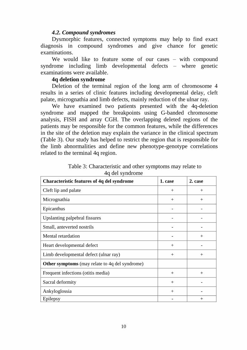
4q deletion syndrome
Deletion of the terminal region of the long arm of chromosome 4
results in a series of clinic features including developmental delay, cleft
palate, micrognathia and limb defects, mainly reduction of the ulnar ray.
We have examined two patients presented with the 4q-deletion
syndrome and mapped the breakpoints using G-banded chromosome
analysis, FISH and array CGH. The overlapping deleted regions of the
patients may be responsible for the common features, while the differences
in the site of the deletion may explain the variance in the clinical spectrum
(Table 3). Our study has helped to restrict the region that is responsible for
the limb abnormalities and define new phenotype-genotype correlations
related to the terminal 4q region.
Table 3: Characteristic and other symptoms may relate to
4q del syndrome
Characteristic features of 4q del syndrome 1. case 2. case
Cleft lip and palate + +
Micrognathia + +
Epicanthus - -
Upslanting palpebral fissures - -
Small, anteverted nostrils - -
Mental retardation - +
Heart developmental defect + -
Limb developmental defect (ulnar ray) + +
Other symptoms (may relate to 4q del syndrome)
Frequent infections (otitis media) + +
Sacral deformity + -
Ankyloglossia + -
Epilepsy - +
11
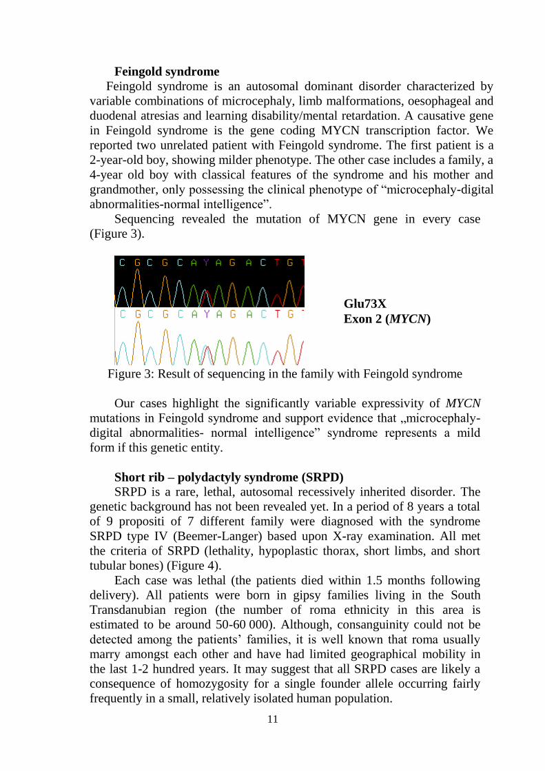
Feingold syndrome
Feingold syndrome is an autosomal dominant disorder characterized by
variable combinations of microcephaly, limb malformations, oesophageal and
duodenal atresias and learning disability/mental retardation. A causative gene
in Feingold syndrome is the gene coding MYCN transcription factor. We
reported two unrelated patient with Feingold syndrome. The first patient is a
2-year-old boy, showing milder phenotype. The other case includes a family, a
4-year old boy with classical features of the syndrome and his mother and
grandmother, only possessing the clinical phenotype of “microcephaly-digital
abnormalities-normal intelligence”.
Sequencing revealed the mutation of MYCN gene in every case
(Figure 3).
Figure 3: Result of sequencing in the family with Feingold syndrome
Our cases highlight the significantly variable expressivity of MYCN
mutations in Feingold syndrome and support evidence that „microcephaly-
digital abnormalities- normal intelligence” syndrome represents a mild
form if this genetic entity.
Short rib – polydactyly syndrome (SRPD)
SRPD is a rare, lethal, autosomal recessively inherited disorder. The
genetic background has not been revealed yet. In a period of 8 years a total
of 9 propositi of 7 different family were diagnosed with the syndrome
SRPD type IV (Beemer-Langer) based upon X-ray examination. All met
the criteria of SRPD (lethality, hypoplastic thorax, short limbs, and short
tubular bones) (Figure 4).
Each case was lethal (the patients died within 1.5 months following
delivery). All patients were born in gipsy families living in the South
Transdanubian region (the number of roma ethnicity in this area is
estimated to be around 50-60 000). Although, consanguinity could not be
detected among the patients’ families, it is well known that roma usually
marry amongst each other and have had limited geographical mobility in
the last 1-2 hundred years. It may suggest that all SRPD cases are likely a
consequence of homozygosity for a single founder allele occurring fairly
frequently in a small, relatively isolated human population.
Glu73X
Exon 2 (MYCN)

12
Figure 4: Thoracic X-ray pictures and photos of the limbs of the SRPD
patients
DNA was obtained from 4 patients (in 2 cases from the parents as
well). Homozygote mapping examination could be performed to find alleles
that are homozygous in all the affected patients, showing the same
haplotype. The examination was performed with a SNP based (16 000
SNP) Affymetrix chip (resolution of 50Kb) that revealed an allele sized
about 4Mb on chromosome 14. 40 genes known to be involved in this
region based upon Human Genome Browser. We have verified the results
with STS marker analysis (Chr 14 bp: 56222543 – 61770134) that was
completely overlapped by the “mapping” examination. Further
examinations, sequence of the target genes and functional examinations of
the encoded proteins are needed to reveal the exact background of this
lethal syndrome.
Brachydactyly E, ulnar-fibular ray defect
Brachydactyly E is characterized by the shortening of the metacarpi
and phalanges in the ulnar ray (mainly 4th
). We examined a family showing
autosomal dominant inherited brachydactyly E in some cases combined
with ulnar and fibular ray defect. The anomaly affected four generation of
the family (Figure 5). In the youngest affected member of the family, a 7-
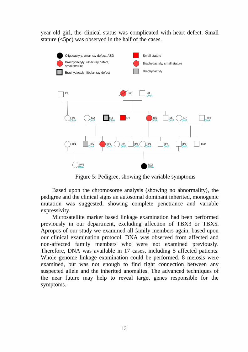
13
year-old girl, the clinical status was complicated with heart defect. Small
stature (<5pc) was observed in the half of the cases.
DNA DNA
DNA DNA
DNA
DNA DNA DNADNA
DNA DNA
DNA
DNA DNA
DNA
III/6 III/7
IV/2IV/1
III/2 III/3 III/8
II/7 II/8II/6II/5II/4II/3II/2
I/3I/2
II/1
I/1
Oligodactyly, ulnar ray defect, ASD
Brachydactyly, ulnar ray defect,
small stature
Brachydactyly, fibular ray defect
Small stature
Brachydactyly, small stature
Brachydactyly
III/4DNA
III/5DNA
III/1 III/9
Figure 5: Pedigree, showing the variable symptoms
Based upon the chromosome analysis (showing no abnormality), the
pedigree and the clinical signs an autosomal dominant inherited, monogenic
mutation was suggested, showing complete penetrance and variable
expressivity.
Microsatellite marker based linkage examination had been performed
previously in our department, excluding affection of TBX3 or TBX5.
Apropos of our study we examined all family members again, based upon
our clinical examination protocol. DNA was observed from affected and
non-affected family members who were not examined previously.
Therefore, DNA was available in 17 cases, including 5 affected patients.
Whole genome linkage examination could be performed. 8 meiosis were
examined, but was not enough to find tight connection between any
suspected allele and the inherited anomalies. The advanced techniques of
the near future may help to reveal target genes responsible for the
symptoms.
14
V. CONCLUSIONS
I summarized our observations and emphasized our scientific
conclusions related to individual cases.
1. Our study brought up new aspects and information related to limb
developmental defects and accompanied symptoms. Our achievements
were summarized in publications that may raise attention of other
professionals who can meet these kinds of abnormalities.
2. We have constructed and now propose a novel classification system that
was created to sort and classify limb developmental defects based upon
Swanson’s recommendation.
3. Our study highlights the importance of the examination of transcription
factors in limb abnormalities, especially in compound syndrome.
4. New results, scientific conclusions:
- We reported on a family with apparentely autosomal dominant
inherited, isolated tetramelic postaxial oligodactyly. We tried to attract
attention for this rare condition that was described to our knowledge only
once before. Additionally we mapped a microduplication that can be found
only in affected family members. Some genes in that region are known to
be involved in limb development. Our findings can highlight the connection
between the genes and this rare developmental abnormality.
- Analysis of two cases with 4q deletion helped us to define some new
phenotype-genotype correlations related to regions involved in the
deletions.
- Detailed clinical examination and results of sequencing in the family
of a Feingold syndrome patient supported evidence that “microcephaly –
digital abnormalities – normal intelligence” syndrome represents a mild
form of Feingold syndrome.
- Analysis and genetic examination of short rib polydactyly cases has
given opportunity to get closer in revealing the background of this rare,
lethal syndrome with unknown origin.
Finally it could be stated that study of genetic background of limb
developmental defects is still in its infancy. Despite of the development in
scientific methods and techniques, declaring exact diagnosis, unravelling
causative mutations is a complicated or – in most of the cases – impossible
task. To this end in view the candidate feels necessary – and is committed
to continue and extend this study to approve cognition of developmental
processes and their abnormalities.

15
LIST OF PUBLICATIONS
Publications related to the thesis:
1. Tészás A, Kárteszi J, Kosztolányi Gy. Új lehetőségek a fejlődési
rendellenességek hátterének feltárására: transzkripciós faktorok klinikai
genetikája. Orv Hetilap 2006; 147: 697-702.
2. Tészás A, Meijer R, Scheffer
H, Kosztolányi
Gy, vanBokhoven
H,
Kellermayer R. Expanding the clinical spectrum of MYCN related Feingold
syndrome. Am J Med Genet A 2006; 140: 2254-2256. IF: 2,063
3. Tészás A. Az array CGH hazai megtelepítését célzó alapképzés. PTE
Orvoskari Hírmondó 2006; Dec: 30.
4. Tészás A, Renner A, Melegh B, Kosztolányi Gy. Veleszületett
végtagfejlődési rendellenességek klinikai osztályozása. Orv Hetilap 2008;
149: 1167-1169.
5. Kaalund SS, Moller RS, Tészás A, Miranda M, Ullmann R, Tommerup
N, Tümer Z. Investigation of the chromosome 4 long arm deletion in two
patients with a cryptic translocation and an interstitial deletion using array
CGH. Am J Med Genet 2008; 146A: 2431-2434. IF: 2,555
6. Tészás A, Møller RS, Tommerup N, Ullmann R, Melegh B, Kosztolányi
Gy, Kjaer KW. Exclusion of TBX2/3 mutations and copy number variations
in a second family with isolated dominant tetramelic oligodactyly. Am J
Med Genet 2009; (accepted for publication).
Published abstracts related to the thesis:
1. Tészás A, Meijer R, Scheffer
H, Kosztolányi
Gy, vanBokhoven
H,
Kellermayer R. Expanding the clinical spectrum of MYCN related Feingold
syndrome. Eur J Hum Genet 2006; 14, Suppl 1, p 124. IF: 3,697
2. Tészás A, Møller R, Tommerup N, Kosztolányi Gy, Kjaer KW. The
second observation of familial congenital tetramelic oligodactyly. Eur J
Hum Genet. 2007; 15, Suppl 1, p 58. IF: 4,003
3. Kaalund SS, Moller RS, Tészás A, Miranda M, Ropers HH, Ullmann R,
Tommerup N, Tümer Z. Delineation of the deletion breakpoints in two
unrelated patients with 4q terminal deletion syndrome. Eur J Hum Genet
2007; 15, Suppl 1, p 98. IF: 4,003

16
Publications and published abstracts in other topics:
1. Kárteszi J, Hollódy K, Bene J, Morava É, Hadzsiev K, Czakó M, Melegh
B, Tészás A, Kosztolányi Gy. Mutation analysis of MECP2 and
determination of the X-inactivation pattern in Hungarian Rett syndrome
patients. Am J Med Genet A 2004; 131: 106 IF: 0,815
2. Kárteszi J, Bene J, Hollódy K, Morava É, Hadzsiev K, Czakó M, Melegh
B, Tészás A, Kosztolányi Gy. Mutation analysis of MECP2 and
determination of the X-inactivation pattern in Hungarian Rett Syndrome
patients. Eur J Hum Genet 2004; 12, p 92. IF: 2,741
3. Kellermayer R, Gyarmati J, Czakó M, Tészás A, Masszi Gy, Ertl T,
Kosztolányi Gy. Mos 46,XX,r(18).ish r(18)(18ptel-,18qtel-)/46,XX.ish
del(18)(18ptel-): An example for successive ring chromosome formation.
Am J Med Genet A 2005; 139A: 234-235. IF: 1,913
4. Tészás A, Pfund Z, Morava E, Kosztolányi Gy, Sistermans E, Wevers RA,
Kellermayer R. Presenile cataract: consider cholestanol. Arch Ophthalmol
2006; 124: 1490-1492. IF: 3,206
5. Willemsen M, Rodenburg RJ, Tészás A, van den Heuvel L, Kosztolányi
Gy, Morava E. Females with PDHA1 gene mutations: A diagnostic
challenge. Mitochondrion 2006; 6: 155-159. IF: 2,191
6. Young AL, Kellermayer R, Szigeti R, Tészás A, Azmi S, Celebi JT. CYLD mutations underlie Brooke-Spiegler, familial cylindromatosis, and
multiple familial trichoepithelioma syndromes. Clin Genet 2006; 70: 246-
249. IF: 3,140 7. Süle N, Tészás A, Kálmán E, Szigeti R, Miseta A, Kellermayer R.
Lithium suppresses epidermal SERCA2 and PMR1 levels in the rat. Pathol
Oncol Res 2006; 12: 234-236. IF: 1,241
8. Szigeti R, Chao S, Szász O, Tészás A, Kosztolányi Gy, Kellermayer R.
Premenstrual exacerbation in calcium ATPase disorders of the skin. J Eur
Acad Dermatol Venereol 2007; 21: 412-413. IF: 1,437
9. Tészás A, Møller RS, Kárteszi J, Czakó M, Kjaer KW, Kosztolányi Gy,
Tommerup N. Significance of molecular chromosome analyses in the
clinical interpretation of apparently balanced translocations. Cellular
Oncology 2007; 29: 161-162. IF: 4,170
10. Tészás A, Møller RS, Kellermayer R, Czakó M, Kjaer KW, Ullmann R,
Melegh B, Tommerup N, Kosztolányi Gy. A cryptic unbalanced
translocation resulting in del13q and dup15q. Am J Med Genet 2008;
146A: 2570-2573. IF: 2,555
11. Hadzsiev K, Tészás A, Kárteszi J, Kosztolányi Gy.
Anyagcserebetegségek és dysmorphia. Gyermekgyógyászat 2008; 59: 25-
28.
12. Czakó M, Tészás A, Kárteszi J, Hadzsiev K, Kosztolányi Gy. A
kromoszómavizsgálatok haszna a szindromatológiában.
Gyermekgyógyászat 2008; 59: 29-33.

17
Congress lectures:
1. Tészás A, Kárteszi J, Kosztolányi Gy. The clinical genetic significance of
transcription factors/ Transzkripciós faktorok klinikai genetikai jelentősége.
VI. Magyar Genetikai Kongresszus (Eger, 2005. April).
2. Tészás A. Genetests in limb developmental defects/ Génvizsgálatok
végtagfejlődési rendellenességekben. Genetikai Műhelyek Magyarországon
(5th
miniconference) (Szeged, 2006. September).
3. Tészás A. Genetests in congenital postaxial limb developmental defects/
Génvizsgálatok öröklődő postaxialis végtagfejlődési rendellenességekben.
Magyar Humángenetikai Társaság VI. Kongresszusa (Győr, 2006.
October).
4. Tészás A. The examination of the genetical background in limb
developmental abnormalities. Marie Curie Conferences and Training
Courses on arrayCGH and Molecular Cytogenetics. (Ghent, Belgium, 2006.
October).
5. Kárteszi J, Bene J, Hollódy K, Morava É, Hadzsiev K, Czakó M, Melegh
B, Tészás A, Kosztolányi Gy. The mutation analysis of the MECP2 gene
and the study of X-inactivation pattern in Rett syndrome patients/ Rett
szindrómás betegek mutáció analízise és X-inaktivációs vizsgálata. Magyar
Humángenetikusok V. Munkakonferenciája (Szeged, 2004. November).
6. Hadzsiev K, Tészás A, Kosztolányi Gy. Syndrome and its background/
Szindróma és ami mögötte áll. Anyagcserebetegségekről
gyermekorvosoknak (course) (Győr, 2007. Februar).
7. Tészás A, Oláh A, Mosdósi B, Kárteszi J, Adamovich K, Kosztolányi
Gy. The severe, lethal form of Smith-Lemli-Opitz syndrome (type II.)/ A
Smith-Lemli-Opitz szindróma súlyos, letális formája (II-es típus).
Anyagcserebetegségekről gyermekorvosoknak (course) (Győr, 2007.
Februar).
Cummulative impact factor (without citated abstracts): 25,286.
18
ACKNOWLEDGEMENTS
First of all I would like to thank my supervisor, Dr. György
Kosztolányi that he supported me continuously during my research and was
always opened to answer my questions.
I am also very grateful to Dr. Richárd Kellermayer for teaching me and
for the opportunity to work together, and to Dr. Klaus W. Kjaer, who
helped my study during the four months I spent in Denmark and afterwards
as well.
I thank to Dr. Béla Melegh, the leader of the PhD programme, for his
support and for the opportunity to spend the last years with my
experiments.
I would like to thank to Dr. Judit Kárteszi and Dr. Márta Czakó that
they taught me and relied on me, and last but not least they offered their
friendship as well.
I also want to express my gratefulness to all my colleagues in the
Department of Genetics for their professional help and for the friendly
atmosphere that surrounded me during my research.
To my family, eminently my parents and my husband, I have so much
to be thankful for: their love, their patience; they supported my plans from
the beginning, without them my achievements would not have been
possible.