Embed Size (px)
Citation preview
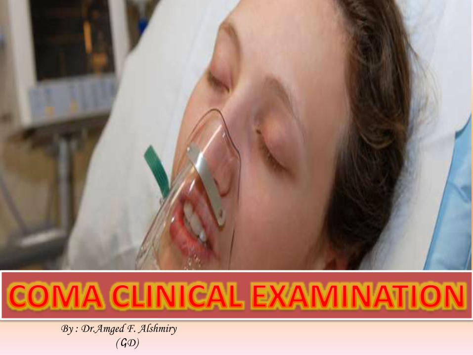
By : Dr.Amged F. Alshmiry
( GD)
Wherever possible an account of eventspreceding coma should be obtained directlyfrom witnesses as family , friends , fromambulance personnel or physicians thattreated the patient before that’s helpful forknow the cause then taking correct diagnosis
Is the PT had Medical illnesses?surgical illnesses?Previous psychological condition?Neurological conditions ?Medications and recreational drugs ?Injured (truma)
Ask them about recent complaints as ,headache , head injury(Don’t move spine until the neurologist see pt), dizziness/vertigo ,seizures, tremors , weakness .
DM , asthma, hypertension ,cancerDepression FALSE COMAEpilepsy ,neurosurgery
HISTORY
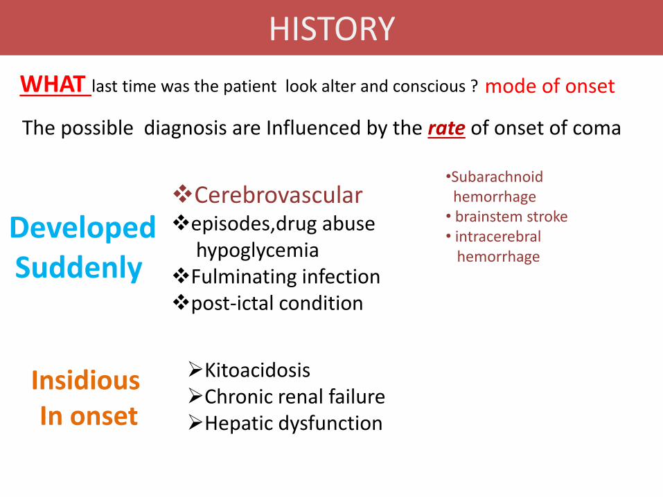
Cerebrovascularepisodes,drug abuse
hypoglycemia Fulminating infection post-ictal condition
Developed Suddenly
KitoacidosisChronic renal failure Hepatic dysfunction
Insidious In onset
mode of onset
•Subarachnoid hemorrhage• brainstem stroke• intracerebral
hemorrhage
WHAT last time was the patient look alter and conscious ?
The possible diagnosis are Influenced by the rate of onset of coma
HISTORY
Priorities
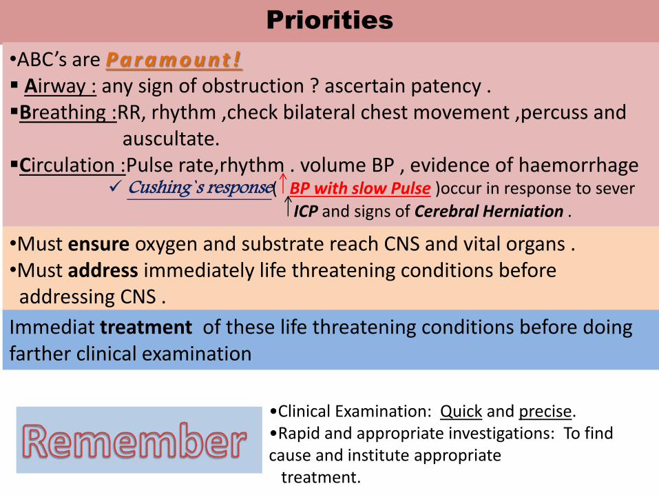
•Clinical Examination: Quick and precise.•Rapid and appropriate investigations: To find cause and institute appropriate
treatment.
•ABC’s are Pa r a m o u nt ! Airway : any sign of obstruction ? ascertain patency .Breathing :RR, rhythm ,check bilateral chest movement ,percuss and
auscultate.Circulation :Pulse rate,rhythm . volume BP , evidence of haemorrhage
Cushing`s response( BP with slow Pulse )occur in response to sever
ICP and signs of Cerebral Herniation .
•Must ensure oxygen and substrate reach CNS and vital organs .•Must address immediately life threatening conditions before addressing CNS .
Immediat treatment of these life threatening conditions before doing farther clinical examination
GENERAL PHYSICAL EXAMINATION
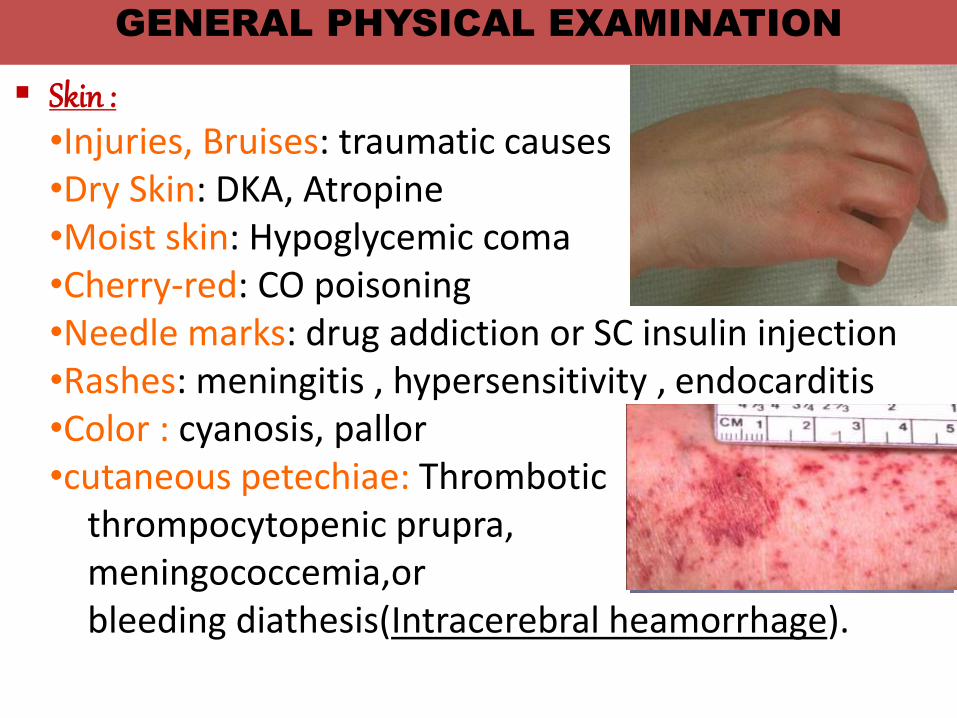
Skin : •Injuries, Bruises: traumatic causes•Dry Skin: DKA, Atropine•Moist skin: Hypoglycemic coma•Cherry-red: CO poisoning•Needle marks: drug addiction or SC insulin injection•Rashes: meningitis , hypersensitivity , endocarditis•Color : cyanosis, pallor•cutaneous petechiae: Thrombotic
thrompocytopenic prupra,meningococcemia,orbleeding diathesis(Intracerebral heamorrhage).
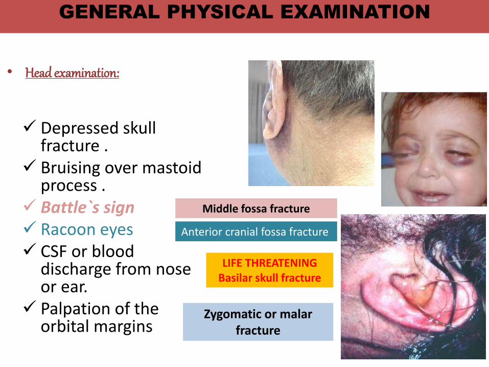
• Head examination:
Depressed skull fracture .
Bruising over mastoid process .
Battle`s sign Racoon eyes CSF or blood
discharge from nose or ear.
Palpation of the orbital margins
Zygomatic or malarfracture
Middle fossa fracture
Anterior cranial fossa fracture
LIFE THREATENINGBasilar skull fracture
GENERAL PHYSICAL EXAMINATION
• Temperature:
I-Hypothermia: causes coma only when the temperature is
• Hypopituitarism• Hypothyroidism• Chlorpromazine• Hypoglycemia• Peripheral circulatory failure • Alcoholic ,barbiturate , sedative, phenothiazine intoxication• Exposure to low temperature environments, cold-water immersion
Risk of hypothermia in the elderly with inadequately heated rooms , exacerbated by immobility.
all vital signs may be decreased(or absent) and all such patients should be
gradual rewarmed before the prognosis is assessed.
<31°C
GENERAL PHYSICAL EXAMINATION
GENERAL PHYSICAL EXAMINATION
• Temperature:II-Hyperthermia (febrile Coma)
• Infective: encephalitis, meningitis
• Vascular: pontine, subarachnoid hge
• Metabolic: thyrotoxic, Addisonian crisis
• Toxic: belladonna, salicylate poisoning
• Sun stroke, heat stroke
• Coma with 2ry infection: UTI, pneumonia, bed sores.• Only rarely is it attributable to a brain lesion that has
disturbed temperature-regulating centers as Lesions in the floor of the third ventricle ,Neuroleptic malignant syndrome.
•High fever(42-44°C) associated with dry skin should arouse the suspicion of heat stroke or anticholinergic drug intoxication.
Other causes•Tetanus•Malignant hyperpyrexia with anaesthetics.
GENERAL PHYSICAL EXAMINATION
• Smell : alcohol , hepatic fetor , ketosis,uraemia
• Tachypnea : systemic acidosis , pneumonia .
• Irregular respiratory pattern :brain stem disorder .
• Breathing
• Blood Pressure• High: hypertensive encephalopathy or rapid rise in ICP• Low: Addisonian crisis, alcohol, barbiturate internal
hemorrhage , MI , sepsis , massive hypothyroidism .
• Pulse
• Bradycardia: brain tumors, opiates,myxedema.
• Tachycardia: hyperthyroidism, uremia
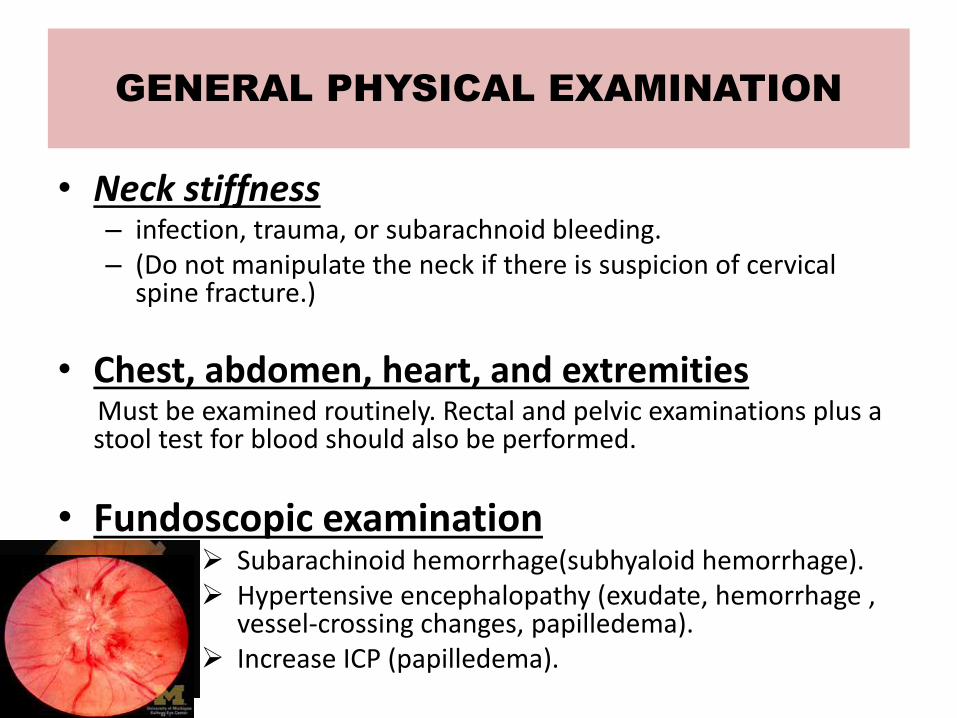
• Neck stiffness– infection, trauma, or subarachnoid bleeding. – (Do not manipulate the neck if there is suspicion of cervical
spine fracture.)
• Chest, abdomen, heart, and extremitiesMust be examined routinely. Rectal and pelvic examinations plus a stool test for blood should also be performed.
• Fundoscopic examination Subarachinoid hemorrhage(subhyaloid hemorrhage). Hypertensive encephalopathy (exudate, hemorrhage ,
vessel-crossing changes, papilledema). Increase ICP (papilledema).
GENERAL PHYSICAL EXAMINATION
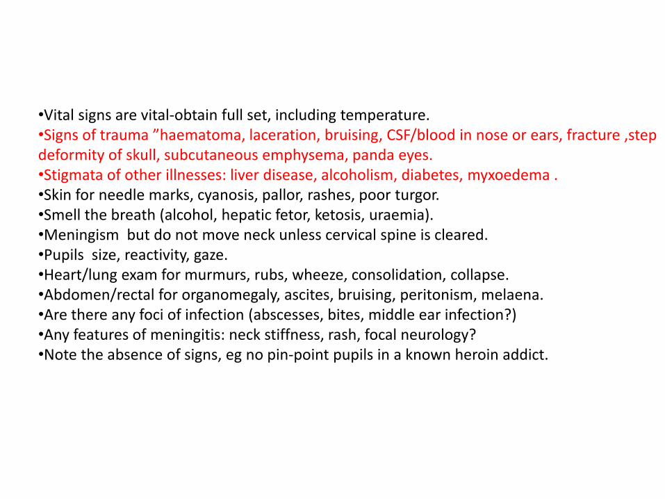
•Vital signs are vital-obtain full set, including temperature.•Signs of trauma ”haematoma, laceration, bruising, CSF/blood in nose or ears, fracture ,step deformity of skull, subcutaneous emphysema, panda eyes.•Stigmata of other illnesses: liver disease, alcoholism, diabetes, myxoedema .•Skin for needle marks, cyanosis, pallor, rashes, poor turgor.•Smell the breath (alcohol, hepatic fetor, ketosis, uraemia).•Meningism but do not move neck unless cervical spine is cleared.•Pupils size, reactivity, gaze.•Heart/lung exam for murmurs, rubs, wheeze, consolidation, collapse.•Abdomen/rectal for organomegaly, ascites, bruising, peritonism, melaena.•Are there any foci of infection (abscesses, bites, middle ear infection?)•Any features of meningitis: neck stiffness, rash, focal neurology?•Note the absence of signs, eg no pin-point pupils in a known heroin addict.
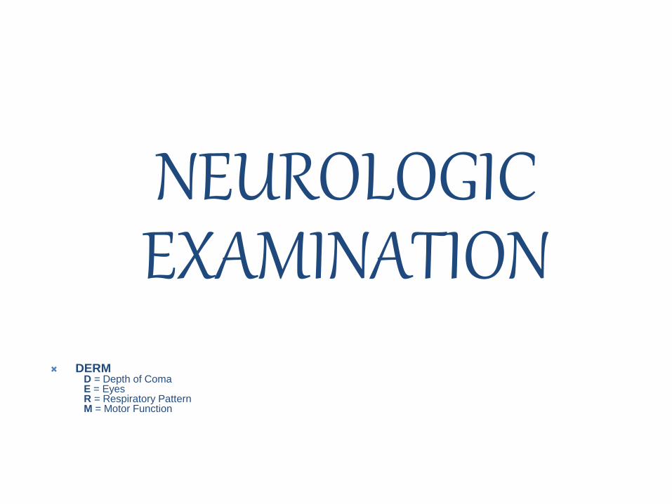
NEUROLOGICEXAMINATION
DERMD = Depth of ComaE = EyesR = Respiratory PatternM = Motor Function
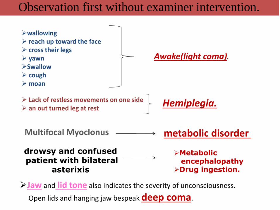
wallowing reach up toward the face cross their legs yawnSwallow coughmoan
Awake(light coma).
Lack of restless movements on one side an out turned leg at rest
Hemiplegia.
Observation first without examiner intervention.
Multifocal Myoclonus metabolic disorder
drowsy and confusedpatient with bilateral
asterixis
Metabolic encephalopathy
Drug ingestion.
Jaw and lid tone also indicates the severity of unconsciousness.
Open lids and hanging jaw bespeak deep coma.
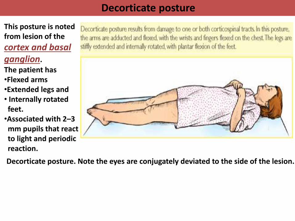
Decorticate posture
Decorticate posture. Note the eyes are conjugately deviated to the side of the lesion.
This posture is noted from lesion of the
cortex and basal ganglion.
The patient has •Flexed arms•Extended legs and• Internally rotated feet. •Associated with 2–3 mm pupils that react to light and periodic reaction.
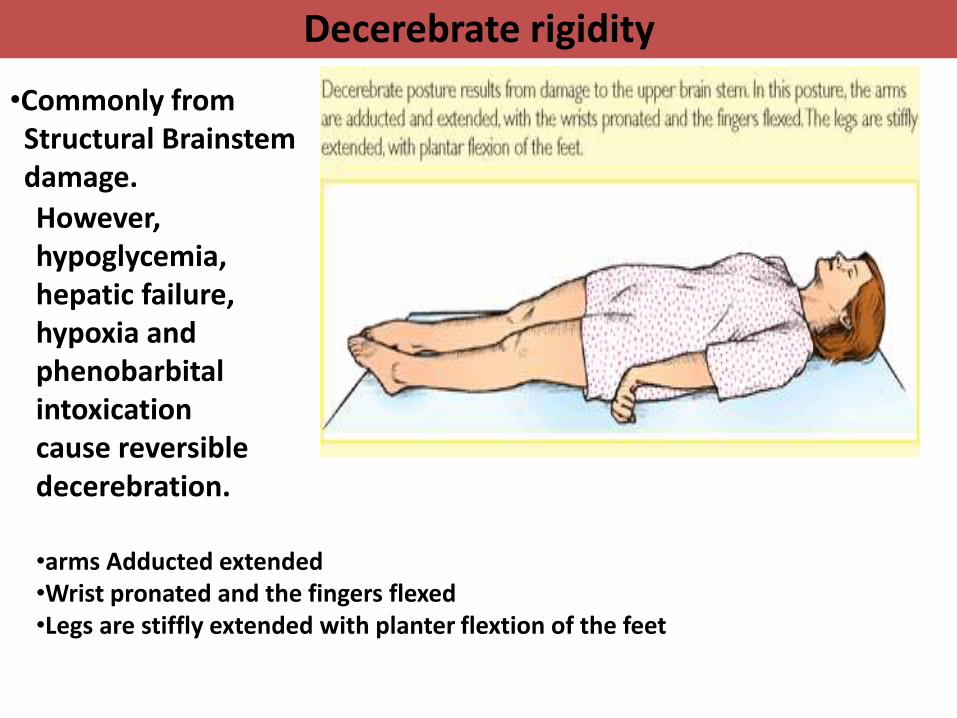
Decerebrate rigidity
•Commonly from Structural Brainstem damage.
•arms Adducted extended •Wrist pronated and the fingers flexed•Legs are stiffly extended with planter flextion of the feet
However, hypoglycemia, hepatic failure, hypoxia and phenobarbitalintoxication cause reversible decerebration.
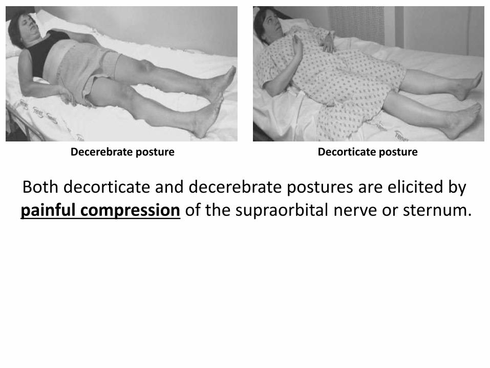
Decorticate postureDecerebrate posture
Both decorticate and decerebrate postures are elicited bypainful compression of the supraorbital nerve or sternum.
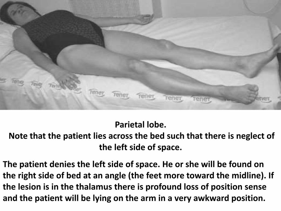
Parietal lobe.Note that the patient lies across the bed such that there is neglect of
the left side of space.
The patient denies the left side of space. He or she will be found on the right side of bed at an angle (the feet more toward the midline). If the lesion is in the thalamus there is profound loss of position sense and the patient will be lying on the arm in a very awkward position.
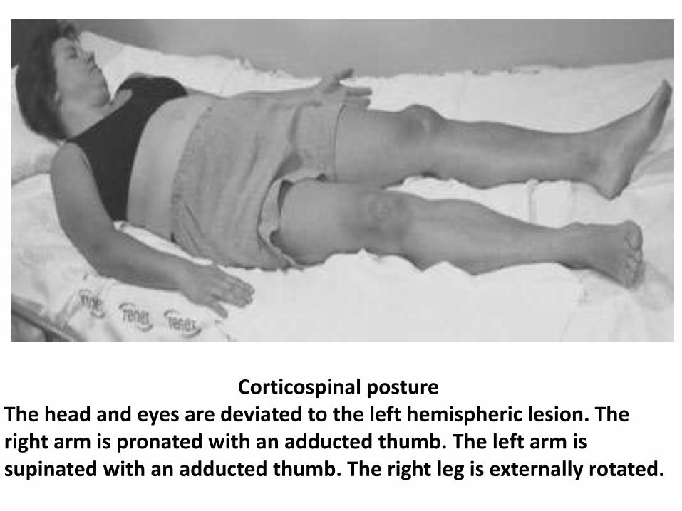
Corticospinal postureThe head and eyes are deviated to the left hemispheric lesion. The right arm is pronated with an adducted thumb. The left arm is supinated with an adducted thumb. The right leg is externally rotated.
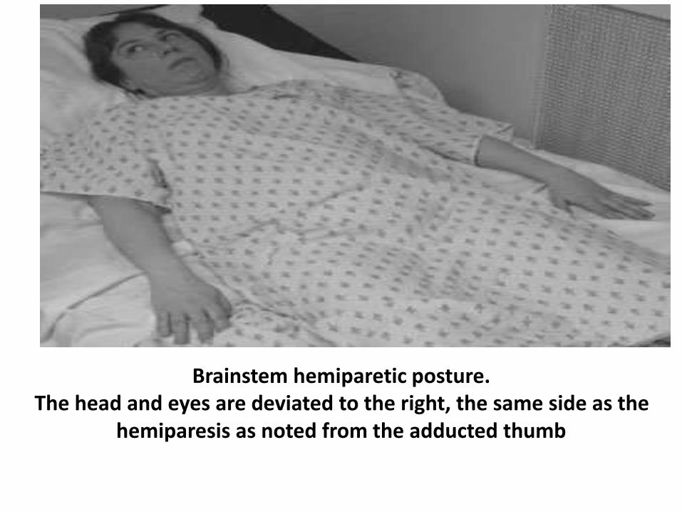
Brainstem hemiparetic posture.The head and eyes are deviated to the right, the same side as the
hemiparesis as noted from the adducted thumb
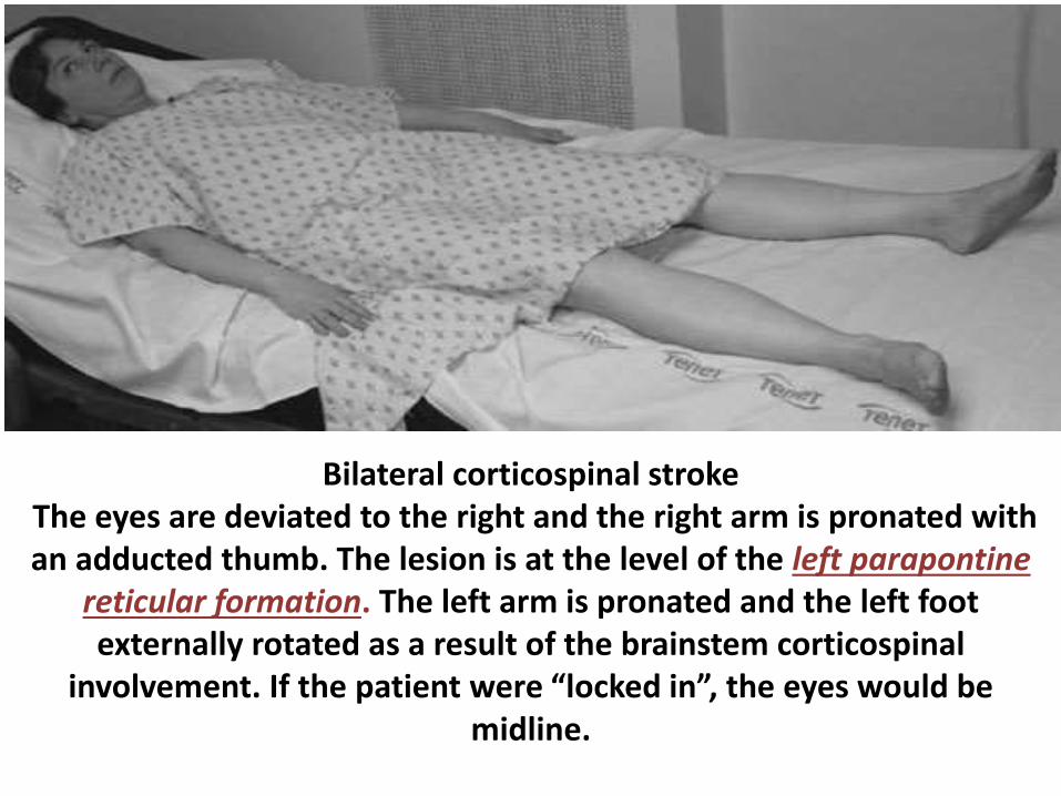
Bilateral corticospinal strokeThe eyes are deviated to the right and the right arm is pronated with an adducted thumb. The lesion is at the level of the left parapontine
reticular formation. The left arm is pronated and the left foot externally rotated as a result of the brainstem corticospinal
involvement. If the patient were “locked in”, the eyes would be midline.
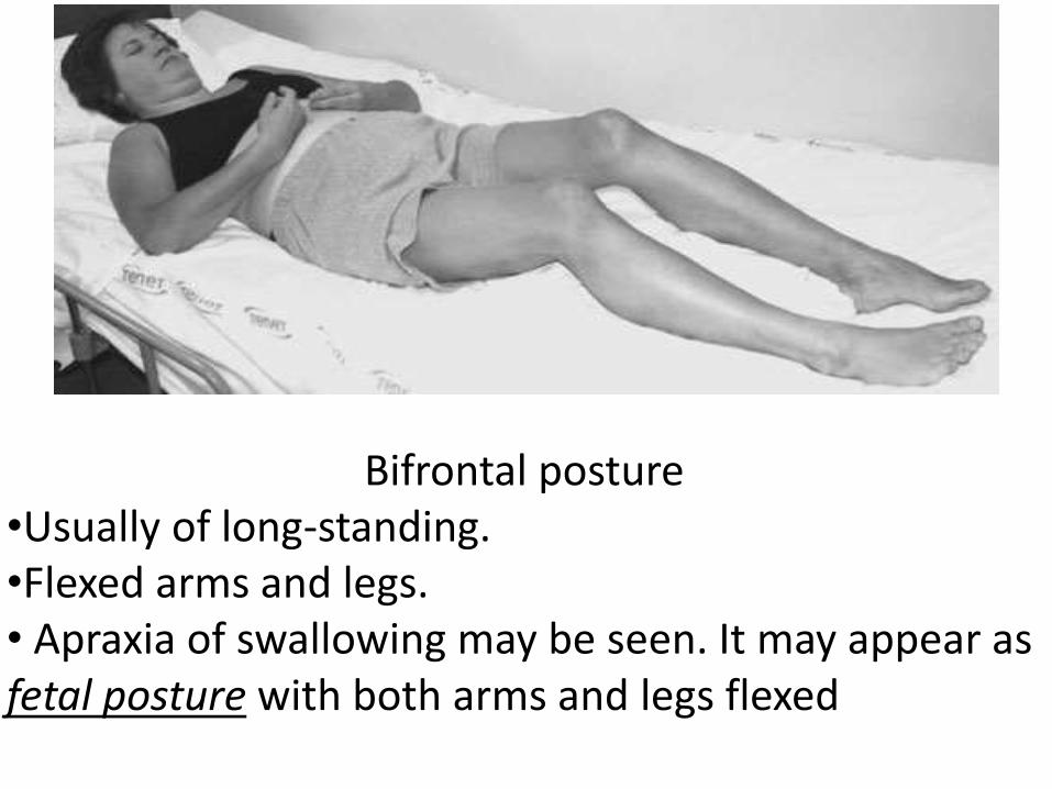
Bifrontal posture•Usually of long-standing. •Flexed arms and legs.• Apraxia of swallowing may be seen. It may appear as fetal posture with both arms and legs flexed
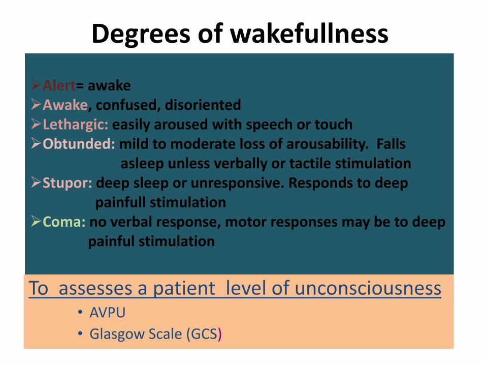
Degrees of wakefullness
To assesses a patient level of unconsciousness• AVPU
• Glasgow Scale (GCS)
Alert= awakeAwake, confused, disorientedLethargic: easily aroused with speech or touchObtunded: mild to moderate loss of arousability. Falls
asleep unless verbally or tactile stimulationStupor: deep sleep or unresponsive. Responds to deep
painfull stimulationComa: no verbal response, motor responses may be to deep
painful stimulation
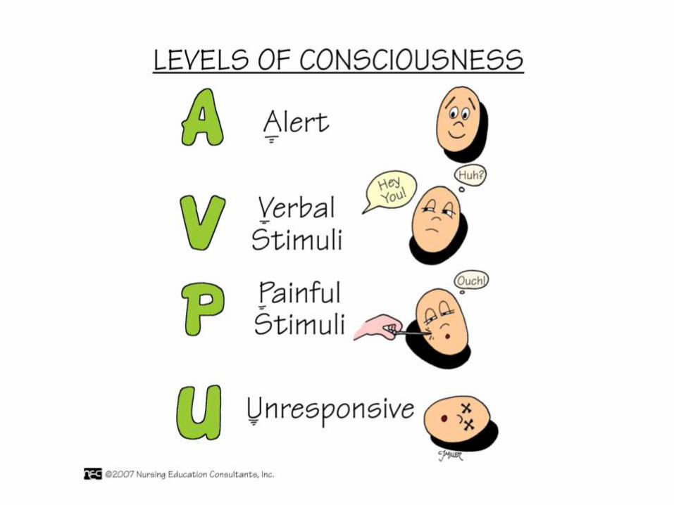
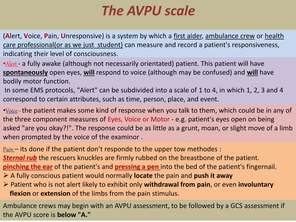
(Alert, Voice, Pain, Unresponsive) is a system by which a first aider, ambulance crew or health care professional(or as we just student) can measure and record a patient's responsiveness, indicating their level of consciousness.
The AVPU scale
•Alert - a fully awake (although not necessarily orientated) patient. This patient will have spontaneously open eyes, will respond to voice (although may be confused) and will have bodily motor function.In some EMS protocols, "Alert" can be subdivided into a scale of 1 to 4, in which 1, 2, 3 and 4
correspond to certain attributes, such as time, person, place, and event.
•Voice - the patient makes some kind of response when you talk to them, which could be in any of the three component measures of Eyes, Voice or Motor - e.g. patient's eyes open on being asked "are you okay?!". The response could be as little as a grunt, moan, or slight move of a limb when prompted by the voice of the examinor .
Pain – its done if the patient don’t responde to the upper tow methodes :Sternal rub the rescuers knuckles are firmly rubbed on the breastbone of the patient.pinching the ear of the patient's and pressing a pen into the bed of the patient's fingernail. A fully conscious patient would normally locate the pain and push it away Patient who is not alert likely to exhibit only withdrawal from pain, or even involuntary
flexion or extension of the limbs from the pain stimulus.
Ambulance crews may begin with an AVPU assessment, to be followed by a GCS assessment if the AVPU score is below "A."
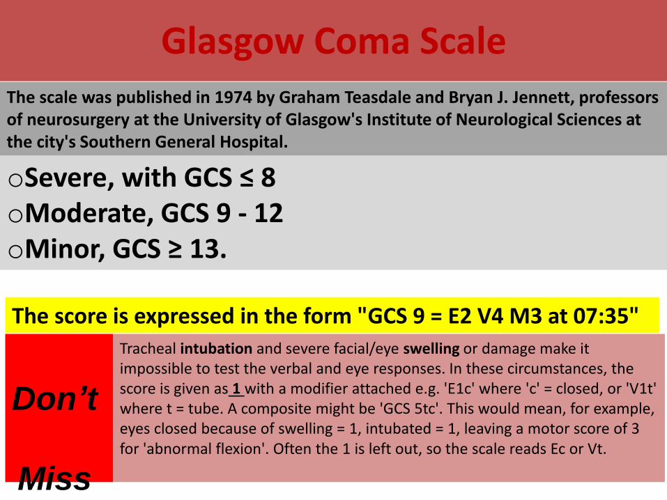
Glasgow Coma ScaleThe scale was published in 1974 by Graham Teasdale and Bryan J. Jennett, professors of neurosurgery at the University of Glasgow's Institute of Neurological Sciences at the city's Southern General Hospital.
oSevere, with GCS ≤ 8oModerate, GCS 9 - 12oMinor, GCS ≥ 13.
The score is expressed in the form "GCS 9 = E2 V4 M3 at 07:35"
Tracheal intubation and severe facial/eye swelling or damage make it impossible to test the verbal and eye responses. In these circumstances, the score is given as 1 with a modifier attached e.g. 'E1c' where 'c' = closed, or 'V1t' where t = tube. A composite might be 'GCS 5tc'. This would mean, for example, eyes closed because of swelling = 1, intubated = 1, leaving a motor score of 3 for 'abnormal flexion'. Often the 1 is left out, so the scale reads Ec or Vt.
Don’t
Miss
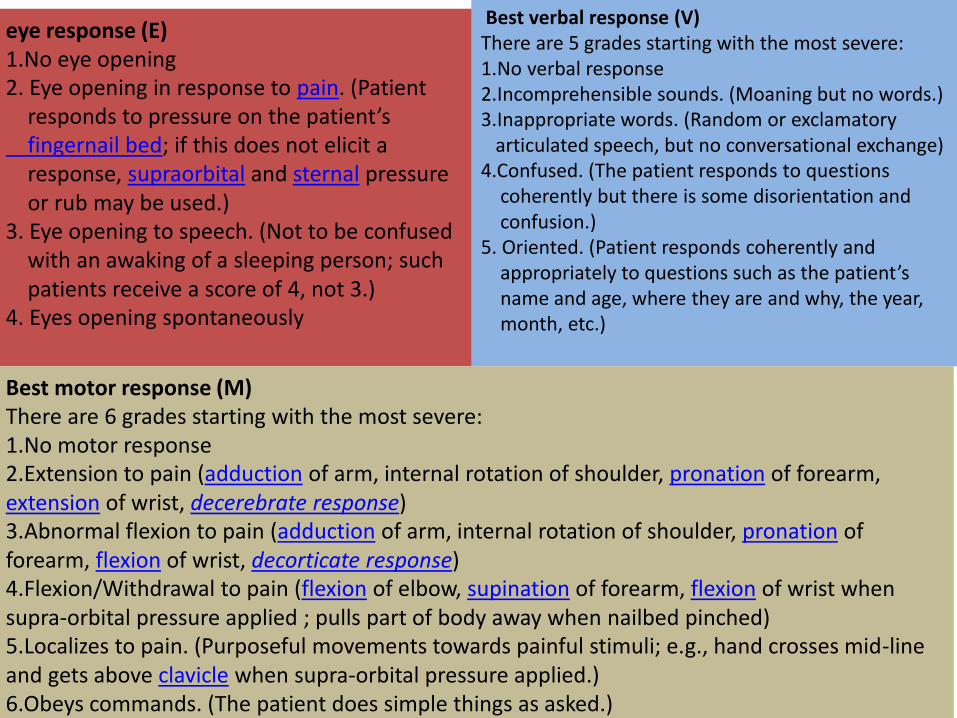
eye response (E)1.No eye opening 2. Eye opening in response to pain. (Patient
responds to pressure on the patient’s fingernail bed; if this does not elicit a response, supraorbital and sternal pressure or rub may be used.)
3. Eye opening to speech. (Not to be confused with an awaking of a sleeping person; such patients receive a score of 4, not 3.)
4. Eyes opening spontaneously
Best verbal response (V)There are 5 grades starting with the most severe:1.No verbal response 2.Incomprehensible sounds. (Moaning but no words.) 3.Inappropriate words. (Random or exclamatory
articulated speech, but no conversational exchange) 4.Confused. (The patient responds to questions
coherently but there is some disorientation and confusion.)
5. Oriented. (Patient responds coherently and appropriately to questions such as the patient’s name and age, where they are and why, the year, month, etc.)
Best motor response (M)There are 6 grades starting with the most severe:1.No motor response 2.Extension to pain (adduction of arm, internal rotation of shoulder, pronation of forearm, extension of wrist, decerebrate response) 3.Abnormal flexion to pain (adduction of arm, internal rotation of shoulder, pronation of forearm, flexion of wrist, decorticate response) 4.Flexion/Withdrawal to pain (flexion of elbow, supination of forearm, flexion of wrist when supra-orbital pressure applied ; pulls part of body away when nailbed pinched) 5.Localizes to pain. (Purposeful movements towards painful stimuli; e.g., hand crosses mid-line and gets above clavicle when supra-orbital pressure applied.) 6.Obeys commands. (The patient does simple things as asked.)
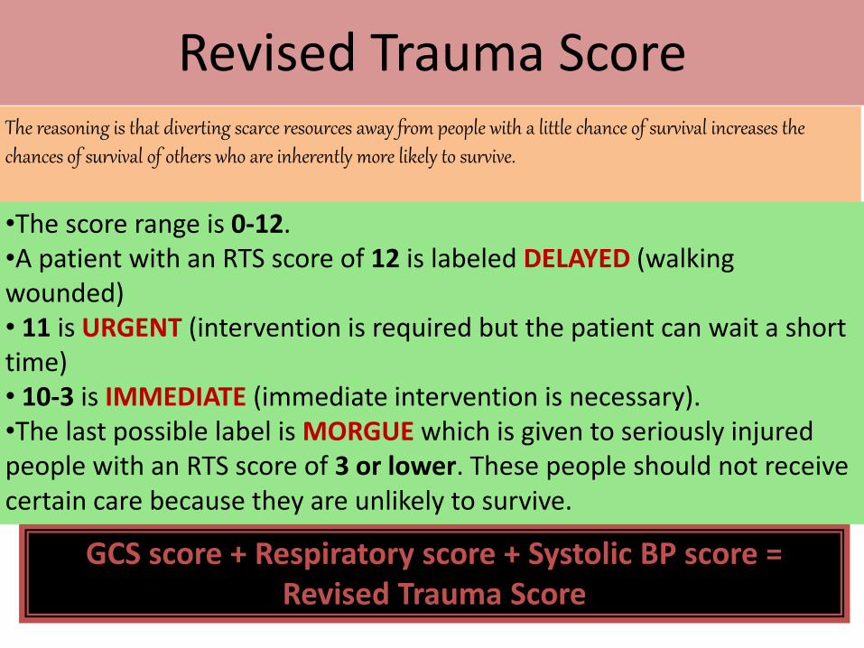
Revised Trauma Score
GCS score + Respiratory score + Systolic BP score = Revised Trauma Score
•The score range is 0-12. •A patient with an RTS score of 12 is labeled DELAYED (walking wounded)• 11 is URGENT (intervention is required but the patient can wait a short time)• 10-3 is IMMEDIATE (immediate intervention is necessary). •The last possible label is MORGUE which is given to seriously injured people with an RTS score of 3 or lower. These people should not receive certain care because they are unlikely to survive.
The reasoning is that diverting scarce resources away from people with a little chance of survival increases the chances of survival of others who are inherently more likely to survive.
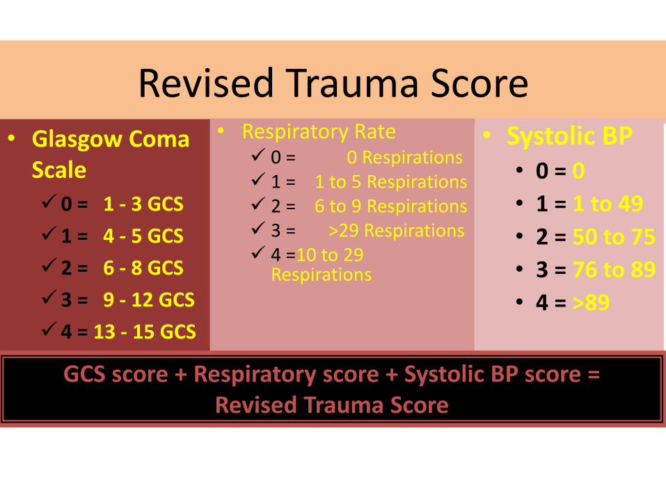
Revised Trauma Score• Glasgow Coma
Scale
0 = 1 - 3 GCS
1 = 4 - 5 GCS
2 = 6 - 8 GCS
3 = 9 - 12 GCS
4 = 13 - 15 GCS
• Respiratory Rate 0 = 0 Respirations 1 = 1 to 5 Respirations 2 = 6 to 9 Respirations 3 = >29 Respirations 4 =10 to 29
Respirations
• Systolic BP• 0 = 0
• 1 = 1 to 49
• 2 = 50 to 75
• 3 = 76 to 89
• 4 = >89
GCS score + Respiratory score + Systolic BP score = Revised Trauma Score
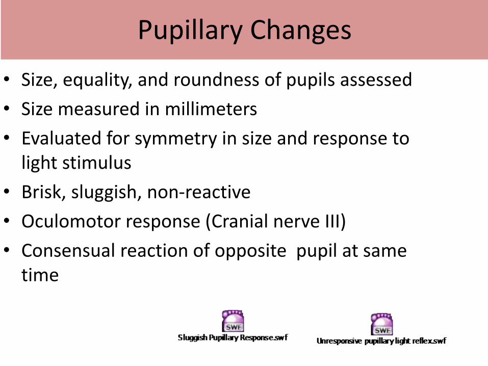
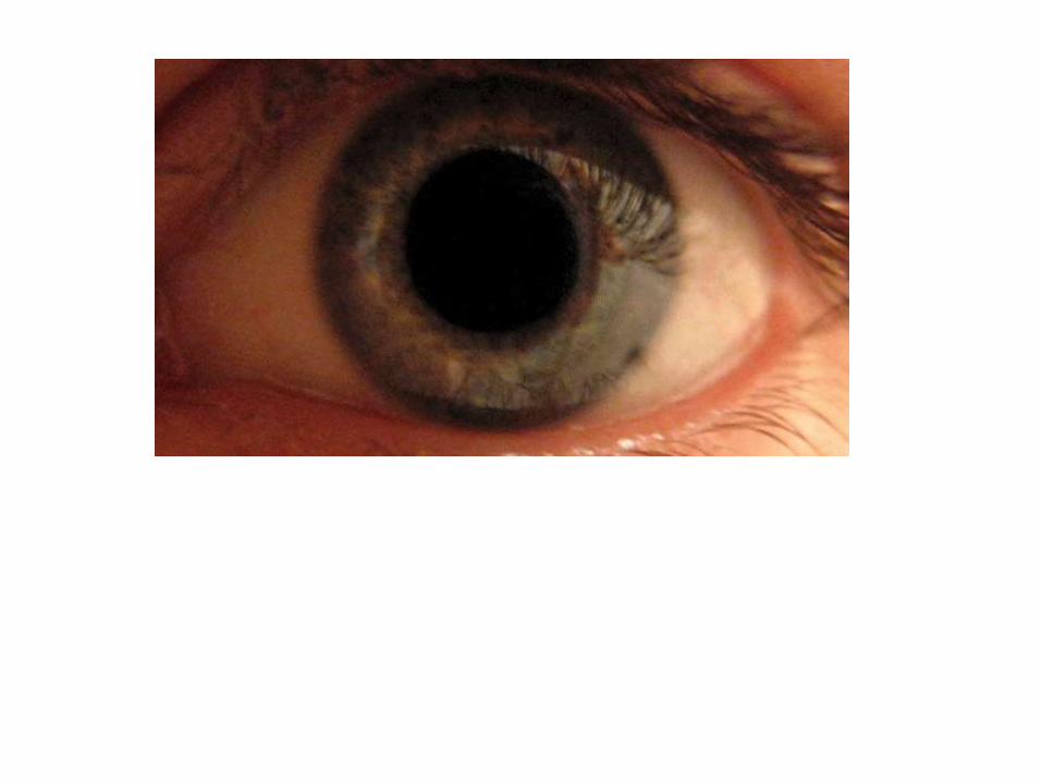
Pupillary Changes
• Size, equality, and roundness of pupils assessed
• Size measured in millimeters
• Evaluated for symmetry in size and response to light stimulus
• Brisk, sluggish, non-reactive
• Oculomotor response (Cranial nerve III)
• Consensual reaction of opposite pupil at same time
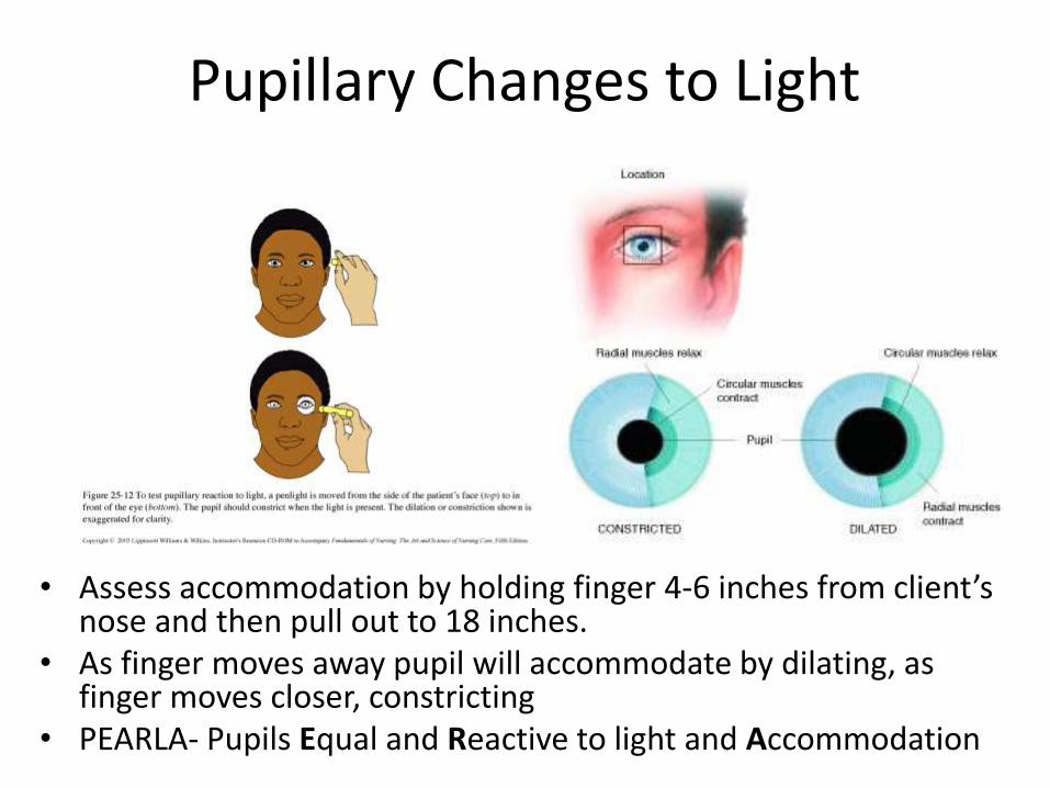
Pupillary Changes to Light
• Assess accommodation by holding finger 4-6 inches from client’s nose and then pull out to 18 inches.
• As finger moves away pupil will accommodate by dilating, as finger moves closer, constricting
• PEARLA- Pupils Equal and Reactive to light and Accommodation
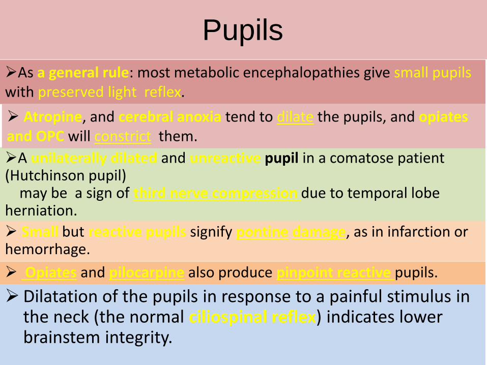
Pupils
A unilaterally dilated and unreactive pupil in a comatose patient (Hutchinson pupil)
may be a sign of third nerve compression due to temporal lobe herniation.
Small but reactive pupils signify pontine damage, as in infarction or hemorrhage.
Opiates and pilocarpine also produce pinpoint reactive pupils.
Dilatation of the pupils in response to a painful stimulus in the neck (the normal ciliospinal reflex) indicates lower brainstem integrity.
As a general rule: most metabolic encephalopathies give small pupils with preserved light reflex.
Atropine, and cerebral anoxia tend to dilate the pupils, and opiates and OPC will constrict them.
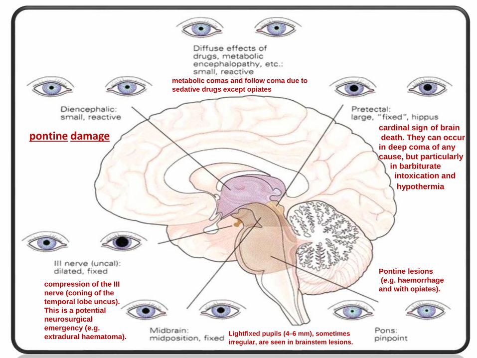
pontine damage
compression of the III
nerve (coning of the
temporal lobe uncus).
This is a potential
neurosurgical
emergency (e.g.
extradural haematoma). Lightfixed pupils (4–6 mm), sometimes
irregular, are seen in brainstem lesions.
Pontine lesions
(e.g. haemorrhage
and with opiates).
cardinal sign of brain
death. They can occur
in deep coma of any
cause, but particularly
in barbiturate
intoxication and
hypothermia
metabolic comas and follow coma due to
sedative drugs except opiates
Eye movements
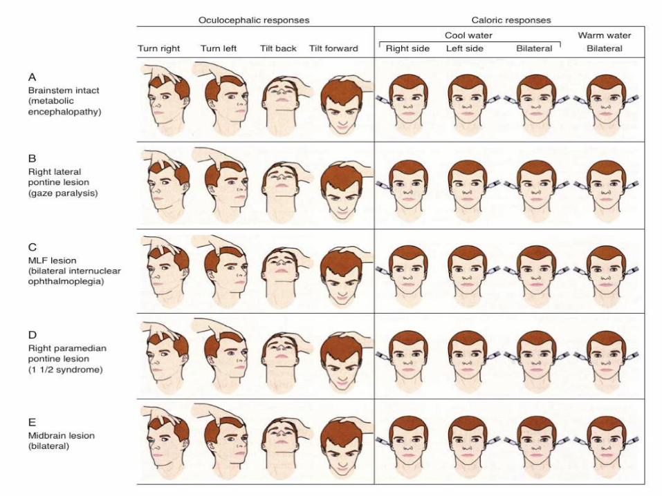
Spontaneous roving, horizontal and conjugate eye movementsintact brain stemdiffuse or metabolic cortical dysfunction
Conjugate lateral deviationmassive hemispheric lesion (eyes toward lesion)pontine lesion (eyes away from lesion)
Doll’s eyes reflexintact brainstem function with depressed cortical influencesnormal sleep, coma, persistent vegetative state
Ice water caloric testeyes toward the side of cold waterabsence in brainstem lesion, inner ear disease, deep drug coma, and anticonvulsants overdose

Extraocular movements
The most important tests are:
• Doll’s-head maneuver
• Ice water calorics
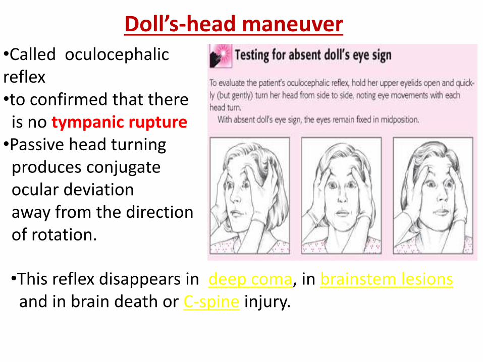
•Called oculocephalicreflex •to confirmed that there
is no tympanic rupture•Passive head turning
produces conjugate ocular deviation away from the direction of rotation.
Doll’s-head maneuver
•This reflex disappears in deep coma, in brainstem lesions and in brain death or C-spine injury.
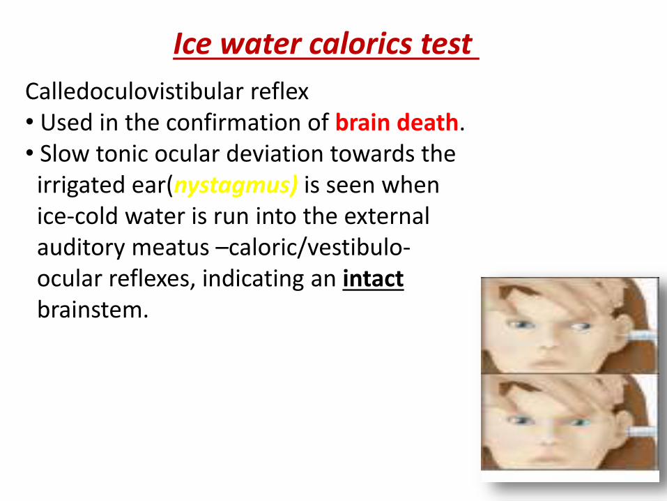
Calledoculovistibular reflex • Used in the confirmation of brain death.• Slow tonic ocular deviation towards the
irrigated ear(nystagmus) is seen when ice-cold water is run into the external auditory meatus –caloric/vestibulo-ocular reflexes, indicating an intactbrainstem.
Ice water calorics test
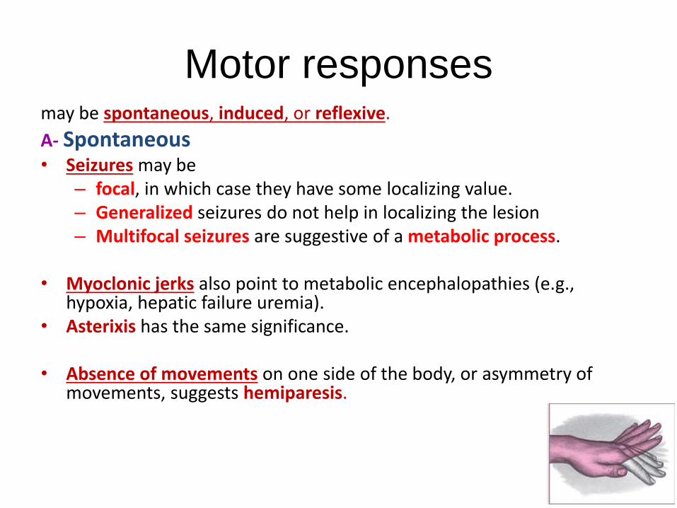
Motor responsesmay be spontaneous, induced, or reflexive.
A- Spontaneous• Seizures may be
– focal, in which case they have some localizing value. – Generalized seizures do not help in localizing the lesion– Multifocal seizures are suggestive of a metabolic process.
• Myoclonic jerks also point to metabolic encephalopathies (e.g., hypoxia, hepatic failure uremia).
• Asterixis has the same significance.
• Absence of movements on one side of the body, or asymmetry of movements, suggests hemiparesis.
Motor responses
B. Induced movements– (e.g., fending-off or other complex, purposeful
movements, such as scratching the nose in response to tickling of the nostril) require integrity of the corresponding corticospinal tract.
– Poorly organized, incomplete movements, especially when unilateral, suggests corticospinal tract dysfunction or damage.

Respiratory patterns
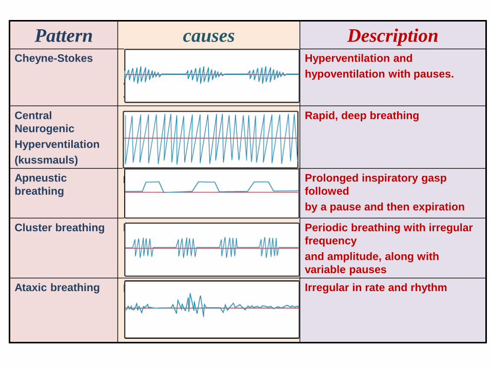
Pattern causes DescriptionCheyne-Stokes • bilateral deep hemispheric and
basal ganglionic dysfunction. •± upper brainstem involvement. •congestive heart failure
Hyperventilation and
hypoventilation with pauses.
Central
Neurogenic
Hyperventilation
(kussmauls)
Systemic acidosis (e.g., diabetic ketoacidosis, lactic acidosis) and hypoxemia should be excluded
Rapid, deep breathing
Apneustic
breathing
pontine damage. Prolonged inspiratory gasp
followed
by a pause and then expiration
Cluster breathing High medullary lesions Periodic breathing with irregular
frequency
and amplitude, along with
variable pauses
Ataxic breathing Imply damageto the medullaryrespiratory centers. Both are agonal events and usually precede respiratory arrest
Irregular in rate and rhythm
Thank you for your attention
Thank you for saving me
from coma