Embed Size (px)
Citation preview
doi:10.1152/ajprenal.00038.2009 298:F365-F380, 2010. First published 25 November 2009;Am J Physiol Renal Physiol
M. Ashcroft and Rajesh V. ThakkerPierre J. Courtoy, M. Andrew Nesbit, Patrik Rorsman, Olivier Devuyst, FrancesPartridge, Juris Galvanovskis, Siân E. Williams, Francois Jouret, Fiona T. F. Wu, Anita A. C. Reed, Nellie Y. Loh, Sara Terryn, Jonathan D. Lippiat, Chrisdiseasetransport: relevance to pathophysiology of Dent'smembrane expression, endocytosis, and microtubular CLC-5 and KIF3B interact to facilitate CLC-5 plasma
You might find this additional info useful...
62 articles, 38 of which can be accessed free at:This article cites http://ajprenal.physiology.org/content/298/2/F365.full.html#ref-list-1
3 other HighWire hosted articlesThis article has been cited by
[PDF] [Full Text] [Abstract]
, June 4, 2010; 285 (23): 17595-17603.J. Biol. Chem.Weinert and Thomas J. JentschGesa Rickheit, Lena Wartosch, Sven Schaffer, Sandra M. Stobrawa, Gaia Novarino, StefanieRequire PY-motif-dependent UbiquitylationRole of ClC-5 in Renal Endocytosis Is Unique among ClC Exchangers and Does Not
[PDF] [Full Text] [Abstract], June 11, 2010; 328 (5984): 1398-1401.Science
Gaia Novarino, Stefanie Weinert, Gesa Rickheit and Thomas J. JentschRenal EndocytosisEndosomal Chloride-Proton Exchange Rather Than Chloride Conductance Is Crucial for
[PDF] [Full Text] [Abstract], April 1, 2011; 22 (7): 1045-1057.Mol. Biol. Cell
Geraldine B. Quinones, Barbara A. Danowski, Anjan Devaraj, Vimla Singh and Lee A. Ligonacetylation as epithelial cells become polarizedThe posttranslational modification of tubulin undergoes a switch from detyrosination to
including high resolution figures, can be found at:Updated information and services http://ajprenal.physiology.org/content/298/2/F365.full.html
can be found at:AJP - Renal Physiologyabout Additional material and information http://www.the-aps.org/publications/ajprenal
This infomation is current as of December 20, 2011.
American Physiological Society. ISSN: 0363-6127, ESSN: 1522-1466. Visit our website at http://www.the-aps.org/.(monthly) by the American Physiological Society, 9650 Rockville Pike, Bethesda MD 20814-3991. Copyright © 2010 by therespective cells and vasculature, as well as to the control of body fluid volume and composition. It is published 12 times a year
publishes original manuscripts on a broad range of subjects relating to the kidney, urinary tract, and theirAJP - Renal Physiology
on Decem
ber 20, 2011ajprenal.physiology.org
Dow
nloaded from
CLC-5 and KIF3B interact to facilitate CLC-5 plasma membrane expression,endocytosis, and microtubular transport: relevance to pathophysiologyof Dent’s disease
Anita A. C. Reed,1* Nellie Y. Loh,1* Sara Terryn,2 Jonathan D. Lippiat,3 Chris Partridge,4
Juris Galvanovskis,4 Siân E. Williams,1 Francois Jouret,2 Fiona T. F. Wu,1 Pierre J. Courtoy,2
M. Andrew Nesbit,1 Patrik Rorsman,4 Olivier Devuyst,2 Frances M. Ashcroft,5 and Rajesh V. Thakker1
1Academic Endocrine Unit and 4Diabetes Research Laboratories, Nuffield Department of Medicine, University of Oxford,Oxford Centre for Diabetes, Endocrinology, and Metabolism, Churchill Hospital, Oxford; 3Institute of Membrane and SystemsBiology, Faculty of Biological Sciences, University of Leeds, Leeds; 5University Laboratory of Physiology, University ofOxford, Oxford, United Kingdom; and 2Division of Nephrology and CELL Unit, de Duve Institute, Université Catholique deLouvain Medical School, Brussels, Belgium
Submitted 23 January 2009; accepted in final form 20 November 2009
Reed AA, Loh NY, Terryn S, Lippiat JD, Partridge C, Gal-vanovskis J, Williams SE, Jouret F, Wu FT, Courtoy PJ, Nesbit MA,Rorsman P, Devuyst O, Ashcroft FM, Thakker RV. CLC-5 andKIF3B interact to facilitate CLC-5 plasma membrane expression, endo-cytosis, and microtubular transport: relevance to pathophysiology ofDent’s disease. Am J Physiol Renal Physiol 298: F365–F380, 2010. Firstpublished November 25, 2009; doi:10.1152/ajprenal.00038.2009.—Re-nal tubular reabsorption is important for extracellular fluid homeostasisand much of this occurs via the receptor-mediated endocytic pathway.This pathway is disrupted in Dent’s disease, an X-linked renal tubulardisorder that is characterized by low-molecular-weight proteinuria, hy-percalciuria, nephrolithiasis, and renal failure. Dent’s disease is due tomutations of CLC-5, a chloride/proton antiporter, expressed in endo-somes and apical membranes of renal tubules. Loss of CLC-5 functionalters receptor-mediated endocytosis and trafficking of megalin andcubilin, although the underlying mechanisms remain to be elucidated.Here, we report that CLC-5 interacts with kinesin family member 3B(KIF3B), a heterotrimeric motor protein that facilitates fast anterogradetranslocation of membranous organelles. Using yeast two-hybrid, gluta-thione-S-transferase pull-down and coimmunoprecipitation assays, theCOOH terminus of CLC-5 and the coiled-coil and globular domains ofKIF3B were shown to interact. This was confirmed in vivo by endoge-nous coimmunoprecipitation of CLC-5 and KIF3B and codistributionwith endosomal markers in mouse kidney fractions. Confocal live cellimaging in kidney cells further demonstrated association of CLC-5 andKIF3B, and transport of CLC-5-containing vesicles along KIF3B micro-tubules. KIF3B overexpression and underexpression, using siRNA, hadreciprocal effects on whole cell chloride current amplitudes, CLC-5 cellsurface expression, and endocytosis of albumin and transferrin. Clcn5Y/�
mouse kidneys and isolated proximal tubular polarized cells showedincreased KIF3B expression, whose effects on albumin endocytosis weredependent on CLC-5 expression. Thus, the CLC-5 and KIF3B interactionis important for CLC-5 plasma membrane expression and for facilitatingendocytosis and microtubular transport in the kidney.
chloride/proton antiporter; kinesin family; proximal tubular reabsorp-tion; hypercalciuric nephrolithiasis; receptor-mediated endocytosis
THE RECEPTOR-MEDIATED ENDOCYTIC pathway (RMEP) plays animportant role in facilitating renal tubular reabsorption and
hence maintaining extracellular fluid homeostasis (4). Thispathway is disrupted in Dent’s disease (OMIM: 300009), anX-linked renal tubular disorder characterized by low-molecu-lar-weight proteinuria, hypercalciuria, nephrolithiasis, and pro-gressive renal failure (28, 37). Dent’s disease is usually causedby mutations in a chloride/proton antiporter, CLC-5 (GenBankaccession number NM_001127899.1), and mice deleted for aClcn5 allele develop the renal tubular defects associated withDent’s disease (37, 45, 47, 48, 55).
CLC-5 belongs to the nine-member family of CLC proteins(CLC-1 to CLC-7, CLC-Ka, and CLC-Kb), which includevoltage-gated chloride channels and chloride/proton antiport-ers that are found at the plasma membrane and membranes ofintracellular organelles. CLCs have diverse functions, whichinclude the control of membrane excitability, the regulation ofcell volume, and transepithelial transport (28). For example,CLC-5, which is an antiporter expressed at multiple sites in thehuman nephron, including the proximal tubules (PT), the thickascending limb (TAL) of the loop of Henle, and the �-inter-calated cells of the collecting duct (CD), is involved in trans-epithelial solute transport (e.g., the RMEP of the PT cells) andis localized primarily in early endosomes and apical (luminal)membranes of renal PT (7, 16, 49). Disruption of Clcn5 in miceleads to defective fluid-phase and receptor-mediated endocy-tosis, by the renal PT, indicating that CLC-5 contributes toendocytosis in vivo (4, 16, 47, 55). Indeed, the renal tubularabnormalities observed in patients with Dent’s disease, whichis caused by mutations in CLC-5, are consistent with a func-tional loss of CLC-5 in such reabsorptive pathways (7, 16).Furthermore, these renal abnormalities have been shown toresult from altered receptor-mediated endocytosis in associa-tion with defective trafficking of the megalin and cubilinreceptor complex (4).
The role of CLC-5 in endosomal trafficking remains to beestablished. Loss of CLC-5 function leads to impaired endo-somal acidification which may contribute to the observeddefects in endosomal trafficking (4). Interestingly, pharmaco-logical inhibition of vacuolar acidification has been reported todecrease endosomal recycling and transfer to lysosomes (1) butnot the rate of endocytosis, consistent with a trafficking defect(6, 52). Any possible role of CLC-5 in endosomal transport islikely to involve the CLC-5 COOH terminus (amino acids 551to 746; Fig. 1A) as this has been shown to be cytoplasmic by
* A. A. C. Reed and N. Y. Loh contributed equally to this work.Address for reprint requests and other correspondence: R. V. Thakker,
Academic Endocrine Unit, Nuffield Dept. of Medicine, Univ. of Oxford,Oxford Centre for Diabetes, Endocrinology, and Metabolism, Churchill Hos-pital, Oxford, OX3 7LJ, UK (e-mail: [email protected]).
Am J Physiol Renal Physiol 298: F365–F380, 2010.First published November 25, 2009; doi:10.1152/ajprenal.00038.2009.
0363-6127/10 $8.00 Copyright © 2010 the American Physiological Societyhttp://www.ajprenal.org F365
on Decem
ber 20, 2011ajprenal.physiology.org
Dow
nloaded from
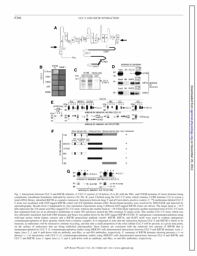
Fig. 1. Interactions between CLC-5 and KIF3B subunits. A: CLC-5 consists of 18 helices (A to R) with the NH2- and COOH-terminus (C-term) domains beingcytoplasmic (membrane boundaries indicated by arrows) (10, 58). B: yeast 2-hybrid using the CLC-5 C-term, which contains 2 CBS domains (11) to screen arenal cDNA library, identified KIF3B as a putative interactor. Interaction between large T and p53 provided a positive control. C: 35S-methionine-labeled CLC-5C-term was incubated with GST-tagged KIF3B coiled coil (CC)/globular domain (GD). Resin-bound proteins were resolved by SDS-PAGE and detected byautoradiography. Results from 2 independent in vitro translation experiments using 2 different GST-tagged KIF3B clones are shown. The larger band at �24.5kDa represents the 216 amino acid Myc-tagged CLC-5 C-term, whereas the smaller band at �18.5 kDa likely represents another translated form of CLC-5 C-termthat originated from use of an alternative methionine at codon 583 and hence lacked the NH2 terminal 32 amino acids. This smaller CLC-5 C-term, which wasless efficiently translated, had both CBS domains and hence was pulled down by the GST-tagged KIF3B CC/GD. D: endogenous coimmunoprecipitation usingwild-type mouse whole kidney extracts and a KIF3B monoclonal antibody (mAb). KIF3B, KIF3A, and KAP3 mAb were used to confirm endogenouscoimmunoprecipitation of these proteins which form a trimeric complex. It is important to note that the interaction between CLC-5 and KIF3B is likely to betransient, as endosomes will be released to continue recycling, and that only a small proportion of the total cellular CLC-5 will be present, at a given time point,on the surface of endosomes that are being trafficked anterogradely; these features are consistent with the relatively low amount of KIF3B that isimmunopreciptiated by CLC-5. E: coimmunoprecipitation studies using HEK293 cells demonstrated interactions between CLC-5 and KIF3B domains. Lane 1:input; lanes 2, 3, and 4: pull-down with no antibody, anti-Myc, or anti-HA antibodies, respectively. F: summary of KIF3B domains showing presence (�) orabsence (�) of interactions with CLC-5. G: coimmunoprecipitation studies using HEK293 cells demonstrated interactions between CLC-4 and KIF3B, andCLC-7 and KIF3B. Lane 1: input; lanes 2, 3, and 4: pull-down with no antibody, anti-Myc, or anti-HA antibodies, respectively.
F366 CLC-5 AND KIF3B INTERACTION
AJP-Renal Physiol • VOL 298 • FEBRUARY 2010 • www.ajprenal.org
on Decem
ber 20, 2011ajprenal.physiology.org
Dow
nloaded from
an analysis of the three-dimensional structures of homologousbacterial CLCs (10, 58). Moreover, the COOH terminus con-tains potential binding motifs for regulatory molecules: theseinclude two cystathionine-�-synthase (CBS1 and CBS2) do-mains, a PY motif (PPLPPY), and a putative PDZ-bindingmotif (11, 25, 49). Indeed, a number of proteins, includingCLC-4, cofilin, WWP2, Nedd4-2, and Na�-H� exchangerregulatory factor 2 (NHERF2), have been reported to interactwith CLC-5.
CLC-4, which is expressed in endosomes, contributes toendosomal acidification and trafficking by forming a complexwith CLC-5 (39, 40). Cofilin, which is an actin depolymerizingprotein, interacts with the COOH terminus of CLC-5, andphosphorylation of cofilin is associated with reduced albuminuptake (26). WWP2, which binds strongly to the PY motif ofCLC-5, belongs to the same protein class as the ubiquitin-protein ligase Nedd4-2 that has also been demonstrated tointeract with the COOH terminus of CLC-5 and to alter CLC-5currents by decreasing the cell surface expression of CLC-5(24, 46, 49). CLC-5, whose cell surface expression increases inopossum kidney (OK) cells that are exposed to albumin,undergoes ubiquitination and a reduction in Nedd4-2 is accom-panied by decreased albumin uptake in OK cells. These resultsindicate that Nedd4-2 may act via ubiquitination to shuntCLC-5 into the endocytic pathway (24). NHERF2, which is anepithelial PDZ scaffold that interacts with and alters the cellsurface levels of CLC-5, links CLC-5 to ezrin and anchors theendocytic complex (23). However, the mechanisms wherebyCLC-5 affects endosomal transport within the RMEP remain tobe elucidated. We therefore used the CLC-5 COOH-terminusdomain (Fig. 1A) as bait to perform a yeast two-hybridscreen of an adult human renal cDNA library with the aimof identifying interacting proteins involved in the endocyticpathway (4, 7).
MATERIALS AND METHODS
Yeast two-hybrid analysis. Partial CLC-5 cDNAs that encode theCOOH terminus (C-term; peptides 551–746), CBS1 (peptides 576–680), and CBS2 (peptides 674–746) were subcloned into pGBKT7,containing the Gal4 DNA binding domain (BD) to generate baitplasmids. Yeast strain PJ69-2A transformed with pGBKT7-CLC-5(C-term) was used to screen a Matchmaker pretransformed adulthuman kidney cDNA library in Y187 yeast strain, according to themanufacturer’s instructions (BD Bioscience) (42). The DNA se-quences of prey plasmids isolated from positive clones were analyzedusing a gapped BLAST search of NCBI databases. To confirm theinteraction, prey plasmids were retransformed into PJ69-2A contain-ing pGBKT7-CLC-5 (C-term) or pGBKT7 and tested for growth onselective plates (42). pGBKT7-p53 and pGADT7-Large T antigenwere used as positive controls (42). KIF3B (FL) was subcloned intopACT2 containing the Gal4 activation domain. All inserts wereconfirmed by DNA sequence analysis (42). PJ69-2A was transformedsequentially with the appropriate CLC-5 and KIF3B constructs andmonitored for growth on selective plates (42). Expression of CLC-5and KIF3B Gal4 fusion proteins in yeast was confirmed by Westernblot analysis using anti-HA (pACT2-KIF3B) or anti-Myc (pGBKT7-CLC-5) antibodies (42).
Glutathione-S-transferase fusion protein and pull-down assay.KIF3B CC/GD was subcloned into pGEX-4T-1 and expressed inEscherichia coli BL21 (42). 35S-labeled CLC-5 (C-term) protein wasprepared by in vitro transcription/translation using pGBKT7-CLC-5(C-term; TNT System, Promega, Madison, WI). Glutathione-S-trans-
ferase (GST)-pulldowns of GST-KIF3B and radiolabeled CLC-5(C-term) proteins were performed as previously described (42).
Coimmunoprecipitations. Full-length CLC-5, CLC-4, CLC-7, andKIF3B, and five KIF3B deletional constructs (Fig. 1), were subclonedinto pCMV-Myc and pCMV-HA, respectively (BD Bioscience), andcotransfected into HEK293 or COS7 cells using FuGene 6 (Roche)according to the manufacturer’s instructions. Cells were harvestedafter 48 h, and lysed in mRIPA (50 mM Tris · HCl, pH 7.4, 150 mMNaCl, 1 mM EDTA, 0.05% SDS, 1% Triton X-100), and supple-mented with protease inhibitors (COMPLETE, Roche) and coimmu-noprecipitation was performed using anti-Myc or -HA antibodies.Protein complexes were captured with protein G sepharose beads,eluted with Laemmli buffer, and analyzed by Western blot usinganti-Myc or HA antibodies as previously described (42). Endogenouscoimmunoprecipitation of CLC-5 and KIF3B was investigated usinga mouse whole kidney that was homogenized in mRIPA. Followingincubation at 4°C for 3 h, a supernatant fraction was obtained bycentrifugation at 12,000 g for 10 min at 4°C and used to investigate forendogenous coimmunoprecipitation. To minimize nonspecific bind-ing, the supernatant was preincubated for 1 h at 4°C with 50 �l ofprotein G sepharose beads (50% slurry). After centrifugation at12,000 g for 1 min at 4°C, 1 �g of CLC-5 polyclonal antibody (7),KIF3B monoclonal antibody (BD Bioscience), or KAP3 monoclonalantibody (BD Bioscience) was added to the supernatant, and thesample was incubated overnight at 4°C. Protein complexes werecaptured with protein G sepharose beads, eluted with Laemmli buffer,and analyzed by Western blot using KIF3B, KIF3A, and KAP3monoclonal antibodies (BD Bioscience).
Confocal imaging. KIF3B (FL) was subcloned into pEGFP-C andCLC-5 (FL) with NH2 terminally tagged mRFP1 (monomeric redfluorescent protein) (3) was subcloned into pcDNA 3.1/Myc-His.HEK293 and/or COS7 cells were cotransfected with CLC-5 taggedwith red fluorescent protein (RFP) and/or KIF3B-GFP constructs,using FuGene 6 (Roche). For Z-stack imaging, some CLC-5-RFP- andKIF3B-GFP-cotransfected HEK293 cells, 24 h posttransfection, werepreextracted with a microtubule stabilizing buffer (MTSB; 80 mMPIPES/KOH, pH 6.8, 1 mM MgCl, 5 mM EGTA, and 0.5% TritonX-100 or 0.1% saponin) and fixed in 4% paraformaldehyde (PFA) orice-cold methanol. CLC-5-Myc-transfected OK cells, 24 h posttrans-fection, and untransfected OK cells were preextracted with MTSB andfixed in 4% PFA and coimmunostained using monoclonal anti-Myc(Santa Cruz Biotechnology) and rabbit polyclonal anti-ZO1 (Invitro-gen) or monoclonal anti-MAPRE1 (EB1; AbCam), which immunos-tains the plus ends of microtubules (27), and rabbit polyclonal anti-ZO1 (Invitrogen), which immunostains tight junctions of polarizedcells (27), and secondary antibodies anti-mouse Alexa Fluor 594 andanti-rabbit Alexa Fluor 488 (Molecular Probes). Some RFP-taggedCLC-5 cells, 24-h posttransfection, were fixed in 4% PFA andimmunostained using monoclonal anti-Kinesin-2 (Covance) or mono-clonal anti-�-tubulin (Santa Cruz Biotechnology) and secondary an-tibody anti-mouse Alexa Fluor 488 (Molecular Probes). The Z stackswere recorded using a confocal laser-scanning microscope (Zeiss,LSM 10 META) with a Plan-Achromat �63/1.4 oil DIC objective(Carl Zeiss, Jena, Germany). An argon laser � 458 nm and 488 nmwas used to excite enhanced green fluorescent protein (EGFP) andAlexa Fluor 488 fluorescence, respectively. The mRFP1 was visual-ized by a HeNe laser � 543 nm. Emission of fluorescent proteinsand Alexa Fluor 488 was detected in the Multitrack mode in METAchannels of the confocal system within the following spectral detec-tion ranges: from 509 to 550 nm for EGFP, from 580 to 620 nm formRFP1 and Alexa Fluor 594, and from 509 to 550 nm for Alexa Fluor488. The Zeiss confocal microscope and software automatically gen-erate a merged image from the original images obtained in the separatechannels, thereby avoiding any errors or shifts that may arise frommanual editing. Colocalization was assessed by using these mergedimages and by intensity profile line scans if there was a largevariability in the intensity of the subcellular structures. Peaks obtained
F367CLC-5 AND KIF3B INTERACTION
AJP-Renal Physiol • VOL 298 • FEBRUARY 2010 • www.ajprenal.org
on Decem
ber 20, 2011ajprenal.physiology.org
Dow
nloaded from
from intensity profile line scans were considered to overlap and henceindicate colocalization if the distance between them was 300 nm,which is the maximum approximate diameter of an endosomal vesiclein the proximal renal tubule (19). For live cell imaging, cells wereobserved 24 h posttransfection under a laser-scanning confocal mi-croscope (Bio-Rad Radiance 2100) using a Nikon Fluor �60 waterimmersion objective lens. Cells were perfused with extracellularsolution containing (in mM) 137 NaCl, 5.6 KCl, 1.2 MgCl2, 2.5CaCl2, and 5 HEPES (pH 7.4 NaOH) at ��34°C. HEK293 cellstransfected with CLC-5-RFP and KIF3B-GFP were treated withnocodazole/DMSO (20 �M/0.12%) or DMSO alone (0.12%) for 4 hand fixed with 4% PFA. Microtubule restoration was verified bywashing in nocodazole free medium (14). GFP was excited using a488-nm Ar/Kr laser and detected with a 515- to 530-nm emissionfilter. mRFP1 was excited using a 543-nm green He-Ne laser anddetected with a 600-nm long pass filter. Simultaneous recordings weremade using a 560-nm dichroic mirror. Live cell scans were sampledat 1 Hz using Lasersharp 2000 software (Bio-Rad Laboratories).
Electrophysiological studies. CLC-5 (FL) and KIF3B (FL) anddeletional constructs (Fig. 1) were subcloned into pEGFP-C andpCMV-HA, respectively (BD Bioscience). The KIF3B-ATP constructwas generated by using site-directed mutagenesis (Quikchange, Strata-gene) to alter the ATP/GTP-binding site motif from GQTGTGKT toGQTGTAAT. HEK293 cells were transfected with CLC-5-GFP withor without KIF3B (FL)-HA, or a KIF3B deletion construct, usingFuGene 6 (Roche). A threefold ratio of KIF3B to CLC-5 was used. Inaddition, cells were transiently transfected with pSUPER (24) con-taining a sequence for siRNA for KIF3B (GAAGCTACCAAGAT-CAACC; KIF3B siRNA) or an unrelated control (i.e., nonsense)sequence (control siRNA). The effects of the siRNA on proteinexpression were assessed by Western blot analysis performed witheither an anti-KIF3B antibody (Santa Cruz Biotechnology), anti-KIF3A antibody (BD Biosciences), or anti-�-tubulin antibody (Ab-Cam). Densitometry of the Western blots was performed using aGS-710 calibrated imaging densitometer and the Quantity One Sys-tem Software (Bio-Rad Laboratories). Transfected cells were identi-fied by EGFP epifluorescence. Patch pipettes were pulled from thin-walled borosilicate glass (Harvard) and fire-polished. Electrode resis-tances ranged from 1 to 3 M� in the experimental solutions. Currentswere recorded using the whole cell patch-clamp configuration (�70%series-resistance compensation) with an EPC-10 amplifier and Patch-master software (HEKA). Currents were filtered at 10 kHz anddigitized at 50 kHz. Cells were held at �30 mV and 10-ms pulsesfrom �100 to �200 mV were applied at 1-s intervals. A P/8leak-subtraction protocol was used. The bath solution contained (inmM) 140 CsCl, 1 CaCl2, 1 MgCl2, 10 HEPES, at pH 7.4. The pipettesolution contained 42 CsCl, 49 Cs2SO4, 10 EGTA, 10 HEPES, at pH7.4. Recordings were analyzed using Fitmaster (HEKA) and MicrocalOrigin 7.5 software. Conductance-voltage relationships were fittedwith the Boltzmann equation, G Gmax/[1 � exp(V0.5 � V)/k], toobtain values for Gmax.
Cell surface expression of CLC-5. Cell surface proteins werebiotinylated using an adaptation of a previously reported method (24).Briefly, HEK293 or OK cells were transfected with CLC-5-Myc andKIF3B-HA or siRNA constructs using FuGene 6 (Roche) or Lipo-fectamine Plus (Invitrogen), grown to confluence, and cell surfaceproteins were biotinylated with 1.22 mg/ml EZ-Link NHS-SS-Biotin(Pierce) at room temperature with gentle agitation. Monolayers werewashed three times in cold phosphate-buffered saline, and the cellswere lysed in mRIPA. The biotinylated proteins were isolated bybinding to ImmunoPure immobilized Streptavidin (Pierce) for 1 h at4°C. The beads were pelleted and the supernatant that contained thecytosolic (unbiotinylated) fraction was recovered by centrifugation at4,500 g for 6 min at 4°C. The membrane (biotinylated) fraction waswashed, and the pellet was resuspended in Laemmli sample buffer.Proteins from the cytosolic (unbiotinylated) and cell surface (biotin-ylated) fractions were resolved on SDS-polyacrylamide gel and trans-
ferred to PVDF membrane and Western blot analysis was performedwith an anti-Myc antibody (Santa Cruz Biotechnology). Densitometryof the Western blots was performed using a GS-710 calibratedimaging densitometer and the Quantity One System Software (Bio-Rad Laboratories).
RT-PCR. PCR was performed on first-strand cDNA generated fromtotal RNA of HEK293 cells, OK cells, human kidney (AmbionEurope), mouse kidney and mouse proximal renal tubular cells(mPTCs) using avian myeloblastosis virus RT and primers specific forcubilin, megalin, KIF3B, calmodulin, caveolin-1, caveolin-2, andGAPDH (primer sequences available on request), using methodspreviously described (42).
Albumin and transferrin uptake. Albumin and transferrin uptakewere measured using modifications of standard methods, as previ-ously described (14, 26). HEK293 or OK cells were seeded in 24-wellplates and transiently transfected with empty vector or KIF3B (FL),MD/CC, CC/GD, -ATP or siRNA constructs using FuGene 6 (Roche)according to the manufacturer’s protocol and grown for 4 or 7 days,respectively. To measure albumin uptake, cells were exposed to 50�g/ml of albumin conjugated to FITC, Alexa Fluor 594, or Texas Red(Sigma or Molecular Probes) for 120 min. To measure transferrinuptake, OK cells were exposed to 50 �g/ml of transferrin conjugatedto Alexa Fluor 594 (Molecular Probes) for 20 min. Nonspecificbinding was determined in cells exposed to albumin or transferrin for1 min. At the end of the uptake period, cells were washed in ice-coldHEPES buffer, pH 6, and solubilized in MOPS lysis buffer (20 mMMOPS, 0.1% Triton X-100, pH 7.4). The FITC, Texas Red, or AlexaFluor 594 fluorescence was determined using a CytoFluor microplatereader (PerSeptive Biosystems) at 480-nm excitation and 520-nmemission or 596-nm excitation and 615-nm emission wavelengths,respectively. Total FITC/Texas Red-albumin or Alexa Fluor 594-transferrin uptake was standardized to total cellular protein. Forcompetition experiments, HEK293 and OK cells were simultaneouslyincubated with either 50 mg/ml unlabeled albumin or transferrinduring the endocytosis assay, and the uptake of fluorescently labeledalbumin or transferrin was compared with that without competition,which was the reference value set at 100%.
Subcellular fractions from Clcn5Y/� kidneys. Mice were kept inaccordance with approved protocols by the Ethics Committee forAnimal Experimentation of the Université Catholique de LouvainMedical School (Brussels) (Ref. no. UCL/MD/2006/036). TheClcn5Y/� mice, generated by targeted deletion of the exon VI ofClcn5, have been previously reported to have features of Dent’sdisease (4, 55). Kidneys from 15-wk-old Clcn5Y/� and Clcn5Y/� malelittermates were obtained. Subcellular fractions from these kidneyswere prepared and used for Western blot analysis as previouslyreported (4). The samples were loaded as 20 �g of total protein perlane, as appropriate for subcellular fractionations (13). The followingantibodies were used: affinity-purified rabbit polyclonal antibodiesagainst the NH2 terminus of human CLC-5 (55), mouse monoclonalantibodies against KIF3B (BD Bioscience), Rab5a (Santa Cruz Bio-technology), goat polyclonal antibodies against cathepsin D (SantaCruz Biotechnology), mouse monoclonal antibodies (E11) against theE1 subunit of V-ATPase (Dr. S. Gluck, University of California, SanFrancisco, CA), and �-actin (Sigma, St. Louis, MO). Specificity wasdetermined by incubation with nonimmune rabbit or mouse IgG(Vector Laboratories, Burlingame, CA). Densitometry analysis wasperformed with a Canon CanoScan8000F using the NIH Image V1.60software.
mPTC primary cultures. Polarized and differentiated mPTC cul-tures were prepared as described previously (51). Kidneys fromClcn5Y/� and Clcn5Y/� mice aged 21–28 days were used to isolatetubules, which were seeded onto collagen-coated PTFE filter mem-branes in culture medium [DMEM:F12 with 15 mM HEPES, 0.55mM Na-pyruvate, 0.1 ml/l nonessential amino acids, and the Single-Quots Kit (Lonza) containing hydrocortisone, hEGF, FBS, epineph-rine, insulin, triiodothyronine, transferrin, gentamicin/amphotericin,
F368 CLC-5 AND KIF3B INTERACTION
AJP-Renal Physiol • VOL 298 • FEBRUARY 2010 • www.ajprenal.org
on Decem
ber 20, 2011ajprenal.physiology.org
Dow
nloaded from
pH 7.4, and osmolality at 325 mosmol/kgH2O] and incubated in ahumidified chamber at 37°C and 5% CO2. After 3–4 days, at �60–80% confluency, primary cell cultures were transfected with anappropriate plasmid (1–2 �g) using FuGene HD (Roche) or Lipo-fectamine (Invitrogen). After 72 h, cells were analyzed by quantitativePCR (qPCR) for the expression of KIF3B (human and mouse) andKIF3A or an endocytosis assay was performed.
qPCR analyses. qPCR was performed as previously described (30).Total RNA from mouse kidney or mPTC primary cultures was extractedand reverse-transcribed into cDNA using SuperScript III RNase H Re-verse Transcriptase (Invitrogen), using the following primers: KIF3B(154 bp) sense 5=-GCCAAAATCAAGGCCATGGAGAGT-3= and antisense5=-GACTCTCCATTTGCTGCTGGATTTC-3=, GAPDH (176 bp) sense 5=-TGCACCACCAACTGCTTAGC-3= and antisense 5=-GGATGCAGGGAT-GATGTTCT-3=. qPCR analyses were performed in duplicate using theSYBR green fluorescence method, as previously reported (31). Foreach assay, standard curves were prepared by serial fourfolddilutions of mouse adult kidney cDNA. Calculations of primerefficiencies [efficiency (10�1/slope) � 1] demonstrated that KIF3Band GAPDH efficiencies were 0.92 0.04 and 1.02 0.03, respectively.mRNA expression was investigated in four or more pairs of Clcn5Y/�
and Clcn5Y/� mouse kidneys, or mPTC primary cultures, after normaliza-tion to GAPDH: ratio 2DCt (KO�WT) KIF3B/2DCt (KO�WT) GAPDH (44).
Endocytosis assay using mPTC cultures. Mock or KIF3B-trans-fected mPTC cultures were washed with warm HBSS (37°C) afterwhich they were incubated with FITC-conjugated bovine serumalbumin at concentrations of 0.5 mg/ml for 15 min at 37°C. After theincubation, mPTCs were washed five times with cold HBSS (4°C) andlysis buffer was added. The fluorescence in the lysate was measuredusing a fluorimeter (Perkin Elmer) with excitation at 490 10 nm andemission at 525 10 nm. Fluorescence was normalized for theamount of proteins in the primary cell cultures. Protein concentrationwas measured using the BCA kit. For metabolic inhibition experi-ments, mPTC cultures were incubated with 10 mM sodium azide or 50mM 2-deoxyglucose (DOG) for 30 min at 37°C before the endocy-tosis assay. For competition experiments, mPTC cultures were simul-taneously incubated with 50 mM unlabeled albumin or transferrinduring the endocytosis assay.
Statistical analysis. Significance of differences was assessed byunpaired, two-tailed Student’s t-test.
RESULTS
Yeast two-hybrid screen identifies KIF3B. Screening of anadult human kidney cDNA library with the COOH terminusof human CLC-5 (amino acids 551 to 746; Fig. 1A) as baitidentified a clone (c73) that coded for a COOH-terminalfragment (amino acids 410 to 747) of a member of theKinesin superfamily, KIF3B (Fig. 1B) (21). KIF3B is amicrotubule-dependent motor protein that exists in a hetero-trimeric complex, known as Kinesin-2, with KIF3A (Gen-Bank accession number NM_007054.5) and Kinesin Asso-ciated Protein 3 (KAP3; GenBank accession numberNM_014970.2) (5, 35). KIF3B has an NH2-terminal motordomain (MD), a coiled-coil (CC) domain that dimerizeswith that of KIF3A, and a COOH-terminal globular domain(GD), that in conjunction with KIF3A-GD binds KAP3.KIF3B is coexpressed with KIF3A and KAP3 in the kidney,and the function of Kinesin-2 as a microtubule-based fastanterograde translocator of membranous organelles is con-sistent with a role for the KIF3B and CLC-5 interaction inendosomal trafficking (61).
Interaction between CLC-5 and KIF3B. To confirm thatKIF3B and CLC-5 interact directly, we performed further yeasttwo-hybrid, GST pull-down, and coimmunoprecipitation studies
with full-length (FL) and deletional constructs (Fig. 1, B–E).Coexpression of CLC-5 and KIF3B constructs in yeast cells wasconfirmed by Western blot analysis (data not shown). Yeasttwo-hybrid analysis showed that the CLC-5 COOH terminus andFL KIF3B interacted (Fig. 1B). However, CLC-5 CBS1 andCBS2 alone were not sufficient for the interaction with KIF3B(FL). The interaction between the CLC-5 COOH terminus and theCC and GD of KIF3B was confirmed by GST pulldown (Fig. 1C).In addition, the interaction between CLC-5 and KIF3B wasestablished to occur in vivo, by demonstrating endogenous coim-munoprecipitation of the two proteins in mouse whole kidneyextracts (Fig. 1D). KIF3B forms a Kinesin-2 complex, whichconsists of KIF3A, KIF3B, and KAP3 (5, 35), and this wasconfirmed to occur in the mouse whole kidney extracts by usingappropriate KIF3A, KIF3B, and KAP3 antibodies to demonstrateendogenous coimmunoprecipitation (Fig. 1D). The structural ba-sis of the CLC-5 and KIF3B interaction in mammalian cells wasfurther investigated by cotransfecting human embryonic kidney(HEK293; Fig. 1E) and African green monkey kidney, COS7cells (data not shown), with FL CLC-5 and KIF3B constructs thatwere tagged with Myc and HA epitopes, respectively. WhenMyc-CLC-5 was immunoprecipitated with anti-Myc antibody, theimmunoreactive band of KIF3B was detected in the immunopre-cipitate. Myc-CLC-5 was reciprocally coimmunoprecipitated withanti-HA antibody. The use of KIF3B deletion constructs contain-ing the MD, CC, or GD alone or in combination (MD/CC andCC/GD) demonstrated that the KIF3B CC and GD could interactindividually or as a fusion protein with CLC-5 (Fig. 1, E and F).These results establish an interaction, in mammalian cells, be-tween the CLC-5 COOH terminus and the fused CC and GD ofKIF3B. The COOH terminus and the CBS domains of CLCs arehighly conserved (11) and to assess the specificity of this inter-action, the interactions between KIF3B and two other CLCs,CLC-4 and CLC-7, were therefore investigated. CLC-4 andCLC-7 were selected because one of them, CLC-4, has a COOHterminus that is highly conserved with an amino acid identity of73% and a similarity of 87% (BlastP) to that of the CLC-5 COOHterminus, and CLC-7 has a COOH terminus that is less highlyconserved with a 27% identity and 49% similarity to that of theCLC-5 COOH terminus. However, CLC-4 has a predominant rolein epithelial endosomal transport (40), whereas CLC-7 has apredominant role in osteoclast late endosomal and lysosomaltransport (28). The interaction between KIF3B and CLC-4 andCLC-7 was investigated by cotransfection of Myc-CLC-4 orMyc-CLC-7 with HA-KIF3B in HEK293 cells (Fig. 1G). KIF3Bwas detected in immunoprecipitates from both Myc-CLC-4- andMyc-CLC-7-transfected cells when immunoprecipitated with anti-Myc antibody. Both Myc-CLC-4 and Myc-CLC-7 were recip-rocally immunoprecipitated with anti-HA antibody. Thesefindings indicate that there is a potential common traffickingmechanism involving KIF3B and the COOH terminus of othermembers of the CLC family.
CLC-5-containing vesicles are transported along KIF3Bmicrotubules. The basis of the CLC-5 and KIF3B interaction, inmammalian cells, was further investigated by colocalization stud-ies of CLC-5 and KIF3B using transient expression of fluores-cently tagged CLC-5 and KIF3B in HEK293 and COS7 (data notshown) cells. This revealed KIF3B to be mostly distributed in apunctate pattern throughout the cytoplasm (Fig. 2A). KIF3B alsoassociated with tubulin (data not shown), and this microtubule-like distribution and colocalization with tubulin are consistent
F369CLC-5 AND KIF3B INTERACTION
AJP-Renal Physiol • VOL 298 • FEBRUARY 2010 • www.ajprenal.org
on Decem
ber 20, 2011ajprenal.physiology.org
Dow
nloaded from
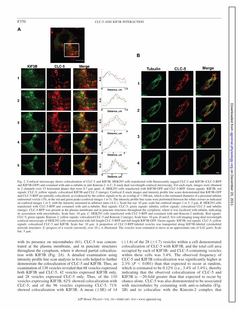
with its presence on microtubules (61). CLC-5 was concen-trated at the plasma membrane, and in punctate structuresthroughout the cytoplasm, where it showed partial colocaliza-tion with KIF3B (Fig. 2A). A detailed examination usingintensity profile line scan analysis in five cells helped to furtherdemonstrate the colocalization of CLC-5 and KIF3B. Thus, anexamination of 138 vesicles revealed that 68 vesicles expressedboth KIF3B and CLC-5, 42 vesicles expressed KIF3B only,and 28 vesicles expressed CLC-5 only. Thus, of the 110vesicles expressing KIF3B, 62% showed colocalization withCLC-5, and of the 96 vesicles expressing CLC-5, 71%showed colocalization with KIF3B. A mean ( SE) of 14
( 1.6) of the 28 ( 1.7) vesicles within a cell demonstratedcolocalization of CLC-5 with KIF3B, and the total cell areaoccupied by each of KIF3B- and CLC-5-containing vesicleswithin these cells was 3.4%. The observed frequency ofCLC-5 and KIF3B colocalization was significantly higher at2.3% (P 0.001) than that expected to occur at random,which is estimated to be 0.12% (i.e., 3.4% of 3.4%), therebyindicating that the observed colocalization of CLC-5 andKIF3B is �20-fold greater than that expected to occur bychance alone. CLC-5 was also demonstrated to be associatedwith microtubules by costaining with anti-�-tubulin (Fig.2B) and to colocalize with the Kinesin-2 complex that
Fig. 2. Confocal microscopy shows colocalization of CLC-5 and KIF3B. HEK293 cells transfected with fluorescently tagged CLC-5 and KIF3B (CLC-5-RFPand KIF3B-GFP) and costained with anti-�-tubulin or anti-Kinesin-2. A-C: Z-stack dual wavelength confocal microscopy. For each stack, images were obtainedin 2 channels over 15 horizontal planes that were 0.7 �m apart. A: HEK293 cells transfected with KIF3B-GFP and CLC-5-RFP. Green signals: KIF3B; redsignals: CLC-5; yellow signals: colocalized KIF3B and CLC-5 (merge). Confocal Z-stack images and intensity profile line scans demonstrated that KIF3B-GFPand CLC-5-RFP are partially colocalized, as evidenced by the yellow signals or by an overlap of 300 nm, which is the estimated diameter of a proximal tubularendosomal vesicle (19), in the red and green peaks (confocal images 1 to 5). The intensity profile line scans were performed between the white crosses as indicatedon confocal images 1 to 5, with the intensity measured in arbitrary units (A.U.). Scale bar top: 10 �m; scale bar confocal images 1 to 5: 2 �m. B: HEK293 cellstransfected with CLC-5-RFP and costained with anti-�-tubulin. Red signals: CLC-5; green signals: tubulin; yellow signals: colocalized CLC-5 and tubulin(merge). CLC-5-RFP was present at the plasma membrane and in punctate structures throughout the cytoplasm, where it was localized with tubulin, indicatingits association with microtubules. Scale bars: 10 �m. C: HEK293 cells transfected with CLC-5-RFP and costained with anti-Kinesin-2 antibody. Red signals:CLC-5; green signals: Kinesin-2; yellow signals: colocalized CLC-5 and Kinesin-2 (merge). Scale bars: 10 �m. D and E: live cell imaging using dual-wavelengthconfocal microscopy of HEK293 cells cotransfected with full-length CLC-5-RFP and full-length KIF3B-GFP. Green signals: KIF3B; red signals: CLC-5; yellowsignals: colocalized CLC-5 and KIF3B. Scale bar: 10 �m. A proportion of CLC-5-RFP-labeled vesicles was transported along KIF3B-labeled cytoskeletalnetwork structures. E: progress of a vesicle (arrowed), over 10 s, is illustrated. The vesicles were estimated to move at an approximate rate of 0.42 �m/s. Scalebar: 5 �m.
F370 CLC-5 AND KIF3B INTERACTION
AJP-Renal Physiol • VOL 298 • FEBRUARY 2010 • www.ajprenal.org
on Decem
ber 20, 2011ajprenal.physiology.org
Dow
nloaded from
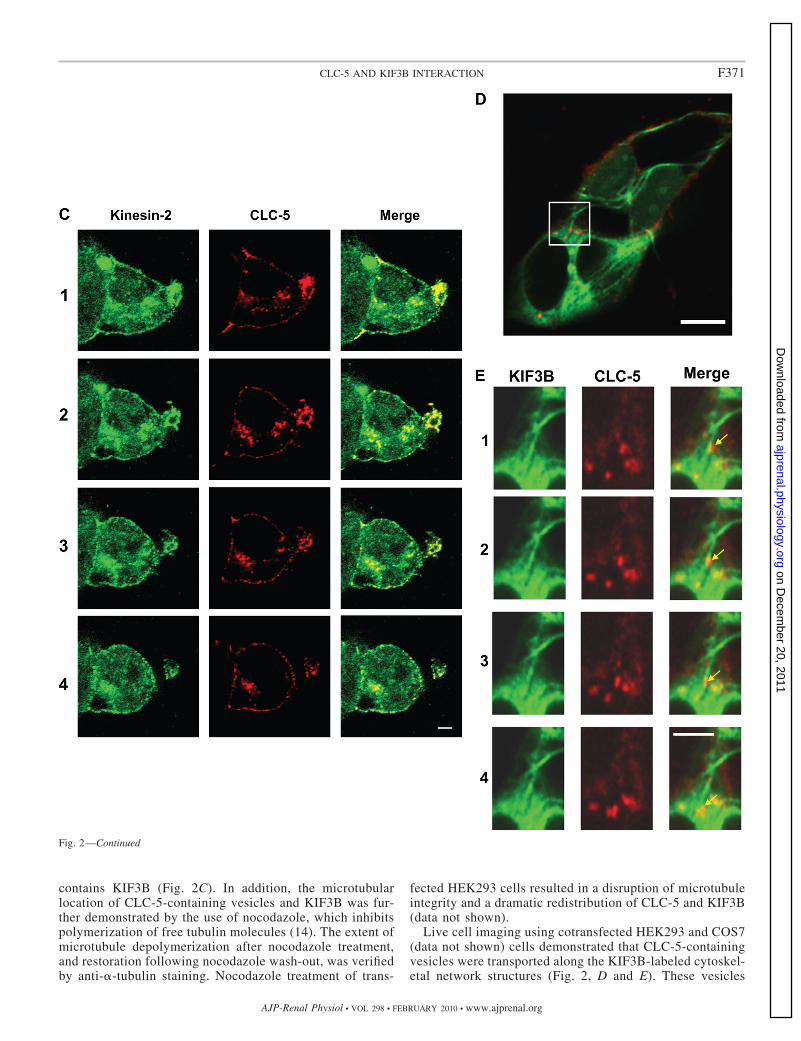
contains KIF3B (Fig. 2C). In addition, the microtubularlocation of CLC-5-containing vesicles and KIF3B was fur-ther demonstrated by the use of nocodazole, which inhibitspolymerization of free tubulin molecules (14). The extent ofmicrotubule depolymerization after nocodazole treatment,and restoration following nocodazole wash-out, was verifiedby anti-�-tubulin staining. Nocodazole treatment of trans-
fected HEK293 cells resulted in a disruption of microtubuleintegrity and a dramatic redistribution of CLC-5 and KIF3B(data not shown).
Live cell imaging using cotransfected HEK293 and COS7(data not shown) cells demonstrated that CLC-5-containingvesicles were transported along the KIF3B-labeled cytoskel-etal network structures (Fig. 2, D and E). These vesicles
Fig. 2—Continued
F371CLC-5 AND KIF3B INTERACTION
AJP-Renal Physiol • VOL 298 • FEBRUARY 2010 • www.ajprenal.org
on Decem
ber 20, 2011ajprenal.physiology.org
Dow
nloaded from
were estimated to move at an approximate rate of 0.42 �m/s.Thus, KIF3B and CLC-5 colocalize within the cell, therebysupporting the yeast two-hybrid and coimmunoprecipitationdata (Fig. 1, B–F), which indicate that the two proteinsphysically interact.
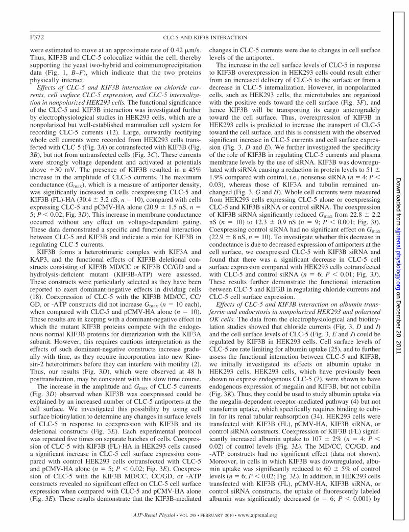
Effects of CLC-5 and KIF3B interaction on chloride cur-rents, cell surface CLC-5 expression, and CLC-5 internaliza-tion in nonpolarized HEK293 cells. The functional significanceof the CLC-5 and KIF3B interaction was investigated furtherby electrophysiological studies in HEK293 cells, which are anonpolarized but well-established mammalian cell system forrecording CLC-5 currents (12). Large, outwardly rectifyingwhole cell currents were recorded from HEK293 cells trans-fected with CLC-5 (Fig. 3A) or cotransfected with KIF3B (Fig.3B), but not from untransfected cells (Fig. 3C). These currentswere strongly voltage dependent and activated at potentialsabove �30 mV. The presence of KIF3B resulted in a 45%increase in the amplitude of CLC-5 currents. The maximumconductance (Gmax), which is a measure of antiporter density,was significantly increased in cells coexpressing CLC-5 andKIF3B (FL)-HA (30.4 3.2 nS, n 10), compared with cellsexpressing CLC-5 and pCMV-HA alone (20.9 1.5 nS, n 5; P 0.02; Fig. 3D). This increase in membrane conductanceoccurred without any effect on voltage-dependent gating.These data demonstrated a specific and functional interactionbetween CLC-5 and KIF3B and indicate a role for KIF3B inregulating CLC-5 currents.
KIF3B forms a heterotrimeric complex with KIF3A andKAP3, and the functional effects of KIF3B deletional con-structs consisting of KIF3B MD/CC or KIF3B CC/GD and ahydrolysis-deficient mutant (KIF3B-ATP) were assessed.These constructs were particularly selected as they have beenreported to exert dominant-negative effects in dividing cells(18). Coexpression of CLC-5 with the KIF3B MD/CC, CC/GD, or -ATP constructs did not increase Gmax (n 10 each),when compared with CLC-5 and pCMV-HA alone (n 10).These results are in keeping with a dominant-negative effect inwhich the mutant KIF3B proteins compete with the endoge-nous normal KIF3B proteins for dimerization with the KIF3Asubunit. However, this requires cautious interpretation as theeffects of such dominant-negative constructs increase gradu-ally with time, as they require incorporation into new Kine-sin-2 heterotrimers before they can interfere with motility (2).Thus, our results (Fig. 3D), which were observed at 48 hposttransfection, may be consistent with this slow time course.
The increase in the amplitude and Gmax of CLC-5 currents(Fig. 3D) observed when KIF3B was coexpressed could beexplained by an increased number of CLC-5 antiporters at thecell surface. We investigated this possibility by using cellsurface biotinylation to determine any changes in surface levelsof CLC-5 in response to coexpression with KIF3B and itsdeletional constructs (Fig. 3E). Each experimental protocolwas repeated five times on separate batches of cells. Coexpres-sion of CLC-5 with KIF3B (FL)-HA in HEK293 cells causeda significant increase in CLC-5 cell surface expression com-pared with control HEK293 cells cotransfected with CLC-5and pCMV-HA alone (n 5; P 0.02; Fig. 3E). Coexpres-sion of CLC-5 with the KIF3B MD/CC, CC/GD, or -ATPconstructs revealed no significant effect on CLC-5 cell surfaceexpression when compared with CLC-5 and pCMV-HA alone(Fig. 3E). These results demonstrate that the KIF3B-mediated
changes in CLC-5 currents were due to changes in cell surfacelevels of the antiporter.
The increase in the cell surface levels of CLC-5 in responseto KIF3B overexpression in HEK293 cells could result eitherfrom an increased delivery of CLC-5 to the surface or from adecrease in CLC-5 internalization. However, in nonpolarizedcells, such as HEK293 cells, the microtubules are organizedwith the positive ends toward the cell surface (Fig. 3F), andhence KIF3B will be transporting its cargo anterogradelytoward the cell surface. Thus, overexpression of KIF3B inHEK293 cells is predicted to increase the transport of CLC-5toward the cell surface, and this is consistent with the observedsignificant increase in CLC-5 currents and cell surface expres-sion (Fig. 3, D and E). We further investigated the specificityof the role of KIF3B in regulating CLC-5 currents and plasmamembrane levels by the use of siRNA. KIF3B was downregu-lated with siRNA causing a reduction in protein levels to 51 1.9% compared with control, i.e., nonsense siRNA (n 4; P 0.03), whereas those of KIF3A and tubulin remained un-changed (Fig. 3, G and H). Whole cell currents were measuredfrom HEK293 cells expressing CLC-5 alone or coexpressingCLC-5 and KIF3B siRNA or control siRNA. The coexpressionof KIF3B siRNA significantly reduced Gmax from 22.8 2.2nS (n 10) to 12.3 0.9 nS (n 9; P 0.001; Fig. 3I).Coexpressing control siRNA had no significant effect on Gmax
(22.9 8 nS, n 10). To investigate whether this decrease inconductance is due to decreased expression of antiporters at thecell surface, we coexpressed CLC-5 with KIF3B siRNA andfound that there was a significant decrease in CLC-5 cellsurface expression compared with HEK293 cells cotransfectedwith CLC-5 and control siRNA (n 6; P 0.01; Fig. 3J).These results further demonstrate the functional interactionbetween CLC-5 and KIF3B in regulating chloride currents andCLC-5 cell surface expression.
Effects of CLC-5 and KIF3B interaction on albumin trans-ferrin and endocytosis in nonpolarized HEK293 and polarizedOK cells. The data from the electrophysiological and biotiny-lation studies showed that chloride currents (Fig. 3, D and I)and the cell surface levels of CLC-5 (Fig. 3, E and J) could beregulated by KIF3B in HEK293 cells. Cell surface levels ofCLC-5 are rate limiting for albumin uptake (25), and to furtherassess the functional interaction between CLC-5 and KIF3B,we initially investigated its effects on albumin uptake inHEK293 cells. HEK293 cells, which have previously beenshown to express endogenous CLC-5 (7), were shown to haveendogenous expression of megalin and KIF3B, but not cubilin(Fig. 3K). Thus, they could be used to study albumin uptake viathe megalin-dependent receptor-mediated pathway (4) but nottransferrin uptake, which specifically requires binding to cubi-lin for its renal tubular reabsorption (34). HEK293 cells weretransfected with KIF3B (FL), pCMV-HA, KIF3B siRNA, orcontrol siRNA constructs. Coexpression of KIF3B (FL) signif-icantly increased albumin uptake to 107 2% (n 4; P 0.02) of control levels (Fig. 3L). The MD/CC, CC/GD, and-ATP constructs had no significant effect (data not shown).Moreover, in cells in which KIF3B was downregulated, albu-min uptake was significantly reduced to 60 5% of controllevels (n 6; P 0.02; Fig. 3L). In addition, in HEK293 cellstransfected with KIF3B (FL), pCMV-HA, KIF3B siRNA, orcontrol siRNA constructs, the uptake of fluorescently labeledalbumin was significantly decreased (n 6; P 0.001) by
F372 CLC-5 AND KIF3B INTERACTION
AJP-Renal Physiol • VOL 298 • FEBRUARY 2010 • www.ajprenal.org
on Decem
ber 20, 2011ajprenal.physiology.org
Dow
nloaded from
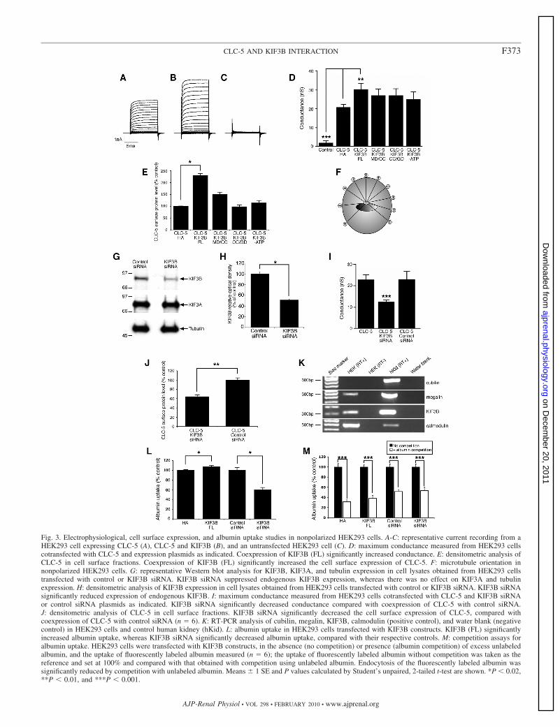
Fig. 3. Electrophysiological, cell surface expression, and albumin uptake studies in nonpolarized HEK293 cells. A-C: representative current recording from aHEK293 cell expressing CLC-5 (A), CLC-5 and KIF3B (B), and an untransfected HEK293 cell (C). D: maximum conductance measured from HEK293 cellscotransfected with CLC-5 and expression plasmids as indicated. Coexpression of KIF3B (FL) significantly increased conductance. E: densitometric analysis ofCLC-5 in cell surface fractions. Coexpression of KIF3B (FL) significantly increased the cell surface expression of CLC-5. F: microtubule orientation innonpolarized HEK293 cells. G: representative Western blot analysis for KIF3B, KIF3A, and tubulin expression in cell lysates obtained from HEK293 cellstransfected with control or KIF3B siRNA. KIF3B siRNA suppressed endogenous KIF3B expression, whereas there was no effect on KIF3A and tubulinexpression. H: densitometric analysis of KIF3B expression in cell lysates obtained from HEK293 cells transfected with control or KIF3B siRNA. KIF3B siRNAsignificantly reduced expression of endogenous KIF3B. I: maximum conductance measured from HEK293 cells cotransfected with CLC-5 and KIF3B siRNAor control siRNA plasmids as indicated. KIF3B siRNA significantly decreased conductance compared with coexpression of CLC-5 with control siRNA.J: densitometric analysis of CLC-5 in cell surface fractions. KIF3B siRNA significantly decreased the cell surface expression of CLC-5, compared withcoexpression of CLC-5 with control siRNA (n 6). K: RT-PCR analysis of cubilin, megalin, KIF3B, calmodulin (positive control), and water blank (negativecontrol) in HEK293 cells and control human kidney (hKid). L: albumin uptake in HEK293 cells transfected with KIF3B constructs. KIF3B (FL) significantlyincreased albumin uptake, whereas KIF3B siRNA significantly decreased albumin uptake, compared with their respective controls. M: competition assays foralbumin uptake. HEK293 cells were transfected with KIF3B constructs, in the absence (no competition) or presence (albumin competition) of excess unlabeledalbumin, and the uptake of fluorescently labeled albumin measured (n 6); the uptake of fluorescently labeled albumin without competition was taken as thereference and set at 100% and compared with that obtained with competition using unlabeled albumin. Endocytosis of the fluorescently labeled albumin wassignificantly reduced by competition with unlabeled albumin. Means 1 SE and P values calculated by Student’s unpaired, 2-tailed t-test are shown. *P 0.02,**P 0.01, and ***P 0.001.
F373CLC-5 AND KIF3B INTERACTION
AJP-Renal Physiol • VOL 298 • FEBRUARY 2010 • www.ajprenal.org
on Decem
ber 20, 2011ajprenal.physiology.org
Dow
nloaded from
incubation in the presence of excess unlabeled albumin (Fig.3M), indicating that the albumin uptake involves a receptor-mediated pathway. Thus, these results are in agreement withthose of the electrophysiology and biotinylation studies andfurther demonstrate the functional interaction between CLC-5and KIF3B. These results clearly show that KIF3B is a specificphysiological regulator of constitutive receptor-mediated albu-min uptake in HEK293 cells.
However, HEK293 cells are nonpolarized cells and havetheir microtubules organized with the positive ends toward thecell surface (Fig. 3F), whereas in polarized renal epithelial
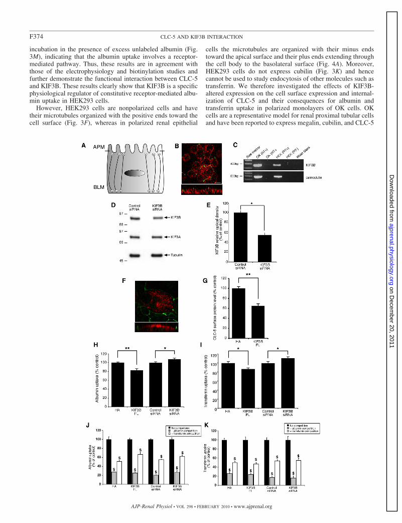
cells the microtubules are organized with their minus endstoward the apical surface and their plus ends extending throughthe cell body to the basolateral surface (Fig. 4A). Moreover,HEK293 cells do not express cubilin (Fig. 3K) and hencecannot be used to study endocytosis of other molecules such astransferrin. We therefore investigated the effects of KIF3B-altered expression on the cell surface expression and internal-ization of CLC-5 and their consequences for albumin andtransferrin uptake in polarized monolayers of OK cells. OKcells are a representative model for renal proximal tubular cellsand have been reported to express megalin, cubilin, and CLC-5
F374 CLC-5 AND KIF3B INTERACTION
AJP-Renal Physiol • VOL 298 • FEBRUARY 2010 • www.ajprenal.org
on Decem
ber 20, 2011ajprenal.physiology.org
Dow
nloaded from
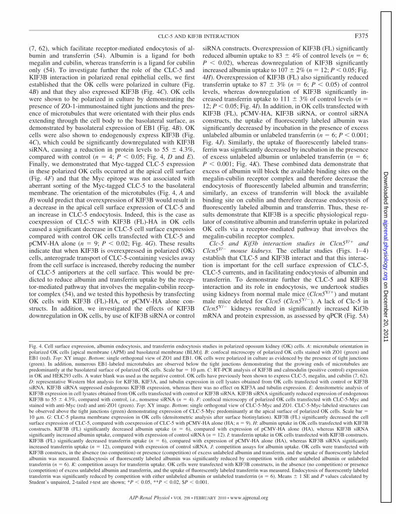
(7, 62), which facilitate receptor-mediated endocytosis of al-bumin and transferrin (54). Albumin is a ligand for bothmegalin and cubilin, whereas transferrin is a ligand for cubilinonly (54). To investigate further the role of the CLC-5 andKIF3B interaction in polarized renal epithelial cells, we firstestablished that the OK cells were polarized in culture (Fig.4B) and that they also expressed KIF3B (Fig. 4C). OK cellswere shown to be polarized in culture by demonstrating thepresence of ZO-1-immunostained tight junctions and the pres-ence of microtubules that were orientated with their plus endsextending through the cell body to the basolateral surface, asdemonstrated by basolateral expression of EB1 (Fig. 4B). OKcells were also shown to endogenously express KIF3B (Fig.4C), which could be significantly downregulated with KIF3BsiRNA, causing a reduction in protein levels to 55 4.3%,compared with control (n 4; P 0.05; Fig. 4, D and E).Finally, we demonstrated that Myc-tagged CLC-5 expressionin these polarized OK cells occurred at the apical cell surface(Fig. 4F) and that the Myc epitope was not associated withaberrant sorting of the Myc-tagged CLC-5 to the basolateralmembrane. The orientation of the microtubules (Fig. 4, A andB) would predict that overexpression of KIF3B would result ina decrease in the apical cell surface expression of CLC-5 andan increase in CLC-5 endocytosis. Indeed, this is the case ascoexpression of CLC-5 with KIF3B (FL)-HA in OK cellscaused a significant decrease in CLC-5 cell surface expressioncompared with control OK cells transfected with CLC-5 andpCMV-HA alone (n 9; P 0.02; Fig. 4G). These resultsindicate that when KIF3B is overexpressed in polarized (OK)cells, anterograde transport of CLC-5-containing vesicles awayfrom the cell surface is increased, thereby reducing the numberof CLC-5 antiporters at the cell surface. This would be pre-dicted to reduce albumin and transferrin uptake by the recep-tor-mediated pathway that involves the megalin-cubilin recep-tor complex (54), and we tested this hypothesis by transfectingOK cells with KIF3B (FL)-HA, or pCMV-HA alone con-structs. In addition, we investigated the effects of KIF3Bdownregulation in OK cells, by use of KIF3B siRNA or control
siRNA constructs. Overexpression of KIF3B (FL) significantlyreduced albumin uptake to 83 4% of control levels (n 6;P 0.02), whereas downregulation of KIF3B significantlyincreased albumin uptake to 107 2% (n 12; P 0.05; Fig.4H). Overexpression of KIF3B (FL) also significantly reducedtransferrin uptake to 87 3% (n 6; P 0.05) of controllevels, whereas downregulation of KIF3B significantly in-creased transferrin uptake to 111 3% of control levels (n 12; P 0.05; Fig. 4I). In addition, in OK cells transfected withKIF3B (FL), pCMV-HA, KIF3B siRNA, or control siRNAconstructs, the uptake of fluorescently labeled albumin wassignificantly decreased by incubation in the presence of excessunlabeled albumin or unlabeled transferrin (n 6; P 0.001;Fig. 4J). Similarly, the uptake of fluorescently labeled trans-ferrin was significantly decreased by incubation in the presenceof excess unlabeled albumin or unlabeled transferrin (n 6;P 0.001; Fig. 4K). These combined data demonstrate thatexcess of albumin will block the available binding sites on themegalin-cubilin receptor complex and therefore decrease theendocytosis of fluorescently labeled albumin and transferrin;similarly, an excess of transferrin will block the availablebinding site on cubilin and therefore decrease endocytosis offluorescently labeled albumin and transferrin. Thus, these re-sults demonstrate that KIF3B is a specific physiological regu-lator of constitutive albumin and transferrin uptake in polarizedOK cells via a receptor-mediated pathway that involves themegalin-cubilin receptor complex.
Clc-5 and Kif3b interaction studies in Clcn5Y/� andClcn5Y/� mouse kidneys. The cellular studies (Figs. 1–4)establish that CLC-5 and KIF3B interact and that this interac-tion is important for the cell surface expression of CLC-5,CLC-5 currents, and in facilitating endocytosis of albumin andtransferrin. To demonstrate further the CLC-5 and KIF3Binteraction and its role in endocytosis, we undertook studiesusing kidneys from normal male mice (Clcn5Y/�) and mutantmale mice deleted for Clcn5 (Clcn5Y/�). A lack of Clc-5 inClcn5Y/� kidneys resulted in significantly increased Kif3bmRNA and protein expression, as assessed by qPCR (Fig. 5A)
Fig. 4. Cell surface expression, albumin endocytosis, and transferrin endocytosis studies in polarized opossum kidney (OK) cells. A: microtubule orientation inpolarized OK cells [apical membrane (APM) and basolateral membrane (BLM)]. B: confocal microscopy of polarized OK cells stained with ZO1 (green) andEB1 (red). Top: XY image. Bottom: single orthogonal view of ZO1 and EB1. OK cells were polarized in culture as evidenced by the presence of tight junctions(green). In addition, numerous EB1-labeled microtubules are observed below the tight junctions demonstrating that the growing ends of microtubules arepredominantly at the basolateral surface of polarized OK cells. Scale bar 10 �m. C: RT-PCR analysis of KIF3B and calmodulin (positive control) expressionin OK and HEK293 cells. A water blank was used as the negative control. OK cells have previously been shown to express CLC-5, megalin, and cubilin (7, 62).D: representative Western blot analysis for KIF3B, KIF3A, and tubulin expression in cell lysates obtained from OK cells transfected with control or KIF3BsiRNA. KIF3B siRNA suppressed endogenous KIF3B expression, whereas there was no effect on KIF3A and tubulin expression. E: densitometric analysis ofKIF3B expression in cell lysates obtained from OK cells transfected with control or KIF3B siRNA. KIF3B siRNA significantly reduced expression of endogenousKIF3B to 55 4.3%, compared with control, i.e., nonsense siRNA (n 4). F: confocal microscopy of polarized OK cells transfected with CLC-5-Myc andstained with anti-Myc (red) and anti-ZO1 (green). Top: XY image. Bottom: single orthogonal view of CLC-5-Myc and ZO1. CLC-5-Myc-labeled structures canbe observed above the tight junctions (green) demonstrating expression of CLC-5-Myc predominantly at the apical surface of polarized OK cells. Scale bar 10 �m. G: CLC-5 plasma membrane expression in OK cells (densitometric analysis after surface biotinylation). KIF3B (FL) significantly decreased the cellsurface expression of CLC-5, compared with coexpression of CLC-5 with pCMV-HA alone (HA; n 9). H: albumin uptake in OK cells transfected with KIF3Bconstructs. KIF3B (FL) significantly decreased albumin uptake (n 6), compared with expression of pCMV-HA alone (HA), whereas KIF3B siRNAsignificantly increased albumin uptake, compared with expression of control siRNA (n 12). I: transferrin uptake in OK cells transfected with KIF3B constructs.KIF3B (FL) significantly decreased transferrin uptake (n 6), compared with expression of pCMV-HA alone (HA), whereas KIF3B siRNA significantlyincreased transferrin uptake (n 12), compared with expression of control siRNA. J: competition assays for albumin uptake. OK cells were transfected withKIF3B constructs, in the absence (no competition) or presence (competition) of excess unlabeled albumin and transferrin, and the uptake of fluorescently labeledalbumin was measured. Endocytosis of fluorescently labeled albumin was significantly reduced by competition with either unlabeled albumin or unlabeledtransferrin (n 6). K: competition assays for transferrin uptake. OK cells were transfected with KIF3B constructs, in the absence (no competition) or presence(competition) of excess unlabeled albumin and transferrin, and the uptake of fluorescently labeled transferrin was measured. Endocytosis of fluorescently labeledtransferrin was significantly reduced by competition with either unlabeled albumin or unlabeled transferrin (n 6). Means 1 SE and P values calculated byStudent’s unpaired, 2-tailed t-test are shown; *P 0.05, **P 0.02, $P 0.001.
F375CLC-5 AND KIF3B INTERACTION
AJP-Renal Physiol • VOL 298 • FEBRUARY 2010 • www.ajprenal.org
on Decem
ber 20, 2011ajprenal.physiology.org
Dow
nloaded from
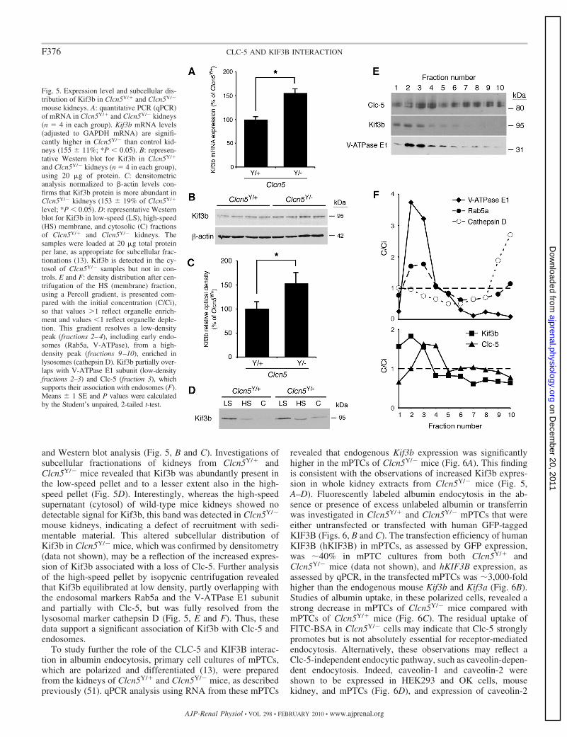
and Western blot analysis (Fig. 5, B and C). Investigations ofsubcellular fractionations of kidneys from Clcn5Y/� andClcn5Y/� mice revealed that Kif3b was abundantly present inthe low-speed pellet and to a lesser extent also in the high-speed pellet (Fig. 5D). Interestingly, whereas the high-speedsupernatant (cytosol) of wild-type mice kidneys showed nodetectable signal for Kif3b, this band was detected in Clcn5Y/�
mouse kidneys, indicating a defect of recruitment with sedi-mentable material. This altered subcellular distribution ofKif3b in Clcn5Y/� mice, which was confirmed by densitometry(data not shown), may be a reflection of the increased expres-sion of Kif3b associated with a loss of Clc-5. Further analysisof the high-speed pellet by isopycnic centrifugation revealedthat Kif3b equilibrated at low density, partly overlapping withthe endosomal markers Rab5a and the V-ATPase E1 subunitand partially with Clc-5, but was fully resolved from thelysosomal marker cathepsin D (Fig. 5, E and F). Thus, thesedata support a significant association of Kif3b with Clc-5 andendosomes.
To study further the role of the CLC-5 and KIF3B interac-tion in albumin endocytosis, primary cell cultures of mPTCs,which are polarized and differentiated (13), were preparedfrom the kidneys of Clcn5Y/� and Clcn5Y/� mice, as describedpreviously (51). qPCR analysis using RNA from these mPTCs
revealed that endogenous Kif3b expression was significantlyhigher in the mPTCs of Clcn5Y/� mice (Fig. 6A). This findingis consistent with the observations of increased Kif3b expres-sion in whole kidney extracts from Clcn5Y/� mice (Fig. 5,A–D). Fluorescently labeled albumin endocytosis in the ab-sence or presence of excess unlabeled albumin or transferrinwas investigated in Clcn5Y/� and Clcn5Y/� mPTCs that wereeither untransfected or transfected with human GFP-taggedKIF3B (Figs. 6, B and C). The transfection efficiency of humanKIF3B (hKIF3B) in mPTCs, as assessed by GFP expression,was �40% in mPTC cultures from both Clcn5Y/� andClcn5Y/� mice (data not shown), and hKIF3B expression, asassessed by qPCR, in the transfected mPTCs was �3,000-foldhigher than the endogenous mouse Kif3b and Kif3a (Fig. 6B).Studies of albumin uptake, in these polarized cells, revealed astrong decrease in mPTCs of Clcn5Y/� mice compared withmPTCs of Clcn5Y/� mice (Fig. 6C). The residual uptake ofFITC-BSA in Clcn5Y/� cells may indicate that Clc-5 stronglypromotes but is not absolutely essential for receptor-mediatedendocytosis. Alternatively, these observations may reflect aClc-5-independent endocytic pathway, such as caveolin-depen-dent endocytosis. Indeed, caveolin-1 and caveolin-2 wereshown to be expressed in HEK293 and OK cells, mousekidney, and mPTCs (Fig. 6D), and expression of caveolin-2
Fig. 5. Expression level and subcellular dis-tribution of Kif3b in Clcn5Y/� and Clcn5Y/�
mouse kidneys. A: quantitative PCR (qPCR)of mRNA in Clcn5Y/� and Clcn5Y/� kidneys(n 4 in each group). Kif3b mRNA levels(adjusted to GAPDH mRNA) are signifi-cantly higher in Clcn5Y/� than control kid-neys (155 11%; *P 0.05). B: represen-tative Western blot for Kif3b in Clcn5Y/�
and Clcn5Y/� kidneys (n 4 in each group),using 20 �g of protein. C: densitometricanalysis normalized to �-actin levels con-firms that Kif3b protein is more abundant inClcn5Y/� kidneys (153 19% of Clcn5Y/�
level; *P 0.05). D: representative Westernblot for Kif3b in low-speed (LS), high-speed(HS) membrane, and cytosolic (C) fractionsof Clcn5Y/� and Clcn5Y/� kidneys. Thesamples were loaded at 20 �g total proteinper lane, as appropriate for subcellular frac-tionations (13). Kif3b is detected in the cy-tosol of Clcn5Y/� samples but not in con-trols. E and F: density distribution after cen-trifugation of the HS (membrane) fraction,using a Percoll gradient, is presented com-pared with the initial concentration (C/Ci),so that values �1 reflect organelle enrich-ment and values 1 reflect organelle deple-tion. This gradient resolves a low-densitypeak (fractions 2–4), including early endo-somes (Rab5a, V-ATPase), from a high-density peak (fractions 9–10), enriched inlysosomes (cathepsin D). Kif3b partially over-laps with V-ATPase E1 subunit (low-densityfractions 2–3) and Clc-5 (fraction 3), whichsupports their association with endosomes (F).Means 1 SE and P values were calculatedby the Student’s unpaired, 2-tailed t-test.
F376 CLC-5 AND KIF3B INTERACTION
AJP-Renal Physiol • VOL 298 • FEBRUARY 2010 • www.ajprenal.org
on Decem
ber 20, 2011ajprenal.physiology.org
Dow
nloaded from
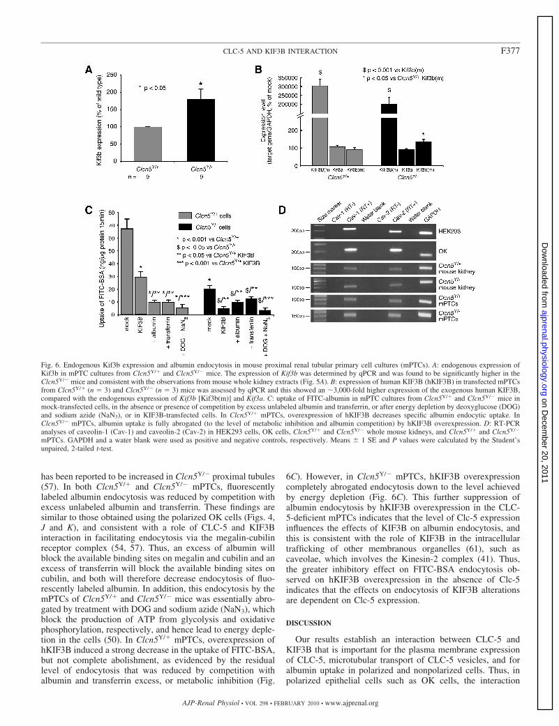
has been reported to be increased in Clcn5Y/� proximal tubules(57). In both Clcn5Y/� and Clcn5Y/� mPTCs, fluorescentlylabeled albumin endocytosis was reduced by competition withexcess unlabeled albumin and transferrin. These findings aresimilar to those obtained using the polarized OK cells (Figs. 4,J and K), and consistent with a role of CLC-5 and KIF3Binteraction in facilitating endocytosis via the megalin-cubilinreceptor complex (54, 57). Thus, an excess of albumin willblock the available binding sites on megalin and cubilin and anexcess of transferrin will block the available binding sites oncubilin, and both will therefore decrease endocytosis of fluo-rescently labeled albumin. In addition, this endocytosis by themPTCs of Clcn5Y/� and Clcn5Y/� mice was essentially abro-gated by treatment with DOG and sodium azide (NaN3), whichblock the production of ATP from glycolysis and oxidativephosphorylation, respectively, and hence lead to energy deple-tion in the cells (50). In Clcn5Y/� mPTCs, overexpression ofhKIF3B induced a strong decrease in the uptake of FITC-BSA,but not complete abolishment, as evidenced by the residuallevel of endocytosis that was reduced by competition withalbumin and transferrin excess, or metabolic inhibition (Fig.
6C). However, in Clcn5Y/� mPTCs, hKIF3B overexpressioncompletely abrogated endocytosis down to the level achievedby energy depletion (Fig. 6C). This further suppression ofalbumin endocytosis by hKIF3B overexpression in the CLC-5-deficient mPTCs indicates that the level of Clc-5 expressioninfluences the effects of KIF3B on albumin endocytosis, andthis is consistent with the role of KIF3B in the intracellulartrafficking of other membranous organelles (61), such ascaveolae, which involves the Kinesin-2 complex (41). Thus,the greater inhibitory effect on FITC-BSA endocytosis ob-served on hKIF3B overexpression in the absence of Clc-5indicates that the effects on endocytosis of KIF3B alterationsare dependent on Clc-5 expression.
DISCUSSION
Our results establish an interaction between CLC-5 andKIF3B that is important for the plasma membrane expressionof CLC-5, microtubular transport of CLC-5 vesicles, and foralbumin uptake in polarized and nonpolarized cells. Thus, inpolarized epithelial cells such as OK cells, the interaction
Fig. 6. Endogenous Kif3b expression and albumin endocytosis in mouse proximal renal tubular primary cell cultures (mPTCs). A: endogenous expression ofKif3b in mPTC cultures from Clcn5Y/� and Clcn5Y/� mice. The expression of Kif3b was determined by qPCR and was found to be significantly higher in theClcn5Y/� mice and consistent with the observations from mouse whole kidney extracts (Fig. 5A). B: expression of human KIF3B (hKIF3B) in transfected mPTCsfrom Clcn5Y/� (n 3) and Clcn5Y/� (n 3) mice was assessed by qPCR and this showed an �3,000-fold higher expression of the exogenous human KIF3B,compared with the endogenous expression of Kif3b [Kif3b(m)] and Kif3a. C: uptake of FITC-albumin in mPTC cultures from Clcn5Y/� and Clcn5Y/� mice inmock-transfected cells, in the absence or presence of competition by excess unlabeled albumin and transferrin, or after energy depletion by deoxyglucose (DOG)and sodium azide (NaN3), or in KIF3B-transfected cells. In Clcn5Y/� mPTCs, overexpression of hKIF3B decreases specific albumin endocytic uptake. InClcn5Y/� mPTCs, albumin uptake is fully abrogated (to the level of metabolic inhibition and albumin competition) by hKIF3B overexpression. D: RT-PCRanalyses of caveolin-1 (Cav-1) and caveolin-2 (Cav-2) in HEK293 cells, OK cells, Clcn5Y/� and Clcn5Y/� whole mouse kidneys, and Clcn5Y/� and Clcn5Y/�
mPTCs. GAPDH and a water blank were used as positive and negative controls, respectively. Means 1 SE and P values were calculated by the Student’sunpaired, 2-tailed t-test.
F377CLC-5 AND KIF3B INTERACTION
AJP-Renal Physiol • VOL 298 • FEBRUARY 2010 • www.ajprenal.org
on Decem
ber 20, 2011ajprenal.physiology.org
Dow
nloaded from
between CLC-5 and KIF3B results in a reduction in the numberof antiporters at the cell surface, as KIF3B is involved infacilitating microtubular transport that removes CLC-5-con-taining vesicles from the cell surface (Fig. 7). In nonpolarizedcells, such as HEK293 cells, the orientation of the microtubulesis reversed (Fig. 3F), and the interaction between CLC-5 andKIF3B facilitates transport of CLC-5-containing vesicles to thecell surface. This situation may be analogous to that reportedfor the interaction between CLC-2 and the microtubule-asso-ciated dynein motor complex, which is also involved in regu-lating CLC-2 cell surface expression through endosomal traf-ficking (8). However, CLC-2 is ubiquitously expressed,whereas CLC-5 is predominantly expressed in the kidney andextrapolations between these two situations may require cau-
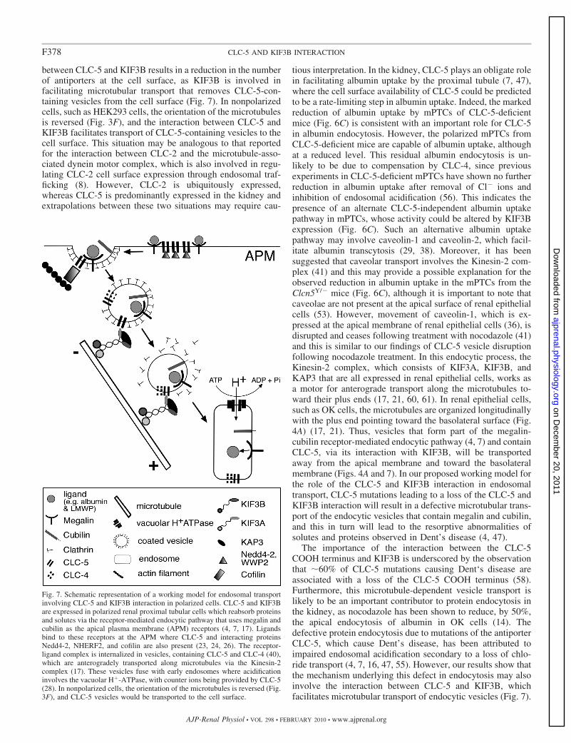
tious interpretation. In the kidney, CLC-5 plays an obligate rolein facilitating albumin uptake by the proximal tubule (7, 47),where the cell surface availability of CLC-5 could be predictedto be a rate-limiting step in albumin uptake. Indeed, the markedreduction of albumin uptake by mPTCs of CLC-5-deficientmice (Fig. 6C) is consistent with an important role for CLC-5in albumin endocytosis. However, the polarized mPTCs fromCLC-5-deficient mice are capable of albumin uptake, althoughat a reduced level. This residual albumin endocytosis is un-likely to be due to compensation by CLC-4, since previousexperiments in CLC-5-deficient mPTCs have shown no furtherreduction in albumin uptake after removal of Cl� ions andinhibition of endosomal acidification (56). This indicates thepresence of an alternate CLC-5-independent albumin uptakepathway in mPTCs, whose activity could be altered by KIF3Bexpression (Fig. 6C). Such an alternative albumin uptakepathway may involve caveolin-1 and caveolin-2, which facil-itate albumin transcytosis (29, 38). Moreover, it has beensuggested that caveolar transport involves the Kinesin-2 com-plex (41) and this may provide a possible explanation for theobserved reduction in albumin uptake in the mPTCs from theClcn5Y/� mice (Fig. 6C), although it is important to note thatcaveolae are not present at the apical surface of renal epithelialcells (53). However, movement of caveolin-1, which is ex-pressed at the apical membrane of renal epithelial cells (36), isdisrupted and ceases following treatment with nocodazole (41)and this is similar to our findings of CLC-5 vesicle disruptionfollowing nocodazole treatment. In this endocytic process, theKinesin-2 complex, which consists of KIF3A, KIF3B, andKAP3 that are all expressed in renal epithelial cells, works asa motor for anterograde transport along the microtubules to-ward their plus ends (17, 21, 60, 61). In renal epithelial cells,such as OK cells, the microtubules are organized longitudinallywith the plus end pointing toward the basolateral surface (Fig.4A) (17, 21). Thus, vesicles that form part of the megalin-cubilin receptor-mediated endocytic pathway (4, 7) and containCLC-5, via its interaction with KIF3B, will be transportedaway from the apical membrane and toward the basolateralmembrane (Figs. 4A and 7). In our proposed working model forthe role of the CLC-5 and KIF3B interaction in endosomaltransport, CLC-5 mutations leading to a loss of the CLC-5 andKIF3B interaction will result in a defective microtubular trans-port of the endocytic vesicles that contain megalin and cubilin,and this in turn will lead to the resorptive abnormalities ofsolutes and proteins observed in Dent’s disease (4, 47).
The importance of the interaction between the CLC-5COOH terminus and KIF3B is underscored by the observationthat �60% of CLC-5 mutations causing Dent‘s disease areassociated with a loss of the CLC-5 COOH terminus (58).Furthermore, this microtubule-dependent vesicle transport islikely to be an important contributor to protein endocytosis inthe kidney, as nocodazole has been shown to reduce, by 50%,the apical endocytosis of albumin in OK cells (14). Thedefective protein endocytosis due to mutations of the antiporterCLC-5, which cause Dent’s disease, has been attributed toimpaired endosomal acidification secondary to a loss of chlo-ride transport (4, 7, 16, 47, 55). However, our results show thatthe mechanism underlying this defect in endocytosis may alsoinvolve the interaction between CLC-5 and KIF3B, whichfacilitates microtubular transport of endocytic vesicles (Fig. 7).
Fig. 7. Schematic representation of a working model for endosomal transportinvolving CLC-5 and KIF3B interaction in polarized cells. CLC-5 and KIF3Bare expressed in polarized renal proximal tubular cells which reabsorb proteinsand solutes via the receptor-mediated endocytic pathway that uses megalin andcubilin as the apical plasma membrane (APM) receptors (4, 7, 17). Ligandsbind to these receptors at the APM where CLC-5 and interacting proteinsNedd4-2, NHERF2, and cofilin are also present (23, 24, 26). The receptor-ligand complex is internalized in vesicles, containing CLC-5 and CLC-4 (40),which are anterogradely transported along microtubules via the Kinesin-2complex (17). These vesicles fuse with early endosomes where acidificationinvolves the vacuolar H�-ATPase, with counter ions being provided by CLC-5(28). In nonpolarized cells, the orientation of the microtubules is reversed (Fig.3F), and CLC-5 vesicles would be transported to the cell surface.
F378 CLC-5 AND KIF3B INTERACTION
AJP-Renal Physiol • VOL 298 • FEBRUARY 2010 • www.ajprenal.org
on Decem
ber 20, 2011ajprenal.physiology.org
Dow
nloaded from
The COOH terminus of the family of nine mammalian CLCs(CLC-1 to CLC-7, CLC-Ka, and CLC-Kb) is highly conservedand includes the two CBS domains (11). The CLCs andKinesin family members are also expressed in multiple tissues(17, 28), including the kidney and brain (43). These featuresindicate that interactions between other CLCs and Kinesinfamily members may be involved in endosomal trafficking andmicrotubule-dependent transport in other tissues (43), and thefindings of our study may have implications for other renal,neural, retinal, muscular, and skeletal disorders associated withCLC mutations (22, 28, 32). The likelihood of such interac-tions between CLCs and Kinesin family members and theirrole in associated diseases is high as it has been established thatother transmembrane channels (e.g., the transient receptorprotein polycystin-2 and polycystin-2), transporter proteins(e.g., the cystic fibrosis transmembrane regulator and sodium-phosphate cotransporter type IIa), and exchangers (e.g., thesodium-hydrogen exchanger, NHE3) can form functional in-teractions with intracellular proteins (e.g., the phosphofurinacidic cluster sorting protein, Kinesin-2, NHERF1/2, merlin-ezrin-moesin, and myosin VIIb) and that these interactions canhave a vital impact on their physiological activity (9, 15, 20,33, 59).
In summary, our studies show that CLC-5 and KIF3Bdirectly interact in vivo and this interaction involves the COOHterminus of CLC-5 and the CC and GDs of KIF3B. Theinteraction with KIF3B alters CLC-5 cell surface expression,whole cell chloride current amplitudes, and is associated withaltered albumin endocytosis. The role of the interaction withKIF3B is to facilitate microtubular transport and endocytosis ofCLC-5-containing vesicles away from the cell surface of po-larized epithelial cells. Thus, our findings, which represent anovel mechanism of ion channel and transporter regulation,have elucidated that an interaction between CLC-5 and KIF3Bis important for the plasma membrane expression of CLC-5,and for the microtubular transport of endosomes.
ACKNOWLEDGMENTS
We are grateful to T. J. Jentsch for helpful discussions and B. Guggino forthe gift of the Clcn5-deleted mice.
GRANTS
This work was supported by the Wellcome Trust (A. A. C. Reed, N. Y. Loh,J. D. Lippiat, C. J. Patridge, J. Galvanovskis, P. Rorsman, F. M. Ashcroft, andR. V. Thakker); the Medical Research Council (M. A. Nesbit, A. A. C. Reed, andR. V. Thakker); the National Kidney Research Fund (S. E. Williams and R. V.Thakker); Nuffield Dominions Trust (F. Wu); Belgian agencies FNRS, FRSM, theFoundation Alphonse et Jean Forton, Concerted Research Actions, Inter-Univer-sity Attraction Poles (P. Courtoy and O. Devuyst), and the EuReGene, FP6,integrated project of the European Community (A. A. C. Reed, N. Y. Loh, S.Terryn, S. E. Williams, F. Jouret, P. Courtoy, O. Devuyst, and R. V. Thakker).
DISCLOSURES
No conflicts of interest are declared by the authors.
REFERENCES
1. Basu SK, Goldstein JL, Anderson RG, Brown MS. Monensin interruptsthe recycling of low density lipoprotein receptors in human fibroblasts.Cell 24: 493–502, 1981.
2. Brown CL, Maier KC, Stauber T, Ginkel LM, Wordeman L, VernosI, Schroer TA. Kinesin-2 is a motor for late endosomes and lysosomes.Traffic 6: 1114–1124, 2005.
3. Campbell RE, Tour O, Palmer AE, Steinbach PA, Baird GS, Zachar-ias DA, Tsien RY. A monomeric red fluorescent protein. Proc Natl AcadSci USA 99: 7877–7882, 2002.
4. Christensen EI, Devuyst O, Dom G, Nielsen R, Van der Smissen P,Verroust P, Leruth M, Guggino WB, Courtoy PJ. Loss of chloridechannel ClC-5 impairs endocytosis by defective trafficking of megalin andcubilin in kidney proximal tubules. Proc Natl Acad Sci USA 100: 8472–8477, 2003.
5. Cole DG, Chinn SW, Wedaman KP, Hall K, Vuong T, Scholey JM.Novel heterotrimeric kinesin-related protein purified from sea urchin eggs.Nature 366: 268–270, 1993.
6. Cupers P, Veithen A, Hoekstra D, Baudhuin P, Courtoy PJ. Threeunrelated perturbations similarly uncouple fluid, bulk-membrane, andreceptor endosomal flow in rat fetal fibroblasts. Biochem Biophys ResCommun 236: 661–664, 1997.
7. Devuyst O, Christie PT, Courtoy PJ, Beauwens R, Thakker RV.Intrarenal and subcellular distribution of the human chloride channel,CLC-5, reveals a pathophysiological basis for Dent’s disease. Hum MolGenet 8: 247–257, 1999.
8. Dhani SU, Mohammad-Panah R, Ahmed N, Ackerley C, RamjeesinghM, Bear CE. Evidence for a functional interaction between the ClC-2chloride channel and the retrograde motor dynein complex. J Biol Chem278: 16262–16270, 2003.
9. Donowitz M, Cha B, Zachos NC, Brett CL, Sharma A, Tse CM, Li X.NHERF family and NHE3 regulation. J Physiol 567: 3–11, 2005.
10. Dutzler R, Campbell EB, Cadene M, Chait BT, MacKinnon R. X-raystructure of a ClC chloride channel at 3.0 A reveals the molecular basis ofanion selectivity. Nature 415: 287–294, 2002.
11. Estevez R, Pusch M, Ferrer-Costa C, Orozco M, Jentsch TJ. Func-tional and structural conservation of CBS domains from CLC chloridechannels. J Physiol 557: 363–378, 2004.
12. Friedrich T, Breiderhoff T, Jentsch TJ. Mutational analysis demon-strates that ClC-4 and ClC-5 directly mediate plasma membrane currents.J Biol Chem 274: 896–902, 1999.
13. Gailly P, Jouret F, Martin D, Debaix H, Parreira KS, Nishita T,Blanchard A, Antignac C, Willnow TE, Courtoy PJ, Scheinman SJ,Christensen EI, Devuyst O. A novel renal carbonic anhydrase type IIIplays a role in proximal tubule dysfunction. Kidney Int 74: 52–61, 2008.
14. Gekle M, Mildenberger S, Freudinger R, Schwerdt G, Silbernagl S.Albumin endocytosis in OK cells: dependence on actin and microtubulesand regulation by protein kinases. Am J Physiol Renal Physiol 272:F668–F677, 1997.
15. Guggino WB. The cystic fibrosis transmembrane regulator forms macro-molecular complexes with PDZ domain scaffold proteins. Proc Am Tho-rac Soc 1: 28–32, 2004.
16. Gunther W, Luchow A, Cluzeaud F, Vandewalle A, Jentsch TJ. ClC-5,the chloride channel mutated in Dent’s disease, colocalizes with the protonpump in endocytotically active kidney cells. Proc Natl Acad Sci USA 95:8075–8080, 1998.
17. Hamm-Alvarez SF, Sheetz MP. Microtubule-dependent vesicle trans-port: modulation of channel and transporter activity in liver and kidney.Physiol Rev 78: 1109–1129, 1998.
18. Haraguchi K, Hayashi T, Jimbo T, Yamamoto T, Akiyama T. Role ofthe kinesin-2 family protein, KIF3, during mitosis. J Biol Chem 281:4094–4099, 2006.
19. Hatae T, Ichimura T, Ishida T, Sakurai T. Apical tubular network in therat kidney proximal tubule cells studied by thick section and scanningelectron microscopy. Cell Tissue Res 288: 317–325, 1997.
20. Hernando N, Gisler SM, Pribanic S, Deliot N, Capuano P, WagnerCA, Moe OW, Biber J, Murer H. NaPi-IIa and interacting partners. JPhysiol 567: 21–26, 2005.
21. Hirokawa N. Stirring up development with the heterotrimeric kinesinKIF3. Traffic 1: 29–34, 2000.
22. Hirokawa N, Takemura R. Biochemical and molecular characterizationof diseases linked to motor proteins. Trends Biochem Sci 28: 558–565,2003.
23. Hryciw DH, Ekberg J, Ferguson C, Lee A, Wang D, Parton RG,Pollock CA, Yun CC, Poronnik P. Regulation of albumin endocytosis byPSD95/Dlg/ZO-1 (PDZ) scaffolds. Interaction of Na�-H� exchange reg-ulatory factor-2 with ClC-5. J Biol Chem 281: 16068–16077, 2006.
24. Hryciw DH, Ekberg J, Lee A, Lensink IL, Kumar S, Guggino WB,Cook DI, Pollock CA, Poronnik P. Nedd4-2 functionally interacts withClC-5: involvement in constitutive albumin endocytosis in proximal tu-bule cells. J Biol Chem 279: 54996–55007, 2004.
F379CLC-5 AND KIF3B INTERACTION
AJP-Renal Physiol • VOL 298 • FEBRUARY 2010 • www.ajprenal.org
on Decem
ber 20, 2011ajprenal.physiology.org
Dow
nloaded from
25. Hryciw DH, Ekberg J, Pollock CA, Poronnik P. ClC-5: a chloridechannel with multiple roles in renal tubular albumin uptake. Int J BiochemCell Biol 38: 1036–1042, 2006.
26. Hryciw DH, Wang Y, Devuyst O, Pollock CA, Poronnik P, GugginoWB. Cofilin interacts with ClC-5 and regulates albumin uptake in proxi-mal tubule cell lines. J Biol Chem 278: 40169–40176, 2003.
27. Jaulin F, Xue X, Rodriguez-Boulan E, Kreitzer G. Polarization-depen-dent selective transport to the apical membrane by KIF5B in MDCK cells.Dev Cell 13: 511–522, 2007.
28. Jentsch TJ, Hubner CA, Fuhrmann JC. Ion channels: function unrav-elled by dysfunction. Nat Cell Biol 6: 1039–1047, 2004.
29. John TA, Vogel SM, Minshall RD, Ridge K, Tiruppathi C, Malik AB.Evidence for the role of alveolar epithelial gp60 in active transalveolaralbumin transport in the rat lung. J Physiol 533: 547–559, 2001.
30. Jouret F, Bernard A, Hermans C, Dom G, Terryn S, Leal T, LebecqueP, Cassiman JJ, Scholte BJ, de Jonge HR, Courtoy PJ, Devuyst O.Cystic fibrosis is associated with a defect in apical receptor-mediatedendocytosis in mouse and human kidney. J Am Soc Nephrol 18: 707–718,2007.
31. Jouret F, Igarashi T, Gofflot F, Wilson PD, Karet FE, Thakker RV,Devuyst O. Comparative ontogeny, processing, and segmental distribu-tion of the renal chloride channel, ClC-5. Kidney Int 65: 198–208, 2004.
32. Kasper D, Planells-Cases R, Fuhrmann JC, Scheel O, Zeitz O, Ru-ether K, Schmitt A, Poet M, Steinfeld R, Schweizer M, Kornak U,Jentsch TJ. Loss of the chloride channel ClC-7 leads to lysosomal storagedisease and neurodegeneration. EMBO J 24: 1079–1091, 2005.
33. Kottgen M, Benzing T, Simmen T, Tauber R, Buchholz B, FeliciangeliS, Huber TB, Schermer B, Kramer-Zucker A, Hopker K, SimmenKC, Tschucke CC, Sandford R, Kim E, Thomas G, Walz G. Traffick-ing of TRPP2 by PACS proteins represents a novel mechanism of ionchannel regulation. EMBO J 24: 705–716, 2005.
34. Kozyraki R, Fyfe J, Verroust PJ, Jacobsen C, Dautry-Varsat A,Gburek J, Willnow TE, Christensen EI, Moestrup SK. Megalin-dependent cubilin-mediated endocytosis is a major pathway for the apicaluptake of transferrin in polarized epithelia. Proc Natl Acad Sci USA 98:12491–12496, 2001.
35. Lawrence CJ, Dawe RK, Christie KR, Cleveland DW, Dawson SC,Endow SA, Goldstein LS, Goodson HV, Hirokawa N, Howard J,Malmberg RL, McIntosh JR, Miki H, Mitchison TJ, Okada Y, ReddyAS, Saxton WM, Schliwa M, Scholey JM, Vale RD, Walczak CE,Wordeman L. A standardized kinesin nomenclature. J Cell Biol 167:19–22, 2004.
36. Lee IH, Campbell CR, Song SH, Day ML, Kumar S, Cook DI,Dinudom A. The activity of the epithelial sodium channels is regulated bycaveolin-1 via a Nedd4-2-dependent mechanism. J Biol Chem 284:12663–12669, 2009.
37. Lloyd SE, Pearce SH, Fisher SE, Steinmeyer K, Schwappach B,Scheinman SJ, Harding B, Bolino A, Devoto M, Goodyer P, RigdenSP, Wrong O, Jentsch TJ, Craig IW, Thakker RV. A commonmolecular basis for three inherited kidney stone diseases. Nature 379:445–449, 1996.
38. Minshall RD, Tiruppathi C, Vogel SM, Niles WD, Gilchrist A, HammHE, Malik AB. Endothelial cell surface gp60 activates vesicle formationand trafficking via G(i)-coupled Src kinase signaling pathway. J Cell Biol150: 1057–1070, 2000.
39. Mohammad-Panah R, Ackerley C, Rommens J, Choudhury M, WangY, Bear CE. The chloride channel ClC-4 colocalizes with cystic fibrosistransmembrane conductance regulator and may mediate chloride fluxacross the apical membrane of intestinal epithelia. J Biol Chem 277:566–574, 2002.
40. Mohammad-Panah R, Harrison R, Dhani S, Ackerley C, Huan LJ,Wang Y, Bear CE. The chloride channel ClC-4 contributes to endosomalacidification and trafficking. J Biol Chem 278: 29267–29277, 2003.
41. Mundy DI, Machleidt T, Ying YS, Anderson RG, Bloom GS. Dualcontrol of caveolar membrane traffic by microtubules and the actincytoskeleton. J Cell Sci 115: 4327–4339, 2002.
42. Nesbit MA, Bowl MR, Harding B, Ali A, Ayala A, Crowe C, DobbieA, Hampson G, Holdaway I, Levine MA, McWilliams R, Rigden S,Sampson J, Williams AJ, Thakker RV. Characterization of GATA3mutations in the hypoparathyroidism, deafness, and renal dysplasia (HDR)syndrome. J Biol Chem 279: 22624–22634, 2004.
43. Nishimura T, Kato K, Yamaguchi T, Fukata Y, Ohno S, Kaibuchi K.Role of the PAR-3-KIF3 complex in the establishment of neuronalpolarity. Nat Cell Biol 6: 328–334, 2004.
44. Pfaffl MW. A new mathematical model for relative quantification inreal-time RT-PCR. Nucleic Acids Res 29: e45, 2001.
45. Picollo A, Pusch M. Chloride/proton antiporter activity of mammalianCLC proteins ClC-4 and ClC-5. Nature 436: 420–423, 2005.
46. Pirozzi G, McConnell SJ, Uveges AJ, Carter JM, Sparks AB, Kay BK,Fowlkes DM. Identification of novel human WW domain-containingproteins by cloning of ligand targets. J Biol Chem 272: 14611–14616,1997.
47. Piwon N, Gunther W, Schwake M, Bosl MR, Jentsch TJ. ClC-5 Cl�
channel disruption impairs endocytosis in a mouse model for Dent’sdisease. Nature 408: 369–373, 2000.
48. Scheel O, Zdebik AA, Lourdel S, Jentsch TJ. Voltage-dependentelectrogenic chloride/proton exchange by endosomal CLC proteins. Na-ture 436: 424–427, 2005.
49. Schwake M, Friedrich T, Jentsch TJ. An internalization signal in ClC-5,an endosomal Cl� channel mutated in dent’s disease. J Biol Chem 276:12049–12054, 2001.
50. Schwoebel ED, Ho TH, Moore MS. The mechanism of inhibition ofRan-dependent nuclear transport by cellular ATP depletion. J Cell Biol157: 963–974, 2002.
51. Terryn S, Jouret F, Vandenabeele F, Smolders I, Moreels M, DevuystO, Steels P, Van Kerkhove E. A primary culture of mouse proximaltubular cells, established on collagen-coated membranes. Am J PhysiolRenal Physiol 293: F476–F485, 2007.
52. Tyteca D, Van Der Smissen P, Mettlen M, Van Bambeke F, TulkensPM, Mingeot-Leclercq MP, Courtoy PJ. Azithromycin, a lysosomo-tropic antibiotic, has distinct effects on fluid-phase and receptor-mediatedendocytosis, but does not impair phagocytosis in J774 macrophages. ExpCell Res 281: 86–100, 2002.
53. Verkade P, Harder T, Lafont F, Simons K. Induction of caveolae in theapical plasma membrane of Madin-Darby canine kidney cells. J Cell Biol148: 727–739, 2000.
54. Verroust PJ, Birn H, Nielsen R, Kozyraki R, Christensen EI. Thetandem endocytic receptors megalin and cubilin are important proteins inrenal pathology. Kidney Int 62: 745–756, 2002.
55. Wang SS, Devuyst O, Courtoy PJ, Wang XT, Wang H, Wang Y,Thakker RV, Guggino S, Guggino WB. Mice lacking renal chloridechannel, CLC-5, are a model for Dent’s disease, a nephrolithiasis disorderassociated with defective receptor-mediated endocytosis. Hum Mol Genet9: 2937–2945, 2000.
56. Wang Y, Cai H, Cebotaru L, Hryciw DH, Weinman EJ, Donowitz M,Guggino SE, Guggino WB. ClC-5: role in endocytosis in the proximaltubule. Am J Physiol Renal Physiol 289: F850–F862, 2005.
57. Wright J, Morales MM, Sousa-Menzes J, Ornellas D, Sipes J, Cui Y,Cui I, Hulamm P, Cebotaru V, Cebotaru L, Guggino WB, GugginoSE. Transcriptional adaptation to Clcn5 knockout in proximal tubules ofmouse kidney. Physiol Genomics 33: 341–354, 2008.
58. Wu F, Roche P, Christie PT, Loh NY, Reed AA, Esnouf RM, ThakkerRV. Modeling study of human renal chloride channel (hCLC-5) mutationssuggests a structural-functional relationship. Kidney Int 63: 1426–1432,2003.
59. Wu Y, Dai XQ, Li Q, Chen CX, Mai W, Hussain Z, Long W,Montalbetti N, Li G, Glynne R, Wang S, Cantiello HF, Wu G, ChenXZ. Kinesin-2 mediates physical and functional interactions betweenpolycystin-2 and fibrocystin. Hum Mol Genet 15: 3280–3292, 2006.
60. Yamazaki H, Nakata T, Okada Y, Hirokawa N. Cloning and charac-terization of KAP3: a novel kinesin superfamily-associated protein ofKIF3A/3B. Proc Natl Acad Sci USA 93: 8443–8448, 1996.
61. Yamazaki H, Nakata T, Okada Y, Hirokawa N. KIF3A/B: a het-erodimeric kinesin superfamily protein that works as a microtubule plusend-directed motor for membrane organelle transport. J Cell Biol 130:1387–1399, 1995.
62. Zhai XY, Nielsen R, Birn H, Drumm K, Mildenberger S, FreudingerR, Moestrup SK, Verroust PJ, Christensen EI, Gekle M. Cubilin- andmegalin-mediated uptake of albumin in cultured proximal tubule cells ofopossum kidney. Kidney Int 58: 1523–1533, 2000.
F380 CLC-5 AND KIF3B INTERACTION
AJP-Renal Physiol • VOL 298 • FEBRUARY 2010 • www.ajprenal.org
on Decem
ber 20, 2011ajprenal.physiology.org
Dow
nloaded from