Embed Size (px)
Citation preview
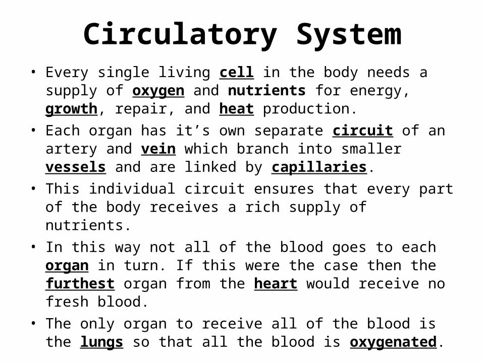
Circulatory System• Every single living cell in the body needs a supply
of oxygen and nutrients for energy, growth, repair, and heat production.
• Each organ has it’s own separate circuit of an artery and vein which branch into smaller vessels and are linked by capillaries.
• This individual circuit ensures that every part of the body receives a rich supply of nutrients.
• In this way not all of the blood goes to each organ in turn. If this were the case then the furthest organ from the heart would receive no fresh blood.
• The only organ to receive all of the blood is the lungs so that all the blood is oxygenated.
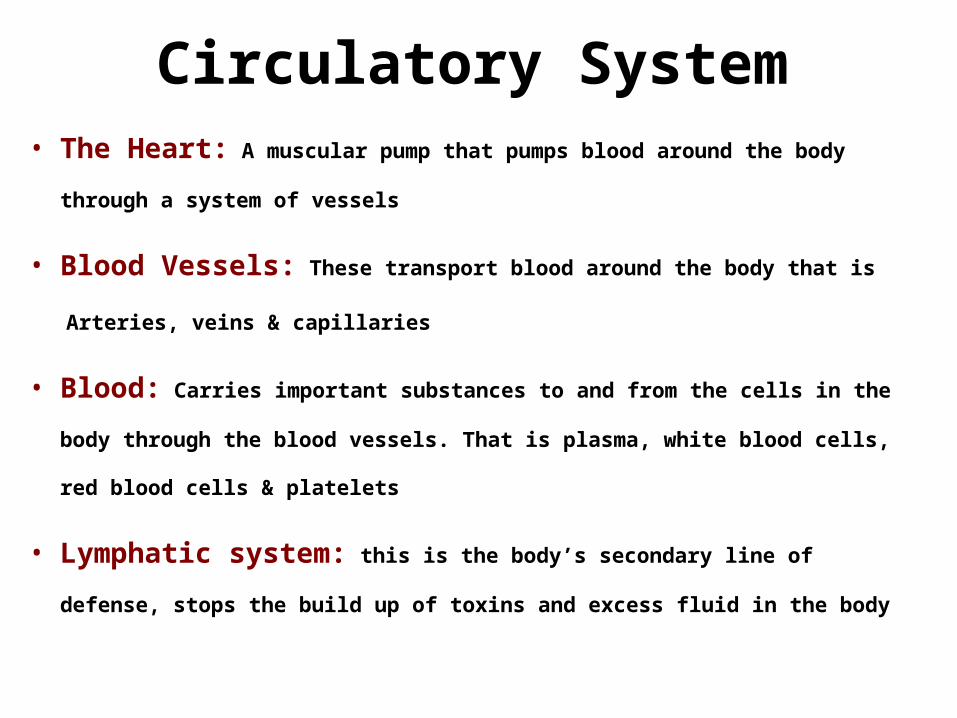
Circulatory System• The Heart: A muscular pump that pumps blood around the body
through a system of vessels
• Blood Vessels: These transport blood around the body that is
Arteries, veins & capillaries
• Blood: Carries important substances to and from the cells in the
body through the blood vessels. That is plasma, white blood cells,
red blood cells & platelets
• Lymphatic system: this is the body’s secondary line of
defense, stops the build up of toxins and excess fluid in the body
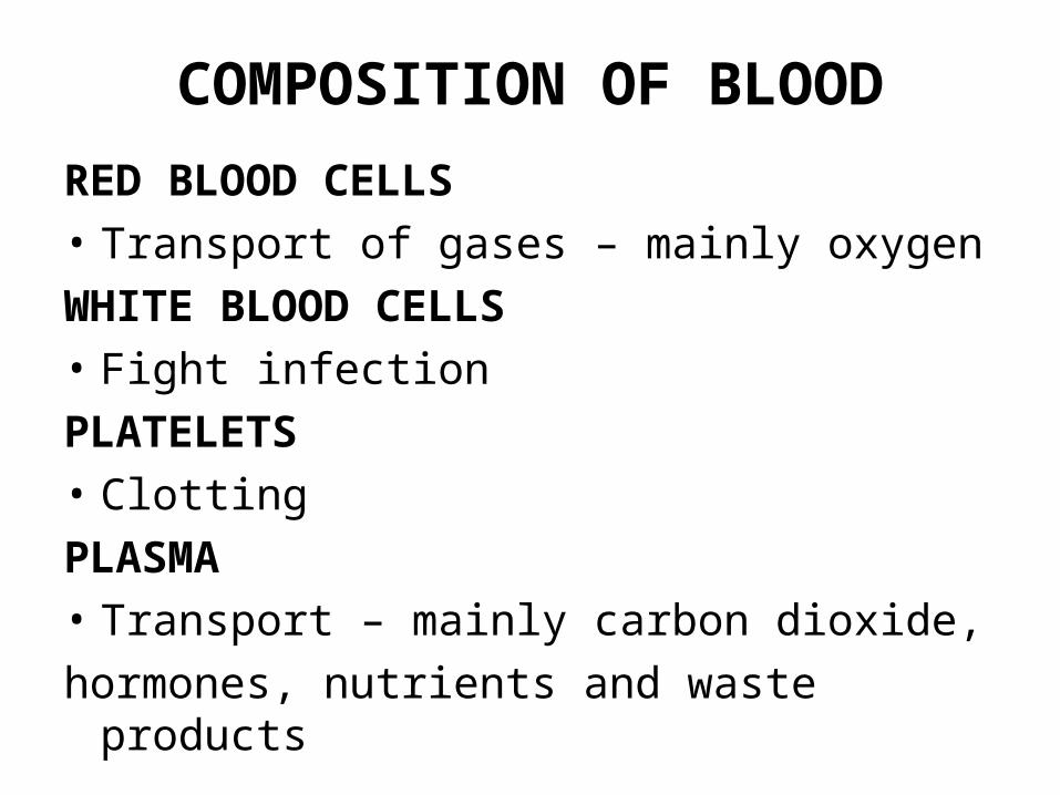
COMPOSITION OF BLOOD
RED BLOOD CELLS• Transport of gases – mainly oxygenWHITE BLOOD CELLS• Fight infectionPLATELETS• ClottingPLASMA• Transport – mainly carbon dioxide, hormones, nutrients and waste products
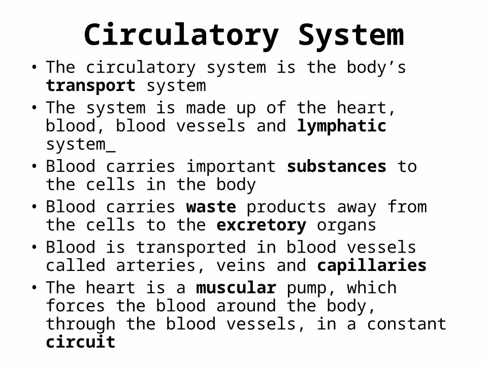
Circulatory System• The circulatory system is the body’s
transport system• The system is made up of the heart, blood,
blood vessels and lymphatic system • Blood carries important substances to
the cells in the body• Blood carries waste products away from
the cells to the excretory organs• Blood is transported in blood vessels
called arteries, veins and capillaries• The heart is a muscular pump, which
forces the blood around the body, through the blood vessels, in a constant circuit
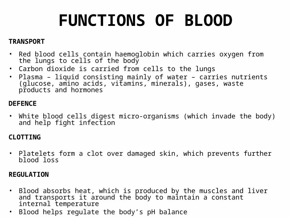
FUNCTIONS OF BLOODTRANSPORT
• Red blood cells contain haemoglobin which carries oxygen from the lungs to cells of the body
• Carbon dioxide is carried from cells to the lungs• Plasma – liquid consisting mainly of water – carries nutrients (glucose,
amino acids, vitamins, minerals), gases, waste products and hormones
DEFENCE
• White blood cells digest micro-organisms (which invade the body) and help fight infection
CLOTTING
• Platelets form a clot over damaged skin, which prevents further blood loss
REGULATION
• Blood absorbs heat, which is produced by the muscles and liver and transports it around the body to maintain a constant internal temperature
• Blood helps regulate the body’s pH balance
Blood Clotting
The process that prevents blood from clotting in an undamaged
vessel is a complex one.
Once a vessel has been damaged, platelets adhere to the damaged
area forming a plug the plasma protein fibrinogen is converted into
fibrin that creates fibres to radiate from the platelets forming a
network which traps red blood cells.
This blood clot stops further blood loss and prevents entry of bacteria.
Once the clot has dried it shrinks to form a scab, which protects the
tissues underneath while healing takes place.
Heparin is a protein normally present in the blood, which is formed in
the liver and it’s function is to prevent blood clotting in the vessels – it
is called an anti – coagulant.
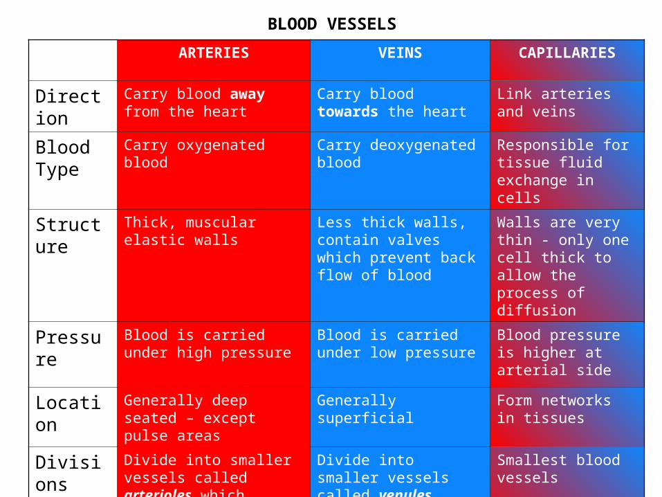
BLOOD VESSELS
ARTERIES VEINS CAPILLARIES
Direction Carry blood away from the heart
Carry blood towards the heart
Link arteries and veins
Blood Type
Carry oxygenated blood Carry deoxygenated blood
Responsible for tissue fluid exchange in cells
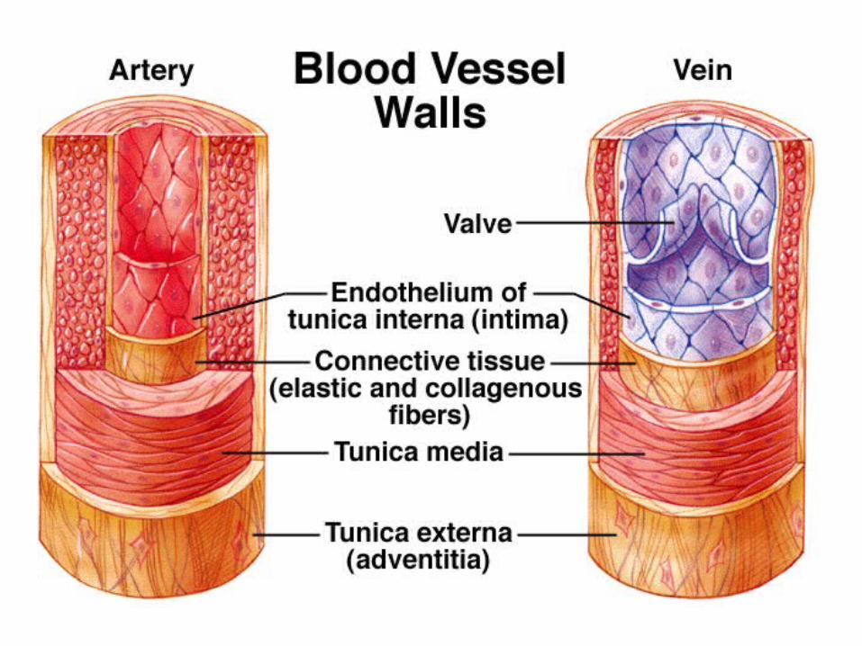
Structure Thick, muscular elastic walls
Less thick walls, contain valves which prevent back flow of blood
Walls are very thin - only one cell thick to allow the process of diffusion
Pressure Blood is carried under high pressure
Blood is carried under low pressure
Blood pressure is higher at arterial side
Location Generally deep seated – except pulse areas
Generally superficial Form networks in tissues
Divisions Divide into smaller vessels called arterioles which deliver blood to capillaries
Divide into smaller vessels called venules which continue from capillaries
Smallest blood vessels
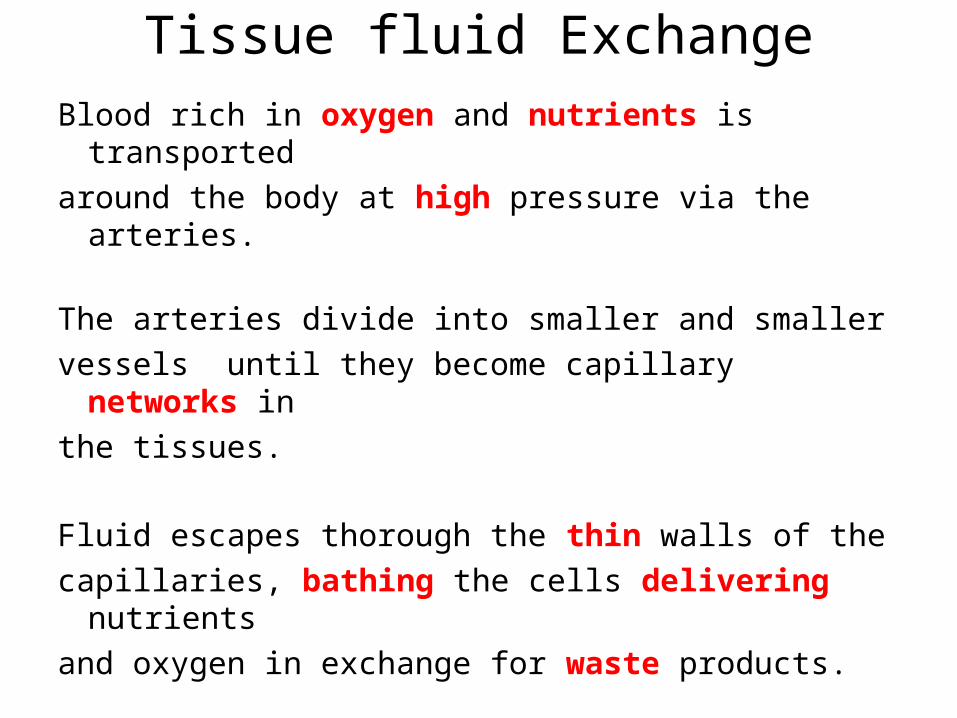
Tissue fluid Exchange
Blood rich in oxygen and nutrients is transported
around the body at high pressure via the arteries.
The arteries divide into smaller and smaller
vessels until they become capillary networks in
the tissues.
Fluid escapes thorough the thin walls of the
capillaries, bathing the cells delivering nutrients
and oxygen in exchange for waste products.
Tissue fluid Exchange
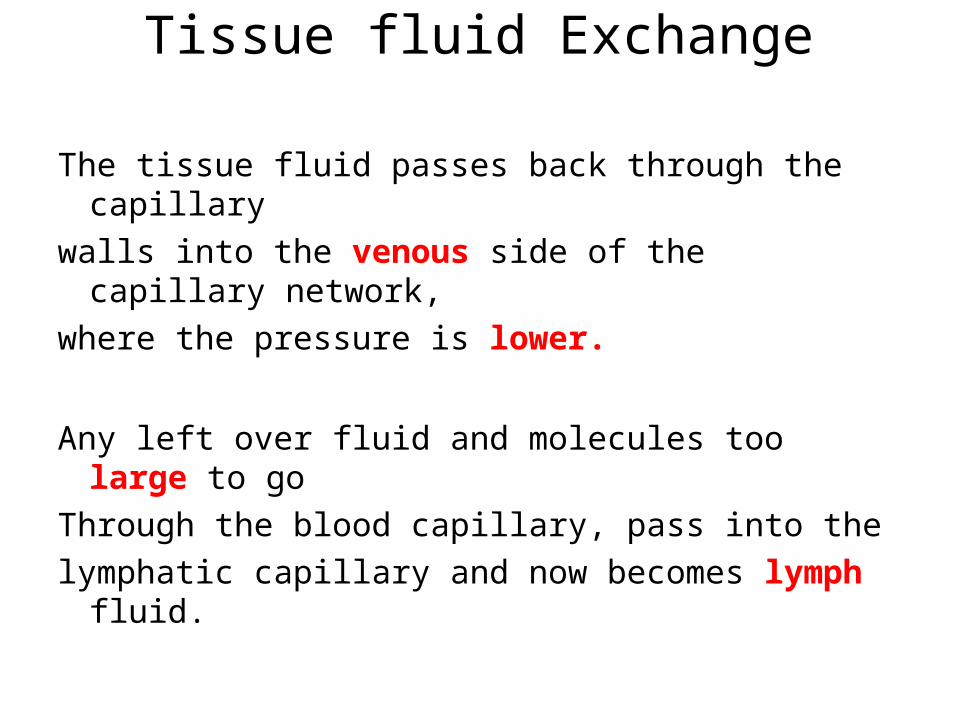
The tissue fluid passes back through the capillary
walls into the venous side of the capillary network,
where the pressure is lower.
Any left over fluid and molecules too large to go
Through the blood capillary, pass into the
lymphatic capillary and now becomes lymph fluid.
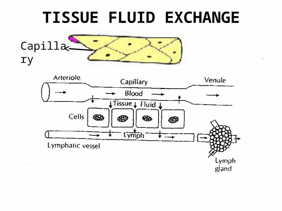
TISSUE FLUID EXCHANGE
Capillary
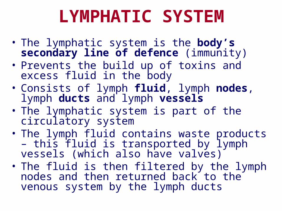
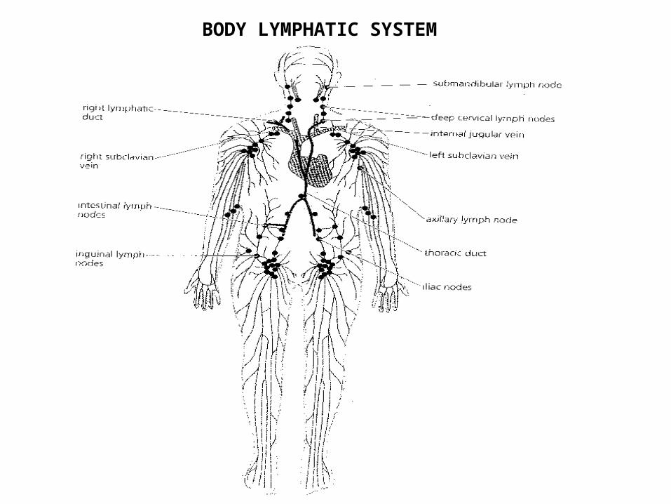
LYMPHATIC SYSTEM• The lymphatic system is the body’s
secondary line of defence (immunity)• Prevents the build up of toxins and excess
fluid in the body• Consists of lymph fluid, lymph nodes,
lymph ducts and lymph vessels• The lymphatic system is part of the
circulatory system• The lymph fluid contains waste products –
this fluid is transported by lymph vessels (which also have valves)
• The fluid is then filtered by the lymph nodes and then returned back to the venous system by the lymph ducts
THE LYMPHATIC SYSTEM
The lymphatic system is really part of the vascularsystem – the blood is contained in a closed systemof vessels which is kept circulating by the heart,from the capillaries some fluid escapes calledplasma, this is by diffusion and filtration.
This fluid then bathes the tissues directly, when thisfluid is collected into the lymph vessels it becomeslymph, which is later returned to the blood stream.
Lymph vessels start as fine, blind ended lymphcapillaries which then join to form lymphatics whichare similar in general course and structure to that ofveins.
THE LYMPHATIC SYSTEM
They are semilunar and numerous through andhave many more finer (pocket) valves, this isbecause the lymph is not pumped by the heart.
The composition of lymph is similar to bloodplasma but has less protein, less food materials andmore waste material – it has no erythrocytes but hasMore leukocytes.
THE LYMPHATIC SYSTEM
Lymph drainage is through lymph nodes
– before the lymph can be returned to the
blood stream it is filtered through one or
more of the lymph nodes, which are bean
shaped structures, situated at strategic
points in the course of the lymph vessels.
THE LYMPHATIC SYSTEM
After passing through the nodes the lymph
is then collected into the main lymph
vessels – vessels – lymph from the
right side of the head and thorax and right
arm, drain into the right lymphatic duct,
while lymph from the rest of the body drains
into the thoracic duct.
THE LYMPHATIC SYSTEM
The lymphatic system acts as the
body’s secondary line of defence
against bacterial invasion, this is
through the presence of
leukocytes at the site of infection.
BODY LYMPHATIC SYSTEM
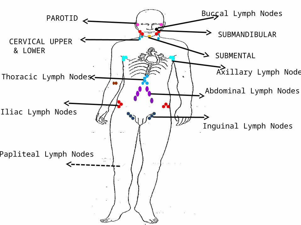
PAROTID
SUBMANDIBULAR
SUBMENTAL
CERVICAL UPPER & LOWER
Abdominal Lymph Nodes
Thoracic Lymph Nodes
Iliac Lymph Nodes
Inguinal Lymph Nodes
Axillary Lymph Nodes
Papliteal Lymph Nodes
Buccal Lymph Nodes
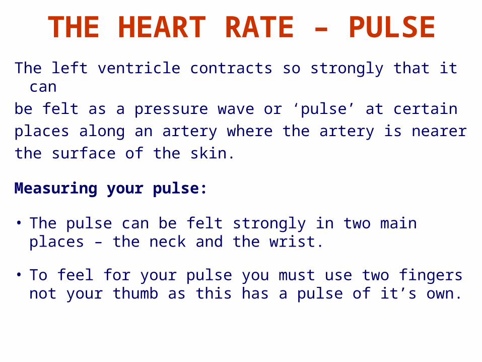
THE HEART RATE – PULSEThe left ventricle contracts so strongly that it canbe felt as a pressure wave or ‘pulse’ at certainplaces along an artery where the artery is nearerthe surface of the skin.
Measuring your pulse:
• The pulse can be felt strongly in two main places – the neck and the wrist.
• To feel for your pulse you must use two fingers not your thumb as this has a pulse of it’s own.
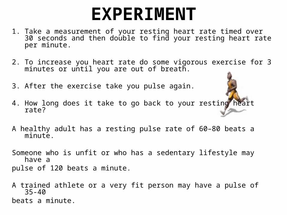
EXPERIMENT1. Take a measurement of your resting heart rate timed over 30
seconds and then double to find your resting heart rate per minute.
2. To increase you heart rate do some vigorous exercise for 3 minutes or until you are out of breath.
3. After the exercise take you pulse again.
4. How long does it take to go back to your resting heart rate?
A healthy adult has a resting pulse rate of 60–80 beats a minute.
Someone who is unfit or who has a sedentary lifestyle may have a
pulse of 120 beats a minute.
A trained athlete or a very fit person may have a pulse of 35-40beats a minute.
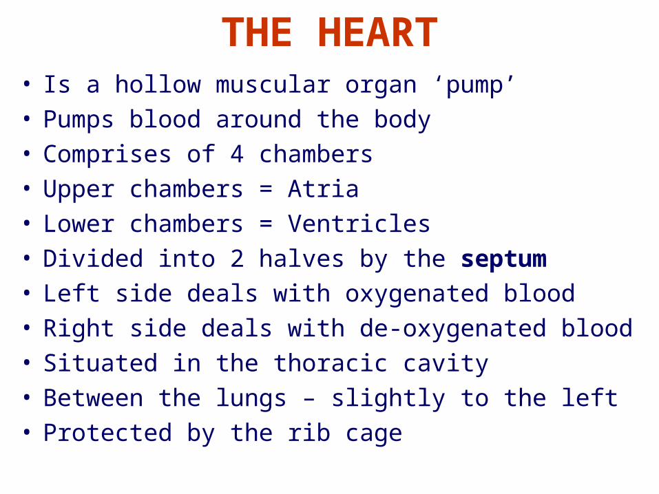
THE HEART• Is a hollow muscular organ ‘pump’• Pumps blood around the body• Comprises of 4 chambers• Upper chambers = Atria• Lower chambers = Ventricles• Divided into 2 halves by the septum• Left side deals with oxygenated blood• Right side deals with de-oxygenated blood• Situated in the thoracic cavity• Between the lungs – slightly to the left• Protected by the rib cage
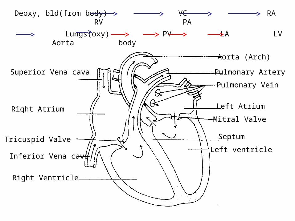
Aorta (Arch)
Pulmonary Artery
Pulmonary Vein
Left Atrium
Mitral Valve
Septum
Left ventricle
Superior Vena cava
Right Atrium
Tricuspid Valve
Inferior Vena cava
Right Ventricle
Deoxy, bld(from body) VC RA RV PA
Lungs(oxy) PV LA LV Aorta body
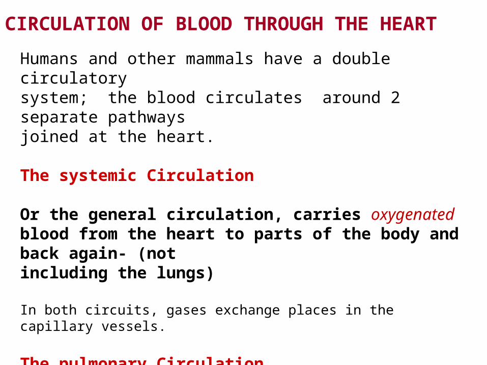
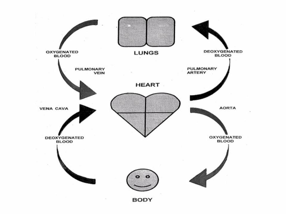
CIRCULATION OF BLOOD THROUGH THE HEART
Humans and other mammals have a double circulatory system; the blood circulates around 2 separate pathways joined at the heart.
The systemic Circulation
Or the general circulation, carries oxygenated blood from the heart to parts of the body and back again- (not including the lungs)
In both circuits, gases exchange places in the capillary vessels.
The pulmonary Circulation
Carries blood from the heart to the lungs be oxygenated and back to the heart.
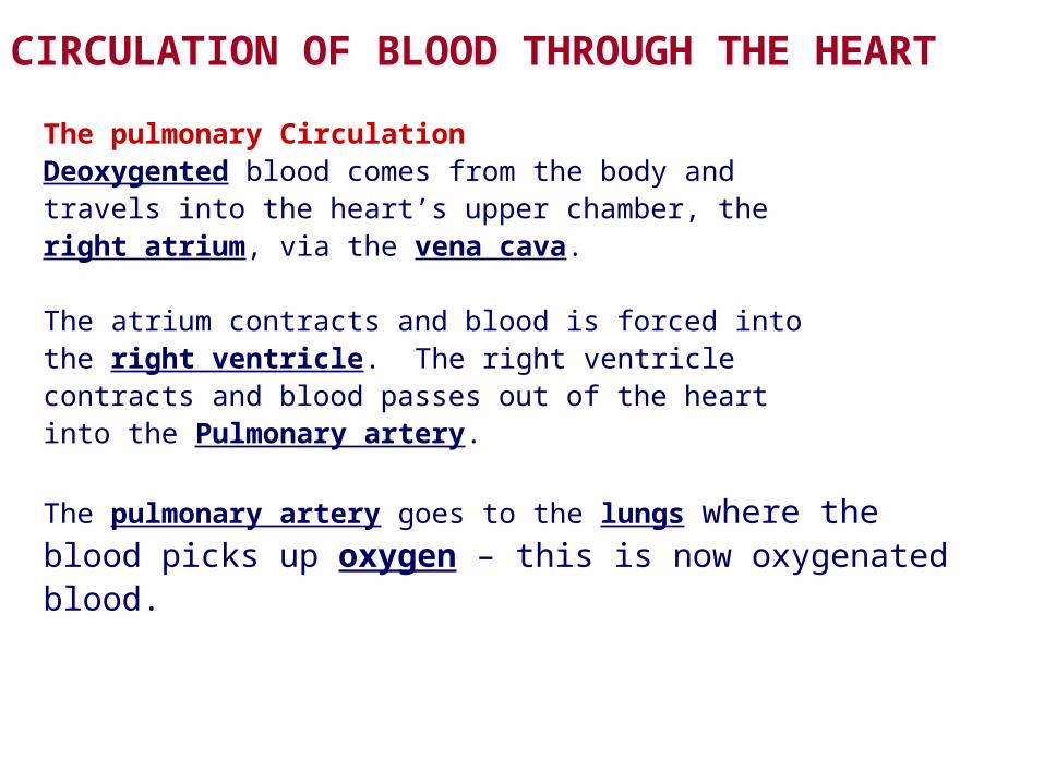
The pulmonary CirculationDeoxygented blood comes from the body andtravels into the heart’s upper chamber, theright atrium, via the vena cava. The atrium contracts and blood is forced intothe right ventricle. The right ventriclecontracts and blood passes out of the heartinto the Pulmonary artery.
The pulmonary artery goes to the lungs where theblood picks up oxygen – this is now oxygenatedblood.
CIRCULATION OF BLOOD THROUGH THE HEART
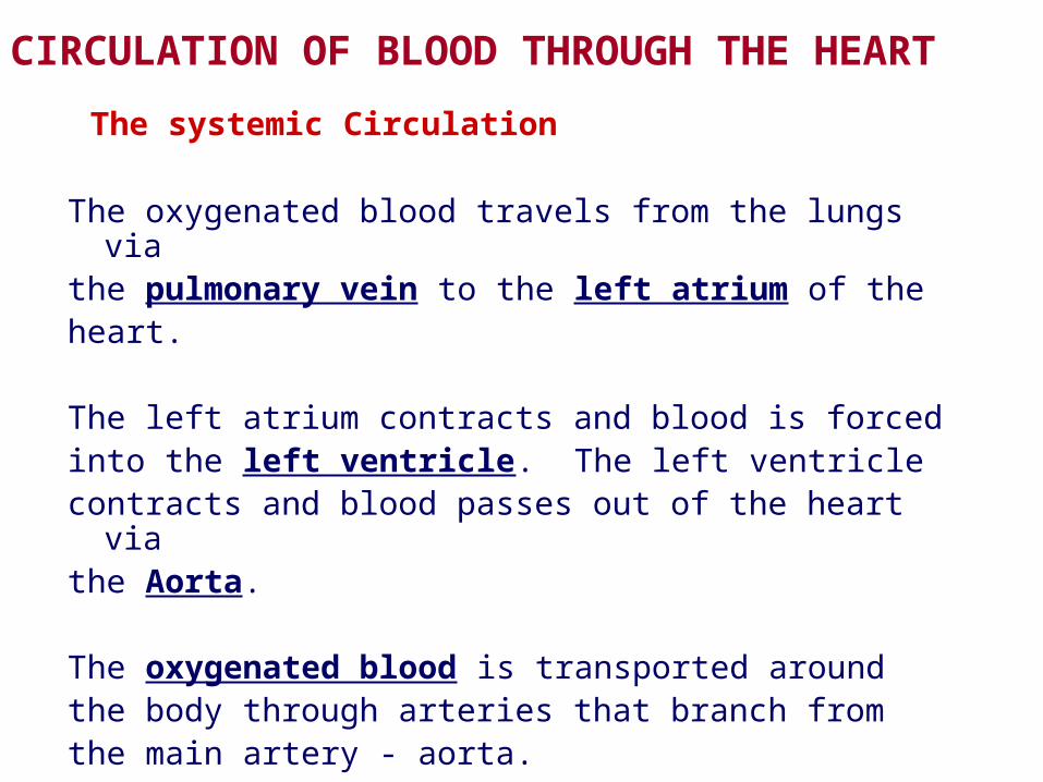
The systemic Circulation
The oxygenated blood travels from the lungs viathe pulmonary vein to the left atrium of theheart.
The left atrium contracts and blood is forcedinto the left ventricle. The left ventriclecontracts and blood passes out of the heart viathe Aorta.
The oxygenated blood is transported aroundthe body through arteries that branch fromthe main artery - aorta.
CIRCULATION OF BLOOD THROUGH THE HEART
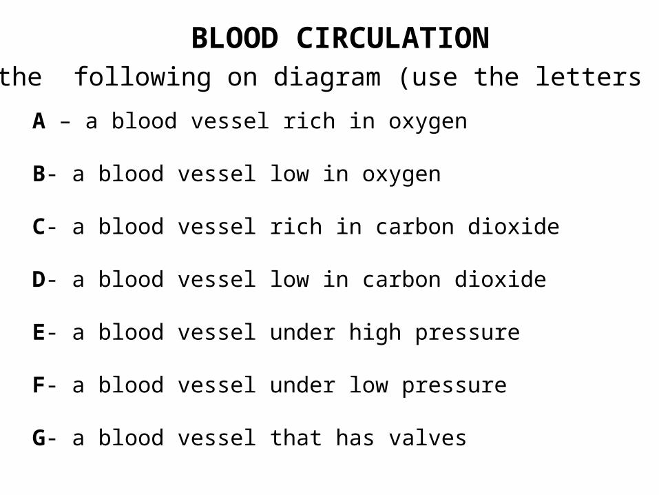
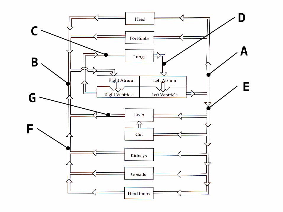
BLOOD CIRCULATIONLabel the following on diagram (use the letters as key)
A – a blood vessel rich in oxygen
B- a blood vessel low in oxygen
C- a blood vessel rich in carbon dioxide
D- a blood vessel low in carbon dioxide
E- a blood vessel under high pressure
F- a blood vessel under low pressure
G- a blood vessel that has valves
AB
CD
E
F
G

Subclavian
Jugular
Axillary
Basilic
Median
Great Saphenous
Short Saphenouse Anterior Tibial Vein
Posterior Tibial Vein
Popliteal
Femoral
Radial
Brachial
Cephalic
Vena Cava
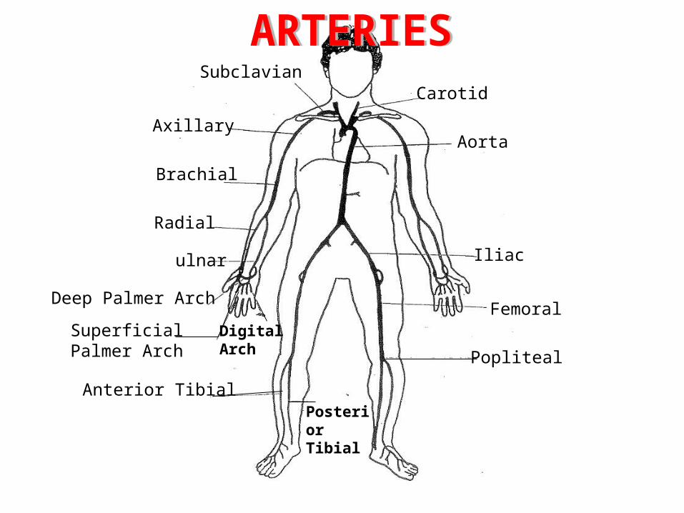
Subclavian
Axillary
Brachial
Radial
ulnar
Deep Palmer Arch
Superficial Palmer Arch
Digital Arch
Anterior TibialPosterior Tibial
Carotid
Aorta
Iliac
Femoral
Popliteal
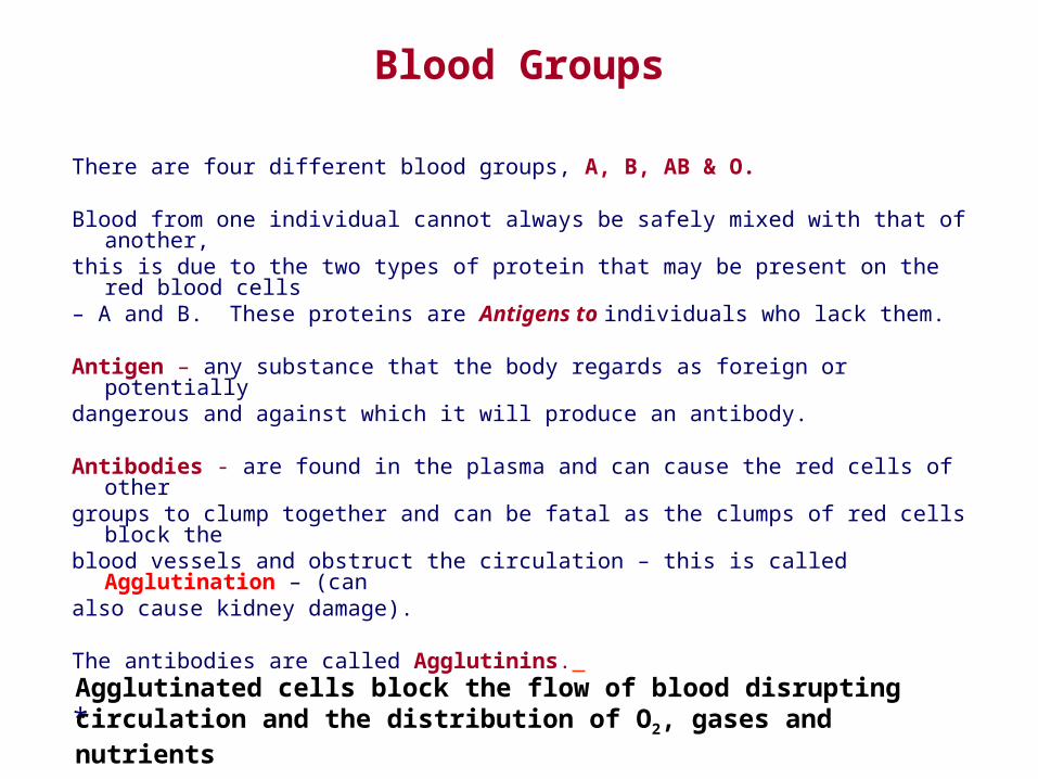
Blood Groups
There are four different blood groups, A, B, AB & O.
Blood from one individual cannot always be safely mixed with that of another,this is due to the two types of protein that may be present on the red blood cells– A and B. These proteins are Antigens to individuals who lack them.
Antigen – any substance that the body regards as foreign or potentiallydangerous and against which it will produce an antibody.
Antibodies - are found in the plasma and can cause the red cells of othergroups to clump together and can be fatal as the clumps of red cells block theblood vessels and obstruct the circulation – this is called Agglutination – (canalso cause kidney damage).
The antibodies are called Agglutinins.
*Agglutinated cells block the flow of blood disrupting circulation and the distribution of O2, gases and nutrients
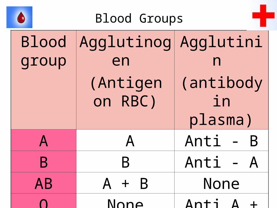
Blood Groups
Blood group
Agglutinogen
(Antigen on RBC)
Agglutinin
(antibody in plasma)
A A Anti - BB B Anti - A
AB A + B NoneO None Anti A + Anti
B
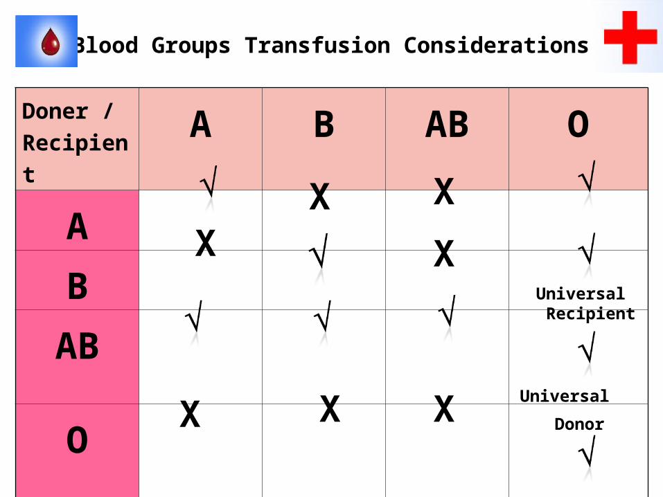
Blood Groups Transfusion Considerations
Doner /
RecipientA B AB O
A
B
AB
O
XX
XXX
X X
Universal Recipient
Universal
Donor
The Rhesus Factor
In addition to ABO grouping there is another antigen present in the blood of about 85% of the population-it is known as the Rhesus Factor, (named after the group of monkeys it was discovered in).
Those who have this factor are known as Rhesus + positive) – 85%
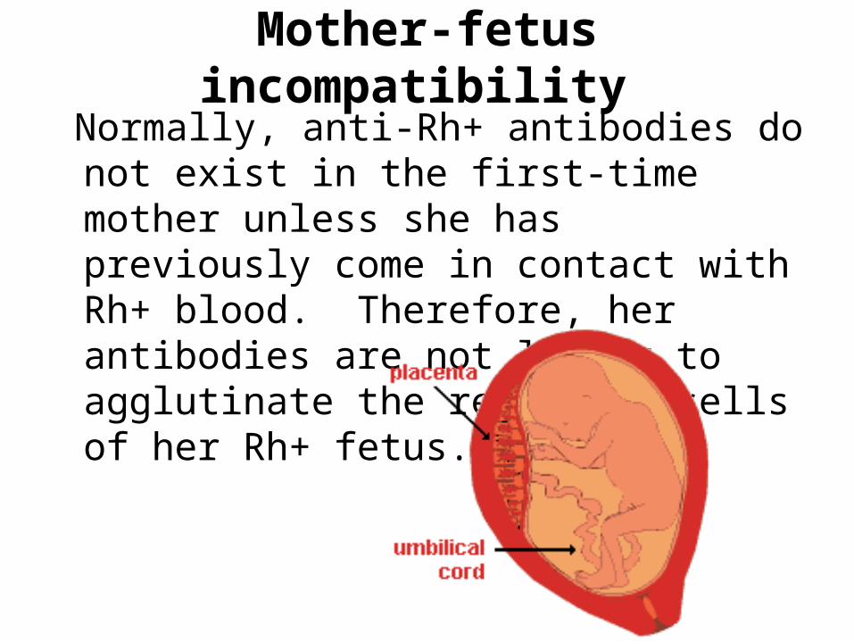
Mother-fetus incompatibility
Normally, anti-Rh+ antibodies do not exist in the first-time mother unless she has previously come in contact with Rh+ blood. Therefore, her antibodies are not likely to agglutinate the red blood cells of her Rh+ fetus.
Rh (Rhesus) Factor
• Up to 8 genes determine if a person is Rh positive.
• The most common of these are the C, D & E genes which determine if a person has C, D and/or E antigens on the walls of the cells.
• People who do not have Rh antigens on their cell membranes are Rh negative.
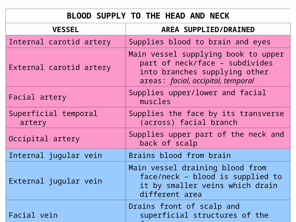
BLOOD SUPPLY TO THE HEAD AND NECK
VESSEL AREA SUPPLIED/DRAINED
Internal carotid artery Supplies blood to brain and eyes
External carotid artery
Main vessel supplying book to upper part of neck/face – subdivides into branches supplying other areas: facial, occipital, temporal
Facial artery Supplies upper/lower and facial muscles
Superficial temporal arterySupplies the face by its transverse
(across) facial branch
Occipital arterySupplies upper part of the neck and back
of scalp
Internal jugular vein Brains blood from brain
External jugular veinMain vessel draining blood from face/neck
– blood is supplied to it by smaller veins which drain different area
Facial veinDrains front of scalp and superficial
structures of the face
Retromandibular vein Drains face below jaw and back of scalp
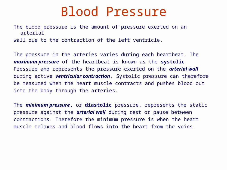
Blood PressureThe blood pressure is the amount of pressure exerted on an arterial
wall due to the contraction of the left ventricle.
The pressure in the arteries varies during each heartbeat. The
maximum pressure of the heartbeat is known as the systolic
Pressure and represents the pressure exerted on the arterial wall
during active ventricular contraction. Systolic pressure can therefore
be measured when the heart muscle contracts and pushes blood out
into the body through the arteries.
The minimum pressure, or diastolic pressure, represents the static
pressure against the arterial wall during rest or pause between
contractions. Therefore the minimum pressure is when the heart
muscle relaxes and blood flows into the heart from the veins.
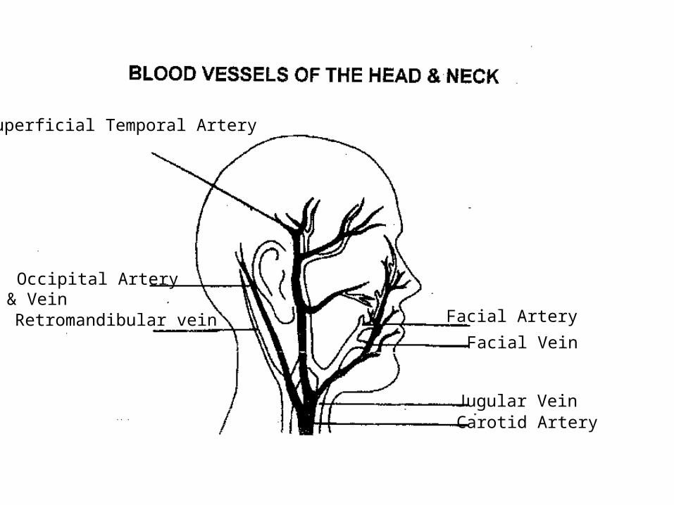
Superficial Temporal Artery
Occipital Artery & Vein
Retromandibular vein Facial Artery
Facial Vein
Jugular VeinCarotid Artery
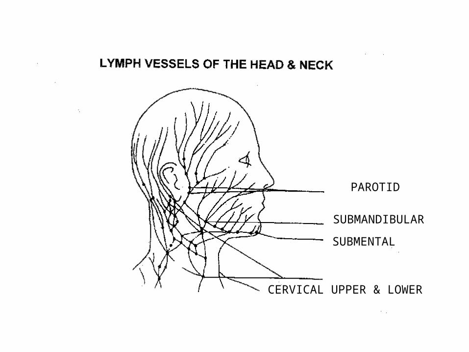
PAROTID
SUBMANDIBULAR
SUBMENTAL
CERVICAL UPPER & LOWER
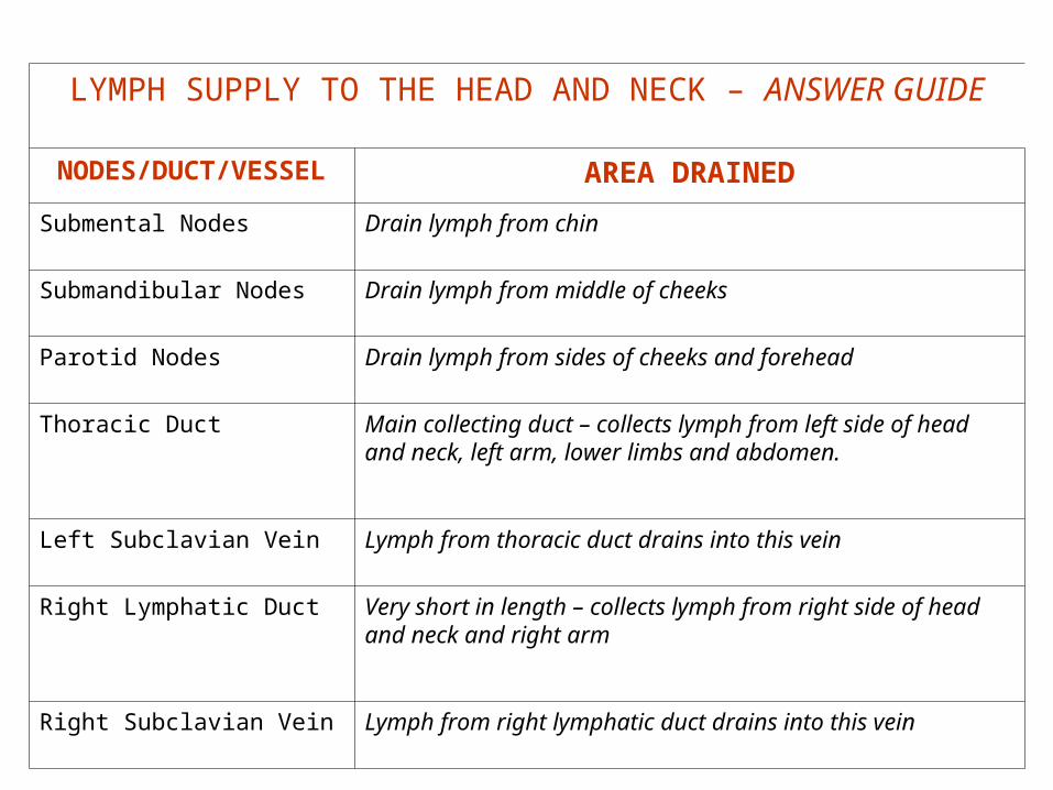
LYMPH SUPPLY TO THE HEAD AND NECK – ANSWER GUIDE
NODES/DUCT/VESSEL
AREA DRAINED
Submental Nodes Drain lymph from chin
Submandibular Nodes Drain lymph from middle of cheeks
Parotid Nodes Drain lymph from sides of cheeks and forehead
Thoracic Duct Main collecting duct – collects lymph from left side of head and neck, left arm, lower limbs and abdomen.
Left Subclavian Vein Lymph from thoracic duct drains into this vein
Right Lymphatic Duct Very short in length – collects lymph from right side of head and neck and right arm
Right Subclavian Vein Lymph from right lymphatic duct drains into this vein