Embed Size (px)
DESCRIPTION
yes
Citation preview
Clinical/Translational Research
Existence of an Endogenous Circadian Blood PressureRhythm in Humans That Peaks in the Evening
Short CommunicationSteven A. Shea, Michael F. Hilton, Kun Hu, Frank A.J.L. Scheer
Rationale: Blood pressure (BP) usually decreases during nocturnal sleep and increases during daytime activities.Whether the endogenous circadian control system contributes to this daily BP variation has not been determinedunder appropriately controlled conditions.
Objective: To determine whether there exists an endogenous circadian rhythm of BP in humans.Methods and Results: In 28 normotensive adults (16 men), we assessed BP across 3 complementary, multiday,
in-laboratory protocols performed in dim light, throughout which behavioral and environmental influences werecontrolled and/or uniformly distributed across the circadian cycle via: (1) a 38-hour “constant routine,” includingcontinuous wakefulness; (2) a 196-hour “forced desynchrony” with 7 recurring 28-hour sleep/wake cycles; and(3) a 240-hour forced desynchrony with 12 recurring 20-hour sleep/wake cycles. Circadian phases were derivedfrom core body temperature. Each protocol revealed significant circadian rhythms in systolic and diastolic BP,with almost identical rhythm profiles among protocols. The peak-to-trough amplitudes were 3 to 6 mm Hg forsystolic BP and 2 to 3 mm Hg for diastolic BP (always P<0.05). All 6 peaks (systolic and diastolic BP in 3protocols) occurred at a circadian phase corresponding to �9:00 PM (ie, the biological evening). Based onsubstantial phase differences among circadian rhythms of BP and other variables, the rhythm in BP appeared to beunrelated to circadian rhythms in cortisol, catecholamines, cardiac vagal modulation, heart rate, or urine flow.
Conclusions: There exists a robust endogenous circadian rhythm in BP. The highest BP occurred at the circadiantime corresponding to �9:00 PM, suggesting that the endogenous BP rhythm is unlikely to underlie thewell-documented morning peak in adverse cardiovascular events. (Circ Res. 2011;108:980-984.)
Key Words: circadian � blood pressure, humans � myocardial infarction � stroke
Numerous epidemiological studies reveal a profoundmorning increase in the incidence of adverse cardiovas-
cular events, including sudden cardiac death, ventriculararrhythmia, stroke,1 and myocardial infarction.2 The extent towhich these peaks are caused by a day/night pattern ofbehaviors and/or endogenous circadian factors is unknown.3
The circadian timing system orchestrates endogenous circa-dian rhythms in physiology and behavior and is composed ofthe master circadian pacemaker located in the suprachias-matic nucleus and circadian oscillators in peripheral tissues.4
The suprachiasmatic nucleus may influence the cardiovascu-lar system via multisynaptic neural projections to the heart,adrenal cortex, adrenal medulla, kidneys, and vasculature andresultant neural or endocrine effects,5 and secondarily bycircadian influences on behaviors, such as activity levels,alertness, and sleep. Moreover, recent animal investigationshave also documented the actions of molecular circadianclocks in peripheral tissues that can affect blood pressure
(BP),6 ischemia/reperfusion tolerance,7 and vascular remod-eling.8 In contrast, mechanistic circadian studies of cardio-vascular function in humans are sparse.
A primary risk factor for adverse cardiovascular events iselevated arterial BP.9 Countless studies have documented theday/night variation of BP in humans, which has been used toclassify hypertensive patients into nocturnal “dippers” (�10%drop in BP overnight) and “nondippers,” and it has been shownthat nondippers are at increased risk for serious adverse events.10
However, no studies have adequately studied the relative con-tributions to this day/night BP variation from the endogenouscircadian cycle and from the daily changes in behaviors, such asthe sleep/wake cycle. Thus, we tested the hypothesis that thereexists an endogenous circadian rhythm in BP in humans.
MethodsAn expanded Methods section is available in the Online DataSupplement at http://circres.ahajournals.org.
Original received September 29, 2010; revision received February 8, 2011; accepted February 14, 2011. In January 2011, the average time fromsubmission to first decision for all original research papers submitted to Circulation Research was 13.34 days.
From the Brigham and Women’s Hospital and Harvard Medical School, Boston, MA.Correspondence to Steven A. Shea, Division of Sleep Medicine, Brigham and Women’s Hospital, 221 Longwood Ave, Boston, MA 02115. E-mail
[email protected]© 2011 American Heart Association, Inc.
Circulation Research is available at http://circres.ahajournals.org DOI: 10.1161/CIRCRESAHA.110.233668
980 by guest on February 23, 2015http://circres.ahajournals.org/Downloaded from by guest on February 23, 2015http://circres.ahajournals.org/Downloaded from by guest on February 23, 2015http://circres.ahajournals.org/Downloaded from by guest on February 23, 2015http://circres.ahajournals.org/Downloaded from by guest on February 23, 2015http://circres.ahajournals.org/Downloaded from by guest on February 23, 2015http://circres.ahajournals.org/Downloaded from by guest on February 23, 2015http://circres.ahajournals.org/Downloaded from by guest on February 23, 2015http://circres.ahajournals.org/Downloaded from by guest on February 23, 2015http://circres.ahajournals.org/Downloaded from
SubjectsSubjects gave informed consent, and the studies were approved bythe local Human Research Committee. We studied 28 adults (16men) who were normotensive, nonobese, and healthy (other thanmild asthma, n�6), and who took no medications (other than oralcontraceptives and �2-agonist rescue inhalers for asthma), and nocaffeine, alcohol, or nicotine products for 2 to 3 weeks immediatelybefore and throughout the laboratory studies. The subjects withasthma participated in only 1 of 3 protocols and all data in the 4hours following any rescue inhaler use were excluded from analyses(see the Online Data Supplement).
ProtocolsQuantifying the effect of the endogenous circadian system on BPrequires controlling all environmental factors and behaviors that canaffect either BP or the circadian cycle (eg, activity, posture, meals,sleep, room temperature, light) while measuring BP across thecircadian cycle and measuring an endogenous circadian phasemarker whose effects can be mathematically isolated from anyinfluences caused by daily behavioral changes (eg, core bodytemperature [CBT], rather than activity levels, which have been usedin most animal studies and greatly affect BP). This was achieved bymeasuring BP throughout 3 separate and complementary circadianprotocols, as shown in Figure 1. To stabilize circadian rhythms,
subjects maintained a regular sleep/wake schedule for 2 to 3 weeksbefore entering the laboratory, followed by 2 baseline days andnights in the laboratory (16-hour scheduled wakefulness, 8-hourscheduled sleep, both at home and in the laboratory). Thereafter, toavoid resetting the phase of the circadian system, all laboratoryprotocols were performed in dim light (�4 lux).11 Subjects com-pleted 1 or 2 of the 3 protocols: (1) 38-hour “constant routineprotocol” (CR), with continuous wakefulness, semirecumbency, and2-hourly isocaloric snacks (Figure 1, top); (2) 196-hour forceddesynchrony with 7 recurring 28-hour sleep/wake cycles (FD28)(Figure 1, middle: 18-hour 40-minute wakefulness, 9-hour 20-minute sleep); or (3) 240-hour forced desynchrony with 12 recurring20-hour sleep/wake cycles (FD20) (Figure 1, bottom: 13-hour20-minute wakefulness, 6-hour 40-minute sleep). In essence, the CRabolished sleep and minimized behaviors, whereas the FD28 andFD20 maintained a normal sleep:wake ratio of 1:2 and scheduled allactivities so that they became uniformly distributed across thecircadian cycle.
Measurements and AnalysesSubjects wore a flexible rectal temperature sensor for measurementof CBT, which was used as the circadian phase marker.12 For eachsubject, the fitted CBT minimum was assigned as a reference phasemarker of 0°. BP was measured repeatedly by automatic oscillomet-ric cuff sphygmomanometer from an upper arm. Heart rate was alsomeasured in each protocol. These measurements were made through-out periods of relaxed wakefulness during each protocol. Data wereassigned a circadian phase and the existence of any circadianrhythms were tested by cosinor mixed-model ANOVA. To assesswhether the rhythms were robust, the phases of peaks and theamplitudes of the circadian rhythms of BP and heart rate werecompared across protocols using unpaired t-tests. To gain insightinto control mechanisms across the circadian cycle, most of thefollowing potentially related variables were measured throughouteach protocol: cardiac vagal modulation (estimated from high fre-quency power of interbeat interval variability), plasma cortisol, urinaryor plasma catecholamines, and urine flow. The phases of peaks andtroughs of these potentially related variables were compared with thecircadian rhythm of BP using paired t-tests. Further details are providedin the Online Methods, Online Table I, Online Figure I.
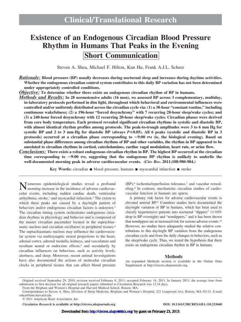
ResultsEach protocol revealed significant circadian rhythms in sys-tolic and diastolic BP, with almost identical rhythm ampli-tudes and phase relationships among protocols (Figures 2 and3; Online Figure I and Online Table I). For the 3 protocols,the peak-to-trough amplitudes were 3 to 6 mm Hg for systolicBP and 2 to 3 mm Hg for diastolic BP (always P�0.05). All6 peaks (systolic and diastolic BP in 3 protocols) occurred inthe circadian phase range of 219° to 265°, with an averagephase of 244°, equivalent to �9:00 PM (Online Table I). Theaverage circadian phase of the peak in heart rate across the 3protocols was 161°, which is �6 hours earlier than the
Figure 1. Three complementary protocols used to examineunderlying circadian rhythmicity of BP. Three protocolsdesigned to keep behaviors constant across the circadian cycle(top graph, 2 baseline days followed by 38-hour constant rou-tine while semirecumbent and awake) or to evenly distributebehaviors across all circadian phases (middle graph, 7 recur-ring 28-hour behavioral cycles [28-hour forced desynchrony];bottom graph, 12 recurring 20-hour behavioral cycles [20-hourforced desynchrony]). In each graph, subsequent days are“double-plotted” to the right and below prior days to visually aidprotocol continuity. The x axes: clock times for an example sub-ject having an habitual wake time of 8:00 AM. Black boxes indi-cate scheduled sleep episodes in darkness; gray/hatched bars,scheduled wakefulness in dim light conditions (�4 lux) to avoidcircadian rhythm resetting.11
Non-standard Abbreviations and Acronyms
BP blood pressure
CR constant routine protocol
FD20 forced desynchrony protocol with 12 recurring 20-hour sleep/wake cycles
FD28 forced desynchrony protocol with 7 recurring 28-hour sleep/wake cycles
CBT core body temperature
Shea et al Endogenous Circadian Blood Pressure Rhythm 981
by guest on February 23, 2015http://circres.ahajournals.org/Downloaded from
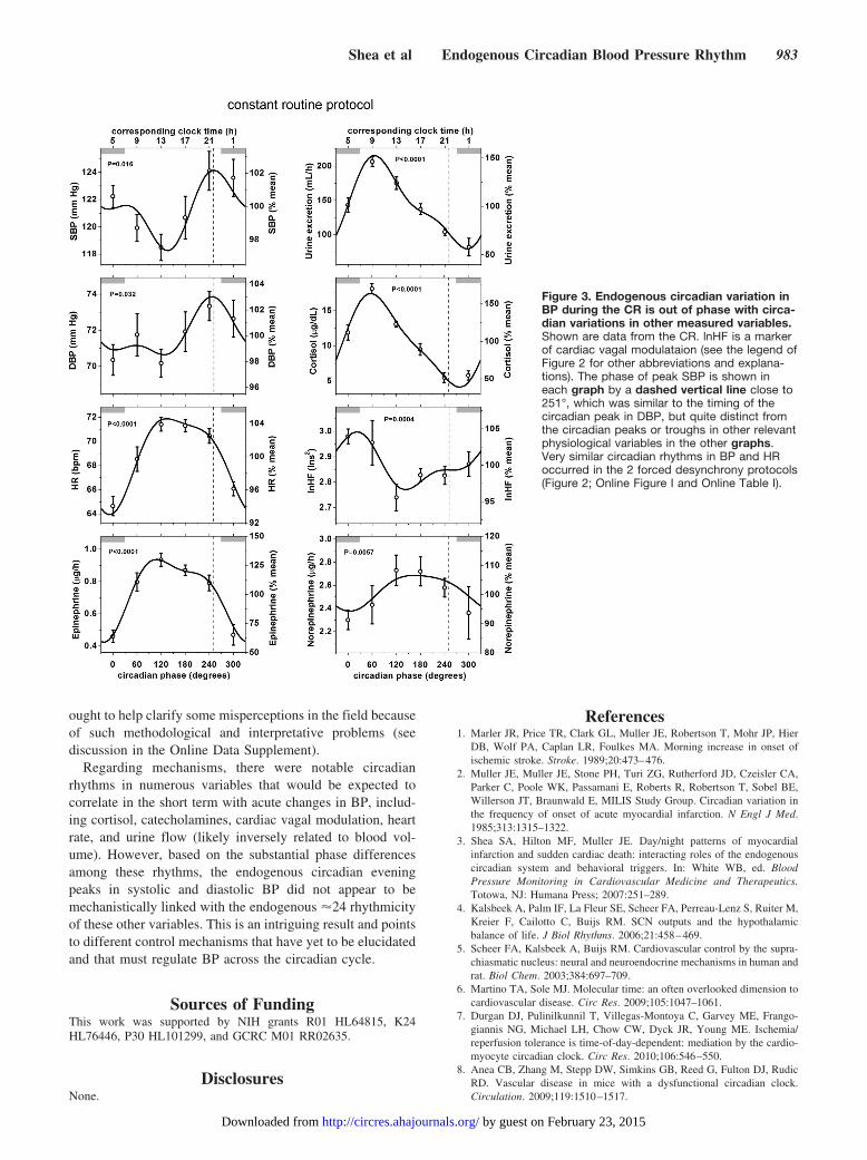
average circadian peak in BP. During the CR, there weresubstantial and significant differences between the timing ofthe circadian peak in BP (systolic and diastolic) and all otherpotentially relevant circadian rhythms measured, includingcircadian peaks in heart rate, urinary catecholamines andplasma cortisol, and circadian troughs in urine flow andcardiac vagal modulation (Figure 3 and Online Table I).Similarly, results for these variables during the 2 forceddesynchrony protocols (where available) were highly consis-tent with the CR data (see Online Table I and Online FigureI). Thus, the endogenous circadian rhythm in BP appeared tobe unrelated to circadian rhythms in cortisol, catecholamines,cardiac vagal modulation, heart rate, or urine flow.
DiscussionThe present study has revealed an endogenous circadianrhythm of BP. This study yielded robust results that werealmost identical in 3 groups of subjects during 3 differentprotocols. This study was performed because of the possiblerelevance of endogenous circadian BP rhythms to the day/night pattern of adverse cardiovascular events. Our results areperhaps unexpected because the timing of the endogenouscircadian peak in BP occurs in the evening, whereas thelowest circadian BP occurs around the most vulnerable timefor adverse cardiovascular events. Thus, our data suggest thatthe morning peaks in adverse cardiovascular events are notcaused by circadian rhythm-related increases in BP. Presum-ably, endogenous circadian rhythms in other cardiovascular
variables (eg, platelet function, sympathovagal balance, andendothelial function) and/or physiological responses to day/night patterns of behavioral changes are more important inthis regard. We note that waking in the evening, as can occurwith shift work, jet lag, and sleep disorders, may result in anexaggerated BP surge attributable to summed behavioral- andcircadian-related increases in BP, perhaps helping to explainthe curious secondary evening peak in incidence of myocar-dial infarction in vulnerable individuals.2 Moreover, theremay be more than simple summation of these effects,whereby the circadian system modulates the BP response to astandardized stress.13–15
This study was performed in healthy subjects, and itremains to be seen whether the endogenous circadian morn-ing trough in BP that we observed could increase the risk ofischemic stroke at that time in people with existing carotid orcerebral artery stenoses and whether the amplitudes or phasesof the endogenous circadian BP rhythms are abnormal inmore vulnerable groups.
There are many studies that refer to a “circadian rhythm” ofBP in humans and other animals without clarifying whetherthis is an endogenous rhythm, as almost all such studies madeBP measurements across the 24-hour period while permittingsleep and altered activity across the day and night, such thatthese behaviors rather than endogenous circadian rhythmicitycould cause much of these observed day/night pattern in BP.Our study overcame many such limitations and quantified theindependent endogenous circadian rhythmicity of BP and
Figure 2. Similar endogenous circadianvariations in BP across 2 complementaryforced desynchrony protocols. Shown are(means�SEM) systolic BP (SBP), diastolic BP(DBP), and heart rate (HR) expressed in absoluteunits (left axes) and as percentages of individualaverages (right axes). Data are aligned accordingto circadian phase (x axis) and plotted in 60° bins(equivalent to �4 hours). Corresponding approxi-mate clock time is shown on the top x axis. 0°represents CBT minimum (�5:00 AM in these sub-jects). Thin gray shaded bars are shown at thetop of each graph to indicate the average equiva-lent clock time when subjects would normallysleep when at home (although all data were col-lected during wakefulness in the laboratory). Solidlines represent the cosinor model fit and the prob-ability values indicate significance of circadianrhythmicity. The phases of the peak SBP areshown by dashed vertical lines in each protocoland occurred close to 240° (equivalent to �9:00PM), which was similar to the timing of the peaks inDBP, but quite distinct from the timing of the cir-cadian peaks in HR.
982 Circulation Research April 15, 2011
by guest on February 23, 2015http://circres.ahajournals.org/Downloaded from
ought to help clarify some misperceptions in the field becauseof such methodological and interpretative problems (seediscussion in the Online Data Supplement).
Regarding mechanisms, there were notable circadianrhythms in numerous variables that would be expected tocorrelate in the short term with acute changes in BP, includ-ing cortisol, catecholamines, cardiac vagal modulation, heartrate, and urine flow (likely inversely related to blood vol-ume). However, based on the substantial phase differencesamong these rhythms, the endogenous circadian eveningpeaks in systolic and diastolic BP did not appear to bemechanistically linked with the endogenous �24 rhythmicityof these other variables. This is an intriguing result and pointsto different control mechanisms that have yet to be elucidatedand that must regulate BP across the circadian cycle.
Sources of FundingThis work was supported by NIH grants R01 HL64815, K24HL76446, P30 HL101299, and GCRC M01 RR02635.
DisclosuresNone.
References1. Marler JR, Price TR, Clark GL, Muller JE, Robertson T, Mohr JP, Hier
DB, Wolf PA, Caplan LR, Foulkes MA. Morning increase in onset ofischemic stroke. Stroke. 1989;20:473–476.
2. Muller JE, Muller JE, Stone PH, Turi ZG, Rutherford JD, Czeisler CA,Parker C, Poole WK, Passamani E, Roberts R, Robertson T, Sobel BE,Willerson JT, Braunwald E, MILIS Study Group. Circadian variation inthe frequency of onset of acute myocardial infarction. N Engl J Med.1985;313:1315–1322.
3. Shea SA, Hilton MF, Muller JE. Day/night patterns of myocardialinfarction and sudden cardiac death: interacting roles of the endogenouscircadian system and behavioral triggers. In: White WB, ed. BloodPressure Monitoring in Cardiovascular Medicine and Therapeutics.Totowa, NJ: Humana Press; 2007:251–289.
4. Kalsbeek A, Palm IF, La Fleur SE, Scheer FA, Perreau-Lenz S, Ruiter M,Kreier F, Cailotto C, Buijs RM. SCN outputs and the hypothalamicbalance of life. J Biol Rhythms. 2006;21:458–469.
5. Scheer FA, Kalsbeek A, Buijs RM. Cardiovascular control by the supra-chiasmatic nucleus: neural and neuroendocrine mechanisms in human andrat. Biol Chem. 2003;384:697–709.
6. Martino TA, Sole MJ. Molecular time: an often overlooked dimension tocardiovascular disease. Circ Res. 2009;105:1047–1061.
7. Durgan DJ, Pulinilkunnil T, Villegas-Montoya C, Garvey ME, Frango-giannis NG, Michael LH, Chow CW, Dyck JR, Young ME. Ischemia/reperfusion tolerance is time-of-day-dependent: mediation by the cardio-myocyte circadian clock. Circ Res. 2010;106:546–550.
8. Anea CB, Zhang M, Stepp DW, Simkins GB, Reed G, Fulton DJ, RudicRD. Vascular disease in mice with a dysfunctional circadian clock.Circulation. 2009;119:1510–1517.
Figure 3. Endogenous circadian variation inBP during the CR is out of phase with circa-dian variations in other measured variables.Shown are data from the CR. lnHF is a markerof cardiac vagal modulataion (see the legend ofFigure 2 for other abbreviations and explana-tions). The phase of peak SBP is shown ineach graph by a dashed vertical line close to251°, which was similar to the timing of thecircadian peak in DBP, but quite distinct fromthe circadian peaks or troughs in other relevantphysiological variables in the other graphs.Very similar circadian rhythms in BP and HRoccurred in the 2 forced desynchrony protocols(Figure 2; Online Figure I and Online Table I).
Shea et al Endogenous Circadian Blood Pressure Rhythm 983
by guest on February 23, 2015http://circres.ahajournals.org/Downloaded from
9. Nielsen WB, Vestbo J, Jensen GB. Isolated systolic hypertension as amajor risk factor for stroke and myocardial infarction and an unexploitedsource of cardiovascular prevention: a prospective population-basedstudy. J Hum Hypertens. 1995;9:175–180.
10. Fagard RH Thijs L, Staessen JA, Clement DL, De Buyzere ML, DeBacquer DA. Night-day blood pressure ratio and dipping pattern aspredictors of death and cardiovascular events in hypertension. J HumHypertens. 2009;23:645–653.
11. Zeitzer JM Dijk DJ, Kronauer R, Brown E, Czeisler C. Sensitivity of thehuman circadian pacemaker to nocturnal light: melatonin phase resettingand suppression. J Physiol. 2000;526:695–702.
12. Czeisler CA Duffy JF, Shanahan TL, Brown EN, Mitchell JF, RimmerDW, Ronda JM, Silva EJ, Allan JS, Emens JS, Dijk DJ, Kronauer RE.
Stability, precision, and near-24-hour period of the human circadianpacemaker. Science. 1999;284:2177–2181.
13. Curtis AM Cheng Y, Kapoor S, Reilly D, Price TS, Fitzgerald GA.Circadian variation of blood pressure and the vascular responseto asynchronous stress. Proc Natl Acad Sci U S A. 2007;104:3450 –3455.
14. Scheer FAJL, Hu K, Evoniuk H, Kelly EE, Malhotra A, Hilton MF, SheaSA. Impact of the human circadian system, exercise and their interactionon cardiovascular function. Proc Nat Acad Sci U S A. 2010;107:20541–20546.
15. Hu K, Scheer AJL, Laker M, Smales C, Shea SA. Endogenous cir-cadian rhythm in vasovagal response to head-up tilt. Circulation. Inpress.
Novelty and Significance
What Is Known?
● Numerous epidemiological studies have shown a profound morningincrease in the incidence of adverse cardiovascular events,including sudden cardiac death, ventricular arrhythmia, stroke,and myocardial infarction.
● A primary risk factor for adverse cardiovascular events is elevatedarterial blood pressure (BP), which usually decreases duringnocturnal sleep and increases during daytime activities.
● The endogenous circadian timing system orchestrates daily rhythmsin physiology (regardless of ongoing behaviors such as sleep andwake cycle) and could potentially contribute to the day/nightpattern of changing BP.
What New Information Does This Article Contribute?
● The data show presence of a robust endogenous circadian rhythm in BP.● The highest BP occurred at the circadian time corresponding to �9
PM, suggesting that the endogenous BP rhythm is unlikely tounderlie the well-documented morning peak in adverse cardio-vascular events.
● The endogenous circadian evening peaks in systolic and diastolic BPdid not appear to be mechanistically linked with the endogenous�24 rhythmicity of numerous variables that would be expected to
correlate in the short term with acute changes in BP, includingcortisol, catecholamines, cardiac vagal modulation, and heart rate.
Arterial BP usually decreases during sleep and increases duringdaytime activities. Many epidemiological studies have shown alarge morning increase in adverse cardiovascular events, po-tentially related to elevated arterial BP around that time. Wediscovered the presence of an endogenous circadian rhythm inBP that can contribute to the day/night BP variation. However,the endogenous circadian peak in BP occurred at �9 PM,suggesting that the circadian BP rhythm is unlikely to underliethe morning peak in adverse cardiovascular events. Presumably,circadian rhythms in other cardiovascular variables (eg, plateletor endothelial function) and/or physiological responses to day/night patterns of behaviors (such as awakening) may be morerelated to the day/night pattern of adverse events. The endog-enous circadian rhythm in BP was not associated with circadianrhythmicity of numerous variables that normally correlate withacute changes in BP, including cortisol, catecholamines, cardiacvagal modulation, and heart rate. This suggests that a differentcontrol mechanism that has yet to be elucidated regulates BPacross the circadian cycle.
984 Circulation Research April 15, 2011
by guest on February 23, 2015http://circres.ahajournals.org/Downloaded from
SUPPLEMENTAL MATERIAL
Existence of an Endogenous Circadian Blood Pressure Rhythm in Humans that Peaks in
the Evening
Steven A Shea, Michael F Hilton, Kun Hu, Frank AJL Scheer
Detailed Methods:
Quantifying the independent effect of the endogenous circadian system on blood pressure (BP) requires controlling environmental factors and behaviors that can affect BP or the circadian cycle (e.g., activity, posture, meals, sleep, room temperature, light) while measuring BP across the entire circadian cycle. A reliable circadian phase marker that is relatively independent of behavior is required. Such a marker is the circadian component of core body temperature (CBT). Circadian rhythms can be extracted from CBT measurements when either minimizing any masking influences of behaviors, as is achieved in the constant routine protocol, or distributing the masking influences uniformly across the circadian cycle, as is achieved in the forced desynchrony protocols (rather than using activity levels as a circadian phase marker which has been used in most animal studiese.g.1). This was achieved by analysis of BP data collected in three separate and complementary valid circadian protocols that had different primary aims,2-6 while all three protocols had the common secondary aim of determining the existence and extent of any endogenous circadian rhythm of BP, as presented herein. Subjects
All subjects gave written informed consent and the studies were approved by the local Human Research Committee. There were 28 subjects (16 men, 12 women; mean age 26±1 year [range 19-44 year]; BMI <30 kg/m2. Prior to enrollment in the studies, participants underwent extensive screening to ensure they had no current physical or mental disorders (other than mild asthma, n = 6). The screening test battery included medical history, physical and psychological examination, psychological questionnaires, electrocardiogram (ECG), and biochemical analysis of blood and urine. To be eligible for participation in the protocols subjects had to be non-obese (BMI <30) and taking no medications (other than rescue inhaler medication in subjects with asthma, and oral contraceptive pills in females). The two groups of subjects for the 20-h forced desynchrony and the constant routine protocols were homogeneous, namely very healthy young, non-obese controls, without any medical disorders or medications. Subjects with asthma were included in only one of the three protocols - the 28-h forced desynchrony protocol (see below). Only subjects with asthma who used an inhaled β2-agonist as their sole medication were recruited. Additional exclusion criteria for subjects with asthma were use of inhaled or topical steroids in the past 8 weeks, use of oral steroids in the past year, or acute asthma exacerbation in the past 6-weeks. Overall, the 6 subjects with mild asthma used 0.2 mg Albuterol β2-agonist rescue inhaler only 13 times over 784 h of wakefulness recording (approximately once per 60 h of wakefulness). Published data indicate that such medication use does not affect BP in healthy subjects, and any effects on heart rate subside within 90 min.7 Thus, to ensure that there were no effects of inhaler use on BP or heart rate in our data, we excluded any data from the 4 h following inhaler from all analyses. A limitation of the 28-h forced desynchrony protocol was that there were insufficient subjects in each group for a meaningful comparison between those subjects with asthma (n = 6) and without asthma (n = 5).
Page 1 of 7
Study Protocols
Ambulatory Protocol: To ensure a stable circadian phase with respect to the time of day at baseline, shift work within three years and crossing more than one time zone within three months of the study was exclusionary. In addition, all subjects maintained a regular sleep/wake schedule with 8-h sleep starting at the same time each night for 2-3 weeks immediately prior to admission to the laboratory, as verified by sleep/wake diaries, call-in times to a time-stamped voice recorder and wrist actigraphy (Actiwatch; Minimitter, Bend, OR). Urine toxicology screens upon admission confirmed that subjects were free of any drugs (except inhaler medication in subjects with asthma), including caffeine, alcohol and nicotine.
Laboratory Protocols: The three complementary protocols are shown in Figure 1 of the main manuscript. To avoid resetting the phase of the circadian system, all protocols were performed in dim light (0 lux during scheduled sleep and <4 lux during wakefulness).8 Following two baseline days and nights (16-h of scheduled wakefulness and 8-h of scheduled sleep), subjects completed one of the three protocols: (i) a 38-h ‘constant routine’ including continuous wakefulness, semi-recumbency, and 2-hourly isocaloric snacks (including identical amounts of ingested fluid each 2-h) (CR; n = 9 subjects); (ii) a 196-h ‘forced desynchrony’ with seven recurring 28-h sleep/wake cycles with 18h 40-min of scheduled wakefulness and 9h 20-min of scheduled sleep (FD28 protocol; n = 11 subjects); or (iii) a 240-h ‘forced desynchrony’ with twelve recurring 20-h sleep/wake cycles with 13h 20-min of scheduled wakefulness and 6h 40-min of scheduled sleep (FD20 protocol, n = 12 subjects). Overall, there were 32 studies performed in the 28 subjects, with 4 subjects performing both the CR and the FD28 studies. In essence, the CR protocol abolished sleep and minimized behavioral changes, whereas the FD20 and FD28 protocols maintained a normal wake:sleep ratio of 2:1, and scheduled all activities, sleep and wake episodes and physiological testing such that these became uniformly distributed across the circadian cycle by the end of the protocol. This uniform distribution of behaviors permits independent assessment of underlying circadian rhythmicity while controlling for any effects of behaviors on BP. Measurements and Analyses
Subjects wore a flexible rectal temperature sensor (Yellow Springs Instrument Company, OH, USA) continuously throughout each protocol (except for during bowel movements and showers) for measurement of core body temperature (CBT), which was used as the circadian phase marker. Non-orthogonal Cosinor analyses of an individual’s CBT data were used to estimate each subject’s circadian period and circadian phase (except for the CR data where circadian period was assumed to be 24.18-h based on prior results from this laboratory9 and/or the circadian period revealed during the FD28 protocol in the 4 subjects who performed both FD28 and CR). The average circadian period in this group of subjects during the two forced desynchrony protocols was 24.17±0.04-h [23.8-24.6-h]. Fitted circadian CBT minimum was assigned as the reference phase marker of 0° for each individual. The average clock time of the circadian CBT minimum was 04.58h ± 50-min for the CR protocol, 04.56h ± 21-min for the FD28 protocol and 4.55h ± 19-min for the FD20 protocol, thus all CBT minima were very close to 5 AM. Using the information on the circadian period and the timing of the circadian phase marker (CBT minimum = 0°), all data were then assigned a specific circadian phase (0°-360°).
Blood pressure was measured repeatedly by automatic oscillometric cuff sphygmomanometer (Dinamap, Critikon INC, Tampa, FL) from a upper arm every 3-6-min throughout test batteries that were scheduled during the wake periods of each protocol. An electrocardiogram was also
Page 2 of 7
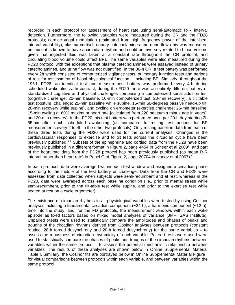
recorded in each protocol for assessment of heart rate using semi-automatic R-R interval detection. Furthermore, the following variables were measured during the CR and the FD28 protocols; cardiac vagal modulation (estimated from high frequency power of the inter-beat interval variability), plasma cortisol, urinary catecholamines and urine flow (this was measured because it is known to have a circadian rhythm and could be inversely related to blood volume given that ingested fluid was taken at a constant rate throughout the CR protocol, and circulating blood volume could affect BP). The same variables were also measured during the FD20 protocol with the exceptions that plasma catecholamines were assayed instead of urinary catecholamines, and urine flow was not quantified. In the 38-h CR, a test battery was performed every 2h which consisted of computerized vigilance tests, pulmonary function tests and periods of rest for assessment of basal physiological function – including BP. Similarly, throughout the 196-h FD28, an identical test and measurement battery was performed every 4-h during scheduled wakefulness. In contrast, during the FD20 there was an entirely different battery of standardized cognitive and physical challenges comprising a computerized serial addition test (cognitive challenge: 20-min baseline, 10-min computerized test, 20-min recovery), a tilt table test (postural challenge; 25-min baseline while supine, 15-min 60-degrees passive head-up tilt, 20-min recovery while supine), and cycling on ergometer (exercise challenge; 25-min baseline, 15-min cycling at 60% maximum heart rate [calculated from 220 beats/min minus age in years], and 20-min recovery). In the FD20 this test battery was performed once per 20-h day starting 2h 55min after each scheduled awakening (as compared to resting test periods for BP measurements every 2 to 4h in the other two protocols). Only resting baseline data from each of these three tests during the FD20 were used for the current analyses. Changes in the cardiovascular responses to exercise and to tilt tests across the circadian cycle have been previously published.5-6 Subsets of the epinephrine and cortisol data from the FD28 have been previously published in a different format in Figure 2, page 4454 in Scheer et al 20093, and part of the heart rate data from the FD28 protocol has been previously published (as mean R-R interval rather than heart rate) in Panel G of Figure 2, page 20704 in Ivanov et al 2007).4
In each protocol, data were averaged within each test window and assigned a circadian phase according to the middle of the test battery or challenge. Data from the CR and FD28 were assessed from data collected when subjects were semi-recumbent and at rest, whereas in the FD20, data were averaged across each baseline condition (i.e., prior to mental stress while semi-recumbent, prior to the tilt-table test while supine, and prior to the exercise test while seated at rest on a cycle ergometer). The existence of circadian rhythms in all physiological variables were tested by using Cosinor analyses including a fundamental circadian component (~24-h), a harmonic component (~12-h), time into the study, and, for the FD protocols, the measurement windows within each wake episode as fixed factors based on mixed model analyses of variance (JMP, SAS Institute). Unpaired t-tests were used to statistically compare the amplitudes and phases of peaks and troughs of the circadian rhythms derived from Cosinor analyses between protocols (constant routine, 28-h forced desynchrony and 20-h forced desynchrony) for the same variables – to assess the robustness of circadian rhythmicity of each variable. Paired t-tests were used were used to statistically compare the phases of peaks and troughs of the circadian rhythms between variables within the same protocol – to assess the potential mechanistic relationship between variables. The results of these analyses are shown below in Online Supplemental Material Table I. Similarly, the Cosinor fits are portrayed below in Online Supplemental Material Figure I for visual comparisons between protocols within each variable, and between variables within the same protocol.
Page 3 of 7
Comparison of results of current study with other published reports:
There are clear methodological and interpretative differences in some of the published literature regarding circadian rhythms and BP regulation. For instance, there are many studies that refer to a ‘circadian rhythm’ of BP in humans and other animals without clarifying whether this is an endogenous rhythm, but almost all such studies made BP measurements across the 24-h period while permitting sleep and altered activity across the day and night. Thus, the known reduction in BP caused by sleep and the increase in BP caused by activity are behavioral rather than endogenous explanations for much of the observed day/night pattern in BP, which is usually characterized by a peak in the middle of the wake/activity period and trough during sleep.10-12 Lesion of the SCN in rats does abolish the day/night pattern of BP but also abolishes the regular activity patterns,13 thus failing to demonstrate a circadian BP rhythm independent of activity (despite opposite claims in the literature12). In contrast, our demonstration of a clear endogenous circadian BP rhythm while controlling for activity4 suggests that the previously observed day/night patterns of BP could be caused by the summation of endogenous circadian and behavioral effects on BP. In contrast to the current results, two studies have attempted to assess endogenous circadian rhythmicity of BP in humans by using a ‘constant routine’ protocol, but with negative results.14-15 These two studies had several methodological limitations including: (1) absence of an endogenous circadian phase marker (clock time was used as a surrogate phase marker, which introduces errors when averaging data from subjects with different relationships between circadian phase and clock time, potentially obscuring an underlying circadian rhythmicity); (2) subjects were exposed to light at up to 100 lux during the ‘constant routines’, and this degree of light exposure has a major effect on circadian phase and amplitudes;8 (3) such light exposure also suppresses melatonin production during the circadian night,8 potentially cancelling any melatonin-mediated nocturnal dip in BP;16 and (4) subjects were allowed to move around between measurements, which presumably introduced uncontrolled and variable effects on BP. Our study overcame these limitations and yielded robust results that were almost identical in three groups of subjects during three different protocols, revealing an endogenous evening peak in systolic and diastolic BP. Supplemental References:
1. Ralph MR, Foster RG, Davis FC, Menaker M. Transplanted suprachiasmatic nucleus determines circadian period. Science. 1990;247:975-978.
2. Shea SA, Hilton MF, Orlova C, Ayers RT, Mantzoros CS. Independent circadian and sleep/wake regulation of adipokines and glucose in humans. J Clin Endocrinol Metab. 2005;90:2537-2544.
3. Scheer FAJL, Hilton, MF, Mantzoros CS, Shea SA. Adverse metabolic and cardiovascular consequences of circadian misalignment. Proc Natl Acad Sci USA. 2009;106:4453-4458.
4. Ivanov PCh, Hu K, Hilton MF, Shea SA, Stanley HE. Endogenous circadian rhythm in human motor activity uncoupled from circadian influences on cardiac dynamics. Proc Natl Acad Sci USA. 2007;104:20702-7.
5. Scheer FAJL, Hu K, Evoniuk H, Kelly EE, Malhotra A, Hilton MF, Shea SA. Impact of the human circadian system, exercise and their interaction on cardiovascular function. Proc Nat Acad Sci USA. 2010;107:20541-6.
6. Hu, K, Scheer, AJL, Laker M, Smales C, Shea, SA. Endogenous circadian rhythm in vasovagal response to head-up tilt. Circulation 2011; 123:961-970.
7. Cekici L, Valipour A, Kohansal R, Burghuber OC. Short-term effects of inhaled salbutamol on autonomic cardiovascular control in healthy subjects: a placebo-controlled study. Br J Clin Pharmacol. 2009;67:394-402.
Page 4 of 7
8. Zeitzer JM, Dijk DJ, Kronauer R, Brown E, Czeisler CA. Sensitivity of the human circadian pacemaker to nocturnal light: melatonin phase resetting and suppression. J Physiol. 2000;526:695-702.
9. Czeisler CA Duffy JF, Shanahan TL, Brown EN, Mitchell JF, Rimmer DW, Ronda, JM, Silva EJ, Allan JS, Emens JS, Dijk DJ, Kronauer RE. Stability, precision, and near-24-hour period of the human circadian pacemaker. Science. 1999:284:2177-81.
10. Hermida RC, Fernández JR, Ayala DE, Mojón A, Alonso I, Calvo C. Circadian time-qualified tolerance intervals for ambulatory blood pressure monitoring in the diagnosis of hypertension. Chronobiol Int. 2004;21:147-60.
11. Pickering TG, Harshfield GA, Kleinert HD, Blank S, Laragh JH. Blood pressure during normal daily activities, sleep, and exercise. Comparison of values in normal and hypertensive subjects. JAMA. 1982;247:992-996.
12. Portaluppi F, Waterhouse J, Minors D. The rhythms of blood pressure in humans. Exogenous and endogenous components and implications for diagnosis and treatment. Ann N Y Acad Sci. 1996;783:1-9.
13. Janssen, B. J., C. M. Tyssen, H. Duindam & W. J. Rietveld. Suprachiasmatic lesions eliminate 24-h blood pressure variability in rats. Physiol. Behav. 1994;55:307–311.
14. Kerkhof GA, Van Dongen HP, Bobbert AC. Absence of endogenous circadian rhythmicity in blood pressure? Am J Hypertens. 1998;11:373-377.
15. Van Dongen HP, Maislin G, Kerhhof GA. Repeated assessment of the endogenous 24-hour profile of blood pressure under constant routine. Chronobiol Int. 2001;18:85-98.
16. Scheer FA, Van Montfrans GA, van Someren EJ, Mairuhu G, Buijs RM. Daily nighttime melatonin reduces blood pressure in male patients with essential hypertension. Hypertension. 2004;43:192-197.
Page 5 of 7
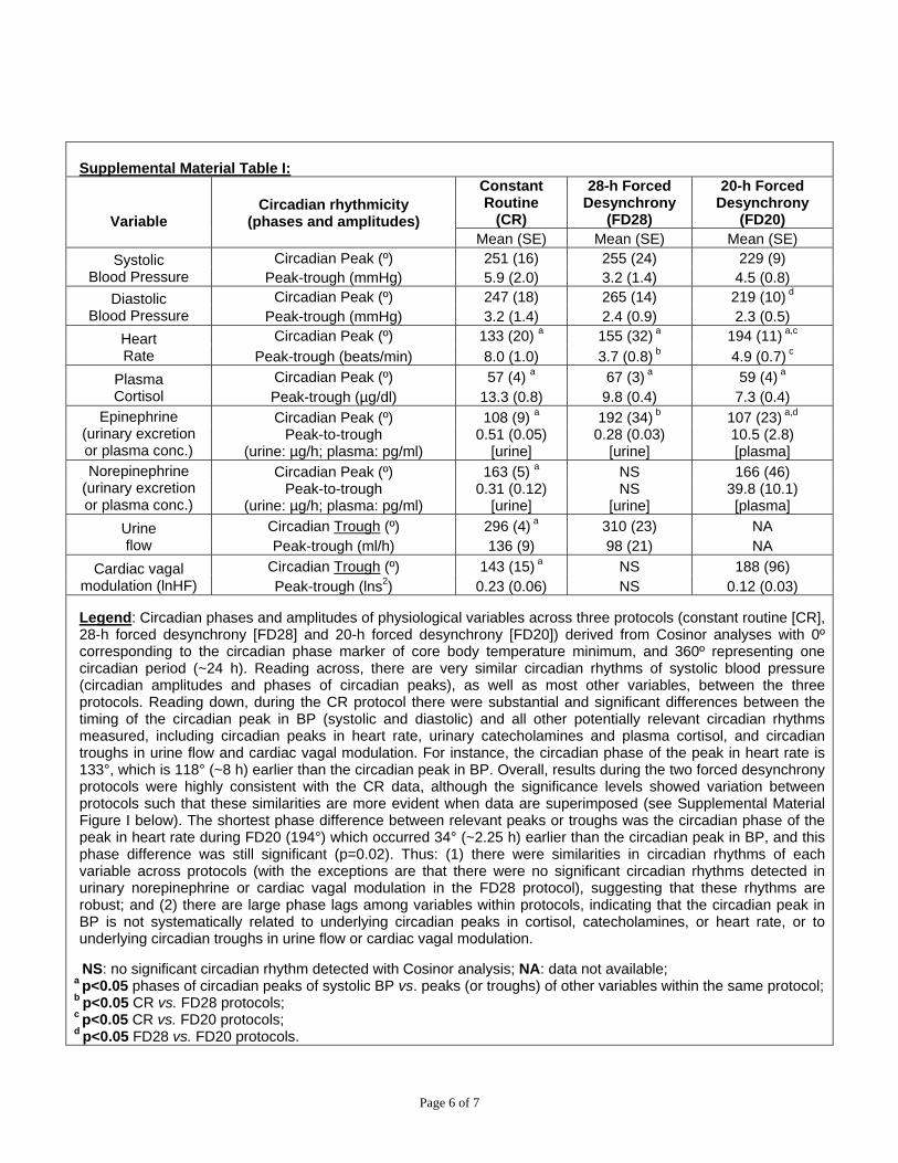
Supplemental Material Table I:
Constant Routine
(CR)
28-h Forced Desynchrony
(FD28)
20-h Forced Desynchrony
(FD20) Variable
Circadian rhythmicity (phases and amplitudes)
Mean (SE) Mean (SE) Mean (SE) Circadian Peak (º) 251 (16) 255 (24) 229 (9) Systolic
Blood Pressure Peak-trough (mmHg) 5.9 (2.0) 3.2 (1.4) 4.5 (0.8) Circadian Peak (º) 247 (18) 265 (14) 219 (10) d Diastolic
Blood Pressure Peak-trough (mmHg) 3.2 (1.4) 2.4 (0.9) 2.3 (0.5) Circadian Peak (º) 133 (20) a 155 (32) a 194 (11) a,c Heart
Rate Peak-trough (beats/min) 8.0 (1.0) 3.7 (0.8) b 4.9 (0.7) c Circadian Peak (º) 57 (4) a 67 (3) a 59 (4) a Plasma
Cortisol Peak-trough (µg/dl) 13.3 (0.8) 9.8 (0.4) 7.3 (0.4) Circadian Peak (º) 108 (9) a 192 (34) b 107 (23) a,d Epinephrine
(urinary excretion or plasma conc.)
Peak-to-trough (urine: µg/h; plasma: pg/ml)
0.51 (0.05) [urine]
0.28 (0.03) [urine]
10.5 (2.8) [plasma]
Circadian Peak (º) 163 (5) a NS 166 (46) Norepinephrine (urinary excretion or plasma conc.)
Peak-to-trough (urine: µg/h; plasma: pg/ml)
0.31 (0.12) [urine]
NS [urine]
39.8 (10.1) [plasma]
Circadian Trough (º) 296 (4) a 310 (23) NA Urine flow Peak-trough (ml/h) 136 (9) 98 (21) NA
Circadian Trough (º) 143 (15) a NS 188 (96) Cardiac vagal modulation (lnHF) Peak-trough (lns2) 0.23 (0.06) NS 0.12 (0.03) Legend: Circadian phases and amplitudes of physiological variables across three protocols (constant routine [CR], 28-h forced desynchrony [FD28] and 20-h forced desynchrony [FD20]) derived from Cosinor analyses with 0º corresponding to the circadian phase marker of core body temperature minimum, and 360º representing one circadian period (~24 h). Reading across, there are very similar circadian rhythms of systolic blood pressure (circadian amplitudes and phases of circadian peaks), as well as most other variables, between the three protocols. Reading down, during the CR protocol there were substantial and significant differences between the timing of the circadian peak in BP (systolic and diastolic) and all other potentially relevant circadian rhythms measured, including circadian peaks in heart rate, urinary catecholamines and plasma cortisol, and circadian troughs in urine flow and cardiac vagal modulation. For instance, the circadian phase of the peak in heart rate is 133°, which is 118° (~8 h) earlier than the circadian peak in BP. Overall, results during the two forced desynchrony protocols were highly consistent with the CR data, although the significance levels showed variation between protocols such that these similarities are more evident when data are superimposed (see Supplemental Material Figure I below). The shortest phase difference between relevant peaks or troughs was the circadian phase of the peak in heart rate during FD20 (194°) which occurred 34° (~2.25 h) earlier than the circadian peak in BP, and this phase difference was still significant (p=0.02). Thus: (1) there were similarities in circadian rhythms of each variable across protocols (with the exceptions are that there were no significant circadian rhythms detected in urinary norepinephrine or cardiac vagal modulation in the FD28 protocol), suggesting that these rhythms are robust; and (2) there are large phase lags among variables within protocols, indicating that the circadian peak in BP is not systematically related to underlying circadian peaks in cortisol, catecholamines, or heart rate, or to underlying circadian troughs in urine flow or cardiac vagal modulation.
NS: no significant circadian rhythm detected with Cosinor analysis; NA: data not available; a p<0.05 phases of circadian peaks of systolic BP vs. peaks (or troughs) of other variables within the same protocol; b p<0.05 CR vs. FD28 protocols; c p<0.05 CR vs. FD20 protocols; d p<0.05 FD28 vs. FD20 protocols.
Page 6 of 7
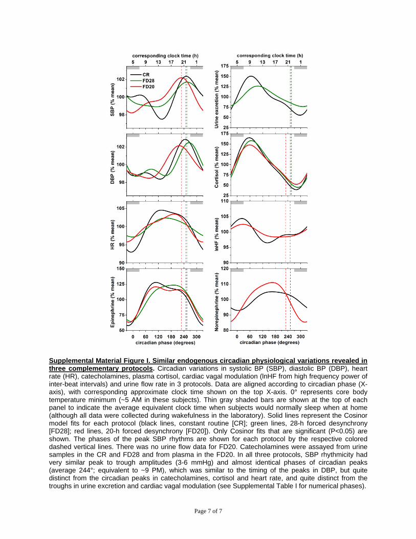
Supplemental Material Figure I. Similar endogenous circadian physiological variations revealed in three complementary protocols. Circadian variations in systolic BP (SBP), diastolic BP (DBP), heart rate (HR), catecholamines, plasma cortisol, cardiac vagal modulation (lnHF from high frequency power of inter-beat intervals) and urine flow rate in 3 protocols. Data are aligned according to circadian phase (X-axis), with corresponding approximate clock time shown on the top X-axis. 0° represents core body temperature minimum (~5 AM in these subjects). Thin gray shaded bars are shown at the top of each panel to indicate the average equivalent clock time when subjects would normally sleep when at home (although all data were collected during wakefulness in the laboratory). Solid lines represent the Cosinor model fits for each protocol (black lines, constant routine [CR]; green lines, 28-h forced desynchrony [FD28]; red lines, 20-h forced desynchrony [FD20]). Only Cosinor fits that are significant (P<0.05) are shown. The phases of the peak SBP rhythms are shown for each protocol by the respective colored dashed vertical lines. There was no urine flow data for FD20. Catecholamines were assayed from urine samples in the CR and FD28 and from plasma in the FD20. In all three protocols, SBP rhythmicity had very similar peak to trough amplitudes (3-6 mmHg) and almost identical phases of circadian peaks (average 244°; equivalent to ~9 PM), which was similar to the timing of the peaks in DBP, but quite distinct from the circadian peaks in catecholamines, cortisol and heart rate, and quite distinct from the troughs in urine excretion and cardiac vagal modulation (see Supplemental Table I for numerical phases).
Page 7 of 7
Steven A. Shea, Michael F. Hilton, Kun Hu and Frank A.J.L. Scheerthe Evening
Existence of an Endogenous Circadian Blood Pressure Rhythm in Humans That Peaks in
Print ISSN: 0009-7330. Online ISSN: 1524-4571 Copyright © 2011 American Heart Association, Inc. All rights reserved.is published by the American Heart Association, 7272 Greenville Avenue, Dallas, TX 75231Circulation Research
doi: 10.1161/CIRCRESAHA.110.2336682011;108:980-984; originally published online April 7, 2011;Circ Res.
http://circres.ahajournals.org/content/108/8/980World Wide Web at:
The online version of this article, along with updated information and services, is located on the
http://circres.ahajournals.org/content/suppl/2011/04/07/CIRCRESAHA.110.233668.DC1.htmlData Supplement (unedited) at:
http://circres.ahajournals.org//subscriptions/
is online at: Circulation Research Information about subscribing to Subscriptions:
http://www.lww.com/reprints Information about reprints can be found online at: Reprints:
document. Permissions and Rights Question and Answer about this process is available in the
located, click Request Permissions in the middle column of the Web page under Services. Further informationEditorial Office. Once the online version of the published article for which permission is being requested is
can be obtained via RightsLink, a service of the Copyright Clearance Center, not theCirculation Researchin Requests for permissions to reproduce figures, tables, or portions of articles originally publishedPermissions:
by guest on February 23, 2015http://circres.ahajournals.org/Downloaded from