Embed Size (px)
Citation preview
LONG-TERM FOLLOW-UP OF EXERCISE REHABILITATION OUTCOMES IN PATIENTS WITH CHRONIC OBSTRUCTIVE PULMONARY DISEASE
By
TAMARA MARIE ARROWOOD
A Thesis Submitted to the Graduate Faculty of
WAKE FOREST UNIVERSITY
in Partial Fulfillment of the Requirements
for the Degree of
MASTER OF SCIENCE
in the Department of Health and Exercise Science
May 2002
Winston-Salem, North Carolina
Approved By: Michael J. Berry, Ph.D., Advisor ___________________________ Examining Committee: Gary D. Miller, Ph.D. ___________________________ Patricia A. Nixon, Ph.D. ___________________________
DEDICATION
This thesis is dedicated to my mom and dad, A.K.A. Gongo and Bubba, who have always believed in me, supported me in every possible way, showed me the value of determination and hard work, inspired me to dream big and go after whatever I wanted, and encouraged me to be the best person that I could possibly be. To my sister, Cindy, and Nader, Nathan, and Megan, who instill in me the true meaning of joy by letting me experience the happiness of childhood all over again and by being such a loving family. To Rick, who over the years has helped me grow, and learn more about myself than I ever thought I wanted to know.
ii
ACKNOWLEDGEMENTS
I wish to thank many people who have made graduate school and the last 2 years such an enjoyable experience for me:
My family, who without their continual encouragement, love, and support, I would be unable to accomplish all that I have in my life.
Bill, for his love, support, and confidence in me to always do my best.
My classmates: Gretchen, Jamie, Stacey, Leigh Anne, and Theresa; I couldn’t ask for a more supportive, caring, and entertaining group. Thanks, I love you guys! Jim, for his friendship, guidance, and never-ending encouragement throughout the past year.
Dr. Berry, for sharing his expertise and outrageous sense of humor with me over the past 2 years, as well as for his ability to somehow make “square wheels roll”.
Dr. Miller and Dr. Nixon, for their valuable input to this project and for serving on my thesis examining committee. Dr. Ribisl and the HES Department for giving me the incredible opportunity to study, learn, teach, lead, and become a part of the Wake Forest family. The Cardiac Rehab Program staff and participants, for making my job not seem like “work”.
The REACT participants who graciously volunteered their time and effort to make this study possible, and also Dave and Eve-Marie for their support. The HES Classes of 2001 and 2003, for all of their help and for reminding me how to have fun!
Ann, Dorothy, Sherry, and Teresa, all very good friends who have stuck by me over the years, through good times and bad.
iii
TABLE OF CONTENTS
DEDICATION................................................................................................................... ii
ACKNOWLEDGEMENTS ............................................................................................ iii
TABLE OF CONTENTS ................................................................................................ iv
LIST OF ABBREVIATIONS ......................................................................................... vi
LIST OF TABLES AND FIGURES.............................................................................. vii
ABSTRACT.................................................................................................................... viii
INTRODUCTION............................................................................................................. 1
LITERATURE REVIEW ................................................................................................ 6
Definition of Chronic Obstructive Pulmonary Disease .................................................. 6
Etiology ........................................................................................................................... 7
Epidemiology .................................................................................................................. 9
Pathophysiology............................................................................................................ 10
Medical Management.................................................................................................... 12 Smoking Cessation.................................................................................................... 12 Medications............................................................................................................... 13 Supplemental Oxygen Therapy................................................................................. 18
Ventilatory Muscle Training ......................................................................................... 20
Lung Surgery................................................................................................................. 24 Bullectomy................................................................................................................ 24 Lung Volume Reduction Surgery ............................................................................. 25 Lung Transplantation................................................................................................ 27
Pulmonary Rehabilitation............................................................................................. 29 Lower Extremity Exercise ........................................................................................ 34 Upper Extremity Exercise......................................................................................... 35 Total Body Resistance Training................................................................................ 36
PURPOSE........................................................................................................................ 40
HYPOTHESIS................................................................................................................. 40
METHODS ...................................................................................................................... 41
Reconditioning Exercise and Chronic Obstructive Pulmonary Disease Trial ............. 41
Participants................................................................................................................... 41
iv
Procedures .................................................................................................................... 42
Outcome Measures........................................................................................................ 43 Pulmonary Function Tests ........................................................................................ 43 Physical Activity Scale for the Elderly..................................................................... 43 Physical Function Questionnaire .............................................................................. 43 Chronic Respiratory Disease Index Questionnaire ................................................... 44 6-Minute Walk Test .................................................................................................. 45
Statistical Analysis ........................................................................................................ 45
RESULTS ........................................................................................................................ 47
Pulmonary Function Tests ............................................................................................ 49
Chronic Respiratory Index Questionnaire………………………………………………… 51
Physical Activity Scale for the Elderly.......................................................................... 54
Physical Function Questionnaire ................................................................................. 55
6-Minute Walk Test ....................................................................................................... 56
DISCUSSION .................................................................................................................. 57
Conclusion .................................................................................................................... 62
REFERENCES................................................................................................................ 63
SCHOLASTIC VITA ..................................................................................................... 78
v
LIST OF ABBREVIATIONS
ADL(s) activity(s) of daily living mg milligram(s) ANCOVA analysis of covariance mmHg millimeter(s) of Mercury ATS American Thoracic Society n number ATT alpha1-antitrypsin NOTT Nocturnal Oxygen Therapy
Trial BMRC British Medical Research
Council NPPV non-invasive positive
pressure ventilation cAMP cyclic adenosine
monophosphate PaCO2 arterial partial pressure of
carbon dioxide CET cycle ergometry training PaO2 arterial partial pressure of
oxygen cm(s) centimeter(s)
PASE Physical Activity Scale for
the Elderly COPD chronic obstructive
pulmonary disease PDE phosphodiesterase
CRQ Chronic Respiratory Index Questionnaire
PEmax maximum expiratory pressure
et al. and others PFQ Physical Function Questionnaire F(s) female(s) PFT (s) pulmonary function test(s) FAST Fitness and Arthritis in
Seniors Trial PImax maximum inspiratory pressure
FEV1 forced expiratory volume in 1 second
REACT Reconditioning and Chronic Pulmonary Disease Trial
FU follow-up RV residual volume FVC forced vital capacity SEM standard error of the mean HRQOL health-related quality
of life SPSS Statistical Package for the Social
l Sciences kg(s) kilogram(s) ST short-term L(s) liter(s) TLC total lung capacity LT long-term TV tidal volume LTOT long-term oxygen therapy U.S. United States LVRS lung volume reduction
surgery VE minute ventilation
m(s) meter(s) VMT ventilatory muscle training M(s) male(s) VO2max maximal oxygen uptake min(s) minute(s) yr(s) year (s)
vi
LIST OF TABLES AND FIGURES
TABLE PAGE I Participant Demographics/Clinical Information ……………………………….48
FIGURES
1 ST vs. LT Mean FEV1 % Predicted…………………………………………….49
2 ST vs. LT Mean FEV1/FVC Ratio …………………………………………….50
3 ST vs. LT Mean RV/TLC Ratio………………………………………………..50
4 ST vs. LT Mean CRQ Dyspnea Score………………………………………….51
5 ST vs. LT Mean CRQ Mastery Score…………………………………………..52
6 ST vs. LT Mean CRQ Emotion Score…………………………………………..53
7 ST vs. LT Mean CRQ Fatigue Score…………………………………………....53
8 ST vs. LT Mean PASE Score…………………………………………………...54
9 ST vs. LT Mean PFQ Score…………………………………………………….55
10 ST vs. LT Mean 6-Minute Walk Test Distance ………….…………………….56
vii
Tamara Marie Arrowood ABSTRACT
LONG-TERM FOLLOW-UP OF EXERCISE REHABILITATION OUTCOMES IN PATIENTS WITH CHRONIC OBSTRUCTIVE PULMONARY DISEASE
Thesis under the direction of Michael J. Berry, Ph.D., Professor Health and Exercise Science The purpose of this study was to compare the long-term outcomes in pulmonary
function, self-reported health–related quality of life, physical activity, and disability,
along with functional exercise capacity in COPD patients either completing a 3-month
(short-term, ST) or an 18-month (long-term, LT) exercise rehabilitation program at 58
months. Thirty-nine patients completed the follow-up study, including 12 from the ST
and 27 from the LT groups. There were no significant differences between the ST and the
LT groups in the adjusted means of FEV1 % predicted, (57.6 ± 3.1 versus 56.6 ± 2.1%),
FEV1/FVC ratio, (51.5 ± 2.1 versus 52.1 ± 1.4%), RV/TLC ratio, (53.7 ± 4.2 versus 58.4
± 2.8%), CRQ: dyspnea (4.9 ± 0.4 versus 5.1 ± 0.2 units), mastery (6.3 ± 0.2 versus 6.4 ±
0.1 units), emotion (5.6 ± 0.2 versus 5.7 ± 0.1 units), fatigue (4.3 ± 0.3 versus 4.7 ± 0.2
units), PASE (103.0 ± 12.2 versus 92.2 ± 8.8 units), PFQ (35.0 ± 3.8 versus 39.4 ± 2.5
units), or 6-minute walk distance, (500.7 ± 26.6 versus 517.9 ± 18.8 m) respectively,
measured at the 58-months. These results showed that at 58 months there are no
significant differences between the ST and the LT exercise rehabilitation groups in any of
the outcome variables. Therefore, an additional 15 months of participation in an exercise
rehabilitation program did not result in a difference in the level of benefits maintained at
58 months.
viii
INTRODUCTION
Chronic obstructive pulmonary disease (COPD), a condition of progressive
deterioration of the respiratory system characterized by the obstruction of pulmonary
airways and decreased airflow, is a major health problem worldwide. COPD is currently
the fourth leading cause of death in the United States (U.S.), following heart disease,
cancer, and stroke, (95) and is the only leading cause of death which has increased in
prevalence (by 71%) over the last several years (68). COPD also has a major influence on
morbidity, accounting for an estimated 668,362 hospital discharges at a rate of 24.5 per
10,000 individuals in 1998 (95).
Individuals with COPD experience functional capacity limitations that adversely
impact the physical activity level and the performance of activities of daily living (ADL’s)
in these patients. A recent national survey of 573 individuals with COPD given by
Schulman, Ronca, and Bucuvalas, Incorporated, and supported by the American Lung
Association (100) revealed that 70% of respondents experienced limitations in activities
requiring physical exertion, 51% had limitations in their occupational abilities, and 56%
were limited in performing household chores. The survey also showed that social
activities and family activities were limited in 53% and 46% respectively, while normal
sleeping patterns were disturbed in 50% of respondents.
As a result of these physical limitations, the quality of life for many COPD
patients and their families is significantly compromised. Twenty-three percent of
respondents described themselves as “invalid”, while 8% remained homebound due to
dyspnea (100), not only because of its physical effects, but also due to the fear and the
1
anxiety associated with the thought of experiencing the sensation of breathlessness
outside of the home.
The medical treatment of individuals with COPD centers on the management of
the current symptoms and the prevention of future exacerbations. Various drug therapies,
supplemental oxygen, pulmonary rehabilitation programs, and surgical interventions all
are currently available treatment options. Smoking cessation is paramount to any
successful treatment plan in COPD patients who continue to smoke, and many alternative
strategies are available to assist with this difficult process (29, 91, 94).
Multidisciplinary pulmonary rehabilitation programs have been successful in
enhancing the physical function and the health related quality of life (HRQOL) in
individuals with COPD. A meta-analysis of 14 randomized controlled clinical trials
looking at the effectiveness of pulmonary rehabilitation revealed that significant
improvements occurred in maximal exercise capacity, functional exercise capacity, and
HRQOL in pulmonary rehabilitation participants as compared to individuals in the
control groups (72).
The long-term (greater than one year post intervention) benefits of participation in
pulmonary rehabilitation programs have also been analyzed (13, 21, 61, 81, 102, 123, 146,
147). The outcomes typically measured in these long-term follow-up studies include
physical performance parameters, HRQOL, morbidity as shown by the number of
hospitalizations or by the total number of days of hospitalization, mortality, as well as the
total utilization of healthcare dollars. In a randomized control trial (128) the long-term
benefits achieved by 50 COPD patients who underwent a 6-month outpatient exercise
therapy only regimen were compared to 50 COPD patients who received “usual care”
2
treatment. The significant improvements in the 6-minute walk test distance, maximal
oxygen uptake (VO2max), quadriceps strength, and HRQOL made by the exercise
therapy group (n=37), as compared to the control group (n=33), were still present and
clinically relevant at the LT follow-up visit.
Strijbos et al. (123) examined the long-term outcomes of COPD patients
participating in a 12-week home-based or a hospital-based outpatient pulmonary
rehabilitation program, as compared to that of a control group that did not receive an
exercise intervention. Their findings showed that both the hospital-based and the home-
based rehabilitation groups had similar significant improvements in exercise capacity,
perceived dyspnea, and well-being at the 6-month follow-up as compared to the control
group. At 18 months, the improvement in exercise capacity began to diminish in the
hospital-based rehabilitation group, as compared to that of the home-based rehabilitation
group. The benefits achieved in both rehabilitation groups were still statistically
significant when compared to the control group at 18 months.
A randomized control trial by Ries et al. (102) found that many of the significant
physical and psychological benefits achieved from participating in an 8-week outpatient
comprehensive pulmonary rehabilitation program diminished within one year of follow-
up. Results from the Reconditioning and Chronic Pulmonary Disease Trial (REACT)
study by Berry et al. (21) also showed that the improvements in self-reported disability,
physical function, and HRQOL made by COPD patients following the completion of a
3-month exercise therapy program had greatly diminished by 18 months, while the
18-month exercise therapy group maintained and improved upon the benefits achieved at
the end of the initial 3-month program.
3
Maintenance exercise programs initiated after the completion of a structured
pulmonary rehabilitation program have been successful in postponing the decline in
benefits achieved following the initial intervention. A study by Guell and colleagues (61)
compared the long-term effects of a 3-month exercise training program followed by a
6-month maintenance program in 30 COPD patients (exercise group) to that of 30 COPD
patients who received standard care only (control group). The results revealed that a
6-month supervised one-time-a-week maintenance therapy program consisting of
breathing and arm/leg coordination exercises helped maintain the benefits achieved in
6-minute walk distance, dyspnea, and HRQOL in the exercise group at 24 months. This
study also showed that the exercise group experienced significantly fewer COPD
exacerbations (p<0.0001) and a trend for fewer hospitalizations than the control group.
Groisbois et al. (59) investigated the effects that a maintenance exercise regimen had on
58 COPD patients who had completed an initial 7-week outpatient pulmonary
rehabilitation program. The patients were self-selected into 1 of 4 groups who had
continued supervised exercise training either once or twice a week, continued an
independent home exercise program, or did not continue any type of exercise program.
Improvements in maximum workload and dyspnea ratings occurred in all patients
following completion of the initial program, but remained significant only in the once-a-
week supervised exercise training group and the independent home exercise group at the
LT follow-up. The non-exercisers showed diminished benefits approaching baseline
measures at 18 months.
The duration of an exercise intervention may also influence the magnitude of
benefits achieved as well as the length of time that they are maintained in COPD patients.
4
The REACT study, a recent unpublished investigation by Berry and colleagues (21)
compared the effects of a 3-month (short-term, ST) versus an 18-month (long-term, LT)
exercise rehabilitation program on self-reported disability, physical function, and
HRQOL in 140 COPD patients. The results showed that both the ST and the LT groups
had similar improvements in these outcomes at 3 months. At 18 months, those patients
randomized into the LT exercise group (n=62) maintained the improvements in self-
reported disability, physical function, and HRQOL obtained at 3 months. Those patients
in the ST exercise group (n=56), showed declining benefits at 18 months, which
approached baseline values. The results of this study present a case for prolonged
exercise rehabilitation programs (greater than 3 months), but also showed that the
benefits achieved from short-term exercise rehabilitation programs are not permanent.
While the results of the REACT study showed diminished benefits in COPD
patients who completed a ST exercise rehabilitation program at 18 months, it is not
known whether this decline in benefits persisted beyond 18 months. It is also not known
whether the improvements made by those patients participating in the LT exercise
rehabilitation program were maintained. Therefore, it was the primary purpose of this
thesis to describe and compare the outcomes of COPD patients who participated in either
a ST or a LT exercise rehabilitation program at 58 months.
5
LITERATURE REVIEW
Definition of Chronic Obstructive Pulmonary Disease
Chronic obstructive pulmonary disease (COPD) is a progressive lung disease
characterized primarily by airway obstruction and decreased airflow. Chronic bronchitis
and emphysema are 2 distinct components of COPD, but may occur simultaneously in the
same individual. Chronic bronchitis is defined as “the presence of a chronic cough for 3
months in each of 2 successive years in a patient in whom other causes of chronic cough
have been excluded” (3). Chronic bronchitis is characterized by chronic inflammation
and edema of the peripheral airways, excessive mucus production and accumulation,
bronchospasm, bronchial airway obstruction hyperinflation of the alveoli distal to the
obstructed airways.
Emphysema is defined as “the abnormal permanent enlargement of the airspaces
distal to the terminal bronchioles, accompanied by the destruction of their walls and
without obvious fibrosis” (3). Emphysema is characterized by alveolar deterioration and
hyperinflation, destruction of pulmonary capillaries, weakened respiratory bronchioles,
and air trapping.
Depending upon the degree of pulmonary system damage and airway
hyperresponsiveness, the following may all occur in an individual with COPD: obstructed
airflow, lung hyperinflation, mismatched ventilation/perfusion ratio, decreased maximum
inspiratory pressure (PImax), increased work of breathing, and a decreased gas diffusion
capacity (33). The primary physical manifestations that result from these
pathophysiologic changes include dyspnea (the sensation of breathlessness), persistent
6
cough, excessive sputum production, fatigue, decreased exercise tolerance, hypoxemia,
and deconditioning. Malnutrition and osteoporosis may also occur, especially in
advanced cases of COPD (54, 109).
Although COPD is a progressive disease that worsens in severity with time, it is
characterized by recurrent “exacerbations” of varying intensity. A COPD exacerbation
commonly occurs following a bacterial infection (in 50% to 70% of cases) (117, 120) or
due to a repeated exposure to an environmental pollutant such as suspended particulate
matter, carbon monoxide, sulfur dioxide, and nitrogen dioxide (in up to 5% of cases). An
acute COPD exacerbation is characterized by any combination of worsening dyspnea, an
increase in sputum production, and/or an increase in sputum purulence (120). Eighteen to
34% of all COPD exacerbations may be related to upper respiratory tract viral infections
(117), while changes in ambient temperature may also trigger an exacerbation (54). In the
U.S. it is estimated that individuals with COPD experience anywhere from 1-4 acute
exacerbations of the disease each year (117).
Thus, exacerbations are detrimental to a COPD patient’s HRQOL and may result
in either temporary or permanent disability, increased emergency room visits and hospital
admissions, respiratory failure, or even death. An important goal of COPD management
therefore is to decrease the number and the severity of exacerbations experienced by
COPD patients, through comprehensive patient education, early aggressive medical
management, and proper follow-up care
Etiology
The etiology of COPD can be attributed to smoking in 80-90% of all cases, to
exposure to second-hand smoke and/or environmental pollutants, to having a history of
7
recurrent respiratory infections in childhood, as well as to a genetic influence in 1-5% of
cases (12). Approximately 15% of all individuals who smoke will develop COPD (117)
and they are 10 times more likely to die of COPD than nonsmokers (11). Smoking
cessation has been related to a decreased number of recurrent respiratory symptoms and
infections in former smokers, as compared to those individuals who continued to smoke
(30).
Smoking is not the only etiology related to the development of COPD, hereditary
influences also play a role in the onset of COPD early in life. The most common genetic
cause of COPD is related to an inherited deficiency in alpha1-antitrypsin (ATT), a protein
that is normally produced by the liver that plays a role in the inhibition of several
proteases, including neutrophil elastase (106). Neutrophil elastase, an enzyme that
degrades lung elastin, causes destruction of lung tissue, which results in many of the
characteristic structural and functional changes associated with emphysema. The
deficiency in ATT results in unchecked elastase activity and further damage to lung
tissue. Alpha1-antitrypsin deficiency is responsible for the early onset of COPD (usually
before the age of 50 years) in approximately 50-100,000 individuals in the U.S., many of
whom are Caucasians of northern European descent (3, 12).
The remaining causes of COPD include prolonged exposure to second-hand
smoke and/or environmental pollutants and recurrent respiratory infections during
childhood. Second-hand smoke can be just as harmful to nonsmokers as smoking is to
smokers. Second-hand smoke contains 200 poisonous chemicals that can cause serious
health problems including respiratory infections, COPD exacerbations, asthma, and
coronary artery disease (11). Other environmental pollutants such as carbon monoxide,
8
sulfur dioxide, nitrogen oxide, the ozone, and suspended particulates may initiate the
inflammatory process and contribute to the development of COPD.
Recurrent respiratory infections during childhood have also been suggested as a
cause of COPD. Early recurrent infections in childhood can stunt the growth of lung
tissue and result in decreased forced expiratory volume in 1 second (FEV1) and forced
vital capacity (FVC) in adulthood (114). Permanent damage and fibrotic scarring of the
airways may occur as a result of the excessive inflammatory response (airway
hyperresponsiveness) potentiated by frequent lower respiratory tract bacterial infections
in childhood (113). Chronic mucus hypersecretion and poor control of lung elastase
activity causing increased lung tissue damage both occur, which contribute to the
development of chronic bronchitis and/or emphysema. All of these pathologic changes
caused by recurrent childhood infections make the individual even more susceptible to
the further lung damage caused by cigarette smoke and other pollutants.
Epidemiology
The World Health Organization ranks the U.S. 12th in COPD mortality for men
and 7th in COPD mortality for women when compared to 28 other industrialized countries
(148). COPD is currently the fourth leading cause of death in the U.S. and is the only
cause of death that has increased in prevalence over the last several years (68).
Approximately 113,000 deaths in the U.S. were attributed to COPD in 1998, while it
encompassed almost 9 million cases of chronic bronchitis and 3 million cases of
emphysema (12). The combined chronic bronchitis and emphysema age-adjusted
9
mortality rates for 1998 were 24.7 per 100,000 men and 16.5 per 100,000 women.
Hospital discharge rates varied among age groups, with 68% of all 1998 discharges
(a rate of 133.3 per 10,000 individuals) attributed to those who were 65 years and older.
Over 16 million physician visits related to COPD occurred in 1995, with chronic
bronchitis accounting for approximately 10 million visits and chronic airways obstruction
accounting for approximately 4 million visits (11, 84).
The economic impact of COPD is tremendous, costing the U.S. approximately
$30.4 billion annually, including $14.7 billion for direct healthcare expenditures and
$15.7 billion in indirect costs (12). An estimated 73% of these healthcare expenditures
were utilized by only approximately 10% of all COPD patients (142).
Pathophysiology
The major pathological consequences of COPD include decreased elastic recoil
and increased compliance of the damaged lung tissue which results in increased work of
breathing, lung hyperinflation both at rest and dynamically during exercise, fixed
expiratory airflow obstruction, decreased inspiratory muscle strength, and decreased
maximum inspiratory pressure (33). Gas exchange and diffusion capacity are also
affected, with arterial hypoxemia commonly occurring mainly due to a combination of
ventilation-perfusion mismatch, alveolar hypoventilation, and low mixed venous partial
pressure of oxygen. Hypercapnia and chronic respiratory acidosis also can occur due to
the inability of individuals with COPD to maintain adequate minute ventilation (VE), and
also due to an increased respiratory dead space to tidal volume ratio. All of these physical
10
manifestations contribute to the intolerance of increased physical activity and exercise as
well as to dyspnea.
Dyspnea is a complex symptom related to many of the above-mentioned physical
manifestations of COPD. Several sensory input mechanisms are involved with the
development of dyspnea including increased activity of central and peripheral
chemoreceptors, along with increased afferent input from various receptors in the
pulmonary system, upper airways, and respiratory muscles (3, 6). Fatigue, as well as the
perceived level of breathlessness that results “when the demand for ventilation is out of
proportion to the patient’s ability to respond to that demand” also plays a role in the
development of dyspnea (144). The quality and the severity of dyspnea experienced by
COPD patients are also influenced by each individual’s experience, emotional state, and
expectations (6). A number of studies have shown that desensitization to dyspnea occurs
following pulmonary rehabilitation program
acidosis as factors involved with peripheral muscle dysfunction in COPD patients (26).
Maltais et al. (76) suggest aging, electrolyte imbalances, and systemic inflammation as
other factors that may play a role in the development of this muscle dysfunction. The
muscles of ambulation are especially affected (18, 25, 58), and may result in decreased
functional mobility, reliance on an assistive device for safe ambulation, or loss of
independent living all together, which would significantly impact the quality of life in
affected COPD patients.
Medical Management
The medical management of COPD is multifactorial and may include all of the
following interventions: smoking cessation, appropriate medications, oxygen therapy,
surgery, and pulmonary rehabilitation programs.
Smoking Cessation
Smoking exposes the bronchial linings to several toxic chemicals including
carbon monoxide, nicotine, hydrocarbons, and other tars, causing irritation and chronic
inflammation. The alkaloid nicotine is the primary addictive compound in cigarette
smoke and is responsible for the feeling of euphoria via a rapid increase in the release of
the central nervous system neurotransmitter, dopamine. Other effects of nicotine include
an antidepressant effect and a feeling of enhanced performance, especially for mundane
activities that rely on memory and focused attention (81). These seemingly “beneficial”
effects of nicotine, coupled with the considerably uncomfortable symptoms of nicotine
withdrawal are what make this addiction so difficult to overcome.
In individuals with mild to moderate COPD, smoking cessation has been shown
to slow the annual age-related rate of decline in FEV1 to that of healthy nonsmokers
12

(107). Therefore, smoking cessation is imperative for individuals with COPD. However,
only 2.5% of all smokers who try to quit are successful (38). Physician counseling on the
negative effects of the continued use of tobacco products and encouragement for smoking
cessation was lacking in 85.7% of all COPD patient office visits in 1996 (38, 83). This
occurred despite the National Cancer Institute’s Recommendation of the “four A’s”
program: “Ask about smoking; Advise about smoking cessation; Assist with smoking
cessation intervention; and Arrange for follow-up” (110).
Several aides for quitting smoking are available including various nicotine
delivery systems (gum, patches, inhalers, and nasal sprays), and pharmacological agents
such as the antidepressants Zyban (bupropion hydrochloride) and Buspar (buspirone
hydrochloride), or the antihypertensive, Catapres (clonidine hydrochloride), (29, 94). The
nicotine patch was found to be the most successful smoking cessation intervention in
1998 (38) and has the lowest potential for continued nicotine addiction due to the slow
delivery rate of nicotine (time to maximum effect= 500 minutes (min)) (94), as compared
to the other systems (time to maximum effect= 10-30 min). A double-blind clinical trial
by Jorenby et al. (45) showed that the combination of Zyban along with a transdermal
nicotine patch, as compared to the use of Zyban alone, had significantly higher
abstinence rates at 1 year than treatment with the nicotine patch or placebo alone
(p<0.001). Behavioral interventions also play an important role in smoking cessation,
with abstinence rates approaching 20% in the most aggressive programs (45).
Medications
Medications commonly used in the management of COPD include
bronchodilators (anticholinergic, sympathomimetic, and xanthine derivatives), anti-
13
inflammatories (steroidal and non-steroidal), antibiotics, antitussives, expectorants, and
mucolytics (43, 90). The primary therapeutic goals of the pharmacological management
of COPD are to increase bronchodilation and mucus expectoration, and to decrease
inflammation along the bronchial linings. Replacement of ATT may also be part of the
medical regimen in patients with ATT deficiency emphysema (43).
Bronchodilators are commonly used in COPD patients but are not as effective as
when used in patients with asthma. Three main classes of bronchodilators are utilized in
COPD patients including anticholinergic, sympathomimetic, and xanthine derivatives
(methylxanthines). Each class of bronchodilator has an independent mechanism for
inducing bronchodilation along with varied primary locations of action. For example,
anticholinergic bronchodilators primarily affect the larger central airways while
sympathomimetic bronchodilators primarily affect the smaller distal airways (89).
Anticholinergic bronchodilators such as Atrovent (ipratropium bromide), and
tiotropium (not approved for use in the U.S.) function by preventing acetylcholine from
binding to the muscarinic receptor sites located in the airway smooth muscle tissue. This
results in decreased bronchoconstriction, mucus secretion, and nocturnal oxygen
desaturation, as well as in improved dyspnea, exercise performance, and quality of sleep
in patients with COPD (43, 79). Atrovent is normally delivered as an inhaled medication
and the recommended dosage is 2 puffs of a metered dose inhaler 4 times per day.
Sympathomimetic bronchodilators are also regularly recommended for COPD
patients, and are administered as inhaled, oral, subcutaneous, or intravenous medications.
The smooth muscle tissue lining the bronchial airways contains beta-2 receptors that
respond to circulating levels of catecholamines by causing smooth muscle relaxation and
14
therefore bronchial dilation. Selective beta-2 agonists function by increasing the
formation of cyclic adenosine monophosphate (cAMP), which results in altered
intracellular calcium concentration (103). Smooth muscle intracellular calcium ion
concentration is a primary regulator of smooth muscle contraction, thus the actions of
sympathomimetic medications result in decreased bronchoconstriction, plus decreased
airway hyperresponsiveness, and decreased inflammatory mediator release by basophils
and mast cells (43). Some examples of sympathomimetic bronchodilators include
Proventil and Ventolin (albuterol), and Serevent (salmeterol xinafoate).
Xanthine derivatives including the methylxanthine, theophylline, have historically
been the first line of medical defense in COPD patients, but their use has significantly
declined since 1993 (132). The exact mechanism of theophylline’s therapeutic effect is
unknown, but there is some evidence to suggest that theophylline acts as a
phosphodiesterase (PDE) inhibitor at higher dosages, causing increased levels of cAMP
and bronchial smooth muscle relaxation (135). Another proposed mechanism of action is
the direct inhibition of selective PDE receptors, which also results in bronchodilation, as
well as improved diaphragm function, vital capacity, cardiac output, and exercise
performance (43). Decreased dyspnea and a decreased inflammatory reaction have also
been found to be associated with theophylline use. Toxicity and serious side effects such
as seizures, cardiac arrhythmias, and respiratory arrest, especially in the elderly and in
individuals with hepatic pathology may occur during the use of theophylline. The narrow
therapeutic range of serum theophylline is 8-15 milligrams (mg) per liter (L), but seizures
have occurred in elderly patients with serum levels as low as 14 mg/L (115).
15
Theophylline or Theo-Dur is commonly administered orally, but can also be given
intravenously.
The combination of anticholinergic and sympathomimetic medications in metered
dose inhalers has also been shown to decrease symptoms, exacerbations, and healthcare
costs in individuals with stable COPD (17, 43). One example is Combivent
(albuterol/ipratropium). The xanthine derivative, theophylline, has also been added to
some bronchodilator inhalers as well. The addition of theophylline to inhaled
bronchodilators has resulted in the reduction in the severity of COPD symptoms (87) and
may also decrease healthcare costs by preventing future COPD exacerbations.
Anti-inflammatory medications are frequently utilized by COPD patients with the
goal of treatment being the reduction of chronic bronchial inflammation, which has been
suggested as an etiology of COPD. Both steroidal and non-steroidal anti-inflammatories
can be prescribed, and are usually administered either orally or inhaled. Corticosteroids
such as prednisone and methylprednisolone, are commonly recommended for COPD
patients experiencing a severe, acute exacerbation requiring hospitalization, but should
not be used for longer than 2 weeks (81). Inhaled corticosteroids have been shown to be
effective in increasing FEV1, reducing COPD symptoms, and shortening the duration of
hospitalization in patients experiencing an acute exacerbation (81, 86). A meta-analysis
of 3 studies by Van Grunsven et al. (133) revealed that relatively high doses of inhaled
corticosteroids utilized by patients with moderate to severe COPD resulted in improved
FEV1 at 2 years and improved long-term prognosis. In contrast, systemic corticosteroids
have not been shown to slow the rate of decline in FEV1 in patients with COPD (24, 81).
Chronic use of systemic corticosteroids has been associated with the development of
16
adverse side effects such as dermal thinning, peptic ulcer disease, adrenal insufficiency,
osteoporosis, diabetes mellitus, hypertension, and myopathy. Despite these risks,
systemic corticosteroids are utilized continuously in approximately 4-10% of “steroid-
dependent” patients with severe COPD (82).
The non-steroidal anti-inflammatory medications, NasalCrom (cromolyn) and
Tilade (nedocromil), have limited success in the treatment of COPD patients, but may be
beneficial when a respiratory tract allergy is present. Newer non-steroidal anti-
inflammatory medications such as PDE 4 inhibitors that inhibit macrophages, neutrophils,
and cytotoxic T-lymphocytes, may be successful in the management of COPD by
decreasing the overall inflammatory response. The results of a clinical trial involving
COPD patients undergoing treatment with the PDE 4 inhibitor, SB 207499, demonstrated
improvement in lung function, symptoms, and quality of life (126). Several other non-
steroidal anti-inflammatory medications are currently under development, which may
prove to be beneficial in the long-term management of COPD.
Antibiotics are not routinely prescribed for patients with COPD, but have been
shown to decrease the duration of acute COPD exacerbations especially when excessive
purulent sputum is present (13). A 10-day cycle of antibiotics, such as tetracycline,
amoxicillin, or erythromycin, is usually recommended for treatment of an acute COPD
exacerbation (89). In COPD patients who experience frequent recurrent infections, the
prolonged use of antibiotics may be justified. The common bacteria involved in acute
respiratory infections include Streptococcus pneumoniae, Hemophilus influenzae, and
Moraxella catarrhalis (3, 89). Patients with COPD who have frequent infections are at
high risk for developing pneumonia, which may result in hospitalization, the use of
17
supplemental oxygen and/or assisted mechanical ventilation, or even respiratory failure.
A meta-analysis of 20 cohort studies (111) revealed that the influenza vaccine was 56%
effective in preventing respiratory illness, 50% effective in preventing hospitalization,
and 68% effective in preventing death in elderly COPD patents. This provides a strong
incentive for the recommendation that all COPD patients obtain yearly immunizations
and vaccinations for protection against both bacterial and viral infections (43, 90).
Antitussives, expectorants, and mucolytics may all be used in the treatment of
COPD patients but are of limited, if any, benefit. The routine use of these medications in
patients with COPD is not recommended, though oral acetylcysteine, a mucolytic, may
decrease the frequency of exacerbations experienced by these patients through its
antioxidant effects (3, 43).
Supplemental Oxygen Therapy
Supplemental oxygen therapy has been shown to improve mortality and HRQOL
in COPD patients with chronic hypoxemia (125). Long-term oxygen therapy (LTOT) is
utilized in 800,000-1 million individuals with COPD in the U.S. (92) and should be
delivered at a rate that maintains an arterial oxygen saturation (SaO2) level of at least
90% or an arterial partial pressure of oxygen (PaO2) of 60 millimeters of Mercury
(mmHg) (51, 125). By maintaining an adequate SaO2 level, supplemental oxygen helps to
decrease pulmonary hypertension (lowers pulmonary artery pressure and pulmonary
vascular resistance), reverses the associated polycythemia, and improves cardiac function
(125). Selinger et al.(110) showed that the removal of LTOT from hypoxic COPD
patients resulted in increased pulmonary vascular resistance and stroke volume index at
rest and during exercise, as well as causing variable effects on oxygen consumption,
18
dependent upon each patient’s resting partial pressure of arterial carbon dioxide (PaCO2).
Long-term oxygen therapy is also associated with increased body weight, improved
physical and neuropsychological function, and prolonged survival (54).
Indications for LTOT according to Medicare guidelines include: PaO2 </= 55
mmHg or SaO2 </= 89% while breathing room air, or PaO2 = 56-59 mmHg or
SaO2 = 89% with any of these accompanying signs: evidence of cor pulmonale by
electrocardiogram, erythrocytosis (hematocrit > 56%), or PaO2 >/= 60 mmHg or
SaO2 >/= 90% with “compelling medical justification” (92). Oxygen delivery systems
typically involve oxygen provided via a nasal cannula by home oxygen concentrators,
and either stationary or portable liquid oxygen tanks, via trans tracheal oxygen, or by
non-invasive positive-pressure ventilation (NPPV) mask systems.
The effect of supplemental oxygen on long-term survival in COPD patients was
the primary focus of 2 early studies, the Nocturnal Oxygen Therapy Trial (NOTT) (1)
and the British Medical Research Council (93) study on domiciliary oxygen. The NOTT
investigated the effects of continuous (17.7 hours/day) versus nocturnal (12 hours/day)
supplemental oxygen therapy on the neuropsychological function, quality of life, and
survival in patients with COPD. The BMRC study looked at the effect of supplemental
oxygen therapy (15 hours/day) versus no supplemental oxygen on survival in COPD
patients. The findings of both of these studies revealed that supplemental oxygen therapy
was effective in improving 3-year (NOTT) and 5-year (BMRC) survival along with
demonstrating that the longer the duration of oxygen therapy per day resulted in better
survival. It was also noted that the availability of a portable ambulatory oxygen supply
was clearly more beneficial for participants in the NOTT continuous oxygen therapy
19
group as compared to a stationary oxygen supply utilized by the BMRC oxygen therapy
group at the 3-year follow-up, in terms of survival.
Patients with COPD who exhibit hypoxia at rest, are typically hypoxic during
periods of increased activity and have been found to benefit from supplemental oxygen
during exercise. A study by Garrod et al. (50) revealed that hypoxic COPD patients who
utilized supplemental oxygen (4 L/min) during a 6-week exercise rehabilitation program
had significant decreases in dyspnea (p=0.02), but not in other outcome measures as
compared to those patients receiving a placebo, though this decrease may have been
related to initial baseline differences. Other studies show improvement in VE and exercise
tolerance at submaximal workloads, as well as increased maximum exercise level when
hypoxemic COPD patients use supplemental oxygen during exercise training (50, 70).
Adverse medical consequences can occur in patients using LTOT including the
development of oxygen toxicity, which may lead to adult respiratory distress syndrome,
and also increased carbon dioxide retention, which can actually cause a depressed
respiratory drive in select COPD patients (3). Physical hazards of LTOT include the
possibility of fires and explosions, due to the high flammability of oxygen. However, the
benefits of LTOT for hypoxemic COPD patients clearly outweigh these potentially
deleterious effects.
Ventilatory Muscle Training
Ventilatory muscle training (VMT) encompasses several techniques that are
performed by individuals with COPD to attempt to increase the strength and endurance of
the respiratory musculature. Maximum inspiratory pressure produced by the muscles of
20
inspiration, especially the diaphragm, is reduced in individuals with COPD as compared
to normal healthy controls. Maximum expiratory pressure (PEmax) may also be reduced
in individuals with COPD, but is not as common a finding (87).
Structural alterations in the chest wall, changes in respiratory physiology, and
overall physical deconditioning associated with COPD all impact the function of the
muscles of inspiration (77). The primary muscle of inspiration is the diaphragm, which is
a thin, dome-shaped muscle with a central tendon. The pathologic changes of COPD,
specifically hyperinflation, cause alterations in total muscle length and the zone of
apposition of the diaphragm, resulting in a decrease in the contractile mechanical
advantage and reduced inspiratory pressure generated during inspiration. This mechanism
may also affect the mechanical advantage of the parasternal intercostals, another
important set of inspiratory muscles. Other changes associated with COPD include an
altered muscle morphology and cellular environment surrounding the muscle tissue.
Studies of the diaphragm in patients with severe COPD show increased type I and
decreased type IIb muscle fibers, as well as muscle atrophy, with up to a 40-60%
reduction in muscle mass (117). Both of these alterations would result in decreased
inspiratory muscle contractile force and thus decreased production of inspiratory pressure.
Electrolyte imbalances, hypercapnia, and/or increased levels of tumor necrosis factor
alpha in the immediate cellular environment surrounding the muscle tissue may
negatively affect contractile function also resulting in decreased inspiratory pressure
production.
Other factors associated with COPD which may have a detrimental influence on
inspiratory pressure production are the increased resistive load, caused by airway
21
resistance and decreased dynamic compliance, along with an altered central control of
respiratory drive and pattern of muscle recruitment, involving both inspiratory as well as
expiratory muscle activation (77).
Improvement of respiratory muscle function may occur either when the resistive
load is decreased or the muscle contractile force and resultant pressure production is
increased. Ventilatory muscle training primarily involves working toward increasing the
force generated by the respiratory muscles by applying the “overload principle” via
resistive exercise training. Inspiratory muscle training may result in improved strength
and endurance of the respiratory muscles both in healthy individuals (110) and in those
with COPD (73, 105, 108, 141). In COPD patients, these strength and endurance
improvements may lessen the perception of dyspnea during rest or exercise and improve
HRQOL (41, 73, 105). The relationship between improved inspiratory muscle strength
and decreased perception of dyspnea may be due to the decrease in motor output (efferent
stimuli) from a smaller proportion of stronger muscle tissue activated in comparison to
the same level of sensory input (afferent stimuli) (37).
Many studies have assessed the efficacy of various techniques of VMT in COPD
patients (20, 73, 105, 108, 141). A randomized control study by Rierra et al. (105)
investigated the effect of target-flow inspiratory muscle training in 20 patients with
severe COPD. The training group underwent a progressive 6-month VMT program
beginning at 60-70% of PImax, while the control group trained at 0% load. No significant
differences were noted in spirometry, VO2max, exercise ventilation, or workload
achieved in or between the 2 groups, while the maximum sustained inspiratory pressure,
the PImax, and functional capacity as determined by the shuttle walking test were all
22
significantly increased in the training group as compared to baseline measures and to the
control group. Dyspnea improved and HRQOL was significantly higher in the training
group at 6-months.
A study by Scherer et al. (108) compared the effectiveness of normocapnic
hyperpnea (increased rate and depth of respiration with normal blood gases) versus
incentive spirometry VMT in an 8-week program completed by 30 COPD patients.
Ventilatory muscle training involving normocapnic hyperpnea resulted in significant
improvements in PEmax, VO2max, respiratory muscle endurance, 6-minute walk test
distance, dyspnea, and the physical component of the Medical Outcomes Survey Short
Form-12 questionnaire. All of these variables were significantly better in the training
group as compared to the control group.
In a study involving 30 patients with advanced COPD, Weiner et al. (141)
investigated the effect of adding a 30-minute VMT session to an existing exercise therapy
program for 6-weeks. Significant improvement was noted in dyspnea, PImax, and
inspiratory muscle endurance, but FEV1 and the 6-minute walk distance were minimally
affected. A study by Larson et al. (73) randomized 53 patients with moderate to severe
COPD into 1 of 4 groups, VMT, cycle ergometry training (CET), CET+VMT, or health
education. Ventilatory muscle training was performed using a threshold loaded breathing
device for 4 months with progressive resisted settings up to 60% of PImax. The results
showed that the addition of VMT to aerobic CET did improve inspiratory muscle strength
but did not improve the perception of dyspnea during exercise, the training effect, or
exercise performance in this group of COPD patients.
23

Berry et al. (20) conducted a similar 12-week study in which 25 patients with
moderate COPD were randomized into 3 groups, a general exercise group, VMT group,
an exercise group + VMT group, and a placebo VMT control group. Ventilatory muscle
training and placebo VMT were performed on a spring-loaded inspiratory muscle trainer.
No significant differences were demonstrated in PImax, PFT’s, or VO2max following the
intervention in any of the groups. In addition, a meta-analysis of 17 randomized clinical
trials on the effectiveness of VMT by Smith and colleagues (119) revealed that VMT
alone did not result in significant clinical improvement in COPD patients.
Lung Surgery
In patients with advanced or end-stage COPD significant disability and poor
quality of life may persist despite maximal medical management. Surgical interventions
that may benefit these individuals include: bullectomy, single or bilateral lung volume
reduction, and single or bilateral lung transplantation surgery. The surgical intervention
recommended for each patient varies according to the type and the severity of COPD, the
level of physiologic and functional disability, associated comorbidity, and other patient
characteristics such as motivation, compliance, and availability of social support.
International guidelines have been established for patient selection for lung transplant
surgery (39) with regard to specific inclusion and exclusion criteria.
Bullectomy
Bullectomy involves the surgical resection of bullae, which are enlarged, thin-
walled, fluid-filled alveoli that are incapable of normal gas exchange and respiration.
Giant bullae may occupy a significant volume of the thoracic cavity resulting in increased
24
alveolar deadspace, the compression of normal lung tissue, and inhibited ventilation. The
removal of this abnormal non-functional lung tissue allows for increased normal lung
tissue expansion during inspiration and improved ventilation.
Lung Volume Reduction Surgery
Lung volume reduction surgery (LVRS) or pneumonectomy involves the removal
of 20-30% of the emphysematous lung tissue from one or both lungs (7, 34). Various
surgical techniques can be utilized including median sternotomy, standard thoracotomy,
video-assisted thorascopic surgery, and laser ablation surgery (47). Single or bilateral
LVRS may be performed, though bilateral LVRS is usually preferred, and bilateral LVRS
may be completed in one or more stage. The primary indications for LVRS include:
age < 75 years; significant disability despite exhausted medical therapy; ex-smoker >3-6
months; FEV1 < 35-40% predicted post bronchodilator; lung hyperinflation; residual
volume (RV) > 200-250%; and total lung capacity (TLC) > 120% predicted (47).
The significant benefits of LVRS on pulmonary physiology involve the promotion
of increased static lung recoil pressure, allowing for increased expiratory airflow and
decreased hyperinflation, as well as improved dynamic compliance (42, 52, 131). Thus
the surgically induced decrease in lung volume and total lung capacity facilitates
improvement in lung function, gas exchange, dyspnea scores, exercise capacity, and
quality of life post-operatively (116). Supplemental oxygen therapy and chronic oral
steroid use is also decreased in many COPD patients following LVRS (47).
Lung volume reduction surgery is not without its inherent risks though, as shown
by post-operative mortality rates at 30 days ranging from 0-15% (47). Common post-
operative complications include air leaks, pneumonia, pneumothorax, prolonged
25
mechanical ventilation, and gastrointestinal disturbances (112). Long-term survival rates
post LVRS vary among studies, but are approximately 85-87% at 1 year and 81-82% at 2
years (109). Gelb et al.(53) demonstrated 4-year and 5-year survival rates of 64% and
42% in a sample of 26 patients with moderate to severe COPD who had undergone
bilateral LVRS.
An interesting study by Criner et al. (34) investigated the benefits of LVRS as
compared to pulmonary rehabilitation on lung function, functional exercise capacity, and
quality of life in patients with severe COPD. All patients completed an 8-week
pulmonary rehabilitation program, then were randomized into 2 groups: the surgery
group (bilateral LVRS with stapling resection), or the medical group, which completed an
additional 3 months of maintenance pulmonary rehabilitation. Thirteen subjects from the
medical group eventually crossed over into the surgery group following the completion of
the additional ST maintenance program. Outcomes were measured at baseline, at the
completion of the 8-week program, and 3 months post either completion of the
maintenance program or LVRS. The results revealed that following the 8-week
rehabilitation program, both groups showed significant improvement in total maximal
exercise time (p<0.001) and a trend toward improved 6-minute walk test distance
(p=0.14), as well as improved quality of life as shown by the scores on the Sickness
Impact Profile. There were no significant changes in any of the variables in the medical
group following an additional 3 months of pulmonary rehabilitation. Three months post
LVRS, subjects in the surgical group demonstrated significant improvement in lung
function, maximum VE, maximum tidal volume (TV), and quality of life as compared to
the pre-operative measures taken at the end of the 8-week pulmonary rehabilitation
26
program. The overall mortality rates for the surgical group and for the medical group
were 9.4% and 2.7% respectively. Thus, LVRS in conjunction with appropriate medical
therapy and pulmonary rehabilitation was more beneficial for COPD patients than the
combination of the 2 non-surgical modalities alone.
Despite the numerous studies supporting the efficacy and safety of LVRS, many
studies reviewed by Flaherty and Martinez (47) demonstrated less positive results. This
has lead to the development of the National Emphysema Treatment Trial (7), which is a
multicenter, prospective, randomized investigation of the outcomes of LVRS. The
primary outcomes of the Nocturnal Emphysema Treatment Trial include maximum
exercise capacity and survival, while the secondary outcomes include pulmonary function,
HRQOL, long-term cost-effectiveness, and comparisons of surgical technique, patient
risk profiles, and patient selection criteria. Early results have demonstrated that COPD
patients who had a pre-operative FEV1 of less than 20% of predicted and either
homogenous emphysema or a very low transfer factor do not benefit from surgical
intervention and exhibit a very high mortality rate (7). As a result of these findings,
patients meeting the above criteria will no longer be randomized into the surgical
intervention (LVRS) group.
Lung Transplantation
For certain individuals with severe COPD symptoms and disability despite
maximal medical management, lung transplant surgery may be an optional treatment. The
Registry of the International Society for Heart and Lung Transplantation reported that
1412 lung transplant surgeries were performed worldwide in 2000, including
approximately 672 single and 740 bilateral lung transplants (10). The number of lung
27
transplant surgeries performed annually represents only a fraction of the number of
patients waiting on a transplant list for an appropriate donor organ. As of April 12, 2002
there were 3811 patients on the national lung transplant waiting list (10), and even with
the acceptance of transplant organs from older donors, the supply of organs simply
cannot keep up with the demand.
Lung transplantation surgery has been performed since 1963, but has been much
more successful since the development of cyclosporine, an immunosuppressive agent, in
1983 (130). Single lung transplants were performed initially, but postoperative
complications and deteriorating pulmonary function occurred due to the native
emphysematous lung becoming even more hyperinflated following surgery. This
increased hyperinflation resulted in the compression of and decreased ventilation in the
allograft. Thoughts of bilateral lung transplantation were initiated as early as 1970 but
came to light in 1986, which technically resolved the native lung hyperinflation issue.
Lung transplant surgery is expensive as shown by the average estimated hospital
cost for bilateral lung transplant surgery alone being $108,000, and the associated
monthly cost for medications being $1000 in 1998 (124). This does not take into account
other charges for postoperative care such as physician fees and ancillary hospital charges.
Patients with severe COPD (FEV1 < 25-30% of predicted), significant hypoxemia and/or
hypercapnia, pulmonary hypertension, life-threatening exacerbations, or an abnormally
rapid deterioration of overall pulmonary function despite maximal medical management
should be referred for lung transplant surgery (5).
The benefits of lung transplantation surgery include improved lung function,
tolerance to physical activity, physical function, and HRQOL (15, 60). The improvement
28
in lung function usually peaks 3-6 months post surgery (15) and is significantly greater
following bilateral lung transplantation as compared to single lung transplantation
(55, 16). HRQOL as measured by the Medical Outcome Health Survey 20 revealed that
lung transplant recipients exhibited significantly higher HRQOL in all dimensions except
for pain post transplant, as well as when compared to the HRQOL of transplant
candidates on a waiting list (60). These improvements in HRQOL were maintained over a
period of 3 years post surgery.
Pulmonary Rehabilitation
Pulmonary rehabilitation programs are multidisciplinary in nature, and include
proper medical management, patient education, exercise therapy, VMT, relaxation and
stress management programs, nutritional advice, and psychological counseling.
Pulmonary rehabilitation programs have varied in the frequency and the duration of
intervention, with the majority of programs being offered 2-3 days per week for 6-8
weeks (75).
The primary goal of pulmonary rehabilitation programs is to “achieve and
maintain an individual’s maximum level of independence and functioning in the
community” (46) by “relieving symptoms, particularly dyspnea; improving functional
ability; and enhancing HRQOL” (75). Many studies have shown that participation in
pulmonary rehabilitation programs is beneficial for individuals with COPD independent
of disease severity (56, 85, 102, 145) and results in decreased health-care costs, primarily
through a decreased number of future hospital readmissions (41, 67, 69, 74) as well as
decreased emergency room and physician office visits. Objective improvement in overall
29
health status, physical activity performance, exercise tolerance, quality of life, and
psychological well being following the completion of pulmonary rehabilitation programs
has been shown by numerous studies (27, 44, 66, 72, 73, 122, 139, 141). However, lung
function as represented by FEV1, which has been shown to be the most significant
predictor of survival in individuals with COPD (14, 102, 127), typically does not improve
following the completion of such programs (51, 102). Other predictors of survival in
COPD patients such as the severity of symptoms and functional status (23, 102) can be
improved following pulmonary rehabilitation programs and thus provide support for
participation in such programs.
Exercise therapy is the cornerstone of any pulmonary rehabilitation program and
has been shown to be the primary modality for improving physical performance and
tolerance to activity (32), although many studies have not isolated exercise therapy from
the multidisciplinary approach of pulmonary rehabilitation when analyzing and
interpreting the results. Aerobic exercise involving the lower extremities has been the
mainstay of exercise therapy for COPD patients. More recently, programs have
incorporated upper extremity resistance training and even more recently, resistive
training for both the upper and lower extremities into the exercise therapy program.
Ventilatory muscle training has also been utilized, but alone has not been proven to
promote significant improvement in lung function or exercise tolerance (119). The
primary purpose of an exercise therapy program is to improve functional exercise
capacity thereby offsetting the detrimental effects of deconditioning, which most
individuals with COPD experience as a consequence of the disease.
30
Deconditioning due to a lack of physical activity is a primary characteristic
associated with COPD, and can be depicted as the “dyspnea spiral” (96). This concept is
based on the effect of the pulmonary impairment causing dyspnea upon physical exertion,
which encourages a more sedentary lifestyle, for example decreased physical activity
level. In turn, the decreased activity level leads to overall deconditioning and then
eventually to the individual with COPD experiencing greater dyspnea upon even very
low levels of physical exertion. Further deconditioning occurs and the process may
continue until functional independence is lost resulting in individuals with COPD
requiring assistance for the most basic of self-care activities. Exercise training has been
shown to decrease the subjective experience of dyspnea at a given workload (125, 146)
allowing the individual with COPD to better tolerate increased physical activity, thereby
disrupting the vicious downward spiral of deconditioning and preventing functional
dependence.
The detrimental effects of physical deconditioning include: decreased muscle
tissue oxidative enzyme levels, decreased numbers of mitochondria, muscle atrophy and
weakness, altered neuromuscular control, decreased blood volume, decreased stroke
volume and cardiac output, and increased resting heart rate (36). As mentioned
previously, the pathophysiologic changes that occur due to COPD include: obstructed
airflow, lung hyperinflation, mismatched ventilation perfusion ratio, decreased PImax,
increased work of breathing, and decreased capacity for gas diffusion. Increased
pulmonary vascular resistance and edema may lead to right-sided ventricular hypertrophy
and/or failure (cor pulmonale), further compromising cardiac output and tolerance to
increased activity. The deficits of deconditioning along with the pathophysiologic
31
changes associated with COPD all contribute to a poor tolerance to increased activity and
exercise, early fatigue, and dyspnea in these patients. Other factors that can reduce
tolerance to exercise in COPD patients are anemia, carboxyhemoglobinemia, chronic
metabolic acidosis, and malnutrition (138). In general, the physical benefits of exercise
training for COPD patients are associated with changes in skeletal muscle morphology
and enzymes, decreased sensitivity to dyspnea, improved ventilation, and the delayed
onset of lactic acidosis. Specific physiological responses to exercise training in COPD
patients include: increased VO2max, increased maximal workrate, decreased heart rate at
submaximal workloads, decreased blood lactate levels, and decreased muscle half-time
phosphocreatine recovery time (104). A study of rigorous exercise training in COPD
patients by Casaburi et al. (27) also showed a reduction in VE and respiratory rate along
with an increased TV, which improved the ventilatory deadspace to tidal volume ratio
thus increasing the potential for more efficient pulmonary gas exchange and better
oxygenation. These benefits of training promote improved exercise performance and a
decreased perception of dyspnea. Two additional mechanisms of positive training
responses in COPD patients are the improved mechanical efficiency in the performance
of an activity, which decreases oxygen demand, and the improved respiratory muscle
function (88), which improves ventilation.
In looking at the evidence for the efficacy of pulmonary rehabilitation programs,
Lacasse et al. (72) performed a meta-analysis of pulmonary rehabilitation studies in 1996.
Fourteen randomized control trials were included in this meta-analysis, which revealed
that pulmonary rehabilitation programs were successful in improving maximum exercise
capacity and the HRQOL in individuals with COPD. Functional exercise capacity, as
32
measured by the 6-minute walk test, improved by 55.7 meters (m) on average, which is
greater than the minimal clinically important distance of 50 m as determined by
Goldstein et al. (57) and the minimal clinically important distance of 54 m as determined
by Redelmeier et al. (98). Improvement in functional exercise capacity was significantly
higher in programs that were 6 months or greater in duration as compared to those less
than 6 months in duration.
Benefits achieved from participation in exercise rehabilitation programs may be
dependent upon the initial level of severity of the COPD. Wedzicha et al. (139)
investigated the effect of an 8-week exercise plus education program versus an 8-week
education only program in 126 randomly assigned COPD patients. These patients were
further grouped according to the severity of dyspnea, either into the moderate or severe
category. Health status and shuttle walking test distance improved significantly in the
exercise plus education group of moderately severe COPD patients, while no significant
improvements were seen in the severely dyspnic exercise plus education group. The
education only groups showed no improvements following intervention. Another study
(73) demonstrated that patients with all stages of COPD (mild, moderate, and severe
based on FEV1) showed significant improvement in functional capacity as determined by
the 6-minute walk test, as well as in the dyspnea and fatigue domains of the Chronic
Respiratory Disease Questionnaire (CRQ) (63) following a 12-week exercise therapy
program. In addition, the mild and moderate COPD patients demonstrated significant
improvement in total treadmill time and overhead task time, while those in the severe
stage did not. This study provided solid evidence that even patients with mild COPD can
benefit from exercise therapy in pulmonary rehabilitation programs.
33
Lower Extremity Exercise
Many early studies on the effects of exercise training in pulmonary rehabilitation
programs involve interventions that primarily utilize lower extremity aerobic exercise
training, either via walking or stationery cycling. Notable dyspnea and impaired
endurance during functional ambulation in COPD patients seems to have prompted this
focus (22). A majority of these studies have demonstrated improvement in exercise
tolerance and endurance as well as in maximal workload and decreased leg fatigue
following exercise training. The results of several randomized controlled trials using
various protocols and modalities have reinforced these results (20, 66, 72, 97, 102, 123,
140).
Weiner et al. (141) utilized supervised outpatient cycle ergometer exercise
training for 6 months in patients with moderate COPD to assess the effects on exercise
endurance and functional capacity. The results demonstrated a significant increase in
cycle ergometer endurance at constant load but did not show an increase in 12-minute
walk test distance. Another study involved 119 COPD patients participating in an 8-week
outpatient multidisciplinary pulmonary rehabilitation program followed by 1 visit
monthly for a year (102). Walking was the primary exercise modality and the results of
this study demonstrated a 9% increase in VO2max, a 33% increase in treadmill maximal
workload, and an 85% increase in treadmill endurance, as well as decreased dyspnea in
the intervention group. Strijbos et al. (123) examined the results of a 12-week outpatient
versus home-based exercise program involving walking, stationary cycling, and stair
climbing exercise intervention. Both groups made improvements in the 4-minute walk
test, maximum cycle ergometry workload, and dyspnea ratings. These benefits were
34
better maintained in the home-based group at 18 months. Berry and colleagues (20)
looked at the effect of a 12-week outpatient exercise therapy walking program on both
functional and treadmill exercise performance in a small sample of COPD patients.
Significant improvement was noted in the 12-minute walk distance, while no significant
changes were seen in total treadmill time or dyspnea ratings. Another study (97)
compared the effects of 2 different 8-week walking programs, one supervised and the
other self-monitored, on the physiological outcomes in 41 COPD patients. The results
revealed that both groups had significant increases in FEV1, VO2max, and exercise
endurance, as well as decreased leg fatigue ratings. There were no significant changes in
dyspnea in either of the 2 groups. Hernandez et al. (66) used a 12-week home-based,
paced walking program to assess the effects of exercise training on a group of COPD
patients. An increase in exercise endurance accompanied by a decreased level of dyspnea
was noted in the intervention group, along with an improved quality of life.
Upper Extremity Exercise
Upper extremity strength and endurance are very important for the independent
performance of ADL’s such as bathing and dressing. COPD patients who rely on
accessory muscle assistance for breathing commonly experience increased dyspnea when
activities involving the use of upper extremities are performed. This is due in part to the
shoulder girdle stabilizing role that some of these accessory muscles play when elevation
of the upper extremities occurs. More of the respiratory burden is then shifted onto the
mechanically disadvantaged, weakened diaphragm (28). The diaphragm, which is unable
to generate sufficient inspiratory pressure for adequate ventilation, becomes overworked
and fatigued which then contributes to the perception of increased dyspnea. Increased
35
dyspnea may also be related to the fact that when upper extremity musculature is active,
oxygen uptake and carbon dioxide production is increased as compared to the metabolic
changes during activity of the lower extremity musculature. This is related to the upper
extremity musculature being smaller in mass and cross-sectional area, composed of more
type 2 than type 1 muscle fibers, and having smaller capillary beds as compared to lower
extremity musculature (143). This increased muscle metabolism results in an increased
ventilatory demand (80) and the increased perception of breathlessness. Another factor is
that dyssynchronous thoracoabdominal breathing, resulting in further compromised
ventilation, is associated with upper extremity activity in some COPD patients (28).
Upper extremity exercise in COPD patients typically involves cranking an upper
body ergometer or performing repetitive therapeutic exercises, either with or without
resistance. Upper extremity exercise may be performed with or without support as well,
though open chain, unsupported activities more closely simulate the performance of
ADL’s and may be more effective (80). Most studies have shown that upper extremity
exercise results in a decreased metabolic and ventilatory requirement specifically for
activities that require arm elevation, but this training effect does not always translate into
improved performance of ADL’s (101). Even so, the ATS recommends that upper
extremity exercise be included in any pulmonary rehabilitation or exercise therapy
program in which patients with COPD are involved (8).
Total Body Resistance Training
More recently pulmonary rehabilitation programs have incorporated both upper
and lower extremity resistance exercise into the training regimen. Peripheral muscle
dysfunction leading to atrophy, weakness, increased fatigue, and decreased endurance is
36

prominent in patients with COPD. Resistance training has been shown to improve muscle
strength and endurance in healthy older adults (4) and has the potential to do the same in
COPD patients.
In fact, resistance training in COPD patients has been shown to improve not only
muscle strength, but also overall endurance. Simpson et al. (118) studied 14 patients with
COPD who performed an 8-week upper and lower extremity resistive exercise program.
Their results demonstrated that those patients who participated in the resistive training
program had a significant (73%) increase in cycling endurance time, while the control
group showed no change. There was also a significant improvement in perceived dyspnea
in the weight-training group but no significant improvements in cycling VO2max or
6-minute walk test distances were observed in either group. Clark and colleagues (31)
investigated the effect of a 12-week outpatient weight-training program on exercise
tolerance and skeletal muscle function in 43 patients with mild COPD. These COPD
patients exhibited decreased upper and lower extremity muscle strength and endurance as
compared to healthy sedentary individuals at baseline except for upper extremity
sustained contraction. Following the weight-training intervention, significant
improvements were noted in maximal strength in 4 of 5 lower extremity exercises, as
well as in the 60-second sustained isokinetic contractions in both the upper and lower
extremity. Maximal workload, VO2max, TV, and VE were all significantly greater in the
training group as compared to the controls following the intervention. Both the training
group and the controls showed improved treadmill endurance, but the training group’s
improvement was significantly higher than the controls’ improvement. The change in
treadmill endurance correlated with the change in isokinetic sustained muscle strength.
37
Gosselink et al. also found a correlation between quadriceps muscle strength and
functional capacity, as determined by cycle ergometer VO2max and the 6-minute walk
test in 41 patients with moderate COPD (58).
In contrast to the above studies, the results of a study conducted by Bernard et al.
(19) demonstrated that the addition of strength training to an aerobic exercise training
program did not result in significant overall increased endurance above that obtained by
the aerobic exercise only group. Patients with COPD were randomized into either 1 of 2
treatment arms, aerobic exercise plus strength training or aerobic exercise alone.
Following the 12-week intervention, both groups showed significantly increased
quadriceps strength, but the aerobic plus strength training group demonstrated
significantly greater quadriceps strength, thigh muscle cross-sectional area, and pectoralis
major strength than the aerobics only group. Maximal work rate, 6-minute walk test
distance, and quality of life improved from baseline in both groups but were not
significantly different between the groups at the completion of the intervention.
The long-term benefits of resistive training can both be maintained for at least
3 years as well as be improved by participating in a comprehensive exercise therapy
program that includes weight training a minimum of 2 days per week (122). Other studies
are underway to assess the long-term maintenance of strength gains made by resistive
exercise training in patients with COPD.
The recommended resistive exercise program for COPD patients involves a
frequency of 2-3 days per week, performing 1-3 sets of 6-10 repetitions beginning with
50-60% of the 1 repetition maximum progressing up to 85% of the one repetition
maximum, by adding a 5-10% increase in lower extremity and a 5-7% increase in upper
38
extremity load as tolerated (122). A resistive exercise program that simulates ADL’s
should be incorporated in order to promote not only improved muscle strength and
endurance, but also improved performance of ADL’s.
Safety considerations for resistive exercise programs in COPD patients include
utilizing proper breathing technique, avoiding the Valsalva maneuver, monitoring SaO2,
dyspnea level, and blood pressure periodically, as well as patient observation for proper
lifting technique in order to protect arthritic joints and osteoporotic bones from possible
injury (122).
39
PURPOSE
The purpose of this study was to compare the long-term outcomes in pulmonary
function, self-reported HRQOL, self-reported physical activity, self-reported disability,
and functional exercise capacity in COPD patients either completing a ST (3-month) or a
LT (18-month) exercise rehabilitation program at 58 months.
HYPOTHESIS
There will be no significant differences in the long-term outcomes between the ST
and the LT exercise rehabilitation groups at the 58-month follow-up.
40
METHODS
Reconditioning Exercise and Chronic Obstructive Pulmonary Disease Trial
The Reconditioning Exercise and Chronic Obstructive Pulmonary Disease Trial
(REACT) (21) served as the original research study from which COPD patients were
recruited for the current follow-up study. The REACT study was conducted from August
1995 through July 1999 and involved 140 patients with COPD (non-reversible airflow
limitation with a FEV1 >/= 20% of predicted and a FEV1/FVC ratio of </= 70%). All
patients participated in a supervised, 3-time per week, facility-based aerobic and upper
extremity resistive group exercise intervention lasting 3 months. Following the 3-month
exercise intervention, the patients were randomly assigned to either one of 2 groups,
those who received an additional 15 months of supervised exercise training (long-term,
LT) or those who did not (short-term, ST). Outcome measures were taken at baseline, 3,
9, 15, and 18 months and included self-reported disability, physical function (as
determined by 6-minute walk test, stair climb time, and overhead task time), pulmonary
function tests (PFT’s), self-reported physical activity, and HRQOL.
Participants
All participants were volunteers recruited from COPD patients who had
completed the initial REACT study. Of the 140 original REACT participants, 39 agreed
to participate in the follow-up study, 5 were known to have expired, while the remainder
of subjects either declined to participate or were unable to be contacted. Informed consent
41
for the follow-up study was obtained from all participants and the Wake Forest
University Institutional Review Board approved the study protocol.
Procedures
All follow-up testing was completed during a 1-time visit to the Wake Forest
University Human Performance Laboratory. Subjects were asked to refrain from using
bronchodilator inhalers within 4 hours prior to testing, but were instructed to take all
other medications as usual. Subjects completed a comprehensive medical history
questionnaire, physical activity readiness questionnaire (9), and a current medication
form.
Each subject’s height and weight (in shoes) were determined using a sliding
balance scale. Pulmonary function tests were then performed using a Medical Graphics
Corporation (St. Paul, MN) 1085D plethysmograph according to the guidelines set by the
ATS (2). Following the PFT’s, subjects completed the physical activity scale for the
elderly (PASE) (137) and the physical function questionnaire (PFQ) (78). A trained
examiner then administered the CRQ (63). Performance of the 6-minute walk test to
assess the functional capacity of each subject completed the follow-up testing protocol.
The 6-minute walk test was not performed in those subjects who had a history of chest
pain while walking or at rest, or who currently had musculoskeletal conditions that
compromised the safe completion of the test.
42
Outcome Measures
Pulmonary Function Tests
Pulmonary function tests determined each subject’s forced expiratory volume in
1 second (FEV1), forced vital capacity (FVC), residual volume (RV), and total lung
capacity (TLC). Forced expiratory volume was expressed as a percent of predicted, based
on the equations of Knudson and colleagues (71) and also as a percent of FVC. All
subjects met reproducibility and acceptable requirements for testing as described by the
ATS (2).
Physical Activity Scale for the Elderly
The PASE (137) is a self-administered questionnaire that is based on a 7-day
recall of occupational, household, and leisure activities. The frequency and duration of
occupational and leisure activities were assessed. The total PASE score is based on
summing the totals of each activity’s empirically derived weight times the duration of
performance, or participation (136). The PASE has been validated and shown to be
reliable as an indicator of physical activity in older adults (136, 137).
Physical Function Questionnaire
The PFQ is a self-administered tool used to measure the level of physical
disability experienced by subjects due to their health over the past month. It is a modified
version of an original questionnaire used in the Fitness and Arthritis in Seniors Trial
(FAST) (99) and is composed of 23 items describing common daily activities. Each item
of the PFQ is scored on a 1-6 scale, with 1 representing “usually did with no difficulty”,
and 6 representing “usually did not do for other reasons”. The scores were categorized
into five activity subscales including basic, transfer, ambulation/climbing, upper
43
extremity, and complex. An overall score was determined by a composite average of
responses for all 23 items.
Chronic Respiratory Disease Index Questionnaire
The CRQ (63) is a disease specific, examiner administered tool that measures the
change in HRQOL over time. The HRQOL is categorized into four categories including
dyspnea, fatigue, emotional function, and mastery of the disease. With respect to the
dyspnea category, subjects listed five important activities in which they experienced
shortness of breath or dyspnea to individualize the baseline CRQ. The same five activities
were used on each subsequent administration of the CRQ to assess the change in dyspnea
over time. Subjects were informed of the answer given for each item during the previous
administration of the CRQ in order to decrease the randomness of their current responses.
The answers were scored on a 1-7 scale, with 1 representing “extremely short of breath”
and 7 representing “not at all short of breath”. The remainder of the questions were not
individualized and dealt with fatigue, emotional function, and mastery of the disease.
These questions were scored on a 1-7 scale, with choices ranging from “all of the time”
to “none of the time”. Scoring is based on the mean responses of each of the 4 categories.
The minimal clinically important difference has been shown to be 0.5 units per question
per dimension (62, 72). Torres and colleagues (35) have determined that the CRQ is one
of the best practical tools for measuring the efficacy of pulmonary rehabilitation due to its
simplicity to administer and sensitivity to detect clinically important differences in COPD
patients.
44
6-Minute Walk Test
The 6-minute walk test was performed to assess the functional capacity of each
subject. Subjects were instructed to cover the greatest distance possible by walking for
6 minutes around the perimeter of a gym that measured 70’x 88’. Subjects’ walking pace
was self-determined and rest periods were allowed if needed. Verbal encouragement was
not provided and subjects were not permitted to time or pace themselves by using a watch.
Performance was based on the measured total distance walked. The minimal clinically
important distance has been established to be 54 m in patients with COPD (98). The
6-minute walk test has been deemed a reliable and valid measure in evaluating the
physical response of COPD patients to pulmonary rehabilitation programs (35). Although
the 6-minute walk test is considered a submaximal exercise test, positive correlations of
between 0.51 and 0.90 have been demonstrated between total walk distance and VO2max
(121), thus making the 6-minute walk test a good measure of functional exercise capacity
when properly performed.
Statistical Analysis
Descriptive statistics were determined for all variables measured in the ST and LT
groups at baseline and 58 months. Independent t-tests were utilized to compare the means
for the ST and LT groups’ descriptive statistics. Independent t-tests were also performed
on selected variables of physical function and disease severity using the original baseline
REACT data. Comparisons of these variables were made between the group of
participants who returned for the 58-month follow-up study and the group of participants
who did not return. Analysis of covariance (ANCOVA), using baseline scores as the
45
covariate, was performed on the PFQ, CRQ, and PASE scores, as well as the PFT values
and the 6-minute walk test distance. All statistical analyses were performed using the
Statistical Package for Social Sciences (SPSS) software for Windows 98 (version 10.1,
SPSS, Incorporated, Chicago, IL). The significance level was set at p = 0.05.
46
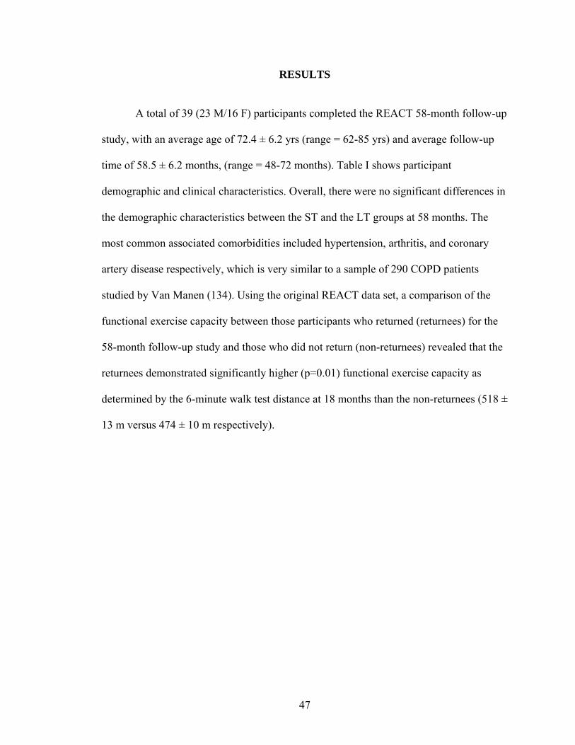
RESULTS
A total of 39 (23 M/16 F) participants completed the REACT 58-month follow-up
study, with an average age of 72.4 ± 6.2 yrs (range = 62-85 yrs) and average follow-up
time of 58.5 ± 6.2 months, (range = 48-72 months). Table I shows participant
demographic and clinical characteristics. Overall, there were no significant differences in
the demographic characteristics between the ST and the LT groups at 58 months. The
most common associated comorbidities included hypertension, arthritis, and coronary
artery disease respectively, which is very similar to a sample of 290 COPD patients
studied by Van Manen (134). Using the original REACT data set, a comparison of the
functional exercise capacity between those participants who returned (returnees) for the
58-month follow-up study and those who did not return (non-returnees) revealed that the
returnees demonstrated significantly higher (p=0.01) functional exercise capacity as
determined by the 6-minute walk test distance at 18 months than the non-returnees (518 ±
13 m versus 474 ± 10 m respectively).
47

TABLE I*
PARTICIPANT DEMOGRAPHICS/CLINICAL INFORMATION
PARTICIPANTS
ST
(3-MONTH)
LT
(18-MONTH)
Completed REACT, n 56 62 Completed 58 mos FU, n (%) 12 (21.4) 27 (43.5) Unable to contact 11 (19.6) 8 (12.9) Contacted but refused FU 17 (30.4) 12 (19.4) Expired 5 (8.9) 0 (0) CHARACTERISTICS
Weight, kg 79.4 ± 4.5 79.1 ± 3.7 Gender, M, n (%)/ F, n (%) 6 (50)/6 (50) 17 (63)/10 (37) Age, yrs 71.3 ± 1.8 72.9 ± 1.2 Height, cm 168.3 ± 3.4 171.7 ± 1.6 FU time, mos 59.3 ± 1.8 58.2 ± 1.2 COMORBIDITY
Arthritis, n (%) 5 (41.7) 14 (51.9) Cancer, n (%) 2 (16.7) 3 (11.1) Circulatory problems, n (%) 1 (8.3) 3 (11.1) Coronary artery disease, n (%) 4 (33.3) 6 (22.2) Diabetes mellitus, n (%) 1 (8.3) 2 (7.4) Hypertension, n (%) 6 (50.0) 13 (48.1) Transient ischemic attack, n (%) 2 (16.7) 2 (7.4) SMOKING STATUS
Current, n (%) 0 (0) 3 (11.1) Past, n (%) 8 (75) 25 (92.6)
*values for FU time, age, height, weight, and pack yrs are means ± SEM; ST= short-term, LT= long-term, mos=months, FU= follow-up, n= number, kg= kilograms, M= male, F= female, yrs= years, cm= centimeters, SEM= standard error of the mean.
48
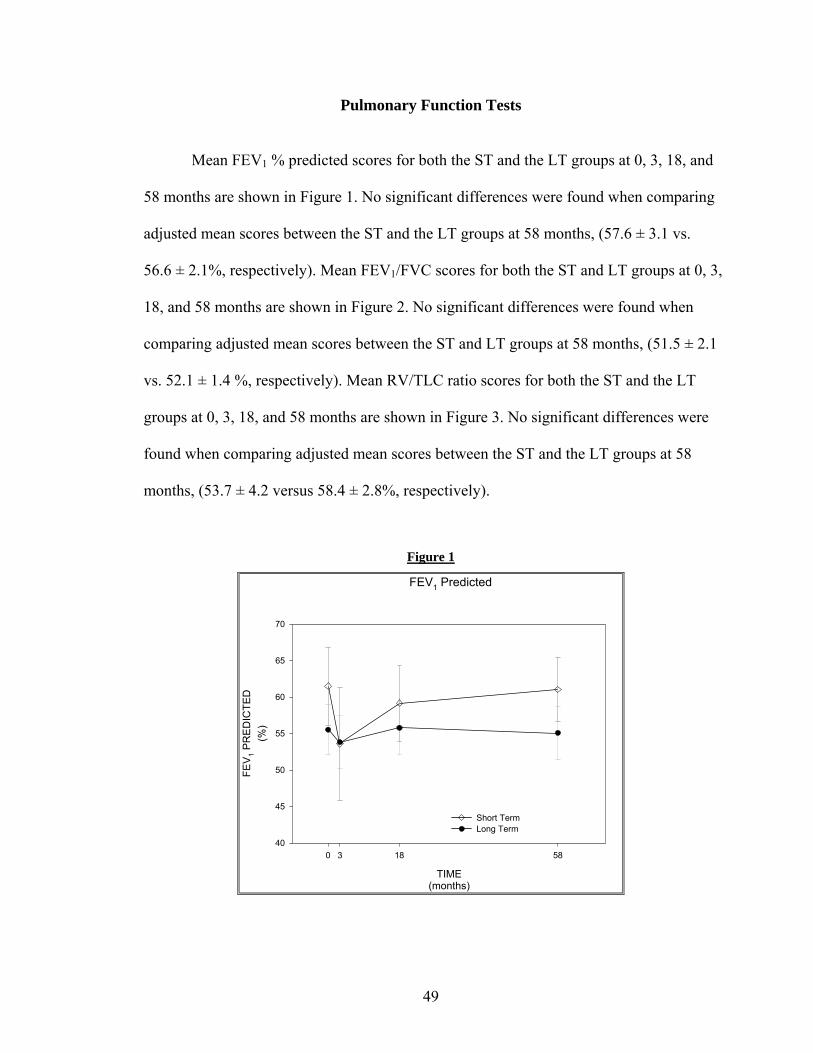
Pulmonary Function Tests
Mean FEV1 % predicted scores for both the ST and the LT groups at 0, 3, 18, and
58 months are shown in Figure 1. No significant differences were found when comparing
adjusted mean scores between the ST and the LT groups at 58 months, (57.6 ± 3.1 vs.
56.6 ± 2.1%, respectively). Mean FEV1/FVC scores for both the ST and LT groups at 0, 3,
18, and 58 months are shown in Figure 2. No significant differences were found when
comparing adjusted mean scores between the ST and LT groups at 58 months, (51.5 ± 2.1
vs. 52.1 ± 1.4 %, respectively). Mean RV/TLC ratio scores for both the ST and the LT
groups at 0, 3, 18, and 58 months are shown in Figure 3. No significant differences were
found when comparing adjusted mean scores between the ST and the LT groups at 58
months, (53.7 ± 4.2 versus 58.4 ± 2.8%, respectively).
Figure 1
FEV1 Predicted
TIME(months)
0 3 18 58
FEV 1 P
RED
ICTE
D(%
)
40
45
50
55
60
65
70
Short Term Long Term
49
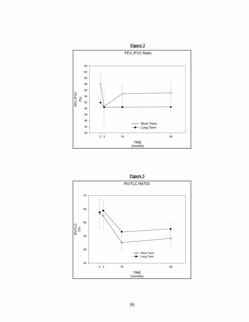
Figure 2
FEV1/FVC Ratio
TIME(months)
0 3 18 58
FEV 1/F
VC(%
)
42
44
46
48
50
52
54
56
58
60
62
64
Short TermLong Term
Figure 3
RV/TLC RATIO
TIME(months)
0 3 18 58
RV/
TLC
(%)
45
50
55
60
65
70
Short TermLong Term
50
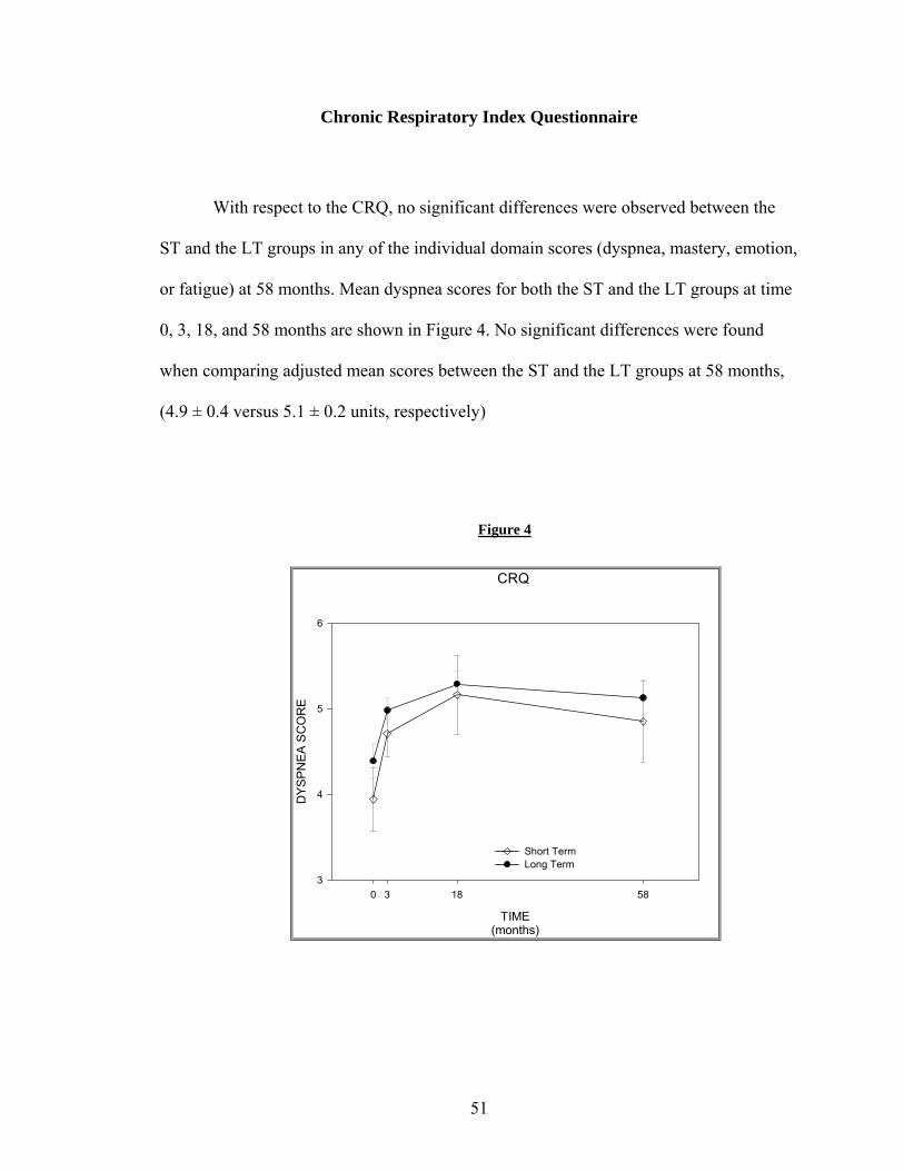
Chronic Respiratory Index Questionnaire
With respect to the CRQ, no significant differences were observed between the
ST and the LT groups in any of the individual domain scores (dyspnea, mastery, emotion,
or fatigue) at 58 months. Mean dyspnea scores for both the ST and the LT groups at time
0, 3, 18, and 58 months are shown in Figure 4. No significant differences were found
when comparing adjusted mean scores between the ST and the LT groups at 58 months,
(4.9 ± 0.4 versus 5.1 ± 0.2 units, respectively)
Figure 4
CRQ
TIME(months)
0 3 18 58
DYS
PNEA
SC
OR
E
3
4
5
6
Short TermLong Term
51
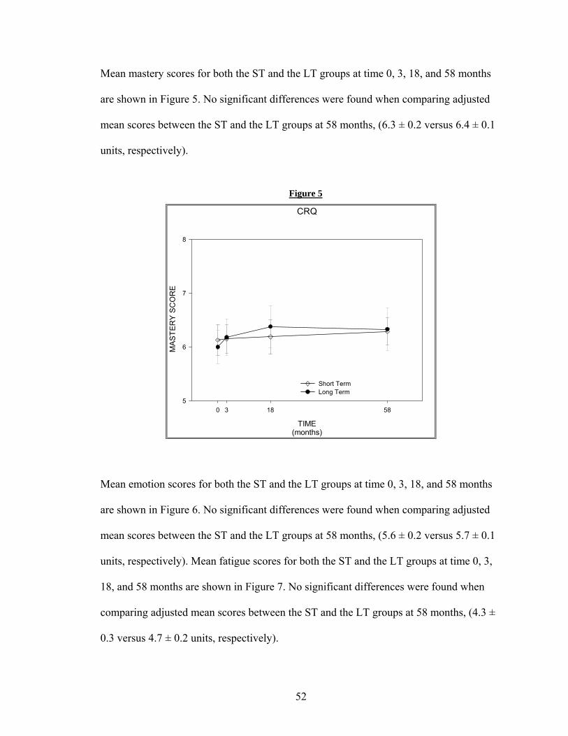
Mean mastery scores for both the ST and the LT groups at time 0, 3, 18, and 58 months
are shown in Figure 5. No significant differences were found when comparing adjusted
mean scores between the ST and the LT groups at 58 months, (6.3 ± 0.2 versus 6.4 ± 0.1
units, respectively).
Figure 5
CRQ
TIME(months)
0 3 18 58
MAS
TER
Y SC
OR
E
5
6
7
8
Short TermLong Term
Mean emotion scores for both the ST and the LT groups at time 0, 3, 18, and 58 months
are shown in Figure 6. No significant differences were found when comparing adjusted
mean scores between the ST and the LT groups at 58 months, (5.6 ± 0.2 versus 5.7 ± 0.1
units, respectively). Mean fatigue scores for both the ST and the LT groups at time 0, 3,
18, and 58 months are shown in Figure 7. No significant differences were found when
comparing adjusted mean scores between the ST and the LT groups at 58 months, (4.3 ±
0.3 versus 4.7 ± 0.2 units, respectively).
52
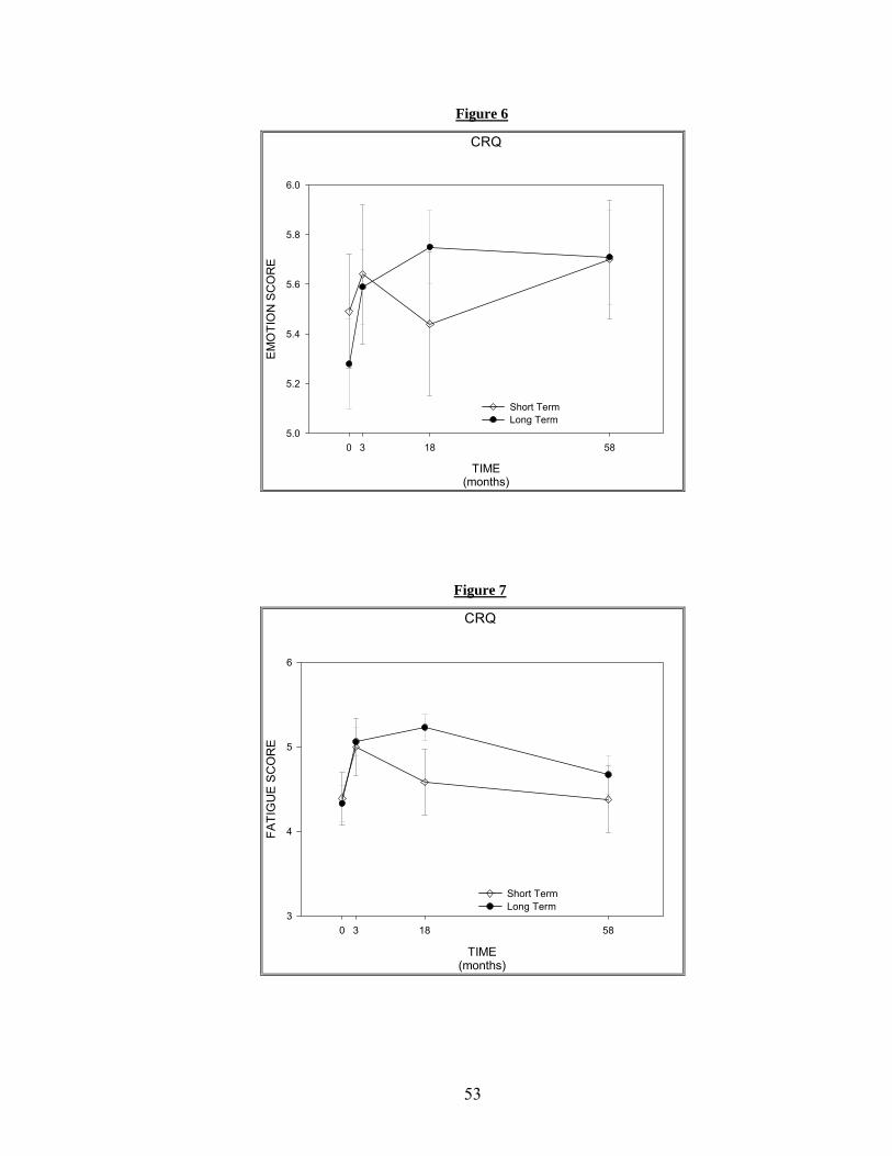
Figure 6
CRQ
TIME(months)
0 3 18 58
EMO
TIO
N S
CO
RE
5.0
5.2
5.4
5.6
5.8
6.0
Short TermLong Term
Figure 7
CRQ
TIME(months)
0 3 18 58
FATI
GU
E SC
OR
E
3
4
5
6
Short Term Long Term
53
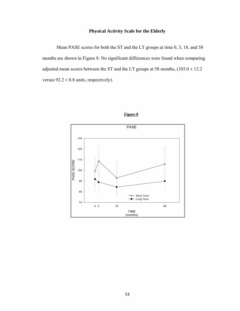
Physical Activity Scale for the Elderly
Mean PASE scores for both the ST and the LT groups at time 0, 3, 18, and 58
months are shown in Figure 8. No significant differences were found when comparing
adjusted mean scores between the ST and the LT groups at 58 months, (103.0 ± 12.2
versus 92.2 ± 8.8 units, respectively).
Figure 8
PASE
TIME(months)
0 3 18 58
PA
SE
SC
OR
E
70
80
90
100
110
120
130
Short TermLong Term
54
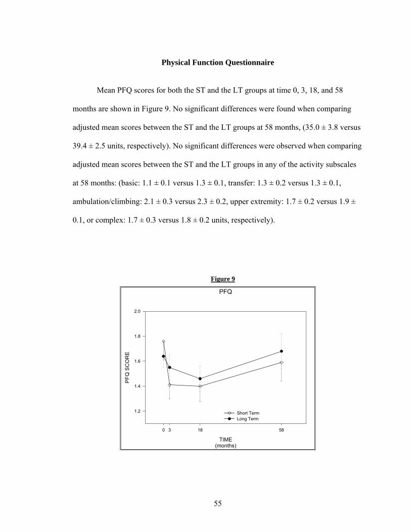
Physical Function Questionnaire
Mean PFQ scores for both the ST and the LT groups at time 0, 3, 18, and 58
months are shown in Figure 9. No significant differences were found when comparing
adjusted mean scores between the ST and the LT groups at 58 months, (35.0 ± 3.8 versus
39.4 ± 2.5 units, respectively). No significant differences were observed when comparing
adjusted mean scores between the ST and the LT groups in any of the activity subscales
at 58 months: (basic: 1.1 ± 0.1 versus 1.3 ± 0.1, transfer: 1.3 ± 0.2 versus 1.3 ± 0.1,
ambulation/climbing: 2.1 ± 0.3 versus 2.3 ± 0.2, upper extremity: 1.7 ± 0.2 versus 1.9 ±
0.1, or complex: 1.7 ± 0.3 versus 1.8 ± 0.2 units, respectively).
Figure 9
PFQ
TIME(months)
0 3 18 58
PFQ
SC
OR
E
1.2
1.4
1.6
1.8
2.0
Short TermLong Term
55
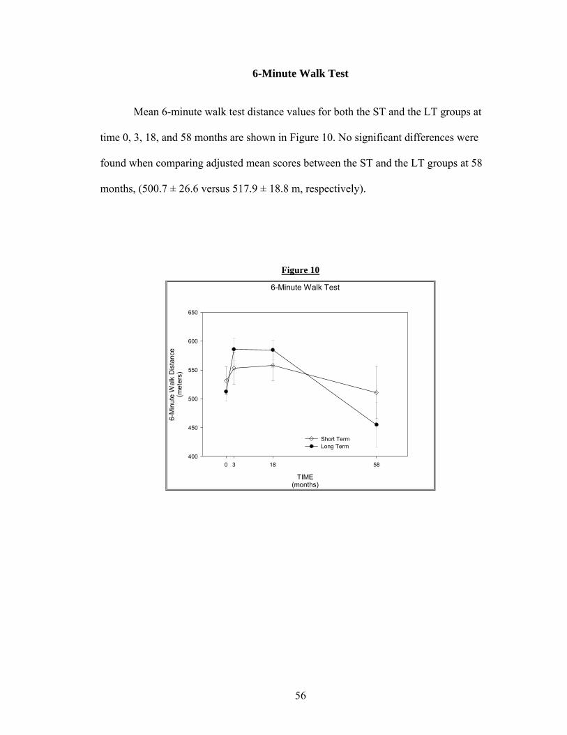
6-Minute Walk Test
Mean 6-minute walk test distance values for both the ST and the LT groups at
time 0, 3, 18, and 58 months are shown in Figure 10. No significant differences were
found when comparing adjusted mean scores between the ST and the LT groups at 58
months, (500.7 ± 26.6 versus 517.9 ± 18.8 m, respectively).
Figure 10
6-Minute Walk Test
TIME(months)
0 3 18 58
6-M
inut
e W
alk
Dis
tanc
e(m
eter
s)
400
450
500
550
600
650
Short TermLong Term
56
DISCUSSION
The original REACT study was a clinical trial in which patients with COPD
participated in a 3-month facility-based exercise therapy program and then were either
randomized to continue the supervised program for an extra 15 months or were
encouraged to continue to exercise on their own, independent of the study. At 18 months,
study participants returned for follow-up assessments. In general, the results of the
REACT study revealed that both the ST and the LT exercise therapy groups showed
improvements in the HRQOL, self-reported disability, and functional exercise capacity at
the completion of the initial ST exercise therapy program. At 18 months, these benefits
were maintained in the LT group, but were not maintained in the ST group whose
outcome variables approached the initial baseline measures. The primary purpose of this
follow-up study was to describe and compare the long-term outcomes of COPD patients
who had participated in either a ST or an LT exercise rehabilitation program (REACT) at
58 months. The outcomes assessed included: lung function (PFT’s- FEV1 % predicted,
FEV1/FVC ratio, and RV/TLC ratio); HRQOL (CRQ); self-reported physical activity
(PASE); self-reported disability (PFQ); and functional capacity (6-minute walk test).
Overall, the results of this study showed no significant differences between the ST and
the LT groups in any of these variables measured at 58 months. These findings suggest
that even patients who participated in a long-term formal exercise rehabilitation program
will not maintain the benefits achieved once the program ends.
Research has demonstrated that the benefits gained from participation in
pulmonary rehabilitation programs that include exercise therapy, can be maintained for
57
up to 2 years (48)and even 3 years in select COPD patients (122). Other studies have
shown that the benefits gained from participation in pulmonary rehabilitation programs
diminish by 18 months following completion of these programs (21;102;123). To our
knowledge, this is the first study to look at the long-term (58 months) outcomes in COPD
patients following completion of an 18-month exercise rehabilitation therapy program.
Lung function (FEV1% predicted, FEV1/FVC ratio, and RV/TLC ratio), as
measured by PFT’s, was not significantly different between the ST and LT groups at 58
months. Many studies investigating the efficacy of pulmonary rehabilitation programs
demonstrate that lung function is typically not affected by exercise therapy interventions
(51;102).
Results from the present investigation show that HRQOL, as measured by the
four domains of the CRQ (dyspnea, mastery, emotion, and fatigue) was not significantly
different between patients from the ST and the LT exercise therapy groups at 58 months.
More specifically, the results of this follow-up study showed that the dyspnea, mastery,
and emotion scores were similar between the 2 groups at 58 months. Results from the
original REACT study showed a significant difference in all domains of the CRQ when
comparing the ST and the LT exercise therapy groups at the 18-month follow-up (49).
Given the fact that the differences seen at 18 months between the 2 groups were not seen
at 58 months, it does not appear that those patients participating in an 18-month exercise
therapy program will maintain long-term benefits to any greater degree than those
participating in a short-term exercise therapy program.
Self-reported activity, as measured by the PASE, was not significantly different
between the ST and the LT groups at 58 months. Older adult volunteers were recruited
58
from community centers and retirement homes in order to assess the value of the PASE
in measuring physical activity in a study conducted by Harada et al. (65). The mean
PASE score was 50 ± 44 units in 36 adults recruited from retirement homes, while the
mean PASE score for 51 adults recruited from community centers was 158 ± 65 units,
respectively. The adjusted mean PASE scores from the ST and the LT groups this follow-
up study were 103 ± 12 and 92 ± 9 units, which demonstrates that our participants were
more active than those individuals residing in retirement homes and somewhat less active
than those individuals recruited from community centers. The subjects in each study were
all of similar age. These results support the idea that individuals with COPD are less
active on a regular basis than independent-living older adults without COPD.
Self-reported disability, as measured by the PFQ, was not significantly different
between the ST and the LT groups at 58 months. The PFQ was developed specifically for
the Fitness Arthritis and Seniors Trial (FAST), which investigated the effects of aerobic
and resistive exercise training, as well as osteoarthritis education on self-reported
disability in patients with knee osteoarthritis (40). The baseline adjusted mean PFQ score
in this sample of 439 patients was approximately 1.8 units, while the sample of COPD
patients in the current study had an adjusted mean PFQ score of 1.5 units in the ST group
and 1.7 units in the LT group. Thus, patients with COPD reported similar, but slightly
less disability than age-matched patients with knee osteoarthritis. The results of these 2
studies also suggest that COPD patients had less self-reported disability associated with
the transfer and the ambulation/climbing domains than the osteoarthritis patients, and
similar self-reported disability in the remaining domains.
59
Functional capacity, as measured by the 6-minute walk test, was not significantly
different between the ST and the LT groups at 58 months. The participants in this follow-
up study had a mean 6-minute walk test distance of 511 ± 45 m in the ST group, and
455 ± 39 m in the LT group. In healthy age-matched individuals the mean 6-minute walk
distance has been reported to be 631 ± 93 m (129), though these subjects had
standardized verbal encouragement every 30 seconds, which may have positively
influenced the total distance walked (64). In the present study, the participants had no
such verbal encouragement. Our participants were also older which may have also
limited the total distance walked given that age has been shown to be an independent
predictor of the 6-minute walk test distance (129). Gibbons et al. (55)also established
reference 6-minute walk test distance values for 20 healthy older adults aged 61-80 years.
The mean distance covered was 635 ± 71 m, which again is higher than that of our study
participants.
The limitations of this study include: small sample size with a disproportionate
number of subjects representing the LT versus the ST group. This discrepancy somewhat
biases the sample and can be possibly explained by idea that the LT group felt more
dedicated to the study because of participation in the longer initial exercise intervention.
Another potential source of bias involves the inclusion criteria for the original REACT
study. Subjects had to be able to complete the 6-minute walk test and must have not been
consistently exercising prior to entry into the study. These criteria exclude low level
functioning COPD patients as well as those COPD patients who were motivated and
already active, which may have influenced adherence to exercise behavior following the
study intervention resulting in differences in the current follow-up study. The follow-up
60
study participants were probably also higher functioning than those individuals who did
not return for follow-up testing. The original REACT data analysis at 18 months showed
that the 58 month follow-up participants had significantly higher 6-minute walk test
distances as compared to the participants which did not return for the 58-month follow-up
study. This finding lends support to the possibility that similar differences between the
returnees and the non-returnees may have also been present at 58 months as well
resulting in a somewhat biased sample. Also, of the original REACT participants that
were contacted, several were no longer able to live independently at home and/or
required assistance for routine activities. Many did not feel it was appropriate to
participate in the follow-up testing and/or were unable to arrange for assistance and
transportation to the campus. An additional limitation of this study, as well as the original
REACT study, is that there was no control group of healthy individuals involved in order
to track the overall age-associated decline in the different outcome variables.
Further research is indicated to identify the ideal parameters of pulmonary
rehabilitation programs in order to maximize benefits and minimize patient healthcare
costs. Long-term follow-up studies (greater than 2 years) looking at morbidity and
mortality in COPD patients need to be completed in order to provide further evidence for
the efficacy of pulmonary rehabilitation programs. Alternative strategies for developing
more cost effective exercise interventions and social support that could be more
accessible to a larger number of COPD patients need to be developed. Programs that
offer a behavioral component that would promote exercise adherence and individual
accountability along with exercise therapy may be more successful in maintaining
benefits gained from short-term participation in pulmonary rehabilitation programs. The
61
Reconditioning and Chronic Disease Trial 2 (REACT 2) a new study that will incorporate
behavioral change strategies into an exercise therapy program for COPD patients will
attempt to discover the answers to these questions.
Conclusion
Overall, the results of this 58-month follow-up study involving a group of 39
COPD patients who had completed either a 3-month or an 18-month exercise therapy
program demonstrated that there were no significant differences in lung function, self-
reported physical activity level, self-reported disability, self-reported HRQOL, or
functional exercise capacity between the 2 groups. These results indicate that the benefits
gained from participation in either a ST or LT exercise rehabilitation program are not
permanent and diminish considerably over time. Future research must continue to assess
the cost effectiveness of pulmonary rehabilitation programs in order to make the most
efficient use of limited healthcare resources.
62
REFERENCES
1. Continuous or nocturnal oxygen therapy in hypoxemic chronic obstructive lung
disease: a clinical trial. Nocturnal Oxygen Therapy Trial Group. Ann.Intern.Med. 93: 391-398, 1980.
2. Standardization of spirometry--1987 update. Statement of the American Thoracic Society. Am.Rev.Respir.Dis. 136: 1285-1298, 1987.
3. Standards for the diagnosis and care of patients with chronic obstructive pulmonary disease. American Thoracic Society. Am.J.Respir.Crit Care Med. 152: S77-121, 1995.
4. American College of Sports Medicine Position Stand. Exercise and physical activity for older adults. Med.Sci.Sports Exerc. 30: 992-1008, 1998.
5. International guidelines for the selection of lung transplant candidates. The American Society for Transplant Physicians (ASTP)/American Thoracic Society (ATS)/European Respiratory Society(ERS)/International Society for Heart and Lung Transplantation(ISHLT). Am.J.Respir.Crit Care Med. 158: 335-339, 1998.
6. Dyspnea. Mechanisms, assessment, and management: a consensus statement. American Thoracic Society. Am.J.Respir.Crit Care Med. 159: 321-340, 1999.
7. Rationale and design of The National Emphysema Treatment Trial: a prospective randomized trial of lung volume reduction surgery. The National Emphysema Treatment Trial Research Group. Chest 116: 1750-1761, 1999.
8. Skeletal muscle dysfunction in chronic obstructive pulmonary disease. A statement of the American Thoracic Society and European Respiratory Society. Am.J.Respir.Crit Care Med. 159: S1-40, 1999.
9. Health Screening and Risk Stratification. In Franklin, B.A., M.H. Whaley, and E.T. Howley, eds., ACSM's Guidelines for Exercise Testing and Prescription. Philadelphia, Lippincott Williams and Wilkins. 2000, 22-32.
63
10. The International Registry of the Society for Heart and Lung Transplantation Eighteenth Annual Report. 2001.
11. American Lung Association. American Lung Association Fact Sheet Secondhand Smoke. http://www.lungusa.org/tobacco/secondhand_factsheet99.html . 2000.
12. American Lung Association. American Lung Association Fact Sheet: Chronic Obstructive Pulmonary Disease.ttp://www.lungusa.org/diseases/copd_factsheet.html. 2001.
13. Anthonisen, N.R., J. Manfreda, C.P. Warren, E.S. Hershfield, G.K. Harding, and N.A. Nelson. Antibiotic therapy in exacerbations of chronic obstructive pulmonary disease. Ann.Intern.Med. 106: 196-204, 1987.
14. Anthonisen, N.R., E.C. Wright, and J.E. Hodgkin. Prognosis in chronic obstructive pulmonary disease. Am.Rev.Respir.Dis. 133: 14-20, 1986.
15. Arcasoy, S.M. and R.M. Kotloff. Lung transplantation. N.Engl.J.Med. 340: 1081-1091, 1999.
16. Bavaria, J.E., R. Kotloff, H. Palevsky, B. Rosengard, J.R. Roberts, P.M. Wahl, N. Blumenthal, C. Archer, and L.R. Kaiser. Bilateral versus single lung transplantation for chronic obstructive pulmonary disease. J.Thorac.Cardiovasc.Surg. 113: 520-527, 1997.
17. Benayoun, S., P. Ernst, and S. Suissa. The impact of combined inhaled bronchodilator therapy in the treatment of COPD. Chest 119: 85-92, 2001.
18. Bernard, S., P. LeBlanc, F. Whittom, G. Carrier, J. Jobin, R. Belleau, and F. Maltais. Peripheral muscle weakness in patients with chronic obstructive pulmonary disease. Am.J.Respir.Crit Care Med. 158: 629-634, 1998.
19. Bernard, S., F. Whittom, P. LeBlanc, J. Jobin, R. Belleau, C. Berube, G. Carrier, and F. Maltais. Aerobic and strength training in patients with chronic obstructive pulmonary disease. Am.J.Respir.Crit Care Med. 159: 896-901, 1999.
64
20. Berry, M.J., N.E. Adair, K.S. Sevensky, A. Quinby, and H.M. Lever. Inspiratory muscle training and whole-body reconditioning in chronic obstructive pulmonary disease. Am.J.Respir.Crit Care Med. 153: 1812-1816, 1996.
21. Berry, M.J., Rejeski, W.J., Adair, N.E., Ettinger, W.H., Zacarro, D.J., and Sevick, M.A. A Randomized Controlled Trial Comparing Long- and Short-Term Exercise in COPD Patients. 2002.
22. Bourjeily, G. and C.L. Rochester. Exercise training in chronic obstructive pulmonary disease. Clin.Chest Med. 21: 763-781, 2000.
23. Bowen, J.B., J.J. Votto, R.S. Thrall, M.C. Haggerty, R. Stockdale-Woolley, T. Bandyopadhyay, and R.L. ZuWallack. Functional status and survival following pulmonary rehabilitation. Chest 118: 697-703, 2000.
24. Burge, P.S., P.M. Calverley, P.W. Jones, S. Spencer, J.A. Anderson, and T.K. Maslen. Randomised, double blind, placebo-controlled study of fluticasone propionate in patients with moderate to severe chronic obstructive pulmonary disease: the ISOLDE trial. BMJ 320: 1297-1303, 2000.
25. Casaburi, R. Skeletal muscle function in COPD. Chest 117: 267S-271S, 2000.
26. Casaburi, R. Skeletal muscle dysfunction in chronic obstructive pulmonary disease. Med.Sci.Sports Exerc. 33: S662-S670, 2001.
27. Casaburi, R., J. Porszasz, M.R. Burns, E.R. Carithers, R.S. Chang, and C.B. Cooper. Physiologic benefits of exercise training in rehabilitation of patients with severe chronic obstructive pulmonary disease. Am.J.Respir.Crit Care Med. 155: 1541-1551, 1997.
28. Celli, B., G. Criner, and J. Rassulo. Ventilatory muscle recruitment during unsupported arm exercise in normal subjects. J.Appl.Physiol 64: 1936-1941, 1988.
29. Celli, B.R. Standards for the optimal management of COPD: a summary. Chest 113: 283S-287S, 1998.
30.Centers for Disease Control. 1990. Surgeon General's Report.
65
31. Clark, C.J., L.M. Cochrane, E. Mackay, and B. Paton. Skeletal muscle strength and endurance in patients with mild COPD and the effects of weight training. Eur.Respir.J. 15: 92-97, 2000.
32. Colt, H.G., A.L. Ries, N. Brewer, and K. Moser. Analysis of chronic obstructive pulmonary disease referrals for lung volume reduction surgery. The University of California San Diego Emphysema Treatment Group. J.Cardiopulm.Rehabil. 17: 248-252, 1997.
33. Corbridge, T. and C. Irvin. Pathophysiology of Chronic Obstructive Pulmonary Disease. In Casaburi, R. and T. Petty, eds., Principles and Practice of Pulmonary Rehabilitation. Philadelphia, W.B. Saunders. 1993, 18-32.
34. Criner, G.J., F.C. Cordova, S. Furukawa, A.M. Kuzma, J.M. Travaline, V. Leyenson, and G.M. O'Brien. Prospective randomized trial comparing bilateral lung volume reduction surgery to pulmonary rehabilitation in severe chronic obstructive pulmonary disease. Am.J.Respir.Crit Care Med. 160: 2018-2027, 1999.
35. De Torres, J.P., V. Pinto-Plata, E. Ingenito, P. Bagley, A. Gray, R. Berger, and B. Celli. Power of outcome measurements to detect clinically significant changes in pulmonary rehabilitation of patients with COPD. Chest 121: 1092-1098, 2002.
36. Dudley, G.A. and L.L. Ploutz-Snyder. Deconditioning and Bed Rest: Musculoskeletal Response. In Roitman, J.L. et al., ed., ACSM's Resource Manual for Guidelines for Exercise Testing and Prescription. Philadelphia, Lippincott Williams and Wilkins. 2001, 203-208.
37. El Manshawi, A., K.J. Killian, E. Summers, and N.L. Jones. Breathlessness during exercise with and without resistive loading. J.Appl.Physiol 61: 896-905, 1986.
38. Epidemiology and Statistics Unit. Trends in Tobacco Use. 2001.
39. Estenne, M., J.R. Maurer, A. Boehler, J.J. Egan, A. Frost, M. Hertz, G.B. Mallory, G.I. Snell, and S. Yousem. Bronchiolitis obliterans syndrome 2001: an update of the diagnostic criteria. J.Heart Lung Transplant. 21: 297-310, 2002.
66
40. Ettinger, W.H., Jr., R. Burns, S.P. Messier, W. Applegate, W.J. Rejeski, T. Morgan, S. Shumaker, M.J. Berry, M. O'Toole, J. Monu, and T. Craven. A randomized trial comparing aerobic exercise and resistance exercise with a health education program in older adults with knee osteoarthritis. The Fitness Arthritis and Seniors Trial (FAST). JAMA 277: 25-31, 1997.
41. Ezzell, L. and G.L. Jensen. Malnutrition in chronic obstructive pulmonary disease. Am.J.Clin.Nutr. 72: 1415-1416, 2000.
42. Ferguson, G.T. Recommendations for the management of COPD. Chest 117: 23S-28S, 2000.
43. Ferguson, G.T. Update on pharmacologic therapy for chronic obstructive pulmonary disease. Clin.Chest Med. 21: 723-738, 2000.
44. Finnerty, J.P., I. Keeping, I. Bullough, and J. Jones. The effectiveness of outpatient pulmonary rehabilitation in chronic lung disease: a randomized controlled trial. Chest 119: 1705-1710, 2001.
45. Fiore, M.C., D.E. Jorenby, and T.B. Baker. Smoking cessation: principles and practice based upon the AHCPR Guideline, 1996. Agency for Health Care Policy and Research. Ann.Behav.Med. 19: 213-219, 1997.
46. Fishman, A.P. Pulmonary rehabilitation research. Am.J.Respir.Crit Care Med. 149: 825-833, 1994.
47. Flaherty, K.R. and F.J. Martinez. Lung volume reduction surgery for emphysema. Clin.Chest Med. 21: 819-848, 2000.
48. Foglio, K., L. Bianchi, and N. Ambrosino. Is it really useful to repeat outpatient pulmonary rehabilitation programs in patients with chronic airway obstruction? A 2-year controlled study. Chest 119: 1696-1704, 2001.
49. Foy, C.G., W.J. Rejeski, M.J. Berry, D. Zaccaro, and C.M. Woodard. Gender moderates the effects of exercise therapy on health-related quality of life among COPD patients. Chest 119: 70-76, 2001.
67
50. Garrod, R., E.A. Paul, and J.A. Wedzicha. Supplemental oxygen during pulmonary rehabilitation in patients with COPD with exercise hypoxaemia. Thorax 55: 539-543, 2000.
51.Garvey, C. Pulmonary Rehabilitation for the Elderly Client. http://www.medscape.com/Medscape/Nurses/journal/2001/v01.n02/mns0817.01.garv/mns0817.01.garv-01.html. 2001.
52. Gelb, A.F., M. Brenner, R.J. McKenna, Jr., N. Zamel, R. Fischel, and J.D. Epstein. Lung function 12 months following emphysema resection. Chest 110: 1407-1415, 1996.
53. Gelb, A.F., R.J. McKenna, Jr., M. Brenner, J.D. Epstein, and N. Zamel. Lung function 5 yr after lung volume reduction surgery for emphysema. Am.J.Respir.Crit Care Med. 163: 1562-1566, 2001.
54. Gerald, L.B., B. Sanderson, D. Redden, and W.C. Bailey. Chronic obstructive pulmonary disease stage and 6-minute walk outcome. J.Cardiopulm.Rehabil. 21: 296-299, 2001.
55. Gibbons, W.J., N. Fruchter, S. Sloan, and R.D. Levy. Reference values for a multiple repetition 6-minute walk test in healthy adults older than 20 years. J.Cardiopulm.Rehabil. 21: 87-93, 2001.
56. Goldstein, R.S., E.H. Gort, D. Stubbing, M.A. Avendano, and G.H. Guyatt. Randomised controlled trial of respiratory rehabilitation. Lancet 344: 1394-1397, 1994.
57.Goldstein, R.S., Redelmeier, D.A., Baksh, L., and Guyatt, G.H. Subjective comparison ratings of walking ability in patients with COPD. Proceedings of the 5th International Conference on Pulmonary rehabilitation and Home Ventilation. 1995.
58. Gosselink, R., T. Troosters, and M. Decramer. Peripheral muscle weakness contributes to exercise limitation in COPD. Am.J.Respir.Crit Care Med. 153: 976-980, 1996.
68
59. Grosbois, J.M., C. Lamblin, B. Lemaire, H. Chekroud, J.M. Dernis, B. Douay, and F. Fortin. Long-term benefits of exercise maintenance after outpatient rehabilitation program in patients with chronic obstructive pulmonary disease. J.Cardiopulm.Rehabil. 19: 216-225, 1999.
60. Gross, C.R., K. Savik, R.M. Bolman, III, and M.I. Hertz. Long-term health status and quality of life outcomes of lung transplant recipients. Chest 108: 1587-1593, 1995.
61. Guell, R., P. Casan, J. Belda, M. Sangenis, F. Morante, G.H. Guyatt, and J. Sanchis. Long-term effects of outpatient rehabilitation of COPD: A randomized trial. Chest 117: 976-983, 2000.
62. Guyatt, G.H., L.B. Berman, and M. Townsend. Long-term outcome after respiratory rehabilitation. CMAJ. 137: 1089-1095, 1987.
63. Guyatt, G.H., C. Bombardier, and P.X. Tugwell. Measuring disease-specific quality of life in clinical trials. CMAJ. 134: 889-895, 1986.
64. Guyatt, G.H., S.O. Pugsley, M.J. Sullivan, P.J. Thompson, L. Berman, N.L. Jones, E.L. Fallen, and D.W. Taylor. Effect of encouragement on walking test performance. Thorax 39: 818-822, 1984.
65. Harada, N.D., V. Chiu, A.C. King, and A.L. Stewart. An evaluation of three self-report physical activity instruments for older adults. Med.Sci.Sports Exerc. 33: 962-970, 2001.
66. Hernandez, M.T., T.M. Rubio, F.O. Ruiz, H.S. Riera, R.S. Gil, and J.C. Gomez. Results of a home-based training program for patients with COPD. Chest 118: 106-114, 2000.
67. Hudson, L.D., M.L. Tyler, and T.L. Petty. Hospitalization needs during an outpatient rehabilitation program for severe chronic airway obstruction. Chest 70: 606-610, 1976.
68. Hurd, S. The impact of COPD on lung health worldwide: Epidemiology and incidence. Chest 117: 1S-4S, 2000.
69
69. Jensen, P.S. Risk, protective factors, and supportive interventions in chronic airway obstruction. Arch.Gen.Psychiatry 40: 1203-1207, 1983.
70. Jolly, E.C., B. Di, V.L. Aguirre, C.M. Luna, S. Berensztein, and R.J. Gene. Effects of supplemental oxygen during activity in patients with advanced COPD without severe resting hypoxemia. Chest 120: 437-443, 2001.
71. Knudson, R.J., Slatin R.C., M.D. Lebowitz, and B. Burrows. The maximal expiratory flow-volume curve: normal standards, variability, and effects of age. Am.Rev.Respir.Dis. 113: 587, 1976.
72. Lacasse, Y., E. Wong, G.H. Guyatt, D. King, D.J. Cook, and R.S. Goldstein. Meta-analysis of respiratory rehabilitation in chronic obstructive pulmonary disease. Lancet 348: 1115-1119, 1996.
73. Larson, J.L., M.K. Covey, S. E. Wirtz, J.K. Berry, C. G. Alex, W.E. Langbein, and L. Edwards. Cycle ergometer and inspiratory muscle training in chronic obstructive pulmonary disease. Am.J.Respir.Crit Care Med. 160: 500-507, 1999.
74. Lertzman, M.M. and R.M. Cherniack. Rehabilitation of patients with chronic obstructive pulmonary disease. Am.Rev.Respir.Dis. 114: 1145-1165, 1976.
75. Mahler, D.A. Pulmonary rehabilitation. Chest 113: 263S-268S, 1998.
76. Maltais, F., P. LeBlanc, J. Jobin, and R. Casaburi. Peripheral muscle dysfunction in chronic obstructive pulmonary disease. Clin.Chest Med. 21: 665-677, 2000.
77. Marchand, E. and M. Decramer. Respiratory muscle function and drive in chronic obstructive pulmonary disease. Clin.Chest Med. 21: 679-692, 2000.
78. Martin, K.A., W.J. Rejeski, M.E. Miller, M.K. James, W.H. Ettinger, Jr., and S.P. Messier. Validation of the PASE in older adults with knee pain and physical disability. Med.Sci.Sports Exerc. 31: 627-633, 1999.
79. Martin, R.J., B.L. Bartelson, P. Smith, D.W. Hudgel, D. Lewis, G. Pohl, P. Koker, and J. F. Souhrada. Effect of ipratropium bromide treatment on oxygen saturation and sleep quality in COPD. Chest 115: 1338-1345, 1999.
70
80. Martinez, F.J., P.D. Vogel, D.N. Dupont, I. Stanopoulos, A. Gray, and J.F. Beamis. Supported arm exercise versus unsupported arm exercise in the rehabilitation of patients with severe chronic airflow obstruction. Chest 103: 1397-1402, 1993.
81. McEvoy, C.E. and D.E. Niewoehner. Corticosteroids in chronic obstructive pulmonary disease. Clinical benefits and risks. Clin.Chest Med. 21: 739-752, 2000.
82. Miravitlles, M., C. Mayordomo, M. Artes, L. Sanchez-Agudo, F. Nicolau, and J.L. Segu. Treatment of chronic obstructive pulmonary disease and its exacerbations in general practice. EOLO Group. Estudio Observacional de la Limitacion Obstructiva al Flujo aEreo. Respir.Med. 93: 173-179, 1999.
83. National Center for Health Statistics. National Ambulatory Medical Care Survey. 1996.
84. National Institutes of Health. Morbidity and Mortality: 2000 Chart Book on Cardiovascular, Lung, and Blood Diseases. 2000.
85. Niederman, M.S. Introduction: mechanisms and management of COPD: we can do better--it's time for a re-evaluation. Chest 113: 233S-234S, 1998.
86. Niewoehner, D.E., M.L. Erbland, R.H. Deupree, D. Collins, N.J. Gross, R.W. Light, P. Anderson, and N.A. Morgan. Effect of systemic glucocorticoids on exacerbations of chronic obstructive pulmonary disease. Department of Veterans Affairs Cooperative Study Group. N.Engl.J.Med. 340: 1941-1947, 1999.
87. Nishimura, K., H. Koyama, A. Ikeda, N. Sugiura, K. Kawakatsu, and T. Izumi. The additive effect of theophylline on a high-dose combination of inhaled salbutamol and ipratropium bromide in stable COPD. Chest 107: 718-723, 1995.
88. O'Donnell, D.E., M. McGuire, L. Samis, and K.A. Webb. The impact of exercise reconditioning on breathlessness in severe chronic airflow limitation. Am.J.Respir.Crit Care Med. 152: 2005-2013, 1995.
89. Owens, M.W., W.M. Anderson, and R.B. George. Pharmacologic Therapy. In Casaburi, R. and T. Petty, eds., Principles and Practice of Pulmonary Rehabilitation. Philadelphia, W. B. Saunders. 1993, 152-166.
71
90. Owens, M.W., B.A. Markewitz, and D.K. Payne. Outpatient management of chronic obstructive pulmonary disease. Am.J.Med.Sci. 318: 79-83, 1999.
91. Pauwels, R.A. National and international guidelines for COPD: the need for evidence. Chest 117: 20S-22S, 2000.
92. Petty, T.L. Supportive therapy in COPD. Chest 113: 256S-262S, 1998.
93. Petty, T.L. and R. Casaburi. Recommendations of the Fifth Oxygen Consensus Conference. Writing and Organizing Committees. Respir.Care 45: 957-961, 2000.
94. Petty, T.L. and S.I. Rennard. Introduction: mechanisms of COPD. Chest 117: 219S, 2000.
95. Popovic, J.R. 1999 National Hospital Discharge Survey: annual summary with detailed diagnosis and procedure data. Vital Health Stat.13 i-206, 2001.
96. Prefaunt, C., A. Varray, and G. Vallet. Pathophysiological basis of exercise training in patients with chronic obstructive pulmonary disease. European Respiratory Review 5: 27-32, 1995.
97. Puente-Maestu, L., M.L. Sanz, P. Sanz, J.M. Cubillo, J. Mayol, and R. Casaburi. Comparison of effects of supervised versus self-monitored training programmes in patients with chronic obstructive pulmonary disease. Eur.Respir.J. 15: 517-525, 2000.
98. Redelmeier, D.A., A.M. Bayoumi, R.S. Goldstein, and G.H. Guyatt. Interpreting small differences in functional status: the Six Minute Walk test in chronic lung disease patients. Am.J.Respir.Crit Care Med. 155: 1278-1282, 1997.
99. Rejeski, W.J., W.H. Ettinger, Jr., S. Schumaker, P. James, R. Burns, and J.T. Elam. Assessing performance-related disability in patients with knee osteoarthritis. Osteoarthritis.Cartilage. 3: 157-167, 1995
100. Rennard, S, American Lung Association, and Schulman, Ronca and Bucuvalas Inc. Confronting COPD in America: Executive Summary. 2000.
72
101. Ries, A.L., B. Ellis, and R.W. Hawkins. Upper extremity exercise training in chronic obstructive pulmonary disease. Chest 93: 688-692, 1988.
102. Ries, A.L., R.M. Kaplan, T.M. Limberg, and L.M. Prewitt. Effects of pulmonary rehabilitation on physiologic and psychosocial outcomes in patients with chronic obstructive pulmonary disease. Ann.Intern.Med. 122: 823-832, 1995.
103. Robertson, C. and H. Levison. Bronchodilators in asthma. Chest 87: 64S-68S, 1985.
104. Sala, E., J. Roca, R.M. Marrades, J. Alonso, J.M. Gonzalez De Suso, A. Moreno, J.A. Barbera, J. Nadal, L. de Jover, R. Rodriguez-Roisin, and P.D. Wagner. Effects of endurance training on skeletal muscle bioenergetics in chronic obstructive pulmonary disease. Am.J.Respir.Crit Care Med. 159: 1726-1734, 1999.
105. Sanchez, R.H., R.T. Montemayor, R.F. Ortega, R.P. Cejudo, O.D. Del Castillo, H.T. Elias, and G.J. Castillo. Inspiratory muscle training in patients with COPD: effect on dyspnea, exercise performance, and quality of life. Chest 120: 748-756, 2001.
106. Sandford, A.J. and P.D. Pare. Genetic risk factors for chronic obstructive pulmonary disease. Clin.Chest Med. 21: 633-643, 2000.
107. Scanlon, P.D., J.E. Connett, L.A. Waller, M.D. Altose, W.C. Bailey, and A.S. Buist. Smoking cessation and lung function in mild-to-moderate chronic obstructive pulmonary disease. The Lung Health Study. Am.J.Respir.Crit Care Med. 161: 381-390, 2000.
108. Scherer, T.A., C.M. Spengler, D. Owassapian, E. Imhof, and U. Boutellier. Respiratory muscle endurance training in chronic obstructive pulmonary disease: impact on exercise capacity, dyspnea, and quality of life. Am.J.Respir.Crit Care Med. 162: 1709-1714, 2000.
109. Schulman, L.L. Lung transplantation for chronic obstructive pulmonary disease. Clin.Chest Med. 21: 849-865, 2000.
110. Selinger, S.R., T.P. Kennedy, P. Buescher, P. Terry, W. Parham, D. Gofreed, A. Medinger, S.V. Spagnolo, and J.R. Michael. Effects of removing oxygen from patients with chronic obstructive pulmonary disease. Am.Rev.Respir.Dis. 136: 85-91, 1987.
73
111. Seneff, M.G., D.P. Wagner, R.P. Wagner, J.E. Zimmerman, and W.A. Knaus. Hospital and 1-year survival of patients admitted to intensive care units with acute exacerbation of chronic obstructive pulmonary disease. JAMA 274: 1852-1857, 1995.
112. Sethi, J.M. and M.D. Siegel. Mechanical ventilation in chronic obstructive lung disease. Clin.Chest Med. 21: 799-818, 2000.
113. Sethi, S. Bacterial infection and the pathogenesis of COPD. Chest 117: 286S-291S, 2000.
114. Shaheen, S.O., J.A. Sterne, J.S. Tucker, and C.D. Florey. Birth weight, childhood lower respiratory tract infection, and adult lung function. Thorax 53: 549-553, 1998.
115. Shannon, M. Life-threatening events after theophylline overdose: a 10-year prospective analysis. Arch.Intern.Med. 159: 989-994, 1999.
116. Shapiro, S.D. Evolving concepts in the pathogenesis of chronic obstructive pulmonary disease. Clin.Chest Med. 21: 621-632, 2000.
117. Sherk, P.A. and R.F. Grossman. The chronic obstructive pulmonary disease exacerbation. Clin.Chest Med. 21: 705-721, 2000.
118. Simpson, K., K. Killian, N. McCartney, D.G. Stubbing, and N.L. Jones. Randomised controlled trial of weightlifting exercise in patients with chronic airflow limitation. Thorax 47: 70-75, 1992.
119. Smith, K., D. Cook, G.H. Guyatt, J. Madhavan, and A.D. Oxman. Respiratory muscle training in chronic airflow limitation: a meta- analysis. Am.Rev.Respir.Dis. 145: 533-539, 1992.
120. Snow, V., S. Lascher, and C. Mottur-Pilson. The evidence base for management of acute exacerbations of COPD: clinical practice guideline, part 1. Chest 119: 1185-1189, 2001.
121. Solway, S., D. Brooks, Y. Lacasse, and S. Thomas. A qualitative systematic overview of the measurement properties of functional walk tests used in the cardiorespiratory domain. Chest 119: 256-270, 2001.
74
122. Storer, T.W. Exercise in chronic pulmonary disease: resistance exercise prescription. Med.Sci.Sports Exerc. 33: S680-S692, 2001.
123. Strijbos, J.H., D.S. Postma, R. van Altena, F. Gimeno, and G.H. Koeter. A comparison between an outpatient hospital-based pulmonary rehabilitation program and a home-care pulmonary rehabilitation program in patients with COPD. A follow-up of 18 months. Chest 109: 366-372, 1996.
124. Sullivan, S.D., S.D. Ramsey, and T.A. Lee. The economic burden of COPD. Chest 117: 5S-9S, 2000.
125. Tiep, B.L. Disease management of COPD with pulmonary rehabilitation. Chest 112: 1630-1656, 1997.
126. Torphy, T.J., M.S. Barnette, D.C. Underwood, D.E. Griswold, S.B. Christensen, R.D. Murdoch, R.B. Nieman, and C.H. Compton. Ariflo (SB 207499), a second-generation phosphodiesterase 4 inhibitor for the treatment of asthma and COPD: from concept to clinic. Pulm.Pharmacol.Ther. 12: 131-135, 1999.
127. Traver, G.A., M.G. Cline, and B. Burrows. Predictors of mortality in chronic obstructive pulmonary disease. A 15- year follow-up study. Am.Rev.Respir.Dis. 119: 895-902, 1979.
128. Troosters, T., R. Gosselink, and M. Decramer. Short- and long-term effects of outpatient rehabilitation in patients with chronic obstructive pulmonary disease: a randomized trial. Am.J.Med. 109: 207-212, 2000.
129. Troosters, T., R. Gosselink, and M. Decramer. Exercise training in COPD: how to distinguish responders from nonresponders. J.Cardiopulm.Rehabil. 21: 10-17, 2001.
130. Trulock, E.P., III. Lung Transplantation for COPD. Chest 113: 269S-276S, 1998.
131. Tschernko, E.M., W. Wisser, T. Wanke, M.A. Rajek, M. Kritzinger, H. Lahrmann, M. Kontrus, H. Benditte, and W. Klepetko. Changes in ventilatory mechanics and diaphragmatic function after lung volume reduction surgery in patients with COPD. Thorax 52: 545-550, 1997.
75
132. Van Andel, A.E., C. Reisner, S.S. Menjoge, and T.J. Witek. Analysis of inhaled corticosteroid and oral theophylline use among patients with stable COPD from 1987 to 1995. Chest 115: 703-707, 1999.
133. Van Grunsven, P.M., C.P. van Schayck, J.P. Derenne, H.A. Kerstjens, T.E. Renkema, D.S. Postma, T. Similowski, R.P. Akkermans, P.C. Pasker-de Jong, P.N. Dekhuijzen, C.L. van Herwaarden, and C. van Weel. Long term effects of inhaled corticosteroids in chronic obstructive pulmonary disease: a meta-analysis. Thorax 54: 7-14, 1999.
134. Van Manen, J.G., P.J. Bindels, C.J. IJzermans, J.S. van der Zee, B.J. Bottema, and E. Schade. Prevalence of comorbidity in patients with a chronic airway obstruction and controls over the age of 40. J.Clin.Epidemiol. 54: 287-293, 2001.
135. Vassallo, R. and J.J. Lipsky. Theophylline: recent advances in the understanding of its mode of action and uses in clinical practice. Mayo Clin.Proc. 73: 346-354, 1998.
136. Washburn, R.A., E. McAuley, J. Katula, S.L. Mihalko, and R.A. Boileau. The physical activity scale for the elderly (PASE): evidence for validity. J.Clin.Epidemiol. 52: 643-651, 1999.
137. Washburn, R.A., K.W. Smith, A.M. Jette, and C.A. Janney. The Physical Activity Scale for the Elderly (PASE): development and evaluation. J.Clin.Epidemiol. 46: 153-162, 1993.
138. Wasserman, K. Exercise Tolerance in the Pulmonary Patient. In Casaburi, R. and T. Petty, eds., Principles and Practice of Pulmonary Rehabilitation. Philadelphia, W.B. Saunders. 1993, 115-123.
139. Wedzicha, J.A., J.C. Bestall, R. Garrod, R. Garnham, E.A. Paul, and P.W. Jones. Randomized controlled trial of pulmonary rehabilitation in severe chronic obstructive pulmonary disease patients, stratified with the MRC dyspnoea scale. Eur.Respir.J. 12: 363-369, 1998.
140. Weiner, P., Y. Azgad, and R. Ganam. Inspiratory muscle training combined with general exercise reconditioning in patients with COPD. Chest 102: 1351-1356, 1992.
76
141. Weiner, P., R. Magadle, N. Berar-Yanay, A. Davidovich, and M. Weiner. The cumulative effect of long-acting bronchodilators, exercise, and inspiratory muscle training on the perception of dyspnea in patients with advanced COPD. Chest 118: 672-678, 2000.
142. Weiss, K.B. and S.D. Sullivan. Understanding the costs of asthma: the next step. CMAJ. 154: 841-843, 1996.
143. Wells, C.L. Physiological Response to Upper Extremity Exercise and the Clinical Implications. Cardiopulmonary Physical Therapy 9: 7-9, 1998.
144. West, J.B. Other Tests. In West, J.B., ed., Pulmonary Pathophysiology. Philadelphia, Williams and Wilkins. 1998, 35-45.
145. Wijkstra, P.J., E.M. TenVergert, T.W. van der Mark, D.S. Postma, R. van Altena, J. Kraan, and G.H. Koeter. Relation of lung function, maximal inspiratory pressure, dyspnoea, and quality of life with exercise capacity in patients with chronic obstructive pulmonary disease. Thorax 49: 468-472, 1994.
146. Wijkstra, P.J., R. van Altena, J. Kraan, V. Otten, D.S. Postma, and G.H. Koeter. Quality of life in patients with chronic obstructive pulmonary disease improves after rehabilitation at home. Eur.Respir.J. 7: 269-273, 1994.
147. Wijkstra, P.J., T.W. van der Mark, J. Kraan, R. van Altena, G.H. Koeter, and D.S. Postma. Long-term effects of home rehabilitation on physical performance in chronic obstructive pulmonary disease. Am.J.Respir.Crit Care Med. 153: 1234-1241, 1996.
148. World Health Organization. World Health Statistics Annual. 1999.
77
SCHOLASTIC AND PROFESSIONAL EXPERIENCE: 2000-2001 Exercise Leader Wake Forest University Cardiac Rehab Program
1999-2000 Physical Therapist
Mercy Hospital Charlotte, NC 1997-1999 Physical Therapist Plantation Estates/Genesis Eldercare Matthews, NC 1993-1997 Physical Therapist MeadowHaven Rehab and Specialty Care Center Beverly Enterprises Rock Hill, SC 1988-1993 Physical Therapist II, Cardiac Specialty Carolinas Medical Center Charlotte, NC 1987-1988 Research Assistant University of Maryland at Baltimore Baltimore, MD
1985 Exercise Leader West Virginia University Cardiac Rehabilitation and Adult Fitness Program Morgantown, WV
CERTIFICATIONS: American Heart Association Basic Life Support American Heart Association Advanced Cardiac Life Support American College of Sports Medicine Exercise Specialist North Carolina Physical Therapy Board Licensed Physical
Therapist South Carolina Physical Therapy Board Licensed Physical
Therapist
79
PROFESSIONAL ORGANIZATIONS: American Physical Therapy Association- Cardiopulmonary
Section American College of Sports Medicine American Association of Cardiovascular and Pulmonary Rehabilitation LECTURES GIVEN:
2002 Long-Term Follow-Up of Exercise Rehabilitation Outcomes in Patients with Chronic Obstructive Pulmonary Disease
2002 Peripheral Arterial Disease Rehabilitation: A Summary of the Premier Scientific Symposium
2002 The Acute and Chronic Medical Complications of Diabetes Mellitus
2001 Chronic Obstructive Pulmonary Disease 2001 Epidemiology of Physical Activity and Sudden Cardiac Death 2000 The Effect of Phrenic Nerve Injury and
Diaphragmatic Dysfunction on the Mechanics of Breathing Following Coronary Artery Bypass Surgery
2000 The Effects of Orientation Interventions on State- Anxiety in New Phase II Cardiac Rehab Participants
ABSTRACTS: Oxygen Desaturation During Symptom-Limited Maximal Graded Exercise Tests in Cardiac Patients. T.M. Arrowood, J.H. Ross, M.J. Berry, FACSM, P.H. Brubaker, FACSM. (To be presented at the American College of Sports Medicine Annual Meeting, St. Louis, MO, May/June, 2002).
A Comparison of Fasting Blood Glucose Values Measured with Cholestech And One Touch Glucometer. J.H. Ross, P.H. Brubaker, FACSM, T.M. Arrowood. (To be presented at the American College of Sports Medicine Annual Meeting, St. Louis, MO, May/June, 2002).
80

81


![Chronic Obstructive Pulmonary Diseaseopenaccessebooks.com/chronic-obstructive-pulmonary...Chronic Obstructive Pulmonary Disease 5 a-MCI is made [32]. COPD patients without significant](https://img.pdfslide.us/doc/110x75/5f853ccf82a2412fd65b9e28/chronic-obstructive-pulmonary-dis-chronic-obstructive-pulmonary-disease-5-a-mci.jpg)