Embed Size (px)
Citation preview
Behavioral/Cognitive
Chronic Cocaine Dampens Dopamine Signaling duringCocaine Intoxication and Unbalances D1 over D2 ReceptorSignaling
Kicheon Park,1 Nora D. Volkow,2 Yingtian Pan,1* and Congwu Du1*1Department of Biomedical Engineering, Stony Brook University, Stony Brook, New York 11794, and 2National Institute of Alcohol Abuse and Alcoholismand National Institute on Drug Abuse, National Institutes of Health, Bethesda, Maryland 20892
Dopamine increases triggered by cocaine and consequent stimulation of dopamine receptors (including D1 and D2 ) are associated withits rewarding effects. However, while facilitation of D1 receptor (D1R) signaling enhances the rewarding effects of cocaine, facilitation ofD2R signaling decreases it, which indicates that for cocaine to be rewarding it must result in a predominance of D1R over D2R signaling.Moreover, the transition to compulsive cocaine intake might result from an imbalance between D1R and D2R signaling. To test thehypothesis that chronic cocaine use unbalances D1R over D2R signaling during cocaine intoxication, we used microprobe optical imagingto compare dynamic changes in intracellular calcium ([Ca 2�]i , marker of neuronal activation) to acute cocaine in striatal D1R-EGFP andD2R-EGFP-expressing neurons between control and chronically treated mice. Chronic cocaine attenuated responses to acute cocaine inD1R (blunting Ca 2� increases by 67 � 16%) and D2R (blunting Ca 2� decrease by 72 � 17%) neurons in most D1R and D2R neurons(�75%). However, the dynamics of this attenuation during cocaine intoxication was longer lasting for D2R than for D1R. Thus, whereascontrol mice showed a fast but short-lasting predominance of D1R over D2R signaling (peaking at �8 min) during acute cocaineintoxication, in chronically treated mice D1R predominance was sustained for �30 min (throughout the measurement period). Thus,chronic cocaine use dramatically reduced cocaine-induced DA signaling, shifting the balance between D1R and D2R signalingduring intoxication to a predominance of D1R (stimulatory) over D2R (inhibitory) signaling, which might facilitate compulsiveintake in addiction.
IntroductionDopamine (DA) increases triggered by cocaine and the conse-quent stimulation of DA receptors (including D1 and D2 recep-tors) in brain reward regions are associated with its rewardingeffects (Koob and Bloom, 1988). However, while facilitation ofD1 receptor (D1R) signaling enhances the rewarding effects ofcocaine (Graham et al., 2007), facilitation of D2R signaling de-creases cocaine reward (Lobo et al., 2010), which indicates thatfor cocaine to be rewarding it must result in a predominance ofD1R over D2R signaling. Thus, an imbalance between the signal-ing through D1R (enhancing) and D2R (attenuating) resulting inenhanced D1R or attenuated D2R signaling during cocaine intox-ication could increase its incentive salience as observed in cocaine
addiction (Thompson et al., 2010). Preclinical studies haveshown that the enhancement of D2R signaling decreases cocainereward (Lobo et al., 2010) and interferes with cocaine self-administration (Thanos et al., 2008), whereas interfering withD2R signaling facilitates cocaine sensitization (Luo et al., 2011).This is in contrast with the increased sensitivity to cocaine rewardreported with enhancement of D1R signaling (Lobo et al., 2010).
In the striatum, D1R and D2R are expressed in medium spinyneurons (MSNs) and exert opposing intracellular effects oncAMP signaling; D1R activates it, whereas D2R inhibits it (Sibleyet al., 1993). Repeated cocaine use has been reported to upregu-late the cAMP signaling pathway, which would be suggestive ofenhanced D1R over D2R signaling (Anderson and Pierce, 2005).However, others have reported no changes or decreases in cAMPwith chronic cocaine exposures (Crawford et al., 2004). Thus, theextent to which chronic cocaine use can modify the relative prev-alence of D1R over D2R signaling is still unclear. Moreover, theeffects of chronic cocaine use on the relative changes in D1R overD2R signaling during cocaine intoxication have not been investi-gated. In cocaine abusers, D2R signaling during intoxication withstimulant drugs (methylphenidate and amphetamine) is mark-edly attenuated when compared with controls (for review, seeVolkow et al., 2011). Since the loss of control in cocaine-addictedindividuals is triggered when they are exposed to cocaine or tococaine cues, it is important to study the effects of chronic cocaineexposure on DA signaling during cocaine intoxication. Here, we
Received May 6, 2013; revised Aug. 23, 2013; accepted Aug. 29, 2013.Author contributions: Y.P. and C.D. designed research; K.P. performed research; K.P., N.D.V., and Y.P. contributed
unpublished reagents/analytic tools; K.P. and C.D. analyzed data; K.P., N.D.V., Y.P., and C.D. wrote the paper.This research was supported in part by National Institutes of Health (NIH) Grants K25-DA021200 (C.D.),
1RC1DA028534 (Y.P., C.D.), R21DA032228 (Y.P., C.D.), and R01DA029718 (Y.P., C.D.); and by NIH intramural pro-grams (N.D.V.).
The authors declare no financial conflicts of interest.*Y.P. and C.D. contributed equally to this work.Correspondence should be addressed to either of the following: Dr. Nora D. Volkow, Director, National Institute on
Drug Abuse, National Institutes of Health, Bethesda, MD 20892, E-mail: [email protected]; or Dr. Congwu Du,Associate Professor, Department of Biomedical Engineering, Stony Brook University, Stony Brook, NY 11794, E-mail:[email protected].
DOI:10.1523/JNEUROSCI.1935-13.2013Copyright © 2013 the authors 0270-6474/13/3315827-10$15.00/0
The Journal of Neuroscience, October 2, 2013 • 33(40):15827–15836 • 15827
tested the hypothesis that chronic cocaineexposure reduces cocaine-induced increasesin DA signaling but enhances the predomi-nance of D1R over D2R signaling during co-caine intoxication.
For this purpose, optical imaging wasused to measure the effects of acute co-caine intake on the dynamic changes in[Ca 2�]i (a marker of cell function) usingRhod2 (a fluorescent [Ca 2�]i indicator) instriatal MSNs of transgenic mice that ex-pressed EGFP under the control of eitherthe D1R or the D2R gene (Gong et al.,2003). A custom epifluorescence micro-scope integrated with a microneedle en-doscope [Luo et al., 2011; �, 1 � 25 mm;0.65 numerical aperture (NA)] allowed usto simultaneously image striatal EGFP inindividual D1R- or D2R-expressing neu-rons and their [Ca 2�]i changes in re-sponse to acute cocaine use (8 mg/kg, i.p.)in vivo in naive mice (referred to as “con-trol mice”) and in mice chronically ex-posed to cocaine (30 mg/kg/d, 2 weeks;referred to as “chronic mice”).
Materials and MethodsAnimals. Drd1-EGFP and Drd2-EGFP bacte-rial artificial chromosome (BAC) transgenicmice generated by the Gensat BAC transgenic project were used(Gong et al., 2003). These animals were divided into different exper-imental groups (Table 1, Experiment no.). Protocols were performedin accordance with the National Institutes of Health Guide for theCare and Use of Laboratory Animals and were approved by the Insti-tutional Animal Care and Use Committees of Stony Brook University.
Surgical preparation. Mice (50% male and 50% female) were anesthe-tized with 2% isoflurane mixed in pure O2, and their heads were immo-bilized using a custom stereotaxic frame. A cranial window (�, 1�1.5mm) was then created �2 mm lateral and 0.4 mm anterior to bregma andabove the striatum (i.e., in the caudate–putamen nucleus region (Hof etal., 2000; Paxinos and Franklin, 2004). The [Ca 2�]i fluorescence indica-tor Rhod2/AM (12.5 �g/100 �l; Invitrogen) was slowly infused into thebrain using a microinfusion pump (3 �l/min; CMA 400, Carnegie Med-icine). Image acquisition started after 1 h to ensure maximal intracellularuptake of Rhod2. The labeling distribution profile of Rhod2 was �1 mm� around the loading spot.
Drug treatment. Acute cocaine (cocaine HCl; 8 mg/kg, i.p.) was admin-istered. Chronic cocaine (30 mg/kg, i.p.) was administered once a day for�2 weeks, and the same volume of saline (�0.1 cc/10 g) was adminis-tered once a day for �2 weeks for the control group. In the article, werefer to the chronically cocaine exposed mice as chronic mice and to themice treated chronically with saline as control mice. SCH23390 (75 �g/kg, i.p.; Sigma-Aldrich) and raclopride (3 mg/kg, i.p.; Sigma-Aldrich),both of which were administrated 30 min before acute cocaine injection,
were used to assess the effects of D1R blockade and D2R blockade, respec-tively (Table 1).
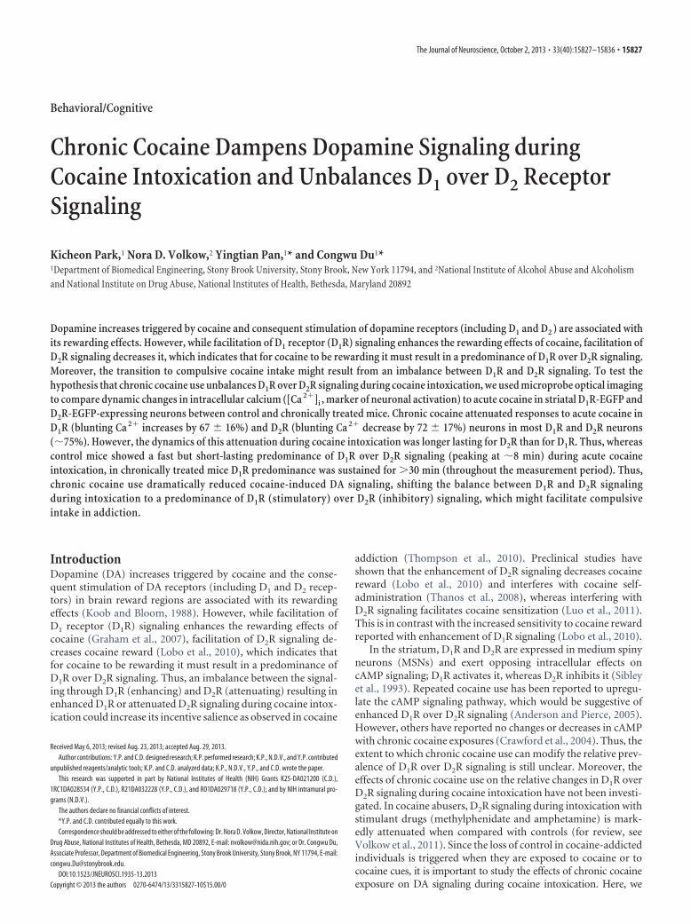
In vivo microprobe fluorescence imaging. The microprobe fluorescenceimaging technique was applied to D1R-EGFP and D2R-EGFP transgenicmice (both control mice and chronic mice) to enable in vivo character-ization of the dynamic [Ca 2�]i changes in striatal D1R and D2R neuronsin response to an acute cocaine challenge. The principle of the endomi-croscopic imaging technique has been outlined previously (Jung et al.,2004; Luo et al., 2011), and Figure 1 illustrates the system setup used inthis study, in which modifications have been implemented to improvefluorescence coupling and image throughput. For instance, additionalPiezoelectrically activated focusing mechanism is used to enable instan-taneous fine focal tracking to maximize [Ca 2�]i-Rhod2 fluorescence de-tection within the field of view (FOV). After anesthesia and surgicalpreparation, the mouse along with the stereotaxic frame was mounted ona motorized 3D stage adapted to a modified upright fluorescence micro-scope (E800, Nikon) as shown in Figure 1a. Figure 1b illustrates a mi-croneedle probe, a custom GRIN lens (�, 1 � 25 mm; 0.65 NA) thatrelayed the image of mouse inner brain (e.g., striatum) at the distal end ofthe needle probe back to the focal plane of the microscope objective (e.g.,PlanFluo 20�, 0.5 NA; Nikon). A motorized 3D microstage facilitatedaccurate light coupling and focal tracking between the microneedleprobe and the microscope objective, allowing for accurate focusing onthe striatal region where there is a high density of GFP-expressing neu-rons (e.g., at �3 mm below mouse cortical surface). Additionally, acustom microsleeve anchoring on mouse cortical bone reduces motion-
Figure 1. Microprobe fluorescence microscope for in vivo deep brain imaging. a, A modified fluorescence microscope enablingsimultaneous EGFP and Rhod2-[Ca 2�]i fluorescence imaging. b, Microneedle (�N) catheter, a 3D motorized microstage assembly,and a PZT-actuated ring holder for deep brain subcellular imaging. Obj, Objective; Ex, excitation; Em, emission. The dashed boxillustrates the time base for dual-channel fluorescence illumination and imaging.
Table 1. Summary of experimental groups
Chronic treatment Acute treatment D1R D2R Experiment no.
In vivo Control mice (0.1 cc/10 g saline, 2 weeks) Cocaine 8 mg/kg Luo et al., 2011Chronic mice (30 mg/kg cocaine, 2 weeks) Cocaine 8 mg/kg 3 4 1
Ex vivo Chronic mice (30 mg/kg cocaine, 2 weeks) 3 3 2Control mice (0.1 cc/10 g saline, 2 weeks) 3 3 3
In vivo Chronic mice (30 mg/kg cocaine, 2 weeks) D1R antagonist SCH23390 (0.075 mg/kg) 30 min before cocaineinjection (8 mg/kg)
3 3 4
In vivo Chronic mice (30 mg/kg cocaine, 2 weeks) D2R antagonist raclopride (3 mg/kg) 30 min before cocaineinjection (8 mg/kg)
4 3 5
15828 • J. Neurosci., October 2, 2013 • 33(40):15827–15836 Park et al. • Unbalance D1R and D2R during Cocaine Intoxication

induced artifacts and potentially enables implantable microprobe to beeasily coupled with the microscope for repeated imaging studies on liveanimals (i.e., time course studies). Special attention was paid to avoiddisruption of large vascular vessels during needle-probe injection, andminor bleeding was occluded by flushing saline solution. For dual-channel EGFP and Rhod2-Ca 2� fluorescence imaging, gated sequentialillumination for the excitation of EGFP (�ex � 460 –500 nm) and Rhod2-Ca 2� (�ex � 530 –550 nm) was used to minimize photon bleaching.EGFP and Rhod2-[Ca 2�]i fluorescence emissions of D1R or D2R neuronswere detected by FITC/EGFP (excitation (EX): 460 –500 nm, dichroicmirror (DM): 505 nm, barrier or emission filter (BA): 510 –560 nm,Chroma #41001) and TRITC/Rhod (EX: 530 –550 nm, DM: 565 nm, BA:590 – 650 nm, Chroma #41002c) epifluorescence cubes, respectively. Theimages were acquired by an EM CCD camera (iXon � 885; 1 m pixels, 8�m/pixel; Andor) synchronized with the fluorescence excitation. Full-field microneedle images (FOV, � 250 �m) were recorded continuouslyat 28 fps from baseline t � �5 min to t � 30 min after cocaineadministration.
Image processing and statistical analysis. The Rhod2-[Ca 2�]i fluores-cence image was coregistered with the corresponding EGFP image toidentify [Ca 2�]i of D1R or D2R EGFP-expressing neurons from non-EGFP cells. We quantified all of the D1R- or D2R-EGFP neurons in thefield of view and measured their time course of [Ca 2�]i changes (i.e.,[Ca 2�]i) in response to acute cocaine for each neuron and normalizedit to their mean basal levels (i.e., t 0). Mean [Ca 2�]i as a function oftime ([Ca 2�]i � t curves) were computed by averaging the curvesamong all these neurons (Fig. 2a5,b5, black curves and bold red or bluecurves, respectively). For statistical analysis, the [Ca 2�]i � t curves ofindividual mice (e.g., m � 3– 4) were further averaged to derive
[Ca 2�]i � t curves for each experimental group, and the results aresummarized in Figure 3, in which the red and blue curves were the globalRhod2-[Ca 2�]i fluorescence changes with their SDs in D1R and D2Rneurons, respectively, for control mice (Fig. 3a) and for chronic mice(Fig. 3b). We also quantified the percentage of D1R-EGFP and D2R-EGFP neurons that showed attenuation of responses to cocaine withinthe field of view. A D1R-EGFP neuron from chronic mice was consideredto have a downregulated response to cocaine if its increase was 50% ofthose in the D1R-EGF neurons of control mice; a D2R-EGFP neuron wasconsidered downregulated if its decrease was 50% of those in the D2R-EGFP neurons of control mice. Similarly, a D1R-EGFP neuron or a D2R-EGFP neuron from chronic mice was considered to have an upregulatedresponse if its increase or decrease was �50% that of control mice,respectively. The differences were assessed using a one-tailed Stu-dent’s t test.
To compare the dynamic differences of [Ca 2�]i in D1R and D2Rneurons between drug-naive and chronic mice, linear regression withleast-squares fitting was applied to the [Ca 2�]i � t curves of each groupto determine their slopes (Luo et al., 2010; k � [Ca2�]i/t), such ask(D1R) or k(D2R) and their ratio, k(D1R)/k(D2R) or [Ca 2�]i(D1R/D2R). We had previously shown that drug-naive mice exhibited biphasic[Ca 2�]i increases in D1R-EGFP neurons; thus, we used piecewise linearregression (e.g., two-piece fitting) to calculate the fast ka
1(D1R) [phase 1,turning point (tp) � 10 min] and the slow ka
2(D1R) (phase 2, tp � 10min) sections on the [Ca 2�]i � t curve. The data collected before andafter the tp for D1R-EGFP were used to quantify the slope ratio betweenthe two phases r' k2/k1. This slope ratio r' k2/k1 was used to statisti-cally evaluate whether the curves for D1R-EGFP and D2R-EGFP followeduniphasic (r3 1) or biphasic (r3 0) dynamics.
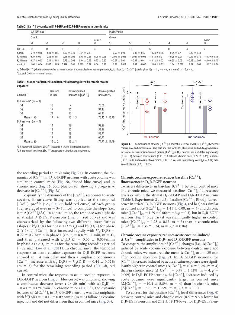
Figure 2. a, b, [Ca 2�]i changes induced by acute cocaine injection (8 mg/kg, i.p.) in striatal D1R-expressing (a) and D2R-expressing (b) neurons in mice chronically exposed to cocaine (30mg/kg/d, i.p., for 2 weeks), which are referred to as chronic mice. 1, 2, [Ca 2�]i images at t � �5 min and t � 25 min before and after cocaine injection, respectively. 3, EGFP image to identifystriatal D1R-/D2R-expressing neurons. 4, [Ca 2�]i overlapped with EGFP image to identify [Ca 2�]i from D1R/D2R neurons. 5, Time course of [Ca 2�]i increases within D1R neurons (black curves,n � 6) and their mean (bold red curve) or [Ca 2�]i decreases within D2R neurons (black curves, n � 6) and their mean (bold blue curve) in response to acute cocaine exposure (also included aredashed curves of previously reported control mice with no history of cocaine exposures before the acute administration (Luo et al., 2011) for comparison. 6, Comparison of mean [Ca 2�]i changes att � 25 min versus baseline (t � 0 min) among the groups, showing less [Ca 2�]i increase for D1R in chronic mice (3.79 � 1.32%) than in control mice (10.6 � 3.2%) and less [Ca 2�]i decreasefor D2R in chronic mice (�3.85 � 1.55%) than in control mice (�10.4 � 5.8%). In vivo simultaneous EGFP and Rhod2-[Ca 2�]i imaging of striatal neurons was enabled by a microneedle approach(Fig. 1) that relayed the subcellular images in the mouse’s striatum at the distal end of a microendoscope (�, 1 � 25 mm; 0.65 NA) back to the focal plane of a modified epifluorescence microscope(E800, Nikon) to be acquired by a time-sharing EM CCD (i885, Andor).
Park et al. • Unbalance D1R and D2R during Cocaine Intoxication J. Neurosci., October 2, 2013 • 33(40):15827–15836 • 15829

Baseline [Ca2�]i assessment and correctionfor changes induced by chronic cocaine. To assesswhether baseline [Ca 2�]i within D1R- andD2R-EGFP neurons changed after chronic co-caine exposure, we performed ex vivo imagingof [Ca 2�]i fluorescence on brain slices (striatalregion) labeled with Rhod2 in Drd1-EGFP orDrd2-EGFP mice (m � 3 for each group) withchronic cocaine exposures (30 mg/kg/d, i.p.;Table 1, Experiment 2) and those (m � 3) ex-posed to saline (0.1 cc/10 g/d, i.p.; Table 1, Ex-periment 3) for 2 weeks. The mice underwentthe same surgical procedure as described aboveand were loaded with Rhod2/AM for intracel-lular Ca 2� labeling. After 1 h to allow for cel-lular uptake of Rhod2, the mice were perfusedwith 20 ml of PBS followed by 20 ml of form-aldehyde (4%). Their heads were embedded in4% formaldehyde for 24 h, and then the brainswere removed and placed in sucrose solution(30%) until the solution was fully absorbed.The striatal region of the mouse’s brain wascryosectioned to �10 �m slices and imaged bythe dual-wavelength microscope setup (Fig. 1)except that the microprobe was removed.[Ca 2�]i-Rhod2 fluorescence within D1R-EGFP or D2R-EGFP neurons was assessed overbackground autofluorescence using a customMATLAB program, by which the influences ofimage location or setup parameters (e.g., expo-sure time) were eliminated (Pan et al., 2010).Differences in [Ca 2�]i fluorescence intensitiesbetween the experimental groups were ana-lyzed using Student’s t test.
The percentage change of baseline [Ca 2�]i
induced by chronic cocaine exposure wasdefined as the coefficient �, which was empiri-cally determined through the ex vivo experi-ments (Experiments 2 and 3) by computing theratio of baseline [Ca 2�]i between chronic miceand control mice using the following linearequation:
�Ca2��t,bo %
� � � 1� � �Ca2��t,b1 %, (1)
where [Ca2�]t,bo % was the intracellular cal-cium change over the baseline of control micebefore cocaine challenge (i.e., t � 0 min),whereas [Ca2�]t,b1 % was the intracellularcalcium change over its baseline in chronicmice before cocaine challenge. � was used toeliminate the influence of the baseline [Ca 2�]i
difference in chronic mice versus control micefor the quantification of the effects of cocainechallenge on [Ca 2�]i.
ResultsThe detailed animal experimental groups are categorized in Table1, and the quantitative results of the in vivo imaging studies aresummarized in Table 2 and Table 3.
Chronic cocaine changes the dynamic profile of [Ca 2�]i inD1R but not in D2RThe dynamic features of [Ca 2�]i in response to acute cocaineexposure (8 mg/kg, i.p.) differed between control mice andchronic mice in D1R-EGFP neurons but not in D2R-EGFP neu-rons (Table 2). Figure 2a (top panels) shows dual-channel fluo-
rescence images of Rhod2-[Ca 2�]i (Fig. 2a1,a2) and D1R-EGFP(Fig. 2a3), and their merged image (Fig. 2a4), which allowed usto trace the temporal [Ca 2�]i profile (Fig. 2a5) of individualD1R-EGFP neurons (Fig. 2a5, black curves; n � 6) and theiraveraged changes (Fig. 2a5, bold red curve); the same strategy wasused for D2R-EGFP neurons (Fig. 2b, bottom panels). Interest-ingly, whereas in control mice (dashed red curve) the [Ca 2�]i
increase in D1R-EGFP neurons was biphasic, with an initialrapid increase to 7.6 � 2.3% within tp � 8.8 � 1.1 min of cocaineinjection followed by a plateau, in chronic mice (Fig. 2a, bold redcurve) the [Ca 2�]i increase was monophasic starting at t � 4min after cocaine injection and gradually increasing throughout
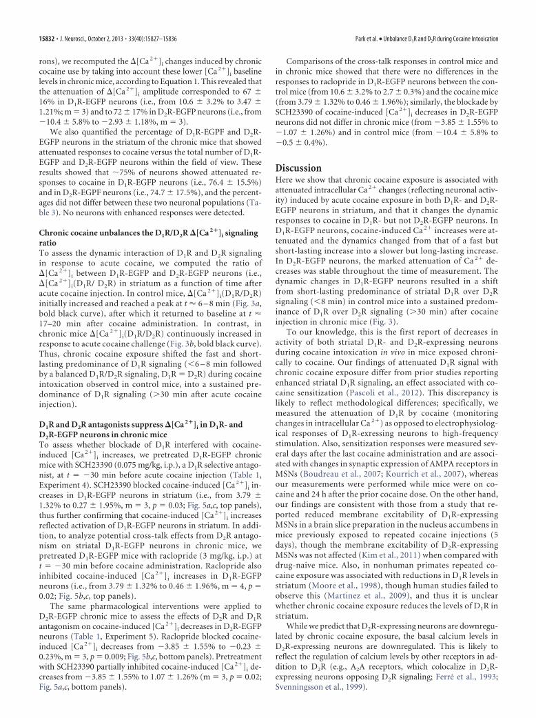
Figure 3. a, b, Dynamic changes in [Ca 2�]i induced by acute cocaine in striatal D1R-expressing neurons (red curve), D2R-expressing neurons (blue curve), and their ratios [[Ca 2�]i(D1R/D2R); bold black curves] in control mice (a) and chronic mice (b).Least-squares fitting (dashed lines) is applied to analyze the slope differences [i.e., k � d([Ca 2�]i)/dt]. In control mice (a),[Ca 2�]i increases in striatal D1R neurons are biphasic (pink lines): an initial steep rise with ka
1(D1R) in phase 1 (t � 8.3 min)followed by a long-lasting, slow increase with ka
2(D1R) in phase 2 (t � 8.3 min) and the [Ca 2�]i in striatal D2R neurons areuniphasic: a gradual decrease with ka(D2R) over t � 30 min. In chronic mice (b), [Ca 2�]i increases in D1R neurons have changedto uniphasic, with a gradual but long-lasting rise with kc(D1R), and those of D2R neurons remain uniphasic with a gradual decrease.As a result, [Ca 2�]i(D1R/D2R) in control mice rapidly increases to a climax at t � 5–7 min, but it then returns to baseline;whereas in chronic mice [Ca 2�]i(D1R/D2R) progressively increases and does not return to baseline.
15830 • J. Neurosci., October 2, 2013 • 33(40):15827–15836 Park et al. • Unbalance D1R and D2R during Cocaine Intoxication
the recording period (t � 30 min; Fig. 2a). In contrast, the dy-namics of [Ca 2�]i in D2R-EGFP neurons with acute cocaine wassimilar in control mice (Fig. 2b, dashed blue curve) and inchronic mice (Fig. 2b, bold blue curve), showing a progressivedecrease in [Ca 2�]i (Fig. 2b).
To quantify the dynamics of the [Ca 2�]i responses to acutecocaine, linear-curve fitting was applied to the temporal[Ca 2�]i profile (i.e., Fig. 2a, bold red curve) of each group(i.e., averaged over m � 3– 4 mice) to compute the slope (i.e.,k � [Ca 2�]i/t). In control mice, the response was biphasicin striatal D1R-EGFP neurons (Fig. 3a, red curve) and wascharacterized by the following two different linear fittings(slopes): ka
1(D1R) for phase 1 (t � tp) and ka2(D1R) for phase
2 (t � tp). [Ca 2�]i first increased rapidly with ka1(D1R) �
0.77 � 0.2%/min in phase 1 (t � tp � 8.8 � 1.1 min, m � 4),and then plateaued with ka
2(D1R) � 0.05 � 0.05%/minin phase 2 (t � tp, m � 4) for the remaining recording period(�22 min; Luo et al., 2011). In chronic mice, the temporalresponse to acute cocaine exposure in D1R-EGFP neuronsshowed an �4 min delay and then a uniphasic continuous[Ca 2�]i increase with kc
1(D1R) � kc2(D1R) � 0.44 � 0.02%
(m � 3) for the remaining recording period (Fig. 3b, redcurve).
In control mice, the response to acute cocaine exposure inD2R-EGFP neurons (Fig. 3a, blue curve) was uniphasic, showinga continuous decrease (over t � 30 min) with ka(D2R) ��0.40 � 0.13%/min. In chronic mice (Fig. 3b), the dynamicfeatures of [Ca 2�]i in D2R-EGFP neurons was also uniphasicwith kc(D2R) � �0.12 � 0.09%/min (m � 3) following cocaineinjection and did not differ from that in control mice (Fig. 3a).
Chronic cocaine exposure reduces baseline [Ca 2�]i
fluorescence in D2R-EGFP neuronsTo assess differences in baseline [Ca 2�]i between control miceand chronic mice, we measured baseline [Ca 2�]i fluorescencelevels ex vivo in the striatal D1R-EGFP and D2R-EGFP neurons(Table 1, Experiments 2 and 3). Baseline [Ca 2�]i-Rhod2 fluores-cence in striatal D1R-EGFP neurons (Fig. 4, red bar) was similarin control mice ([Ca 2�]i/b � 1.41 � 0.08, m � 3) and chronicmice ([Ca 2�]i/b � 1.29 � 0.06; m � 3, p � 0.3), but in D2R-EGFPneurons (Fig. 4, blue bar) it was significantly higher in controlmice ([Ca 2�]i/b � 1.78 � 0.15; m � 3) than in chronic mice([Ca 2�]i/b � 1.35 � 0.24, m � 3, p � 0.04).
Chronic cocaine exposure reduces acute cocaine-induced�[Ca 2�]i amplitudes in D1R- and D2R-EGFP neuronsTo compare the amplitudes of [Ca 2�]i changes (i.e., [Ca 2�]i)induced by acute cocaine exposure between control mice andchronic mice, we measured the mean [Ca 2�]i at t � 25 minafter cocaine injection (Fig. 2). In D1R-EGFP neurons, the[Ca 2�]i increases induced by acute cocaine exposure were signif-icantly higher in control mice ([Ca 2�]i � 10.6 � 3.2%, m � 4)than in chronic mice ([Ca 2�]i � 3.79 � 1.32%, m � 4, p �0.009). In D2R-EGFP neurons, the [Ca 2�]i decreases induced byacute cocaine were significantly larger in control mice([Ca 2�]i � �10.4 � 5.8%, m � 4) than in chronic mice([Ca 2�]i � �3.85 � 1.55%, m � 3, p � 0.005).
To correct for the baseline [Ca 2�]i level differences (Fig. 4)between control mice and chronic mice (8.5 � 9.5% lower forD1R-EGFP neurons and 24.2 � 18.1% lower for D2R-EGFP neu-
Table 2. �Ca 2��i dynamics in D1R-EGFP and D2R-EGFP neurons in chronic mice
D1R EGFP mice D2R EGFP mice
ChronicAcute a
m
ChronicAcutea
mS1 S2 S3 m S1 S2 S3 S4 m
Cells (n) 10 13 6 3 4 6 13 12 15 4 7t0 (min) 6.10 � 0.68 3.83 � 0.85 1.90 � 0.49 3.94 � 2.1 0.59 � 0.98 0.00 � 0.56 0.28 � 0.36 0.75 � 0.7 0.40 � 0.33k2 (%/min) 0.29 � 0.01 0.32 � 0.01 0.68 � 0.03 0.43 � 0.01 0.05 � 0.05 �0.071 � 0.003 �0.029 � 0.004 �0.12 � 0.01 �0.26 � 0.01 �0.12 � 0.10 �0.39 � 0.15k1 (%/min) 0.27 � 0.03 0.33 � 0.03 0.72 � 0.02 0.44 � 0.02 0.77 � 0.20 �0.07 � 0.01 �0.03 � 0.01 �0.12 � 0.02 �0.25 � 0.02 �0.12 � 0.09 �0.40 � 0.13r � k2 /k1 1.08 � 0.14 0.967 � 0.09 0.944 � 0.06 0.999 � 0.07 0.06 � 0.23 1.08 � 0.013 1.07 � 0.047 1.00 � 0.025 1.04 � 0.012 1.04 � 0.03 0.97 � 0.26
t0 , Delay of �Ca 2��i change to acute cocaine; m, animal number; n, number of selected neurons per mouse; k1 , k2 , slope Kp � �Ca 2��i /t for phase 1 ( p � 1, t0 � t � tp) and phase 2 ( p � 2, t � tp).aLuo, et al. (2011); m � animal numbers.
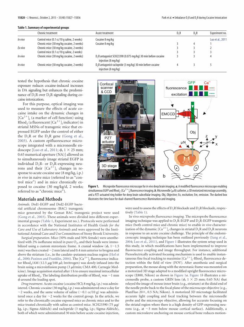
Table 3. Numbers of D1R cells and D1R cells downregulated by chronic cocaineexposure
Animal IDNeuronsin FOV
Downregulatedneurons in �Ca 2��i
Downregulatedneurons (%)
D1R neurons* (m � 3)S1 10 7 70.00S2 17 16 94.12S3 23 15 65.22
Mean � SD 17 � 6 13 � 5 76.45 � 15.49D2R neurons † (m � 4)
S1 14 13 92.86S2 18 10 55.56S3 14 12 85.71S4 17 11 64.71
Mean � SD 16 � 2 12 � 1 74.71 � 17.49
*D1R neuron with 50% lower �Ca 2��i response to cocaine than that in naive mice.†D2R neuron with 50% lower �Ca 2��i response to cocaine than that in naive mice.
Figure 4. Comparison of baseline [Ca 2�]i-Rhod2 fluorescence levels (�[Ca 2�]i) betweencontrol mice and chronic mice. Red/blue lines are for D1R/D2R neurons, and white/gray bars arefor saline- versus cocaine-treated groups. [Ca 2�]i in D1R neurons did not differ significantly( p � 0.3) between control mice (1.41 � 0.08) and chronic mice (1.29 � 0.06), whereas[Ca 2�]i in D2R neurons in chronic mice (1.35 � 0.24) was significantly lower ( p � 0.04) thanin control mice (1.78 � 0.15).
Park et al. • Unbalance D1R and D2R during Cocaine Intoxication J. Neurosci., October 2, 2013 • 33(40):15827–15836 • 15831
rons), we recomputed the [Ca 2�]i changes induced by chroniccocaine use by taking into account these lower [Ca 2�]i baselinelevels in chronic mice, according to Equation 1. This revealed thatthe attenuation of [Ca 2�]i amplitude corresponded to 67 �16% in D1R-EGFP neurons (i.e., from 10.6 � 3.2% to 3.47 �1.21%; m � 3) and to 72 � 17% in D2R-EGFP neurons (i.e., from�10.4 � 5.8% to �2.93 � 1.18%, m � 3).
We also quantified the percentage of D1R-EGPF and D2R-EGFP neurons in the striatum of the chronic mice that showedattenuated responses to cocaine versus the total number of D1R-EGFP and D2R-EGFP neurons within the field of view. Theseresults showed that �75% of neurons showed attenuated re-sponses to cocaine in D1R-EGFP neurons (i.e., 76.4 � 15.5%)and in D2R-EGPF neurons (i.e., 74.7 � 17.5%), and the percent-ages did not differ between these two neuronal populations (Ta-ble 3). No neurons with enhanced responses were detected.
Chronic cocaine unbalances the D1R/D2R �[Ca 2�]i signalingratioTo assess the dynamic interaction of D1R and D2R signalingin response to acute cocaine, we computed the ratio of[Ca 2�]i between D1R-EGFP and D2R-EGFP neurons (i.e.,[Ca 2�]i(D1R/ D2R) in striatum as a function of time afteracute cocaine injection. In control mice, [Ca 2�]i(D1R/D2R)initially increased and reached a peak at t � 6 – 8 min (Fig. 3a,bold black curve), after which it returned to baseline at t �17–20 min after cocaine administration. In contrast, inchronic mice [Ca 2�]i(D1R/D2R) continuously increased inresponse to acute cocaine challenge (Fig. 3b, bold black curve).Thus, chronic cocaine exposure shifted the fast and short-lasting predominance of D1R signaling (6 – 8 min followedby a balanced D1R/D2R signaling, D1R � D2R) during cocaineintoxication observed in control mice, into a sustained pre-dominance of D1R signaling (�30 min after acute cocaineinjection).
D1R and D2R antagonists suppress �[Ca 2�]i in D1R- andD2R-EGFP neurons in chronic miceTo assess whether blockade of D1R interfered with cocaine-induced [Ca 2�]i increases, we pretreated D1R-EGFP chronicmice with SCH23390 (0.075 mg/kg, i.p.), a D1R selective antago-nist, at t � �30 min before acute cocaine injection (Table 1,Experiment 4). SCH23390 blocked cocaine-induced [Ca 2�]i in-creases in D1R-EGFP neurons in striatum (i.e., from 3.79 �1.32% to 0.27 � 1.95%, m � 3, p � 0.03; Fig. 5a,c, top panels),thus further confirming that cocaine-induced [Ca 2�]i increasesreflected activation of D1R-EGFP neurons in striatum. In addi-tion, to analyze potential cross-talk effects from D2R antago-nism on striatal D1R-EGFP neurons in chronic mice, wepretreated D1R-EGFP mice with raclopride (3 mg/kg, i.p.) att � �30 min before cocaine administration. Raclopride alsoinhibited cocaine-induced [Ca 2�]i increases in D1R-EGFPneurons (i.e., from 3.79 � 1.32% to 0.46 � 1.96%, m � 4, p �0.02; Fig. 5b,c, top panels).
The same pharmacological interventions were applied toD2R-EGFP chronic mice to assess the effects of D2R and D1Rantagonism on cocaine-induced [Ca 2�]i decreases in D2R-EGFPneurons (Table 1, Experiment 5). Raclopride blocked cocaine-induced [Ca 2�]i decreases from �3.85 � 1.55% to �0.23 �0.23%, m � 3, p � 0.009; Fig. 5b,c, bottom panels). Pretreatmentwith SCH23390 partially inhibited cocaine-induced [Ca 2�]i de-creases from �3.85 � 1.55% to 1.07 � 1.26% (m � 3, p � 0.02;Fig. 5a,c, bottom panels).
Comparisons of the cross-talk responses in control mice andin chronic mice showed that there were no differences in theresponses to raclopride in D1R-EGFP neurons between the con-trol mice (from 10.6 � 3.2% to 2.7 � 0.3%) and the cocaine mice(from 3.79 � 1.32% to 0.46 � 1.96%); similarly, the blockade bySCH23390 of cocaine-induced [Ca 2�]i decreases in D2R-EGFPneurons did not differ in chronic mice (from �3.85 � 1.55% to�1.07 � 1.26%) and in control mice (from �10.4 � 5.8% to�0.5 � 0.4%).
DiscussionHere we show that chronic cocaine exposure is associated withattenuated intracellular Ca 2� changes (reflecting neuronal activ-ity) induced by acute cocaine exposure in both D1R- and D2R-EGFP neurons in striatum, and that it changes the dynamicresponses to cocaine in D1R- but not D2R-EGFP neurons. InD1R-EGFP neurons, cocaine-induced Ca 2� increases were at-tenuated and the dynamics changed from that of a fast butshort-lasting increase into a slower but long-lasting increase.In D2R-EGFP neurons, the marked attenuation of Ca 2� de-creases was stable throughout the time of measurement. Thedynamic changes in D1R-EGFP neurons resulted in a shiftfrom short-lasting predominance of striatal D1R over D2Rsignaling (8 min) in control mice into a sustained predom-inance of D1R over D2R signaling (�30 min) after cocaineinjection in chronic mice (Fig. 3).
To our knowledge, this is the first report of decreases inactivity of both striatal D1R- and D2R-expressing neuronsduring cocaine intoxication in vivo in mice exposed chroni-cally to cocaine. Our findings of attenuated D1R signal withchronic cocaine exposure differ from prior studies reportingenhanced striatal D1R signaling, an effect associated with co-caine sensitization (Pascoli et al., 2012). This discrepancy islikely to reflect methodological differences; specifically, wemeasured the attenuation of D1R by cocaine (monitoringchanges in intracellular Ca 2�) as opposed to electrophysiolog-ical responses of D1R-exressing neurons to high-frequencystimulation. Also, sensitization responses were measured sev-eral days after the last cocaine administration and are associ-ated with changes in synaptic expression of AMPA receptors inMSNs (Boudreau et al., 2007; Kourrich et al., 2007), whereasour measurements were performed while mice were on co-caine and 24 h after the prior cocaine dose. On the other hand,our findings are consistent with those from a study that re-ported reduced membrane excitability of D1R-expressingMSNs in a brain slice preparation in the nucleus accumbens inmice previously exposed to repeated cocaine injections (5days), though the membrane excitability of D2R-expressingMSNs was not affected (Kim et al., 2011) when compared withdrug-naive mice. Also, in nonhuman primates repeated co-caine exposure was associated with reductions in D1R levels instriatum (Moore et al., 1998), though human studies failed toobserve this (Martinez et al., 2009), and thus it is unclearwhether chronic cocaine exposure reduces the levels of D1R instriatum.
While we predict that D2R-expressing neurons are downregu-lated by chronic cocaine exposure, the basal calcium levels inD2R-expressing neurons are downregulated. This is likely toreflect the regulation of calcium levels by other receptors in ad-dition to D2R (e.g., A2A receptors, which colocalize in D2R-expressing neurons opposing D2R signaling; Ferre et al., 1993;Svenningsson et al., 1999).
15832 • J. Neurosci., October 2, 2013 • 33(40):15827–15836 Park et al. • Unbalance D1R and D2R during Cocaine Intoxication

Our results in D2R-expressing neurons are consistent withstudies showing decreases in striatal D2R signaling with chroniccocaine exposure (baseline studies), including preclinical studiesreporting reductions in striatal D2R levels (Nader et al., 2006;Thanos et al., 2007) and clinical studies showing reductions in
striatal D2R in cocaine abusers (for review, see Volkow et al.,2009). In human addicts, the reduction in striatal D2R has beenimplicated in compulsive drug intake and impulsivity (Volkow etal., 1993, 2006, 2007). Similarly, an in vitro study reported atten-uation of cocaine-induced Ca 2� decreases in D2R-expressing
Figure 5. I, Top panels, II, Right panels, Dynamic [Ca 2�]i changes induced by acute cocaine (8 mg/kg, i.p.) in striatal D1R-expressing neurons (I, top panels) and D2R-expressing neurons (II, rightpanels), with D1R antagonist (SCH23390, i.p.; a) and with D2R antagonist (raclopride, i.p.; b) in chronic mice. 1, 2,[Ca 2�]i images at t ��5 min and t �25 min before and after cocaine injection,respectively. 3, EGFP image to identify striatal D1R-expressing (I ) or D2R-expressing (II ) neurons. 4, [Ca 2�]i overlapped with EGFP image to identify [Ca 2�]i from D1R (I ) or D2R (II ) neurons. 5,[Ca 2�]i increases within D1R (I ) or decreases within D2R (II ) neurons (black curves, n � 5, 6, respectively) and their mean time course (bold red or blue curves) in response to acute cocaine. c,Comparison of mean [Ca 2�]i increases at t � 25 min versus baseline (t � 0 min) among groups. D1R (I ) shows less [Ca 2�]i increase in chronic mice (3.79 � 1.32%) than in the control mice(10.6 � 3.2%), which was further reduced by D1R and D2R antagonists to 0.2 � 1.8% and 0.46 � 1.96%, respectively. D2R (II ) shows less [Ca 2�]i decrease in chronic mice (�3.85 � 1.55%)than in control mice (�10.4 � 5.8%), which was further reduced by D2R and D1R antagonists to �0.23 � 0.23% and �1.07 � 1.26%, respectively.
Park et al. • Unbalance D1R and D2R during Cocaine Intoxication J. Neurosci., October 2, 2013 • 33(40):15827–15836 • 15833

neurons in nucleus accumbens with chronic cocaine exposure(Perez et al., 2011), and imaging studies have reported markedreductions in stimulant-induced DA increases in cocaine abusers(for review, see Volkow et al., 2009).
Since activation of striatal D1R- or inhibition of striatal D2R-expressing neurons enhances cocaine reward (Hikida et al., 2010;Lobo et al., 2010; Ferguson et al., 2011) and upregulation of D2Rinterferes with cocaine consumption (Thanos et al., 2008), animbalance between the stimulatory direct D1R pathway and theinhibitory D2R indirect pathway during cocaine intoxicationwould be expected to facilitate compulsive cocaine intake evenwhen DA signaling is markedly attenuated (for review, seeVolkow et al., 2009). This would be akin to the improvement inmovement observed in patients with Parkinson’s disease aftersurgical lesioning of the subthalamic nucleus (to remove controlfrom the D2R-modulated indirect pathway, which inhibits loco-motor activity), even when they have very low DA levels (Alvarezet al., 2009). So, while we showed a significant decrease in signal-ing through the striatal D1R direct pathway with chronic cocaineexposure, the longer-lasting inhibition of the striatal D2R indirectpathway during cocaine intoxication could analogously be ex-pected to allow the direct pathway to drive the behavior of co-caine reward and consumption.
In this study, we also showed that chronic cocaine exposuredisrupted the dynamics of cocaine-induced Ca 2� increases inD1Rs from an abrupt increase followed by a leveling off to adelayed and progressive increase in D1R signaling. Though itsbehavioral significance is unclear, we postulate that it mightresult in an attenuation of reward, since the speed at whichdrugs stimulate DA signaling modulates their rewarding ef-fects (Beardsley and Balster, 1993; Volkow et al., 1995, 2000;Samaha and Robinson, 2005). We also postulate that the long-lasting predominance of D1R over D2R signaling might resultin enhanced motivational drive for the drug despite a decreasein reward intensity. This is because D1R, which is stimulatoryand has a low affinity for DA, will be activated when exposed tolarge DA increases, as occur during cocaine intoxication,whereas D2R, which is inhibitory and has a high affinity forDA, will be activated both by the sharp DA increases as well asby the lower tonic DA levels that follow ( Beaulieu and Gainet-dinov, 2011). Thus, the relative predominance of D1R overD2R following cocaine administration (as seen in controlmice) probably contributes to the dynamics of the behavioraleffects of cocaine (Volkow et al., 1997a). That is, immediatelyafter cocaine injection, full signaling from D1R and D2R isexpected, but as DA levels start to decline then D1R signalingwould decrease faster than D2R, eventually resulting in a D2Rpredominance and a consequent decrease in the rewardingeffects of cocaine. Indeed, in control mice, acute cocaine ex-posure abruptly stimulated striatal D1R-expressing neurons,whereas it slowly and continuously inhibited D2R-expressingneurons. Thus, the persistent predominance of D1R signalingseen in the chronic mice might sustain the incentive and mo-tivation for cocaine intake without the opposing effects ofstriatal D2R signaling.
The molecular mechanisms underlying the attenuated responsesto cocaine in D2R- and D1R-expressing MSNs during intoxicationare unclear. They could reflect blunted cocaine-induced DA in-creases, as shown by neuroimaging studies reporting reduced DAincreases to stimulant drugs in cocaine abusers (Volkow et al.,1997b, 2011; Martinez et al., 2007). In D2R-expressing MSNs,they could also reflect downregulation of D2R levels with chronic
cocaine exposure, as reported by prior studies (for review, seeVolkow et al., 2012). For D1R-expressing MSNs, this is less clearsince studies have reported increases, decreases, or no changes inD1R levels with chronic cocaine exposure (for review, see Volkowet al., 2012). As for the mechanisms underlying the differentialeffects of chronic cocaine use on the dynamic changes in D1Rversus D2R signaling, we can only speculate that they could reflectinteractions with other receptors (i.e., D3R, which heteromerizeswith D1R; Marcellino et al., 2008), that they are upregulated withchronic cocaine use (Staley and Mash, 1996), and that differentialchanges occur in synaptic plasticity in D1R versus D2R-expressing MSNs with chronic cocaine use.
The cross talk between D1Rs and D2Rs observed with the ad-ministration of the D1R antagonist in D2R-EGFP neurons andthe D2R antagonist in D1R-EGFP neurons is consistent with syn-ergism between D1R- and D2R-expressing neurons in striatum(LaHoste et al., 2000), including interactions of both receptorswithin striatal neurons that coexpress D1R and D2R (Lester et al.,1993; Bertran-Gonzalez et al., 2008; Matamales et al., 2009,Rashid et al., 2007; Aizman et al., 2000). Interestingly, raclopridewas more effective in blocking cocaine-induced D1R signalingthan SCH23390 was in blocking D2R signaling. Though themechanisms are unclear, we postulate that they might reflect theblockade by raclopride of D3R (Malmberg et al., 1994), whichheteromerizes with D1R (Marcellino et al., 2008).
Limitations of this study include measurements in dorsalstriatum rather than in nucleus accumbens, which is the brainregion implicated in drug reward (Koob and Bloom, 1988). How-ever, the dorsal striatum is implicated in the neuroadaptationsassociated with the transition from controlled to compulsive co-caine intake (for review, see Belin and Everitt, 2008; Volkow et al.,2012), and thus our findings are pertinent to addiction. In thisstudy, we measured the dynamic changes over a 30 min period,but further studies are needed to determine whether dynamics inD2R-expressing MSN are affected by chronic cocaine exposureover a longer imaging period.
Here we show that chronic cocaine exposure markedly de-creased the responses to acute cocaine exposure both in D1R- andD2R-expressing MSNs and changed the dynamics of their signal-ing during cocaine intoxication. Chronic cocaine exposure re-sulted in a sustained predominance of D1R over D2R signalingduring cocaine intoxication, which, by removing the reward-decreasing effects of D2R stimulation, might leave the D1R directpathway unopposed.
ReferencesAizman O, Brismar H, Uhlen P, Zettergren E, Levey AI, Forssberg H, Green-
gard P, Aperia A (2000) Anatomical and physiological evidence for D1
and D2 dopamine receptor colocalization in neostriatal neurons. NatNeurosci 3:226 –230. CrossRef Medline
Alvarez L, Macias R, Pavon N, Lopez G, Rodríguez-Oroz MC, Rodríguez R,Alvarez M, Pedroso I, Teijeiro J, Fernandez R, Casabona E, Salazar S,Maragoto C, Carballo M, García I, Guridi J, Juncos JL, DeLong MR,Obeso JA (2009) Therapeutic efficacy of unilateral subthalamotomy inParkinson’s disease: results in 89 patients followed for up to 36 months.J Neurol Neurosurg Psychiatry 80:979 –985. CrossRef Medline
Anderson SM, Pierce RC (2005) Cocaine-induced alterations in dopaminereceptor signaling: implications for reinforcement and reinstatement.Pharmacol Ther 106:389 – 403. CrossRef Medline
Beardsley PM, Balster RL (1993) The effects of delay of reinforcement anddose on the self-administration of cocaine and procaine in rhesus mon-keys. Drug Alcohol Depend 34:37– 43. CrossRef Medline
15834 • J. Neurosci., October 2, 2013 • 33(40):15827–15836 Park et al. • Unbalance D1R and D2R during Cocaine Intoxication

Beaulieu JM, Gainetdinov RR (2011) The physiology, signaling, and phar-macology of dopamine receptors. Pharmacol Rev 63:182–217. CrossRefMedline
Belin D, Everitt BJ (2008) Cocaine seeking habits depend upon dopamine-dependent serial connectivity linking the ventral with the dorsal striatum.Neuron 57:432– 441. CrossRef Medline
Bertran-Gonzalez J, Bosch C, Maroteaux M, Matamales M, Herve D, ValjentE, Girault JA (2008) Opposing patterns of signaling activation in dopa-mine D1 and D2 receptor-expressing striatal neurons in response to co-caine and haloperidol. J Neurosci 28:5671–5685. CrossRef Medline
Boudreau AC, Reimers JM, Milovanovic M, Wolf ME (2007) Cell surfaceAMPA receptors in the rat nucleus accumbens increase during cocainewithdrawal but internalize after cocaine challenge in association withaltered activation of mitogen-activated protein kinases. J Neurosci 27:10621–10635. CrossRef Medline
Crawford CA, Choi FY, Kohutek JL, Yoshida ST, McDougall SA (2004)Changes in PKA activity and Gs alpha and Golf alpha levels afteramphetamine- and cocaine-induced behavioral sensitization. Synapse 51:241–248. CrossRef Medline
Ferguson SM, Eskenazi D, Ishikawa M, Wanat MJ, Phillips PE, Dong Y,Roth BL, Neumaier JF (2011) Transient neuronal inhibition revealsopposing roles of indirect and direct pathways in sensitization. NatNeurosci 14:22–24. CrossRef Medline
Ferre S, O’Connor WT, Fuxe K, Ungerstedt U (1993) The striatopallidalneuron: a main locus for adenosine– dopamine interactions in the brain.J Neurosci 13:5402–5406. Medline
Gong S, Zheng C, Doughty ML, Losos K, Didkovsky N, Schambra UB, NowakNJ, Joyner A, Leblanc G, Hatten ME, Heintz N (2003) A gene expressionatlas of the central nervous system based on bacterial artificial chromo-somes. Nature 425:917–925. CrossRef Medline
Graham DL, Hoppenot R, Hendryx A, Self DW (2007) Differential ability ofD1 and D2 dopamine receptor agonists to induce and modulate expres-sion and reinstatement of cocaine place preference in rats. Psychophar-macology (Berl) 191:719 –730. CrossRef Medline
Hikida T, Kimura K, Wada N, Funabiki K, Nakanishi S (2010) Distinct rolesof synaptic transmission in direct and indirect striatal pathways to rewardand aversive behavior. Neuron 66:896 –907. CrossRef Medline
Hof PR, Young WG, Bloom FE, Belichenko PV, Celio MR (2000) Compar-ative cytoarchitectonic atlas of the C57BL/6 and 129/Sv mouse brains.New York: Elsevier.
Jung JC, Mehta AD, Aksay E, Stepnoski R, Schnitzer MJ (2004) In vivomammalian brain imaging using one- and two-photon fluorescence mi-croendoscopy. J Neurophysiol 92:3121–3133. CrossRef Medline
Kim J, Park BH, Lee JH, Park SK, Kim JH (2011) Cell type-specific altera-tions in the nucleus accumbens by repeated exposures to cocaine. BiolPsychiatry 69:1026 –1034. CrossRef Medline
Koob GF, Bloom FE (1988) Cellular and molecular mechanisms of drugdependence. Science 242:715–723. CrossRef Medline
Kourrich S, Rothwell PE, Klug JR, Thomas MJ (2007) Cocaine experiencecontrols bidirectional synaptic plasticity in the nucleus accumbens.J Neurosci 27:7921–7928. CrossRef Medline
LaHoste GJ, Henry BL, Marshall JF (2000) Dopamine D1 receptors syner-gize with D2, but not D3 or D4, receptors in the striatum without theinvolvement of action potentials. J Neurosci 20:6666 – 6671. Medline
Lester J, Fink S, Aronin N, DiFiglia M (1993) Colocalization of D1 and D2
dopamine receptor mRNAs in striatal neurons. Brain Res 621:106 –110.CrossRef Medline
Lobo MK, Covington HE 3rd, Chaudhury D, Friedman AK, Sun H, Damez-Werno D, Dietz DM, Zaman S, Koo JW, Kennedy PJ, Mouzon E, MogriM, Neve RL, Deisseroth K, Han MH, Nestler EJ (2010) Cell type-specificloss of BDNF signaling mimics optogenetic control of cocaine reward.Science 330:385–390. CrossRef Medline
Luo Z, Volkow ND, Heintz N, Pan Y, Du C (2011) Acute cocaine inducesfast activation of D1 receptor and progressive deactivation of D2 receptorstriatal neurons: in vivo optical microprobe [Ca 2�]i imaging. J Neurosci31:13180 –13190. CrossRef Medline
Malmberg A, Nordvall G, Johansson AM, Mohell N, Hacksell U (1994) Mo-lecular basis for the binding of 2-aminotetralins to human dopamine D2Aand D3 receptors. Mol Pharmacol 46:299 –312. Medline
Marcellino D, Ferre S, Casado V, Cortes A, Le Foll B, Mazzola C, Drago F,Saur O, Stark H, Soriano A, Barnes C, Goldberg SR, Lluis C, Fuxe K,Franco R (2008) Identification of dopamine D1-D3 receptor hetero-
mers. Indications for a role of synergistic D1-D3 receptor interactions inthe striatum. J Biol Chem 283:26016 –26025. CrossRef Medline
Martinez D, Narendran R, Foltin RW, Slifstein M, Hwang DR, Broft A,Huang Y, Cooper TB, Fischman MW, Kleber HD, Laruelle M (2007)Amphetamine-induced dopamine release: markedly blunted in cocainedependence and predictive of the choice to self-administer cocaine. Am JPsychiatry 164:622– 629. CrossRef Medline
Martinez D, Slifstein M, Narendran R, Foltin RW, Broft A, Hwang DR, PerezA, Abi-Dargham A, Fischman MW, Kleber HD, Laruelle M (2009) Do-pamine D1 receptors in cocaine dependence measured with PET and thechoice to self-administer cocaine. Neuropsychopharmacology 34:1774 –1782. CrossRef Medline
Matamales M, Bertran-Gonzalez J, Salomon L, Degos B, Deniau JM, ValjentE, Herve D, Girault JA (2009) Striatal medium-sized spiny neurons:identification by nuclear staining and study of neuronal subpopulationsin BAC transgenic mice. PLoS One 4:e4770. CrossRef Medline
Moore RJ, Vinsant SL, Nader MA, Porrino LJ, Friedman DP (1998) Effect ofcocaine self-administration on striatal dopamine D1 receptors in rhesusmonkeys. Synapse 28:1–9. CrossRef Medline
Nader MA, Morgan D, Gage HD, Nader SH, Calhoun TL, Buchheimer N,Ehrenkaufer R, Mach RH (2006) PET imaging of dopamine D2 recep-tors during chronic cocaine self-administration in monkeys. Nat Neuro-sci 9:1050 –1056. CrossRef Medline
Pan R, Yan ZJ, Luo Z, Du C (2010) Optical discrimination of intracellularCa 2� changes of brain induced by cocaine and ischemia. Paper presentedat Biomedical Optics, Miami, FL, April.
Pascoli V, Turiault M, Luscher C (2012) Reversal of cocaine-evoked syn-aptic potentiation resets drug-induced adaptive behaviour. Nature481:71–75. CrossRef Medline
Paxinos G, Franklin KBJ (2004) The mouse brain in stereotaxic coordinates,Ed 2. San Diego, CA: Academic.
Perez MF, Ford KA, Goussakov I, Stutzmann GE, Hu XT (2011) Repeatedcocaine exposure decreases dopamine D2-like receptor modulation ofCa 2� homeostasis in rat nucleus accumbens neurons. Synapse 65:168 –180. CrossRef Medline
Rashid AJ, So CH, Kong MM, Furtak T, El-Ghundi M, Cheng R, O’Dowd BF,George SR (2007) D1-D2 dopamine receptor heterooligomers withunique pharmacology are coupled to rapid activation of Gq/11 in thestriatum. Proc Natl Acad Sci U S A 104:654 – 659. CrossRef Medline
Samaha AN, Robinson TE (2005) Why does the rapid delivery of drugs tothe brain promote addiction? Trends Pharmacol Sci 26:82– 87. CrossRefMedline
Sibley DR, Monsma FJ Jr, Shen Y (1993) Molecular neurobiology of dopa-minergic receptors. Int Rev Neurobiol 35:391– 415. CrossRef Medline
Staley JK, Mash DC (1996) Adaptive increase in D3 dopamine receptors inthe brain reward circuits of human cocaine fatalities. J Neurosci 16:6100 –6106. Medline
Svenningsson P, Le Moine C, Fisone G, Fredholm BB (1999) Distribution,biochemistry and function of striatal adenosine A2A receptors. Prog Neu-robiol 59:355–396. CrossRef Medline
Thanos PK, Michaelides M, Benveniste H, Wang GJ, Volkow ND (2007)Effects of chronic oral methylphenidate on cocaine self-administrationand striatal dopamine D2 receptors in rodents. Pharmacol Biochem Be-hav 87:426 – 433. CrossRef Medline
Thanos PK, Michaelides M, Umegaki H, Volkow ND (2008) D2R DNAtransfer into the nucleus accumbens attenuates cocaine self-administration in rats. Synapse 62:481– 486. CrossRef Medline
Thompson D, Martini L, Whistler JL (2010) Altered ratio of D1 and D2
dopamine receptors in mouse striatum is associated with behavioral sen-sitization to cocaine. PLoS One 5:e11038. CrossRef Medline
Volkow ND, Fowler JS, Wang GJ, Hitzemann R, Logan J, Schlyer DJ, Dewey SL,Wolf AP (1993) Decreased dopamine D2 receptor availability is associatedwith reduced frontal metabolism in cocaine abusers. Synapse 14:169–177.CrossRef Medline
Volkow ND, Ding YS, Fowler JS, Wang GJ, Logan J, Gatley JS, Dewey S, AshbyC, Liebermann J, Hitzemann R (1995) Is methylphenidate like cocaine?Studies on their pharmacokinetics and distribution in the human brain.Arch Gen Psychiatry 52:456 – 463. CrossRef Medline
Volkow ND, Wang GJ, Fischman MW, Foltin RW, Fowler JS, Abumrad NN,Vitkun S, Logan J, Gatley SJ, Pappas N, Hitzemann R, Shea CE (1997a)Relationship between subjective effects of cocaine and dopamine trans-porter occupancy. Nature 386:827– 830. CrossRef Medline
Park et al. • Unbalance D1R and D2R during Cocaine Intoxication J. Neurosci., October 2, 2013 • 33(40):15827–15836 • 15835

Volkow ND, Wang GJ, Fowler JS, Logan J, Gatley SJ, Hitzemann R, Chen AD,Dewey SL, Pappas N (1997b) Decreased striatal dopaminergic respon-siveness in detoxified cocaine-dependent subjects. Nature 386:830 – 833.CrossRef Medline
Volkow ND, Wang GJ, Fischman MW, Foltin R, Fowler JS, Franceschi D,Franceschi M, Logan J, Gatley SJ, Wong C, Ding YS, Hitzemann R, PappasN (2000) Effects of route of administration on cocaine induced dopa-mine transporter blockade in the human brain. Life Sci 67:1507–1515.CrossRef Medline
Volkow ND, Wang GJ, Begleiter H, Porjesz B, Fowler JS, Telang F, Wong C, MaY, Logan J, Goldstein R, Alexoff D, Thanos PK (2006) High levels of dopa-mine D2 receptors in unaffected members of alcoholic families: possible pro-tective factors. Arch Gen Psychiatry 63:999–1008. CrossRef Medline
Volkow ND, Wang GJ, Telang F, Fowler JS, Logan J, Jayne M, Ma Y, PradhanK, Wong C (2007) Profound decreases in dopamine release in striatumin detoxified alcoholics: possible orbitofrontal involvement. J Neurosci27:12700 –12706. CrossRef Medline
Volkow ND, Fowler JS, Wang GJ, Baler R, Telang F (2009) Imaging dop-amine’s role in drug abuse and addiction. Neuropharmacology 56 [Suppl1]:3– 8. CrossRef Medline
Volkow ND, Wang GJ, Fowler JS, Tomasi D, Telang F (2011) Addiction:beyond dopamine reward circuitry. Proc Natl Acad Sci U S A 108:15037–15042. CrossRef Medline
Volkow ND, Wang GJ, Fowler JS, Tomasi D (2012) Addiction circuitry inthe human brain. Annu Rev Pharmacol Toxicol 52:321–336. CrossRefMedline
15836 • J. Neurosci., October 2, 2013 • 33(40):15827–15836 Park et al. • Unbalance D1R and D2R during Cocaine Intoxication