Embed Size (px)
Citation preview
Objective: Human epidermal growth factor receptor 2 (HER2) oncoprotein is overexpressed in 15% to 20% of invasive breast cancers, with HER2 gene amplification being responsible for overexpression in the vast majority of cases. A subset of breast cancers harbors increased chromosome 17 copy number (polysomy), frequently associated with comparable HER2 copy number increase. We investigated the clinicopathologic significance of chromosome 17 polysomy in correlation with various histological parameters and HER2 gene amplification in breast invasive carcinomas. Materials and Methods: Surgical staging specimens of 266 consecutive cases of primary invasive breast carcinomas were selected from a single tertiary medical center. HER2 gene status and chromosome 17 copy numbers were assessed by dual-color fluorescent in situ hybridization (FISH). Chromosome 17 polysomy was determined by the presence of ≥3 average CEP 17 (chromosome enumeration probe 17) signals per average nucleus of 30 invasive tumor cells. Results: Of 266 cases, 63 tumors (23.7%) harbored chromosome 17 polysomy. Invasive breast carcinomas with polysomy 17 were associated with several adverse histological indicators including high nuclear grade, high tumor grade, high Nottingham Prognostic Index (NPI), high tumor stage and lymphovascular invasion. Moreover, cases with polysomy 17, compared to non-polysomy cases, had greater estrogen receptor (ER) positivity and progesterone receptor (PR) negativity. Polysomy 17 was more frequently observed in HER2/neu non-amplified cases (71.4%) than in HER2 amplified cases (23.8%). However, polysomic cases were more often HER2 3+ by immunohistochemistry (17.5%) than non-polysomy cases (5.9%). A total of 11 (17.5%, 11/63) polysomy cases had 3+ HER2 immunohistochemistry, compared with 12 (5.9%, 12/203) non-polysomy cases (p=0.0202). Five cases (2%, 5/266) showed HER2 protein overexpression (3+ immunohistochemistry) but failed to demonstrate HER2 gene amplification by FISH, none of which had more than 6 CEP17 signals per average nucleus. Conclusions: The presence of polysomy 17 is closely correlated with adverse histological parameters including high nuclear grade, high tumor grade, high NPI, high tumor stage and lymphovascular invasion. Polysomy 17 is common to both HER2 amplified and non-amplified tumors. Upregulation of the transcription by a mechanism other than polysomy 17 is likely responsible for the overexpression of HER2 protein in tumors without the gene amplification. In conjunction with the literature, our findings further highlight the need to incorporate the determination of chromosome 17 copy number into the pathology assessment of invasive breast carcinoma.
Chromosome 17 Polysomy: Correlation with Histological Parameters and HER2/neu Gene Amplification
Overexpression of HER2 protein and/or amplification of the gene has been reported in 15% to 20% of breast cancers. HER2 gene amplifi-cation is responsible for the protein overexpression in approximately 90% of breast carcinomas. Although associated with a poor prognosis, HER2 protein overexpression and/or gene amplification are the ther-apeutic basis of the treatment by humanized monoclonal antibody trastuzumab. A subset of breast cancers (10 to 50%) harbor increased chromosome 17 copy number (polysomy). Although studies have indicated that the presence of polysomy 17 is correlated with tumor aggressiveness – including high histological grade, nuclear atypia, lymphovascular invasion and lymph node metastasis – and hormonal status, the clinical relevance of chromosome 17 polysomy is far from being clear. It has been hypothesized that an increased chromosome 17 copy number without true HER2 gene amplification may explain a small percentage of breast carcinomas with HER2 protein over-expression. Moreover, it has become clear that a simple increase of CEP17 copy number does not necessarily represent the presence of multiple copies of chromosome 17.
Surgical staging specimens of 266 consecutive cases of primary invasive breast carcinoma from a 13 months period (1/1/2010 to 2/8/2011) were included in this study. H&E and immunohistochemical stains (ER, PR, HercepTest) were reviewed. Histological parameters were extracted from the final pathology report, including nuclear grade, tumor grade, NPI, lympho-vascular invasion, lymph node metastasis, tumor stage, ER, PR and HER2 protein expression. HER2 gene status and chromosome 17 copy numbers were assessed by dual-color FISH. Chromosome 17 polysomy was deter-mined by the presence of ≥ 3 average CEP 17 signals per nucleus of 30 invasive tumor cells. Statistical analysis was performed using Z test for two proportions to interrogate the correlative relationships between the presence of chromosome 17 polysomy and various histological parameters and HER2 gene status by FISH and immunohistochemistry.
ABSTRACT
BACKGROUND
METHODS
RESULTS
Department of Pathology, Yale School of Medicine, New Haven, CT, USA
Maria Orsaria, MD, Sihem Khelifa, MD, Natalia Buza, MD, and Pei Hui, MD, PhD
Table 1. Correlation of Chromosome 17 Polysomy and HER2 Gene Amplification with Various Histological Parameters in Breast Invasive Carcinomas.
REFERENCES A B
Table 2. Correlation of Chromosome 17 Polysomy with HER2 IHC and Gene Amplification by FISH (N=266).
Table 3. Summary of statistical findings.
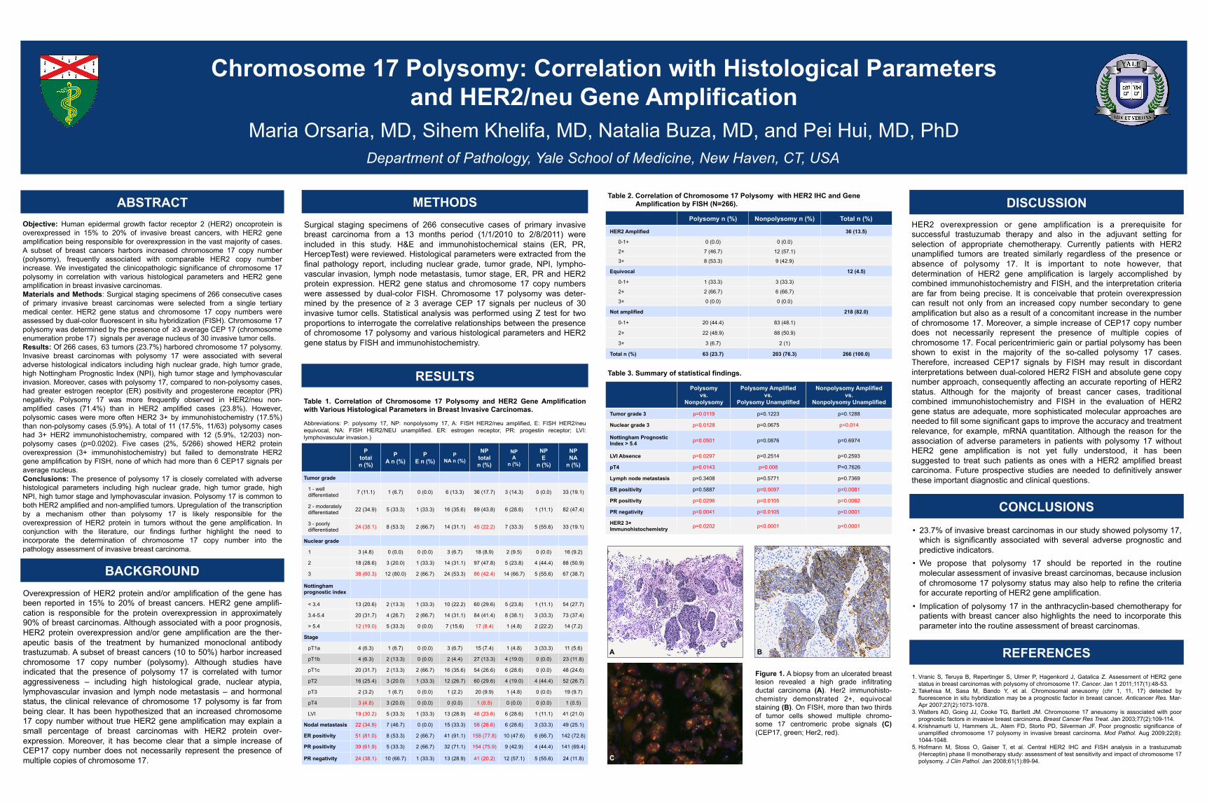
Figure 1. A biopsy from an ulcerated breast lesion revealed a high grade infiltrating ductal carcinoma (A). Her2 immunohisto-chemistry demonstrated 2+, equivocal staining (B). On FISH, more than two thirds of tumor cells showed multiple chromo-some 17 centromeric probe signals (C) (CEP17, green; Her2, red).
C
CONCLUSIONS • 23.7% of invasive breast carcinomas in our study showed polysomy 17,
which is significantly associated with several adverse prognostic and predictive indicators.
• We propose that polysomy 17 should be reported in the routine molecular assessment of invasive breast carcinomas, because inclusion of chromosome 17 polysomy status may also help to refine the criteria for accurate reporting of HER2 gene amplification.
• Implication of polysomy 17 in the anthracyclin-based chemotherapy for patients with breast cancer also highlights the need to incorporate this parameter into the routine assessment of breast carcinomas.
DISCUSSION
1. Vranic S, Teruya B, Repertinger S, Ulmer P, Hagenkord J, Gatalica Z. Assessment of HER2 gene status in breast carcinomas with polysomy of chromosome 17. Cancer. Jan 1 2011;117(1):48-53.
2. Takehisa M, Sasa M, Bando Y, et al. Chromosomal aneusomy (chr 1, 11, 17) detected by fluorescence in situ hybridization may be a prognostic factor in breast cancer. Anticancer Res. Mar-Apr 2007;27(2):1073-1078.
3. Watters AD, Going JJ, Cooke TG, Bartlett JM. Chromosome 17 aneusomy is associated with poor prognostic factors in invasive breast carcinoma. Breast Cancer Res Treat. Jan 2003;77(2):109-114.
4. Krishnamurti U, Hammers JL, Atem FD, Storto PD, Silverman JF. Poor prognostic significance of unamplified chromosome 17 polysomy in invasive breast carcinoma. Mod Pathol. Aug 2009;22(8):1044-1048.
5. Hofmann M, Stoss O, Gaiser T, et al. Central HER2 IHC and FISH analysis in a trastuzumab (Herceptin) phase II monotherapy study: assessment of test sensitivity and impact of chromosome 17 polysomy. J Clin Pathol. Jan 2008;61(1):89-94.
HER2 overexpression or gene amplification is a prerequisite for successful trastuzumab therapy and also in the adjuvant setting for selection of appropriate chemotherapy. Currently patients with HER2 unamplified tumors are treated similarly regardless of the presence or absence of polysomy 17. It is important to note however, that determination of HER2 gene amplification is largely accomplished by combined immunohistochemistry and FISH, and the interpretation criteria are far from being precise. It is conceivable that protein overexpression can result not only from an increased copy number secondary to gene amplification but also as a result of a concomitant increase in the number of chromosome 17. Moreover, a simple increase of CEP17 copy number does not necessarily represent the presence of multiple copies of chromosome 17. Focal pericentrimieric gain or partial polysomy has been shown to exist in the majority of the so-called polysomy 17 cases. Therefore, increased CEP17 signals by FISH may result in discordant interpretations between dual-colored HER2 FISH and absolute gene copy number approach, consequently affecting an accurate reporting of HER2 status. Although for the majority of breast cancer cases, traditional combined immunohistochemistry and FISH in the evaluation of HER2 gene status are adequate, more sophisticated molecular approaches are needed to fill some significant gaps to improve the accuracy and treatment relevance, for example, mRNA quantitation. Although the reason for the association of adverse parameters in patients with polysomy 17 without HER2 gene amplification is not yet fully understood, it has been suggested to treat such patients as ones with a HER2 amplified breast carcinoma. Future prospective studies are needed to definitively answer these important diagnostic and clinical questions.
P total n (%)
P A n (%)
P E n (%)
P NA n (%)
NP total n (%)
NP A
n (%)
NP E
n (%)
NP NA
n (%)
Tumor grade
1 - well differentiated 7 (11.1) 1 (6.7) 0 (0.0) 6 (13.3) 36 (17.7) 3 (14.3) 0 (0.0) 33 (19.1)
2 - moderately differentiated 22 (34.9) 5 (33.3) 1 (33.3) 16 (35.6) 89 (43.8) 6 (28.6) 1 (11.1) 82 (47.4)
3 - poorly differentiated 24 (38.1) 8 (53.3) 2 (66.7) 14 (31.1) 45 (22.2) 7 (33.3) 5 (55.6) 33 (19.1)
Nuclear grade
1 3 (4.8) 0 (0.0) 0 (0.0) 3 (6.7) 18 (8.9) 2 (9.5) 0 (0.0) 16 (9.2)
2 18 (28.6) 3 (20.0) 1 (33.3) 14 (31.1) 97 (47.8) 5 (23.8) 4 (44.4) 88 (50.9)
3 38 (60.3) 12 (80.0) 2 (66.7) 24 (53.3) 86 (42.4) 14 (66.7) 5 (55.6) 67 (38.7)
Nottingham prognostic index
< 3.4 13 (20.6) 2 (13.3) 1 (33.3) 10 (22.2) 60 (29.6) 5 (23.8) 1 (11.1) 54 (27.7)
3.4-5.4 20 (31.7) 4 (26.7) 2 (66.7) 14 (31.1) 84 (41.4) 8 (38.1) 3 (33.3) 73 (37.4)
> 5.4 12 (19.0) 5 (33.3) 0 (0.0) 7 (15.6) 17 (8.4) 1 (4.8) 2 (22.2) 14 (7.2)
Stage
pT1a 4 (6.3) 1 (6.7) 0 (0.0) 3 (6.7) 15 (7.4) 1 (4.8) 3 (33.3) 11 (5.6)
pT1b 4 (6.3) 2 (13.3) 0 (0.0) 2 (4.4) 27 (13.3) 4 (19.0) 0 (0.0) 23 (11.8)
pT1c 20 (31.7) 2 (13.3) 2 (66.7) 16 (35.6) 54 (26.6) 6 (28.6) 0 (0.0) 48 (24.6)
pT2 16 (25.4) 3 (20.0) 1 (33.3) 12 (26.7) 60 (29.6) 4 (19.0) 4 (44.4) 52 (26.7)
pT3 2 (3.2) 1 (6.7) 0 (0.0) 1 (2.2) 20 (9.9) 1 (4.8) 0 (0.0) 19 (9.7)
pT4 3 (4.8) 3 (20.0) 0 (0.0) 0 (0.0) 1 (0.5) 0 (0.0) 0 (0.0) 1 (0.5)
LVI 19 (30.2) 5 (33.3) 1 (33.3) 13 (28.9) 48 (23.6) 6 (28.6) 1 (11.1) 41 (21.0)
Nodal metastasis 22 (34.9) 7 (46.7) 0 (0.0) 15 (33.3) 58 (28.6) 6 (28.6) 3 (33.3) 49 (25.1)
ER positivity 51 (81.0) 8 (53.3) 2 (66.7) 41 (91.1) 158 (77.8) 10 (47.6) 6 (66.7) 142 (72.8)
PR positivity 39 (61.9) 5 (33.3) 2 (66.7) 32 (71.1) 154 (75.9) 9 (42.9) 4 (44.4) 141 (69.4)
PR negativity 24 (38.1) 10 (66.7) 1 (33.3) 13 (28.9) 41 (20.2) 12 (57.1) 5 (55.6) 24 (11.8)
Polysomy n (%) Nonpolysomy n (%) Total n (%)
HER2 Amplified 36 (13.5)
0-1+ 0 (0.0) 0 (0.0)
2+ 7 (46.7) 12 (57.1)
3+ 8 (53.3) 9 (42.9)
Equivocal 12 (4.5)
0-1+ 1 (33.3) 3 (33.3)
2+ 2 (66.7) 6 (66.7)
3+ 0 (0.0) 0 (0.0)
Not amplified 218 (82.0)
0-1+ 20 (44.4) 83 (48.1)
2+ 22 (48.9) 88 (50.9)
3+ 3 (6.7) 2 (1)
Total n (%) 63 (23.7) 203 (76.3) 266 (100.0)
Polysomy vs.
Nonpolysomy
Polysomy Amplified vs.
Polysomy Unamplified
Nonpolysomy Amplified vs.
Nonpolysomy Unamplified
Tumor grade 3 p=0.0119 p=0.1223 p=0.1288
Nuclear grade 3 p=0.0128 p=0.0675 p=0.014
Nottingham Prognostic Index > 5.4 p=0.0501 p=0.0876 p=0.6974
LVI Absence p=0.0297 p=0.2514 p=0.2593
pT4 p=0.0143 p=0.008 P=0.7626
Lymph node metastasis p=0.3408 p=0.5771 p=0.7369
ER positivity p=0.5887 p=0.0097 p=0.0081
PR positivity p=0.0296 p=0.0105 p=0.0062
PR negativity p=0.0041 p=0.0105 p<0.0001
HER2 3+ Immunohistochemistry p=0.0202 p<0.0001 p<0.0001
Abbreviations: P: polysomy 17, NP: nonpolysomy 17, A: FISH HER2/neu amplified, E: FISH HER2/neu equivocal, NA: FISH HER2/NEU unamplified. ER: estrogen receptor, PR: progestin receptor; LVI: lymphovascular invasion.)