Embed Size (px)
Citation preview
Chromatin Immunoprecipitation Protocol (Yeast) Harvesting Cells
1. Grow a 10 mL culture of yeast O/N at 37°C in YPD 2. The next day, inoculate a 50-100 mL of media in a 250 mL flask to an OD600 of ~0.3
a. Use selective media if maintaining a plasmid with a selective marker 3. Grow cells to an OD600 of 0.800-1.000
Fixing Cells
4. Slowly add 37% Paraformaldehyde (in hood) to the cells to a final concentration of 1% a. 1.35 mL for 50 mL sample b. 2.70 mL for 100 mL sample
5. Shake at RT for 15 minutes at 100 rpm in shaker 6. Add glycine to a final concentration of 125 mM and shake sample again for 5 minutes at RT 7. Spin cells down in tabletop centrifuge at 4°C for 5 minutes 8. Wash cells 2x with 30-40 mL of ice cold 1xTBS 9. Snap freeze cells
a. At this point you can freeze the cells O/N at -80°C or keep going Lysing the Cells
10. Resuspend the cells in 400 µl of ChIP lysis buffer for every 50 mL of sample grown 11. Weigh out 0.5 grams of acid washed beads in a 1.5 mL microcentrifuge tube and place
resuspended sample in tube 12. Bead beat (using Smth lab mini-bead beater in cold room) 8x at 40 seconds ON and 1 minute
OFF 13. Poke a hole in the bottom of the tube with a hot 25 gauge needle and place the tube in another
1.5 mL tube 14. Spin the tubes for 5 minutes at 5000 rpm at 4°C
a. You will have to leave the cap off of the rotor when spinning because the stacked tubes will not fit under the cap
15. Make sure all of the cells and lysate has passed into the new tube. If not, wash the beads with 100 µl of ChIP lysis buffer and spin into a new tube. You can recombine the wash with the previous lysate.
Sonicating the cells
16. Resuspend the cells/debris and lysate and replenish protease inhibitors 17. Split resuspension into new 1.5 mL tubes containing NO MORE THAN 300 µl of sample
a. For 50 mL, I usually get 2-3 tubes b. For 100 mL, I usually get 4 tubes
18. Place samples in Bioruptor rotors a. Each spot in the rotors should be filled (with blanks or samples) to give equal sonication
19. Sonicate using the Twin Bioruptor on HIGH 20x for 30 seconds ON/OFF a. I change the water bath once in between the cycles so the bath doesn’t get too warm b. This means I do 10x water change 10x
20. Spin down the samples at 4°C for 5 minutes at maximum speed 21. Transfer the supernatant to a new tube and repeat the spin for 15 minutes 22. Transfer the supernatant to a new tube
a. This is your lysate you will used for IP Pre-clearing the Lysate
23. Determine your protein concentration using a BCA kit and BSA standards
24. Measure out ~1.0 mg of protein and add ChIP lysis buffer so final volume is 500 µl a. I have found that ~250 µl of sample gives about 1.0 mg of protein b. I usually just measure the OD562 of the samples and use 250 µl of the most
concentrated sample and adjust accordingly for the others 25. Add ~40 µl of beads without antibody for preclearing
a. I use protein A beads for antibody/protein A IPs b. I use Glutathione Sepharose for TAP/IgG IPs
26. Rotate at 4°C for 2 hours Immunoprecipitation
27. Spin down samples at 1000 g for 2 minutes at 4°C 28. Transfer supernatant (DO NOT SUCK UP BEADS) to a new tube 29. Save 1/10th of the volume for input samples used in quantitative PCR
a. The volume should be ~50 µl b. Input can be kept at -20°C until reversing crosslinks
30. Add antibody or IgG beads and rotate O/N at 4°C a. Add 5 µg of primary antibody b. Add ~40-50 µl of IgG beads if ChIPping TAP tagged protein
31. The following day, add ~40-50 µl of protein A beads to antibody/lysate mixture if you are using a primary antibody
32. Rotate for 2 hours at 4°C Washing
33. Spin down the beads at 1000 g for 2 minutes at 4°C 34. Decant supernatant 35. Wash beads at 4°C in the following manner for 5 minutes each
a. 2x ChIP lysis buffer b. 1x IP1 c. 1x IP2 d. 1x TE e. 1x TE (at RT)
Reversing Crosslinking
36. Add 100 µl of elution buffer and incubate at 65°C for 15 minutes with brief vortexing every 5 minutes
37. Spin beads down at maximum speed and transfer elution to a new tube 38. Add ~70-80 µl to beads and repeat elution
a. Final elution volume should be 200 µl 39. DON’T FORGET! Add 150 µl of elution buffer to inputs from step 29 40. Leave samples O/N at 65°C
DNA Purification
41. Add 800 µl of binding buffer to samples and use Invitrogen PCR purification kit to isolate DNA 42. Measure DNA concentration of inputs
a. Mine are usually 40-100 ng/µl Quantitative PCR I prepare 1:10 dilutions of my inputs and use 2 µl of my samples and diluted inputs for each well. I make five 5-fold dilution standards with undiluted input. You can use any input for the standards. Below is an example of a plate with 2 primer sets and 2 samples (e.g. wild-type and mutant).Rows A
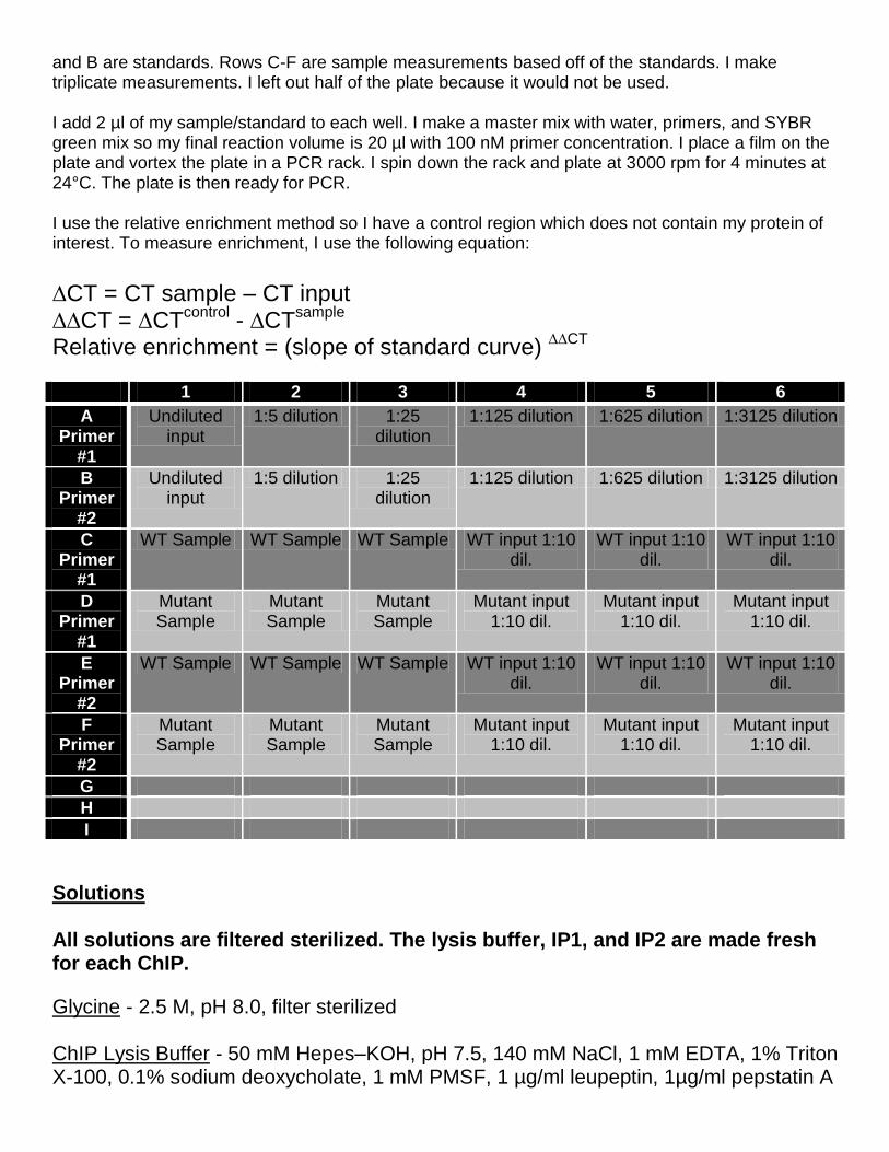
and B are standards. Rows C-F are sample measurements based off of the standards. I make triplicate measurements. I left out half of the plate because it would not be used. I add 2 µl of my sample/standard to each well. I make a master mix with water, primers, and SYBR green mix so my final reaction volume is 20 µl with 100 nM primer concentration. I place a film on the plate and vortex the plate in a PCR rack. I spin down the rack and plate at 3000 rpm for 4 minutes at 24°C. The plate is then ready for PCR. I use the relative enrichment method so I have a control region which does not contain my protein of interest. To measure enrichment, I use the following equation:
∆CT = CT sample – CT input ∆∆CT = ∆CT
control - ∆CT
sample
Relative enrichment = (slope of standard curve) ∆∆CT
1 2 3 4 5 6
A Primer
#1
Undiluted input
1:5 dilution 1:25 dilution
1:125 dilution 1:625 dilution 1:3125 dilution
B Primer
#2
Undiluted input
1:5 dilution 1:25 dilution
1:125 dilution 1:625 dilution 1:3125 dilution
C Primer
#1
WT Sample WT Sample WT Sample WT input 1:10 dil.
WT input 1:10 dil.
WT input 1:10 dil.
D Primer
#1
Mutant Sample
Mutant Sample
Mutant Sample
Mutant input 1:10 dil.
Mutant input 1:10 dil.
Mutant input 1:10 dil.
E Primer
#2
WT Sample WT Sample WT Sample WT input 1:10 dil.
WT input 1:10 dil.
WT input 1:10 dil.
F Primer
#2
Mutant Sample
Mutant Sample
Mutant Sample
Mutant input 1:10 dil.
Mutant input 1:10 dil.
Mutant input 1:10 dil.
G
H
I
Solutions All solutions are filtered sterilized. The lysis buffer, IP1, and IP2 are made fresh for each ChIP.
Glycine - 2.5 M, pH 8.0, filter sterilized ChIP Lysis Buffer - 50 mM Hepes–KOH, pH 7.5, 140 mM NaCl, 1 mM EDTA, 1% Triton X-100, 0.1% sodium deoxycholate, 1 mM PMSF, 1 µg/ml leupeptin, 1µg/ml pepstatin A
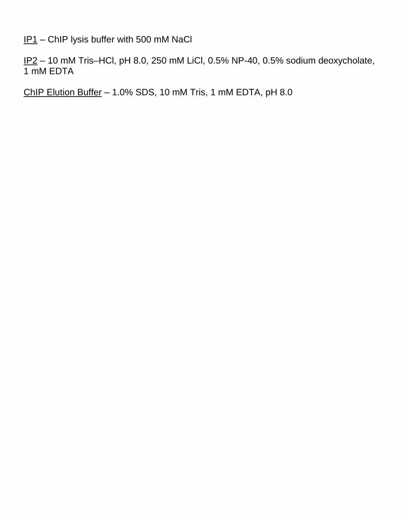
IP1 – ChIP lysis buffer with 500 mM NaCl IP2 – 10 mM Tris–HCl, pH 8.0, 250 mM LiCl, 0.5% NP-40, 0.5% sodium deoxycholate, 1 mM EDTA ChIP Elution Buffer – 1.0% SDS, 10 mM Tris, 1 mM EDTA, pH 8.0