Embed Size (px)
Citation preview
PROTOCOL: Chromatin Immunoprecipitation from Arabidopsis Tissues
Authors: Nobutoshi Yamaguchi, Cara M. Winter, Miin-Feng Wu, Chang Seob Kwon, Dilusha A.William, et. al.
Source: The Arabidopsis Book, 2014(12)
Published By: The American Society of Plant Biologists
URL: https://doi.org/10.1199/tab.0170
BioOne Complete (complete.BioOne.org) is a full-text database of 200 subscribed and open-access titles in thebiological, ecological, and environmental sciences published by nonprofit societies, associations, museums, institutions,and presses.
Your use of this PDF, the BioOne Complete website, and all posted and associated content indicates your acceptance ofBioOne’s Terms of Use, available at www.bioone.org/terms-o-use.
Usage of BioOne Complete content is strictly limited to personal, educational, and non - commercial use. Commercialinquiries or rights and permissions requests should be directed to the individual publisher as copyright holder.
BioOne sees sustainable scholarly publishing as an inherently collaborative enterprise connecting authors, nonprofit publishers, academic institutions,research libraries, and research funders in the common goal of maximizing access to critical research.
Downloaded From: https://bioone.org/journals/The-Arabidopsis-Book on 06 Jan 2020Terms of Use: https://bioone.org/terms-of-use
The Arabidopsis Book © 2014 American Society of Plant Biologists
First published on February 17, 2014: e0170. doi: 10.1199/tab.0170
PROTOCOLChromatin Immunoprecipitation from Arabidopsis Tissues
Nobutoshi Yamaguchi,a Cara M. Winter,a,b Miin-Feng Wu,a Chang Seob Kwon,a Dilusha A. William,a and Doris Wagnera,1
aDepartment of Biology, University of Pennsylvania, 415 S. University Avenue, Philadelphia, PA, USA 19104bDepartment of Biology and Duke Center for Systems Biology, 4128 French Family Science Center, Duke University, Durham, NC 277081 Address correspondence to [email protected]
The ability of proteins to associate with genomic DNA in the context of chromatin is critical for many nuclear processes includ-ing transcription, replication, recombination, and DNA repair. Chromatin immunoprecipication (ChIP) is a practical and useful technique for characterizing protein / DNA association in vivo. The procedure generally includes six steps: (1) crosslinking the protein to the DNA; (2) isolating the chromatin; (3) chromatin fragmentation; (4) imunoprecipitation with antibodies against the protein of interest; (5) DNA recovery; and (6) PCR identification of factor associated DNA sequences. In this protocol, we describe guidelines, experimental setup, and conditions for ChIP in intact Arabidopsis tissues. This protocol has been used to study association of histone modifications, of chromatin remodeling ATPases, as well as of sequence-specific transcription factors with the genomic DNA in various Arabidopsis thaliana tissues. The protocol described focuses on ChIP-qPCR, but can readily be adapted for use in ChIP-chip or ChIP-seq experiments. The entire procedure can be completed within 3 days.
I. INTRODUCTION
In eukaryotes, DNA is packaged into chromatin. Chromatin is composed of DNA, proteins, and RNA and is the substrate for nuclear processes such as transcription, replication, recombina-tion and repair (Rando and Ahmad, 2007; Margueron and Rein-berg 2010). Many proteins directly or indirectly contact genomic DNA in the context of chromatin. For example, gene expression is modulated by proteins with diverse functions such as structural proteins that make up the nucleosome, histone modification read-ers, enzymes modulating chromatin structure, sequence specific binding proteins and their cofactors, or the general transcriptional machinery (Li et al., 2007; Clapier and Cairns, 2009; Alabert and Groth 2012; Spitz and Furlong 2012).
To understand the contribution of these factors to the regula-tion of key nuclear processes, techniques that probe their associ-ation with the genomic DNA are of central importance. Several in vitro techniques, such as electrophoretic mobility shift assays or yeast one-hybrid assays are commonly used to determine DNA-protein interactions. However, in vitro interactions could be forced and may not reflect the situation in cells. Chromatin immunopre-cipitation (ChIP) represents a valuable alternative for probing such interactions in vivo (in isolated cells, tissues or entire seed-lings) under physiological conditions and to estimate the density of protein at specific sites (Kuo and Allis, 1999). In addition, this
technique allows investigation of change in protein association with the DNA in response to cue and the temporal relationship of this change relative to changes in transcription.
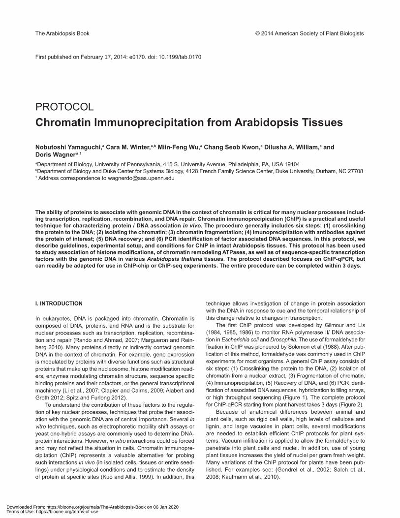
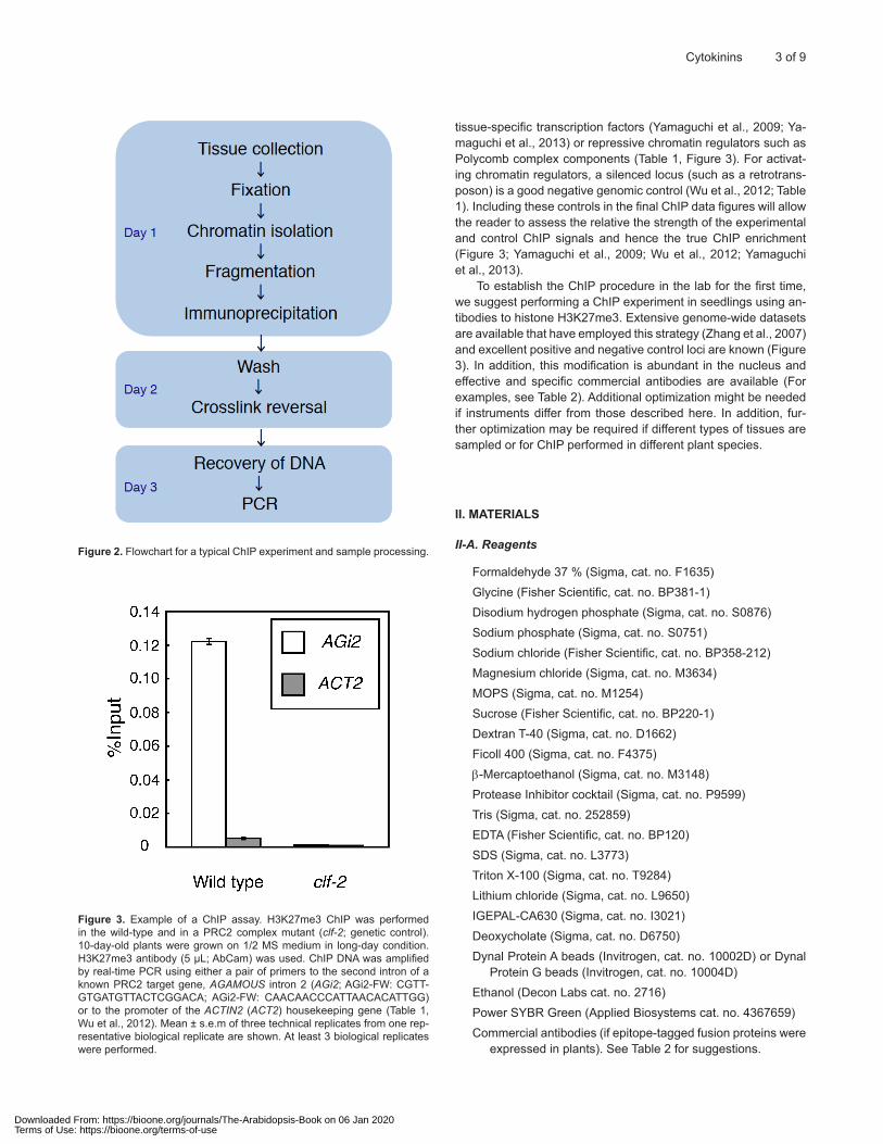
The first ChIP protocol was developed by Gilmour and Lis (1984, 1985, 1986) to monitor RNA polymerase II/ DNA associa-tion in Escherichia coli and Drosophila. The use of formaldehyde for fixation in ChIP was pioneered by Solomon et al (1988). After pub-lication of this method, formaldehyde was commonly used in ChIP experiments for most organisms. A general ChIP assay consists of six steps: (1) Crosslinking the protein to the DNA, (2) Isolation of chromatin from a nuclear extract, (3) Fragmentation of chromatin, (4) Immunoprecipitation, (5) Recovery of DNA, and (6) PCR identi-fication of associated DNA sequences, hybridization to tiling arrays, or high throughput sequencing (Figure 1). The complete protocol for ChIP-qPCR starting from plant harvest takes 3 days (Figure 2).
Because of anatomical differences between animal and plant cells, such as rigid cell walls, high levels of cellulose and lignin, and large vacuoles in plant cells, several modifications are needed to establish efficient ChIP protocols for plant sys-tems. Vacuum infiltration is applied to allow the formaldehyde to penetrate into plant cells and nuclei. In addition, use of young plant tissues increases the yield of nuclei per gram fresh weight. Many variations of the ChIP protocol for plants have been pub-lished. For examples see: (Gendrel et al., 2002; Saleh et al., 2008; Kaufmann et al., 2010).
Downloaded From: https://bioone.org/journals/The-Arabidopsis-Book on 06 Jan 2020Terms of Use: https://bioone.org/terms-of-use
2 of 9 The Arabidopsis Book
Figure 1. Outline of the ChIP procedure. After cross-linking with formaldehyde, the cells are lysed and the nuclei-containing fraction is isolated. After chromatin shearing, the samples are incubated with antibodies for immunoprecipitation. After crosslink reversal, PCR-ready DNA is collected. Binding of the protein of interest to genomic DNA is examined by real-time PCR.
Here, we provide guidelines and conditions for highly efficient and specific ChIP using Arabidopsis tissues. We have reduced the number of experimental steps required to shorten preparation time. We suggest that a well-controlled ChIP experiment requires two types of negative controls: a) a genetic control, b) a genomic control. For the former, we recommend use of null mutant plants, if gene-specific antibodies are employed (Wu et al., 2012), or use of a non-transformed parental line, if an epitope tagged protein is
used (Yamaguchi et al., 2013). These controls are more stringent than use of IgG antibody or no antibody, because the experimen-tal and control samples are subjected to identical conditions. For the genomic control, a locus unlikely to be bound by the factor of interest should be tested for each experimental and control ChIP reaction. Even if no prior ChIP data is available for the factor of interest, suitable genomic controls can be chosen. For example, housekeeping genes can serve as genomic controls for both
Downloaded From: https://bioone.org/journals/The-Arabidopsis-Book on 06 Jan 2020Terms of Use: https://bioone.org/terms-of-use
Cytokinins 3 of 9
tissue-specific transcription factors (Yamaguchi et al., 2009; Ya-maguchi et al., 2013) or repressive chromatin regulators such as Polycomb complex components (Table 1, Figure 3). For activat-ing chromatin regulators, a silenced locus (such as a retrotrans-poson) is a good negative genomic control (Wu et al., 2012; Table 1). Including these controls in the final ChIP data figures will allow the reader to assess the relative the strength of the experimental and control ChIP signals and hence the true ChIP enrichment (Figure 3; Yamaguchi et al., 2009; Wu et al., 2012; Yamaguchi et al., 2013).
To establish the ChIP procedure in the lab for the first time, we suggest performing a ChIP experiment in seedlings using an-tibodies to histone H3K27me3. Extensive genome-wide datasets are available that have employed this strategy (Zhang et al., 2007) and excellent positive and negative control loci are known (Figure 3). In addition, this modification is abundant in the nucleus and effective and specific commercial antibodies are available (For examples, see Table 2). Additional optimization might be needed if instruments differ from those described here. In addition, fur-ther optimization may be required if different types of tissues are sampled or for ChIP performed in different plant species.
II. MATERIALS
II-A. Reagents
Formaldehyde 37 % (Sigma, cat. no. F1635)Glycine (Fisher Scientific, cat. no. BP381-1)Disodium hydrogen phosphate (Sigma, cat. no. S0876)Sodium phosphate (Sigma, cat. no. S0751)Sodium chloride (Fisher Scientific, cat. no. BP358-212)Magnesium chloride (Sigma, cat. no. M3634)MOPS (Sigma, cat. no. M1254)Sucrose (Fisher Scientific, cat. no. BP220-1)Dextran T-40 (Sigma, cat. no. D1662)Ficoll 400 (Sigma, cat. no. F4375)b-Mercaptoethanol (Sigma, cat. no. M3148)Protease Inhibitor cocktail (Sigma, cat. no. P9599)Tris (Sigma, cat. no. 252859)EDTA (Fisher Scientific, cat. no. BP120)SDS (Sigma, cat. no. L3773)Triton X-100 (Sigma, cat. no. T9284)Lithium chloride (Sigma, cat. no. L9650)IGEPAL-CA630 (Sigma, cat. no. I3021)Deoxycholate (Sigma, cat. no. D6750)Dynal Protein A beads (Invitrogen, cat. no. 10002D) or Dynal
Protein G beads (Invitrogen, cat. no. 10004D)Ethanol (Decon Labs cat. no. 2716)Power SYBR Green (Applied Biosystems cat. no. 4367659)Commercial antibodies (if epitope-tagged fusion proteins were
expressed in plants). See Table 2 for suggestions.
Figure 2. Flowchart for a typical ChIP experiment and sample processing.
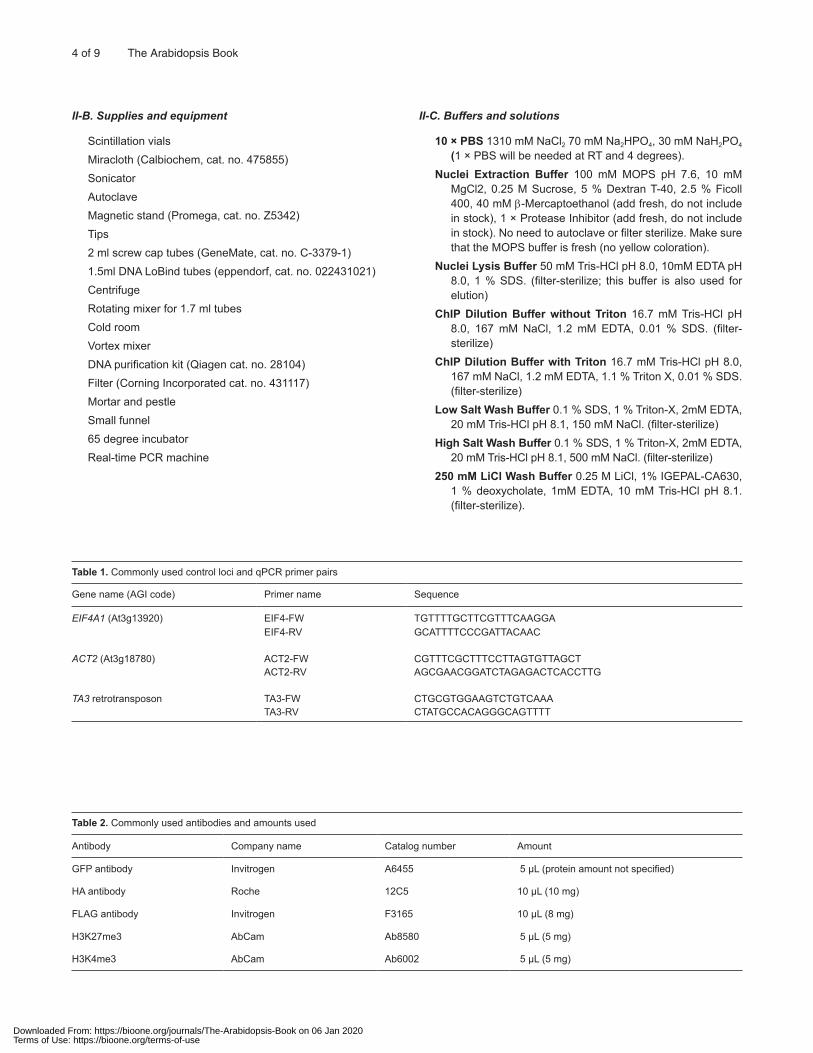
Figure 3. Example of a ChIP assay. H3K27me3 ChIP was performed in the wild-type and in a PRC2 complex mutant (clf-2; genetic control). 10-day-old plants were grown on 1/2 MS medium in long-day condition. H3K27me3 antibody (5 µL; AbCam) was used. ChIP DNA was amplified by real-time PCR using either a pair of primers to the second intron of a known PRC2 target gene, AGAMOUS intron 2 (AGi2; AGi2-FW: CGTT-GTGATGTTACTCGGACA; AGi2-FW: CAACAACCCATTAACACATTGG) or to the promoter of the ACTIN2 (ACT2) housekeeping gene (Table 1, Wu et al., 2012). Mean ± s.e.m of three technical replicates from one rep-resentative biological replicate are shown. At least 3 biological replicates were performed.
Downloaded From: https://bioone.org/journals/The-Arabidopsis-Book on 06 Jan 2020Terms of Use: https://bioone.org/terms-of-use
4 of 9 The Arabidopsis Book
Table 1. Commonly used control loci and qPCR primer pairs
Gene name (AGI code) Primer name Sequence
EIF4A1 (At3g13920) EIF4-FW TGTTTTGCTTCGTTTCAAGGAEIF4-RV GCATTTTCCCGATTACAAC
ACT2 (At3g18780) ACT2-FW CGTTTCGCTTTCCTTAGTGTTAGCTACT2-RV AGCGAACGGATCTAGAGACTCACCTTG
TA3 retrotransposon TA3-FW CTGCGTGGAAGTCTGTCAAATA3-RV CTATGCCACAGGGCAGTTTT
Table 2. Commonly used antibodies and amounts used
Antibody Company name Catalog number Amount
GFP antibody Invitrogen A6455 5 µL (protein amount not specified)
HA antibody Roche 12C5 10 µL (10 mg)
FLAG antibody Invitrogen F3165 10 µL (8 mg)
H3K27me3 AbCam Ab8580 5 µL (5 mg)
H3K4me3 AbCam Ab6002 5 µL (5 mg)
II-B. Supplies and equipment
Scintillation vials Miracloth (Calbiochem, cat. no. 475855)Sonicator AutoclaveMagnetic stand (Promega, cat. no. Z5342)Tips2 ml screw cap tubes (GeneMate, cat. no. C-3379-1)1.5ml DNA LoBind tubes (eppendorf, cat. no. 022431021)CentrifugeRotating mixer for 1.7 ml tubesCold roomVortex mixerDNA purification kit (Qiagen cat. no. 28104)Filter (Corning Incorporated cat. no. 431117)Mortar and pestleSmall funnel65 degree incubatorReal-time PCR machine
II-C. Buffers and solutions
10 × PBS 1310 mM NaCl2 70 mM Na2HPO4, 30 mM NaH2PO4 (1 × PBS will be needed at RT and 4 degrees).
Nuclei Extraction Buffer 100 mM MOPS pH 7.6, 10 mM MgCl2, 0.25 M Sucrose, 5 % Dextran T-40, 2.5 % Ficoll 400, 40 mM b-Mercaptoethanol (add fresh, do not include in stock), 1 × Protease Inhibitor (add fresh, do not include in stock). No need to autoclave or filter sterilize. Make sure that the MOPS buffer is fresh (no yellow coloration).
Nuclei Lysis Buffer 50 mM Tris-HCl pH 8.0, 10mM EDTA pH 8.0, 1 % SDS. (filter-sterilize; this buffer is also used for elution)
ChIP Dilution Buffer without Triton 16.7 mM Tris-HCl pH 8.0, 167 mM NaCl, 1.2 mM EDTA, 0.01 % SDS. (filter-sterilize)
ChIP Dilution Buffer with Triton 16.7 mM Tris-HCl pH 8.0, 167 mM NaCl, 1.2 mM EDTA, 1.1 % Triton X, 0.01 % SDS. (filter-sterilize)
Low Salt Wash Buffer 0.1 % SDS, 1 % Triton-X, 2mM EDTA, 20 mM Tris-HCl pH 8.1, 150 mM NaCl. (filter-sterilize)
High Salt Wash Buffer 0.1 % SDS, 1 % Triton-X, 2mM EDTA, 20 mM Tris-HCl pH 8.1, 500 mM NaCl. (filter-sterilize)
250 mM LiCl Wash Buffer 0.25 M LiCl, 1% IGEPAL-CA630, 1 % deoxycholate, 1mM EDTA, 10 mM Tris-HCl pH 8.1. (filter-sterilize).
Downloaded From: https://bioone.org/journals/The-Arabidopsis-Book on 06 Jan 2020Terms of Use: https://bioone.org/terms-of-use
Cytokinins 5 of 9
III. PROCEDURE
III-A. Tissue collection and fixation Duration: 1 h
1*. Harvest 300-600 mg of plant tissues into a 25 mL scintillation vial containing 10 mL of 1 × PBS solution. Preferably young and healthy tissue should be used.NOTES
2*. Remove 1 × PBS with 10 mL pipette and replace with 10 mL room temperature 1 % formaldehyde solution in 1× PBS (270 µL 37 %) formaldehyde per 10 mL 1 × PBS) to cross-link tissue. NOTES
CAUTION: Formaldehyde is toxic; work in the fume hood.
3*. Vacuum infiltrate the tissue until it sinks (3 times for seedlings; 3-5 times for inflorescences), not exceeding 15 min total ex-posure time to formaldehyde. A typical seedling vacuum in-filtration: pre-incubate in the fume hood: 4 min; first vacuum: 1.5 min (or stop when bubbles start to boil over); second or more vacuum: 1 min (bubbles should slow down by the end of vacuum time); post-incubate in hood: ~ 3 min). NOTES
4. Remove formaldehyde in hood with 10 mL pipette and add 10 mL 0.125 M glycine solution (quenches the cross-linker). Vacuum once for ~1.5 min. The total glycine incubation time is 5 min.
5. Remove glycine in hood and rinse 2 times with 10 mL cold 1 × PBS solution on the lab bench (PBS should be cold to preserve cross-linking).
6*. Dry plant tissues briefly (blot on paper towel), weigh (should be 300-600 mg), and insert into 2 mL tubes. Freeze in liquid nitrogen. NOTES
STOPPING POINT: The tissue can be stored at -80 ºC for up to 3 months.
NOTES AND TROUBLESHOOTING
N1. For samples that trap air between tissues (like the flower bearing stem of Arabidopsis plants called the inflorescence), we incubate the sample in PBS at room temperature for 2-3 h to ensure that most airspaces are occupied by liquid. This step is not necessary for leaves, stems, roots, or seedlings.
300 mg of tissue is sufficient for one ChIP reaction using either entire seedlings or dissected inflorescences.
N2. The formaldehyde needs to be fresh. Prepare the PBS form-aldehyde solution immediately prior to use.
N3. Tissue needs to be submerged in the solution before starting the vacuum infiltration and throughout the infiltration.
Vacuum infiltration allows the formaldehyde to penetrate into plant cells.
Tissues should sink to the bottom of the vials after the sec-ond vacuum release. If they still float, vacuum again.
N6. Do not freeze wet samples as ice will reduce the grinding efficiency.
III-B. Nuclei isolation and shearing of chromatin Duration: 2 h
7. Add Protease Inhibitors (from 100 × stock) and b-Me (3.12 µL /mL) to Nuclei Extraction Buffer. Make 3.0 mL per sample.
CAUTION: b-Me-containing solution; work in the fume hood.
8. Grind the samples to a fine powder using a mortar and pes-tle, keeping the tissue frozen throughout. After completion, add 2.5 mL nuclei extraction buffer with protease inhibitors and b-ME and keep grinding until the tissue is thawed.
9. Set up a small funnel over a 50 mL Falcon tube with two layers of Miracloth. Pass the filtrate through the Miracloth twice and transfer the filtrate to a fresh 2 mL tube. Be sure to squeeze the Miracloth after the second pass-through to extract all of the liquid (change gloves after each sample). Keep samples on ice until all samples have been processed.
10. Spin for 5 min at 10,000 g at 4 ºC. Remove the supernatant and resuspend the pellet in 75 µL nuclei lysis buffer. This will require stirring with a pipette tip. Try to avoid foaming.
11. Leave sitting on ice for 30 min with occasional stirring. Sub-sequently bring up to a final volume of 700 µL with ChIP dilu-tion buffer without Triton (add 625 µL). Transfer the solution to a new 1.5 mL tube.
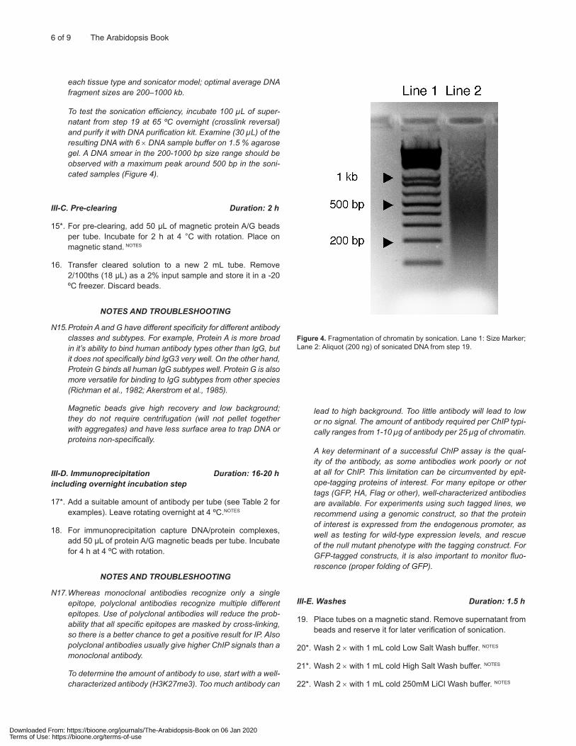
12*. Sonicate five times for 10 seconds each (~ P3.2) with a Fisher Dismembrator or other sonicator (the size range of sheared DNA should be around 200-1000 bp; See Figure 4). Incubate the samples on ice for 50 sec between sonications to allow them to cool. NOTES
13. Transfer the sonicated solution to a new 2 mL tube. A typical yield after sonication is 665 µL. Add 200 µL ChIP dilution buf-fer with Triton X-100 and 35 µL of 22 % Triton X-100 to attain 900 µL of sample with a 1.1 % Triton X-100 concentration.
14. Spin at full speed (13,200 rpm) for 5 min at 4 °C. Remove from the centrifuge immediately, and transfer the clean su-pernatant to a new 2 mL tube. Repeat this step one more time to remove as much of the pellet (debris) as possible.
NOTES AND TROUBLESHOOTING
N12. If samples get too hot, the proteins will be denatured. Try to avoid foaming. The formation of bubbles will reduce the sonication efficiency. Make sure the sonicator probe tip does not touch the side of the tube.
Fragmentation efficiency determines the ChIP resolution. Sonication conditions must be determined empirically for
Downloaded From: https://bioone.org/journals/The-Arabidopsis-Book on 06 Jan 2020Terms of Use: https://bioone.org/terms-of-use
6 of 9 The Arabidopsis Book
each tissue type and sonicator model; optimal average DNA fragment sizes are 200–1000 kb.
To test the sonication efficiency, incubate 100 µL of super-natant from step 19 at 65 ºC overnight (crosslink reversal) and purify it with DNA purification kit. Examine (30 µL) of the resulting DNA with 6 × DNA sample buffer on 1.5 % agarose gel. A DNA smear in the 200-1000 bp size range should be observed with a maximum peak around 500 bp in the soni-cated samples (Figure 4).
III-C. Pre-clearing Duration: 2 h
15*. For pre-clearing, add 50 µL of magnetic protein A/G beads per tube. Incubate for 2 h at 4 °C with rotation. Place on magnetic stand. NOTES
16. Transfer cleared solution to a new 2 mL tube. Remove 2/100ths (18 µL) as a 2% input sample and store it in a -20 ºC freezer. Discard beads.
NOTES AND TROUBLESHOOTING
N15. Protein A and G have different specificity for different antibody classes and subtypes. For example, Protein A is more broad in it’s ability to bind human antibody types other than IgG, but it does not specifically bind IgG3 very well. On the other hand, Protein G binds all human IgG subtypes well. Protein G is also more versatile for binding to IgG subtypes from other species (Richman et al., 1982; Akerstrom et al., 1985).
Magnetic beads give high recovery and low background; they do not require centrifugation (will not pellet together with aggregates) and have less surface area to trap DNA or proteins non-specifically.
III-D. Immunoprecipitation Duration: 16-20 h including overnight incubation step
17*. Add a suitable amount of antibody per tube (see Table 2 for examples). Leave rotating overnight at 4 ºC.NOTES
18. For immunoprecipitation capture DNA/protein complexes, add 50 µL of protein A/G magnetic beads per tube. Incubate for 4 h at 4 ºC with rotation.
NOTES AND TROUBLESHOOTING
N17. Whereas monoclonal antibodies recognize only a single epitope, polyclonal antibodies recognize multiple different epitopes. Use of polyclonal antibodies will reduce the prob-ability that all specific epitopes are masked by cross-linking, so there is a better chance to get a positive result for IP. Also polyclonal antibodies usually give higher ChIP signals than a monoclonal antibody.
To determine the amount of antibody to use, start with a well-characterized antibody (H3K27me3). Too much antibody can
lead to high background. Too little antibody will lead to low or no signal. The amount of antibody required per ChIP typi-cally ranges from 1-10 µg of antibody per 25 µg of chromatin.
A key determinant of a successful ChIP assay is the qual-ity of the antibody, as some antibodies work poorly or not at all for ChIP. This limitation can be circumvented by epit-ope-tagging proteins of interest. For many epitope or other tags (GFP, HA, Flag or other), well-characterized antibodies are available. For experiments using such tagged lines, we recommend using a genomic construct, so that the protein of interest is expressed from the endogenous promoter, as well as testing for wild-type expression levels, and rescue of the null mutant phenotype with the tagging construct. For GFP-tagged constructs, it is also important to monitor fluo-rescence (proper folding of GFP).
III-E. Washes Duration: 1.5 h
19. Place tubes on a magnetic stand. Remove supernatant from beads and reserve it for later verification of sonication.
20*. Wash 2 × with 1 mL cold Low Salt Wash buffer. NOTES
21*. Wash 2 × with 1 mL cold High Salt Wash buffer. NOTES
22*. Wash 2 × with 1 mL cold 250mM LiCl Wash buffer. NOTES
Figure 4. Fragmentation of chromatin by sonication. Lane 1: Size Marker; Lane 2: Aliquot (200 ng) of sonicated DNA from step 19.
Downloaded From: https://bioone.org/journals/The-Arabidopsis-Book on 06 Jan 2020Terms of Use: https://bioone.org/terms-of-use
Cytokinins 7 of 9
23*. Wash 2 × with 1 mL cold 0.5 × TE. NOTES
24. Pipette off all liquid and add 50 µL Nuclei Lysis Buffer.
25. Incubate for 30 min at 65 ºC in large incubator in Eppendorf tubes to separate the sample from the beads.
26. Remove and store the elution buffer, add another 50 µL elu-tion buffer to beads and repeat heating and suspension. Combine both sets of elution buffer. Discard beads.
NOTES AND TROUBLESHOOTING
N20-23. Rotate all washes in a rotator for 5 min at 4 ºC. Betweenwashes place tubes on the magnetic stand until all magnetic beads have collected at the side of the tube and remove solution. Discard all washes.
III-F. Reverse crosslinking Duration: overnight
27. Add 82 µL Nuclei Lysis Buffer to reserved Input samples (Step 16) to bring them up to 100 µL.
28. Add 6 µL of 5 M NaCl to both Input and ChIP samples. Re-verse crosslinks overnight at 65 ºC.
III-G. DNA purification Duration: 1 h
29. Add 5 volumes (550 µL) Buffer PB to each tube.
30. Shake samples for 30 min at room temperature.
31. Load QIAquick spin columns with samples and centrifuge for 1 min at 13,200 rpm.
32. Reload flow-through and spin again. Discard flow-thru.
33. Add 750 µL Buffer PE to column and centrifuge 1 min at 13,200 rpm.
34. Discard flow-thru and spin again 1 min at 13,200 rpm.
35. Place column in a clean 1.5 mL tube and add 100 µL 0.5 × EB Buffer to center of column. Let sit 5 min.
36. Centrifuge at 13,200 rpm 1 min.
37. Reload flow-thru, let sit 5 min, and spin again for 1 min. Keep eluate.
STOPPING POINT: The purified ChIP sample can be stored at -20 ºC for at least
one month. Do not dilute prior to storage.
III-H. Quantitative PCR and data analysis Duration: 3 h
38*. Proceed to quantitative PCR using gene specific primers. NOTES
PCR is carried out in a final volume of 25 µL containing
12.5 µL Power SYBR Green PCR Master Mix (2×)
2 µL ChIP DNA template
0.5 µL primers (forward and reverse, 10 µM each)
9.5 µL sterile ddH2O.
The PCR products are amplified with the following condi-tions: 95 4 ºC for 10 min; 40 cycles of 95 4 ºC for 15 sec and 60 4 ºC for 1 min (Cycling stage); 95 4 ºC for 15 sec, 60 4 ºC for 1 min, and 95 4 ºC for 15 sec (Melting Curve Stage). At least three technical replicates are needed. We include a 5-fold dilution series of input to be amplified by the same primer set to calculate % input. Four or more input dilutions are recommended. The eukaryotic initiation factor (EIF4), ACTIN2, and TA3 are often used negative controls. For posi-tive controls, use published data or test several candidate genes that could be regulated by the protein of interest. At least three independent biological replicate ChIP experi-ments should be performed. NOTES
NOTES AND TROUBLESHOOTING
N38. Test your primers on genomic DNA for efficient amplification prior to using them on ChIP reactions.
IV. OPTIMIZATION
If your ChIP-qPCR signal is very low relative to that obtained for input DNA (see ANTICIPATED RESULTS below), optimization of the ChIP protocol is required.
IV-A. Low abundance of transcription factor (Step 1, Step 35-37)
If your transcription factor of interest is low abundance (lower than 1 in 500,000 cDNA), we recommend using 600 mg tissues (more than 600 mg causes increased background) and we suggest pre-paring 2 biological replicates. After step 34, the two samples will be combined into one by using the eluate from one spin column for dual elution of the second sample.
IV-B. Insufficient reversal of the crosslink and subsequent loss of protein-associated DNA (Step 3)
A nice test for full crosslink reversal is the FAIRE (Formaldehyde Assisted Isolation of Regulatory Elements; Simon et al., 2012) method. Briefly, this technique relies on phenol chloroform ex-traction of sheared crosslinked DNA; only DNA not associated with proteins is recovered. Equal amounts of sheared genomic DNA, sheared crosslinked DNA and sheared crosslinked DNA after crosslink reversal should be subjected to FAIRE. The ex-pected result is near 100 % recovery of the sheared genomic DNA and the sheared crosslinked DNA after crosslink reversal. By contrast the recovery of sheared crosslinked DNA should be very low. DNA recovery can be measured directly (by Qubit) or by qPCR of a single copy locus.
Downloaded From: https://bioone.org/journals/The-Arabidopsis-Book on 06 Jan 2020Terms of Use: https://bioone.org/terms-of-use
8 of 9 The Arabidopsis Book
IV-C. Sonication efficiency (Step 12)
(See Figure 4). The sonicated DNA should be a smear with a ca. 500 bp maximum signal intensity. If the sonication was insuf-ficient, you will see broader binding peak.
IV-D. Efficient IP (Step 17)
One reason for the low ChIP signal could be that insufficient an-tibody was used or that the antibody was of poor quality. Alterna-tively, the protein of interest may not be expressed at high levels in the tissue tested. ChIP reactions should be repeated with different amounts of antibody and antibody from different lots and providers. Of note, individual lots of polyclonal antisera can differ greatly in avidity and specificity, even from the same provider. This website reports functional testing antibodies commonly used in epigenetic research: http://compbio.med.harvard.edu/antibodies/. If the anti-body to be tested also recognizes a protein that is more abundant than the protein of interest (for example both share the same epit-ope tag), we recommend first optimizing the antibody amount and source first with the more abundant protein. It is a good idea to test for protein recovery after IP by Western blotting and to check how much of the protein of interest remains in the supernatant after IP. To enrich for factors only present in a small number of cells, enhanced ChIP signal can be achieved by dissecting the tissue of interest, by using a genetic background that enriches for the tissue of interest (Kwon et al., 2005; Wu et al., 2012), or by employing cell or nuclei sorting (Birnbaum et al., 2005; Deal and Henikoff, 2010).
IV-E. Fresh solutions or reagents (Step 1-38)
Presence of a faulty solution or reagent besides the antibody of choice can significantly reduced ChIP efficacy (especially, formal-dehyde). In this case, we recommend the user repeat a ChIP that has worked in the past (such as the H3K27me3 ChIP in Figure 3) and test if the same % input enrichment is observed.
IV-F. Optimize qPCR primer sets (Step 38)
In order to obtain high quality qPCR data, the primer sets need to meet several criteria (e.g., primer Tm = 60 ± 5 ºC, length 18 to 25 bases, GC content between 40 and 60 %). If the primer sets are not optimized, this may result in amplification artifacts and/or inaccurate quantification. To design primer sets, we recommend use of Primer3 (bioinfo.ut.ee/primer3-0.4.0/). Primer sets should be tested by performing qPCR with sonicated input DNA. The amplification efficiency can be improved considerably if the length of the amplified fragment is between 60 and 150 bp. The ChIP-qPCR should be in the linear range of amplification. To confirm that the primer sets amplify a single fragment of the correct size it is recommended to run the qPCR product on an agarose gel.
IV-G. Data normalization (Step 38)
The normalization methods often used for ChIP assay are (I) % in-put, and (II) fold enrichment. (I) In the % input method, the qPCR sig-nals derived from the ChIP samples are divided by the qPCR signals derived from the input sample taken early during the ChIP proce-dure. Normalizing over input amplification gives a reliable readout of the ChIP efficacy. We recommend using this normalization method.
(II) If the ChIP signals for the genetic controls vary greatly between the different primer sets used, it may be advisable to use the fold en-richment method. In this method the signal obtained for the genetic control ChIP is set to one and the signal for the experimental ChIP is expressed as fold-increase above the genetic control signal.
V. ANTICIPATED RESULTS
Using this protocol, we obtain approximately 15 ng of good-qual-ity genomic DNA from 300 to 500 mg of fresh tissue per immu-noprecipitated sample. This amount of ChIP DNA is sufficient for 50 10 µL real-time PCRs, which allows us to monitor as many as 15 genomic regions plus the control locus in triplicate. The expected % input yield differs for the different types of factors that contact the genomic DNA. In general, we expect ca. 0.1-1 % in-put recovery for histone modification ChIP (Figure 3), 0.05-0.3 % input for transcription factors, and 0.009-0.002 % input for chro-matin regulators (such as SPLAYED or BRAHMA). The difference in recovery is due to several variables, which include cell-type specificity of the factor of interest, its abundance in the nucleus, its proximity to the DNA, and its residence time on the DNA. This protocol has been used to perform ChIP-PCR, ChIP-qPCR, and ChIP-chip (William et al., 2004; Yamaguchi et al., 2009; Winter et al., 2011; Yamaguchi et al., 2013). We expect that it can be easily adapted for ChIP-seq.
ACKNOWLEDGEMENTS
We acknowledge the members of the Wagner lab for constructive discus-sions during the method development. We thank the members of the Penn Genomics Frontiers Institute ChIP class taught by N.Y. and D.W. for the data used to generate Figure 3. This research was supported by NSF grants IOS- 1257111 and NIH grant R01 GM64650-01.
REFERENCES
Akerstrom, B., Brodin, T., Reis, K., and Bjork, L. (1985). Protein G: A powerful tool for binding and detection of monoclonal and polyclonal antibodies. J. Immunol. 135: 2589-2592.
Alabert, C., and Groth, A. (2012). Chromatin replication and epigenome maintenance. Nat. Rev. Genetics 13: 153-167.
Birnbaum, K., Jung, J.W., Wang, J.Y., Lambert, G.M., Hirst, J.A., Gal-braith, D.W., and Benfey, P.N. (2005). Cell type-specific expression profiling in plans via cell sorting of protoplasts from fluorescent reporter lines. Nat. Methods 2: 615-619.
Clapier, C.R., and Cairns, B.R. (2009). The biology of chromation remod-eling complexes. Annu. Rev. Biochem. 78: 273-304.
Deal, R.B. and Henikoff, S. (2010). The INTACT method for cell type-specific gene expression and chromatin profiling in Arabidopsis thali-ana. Nat. Protoc. 6: 56-68.
Gilmour, D.S., and Lis, J.T. (1984). Detecting protein-DNA interactions in vivo: Distribution of RNA polymerase on specific bacterial genes. Proc. Natl. Acad. Sci. 81: 4275-4279.
Gilmour,D.S., and Lis, J.T. (1985). In vivo interactions of RNA polymerase II with genes of Drosophila melanogaster. Mol. Cell Biol. 5: 2009–2018.
Gilmour, D.S., and Lis, J.T. (1986). RNA polymerase II interacts with the
Downloaded From: https://bioone.org/journals/The-Arabidopsis-Book on 06 Jan 2020Terms of Use: https://bioone.org/terms-of-use
Cytokinins 9 of 9
promoter region of the noninduced hsp70 gene in Drosophila melano-gaster cells. Mol. Cell Biol. 6: 3984–3989.
Kaufmann, K., Muino, J.M., Osteras,M., Farinelli, L., Krajewsli, P., and Angenent, G.C. (2010). Chromatin immunoprecipitation (ChIP) of plant transcription factors followed by sequencing (ChIP-SEQ) or hybridiza-tion to whole genome arrays (ChIP-CHIP). Nat. Protoc. 5: 457-472.
Kuo, M.H., and Allis, C.D. (1999). In vivo cross-linking and immunopre-cipitation for studying dynamic protein: DNA associations in a chromatin environment. Methods 19: 425-433.
Kwon, C.S., Chen, C., and Wagner, D. (2005). WUSCHEL is a primary target for transcriptional regulation by SPLAYED in dynamic control of stem cell fate in Arabidopsis. Genes Dev. 19: 992-1003.
Li, B., Carey, M., and Workman, J.L. (2007). The role of chromatin during transcription. Cell 128: 707-719.
Margueron, R., and Reinberg, D. (2010). Chromatin structure and the inheritance of epigenetic information. Nat. Rev. Genet. 11: 285-296.
Rando, O.J., and Ahmad, K. (2007). Rules and regulation in the primary structure of chromatin. Curr. Opin. Cell Biol. 19: 250-256.
Richman, D.D., Cleveland, P.H., Oxman, M.N., and Johnson, K.M. (1982). The binding of staphylococcal protein A by the sera of different animal species. J. Immunol. 128: 2300-2305.
Saleh, A., Alvarez-Venegas, R., and Avramova Z. (2008). An efficient chromatin immunoprecipitation (ChIP) protocol for studying histone modifications in Arabidopsis plants. Nat. Protoc. 3: 1018-1025.
Simon, J.M., Giresi, P.G., Davis, I.J., and Lieb, J.D. (2012). Using fol-maldehyde-assisted isolation of regulatory elements (FAIRE) to isolate
active regulatory DNA. Nat. Protoc. 19: 256-267.Solomon, M.J., Larsen, P.L., Varshavsky, A. (1988). Mapping protein-
DNA interactions in vivo with formaldehyde: Evidence that histone H4 is retained on a highly transcribed gene. Cell 53: 937–947.
Winter, C.M., Austin, R.S., Blanvillain-Baufume, S., Reback, M.A., Monniaux, M., Wu, M.F., Sang, Y., Yamaguchi, A., Yamaguchi, N., Parker, J.E., Parcy, F., Jensen, S.T., Li, H., and Wagner, D. (2011). LEAFY Target Genes Reveal Floral Regulatory Logic, cis Motifs, and a Link to Biotic Stimulus Response. Dev. Cell 20: 430-443.
Wu, M-F., Sang, Y., Bezhani, S., Yamaguchi, N., Han S.K., Li, Z., Su, Y., Slewinski, T.L., and Wagner, D. (2012). SWI2/SNF2 Chromatin Re-modelers Overcome Polycomb Repression and Control Floral Organ Identity Together with LEAFY and SEPALLATA3. Proc. Natl. Acad. Sci. USA 109: 3576-3581.
Yamaguchi, A., Wu, M.F., Yang, L., Wu, G., Poethig, R.S., and Wagner, D. (2009). The microRNA-regulated SBP-Box transcription factor SPL3 is a direct upstream activator of LEAFY, FRUITFULL, and APETALA1. Dev. Cell 17: 268-278.
Yamaguchi, N., Wu, M-F., Winter, C., Berns, M., Nole-Wilson, S., Ya-maguchi, A., Coupland, G., Krizek, B., and Wagner, D. (2013). A mo-lecular framework for auxin-mediated initiation of floral promordia. Dev. Cell 24: 271-282.
Zhang, X., Clarenz, O., Cokus, S., Bernatavichute, Y.V., Pellegrini, M., Goodrich, J., and Jacobsen, S.E. (2007). Whole-genome analysis of Histone H3 Lysine 27 Trimethylation in Arabidopsis. PLoS Biol. 5: e129.
Downloaded From: https://bioone.org/journals/The-Arabidopsis-Book on 06 Jan 2020Terms of Use: https://bioone.org/terms-of-use