Embed Size (px)
Citation preview

PERSPECTIVES
Chromatin Conformation of Yeast Centromeres
KERRY S. BLOOM, ENRIQUE AMAYA, JOHN CARBON,* LOUISE CLARKE,* ALISON HILL, and ELAINE YEH Department of Biology, University of North Carolina, Chapel Hill 27514; and *Department of Biological Sciences, University of California, Santa Barbara 93106
ABSTRACT The centromere region of Saccharomyces cerevisiae chromosome III has been replaced by various DNA fragments from the centromere regions of yeast chromosomes III and Xl. A 289-base pair centromere (CEN3) sequence can stabilize yeast chromosome III through mitosis and meiosis. The orientation of the centromeric fragments within chromosome III has no effect on the normal mitotic or meiotic behavior of the chromosome. The structural integrity of the centromere region in these genomic substitution strains was examined by mapping nucleolytic cleavage sites within the chromatin DNA. A nuclease-protected centro- mere core of 220-250 base pairs was evident in all of the genomic substitution strains. The position of the protected region is determined strictly by the centromere DNA sequence. These results indicate that the functional centromere core is contained within 220-250 base pairs of the chromatin DNA that is structurally distinct from the flanking nucleosomal chro- matin.
A mechanism to transmit genetic material to daughter cells during cell division is required by both procaryotic and eu- caryotic organisms. Although the mitotic apparatus may range in complexity from a simple DNA-membrane attachment in procaryotes to the highly elaborate processes of the eucaryotic spindle, one common feature is the establishment of a specific DNA locus that interacts with the segregation mechanism.
In procaryotes, plasmid systems have proved the most tractable in identifying DNA sequences involved in segrega- tion phenomena. The par locus is a cis-acting DNA element needed to promote the equipartition of replicating plasmid DNA molecules in Escherichia coli (l, 2). The par sequences are functional in either orientation in the plasmid molecule and par loci from different plasmids are interchangeable (2, 3).
The experimental strategy for isolation of DNA elements required for the proper segregation of eucaryotic chromo- somes also depends upon the relative simplicity of plasmid systems. The yeast Saccharomyces cerevisiae can be trans- formed with plasmid molecules containing a selectable genetic marker and DNA sequences providing autonomous replica- tion. These molecules can be followed through many succes- sive generations of mitotic cell division, and upon sporulation of the diploid yeast host, plasmid segregation can be followed through meiosis. Plasmids containing a selectable genetic marker plus an autonomous replication sequence are not efficiently distributed from mother to daughter cells during mitosis (4) and are rapidly lost from the population. Frag-
ments of chromosomal DNA isolated by virtue of their ability to confer mitotic stability on these plasmids map to the centromere region of yeast chromosomes (5). The centromere DNAs (CEN) isolated from five chromosomes in yeast, CEN3( 5), CEN4(6), CEN5(7), CEN6(8), and CENl l(9), have the common ability to stabilize autonomously replicating plasmids through mitosis, and direct the segregation of plas- mid molecules in a predominantly Mendelian fashion through meiosis.
The nucleotide sequences of DNA fragments carrying CEN3(10), CEN4 (Mann, C., and R. Davis, Stanford Univer- sity, personal communication), CEN6(8), and CENll(IO) have been determined. Sequence comparison reveals that short regions of DNA are conserved in their nucleotide se- quence and spatial arrangement in the centromere regions from the different chromosomes (Fig. 1). Sequence element III (11 base pairs [bp] ~ is completely homologous in CEN3 and CEN11 and exhibits strong homology (10/11 bp) to similar elements in CEN4 and CEN6. An extremely (A+T)- rich region (>93% A+T, element II) spanning 82-89 bp occurs immediately adjacent to element III in all the centro- meres. The element II region is flanked on the other side by sequence element I (14 bp), which is completely homologous in CEN3 and CENll, and exhibits partial homology in CEN4
Abbreviations used in this paper: bp, base pair; kb, kilobase pair; SPCM, standard digestion buffer as described in Materials and Meth- ods.
THE JOURNAL OF CELL BIOLOGY • VOLUME 99 NOVEMBER 1984 1559-1568 © The Rockefeller University Press • 0021-9525/84/11/1559/10 $1.00 1559
on Novem
ber 14, 2007 w
ww
.jcb.orgD
ownloaded from

CEN J
T ]1 ]]1
ATAAGTCACATGAT TGATTTCCGAA TATT CAGTGTACTA 'a ~ bp (93%ATl PACTAAAGG CTT
CEN 4 AAAGGTCACATGCT TGATTACCGAA TTTCCAGTGTACGA 'Q 82 bp(93 %AT) m, ACTAATG GC T T
CEN I I ATAAGTCACATGAT TATTCAGTGTACTA !
I ATAAGTAAAATAAT TTTCATCACGTGCT
CEN~ T A T T C A T T T T A T T A " - 4 O b P ~ A A A G T A G T G CACGA 9
• • • • A&& • •
TGATTTCCGAA 89 bp(94 %AT) ~ACTAAAGGCTT
89 bp (94 %AT)
(11/14 bp) and CEN6 (11/14 and 8/14 bp). Deletion of the sequence element I-III region completely inactivates the cen- tromere (11), indicating that these sequences comprise all or an important part of the functional yeast centromere.
A more detailed description of the eucaryotic centromere requires an assay that is not encumbered by problems that may be unique to a plasmid molecule. For example, the centromeric plasmids do not segregate with the fidelity of the parental chromosomes; the frequency of centromeric plasmid loss is about once in every 100 cell divisions (5), whereas mitotic chromosomes are lost about once in every 50,000 cell divisions (12). A recent development in the yeast system allows specific DNA sequences, isolated and manipulated in vitro, to be directed into the yeast cell to substitute for sequences normally occurring within the host genome (13). By using this technique, a series of genomic substitution strains were constructed by replacing the host centromere region in chromosome III with altered DNA fragments (14). When the 624-bp CEN3 fragment is deleted from chromo- some III, an acentric chromosome results and is rapidly lost from the population. Thus the CEN3 fragment is required to stabilize the entire yeast chromosome. Chromosome stability is recovered when the centromere DNAs from chromosomes III (624-bp fragment) or XI (858-bp fragment), are substituted in either orientation for the CEN3 region in chromosome III. The resulting chromosome segregates normally through mi- tosis and meiosis in a manner identical to that of the normal chromosome III. These results indicate the yeast centromeres are fully functional in either orientation and are not necessar- ily chromosome specific. Thus the CEN sequences in eucary- otes, as well as the par loci in procaryotes, function as auton- omous units stabilizing the plasmid or chromosome in which they reside.
To begin to understand how the centromere DNA interacts with the segregation machinery, we have initiated studies on the conformation of the centromere DNA sequences in the yeast chromosomes. The DNA in eucaryotic cells is wrapped around histone core particles to create a periodic array of 146- bp nucleosomal subunits and 20-50-bp linker sequences. A consequence of the histone-core linker organization of DNA in chromatin is that the linker DNA is more accessible to nucleolytic cleavage than is the DNA in the nucleosomal core particles. Mild digestion of chromatin with the enzyme, mi- crococcal nuclease, generates a series of nucleoprotein parti- cles containing DNA fragments whose molecular weights are multiples of the basic nucleosomal subunit. Digestion of the chromatin DNA from yeast with micrococcal nuclease reveals this typical nucleosomal subunit structure. Analysis of cen- tromeric chromatin, in contrast, indicates that the centromere DNA sequences are in a nuclease-protected structure that
1560 THE JOURNAL OF CELL BIOLOGY . VOLUME 99, 1984
TGTTTTCCGAA BACAAAAGGCTT
FIGURE I Regions of sequence homology between CEN3(IO), CEN4 (C. Mann and R. Davis, Stanford University, personal communication), CEN6(8), and CEN11 (I0) in the yeast, Saccharomyces cerevisiae. The sequence elements I-Ill are arranged in an almost identical spatial arrangement in the centro- meres of four different chromosomes. The regions of nonhomology are indicated by the arrowheads below the nucleotide pair
encompasses 220-250 base pairs of DNA (15). This protected region is distinct from the nucleosomal core particle and maps to the sequence element I-III region in both chromosomes III and XI. This region of structural differentiation is associated with the centromere sequences whether they are present within the yeast genome or on autonomously replicating plasmids. We now demonstrate that the protected centromere core seen in the parental chromosomes and on centromeric plasmid molecules is determined strictly by the element I-III DNA sequences, and includes these sequences regardless of where they are positioned in the genome. This protected 220- 250 bp of centromeric chromatin may therefore be the fun- damental structural unit of the yeast centromere.
MATERIALS AND METHODS
Yeas t Strains: s. cerevisiae genomie substitution strains were con- structed as previously described (14) by transformation of strain SB9882-4 (a trpl-289 ura3-52 leu2-3 leu2-112 his4-519/ct trpl-289 ura3-52 canl) with the appropriate centromere-substitution fragments diagrammed in Fig. 3 (g.v.). The haploid strains used for the chromatin-mapping experiments were derived by sporulation of the resulting stable URA3 + diploid transformants. The haploid genotypes are: SB303-6A(3C), a URA3+(III) ura3(V) trpl leu2 his4; SB303- 4A(2C), a URA3+(III) ura3(V) trpl leu2 his4; SB311-9B(1A), a URA3+(III) ura3(V) trpl leu2 his4; SB311-11A(10D), a URA3+(III) ura3(V) trpl LEU2 + HIS4 + can; SB303-14(29D), a URA3÷(III) ura3(V) trpl leu2 his4; SB303- 12(2B), a URA3+(III) ura3(V) trpl leu2 his4. Yeast strain X2180a was obtained from the Yeast Genetic Stock Center, University of California, Berkeley. Yeast stain ]17 (a his2 adel trpl metl4 ura3) has been described previously (9). A proteinase deficient strain 20B-12~, carrying the pep4-3 mutation (16), was used for preparing the protein extracts in DNA-binding experiments.
Isolation and Digestion of Yeast Nuclei: Yeast cells were grown in rich media containing 1% yeast extract, 2% bacto-peptone, and 2% glucose. Cells were harvested in mid-logarithmic growth phase, washed, and converted to spheroplasts by treatment with I% Glusulase (DuPont Pharmaceuticals, Wilmington, DE), as described by Forte and Fangman (17). Nuclei were isolated from spheroplasts as described by Nelson and Fangman (18). The nuclei were resuspended in standard digestion buffer (SPCM) that contains 1 M sorbitol, 20 mM 1,4-piperazone-diethanesulfonic acid (pH 6.3), 0.1 mM CaCI2, 0.5 mM MgCI2, and I mM phenylmethylsulfonyl fluoride. The suspension was pre- warmed to 32*(= for 3 rain, and DNAase I (5 tLg/ml) was added for the times (in minutes) indicated in the text. After incubation, samples were adjusted to 1% SDS, 1 M NaC1, and 20 mM EDTA to stop the digestion.
Preparation and Analysis of DNA: DNA was extensively depro- teinized and treated with RNase A as described by Bloom and Carbon (15). DNA samples were digested with restriction enzymes according to the specifi- cations provided by the suppliers. DNA fragments were analyzed on 1.4% agarose slab gels containing 0.09 M Tris-borate (pH 8.3) and 2.5 mM EDTA. To visualize unique DNA sequences, DNA fragments were transferred to nitrocellulose filters (19) and hybridized to radiolabcled DNA probes as de- scribed previously (15). Autoradiography was performed for 24-72 h at -80"C with Kodak XAR-5 film (Eastman Kodak Co., Rochester, NY) and a Du Pont Cronex Lightning-Plus intensifying screen (DuPont Instruments, Wilmington, DE).
Isolation and Analysis of DNA-binding Proteins: Extracts used for isolation of centromere-binding proteins were prepared from nuclei isolated from 200 ml of mid-logarithmic growth-phase cultures of yeast strain
on Novem
ber 14, 2007 w
ww
.jcb.orgD
ownloaded from

20B-12a as described above. Nuclei were resuspended in 2 ml of SPCM and sonicated using three 15-s bursts with intermittent cooling to an average double- stranded DNA length of 1,000 base pairs, as determined by agarose gel dectro- phoresis. The samples were diluted to 5 ml with 10 mM sodium phosphate (pH 7.0) and centrifuged at 20,000 g for 15 rain. The solubilized ehromatin supernatant was applied to a hydroxyapatite column equilibrated with 10 mM sodium phosphate (pH 7.0). The proteins were selectively dissociated by during the column with solutions containing increasing concentrations of NaCI as described by Bloom and Anderson (20). Centrnmere-binding proteins were eluted with 2 M NaCI and dialyzed against 10 mM Tris-HCl (pH 7.0), 50 mM NaC1, 5 mM MgCI2, and 0.1 mM EDTA. Proteins were reconstituted with restriction digested yeast genomic DNA for 45 min at 32°C. After incubation the samples were diluted to 500 al in dialysis buffer and passed through a nitrocellulose filter. The filters were sequentially washed with 0.4 M NaCI and 1.0 M NaCI followed by 1.0 M NaC1 in the presence of 1% SDS as described by Bloom et al. (11). The eluants were precipitated with ethanol, and the pelleted DNAs were redissolved and electrophoresed on a 1.4% agarose gel. Specific DNA fragments were visualized after Southern blot transfer and hybridization as described above.
RESULTS
Chrornatin Structure of Yeast Centromeres We have previously assayed the structure of centromeric
regions in chromatin from yeast chromosomes III and XI by examining the susceptibility of centromeric DNA to nucleo- lyric cleavage (15). Here, we have utilized the enzyme, DNAase I, to map specific nuclease cutting sites in the cen- tromeric region of chromosome IV in yeast (Fig. 2). To determine if the enzyme is actually recognizing structural parameters of the chromatin template, rather than specific DNA sequences, we have also examined the cutting sites in the centromeric region of protein-free chromosomal DNA. Chromatin DNA from yeast nuclei was partially digested with DNAase I, before (Fig. 2, chromatin) or after (Fig. 2, naked DNA) extraction of chromosomal proteins. The purified DNA samples were digested to completion with a restriction enzyme, XhoI, which cleaves at a fixed site close to the sequence element I-III region of CEN4 (6) (Fig. 2, left). The lengths of the sequences that hybridize to a radiolabeled probe extending from the restriction site towards the centromere therefore provide a direct map of the points of nucleolytic cleavage within the chromatin or DNA fiber relative to the restriction site (21).
The most striking feature of the fragment pattern shown in Fig. 2 is the 220-250-bp nudease-resistant region of DNA, occurring between 720 and 950 bp in a centromere-proximal direction from the XhoI site (open arrow in Fig. 2). The molecular weight standards on the gel (Fig. 2, lane MW) confirm the restriction map of the chromosome, and allow the nuclease cleavage sites to be mapped relative to the DNA sequence. Strong nuclease cleavage sites occur on both sides of the region of sequence elements I-II1, leaving it in a protected region of ~220-250 bp. These results indicate the regions of highest sequence homology, elements I-III, are protected in a centromere core particle. In the control lanes (Fig. 2, naked DNA), there were no specific nuclease cutting sites visualized. The protected region therefore reflects chro- matin components associated with the centromere DNA in the yeast cell nucleus. A similar protected region was also found around the centrornere region in chromosomes III and XI, and was determined to include sequence elements I-III (15). In fact, no matter where we map along the chromatin fiber in the centromere regions of chromosomes III, IV, and XI, we find a protected region of 220-250 bp which includes sequence elements I and III.
FIGURE 2 Mapping nuclease-sensitive sites on centromeric chro- matin from yeast chromosome IV. Nuclei were prepared from yeast strain J17 and digested with DNAase I (5 #g/ml) for the times (in minutes) indicated, as described in Materials and Methods. For the experiments with naked DNA, nuclei were prepared as described for the chromatin digests, but immediately before nuclease cleav- age, the DNA was extensively deproteinized. The naked DNA samples were resuspended in SPCM and digested with DNAase I (50 ng/ml) for the times (in minutes) indicated. After partial DNAase I cleavage, DNA samples were incubated in the presence (+), or absence (-) of Xhol, and electrophoresed on a 1.4% agarose gel. The DNAs were transferred to nitrocellulose filters and hybridized to the radiolabeled 603-bp probe from chromosome IV that extends from the Xhol site (large arrow) toward the CEN4 region (elements I-III). At left is a partial restriction-site map of the centromere region of yeast chromosome IV (6). The elements of sequence homology (I-Ill) between the different centromeres (Fig. 1) are indicated by darkened boxes. Sequence element IV is homologous to similar elements in the other centromeres, but is expendable with respect to active CEN function. Molecular weight markers (MW) indicate yeast nuclear DNA fragments cut with Xhol, and XhoI-Hpal. These fragments contain regions complementary to the radiolabeled probe (6). Restriction enzyme sites are Xhol (x).
Genomic Substitution of Yeast Centromeres To define the functional boundaries of the centromere in
the chromosome, we have utilized a unique property of the yeast system that allows replacement of DNA sequences in the host chromosome (13). DNA fragments constructed in vitro were introduced into yeast by transformation (14). The transforming DNA fragments were directed into the centro- meric region of chromosome III by virtue of the DNA se- quence homology between their free ends and regions flanking
BLOOM ET AL. Structure ot a Yeast Centromere 156"[
on Novem
ber 14, 2007 w
ww
.jcb.orgD
ownloaded from

the centromere in chromosome III (Fig. 3, regions A and B). The internal portion of each transforming fragment contains a genetically selectable marker, URA3 +, and an inverted, deleted, or foreign centromeric DNA sequence. A number of these transforming fragments containing different internal sequences are diagrammed in the inset to Fig. 3. These include the 624-bp CEN3 or 858-bp CEN11 fragments, either prop- erly oriented or inverted, or a 289-bp fragment containing CEN3, including sequence elements I-III, in either orienta- tion.
When diploid yeast cells are transformed with any one of these fragments, a recombination event occurs between re- gions A and B from the transforming fragment and the corresponding regions in the host chromosome. Using a ge-
a chromosome !i l CEN3
!li.l t t c ~" 0.63 1.9
netic selection for the URA3 + gene, the transformants with a genomic substitution in one copy of chromosome III are identified. When a fragment lacking the 624-bp CEN3 se- quence replaces CEN3 in one of the host chromosomes no. III, an acentric chromosome results and thus, missing a spindle attachment site, is rapidly lost from the population, presumably through mitotic nondisjunction (Table I and reference 14). Thus the centromere sequence is required to stabilize the entire yeast chromosome. If the 624-bp CEN3 or 858-bp CEN11 fragments, in either orientation, replaces CEN3 in the host chromosome, the substituted chromosome behaves completely normally in both mitosis and meiosis (Table I and reference 14).
The fragment-mediated transformation system has recently
--- B -"
'. x 0.9 1.6
b Transforming fragment
-, A
I 1.9
chromosome II I
' ~ ~ I - - ( A ) . . . . . o
CEN3
S
x I 0.9 1.6
Centromere fragment plasmid
I I I I 063 zx . . . . . (B) p J C 3 0 3 - 4
I I I I o pJC303-6 0.63
I I I I .~ pJC311-9 0 0.89 CEN11 I III
o pJC311-11 A 089
I I I I f p J C 3 0 3 - 1 4 ~ 0.29
CEN3 I I I I pJC303-12 0.29
URA3 CEN
1.9 1.1 0 .9 1.6 FIGURE 3 Schematic representation of the centromere region of yeast chromosome III and conversion of this region in the genomic substitution strains. (a) The physical map gives the location of the 624-bp CEN3 fragment (darkened line) along the wild- type yeast chromosome III. Restriction sites are EcoRI (I), BamHI (LX), Hindlll (x), and selected A/ul (~,), Rsal ('[), and Sau3A (0) sites. Numbers denote kilobase pairs (kb). The flanking regions A (1.9 kb) and B (2.5 kb) are as indicated (5). Panel b illustrates the genomic substitution fragments that are utilized in construction of the CEN3 substitution strains (14). The transforming fragment contains a selectable genetic marker, URA3, regions A and B flanking the centromere in chromosome III, and any one of the centromere fragments in the inset at right. These insertions include the 624-bp CEN3 fragment in either orientation (IC303- 4, JC303-6), the 858-bp CEN11 fragment in either orientation (JC311-9, JC311-11), and the 289-bp RsaI-Alul CEN3 fragment in either orientation (JC303-12, IC303-14). Roman numerals above the centromere fragments give the orientation of centromere sequence elements I and III. Transformation of diploid yeast strains with EcoRI-restricted pJC303-4, pjC303-6, plC311-9, plC311- 11, p/C303-12, and pjC303-14 DNAs yield conversion constructions 303-4, 303-6, 311-9, 311-11, 303-12, and 303-14, which include at the CEN3 region URA3 plus the various CEN fragments in the orientations as shown.
1562 THE JOURNAL OF CELL BIOLOGY • VOLUME 99, 1984
on Novem
ber 14, 2007 w
ww
.jcb.orgD
ownloaded from

been used to more accurately define the structural features necessary for proper functioning of CEN3. Various restriction fragments contained within the 624-bp CEN3 BamHI-Sau3A fragment were used individually or in various combinations as substitutes for the 624-bp CEN3 region in the genome (22). For example, cleavage of the 624-bp fragment with both RsaI and AluI yields a 289-bp segment that extends from a point 4 bp immediately to the left of element I through elements I- III, and ends 172 bp to the right of element III (see Fig. 3 b). Substitution of the 624-bp CEN3 sequence in the genome with this 289-bp fragment effectively deletes sequences occur- ring on both sides of the key element I-III region. Plasmid pJC303-14 contains the 289-bp sequence in the same orien- tation as occurs in the yeast genome, whereas the orientation is reversed in pJC303-12. Plasmid pJC303-12 and pjC303-14 DNAs were individually cleaved with EcoRI, and the resulting DNA fragments were used to transform diploid yeast strain
TABLE I
Chromosome III Nondisjunction in Strains with Altered Centromeres
Sequences at centro- meres III of diploid
Frequency of appear-
ance of competent a or a cells
Structural alteration of (per cell di- CEN3 (orientation) vision)
XIO s
CEN3/CEN3(SB9882-4) Wild-type 0.7 CEN3/CEN3(SB9882-4CR) Wild-type 0.1 303-4/CEN3 624 bp CEN3 (correct) 0.9 303-6/CEN3 624 bp CEN3 (reverse) 0.3 303-9/CEN3 858 bp CEN11 (reverse) 1.1 303-11/CEN3 858 bp CEN11 (correct) 0.6 303-12/CEN3 289 bp CEN3 (reverse) 0.2 303-14/CEN3 289 bp CEN3 (correct) 0.5
The observed frequencies of appearance of mating-competent cells represent the sum of mitotic gene conversion and recombination at the MAT locus, plus nondisjunction of chromosome Ill to form 2n-1 cells. Some of these data are taken from reference 14. See that reference for a more detailed description of experimental methods.
SB9882-4CR (see the legend to Table II for genotype) to URA3 ÷. The transformant colonies (~ 1,000 per microgram of transforming DNA) were normal in appearance and growth rate, and the Ura ÷ phenotype was mitotically stable. After growth of individual transformants in nonselective media for several generations, no Ura- cells could be detected among the 1,000 cells that were examined. The rate of loss of chro- mosome IlI in these transformants was determined by scoring for the number of mating-competent cells in a growing pop- ulation. The loss of one copy of chromosome III in an a/a diploid (sterile) results in a competent mater, because the mating type locus (MATa or MATa) is located on that chromosome (see reference 14 for details). As shown in Table I, the chromosome III loss rate in these transformants was no greater than 10 -5 per cell division, a value not significantly different from that obtained with the untransformed parent diploid strain.
The meiotic behavior of the altered chromosomes no. III in the transformants of the 303-12 and 303-14 classes was examined by the classical methods of yeast genetic analysis. The diploid transformants were induced to sporulate, the individual spores in the tetrads were separated by microdis- section, and the resulting haploids were scored for the distri- bution of appropriate genetic markers. In addition, haploid progeny containing the altered CEN3 region (Ura3 ÷) were back-crossed to examine the effect of having two altered CEN3 regions in opposition. The results of these meiotic analyses, summarized in Table II, indicate normal behavior of the chromosome III containing the 289-bp CEN3 fragment in place of the normal 624-bp sequence. Spore viability was uniformly high (>90% in most cases), and the URA3 marker on the altered chromosome III segregated as a centromere- linked gene, tightly linked to LEU2 on chromosome III. Recombination frequencies between markers on chromosome II1, and the number of gene conversions observed also fell within normal ranges. Finally, the predicted structure of the DNA in the CEN3 region in the URA3 haploids was verified by using a standard genomic Southern blot hybridization analysis (data not shown, see reference 14 for details).
The results described above suggest the functional centro- mere to be completely contained within the 289-bp RsaI-AluI
TABLE II
Meiotic Behavior of Chromosome III Containing a 289-bp CEN3 Substitution
Spore Tetrads viabil-
a /a diploid strain scored ity HIS4-LEU2
Apparent map distance Gene conversions
LEU2-URA3 LEU2-CEN3 URA3-MAT HIS4 LEU2 MAT
n %
303-14/CEN3 33 95 303-121CEN3 41 98
Back-crosses 303-14(29D) X BF305-18A 22 91 303-14(38A) x 303-14(18B) 26 95 303-12(2A) x 303-14(18B) 13 77
Literature values
centimorgans %
21 9 33 10 6 0 20 10 45 18 6 0
5 0 32 9 0 0 21 9 31" 8 8 0 29 8 21" 0 8 0
(LEU2-MAT) 9.9-19.9 3.4-11.6 20-23.1 8.4 2.5 0.6
(LEU-2-CEN3) (CEN3-MAT)
The URA3/ura3 heterozygous diploids, 303-14/C£N3 and 303-121CEN3, were obtained by transformation of yeast strain SB9882-4CR (a ura3-52 tripl-289 leu2- 3 leu2-112 his4-519 cryl/a ura3-52 trpl-289 canl) with the centromere-substitution EcoRI fragments as described in the text. The 303-14 construction contains the URA3 gene plus the 289 bp RsaI-Alul CEN3 fragment in the correct orientation, whereas the 303-12 construction contains theCEN3 sequence inserted in the orientation opposite from that seen in normal chromosome III. The back-cross diploids were constructed by mating selected Ura+ haploid progeny from the first two groups of tetrads. The genotypes are: 303-14 (29D), a URA3(III) ura3(V) trpl leu2 his4; BF305-18A, a arg5,6 ura3 metl4 ariel; 303-14(38A), a URA3(III) ura3(V) trpl ; 303-14(18B), a URA3(III) ura3(V) trpl leu2 his 4 metl4 adel ; 303-12(2A), a URA(III) ura3(V) trial. As expected, UR, A3+ segregated 2+:2- always in sister spores in all tetrads from the first three diploids (URA3 thus maps 0 cM from CEN3). In the homozygous URA3 back-cross diploids, URA3 segregated 4+:0- in all tetrads. For experimental details, see reference 14.
BLOOM [T AL. Structure of a Yeast Centromere 1 5 6 3
on Novem
ber 14, 2007 w
ww
.jcb.orgD
ownloaded from

fragment. Sequences within flanking regions A and B (Fig. 3) may be required, but need not be contiguous with the cen- tromere core.
Chromat in Structure of the Al tered CEN3 Regions
We have examined the chromatin conformation associated with the altered centromeres in the genomic substitution strains described above. Chromatin was isolated from the haploid genomic substitution strains with CEN3, CEN11, or the 289-bp CEN3 fragments in either orientation in chromo- some III (Fig. 3 b), and was partially digested with the nucleo- lyric enzyme, DNAase I, as described above. The DNA was purified and cut to completion with the restriction enzyme, HindlII. The DNA fragments were separated on agarose gels, transferred to nitrocellulose, and probed with a radiolabeled
DNA sequence originating from the HindlII site in region B (Fig. 3 a) and extending 900 bp toward the centromere region. This region remains intact in all of the genomic substitutions shown in Fig. 3, thereby enabling the same mapping strategy to be employed for each strain.
The chromatin mapping results from the genomic substi- tution strains are shown in Fig. 4. The fragment patterns obtained by using chromatin from strains containing the 624- bp CEN3 fragment in the proper orientation (strain 303-4, left) and an inverted orientation (strain 303-6, right) are shown in Fig. 4 a. The orientation of the CEN fragments can be visualized by the change in position of selected restriction sites. In strain 303-4, the BaraHI site to the element III side of the 624-bp CEN3 fragment occurs 900 bp from the HindlII restriction site, whereas when the 624-bp CEN3 fragment is inverted (strain 303-6), the BamHI site is 1,500 bp from the
FIGURE 4 Mapping nuclease-sensitive sites on the centromere chromatin from various genornic substitution strains. Nuclei were prepared from genomic substitution strains 303-4 and 303-6 (a); 31 I-9 and 311-11 (b); and 303-12 and 303-14 (c). Chromatin DNA and naked, deproteinized DNA were digested with DNAase I for the times (in minutes) indicated, as described in Fig. 2 and Materials and Methods. The DNA fragments were purified and incubated in the presence (+) or absence (-) of Hindlll. Samples were electro- phoresed on a 1.4% agarose gel, blotted, and hybridized to a 900- bp radiolabeled probe originating from the Hindlll site (large arrow) and extending toward the centromere region, elements I-III. The 900-bp probe is the centromere proximal fragment of region B shown in Fig. 3. The relevant portions of each restriction-site map for the substitution strains are shown to the side of the appropriate autoradiograph. The centromere fragment is denoted by the dark- ened line. The regions of centromere sequence homology, ele- ments I-III, are indicated by darkened boxes. Molecular weight markers (MW) were prepared from DNA isolated from each strain and digested with Hindlll-BamHl, Hindlll, and BamHI-EcoRl for 303-4 and 303-6, Hindlll-BamHl, Hindlll, and BamHI-EcoRl for 31 I-9, Hindlll-BamHl and BamHI-EcoRl for 311-11, Hindlll and
EcoRl for 303-I 2, and Hind lll-BamHl and Barn HI-EcoRI for 303-14. The size of these DNA fragments serves to confirm the actual restriction map of the genomic substitution chromosomes in the yeast cell. Restriction sites are BamHl (A), Hindlll (x), and selected Sau3A (O) sites.
1564 THE JOURNAL OF CELL BIOLOGY . VOLUME 99, 1984
on Novem
ber 14, 2007 w
ww
.jcb.orgD
ownloaded from

same HindlII site (Fig. 4 a). The chromatin structure in the CEN3 regions of strains 303-4 and 303-6 was revealed after secondary restriction endonuclease digestion (Fig. 4a, chro- matin). The nucleolytic cleavage pattern within the centro- meric region reveals two prominent cutting sites that delineate a nuclease resistant region. With molecular weight standards on the gel, these cutting sites could be mapped relative to the DNA sequence. In the wild-type orientation (303-4), the nuclease cutting sites occur ~1250 and 1500 bp from the HindlII site. The sequence clement I-III region occurs be- tween 1350 and 1450 bp from the HindlII site; thus, the cleavage sites in chromatin flank a 220-250-bp protected centromere core that encompasses sequence elements I-III. This cleavage pattern is clearly absent in naked DNA, and therefore reflects the chromatin conformation at this chro- mosomal locus. In the inverted orientation (303-6), the pro- tected region ofcentromere chromatin is altered in its position in the gel (Fig. 4a, right), corresponding to the altered position of the inverted element I-III region.
A protected chromatin structure is also maintained on the element 1-III region from chromosome XI when these se- quences are used to replace CEN3 in chromosome III (Fig. 4b). The orientation of the 858-bp CEN11 fragment can be visualized by the position of the BamHI site 900 bp from the HindlII site in strain 311-9, and 1,800 bp from the HindlII site in strain 311-I 1. The chromatin mapping lanes (Fig. 4 b, chromatin), again reveal two prominent cutting sites that delineate a nuclease resistant core, and map to either side of the sequence element I-III region. Again, the protein-free DNA does not contain these specific nuclease-sensitive sites (Fig. 4 b, naked DNA).
When the 289-bp CEN3 fragment was substituted into chromosome III (Fig. 4c), the same protected region encom- passing elements I-III is seen as occurs in the wild-type chromosome. The truncated 289-bp CEN3 fragment contains only the DNA sequences from element I to ~150 bp past element III (Fig. 3). The DNA sequences normally present at the nuclease-sensitive site flanking element I are deleted in the 289-bp CEN3 fragment, and foreign DNA sequences originating either from the bacterial vector or flanking yeast chromosomal DNA juxtapose element I in strains 303-14 and 303-12. Nevertheless, the nuclease cleavage pattern of the 289-bp centromeric chromatin (Fig. 4 c) exhibits striking sim- ilarities to the patterns shown in Fig. 4, a and b. Two promi- nent nuclease cleavage sites flank the element I-III sequences regardless of the orientation of this region in the chromosome. The substitution of foreign DNA sequences for the DNA normally located adjacent to sequence element I apparently does not effect the structural integrity of the centromere core in the chromosome.
Protein Binding to the Centromere Core In Vivo
Because the yeast centromeres do not appear to be chro- mosome specific, and the chromatin structure surrounding the element I-III region in the various chromosomes is very similar or identical, it seems likely that the same centromeric proteins and/or RNA molecules recognize and bind to the different centromeres. We have measured the strength of the protein-DNA interaction at the centromere core in both the wild-type and structurally altered CEN3 regions by dissociat- ing chromosomal proteins with NaCl and by subsequently determining the structure of the protein-depleted chromatin complex. Chromatin from isolated yeast nuclei was washed
extensively with SPCM or with buffer containing 0.4, 0.75, or 1.25 M NaCI. After the salt washes, chromatin was re- equilibrated with the standard digestion buffer and partially cleaved with DNAase I. The DNA was isolated from these samples, deproteinized and restricted with HindIII. The DNA fragments were separated electrophoretically, blotted to nitro- cellulose, and probed with the 900-bp HindIII-BamHI frag- ment from chromosome III (Fig. 3), as described in Fig. 4. The dissociation pattern of nucleoproteins from the centro- meric chromatin of the wild-type strain, X2180a, is shown in Fig. 5. The pattern visualized after exhaustive washing in standard digestion buffer (Fig. 5, X2180a, no NaCI) revealed two prominent cutting sites flanking elements I-III, with the characteristic 220-250-bp spacing indistinguishable from the pattern previously obtained with the conventional nuclei preparations (see Fig. 6B in reference 15). The pattern visu- alized after treatment of the chromatin complex with 0.4 M NaCI was comparable to the "no salt" lanes. Thus, the protein or RNA components that confer this unique structure to the centromeric chromatin remain bound after dissociation of loosely bound chromosomal proteins. More tightly bound chromosomal proteins, including the core historic proteins, are not dissociated until higher salt concentrations (1-2 M NaCI) are employed (20, 23). Upon dissociation of these more tightly bound chromosomal proteins, the protected region of chromatin becomes accessible to nucleolytic digestion (Fig. 5, X2180a, 0.75 and 1.25 M NaCI lanes), and the specific cleavage pattern in chromatin begins to resemble the cleavage pattern of naked DNA. Similar results were obtained by using chromatin from the genomic substitution stains. An example using the 289-bp CEN3 substitution in chromosome III (303- 12) is shown in Fig. 5. The protected centromere core was intact after exhaustive washing in SPCM (Fig. 5, 303-12, no NaC1 lanes) and was very similar to the pattern visualized in Fig. 4c. After treatment with 0.4 M NaCI, again dissociating loosely bound chromosomal proteins, centromeric chromatin in this altered strain was unaffected. Washes with higher salt concentrations disrupted the protected centromere core (Fig. 5, 303-12, 0.75 and 1.25 M NaCI lanes). These results indicate that upon dissociation of tightly bound chromosomal pro- teins, the centromeric chromatin DNA becomes more acces- sible to nucleolytic digestion. The unique structure of the centromere is therefore dependent on the association ofchro- matin components in the cell nucleus. Furthermore, the chro- matin components protecting the centromere DNA from cleavage bind with equal affinity to the various structurally altered CEN3 regions studied in this work.
Isolation of Centromeric DNA-binding Proteins The disruption ofcentromeric structure with high-salt treat-
ment indicates that at least some of the essential centromere DNA-binding proteins are concomitantly released from the chromatin fiber. We have isolated the protein fraction that dissociates from the chromatin complex at the same salt concentrations required to dissociate the centromere chro- matin structure. A soluble chromatin fraction was prepared as described in Materials and Methods, and immobilized on hydroxyapatite. The chromosomal proteins were subse- quently dissociated by washing the column with increasing concentrations of NaCl. This method is useful for the isolation of specific chromosomal proteins, in that the protein fractions obtained by dissociation of chromatin in higher ionic strengths are devoid of proteins dissociated at lower NaC1
BLOOM ET At. Structure of a Yeast Centromere 1565
on Novem
ber 14, 2007 w
ww
.jcb.orgD
ownloaded from
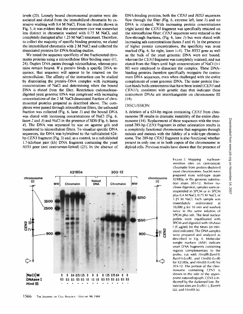
levels (20). Loosely bound chromosomal proteins were dis- sociated and eluted from the immobilized chromatin by ex- tensive washing with 0.4 M NaC1. From the results shown in Fig. 5, it was evident that the centromere core was somewhat less distinct in chromatin washed with 0.75 M NaCI, and completely disrupted after 1.25 M NaC1 treatment. Therefore, to collect the majority of specific binding proteins, we eluted the immobilized chromatin with 2 M NaC1 and collected the dissociated proteins for DNA-binding studies.
We tested the sequence specificity of the fractionated chro- matin proteins using a nitrocellulose filter binding assay (11, 24). Duplex DNA passes through nitrocellulose, whereas pro- teins remain bound. If a protein binds a specific DNA se- quence, that sequence will appear to be retained on the nitrocellulose. The affinity of the interaction can be studied by dissociating the protein-DNA complex with increasing concentrations of NaC1 and determining when the bound DNA is eluted from the filter. Restriction endonuclease- digested yeast genomic DNA was complexed with increasing concentrations of the 2 M NaCl-dissociated fraction of chro- mosomal proteins prepared as described above. The com- plexes were passed through nitrocellulose filters, the unbound fraction was collected (Fig. 6, lane 1) and the bound DNA was eluted with increasing concentrations of NaCI (Fig. 6, lanes 2 and 3) and NaCI in the presence of SDS (Fig. 6, lanes 4). The DNA was separated by size on agarose gels and transferred to nitrocellulose filters. To visualize specific DNA sequences, the DNA was hybridized to the radiolabeled 624- bp CEN3 fragment (Fig. 3) and, as a control, to a radiolabeled 1.7-kilobase pair (kb) DNA fragment containing the yeast HIS3 gene (not centromere-linked) (25). In the absence of
DNA-binding proteins, both the CEN3 and HIS3 sequences flow through the filter (Fig. 6, extreme left, lane 1) and no DNA is retained. With increasing protein concentrations (right lanes) the CEN3 fragment was specifically retained on the nitrocellulose filter; CEN3 sequences were reduced in the flow-through fractions, (Fig. 6, lane 1) but were eluted with increasing salt concentrations (lanes 3 and 4). In the presence of higher protein concentrations, the specificity was most marked (Fig. 6, far right, lanes 1-4). The HIS3 gene as well as the bulk of the yeast genomic DNA were not bound, whereas the CEN3 fragment was completely retained, and not eluted from the filters until high concentrations of NaC1 (>1 M) were employed to dissociate the complex. These DNA- binding proteins therefore specifically recognize the centro- mere DNA sequences, even when challenged with the entire complement of yeast genomic DNA. The same protein frac- tion binds both centromeres that have been tested (CEN3 and CENll) , consistent with genetic data that indicates these centromere DNAs are interchangeable on chromosome III (14).
D ISCUSSION
A deletion of a 624-bp region containing CEN3 from chro- mosome III results in dramatic instability of the entire chro- mosome (14). Replacement of these sequences with the trun- cated 289-bp CEN3 fragment in either orientation results in a completely functional chromosome that segregates through mitosis and meiosis with the fidelity of a wild-type chromo- some. The 289-bp CEN3 fragment is also functional whether present in only one or in both copies of the chromosome in diploid cells. Previous results have shown that the presence of
FiGUrE 5 Mapping nuclease- sensitive sites on centromeric chromatin from protein-depleted yeast chromosomes. Nuclei were prepared from wild-type strain X2180a, or the genomic substitu- tion strain 303-12. Before nu- clease digestion, samples were re- suspended in SPCM or in SPCM plus 0.4 M NaCI, 0.75 M NaCI, or 1.25 M NaCI. Each sample was immediately sedimented at 10,000 g for 10 rain and washed twice in the same solution of SPCM plus salt. The final nuclear pellets were equilibrated with SPCM and digested with DNAase I (5 ~,g/ml) for the times (in min- utes) indicated. The DNA samples were prepared and analyzed as described in Fig. 4. Molecular weight markers (MW) indicate yeast DNA fragments containing regions complementary to the probe, cut with HindllI-BamHI, BamHI-EcoRI, and HindllI-EcoRI for X2180a, and Hindlll EcoRI for 303-12. The portion of the chro- mosome containing CEN3 is shown to the side of the appro- priate autoradiograph. CEN3 is in- dicated by the darkened line. Re- striction sites are EcoRI (-), BamHI (A), and F/indlll (x).
1566 THE JOURNAL OF CELL BIOLOGY • VOLUME 99, 1984
on Novem
ber 14, 2007 w
ww
.jcb.orgD
ownloaded from

FIGURE 6
Selective affinity of centromere binding proteins to CEN DNAs . A protein fraction was dissociated from immobilizedchromatin as described in the Materials and Methods with the salt solutions that dissociate the in vivo centromeric complex (Fig.5) . Increasing concentrations of this protein fraction (0, 0.2, 0 .5, and 1 .0 jug) were incubated with 1 .0 wg of BamHI restrictedgenomic DNA from yeast strain RH218, as described in Materials and Methods . The complexes were passed through nitrocellulosefilters and the unbound fraction (lanes 1), 0 .4 M NaCl eluants (lanes 2), 1 .0 M NaCl eluants (lanes 3), and 1 .0 M NaCI plus 1%SDS eluants (lanes 4) were collected . DNA samples were deproteinized and electrophoresed on a 1 .0% agarose gel . The DNAfragments were transferred to nitrocellulose and hybridized with the 624-bp CEN3 fragment shown in Fig . 3, and as a control, the1 .7-kb HISS gene fragment (25) . The 8.2-kb band containing the CEN3 sequences (5) and the 1 .7-kb band containing the HISSsequences (25) are indicated . At left, lanes I and 2 show the elution pattern of DNA fragments incubated in the absence ofbinding protein . The three sets of lanes 1-4, from left to right, show the elution pattern of DNA fragments incubated with 0 .2,0 .5, and 1 .0,ug of binding proteins, respectively .
different centromeres, or the same centromere in oppositeorientations in chromosomes no. III ofdiploid cells, does notseem to affect the fidelity of chromosome pairing and segre-gation in a cell undergoing the complicated processes ofmeiosis (14). The centromeres may therefore be structurallyautonomous units whose function is chromosome interspe-cific such that they are able to interact with the same com-ponents of the segregation apparatus regardless of the chro-mosome in which they reside.We have examined the folding of centromere DNA in the
yeast cell nucleus with the nucleolytic enzymes, micrococcalnuclease and DNAase I . A 220-250-bp region that includesthe key sequence elements I-III was refractory to nucleolyticdigestion (11, 15). This 220-250-bp centromere core is foundto be associated with the centromere sequences on all thechromosomes that have been examined to date (Fig. 7) . Themaintenance of this distinctive structural differentiation inchromatin containing the 289-bp CEN3 replacement (Fig. 4 c)indicates that the DNA sequences within this fragment pro-vide the necessary information for binding of centromere-specific chromatin components, independent of flanking se-quence information . This region of DNA is characterized byshort sequence elements whose spacing is conserved in thecentromeres of different chromosomes (Figs . 1 and 7 andreference 10) . Ifthis region ofchromatin is wound in a similarfashion to nucleosomal chromatin, these elements may bejuxtaposed in the cell nucleus and could provide a commonbinding site for components of the mitotic apparatus. Thecomponents required for maintenance ofthe chromatin struc-ture surrounding the centromere core are dissociated fíomthe DNA at the same ionic strengths used to remove thehistones from bulk chromatin (15, 20) . Although chromatincomponents other than histones must be bound to the cen-tromere at some point in the cell division cycle, this regionmay be wound around histone or histonelike proteins thattogether serve as a chromatin template for the components ofthe mitotic apparatus.The ability to isolate the mitotic spindle from the yeast,
Saccharomyces cerevisiae (27, 28), has provided a cytologicalview of the mitotic apparatus in a cell that is not particularlyamenable to such an analysis. Although the role the spindle
FIGURE 7
Structural organization at the centromere core in yeastchromosomes III, IV, and XI . A map of the centromere regions fromchromosomes III, IV, and XI shows the region of structural differ-entiation encompassing the elements of sequence homology I-IIIin each centromere . DNAase I sites (vertical arrows) that bound thenuclease-resistant centromere core (stippled box) are indicated .Restriction enzyme sites are BamHI (A), Xhol (A), and Sau3A (O) .The DNA fiber is represented in linear form to visualize the positionof the 220-250-bp nuclease-resistant core and the elements 1-111 ofcentromere sequence homology . In chromatin, the bulk of theDNA is wrapped around histone proteins to form a subunit structureof 160-bp repeats . The centromere core, in contrast, is in a pro-tected particle that contains 1 .3-1 .5 times more DNA than doesthe typical nucleosomal subunit, Although the conformation ofDNA within the 220-250-bp protected core particle remains to beelucidated, a direct extrapolation from the dimensions of a nucleo-some (11 x 5 nm, reference 26) would give a diameter of 15-20nm for the centromeric core .
plays in yeast mitosis remains to be elucidated, there is astrong correlation between the number of discontinuous mi-crotubules and the number ofgenetic linkage groups in yeast
BLOOM ET AL.
Structure of a Yeast Centromere
1567
on Novem
ber 14, 2007 w
ww
.jcb.orgD
ownloaded from

(27, 29). If a single microtubule interacts with a unique point along the chromatin fiber of each chromosome, a structural discontinuity may be characteristic of that region in chroma- tin. The structural parameters of yeast microtubules and the nuclease-resistant centromere core (Fig. 7) are consistent with the notion that the centromere core is such a binding site. With the isolation of specific DNA-binding proteins, yeast mitotic spindles, and centromere DNA, we should eventually be able to characterize their interaction and begin to under- stand the molecular mechanisms that govern chromosome segregation and cell division.
This work was supported by research grant GM-32238 from the National Institutes of Health to Dr. Bloom and CA-11034 from the National Cancer Institute to Dr. Carbon.
Received for publication 30 May 1984, and in revised form 20 July 1984.
REFERENCES
1. Meacock, P. A., and S. N. Cohen. 1980. Partitioning of bacterial plasmids during cell division: a cis-acfing locus that accomplishes stable plasmid inheritence. Cell. 20:529- 542.
2. Austin, S., and A. Abeles. 1983. Partition of unit-copy miniplasmids to daughter cells. I. P1 and F miniplasmids contain discrete, interchangeable sequences sufficient to promote equilmrtition. £ Mol. Biol. 169:353-372.
3. Austin, S., and A. Abeles. 1983. Partition of unit-copy miniplasmids to daughter cells. I1. The partition region of miniplasmid PI encodes an essential protein and a centromere- like site at which it acts. J. MoL Biol. 169:373-387.
4. Murray, A. W., and J. W. Szostak. 1983. Pedigree analysis of plasmid segregation in yeast. Cell. 34:961-970.
5. Clarke, L., and J. Carbon. 1980. Isolation of a yeast centromere and construction of functional small circular minichromosomes. Nature (Lond.). 287:504-509.
6. Stinchcomb, D. T., C. Mann, and R. W. Davis. 1982. Centromeric DNA from Saccha- rom yces cerevisiae. Z Mol. Biol. 158:157-179.
7. Maine, G. T., R. T. Surosky, and B-K. Tye. 1984. Isolation and characterization of the centromere from chromosome V (CEN5) of Saccharomyces cerevisiae. Mol. Cell. BioL 4:86-91.
8. Panzeri, L., and P. Philippsen. 1982. Centromeric DNA from chromosome Vl in Saccharomyces cerevisiae strains. EMBO (Eur. Mol. Biol. Organ.) ,£ 1:1605-1611.
9. Fitzgerald-Hayes, M. J.-M. Buhler, T. G. Cooper, and J. Carbon. 1982. Isolation and subcloning analysis of functional centromere DNA (CENII) from Saccharomyces cerevisiae chromosome Xl. Mol. Cell. BioL 2:82-87.
10. Fitzgarald-Hayes, M., L. Clarke, and J. Carbon. 1982. Nucleotide sequence comparisons and functional analysis of yeast centromerc DNAs. Cell. 29:235-244.
11. Bloom, K. S., M. Fitzgerald-Hayes, and J. Carbon. 1983. Structural analysis and sequence organization of yeast ceotromeres. Cold Spring Harbor Syrup. Quam BioL 47:1175-1185.
12. Hartwell, L. H., S. K. Dutcher, J. S. Wood, and B. Garvik. 1982. The fidelity of mitotic chromosome reproduction in S. cerevisiae. Recent Adv. Yeast MoL Biol. 1:28-38.
13. Rothstein, R. J. 1983. One-step gene disruption in Yeast. Methods Enzymol. 101:202- 211.
14. Clarke, L., and J. Carbon. 1983. Genomic substitutions of ceotromeres in Saccharo- myces cerevisiae. Nature (Lond.). 305:23-28.
15. Bloom, K. S., and J. Carbon. 1982. Yeast centromere DNA is in a unique and highly ordered structure in chromosomes and small circular miniehromosomes. Cell. 29:305- 317.
16. Jones, E. W. 1977. Proteinase mutants ofSaecharomyces cerevisiae. Genetics. 35:23- 33.
17. Forte, M. A., and W. L. Fangman. 1976. Naturally occurring cross-links in yeast chromosomal DNA. Cell. 8:425--431.
18. Nelson, R. G., and W. L. Fangman. 1979. Nucleosome organization of the yeast 2-~m DNA plasmid: a eukaryotic minichromosome. Proc. Natl. ~Iced. Sci. USA. 76:6515- 6519.
19. Southern, E. M. 1975. Detection of specific sequences among DNA fragments separated by gel electrophoresis. ,L Mol. Biol. 98:503-517.
20. Bloom, K. S., and J. N. Anderson. 1978. Fractionation and characterization of chro- mosomal proteins by the hydroxyapatite dissociation method. Z Biol Chem. 253:4446- 4450.
21. Wu, C. 1980. The 5' ends of Drosophila heat shock genes in chromatin are hypersensitive to DNase I. Nature (Lond.). 286:854-860.
22. Carbon, J., and L Clarke. 1984. Structural and functional analysis of a yeast centromere ( CEN3)..L Cell Science. In press.
23. Paulson, J. R., and U. K. Laemmli. 1977. The structure of historic-depleted metaphase chromosomes. Cell. 12:817-828.
24. Riggs, A. O., H. Suzuki, and S. Bourgeois. 1970. lac repressor-opemtor interaction. I. Equilibrium studies. J. Mol. Biol. 48:67-83.
25. Struhl, K., and R. W. Davis. 1980. A physical, genetic and transcriptional map of the cloned his3 gene region of Saccharomyces cerevisiae. J. Mol. Biol. 136:309-332.
26. McGhce, J. D., and G. Felsenfeld. 1980. Nucleosome structure. Annu. Rev. Biochem. 59:1115-1156.
27. King, S. M., J. S. Hyams, and A. Luba. 1982. Ultrastructure of mitotic spindles isolated from a cell division cycle mutant of the yeast, Saccharomyces cerevisiae. Eur. J. Cell Biol. 28:98-102.
28. King, S. M., J. S. Hyams, and A. Luba. 1982. Absence of microtubule sliding and an analysis of spindle formation and elongation in isolated mitotic spindles from the yeast Saccharomyces cerevisiae. ,L Cell Biol. 94:341-349.
29. Peterson, J. B., and H. Ris. 1976. Electron microscope study of the spindle and chromosome movement in the yeast Saccharomyces cerevisiae. J. Cell. Sci. 22:219-242.
1 5 6 8 THE JOURNAL OF CELL BIOLOGY . VOLUME 99, 1984
on Novem
ber 14, 2007 w
ww
.jcb.orgD
ownloaded from