Embed Size (px)
Citation preview

C H R I S T I A N S O N N I E R M D
INTRODUCTION TO HOSPITAL MEDICINE:
COMMON RESPIRATORY ADMITS

LEARNING OBJECTIVES
• Discuss common respiratory admits and their management• Pneumonia: community acquired vs HCAP vs aspiration
pneumonia• COPD exacerbation• Asthma exacerbation• CHF exacerbation (“cardiac asthma”)• Pulmonary embolism/dvt
• Recognizing respiratory distress and failure• When to consider ICU• When to consider intubation• RSI basics

COMMUNITY ACQUIRED PNEUMONIA
• Not all CAP needs hospitalization. Need to use clinical judgment when deciding if admission is needed• Admit if:• Failed outpatient therapy• hemodynamically unstable• Sepsis • Pneumonia severity index• Curb-65 score• Severe community acquired pneumonia score• SMART-COP

COMMUNITY ACQUIRED PNEUMONIA
• PSI (pneumonia severity index• If one or more Step 1 risk factors are present then
proceed to step 2• Step 2 stratifies risk into class II, III, IV or V based on total
points• Total point score is calculated by adding:• Male: pt age in years• Female: pt age in years -10
• 70 pts and under= class II• 71-90 pts= class III• 91-130 pts = class IV• 130+ pts= class V

PSI STEP 1
• Age over 50• Presence of any of the following comorbid conditions• Cancer• Heart failure• Stroke• Renal dz• Liver dz
• Presence of any of the following physical exam• Ams• Pulse over 125• Resp rate over 30• Sbp under 90• fever


PSI STRATEGY
The higher the class the greater the indication for admission

CURB-65 SCORE
• Similar validation to PSI and simpler to use• C confusion• Based on specific mental status test or any altered mentation
• U urea• BUN over 7 mmol/L or 20mg/dL
• R respiratory rate• Over 30/min
• B Blood pressure• Sbp under 90 or dbp under 60
• 65 age 65 or over
• Score of 0-1= outpatient• Score of 2= admission• Score of 3+= strongly consider ICU especially if 4 or 5

SEVERE COMMUNITY ACQUIRED PNEUMONIA SCORE
• SCAP• Score 10+ points = admission and likely ICU• Has the worst validation of all scores…needs more study
• Major criteria• Arterial pH <7.30 = 13 points• Sbp < 90mmHg = 11 points
• Minor criteria• Resp rate >30 bpm = 9 points• PaO2/FiO2 <250 = 6 points• BUN >30 mg/dL or > 10.7mmol/L = 5 points• AMS = 5 points• Age 80+ = 5 points• Multilobar or bilateral infiltrates cxr = 5 points
SMART-COP
• Uses set of clinical criteria to predict who needs ICU care (score of 3+=increasing need for ICU)• Age• Albumin• Resp rate• Arterial pH• PaO2 on room air• Sbp• Multilobar infiltrates• Pulse • confusion

COMMUNITY ACQUIRED PNEUMONIA
• Dx: clinical, imaging and lab based• Clinically:• Cough, rhonchi, rales, fever, chills• No long term hospital or health care exposure in past 30-
90 days
• Imaging• Evidence of infiltrates on cxr
• Lab• Leukocytosis• Sputum cx• Blood cx• Urine studies (cx and legionella antigen)

COMMUNITY ACQUIRED PNEUMONIA
• Can be caused by variety of pathogens including viral• Most commonly but not limited to• Strep pneumo• H influ• Mycoplasma pneumonia• Chlamydia pneumo• Legionella
Just as with any infection start with broad abx coverage and decrease as needed based on culture reports


COMMUNITY ACQUIRED PNEUMONIA
• Additional tips• Duration of therapy is usually 5-7 days of po abx• Cxr may lag by 24-48 hours behind clinical picture• Upon admission if patient is dehydrated, rehydration may
“fluff out” infiltrates and make them more apparent• If patient does not respond to tx, may need stronger abx
coverage and mechanical ventilation• The more co-morbidites the greater the re-admission risk• Glucocorticoids have some evidence in shortening
hospital stays• Tissue factor pathway inhibitors do not show benefit• Statins have limited anti-inflamatory properties and show
limited benefit in studies

COMMUNITY ACQUIRED PNEUMONIA
• Prevention• Vaccination in patients 65+• Smoking cessation


HEALTH CARE ASSOCIATED PNEUMONIA
• HCAP= pneumonia in non-hospitalized patient who has extensive healthcare contact• IV therapy, wound care, chemo with in 30 days• Resident of long term care facility (pinecrest, nh ect)• Hospitalization for acute care for 2+ days with in last 90 days• HD in clinic or hospital within past 30 days.• Some argue hospital employees qualify
• HAP: hospital acquired (nosocomial) pneumonia: pneumonia that occurs 48 hrs or more after admission and was not noted on admission
• VAP: ventilator-associated pneumonia: HAP that develops 48-72 hours after intubation

HCAPTX
• Patients are usually septic therefore begin EGDT asap• Obtain cbc, cmp, lactic acid, procalcitonin, bld cx,
urine cx, sputum cx• Begin empiric abx therapy• Vancomycin, levaquin, zosyn• Goal is to cover MRSA and double cover pseudomonas• Maintain these meds until specific organism has been
identified then de-escelate to monotherapy if possible • Then to po and discharge• Once de-escelating from empiric to focused therapy the tx is
very similar to CAP (see prior slides)• Usually duration of therapy is at least 14-15 days
HCAP
• Tips: consider the following (for any pneumonia)• Albuterol, duonebs, xopenex (q4q2prn)• Incentive spirometry• Chest physiotherapy• Lactinex• Pneumovax if age appropriate• Smoking cessation• Good pulm hygiene and toilet (suctioning ect)• If copious secretions mucomyst and or robinol may be
helpful.

ASPIRATION PNEUMONIA
• Definition: pneumonia following aspiration of gastric contents• Difficult to distinguish between chemical
pneumonitis and true pneumonia therefore we usually treat as pneumonia• If aspiration pneumonia occurs always get a
swallow study or evaluate for dysphagia before feeding

ASPIRATION PNEUMONIA
• Clinical signs of anaerobic bacterial infection• Indolent/smoldering sx (low grade fevers ect)• Absence of chills/rigors• Foul odor to sputum• Infiltrates worse on the right (remember anatomy…right
mainstem is straight shot)

ASPIRATION PNEUMONIA
• If anaerobic organisms are suspected/assumed• First line is clindamycin 600mg iv q8hrs then 300mg po
qid• Alternatives• Augmentin 875mg po bid• Flagyl (500mg po/iv tid) +amoxicillin (500mg tid) or penicillin
G (1-2million units IV q4-6hrs)
• If hcap suspected: go to vanc, levaquin and zosyn as discussed earlier
• Duration of tx: 7-10 days unless case becomes more complicated
• In the event of mechanical obstruction may need bronchoscopy

COPD
• Chronic obstructive pulmonary disease: group of related disorders that all cause airflow limitation• Emphyseme• Chronic bronchitis• Chronic obstructive asthma
• Main difference between copd and asthma is airway (bronchial constriction is more reversible in asthma.

COPD
• GOLD Classification

COPD
• For the purposes of this lecture we will not discuss dx of copd as this usually is an outpatient event and chronic management of the disorder is also outpatient.• We will focus on how to tell a patient may have
copd and how to handle acute exacerbations

COPD
• Copd patient:• Usually heavy smoking history• Usually have sedintary lifestyle due to respiratory
limitations• Have baseline non-productive cough, wheeze• Barrel chest, blue bloater, pink puffer is not a reliable
characteristic• Intermittent exacerbations caused by physiological stress
(cold, flu, pneumonia) “peaks in symptoms however upon resolution patient is never quite back to baseline”
COPD
• Mild exacerbations can be managed at home by providing:• Bronchodilator• Oral glucocorticoids• Antibiotics • Continuation of chronic meds• Inhaled short acting beta-agonists (albuterol)• Inhaled short acting anticholinergic agents (ipratropium)• Nebulizers may be easier for patient’s with exacerbations

COPD
• Moderate to severe exacerbations as well as any exacerbation with hemodynamic instability need hospital treatment. • Oxygen therapy• Beta-adrenergic agonists• Anticholinergic agents• Systemic glucocorticoids• Antibiotics and antiviral agents• Supportive care• Bipap vs mechanical ventilation

COPD
• Oxygen therapy• Critical component of acute therapy however care should be
taken to avoid excess oxygen therapy• Excess oxygen can worsen condition because COPD patient’s
tend to be CO2 retainers• These patient’s have a baseline hypercapnia which supports their
respiratory drive.• Excess O2 over 60-70mmHg causes decrease in hypercapnia
which results in respiratory depression
• Pox goal of 88-92% is adequate for copd patient’s• Nurses and RT may not understand this, specify to them that
this is adequate and not to be concerned.• DO NOT PLACE COPD PATIENT ON HIGH FLOW O2 THERAPY
AND LEAVE THEM ALONE… THEY WILL STOP BREATHING.
COPD
• Every copd exacerbation should receive inhaled short acting beta adrenergic agonists• Albuterol• Use caution as albuterol can cause tachyarrhythmias such as
afib. In patient’s prone to these arrythmias use levalbuterol
• Levalbuterol (xopenex)• These agents are commonly combined with anticholinergic
agents such as ipratoprium • This is duoneb
MDI therapy has equal efficacy as nebulizers however patients are often comforted by nebulizer therapy and this therapy can be combined with oxygen therefore we tend to use these more in the hospital

COPD
• Albuterol, duonebs, xopenex are usually dosed as follows• Q4q2prn• Scheduled every 4 hours while awake• Every 2 hours as needed
• There has not been shown to be a difference in preventing hospital admission using duonebs vs albuterol in an outpatient bases therefore which ever is cheaper for the patient is the right answer at discharge

COPD
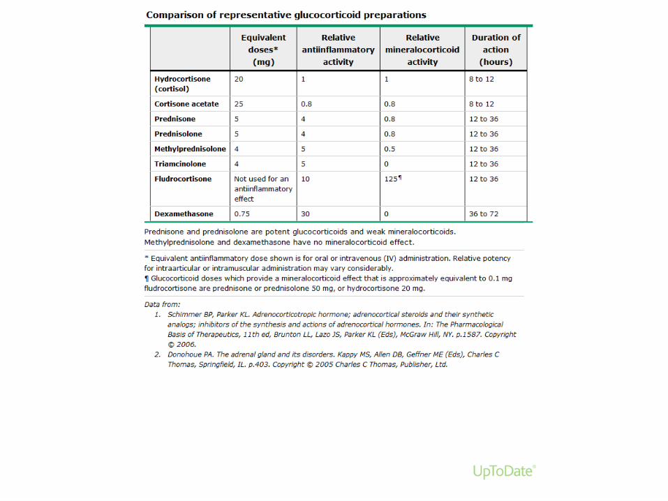
• Systemic glucocorticoids• Shown to improve the following when added to
bronchodilator therapies • Improve lung function• Decrease hospital lenghts of stay• Reduced failure rate of therapies• Does not matter if it is oral or IV• Doses (optimal doses are unknown) the following are
common doses• Prednisone 40mg qday (this is the prototypical dose)• Methylprednisolone 60-125mg bid-qid

COPD
• Anti-microbial therapy• If suspected infection is cause exacerbation then treat
accordingly• this could be anything from pneumonia, uri, flu, cellulitis or
any other infection…the physiological stress can trigger an exacerbation
• In case of pneumonias see earlier sections of this presentation


COPD
• Supportive care during exacerbation• Stop smoking• Dvt/pe prevention• Nutritional support• Mucolytic agents• Rehydration• Pain control (specifically for air-hunger)• Chest physiotherapy• Mechanical ventilation (progressive support from
cpapbipapintubation and vent support


ASTHMA
• Definition: reversable bronchoconstriction and inflammation. An allergic reaction usually.
• Exacerbation is characterized by• Sob, wheezing• Cough, chest pain• Fatigue
• Risk factors for fatal asthma • Previous severe episodes requiring ICU and intubation• Hospitalization/ER within past year the more the worse• Not on inhaled steroids• More than 1 canister of SABA a month• Comorbidities such as CAD, copd, drug abuse ect


ASTHMA
• Rest of treatment is similar to COPD exacerbation• Oxygen therapy (with out the risk of respiratory
depression)• Systemic steroids• Antibiotics, antivirals• Progressive vent support• Chest physiotherapy• Mucolytic• Supportive therapy

CHF
• See cardio lecture• Goal here is to provide oxygen/ventilator support
until chf exacerbation is resolving• Take the opportunity to optimize therapy• ACE/ARB• B-Blocker• Diruetics (especially lasix)• Digoxin• Acid• Salt restriction

PE/DVT
• DVT is the leading cause of PE• Deep vein thrombosis • Most commonly occurs in the lower extremity however upper
extremity and deep abdominal vessels are possible as well• Pieces of the clot can dislodge and travel via venous system
through right side of heart and lodge in the pulmonary arteries causing PE

DVT/PE
• Diagnosis• Providers in the ER/hospital must have high index of
suspicion…this means always look for it!• Classic triad: hemostasis, endothelial injury,
hypercoagulable states• Immobility• Trauma• malignancy

DVT/PE
• Physical exam findings• Disproportionate swelling of the limbs• Measure above and below the knee/elbow• Difference of 2 or more cm between limbs
• Pain on deep palpation of the limb • Palpable cord• Homan sign

DVT/PE
• Diagnosis• Well criteria (there is one for both dvt and pe) • D-dimer• Only clinically useful if it is negative as this effectively rules
out clot• Positive d-dimer should always prompt further investigation
with bilateral venous ultrasound • ct pe protocol or vq scan if pt is also sob

DVT/PE
• Ct pe protocol• Useful in actually visualizing the clot• Will yield a definitive yes or no answer
• Vq scan• Useful if iv contrast can not be used or not available• Will yield a probability but can not 100% rule out a clot• If clinical suspicion still remains then treat for it

DVT/PE
• Management• Prophylaxis• Scd• Compression hose• Lovenox 30-40mg (1mg/kg) subq qday
• Treatment• Lovenox 1mg/kg subq bid while bridging to warfarin• Xarelto• Heparin• Direct TPA per catheter placed by IR
• Indicated for• Saddle embolism• Hemodynamic instability with large clot• Compartment syndrome of the leg

ACUTE RESPIRATORY FAILURE
• Definition: Inability of the respiratory system to meet the oxygenation, ventilation and metabolic demand of the patient’s current condition• Two types of failure per definition above• Oxygenation failure: aka Type 1: • PaO2 less than 60mmhg therefore first priority is to correct
hypoxemia
• Ventilation failure: aka Type 2:• PACO2 over 50mmhg with pH under 7.3. need to determine if
patient has chronic failure like copd or if this is acute.

ACUTE RESPIRATORY FAILURE:
• Dx is clinical: too fast, too slow, unable to protect airway, hypoxic or hypoxemia • Hypoxic: refers to the POX monitor or cyanosis• Hypoxemia: is on the ABG• Dx is also lab based: • ABG, CXR, CT scan, broncoscopy

ACUTE RESPIRATORY FAILURE
• In reference to the two types mentioned earlier; these are also referred to as hypoxemic respiratory failure vs Hypercapnic respiratory failure.• each of these has several possible causes
therefore early in the tx course it is important to determine what you are dealing with, thus the importance of the abg ect.

HYPOXEMIC RESPIRATORY FAILURE
• Hypoxemia is the most immediate threat to life, more immediately life threatening than hypercapnea• Pathophysiologic causes of hypoxemic
respiratory failure• Shunting• V/Q mismatch• Diffusion limitation• Dead spaces• Low FiO2• Low atmospheric pressure• Hypoventilation of alveoli

HYPOXEMIC RESPIRATORY FAILURE
• In any respiratory failure it is important to calculate the A-a gradient because a normal gradient means there is no problem with the lungs or pulmonary circulation and you should start looking else where for the problem ie cardiac or muscular fatigue• An increased A-a gradient means there is a
venous admixture and can only have one of 3 causes• Shunt• Deadspace• V/Q mismatch

HYPOXEMIC RESPIRATORY FAILURE: SHUNT
• Shunt: blood goes through pulmonary circulation without being exposed to oxygen areas of lung have no ventilation• Can be intracardiac: ASD, VSD ect• Can be pulmonary: something is preventing
inspired gas from reaching the alveoli• Atelectasis, ARDS, pulmonary edema, pneumonia
consolidation

SHUNT
• Main characteristic is hypoxemia resistant to increased FIO2• PaO2/FIO2 ratio is lower the worse the
shunt gets• Shunts do respond well to positive pressure
ventilation to recruit alveoli

HYPOXEMIC RESPIRATORY FAILURE: VQ MISMATCH
• Blood is not flowing to an area of the lung that is being ventilated: • asthma, copd, interstitial lung disease, pneumonitis can
also cause this
• Readily corrects with increased FIO2 as there is nothing wrong with the oxygen reaching the alveoli and diffusing

HYPOXEMIC RESPIRATORY FAILURE: DEAD SPACE VENTILATION
• Opposite of a shunt, the air is getting to the alveoli but the blood isn’t. Think PE!• If no blood can get to the alveoli then it doesn’t
matter how good they are…there is no gas exchange.

HYPERCAPNIC/HYPERCARBIC RESPIRATORY FAILURE
• Type II failure: too much PaCO2 and low pH• There is an inability of the body to rid itself of
enough CO2. • Can have a variety of different causes• Brain• Spinal cord• Peripheral nerves • Neuromuscular junction• Muscles• Thorax• lungs
HYPERCAPNIC RESPIRATORY FAILURE:
• Easiest way to find the problem is by physical exam and history looking for problems with any of the systems listed previously.• It is important to remember that COPD pt will be
chronic CO2 retainers and can mimic lab values of this respiratory failure as a base line. However COPD exacerbation can be hypercapnic respiratory failure.

OTHER RESP FAILURES
• Trauma, obstruction ect
GENERAL TX OF RESPIRATORY FAILURE
• Identify and treat underlying cause• Provide simple/non-invasive support and move up
towards mechanical ventilation as needed.• Always be on the look out for progression to ARDS
or other severe manifestations of respiratory failure.
WHEN TO CONSIDER ICU
• Profound hemodynamic instability• AMS in the presence of respiratory distress• Intubated patient or patient with high likelihood of
sudden decompensation• Use your clinical judgement…if the patient makes
you nervous it is better to move to ICU early then to have them decompensate and code

WHEN TO CONSIDER INTUBATION
• Impending respiratory failure• Gcs less than 8 (or if you feel patient is unable to
adequately protect airway)• Worsening abg• Severe respiratory distress or depression that is
not responding to non-invasive support

INTUBATION
• For the purpose of this lecture we will not go into specifics of intubation. At this point (beginning of intern year) you should have an upper level resident or attending with adequate experience assist you with intubation and vent setup/management• For further information please review the
respiratory failure and mechanical vent lectures in the ICU lecture set

BASICS OF RSI
• Determine need for intubation• If patient is coding or there is no time for admit of
meds then proceed with intubation• If time• Sedation ie etomidate, versed, ativan • Paralytic ie succynocholine, vecuronium, rocuronium• Directly or indirectly visualize the vocal cords• pass et tube cuff just beyond vocal cords• Inflate tube and check for placement• Verify with cxr• Set the vent

REFERENCES
• Cecil’s medicine• Harrison’s textbook of medicine• Uptodate• Ardsnet protocol