Embed Size (px)
Citation preview
CHEN, WEI-YANG, M.S. Inhibition of Cytochrome P4502E1 by Lemongrass and the
Primary Aldehyde Constituent of Lemongrass, Citral. (2009)
Directed by Dr. Gregory M. Raner. 71pp
Cytochrome P4502E1, the ethanol-inducible form, metabolizes and
activates a significant number of substrates to more toxic products and the induction
of CYP2E1 by ethanol is thought to result in increased oxidative stress in hepatocytes.
One proposed mechanism for this increased oxidative stress is the increased
production of hydrogen peroxide by CYP2E1 via a so called “uncoupling’’ of its
NADPH oxidation activity. A main hypothesis of this research is that the main
aldehyde constituent found in Lemongrass, citral, will be able to block the activity of
CYP2E1, and consequently demonstrate physiological antioxidant properties.
The induction of the phase II enzyme is an important process involved in
cellular oxidative stress response, by which the oxidative toxicants can be eliminated
or inactivated before they damage the critical cellular macromolecules. Phase II
detoxifying genes provide protection to the cell against the toxicities of ROS and
reactive intermediates produced during phase I metabolism.
In this research, cell culture, RT-PCR and electrophoresis gel technology
will be used for monitoring the induction of antioxidant genes by a variety of different
natural products.
INHIBITION of CYTOCHROME P4502E1 BY LEMONGRASS AND THE
PRIMARY ALDEHYDE CONSTITUENT OF LEMONGRASS, CITRAL
by
Wei-Yang Chen
A Thesis Submitted to
the Faculty of The Graduate School at
The University of North Carolina at Greensboro
in Partial Fulfillment
of the Requirements for the Degree
Master of Science
Greensboro
2009
Approved by
___________________________
Committee Chair

ii
To almighty God, my family, friends of church in Taiwan, and friends in I-HOUSE
of UNCG Fall2007~Spring2009
iii
APPROVAL PAGE
This thesis has been approved by the following committee of the Faculty of The
Graduate School at The University of North Carolina at Greensboro.
Committee Chair______________________
Committee Members______________________
______________________
______________________________
Date of Acceptance by Committee
______________________________
Date of Final Oral Examination

iv
ACKNOWLEGEMENTS
First of all, I would like to thank my advisor and the chair of the thesis
committee, Dr. Gregory M. Raner, for instruction, guidance, and support for my two
years master degree in academic and research work. He is always supportive,
optimistic, helpful, and encourage me to pursue higher knowledge in biochemistry.
His teaching in the class is always lead me to be more interested and positive in the
road of research.
I also would like to appreciate the members of my committee, Dr.
Norman Chiu and Dr. Jason J. Reddick for their suggestions and guidance. They
always give advice and encouragement for my academic life.
Thanks all the faculty and fellow students in the department of Chemistry
and Biochemistry at the University of North Carolina at Greensboro for the teaching
experience, excellent research resources, and friendship. Words cannot describe how
grateful I am for everything at UNCG.
v
TABLE OF CONTENTS
Page
LIST OF TABLES ...................................................................................................... viii
LIST OF FIGURES ...................................................................................................... ix
CHAPTER
I.INTRODUCTION ........................................................................................... 1
1.1 Cytochrome P450 enzymes .............................................................. 2
1.1.1 General overview .............................................................. 2
1.1.2 The aldehydes: Citral, Citroneallal, and Decanal ............. 4
1.1.2.A. Citral ....................................................... 4
1.1.2.B. Citronellal ....................................................... 5
1.1.2.C. Decanal ........................................................... 5
1.1.3.Aldehyde deformylation ................................................. 7
1.2 Cytochrome P4502E1 .................................................................... 8
1.3 Phase II detoxifying genes ............................................................. 9
1.4 Inhibition of Cytochrome P450 3A4 ............................................ 12
1.5 Nifedipine ...................................................................................... 13
1.6 Oxidized Nifedipine ....................................................................... 15
1.7 HepG2 human liver cells ........................................................... 15
1.8 Proposed research ........................................................................ 16
II.MATERIALS AND METHODS ............................................................... 18
2.1 CYP2E1 by lemongrass and other essential oils .......................... 18
2.2 Growth and treatment of HepG2 cells ......................................... 19
2.2.1 Total RNA isolation and RT-PCR analysis .................. 20
2.2.2 RT-PCR ....................................................................... 22

vi
2.2.3 Induction of phase II genes ........................................... 23
2.2.3.1 Determination of effective dose of lemongrass for
induction of phase II genes ...................................... 23
2.2.4 Time dependent induction of phase II genes ................ 25
2.2.5 Effect of essential oil of Lemongrass on CYP3A4
activity .......................................................................... 25
III.RESULTS AND DISCUSSION ............................................................... 29
3.1 Inhibition of CYP2E1 by Lemongrass oil and other essential
oils .............................................................................................. 29
3.1.1 Introduction ................................................................... 29
3.1.2 Michaelis-Menten kinetic analysis ............................... 30
3.1.2.A. Inhibition of CYP2E1 by Lemongrass oil
and the aldehyde citral ................................. 30
3.1.2.B. Inhibition of CYP2E1 by Eucalyptus Lemon
oil, Citronella oil, and the aldehyde
citronellal ................................................... 34
3.1.2.C. Inhibition of CYP2E1 by Eucalyptus
Globulous oil and it’s main constituent
1,8 Cineole ................................................. 37
3.1.2.D. Inhibition of CYP2E1 by Cassia oil ............. 40
3.1.2.E. Inhibition of CYP2E1 by the aldehyde
decanal ....................................................... 41
3.2 Growth and treatment of HepG2 cells ......................................... 44
3.2.1. The gel electrophoresis result ........................................ 44
3.3 Inhibition of CYP3A4 by nifedipine oxidation ........................... 55
3.3.1. The retention time of Nifedipine and Oxidized
nifedipine was identified .......................................... 56
3.3.2. The screen experiments of nifedipine oxidation by
CYP3A4 with and without Lemongrass oil and the
vii
aldehyde citral ........................................................................ 62
IV.DISCUSSION ........................................................................................... 65
REFERENCES .......................................................................................................... 69

viii
LIST OF TABLES
Page
Table 1. The primer sequences and base pairs……………………………………….24
Table 2. KI values of essential oils and the aldehydes……………………………….43
ix
LIST OF FIGURES
Page
Figure 1. The P450 catalytic cycle ................................................................................. 3
Figure 2. Aldehyde deformylation scheme showing heme alkylation mechanism
involved in P450 inactivation by aldehydes ................................................ 7
Figure 3. Reaction intermediates in the conversion of heme to biliverdin and CO
by heme oxygenase .................................................................................... 12
Figure 4. Michaelis-Menten plot of 50mM p-nitrophenol oxidation by CYP2E1
and inhibition by 5ug/ml Lemongrass oil in 20ul human liver
microsomes. ............................................................................................. 31
Figure 5. The Lineweaver-Burk plot of p-nitrophenol oxidation by CYP2E1 and
inhibition by 5ug/ml Lemongrass oil in 20ul human liver microsomes,
the x-axis= 1/(S) and the y-axis=1/(V), each data point is the average
of two samples ........................................................................................... 31
Figure 6. Michaelis-Menten plot of 50mM p-nitrophenol oxidation by CYP2E1
and inhibition by 0.033mM the aldehyde citral in 20ul human liver
microsomes ................................................................................................ 33
Figure 7. The Lineweaver-Burk plot of p-nitrophenol oxidation by CYP2E1 and
inhibition by the 0.033mM aldehyde citral in 20ul human liver
microsomes, the x-axis= 1/(S) and the y-axis=1/(V), each data point
is the average of two samples .................................................................... 33
Figure 8. Michaelis-Menten plot of 50mM p-nitrophenol oxidation by CYP2E1
and inhibition by 5ug/ml Eucalyptus Lemon oil in 20ul human liver

x
microsomes ................................................................................................ 34
Figure 9. Michaelis-Menten plot of 50mM p-nitrophenol oxidation by CYP2E1
and inhibition by 5ug/ml Citronella oil in the 20ul human liver
microsomes ................................................................................................ 35
Figure 10. Michaelis-Menten plot of 50mM p-nitrophenol oxidation by CYP2E1
and inhibition by 5ug/ml Citronellal in 20ul human liver
microsomes .............................................................................................. 36
Figure 11. Michaelis-Menten plot of 50mM p-nitrophenol oxidation by CYP2E1
and inhibition by 5ug/ml Eucalyptus Globulous oil in 20ul human liver
microsomes .............................................................................................. 38
Figure 12. Michaelis-Menten plot of 50mM p-nitrophenol oxidation by CYP2E1
and inhibition by 5ug/ml 1,8 Cineole in 20ul human liver microsomes ... 39
Figure 13. Michaelis-Menten plot of 50mM p-nitrophenol oxidation by CYP2E1
and inhibition by 5ug/ml Cassia oil in 20ul human liver microsomes ...... 30
Figure 14. Michaelis-Menten plot of 50mM p-nitrophenol oxidation by CYP2E1
and inhibition by 5ug/ml Decanal in 20ul human liver microsomes ....... 42
Figure 15. The Lineweaver-Burk plot of p-nitrophenol oxidation by CYP2E1
and inhibition by 5ug/ml Decanal in 20ul human liver microsomes,
the x-axis= 1/(S) and the y-axis=1/(V), each data point
is the average of two samples .................................................................. 42
xi
Figure 16. 1.5% agarose gel showing the resulting band from RT-PCR
amplification of β-actin RNA produced in HepG2 cells with
increasing doses of Lemongrass oil. ...................................................... 44
Figure 17. 1.5% agarose gel showing the resulting band from RT-PCR
amplification of β-actin RNA produced in HepG2 cells at 20ug/ml
Lemongrass oil time-dependent experiment. ........................................... 46
Figure 18. 1.5% agarose gel showing the resulting band from RT-PCR
amplification of β-actin RNA produced in HepG2 cells with increasing
doses of the aldehyde citral. ................................................................... 47
Figure 19. 1.5% agarose gel showing the resulting band from RT-PCR
amplification of β-actin RNA produced in HepG2 cells at 20ug/ml
Citral time-dependent experiment............................................................ 48
Figure 20. 1.5% agarose gel showing the resulting band from RT-PCR
amplification of HO-1 RNA produced in HepG2 cells with increasing
doses of Lemongrass oil. .......................................................................... 49
Figure 21. 1.5% agarose gel showing the resulting band from RT-PCR
amplification of HO-1 RNA produced in HepG2 cells at 20ug/ml
Lemongrass oil time-dependent experiment. ......................................... 51
Figure 22. 1.5% agarose gel showing the resulting band from RT-PCR
amplification of HO-1 RNA produced in HepG2 cells with
increasing doses of the aldehyde citral. ................................................. 53
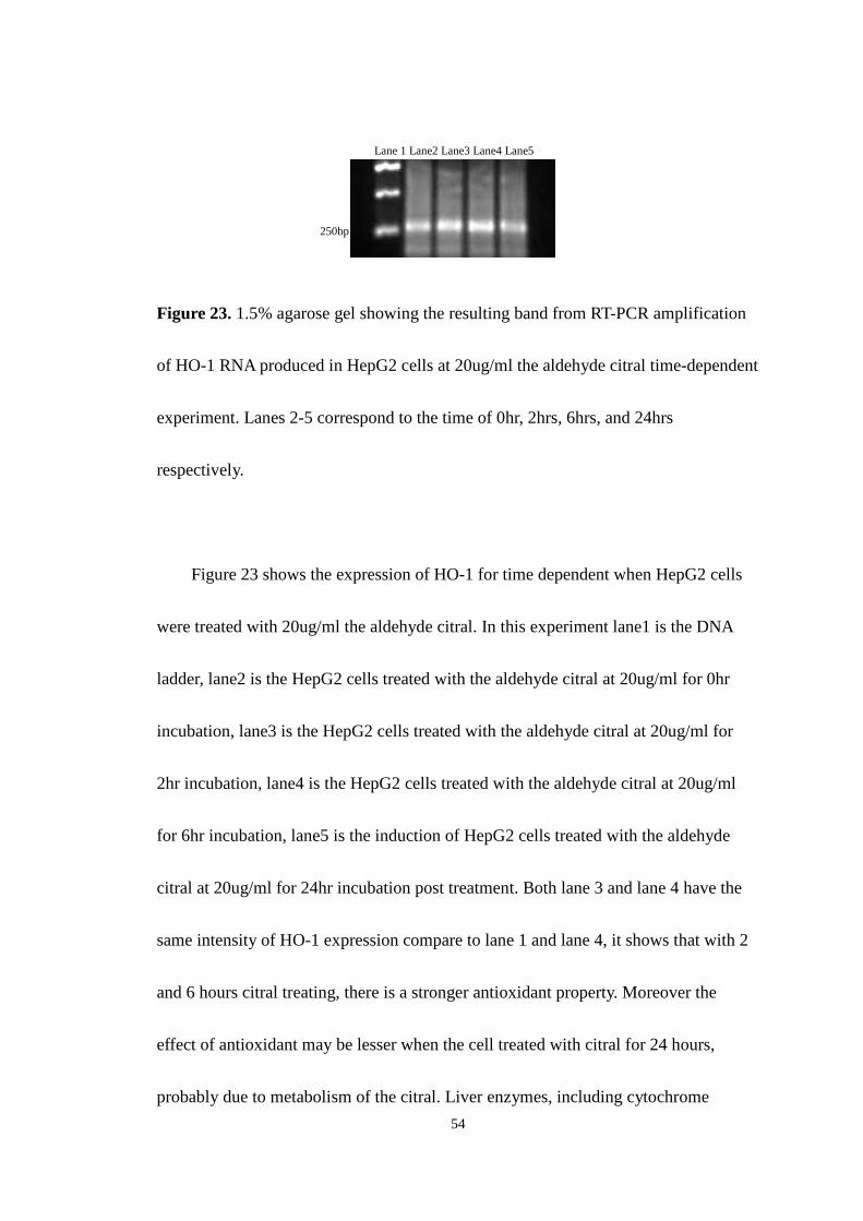
Figure 23. 1.5% agarose gel showing the resulting band from RT-PCR
xii
amplification of HO-1 RNA produced in HepG2 cells at 20ug/ml
the aldehyde citral time-dependent experiment. ...................................... 54
Figure24. HPLC-UV chromatogram of standard oxidized nifedipine(4.0minute)
and nifedipine(6.2minute). ......................................................................... 57
Figure25. HPLC-UV chromatogram of nifedipine(6.2minute) when the reaction
incubated without the addition of NADPH. ............................................... 59
Figure26. HPLC-UV chromatogram of oxidized nifedipine when the reaction
ncubated with the addition of NADPH(4.0minute) and
nifedipine(6.2minute). .............................................................................. 61
Figure 27. The screen experiment of Lemongrass oil with 0ul, 5ul, 50ul, and
350ul correspond to the oil concentration of 0ug/ml, 0.5ug/ml, 5ug/ml,
and 35ug/ml respectively. .......................................................................... 62
Figure28. The screen experiment of citral with 0ul, 5ul, 50ul, and 350ul
correspond to the oil concentration of 0ug/ml, 0.5ug/ml, 5ug/ml,
and 35ug/ml respectively. .......................................................................... 64
1
CHAPTER I
INTRODUCTION
Recent medical studies have shown that oxidative stress is at the root of a
number of human diseases such as Parkinson's disease (1), Alzheimer's disease (2)
and cancer (3) which directly or indirectly relates to most of human illness and death
in the world. Oxidative stress also contributes to aging in the human body (4) and
causes many general health problems for humans. The production of reactive oxygen
species (ROS) in the human body damages components of the cell including DNA (5),
lipids (6) and proteins (7). ROS are ions or molecules including free radicals, oxygen
ion or peroxides that can oxidize biological molecules. ROS are formed from a variety
of biochemical reactions and cellular functions (8), or through exposure to chemicals
(9). The formation and consumption of free radicals can be balanced by antioxidants
in the cell. Oxidative stress is caused by an imbalance in rate of formation and
consumption of free radicals. In addition, there are some common factors responsible
for the generation of free radicals such as air pollution (10), sunlight (11) and smoking
(12). In order to reduce oxidative stress, the body produces antioxidants to neutralize
2
free radicals which otherwise can destroy the human cells. The purpose of this
research is to show that specific compounds in the essential oil of lemongrass and
the primary aldehyde constituent of lemongrass, citral, have the ability to reduce
oxidative stress by scavenging ROS and/or by preventing their formation. The
project also addressed the ability of certain essential oils to induce cellular systems
that remove ROS in the liver and possibly in other tissues. The hypothesis of this
paper is that lemongrass oil stimulates phase II drug metabolizing enzymes in the
human liver and reduces the formation of ROS through inhibition of cytochrome
P4502E1 activity, which is a monooxygenase enzyme that has been implicated in the
generation of hydrogen peroxide and superoxide radicals.
1.1 Cytochrome P450 enzymes
1.1.1. General overview
Cytochrome P450 enzymes are most responsible for the metabolism of foreign
chemicals. Their primary purpose is the hydroxylation of non-polar molecules, and
making the drug more polar to facilitate their elimination in urine. Cytochrome P450
enzymes are commonly found in both prokaryotic and eukaryotic cells in nature. In
eukaryotic cells, the cytochrome P450 enzymes are mostly found in the membrane
3
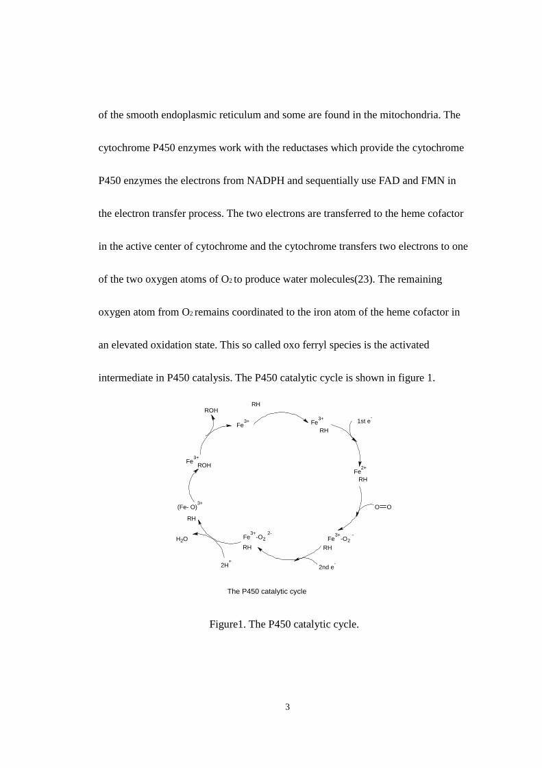
of the smooth endoplasmic reticulum and some are found in the mitochondria. The
cytochrome P450 enzymes work with the reductases which provide the cytochrome
P450 enzymes the electrons from NADPH and sequentially use FAD and FMN in
the electron transfer process. The two electrons are transferred to the heme cofactor
in the active center of cytochrome and the cytochrome transfers two electrons to one
of the two oxygen atoms of O2 to produce water molecules(23). The remaining
oxygen atom from O2 remains coordinated to the iron atom of the heme cofactor in
an elevated oxidation state. This so called oxo ferryl species is the activated
intermediate in P450 catalysis. The P450 catalytic cycle is shown in figure 1.
Fe3+ Fe
3+
Fe3+
-O2 -
Fe3+
Fe2+
RH
RH
RH
RHRH
RH
ROH
ROH
(Fe- O)3+
1st e-
O O
2nd e-
Fe3+
-O2 2-
2H+
OH2
The P450 catalytic cycle
Figure1. The P450 catalytic cycle.
4
1.1.2. The aldehydes: Citral, Citronellal, and Decanal
1.1.2.A. Citral
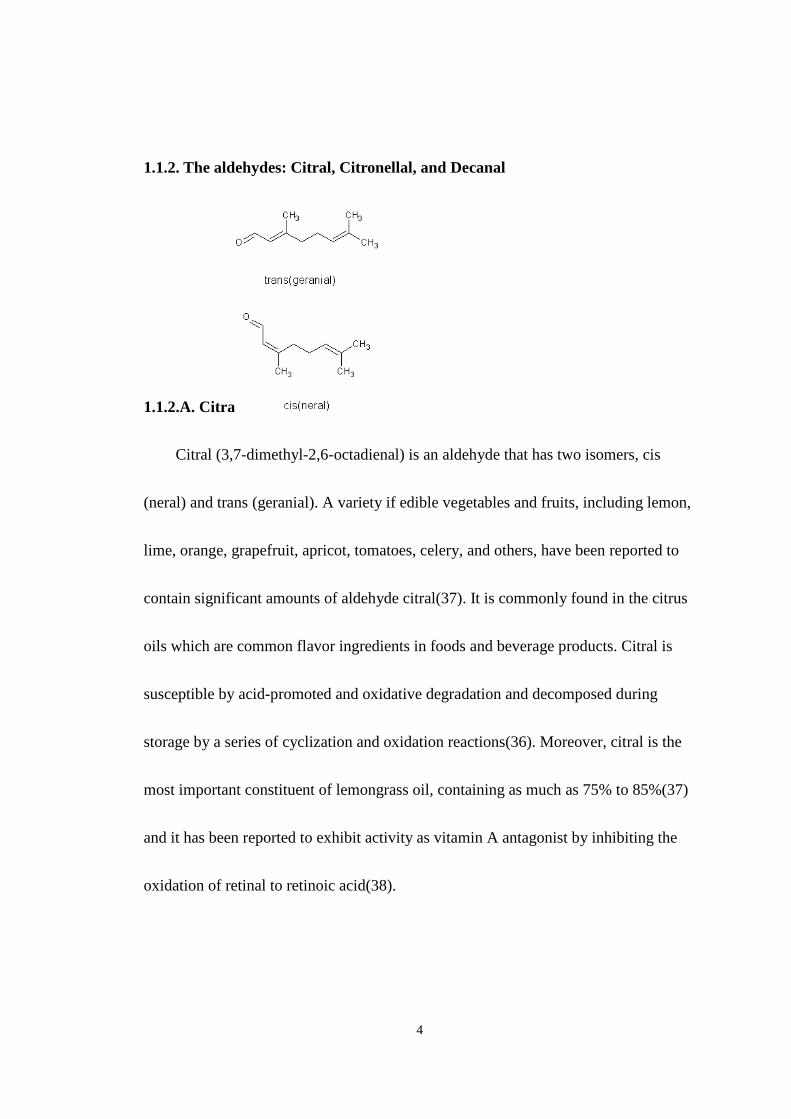
Citral (3,7-dimethyl-2,6-octadienal) is an aldehyde that has two isomers, cis
(neral) and trans (geranial). A variety if edible vegetables and fruits, including lemon,
lime, orange, grapefruit, apricot, tomatoes, celery, and others, have been reported to
contain significant amounts of aldehyde citral(37). It is commonly found in the citrus
oils which are common flavor ingredients in foods and beverage products. Citral is
susceptible by acid-promoted and oxidative degradation and decomposed during
storage by a series of cyclization and oxidation reactions(36). Moreover, citral is the
most important constituent of lemongrass oil, containing as much as 75% to 85%(37)
and it has been reported to exhibit activity as vitamin A antagonist by inhibiting the
oxidation of retinal to retinoic acid(38).
5
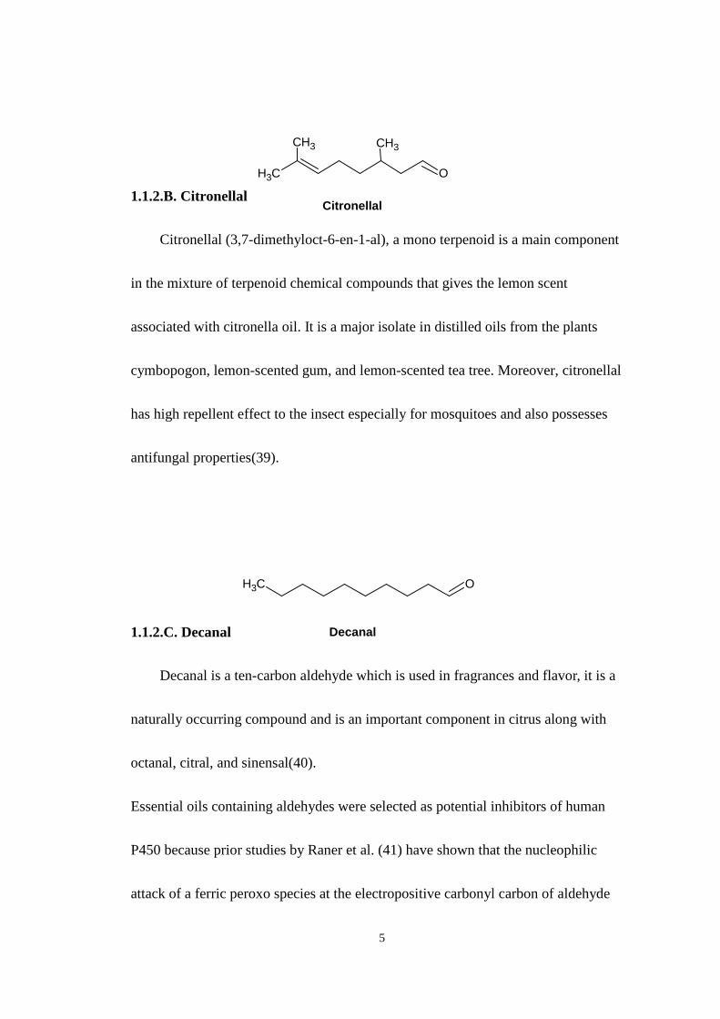
1.1.2.B. Citronellal
Citronellal (3,7-dimethyloct-6-en-1-al), a mono terpenoid is a main component
in the mixture of terpenoid chemical compounds that gives the lemon scent
associated with citronella oil. It is a major isolate in distilled oils from the plants
cymbopogon, lemon-scented gum, and lemon-scented tea tree. Moreover, citronellal
has high repellent effect to the insect especially for mosquitoes and also possesses
antifungal properties(39).
1.1.2.C. Decanal
Decanal is a ten-carbon aldehyde which is used in fragrances and flavor, it is a
naturally occurring compound and is an important component in citrus along with
octanal, citral, and sinensal(40).
Essential oils containing aldehydes were selected as potential inhibitors of human
P450 because prior studies by Raner et al. (41) have shown that the nucleophilic
attack of a ferric peroxo species at the electropositive carbonyl carbon of aldehyde
CH3 O
CH3 CH3
Citronellal
CH3 O
Decanal

6
forms a transient peroxohemiacetal. The peroxy-hemiacetal intermediate in aldehyde
deformylation resulted heme modification in CYP450.

7
1.1.3.Aldehyde deformylation
CH3
CHO
CH3
O-
H
OO
Fe3+
R
CH3
CH2-
+
O
O-
H
Nucleophilic addition
Homolytic cleavage and radical rearrangement
Adduct formation
OO-
Fe3+
R
O-
Fe4+
R
Figure2. Aldehyde deformylation scheme showing heme alkylation mechanism
involved in P450 inactivation by aldehydes.
8
1.2 Cytochrome P4502E1
Cytochrome P4502E1, the ethanol-inducible form, metabolizes and activates a
significant number of substrates to more toxic products and the induction of CYP2E1
by ethanol is thought to result in increased oxidative stress in hepatocytes(15). One
proposed mechanism for this increased oxidative stress is the increased production of
hydrogen peroxide by 2E1 via a so called “uncoupling’’ of its NADPH oxidation
activity. A main hypothesis of this research is that the main aldehyde constituent
found in lemongrass, citral is able to block the activity of CYP2E1, and consequently
demonstrate physiological antioxidant properties. Many studies have been carried out
involving inhibition of CYP2E1and the potential beneficial effects related to a
reduction in the activation of carcinogens or hepatotoxins(32). For example
Cedarbaun et al. showed NO can effectively inhibite arachidonic acid(AA) toxicity in
liver cells which express high levels of CYP2E1. They treated pyrazole-induced rat
hepatocytes with AA in the presence of an inhibitor of nitric oxide synthase,
L-NG-Nitroarginine Methylester (L-NAME) and added the NO donors
S-nitroso-N-acetylpenicillamine (SNAP) to increase NO level to demonstrate that NO
can be hepatoprotective against CYP2E1-dependent toxicity and prevent AA-induced
oxidative stress(31). In order to demonstrate the inhibition of cytochrome P4502E1

9
activity by these natural products, the Michaelis-Menton model will be used in
conjunction with a HPLC-based P4502E1 catalytic assay involving the oxidation of
p-nitrophenol to nitro catechol.
1.3 Phase II detoxifying genes
The induction of the phase II enzyme is an important process involved in
cellular oxidative stress response, by which the oxidative toxicants can be eliminated
or inactivated before they damage the critical cellular macromolecules(13). Phase II
detoxifying genes provide protection to the cell against the toxicities of ROS and
reactive intermediates produced during phase I metabolism(13). Members of the
phase II type enzymes include histone acetyltransferase-1(HAT-1), choline
acetyltransferase-1(CHAT-1), histamine N-methyltransferase-1(HNMT-1), Epoxide
hydrolase-1(EPHX-1), Heme oxygenase-1(HO-1) and NAD(P)H: quinone
oxidoreductase -1(NQO-1). Each of the corresponding gene products play an
important role in the cell in quenching ROS and preparing them for elimination.
Several recent studies have shown that some novel antioxidant chemicals induce the
synthesis of phase II detoxifying enzymes as a mechanism for increasing the ratio of
reduced GSH/oxidized GSSG. What appears to be happening is that the transcription
factor Nrf2 is activated by these chemicals, allowing it to stimulate the transcription
10
of a large number of genes in the nucleus. Nrf2 normally binds to the cytoskeleton
associated protein Keap1 which is located in the cytoplasm(14). When released from
its complex, Nrf2 enters the nucleus and binds to Antioxidant response
element(ARE) and regulate the expression and induction of a battery of genes
encoding detoxifying/chemopreventive proteins, which are activated in response to
oxidants, xenobiotics, UV light, and radiation(14). The increase of the
Nrf2-dependent transcription is correlated to the response associated with
electrophilic chemicals and oxidative stress. According to a recent study, the
activation of Nrf2 involving several kinases such as MAPKs, PKC and PI3K. For
example PKC, p38 , JNK, ERK1/2 and Nrf2, all have a role in the induction of
HO-1 by oxidized LDL in human muscle cells(13). The study showed that MAPK
pathways played an important role in the Nrf2-regulated phase II gene
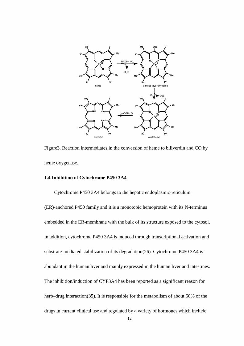
expression(13). The gene product of HO-1 catalyzes the rate-limiting step in heme
degradation, which is transformation of heme into biliverdin, carbon monoxide, and
free ion(Fe2+)(15). The importance of HO-1 expression is that it mediates
antioxidant, anti-inflammatory, and antiapoptotic effects within the cell(16-18). The
increasing activity of HO-1 correspond to the degradation of the heme moiety which
is a potential toxic prooxidant. In addition, increased activity of HO-1 generates

11
bilirubin which is an antioxidant with the ability to scavenge peroxy radicals and
inhibit lipid peroxidation(19-21) (Fig 3). CO is another product generated by the
HO-1 induction, it has the vasodilatory effects, anti-apoptotic effect, and
anti-inflammatory effect which are mediated by cGMP(15). As for Fe2+ ion, Ferritin,
an intracellular iron repository, is induced along with HO-1, therefore ferritin binds
the unbound iron from heme degradation(15). Cell culture, RT-PCR and
electrophoresis gel technology will be used for monitoring the induction of
antioxidant genes by a variety of different natural products. The detoxification of
foreign substances can be classified into two reaction processes; phase I and phase II.
For phase I reactions, foreign chemicals are mainly oxidized by cytochrome P450
(CYP) enzymes to become polarized metabolites. Phase II metabolism involves a
variety of enzymes that catalyze group transfer reactions of reductive reactions such
as glutathione S-transferase (GST) and NADPH quinone oxidoreductase (NQO1).
These enzymes convert the reactive Phase I products to more inert hydrophilic
products(22).
12
Figure3. Reaction intermediates in the conversion of heme to biliverdin and CO by
heme oxygenase.
1.4 Inhibition of Cytochrome P450 3A4
Cytochrome P450 3A4 belongs to the hepatic endoplasmic-reticulum
(ER)-anchored P450 family and it is a monotopic hemoprotein with its N-terminus
embedded in the ER-membrane with the bulk of its structure exposed to the cytosol.
In addition, cytochrome P450 3A4 is induced through transcriptional activation and
substrate-mediated stabilization of its degradation(26). Cytochrome P450 3A4 is
abundant in the human liver and mainly expressed in the human liver and intestines.
The inhibition/induction of CYP3A4 has been reported as a significant reason for
herb–drug interaction(35). It is responsible for the metabolism of about 60% of the
drugs in current clinical use and regulated by a variety of hormones which include
13
glucocorticoids, growth hormone, and triiodothyronine. Furthermore, drugs such as
Phenobarbital, clotrimazole, mifepristone, and rifamycin have been shown to induce
cytochrome P450 3A4 via a transcriptional upregulation(24,25).
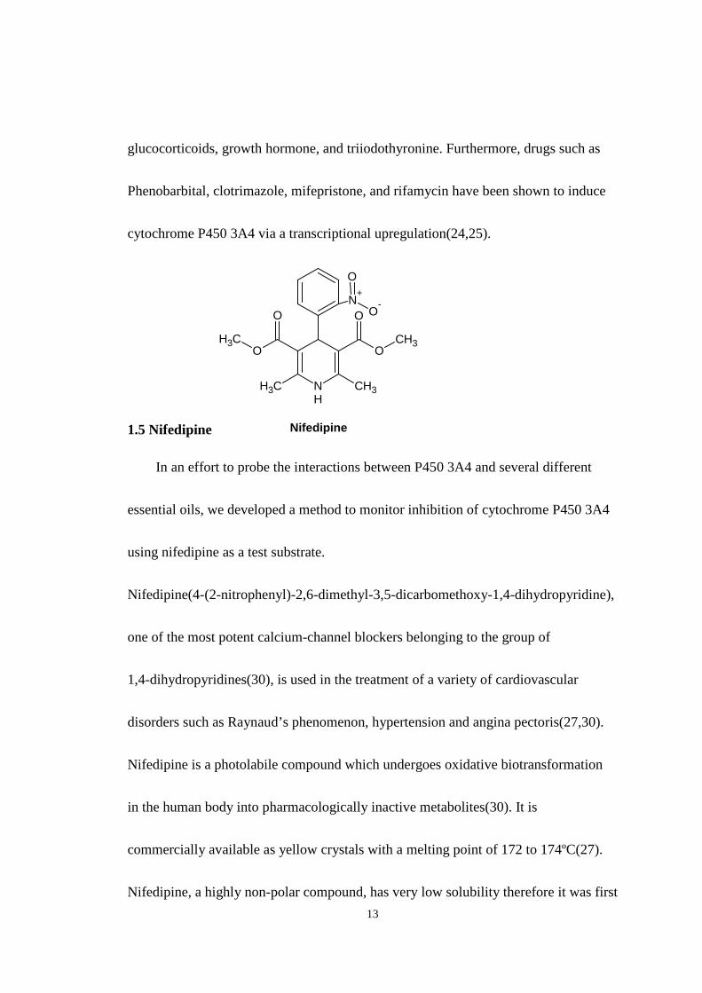
1.5 Nifedipine
NH
OO
CH3CH3
CH3OO
CH3
N+
O-
O
Nifedipine
In an effort to probe the interactions between P450 3A4 and several different
essential oils, we developed a method to monitor inhibition of cytochrome P450 3A4
using nifedipine as a test substrate.
Nifedipine(4-(2-nitrophenyl)-2,6-dimethyl-3,5-dicarbomethoxy-1,4-dihydropyridine),
one of the most potent calcium-channel blockers belonging to the group of
1,4-dihydropyridines(30), is used in the treatment of a variety of cardiovascular
disorders such as Raynaud’s phenomenon, hypertension and angina pectoris(27,30).
Nifedipine is a photolabile compound which undergoes oxidative biotransformation
in the human body into pharmacologically inactive metabolites(30). It is
commercially available as yellow crystals with a melting point of 172 to 174ºC(27).
Nifedipine, a highly non-polar compound, has very low solubility therefore it was first
14
dissolved in methanol to facilitate delivery in the current study. In the human body,
nifedipine is absorbed completely from the gastrointestinal tract and mostly from
jejunum. After absorption, nifedipine is further metabolized in the small intestine and
liver to form more polar compounds which enable the kidney to eliminate it via the
urine(30). The first analytical instrumental method to detect nifedipine in biological
fluids level is gas chromatographic(GC). The disadvantage of GC is that it lacks
specificity and selectivity even though the amounts of volumes are in the level of
micro liter and the limit of detection could go as low as 2ng/ml. In addition, most GC
methods require liquid-liquid and solid-phase extraction which increases the
complexity and time required for analysis. Therefore, in order to improve the
sensitivity, specificity, and efficiency, a high performance liquid
chromatographic(HPLC) method has been developed to detect nifedipine in plasma,
however, many of these methods still involved complicated and time-consuming
sample extraction. So a major goal has been to determine the plasma level of
nifedipine to yield a reliable estimate of its pharmacokinetic parameters for
therapeutic drug monitoring and bioavailability/bioequivalence purposes. These
estimates rely on the ability to measure the drug level at the lower end of the plasma
concentration range found in pharmacokinetic studies following the administration of
15
therapeutic doses of the drug(30). The method described in this thesis allows rapid
accurate measurements of oxidized nifedipine.
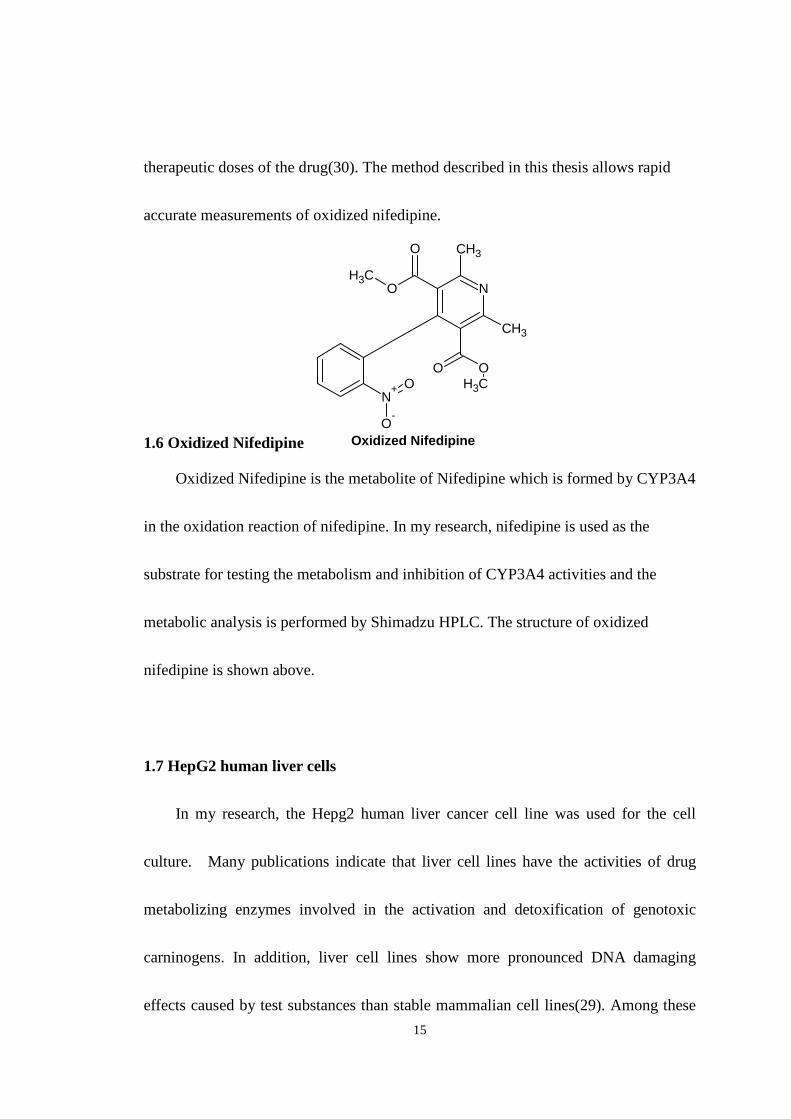
1.6 Oxidized Nifedipine
N
CH3O
OCH3
CH3
OOCH3
N+
O-
O
Oxidized Nifedipine
Oxidized Nifedipine is the metabolite of Nifedipine which is formed by CYP3A4
in the oxidation reaction of nifedipine. In my research, nifedipine is used as the
substrate for testing the metabolism and inhibition of CYP3A4 activities and the
metabolic analysis is performed by Shimadzu HPLC. The structure of oxidized
nifedipine is shown above.
1.7 HepG2 human liver cells
In my research, the Hepg2 human liver cancer cell line was used for the cell
culture. Many publications indicate that liver cell lines have the activities of drug
metabolizing enzymes involved in the activation and detoxification of genotoxic
carninogens. In addition, liver cell lines show more pronounced DNA damaging
effects caused by test substances than stable mammalian cell lines(29). Among these

16
cell lines, human HepG2 cell line is the most promising one which has been used in a
number of genotoxicity tests. The HepG2 cells possess different phase I and phase II
enzymes involved in the activation/detoxification of genotoxic carcinogens(29) and
HepG2 cell line is very valuable for the screening purposed in the early phase of
pharmaceutical development(28).
1.8 Proposed research
The following two projects were initiated in order to probe the potential
antioxidant mechanisms associated with essential oil of lemongrass.
Project 1: To establish the mechanism and potency of inhibition of human
cytochrome P4502E1 and P4503A4 by essential oil of lemongrass and the primary
aldehyde constituent of lemongrass, citral. According to preliminary studies,
lemongrass has promising inhibitory properties toward P4502E1 in vitro. The major
aldehyde component of lemongrass (citral) appears to be the primary inhibitory
constituents of that oil. The experimental method to be used is a HPLC-based assay
in which p-nitrophenol(PNP) is oxidized by cytochrome P4502E1 by human liver
tissue in the presence of the essential oil of Lemongrass or its major constituents. A
similar approach using Nifedipine oxidation reaction will be used to show the
analogous effects on P4503A4.
17
Project 2: To show induction of antioxidant genes by essential oil of lemongrass
and primary aldehyde constituent of lemongrass, citral, and to identify those
constituents responsible for induction. The hypothesis of this objective is that the
Lemongrass aldehyde constituent, citral, can activate the antioxidant response
elements found in a variety of antioxidant /phase II drug metabolizing genes.
Cultured human liver cells ( HepG2 ) will be used to test this hypothesis where cells
will be treated with oil or aldehyde and RT-PCR will be used to monitor antioxidant
gene expression.

18
CHAPTER II
MATERIALS AND METHODS
2.1 CYP2E1 by lemongrass and other essential oils
Several different essential oils along with purified aldehyde constituents were
tested for their abilities to inhibit P4502E1. In particular Citral, Nonenal, Nonyl
aldehyde, Citronellal and Decanal were evaluated with the oils Lemongrass,
Eucalyptus Globulous, Cassia, Citronella and Eucalyptus lemon. The following
experimental procedure was used for the evaluation of P4502E1 inhibition: 5ul of the
oil or aldehyde was diluted to 100ml in deionized water and this solution, along with
20ul of microsomes, 50ul of 1M potassium phosphate buffer solution (pH 7.4), 50mM
p-nitrophenol(PNP) and deionized water were combined in a final volume of 0.5ml.
25ul of 1mM NADPH was added to initiate the reaction in a 37oC water bath for 1 hr,
then the reaction was quenched with the addition of 200ul of 6% perchloric acid.
Samples were incubated in an ice bath for 10 minutes then centrifuged at 4000 rpm
for 10 minutes and 400ul of supernatant was transferred to HPLC vials and analyzed
by HPLC(SHIMADZU) for product formation. The product of p-nitrophenol
19
oxidation, nitrocatechol, was monitored using a Shimadzu LC 20A Series HPLC
system consisting of an SPD-20A UV/Vis detector, LC 20AT solvent delivery, and a
Sil 20A autosampler, all controlled using the Shimadzu EZStart version 7.3 SP1
software package. Absorbance detection was set to 340 nm with a mobile phase
consisting of 35% acetonitrile, 64.5% H2O, 0.5% acetic acid. The volume of
injection is 40 ul for each sample and the column used was a RP-C18 HPLC column.
For the CYP2E1 assay, total oil content in the experiments ranged from 0.5 mg/ml
up to 37 mg/ml. Graphical analysis of the Michaelis-Menten data was processed
using the non-linear regression analysis function of Slidewrite version 4.1 (advanced
Graphics Software Inc), in order to generate KI values for each of the inhibitors used.
All assays were carried out in duplicate and repeated at least twice giving a
minimum of four independent experimental values that could be averaged. The
Lemongrass oil was purchased from Birch Hill Happenings Aromatherapy, LLC and
the aldehyde citral, 95%, mixture of cis-Citral and trans-Citral, was purchased from
ACROS ORGANICS.
2.2 Growth and treatment of HepG2 cells
HepG2 cells were grown in 75 cm2 tissue culture flasks at 37oC 5% CO2 under
20
standard conditions: (Dulbecco's Modified Eagle Medium(DMEM) 1X with 1g/L
glucose, 584 mg/L L-glutamine & 110 mg/L sodium pyruvate+ 10% Fetal bovine
serum). When the cells were about 80% confluency, they were treated with diluted
essential oils at concentrations between 20ug/ml and 200ug/ml for 5h. The cells were
then treated with DMEM containing trypsin and incubated in the 37oC environment
for 5 minutes until they separated from the flask. Following this incubation, cells were
harvested by scraping from the plate and were collected in a 15ml Falcon tube. The
samples were then subjected total RNA isolation which is described in the following
section.
2.2.1 Total RNA isolation and RT-PCR analysis
The RNA of the cells was isolated by using the SV Total RNA Isolation
System protocol which includes disrupting the cells, denaturing the nucleoprotein
complexes, inactivation of the RNase activity and removal of the proteins and DNA.
The protocol used was as follows: In the RNA isolation section, 1. 175ul cell lysate
was placed into the centrifuge tube then 350ul of SV RNA Dilution Buffer was
added. The sample was mixed by inverting the tube 3~4 times and centrifuged at
14000g for 10 minutes at room temperature. 2. The cleared lysate solution was
transferred to a fresh microcentrifuge tube by pipeting, while avoiding disturbing the
21
pelleted debris. A 200ul 95% ethanol aliquot was added to the cleard lysate and
mixed by pipeting 3-4 times. This mixture was transferred to a spin column
assembly and centrifuged for one minute at 14000g. 3. The spin basket was removed
from the spin column assembly and the liquid in the collection tube was discarded.
The spin basket was placed back into the collection tube, and 600ul of SV RNA
Wash Solution was added to the spin column assembly then centrifuged at 14000g
for one minute. 4. The collection tube was emptied as before and set in a rack. The
DNase incubation mix was prepared by combining 40ul yellow core buffer, 5ul
0.09M MnCl2 and 5ul of DNase I enzyme, per sample, in a sterile tube (in this order).
The DNase incubation mix was disbursed by gentle pipeting. A 50ul aliquot of
freshly prepared DNase incubation mix was applied directly to the membrane inside
the spin basket for 15minutes incubation at the room temperature. 5. After the
incubation, 200ul of SV DNase Stop Solution was added to the spin baset and
centrifuged at 14000g for one minute. Then 600ul SV RNA Wash Solution (with
ethanol added) was added and centrifuged at 14000g for one minute. 6. The
collection tube was emptied and 250ul SV RNA Wash Solution (with ethanol added)
was added and the assembly was then centrifuged at 14000g for two minutes.
Finally, 100ul Nuclease-Free Water was added to the membrane and centrifuged for
22
one minute to elute the RNA and stored at -70 oC. A 10 ul sample of the RNA was
used for reverse transcription to the corresponding cDNA, as described in the
protocol provided with the kit.
2.2.2 RT-PCR
The cDNA obtained from the previous step was used as a template for PCR
reactions, which were performed in a final volume of 20 µl according to the
specifications given with the DNA polymerase. The cycling conditions were as
follows: mixtures were heated to 95 °C for 5 min and then cycled 35 times through a
30 sec denaturation step at 95 °C, a 1 min annealing step at a specified annealing
temperature 54°C-62°C, and a 45 sec extension step at 72 °C in a Perkin Elmer 9600
DNA cycler (Wellesley, MA). A 4.0 min extension time at 72 °C was included at the
end of 35 cycles, and this was followed by incubation at 4 °C until the samples were
analyzed. All PCR products were separated by gel electrophoresis on a 1.5% agarose
gel, prepared by dissolving 0.6g agarose into 35ml 1xTAE buffer. A 10ul aliquot of
each RT-PCR was mixed with 5ul 5X loading buffer and the samples were
electrophoresed at 120V until the bromophenol blue dye reached the bottom of the
gel. After running the gel, 50ul of a 0.625mg/ml Ethiudium Bromide solution was
23
added to DI water (100ml) and the gel was placed in this solution for one to two
hours, and the DNA was visualized on a transilluminator with UV excitation.
2.2.3 Induction of phase II genes
2.2.3.1 Determination of effective dose of lemongrass for induction of phase II
genes
Primer sets were used to amplify the cDNA of certain phase II mRNA produced
in the HepG2 cells: the genes of interest consisted of histone
acetyltransferase-1(HAT-1), choline acetyltransferase-1(CHAT-1), histamine
N-methyltransferase-1(HNMT-1), Epoxide hydrolase-1(EPHX-1), Heme
oxygenase-1(HO-1) and NAD(P)H dehydrogenase, quinone-1(NQO-1). These genes
were selected on the basis of their observed induction upon exposure to Green tea
extract in a prior study(34). Cells were treated with the essential oils at concentrations
of 20ug/ml, 50ug/ml, and 200ug/ml. HepG2 cells were grown in 25 cm2 tissue culture
flasks at 37oC. When the cells reached about 80% confluence, they were treated with
the essential oils for 5h. The cells were then treated with trypsin and incubated in the
37oC environment for 5 minutes as described previously. Following this incubation,
cells were harvested by scraping from the flask. The cells were collected in a 15ml
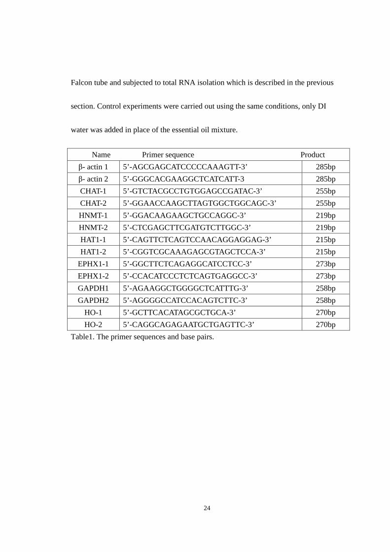
24
Falcon tube and subjected to total RNA isolation which is described in the previous
section. Control experiments were carried out using the same conditions, only DI
water was added in place of the essential oil mixture.
Table1. The primer sequences and base pairs.
Name Primer sequence Product
β- actin 1 5’-AGCGAGCATCCCCCAAAGTT-3’ 285bp
β- actin 2 5’-GGGCACGAAGGCTCATCATT-3 285bp
CHAT-1 5’-GTCTACGCCTGTGGAGCCGATAC-3’ 255bp
CHAT-2 5’-GGAACCAAGCTTAGTGGCTGGCAGC-3’ 255bp
HNMT-1 5’-GGACAAGAAGCTGCCAGGC-3’ 219bp
HNMT-2 5’-CTCGAGCTTCGATGTCTTGGC-3’ 219bp
HAT1-1 5’-CAGTTCTCAGTCCAACAGGAGGAG-3’ 215bp
HAT1-2 5’-CGGTCGCAAAGAGCGTAGCTCCA-3’ 215bp
EPHX1-1 5’-GGCTTCTCAGAGGCATCCTCC-3’ 273bp
EPHX1-2 5’-CCACATCCCTCTCAGTGAGGCC-3’ 273bp
GAPDH1 5’-AGAAGGCTGGGGCTCATTTG-3’ 258bp
GAPDH2 5’-AGGGGCCATCCACAGTCTTC-3’ 258bp
HO-1 5’-GCTTCACATAGCGCTGCA-3’ 270bp
HO-2 5’-CAGGCAGAGAATGCTGAGTTC-3’ 270bp
25
2.2.4 Time dependent induction of phase II genes
Experiments were also carried out in an attempt to establish the kinetics of
induction of these genes. To do this, HepG2 cells were treated with lemongrass oil at
the dose of 20ug/ml, as indicated in the previous section. After treatment, cells were
grown in 25 cm2 tissue culture flasks at 37oC for additional 0hr, 2hrs, 6hrs and 24hrs.
The cells were then trypsinized and incubated in the 37oC environment for 5 minutes.
Following this incubation, cells were harvested by scraping from the flask. The cells
were collected in a 15ml Falcon tube and subjected to total RNA isolation as
described in the previous sections. Samples were analyzed by RT-PCR in an attempt
to determine kinetics of induction. Control experiments were carried out using the
same conditions, only DI water was added in place of the essential oil mixture.
2.2.5 Effect of essential oil of Lemongrass on CYP3A4 activity
As with the 2E1 study, the effect of Lemongrass on CYP3A4 activity was
determined. A specific assay for monitoring CYP3A4 has been developed utilizing
the specific ability of this enzyme to oxidize nifedipine. Initially an assay for the
oxidation of the nifedipine by CYP3A4 had to be developed. Using five different
types of microsomes; human liver microsomes, rabbit liver microsomes 1, rabbit
liver microsomes 2, rat liver microsomes, and supersomes enriched in CYP3A4,
26
experiments were carried out in the absence and presence of NADPH. The
experimental procedure used for the evaluation of CYP3A4 inhibition was as
follows: Along with 20ul of the five different kinds of microsomes, 50ul of 0.1M
potassium phosphate buffer solution (pH 7.4), 40ul of 1mM Nifedipine and
deionized water were initially combined in a final volume of 0.5ml. 25ul of 20mM
NADPH was added to initiate the reaction in a 37oC water bath for 10 minutes, then
the reaction was quenched with the addition of 1ml of dichloromethane and 100ul of
1M Na2CO3 buffer (pH 10.5) containing 2M NaCl. Samples were extracted by
liquid-liquid extraction then centrifuged at 4000 rpm for 10 minutes and 1ml of
organic layer was transferred to centrifuge tube, reduced to dryness at 23oC under
vacuum and 500ul of 45% methanol was added to the centrifuge tube, transferred to
HPLC vials, and analyzed by HPLC(SHIMADZU) for product formation. The
product of Nifedipine oxidation, oxidized Nifedipine, was monitored using a
Shimadzu LC 20A Series HPLC system consisting of an SPD-20A UV/Vis detector,
LC 20AT solvent delivery, and a Sil 20A autosampler, all controlled using the
Shimadzu EZStart version 7.3 SP1 software. Absorbance detection was set to 254
nm with a mobile phase consisting of 60% methanol, 40% H2O, 0.5% acetic acid at
a flow rate of 0.6ml/min. The volume of injection was 40 ul for each sample and the
27
column was a RP-C18 HPLC column 100cm x 3.0mm. All assays were carried out
in duplicate. After the trials with five different microsomes, rat liver microsomes
were chosen to perform the oxidation of the nifedipine by CYP3A4 assay due to the
greater activity of these microsomes in the oxidation of nifedipine. First, the screen
experiments of both Lemongrass oil and the aldehyde citral were carried out to
determine the potency of inhibition of CYP3A4 with 20ul of rat microsomes, 50ul of
1.0M potassium phosphate buffer solution (pH 7.4), 40ul of 1mM Nifedipine , and
deionized water were initially combined in a final volume of 0.5ml. 25ul of 20mM
NADPH was added to initiate the reaction in a 37oC. After establishing the potency
of inhibition of CYP3A4, Michaelis-Menten plots were used for the evaluation of
CYP3A4 inhibition by Lemongrass oil and the aldehyde citral. 5ul of the
Lemongrass oil or the aldehyde citral was diluted to 100ml in deionized water and
5ul of this solution, along with 20ul of rat microsomes, 50ul of 1.0M potassium
phosphate buffer solution (pH 7.4), 0.2mM nifedipine and deionized water were
combined in a final volume of 0.5ml. 25ul of 20mM NADPH was added to initiate
the reaction in a 37oC water bath for 10 minutes, then the reaction was quenched
with the addition of 1ml of dichloromethane and 100ul of 1M Na2CO3 buffer (pH
10.5) containing 2M NaCl. Samples were extracted again by liquid-liquid extraction
28
then centrifuged at 3000 rpm for 10 minutes and 1ml of organic layer was
transferred to centrifuge tube, reduced to dryness at 23oC under vacuum. 500ul of
45% methanol was added to the centrifuge tube, transferred to HPLC vials, and
analyzed by HPLC(SHIMADZU) for product formation. The product of Nifedipine
oxidation, oxidized Nifedipine, was monitored using the Shimadzu HPLC as
described previously.

29
CHAPTER III
RESULTS AND DISCUSSION
3.1 Inhibition of CYP2E1 by Lemongrass oil and other essential oils
3.1.1 Introduction
The inhibition of CYP2E1 by natural compounds may have significant
implications in the field of pharmacology or toxicology, given the role of this isoform
in production of ROS or reactive drug metabolites. CYP2E1 metabolizes and activates
many toxicological substrates to more toxic products and the induction of CYP2E1 by
ethanol is thought to result in increased oxidative stress in hepatocytes. One proposed
mechanism for this increased oxidative stress is the increased production of hydrogen
peroxide by CYP2E1 via uncoupling of its NADPH oxidation activity. The main
hypothesis of this thesis is that the main aldehyde constituent found in Lemongrass,
Citral, will be able to block the activity of CYP2E1, and demonstrate physiological
antioxidant properties. For this reason we initiated studies to probe the interaction
between a variety of oils and pure aldehydes with CYP2E1, specifically to determine
whether these oils or aldehydes possessed inhibitory properties toward this enzyme.
30
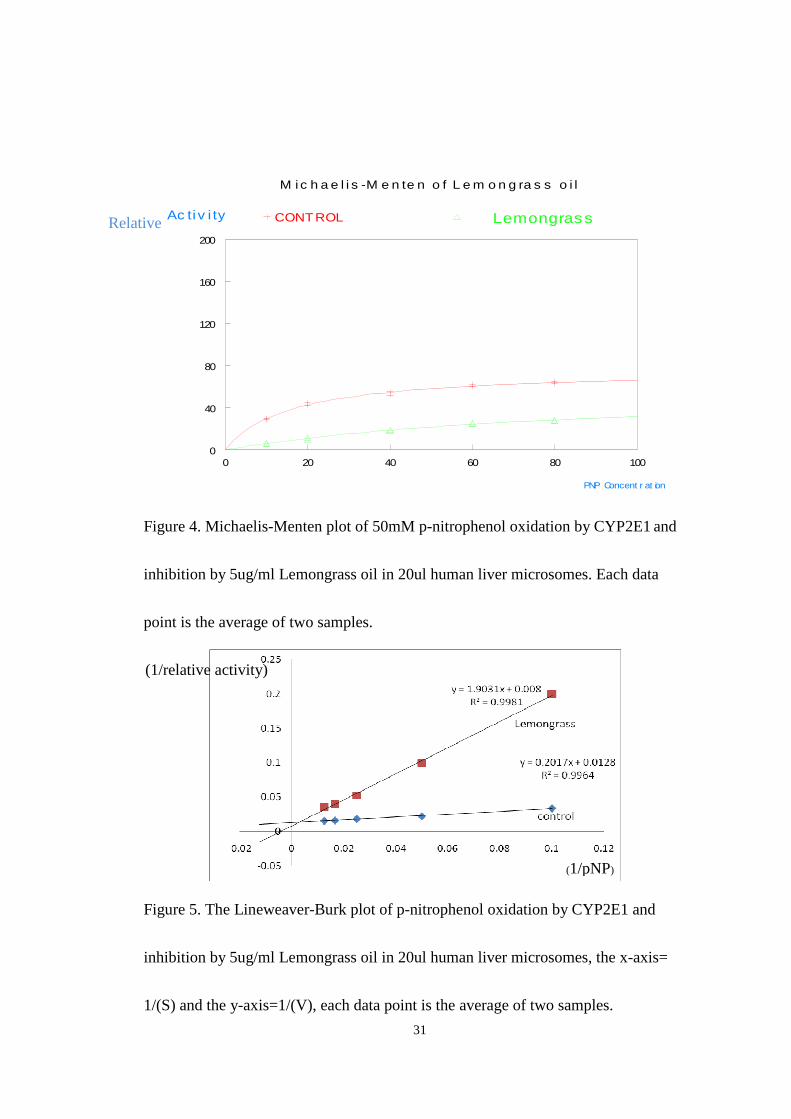
Furthermore the mechanism of inhibition was also addressed.
3.1.2 Michaelis-Menten kinetic analysis
3.1.2.A. Inhibition of CYP2E1 by Lemongrass oil and the aldehyde citral
Figure 4 and figure 5 show the results of inhibition studies carried out by using
a single dose of Lemongrass oil of 5ug/ml. The final concentration of p-nitrophenol
in the reaction was (10-100uM) and that of NADPH was 1.0mM. The
Michaelis-Menten plot for Lemongrass (shown in Fig.4) shows significant enzyme
inhibition across the entire range of substrate concentrations used. Because of the
selectivity of CYP2E1 in the human liver microsomes for p-nitrophenol, the
inhibition effect observed must be due to reduction in activity of this isoform. The
observed value for Vmax of 59 and KM of 87 in the presence of inhibitor compared
to the control Vmax of 76 and KM of 16 suggest a competitive type inhibition. The
K I value was evaluated using the equation KM’=KM(1+[I]/K I) and was found to be
1.1ug/ml. The lineweaver-Burk plot (Fig.5) although the curves do not cross
precisely at the Y axis is consistent with the competitive model of inhibition.
31
0 20 40 60 80 1000
40
80
120
160
200
CONTROL LemongrassAc tiv i ty
PNP Concent r at ion
M i c h a e l i s -M e n te n o f L e m o n g ra s s o i l
Figure 4. Michaelis-Menten plot of 50mM p-nitrophenol oxidation by CYP2E1 and
inhibition by 5ug/ml Lemongrass oil in 20ul human liver microsomes. Each data
point is the average of two samples.
Figure 5. The Lineweaver-Burk plot of p-nitrophenol oxidation by CYP2E1 and
inhibition by 5ug/ml Lemongrass oil in 20ul human liver microsomes, the x-axis=
1/(S) and the y-axis=1/(V), each data point is the average of two samples.
(1/pNP)
(1/relative activity)
Relative
32
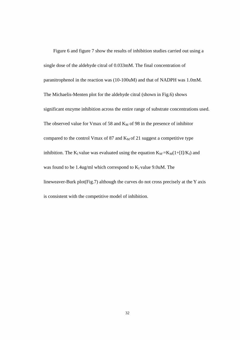
Figure 6 and figure 7 show the results of inhibition studies carried out using a
single dose of the aldehyde citral of 0.033mM. The final concentration of
paranitrophenol in the reaction was (10-100uM) and that of NADPH was 1.0mM.
The Michaelis-Menten plot for the aldehyde citral (shown in Fig.6) shows
significant enzyme inhibition across the entire range of substrate concentrations used.
The observed value for Vmax of 58 and KM of 98 in the presence of inhibitor
compared to the control Vmax of 87 and KM of 21 suggest a competitive type
inhibition. The KI value was evaluated using the equation KM’=KM(1+[I]/K I) and
was found to be 1.4ug/ml which correspond to KI value 9.0uM. The
lineweaver-Burk plot(Fig.7) although the curves do not cross precisely at the Y axis
is consistent with the competitive model of inhibition.

33
0 20 40 60 80 1000
40
80
120
160
200
CONTROL CitralAc tiv i ty
PNP Concent r at ion
M i c h a e l i s -M e n te n o f Ci tra l a l d e h y d e
Figure 6. Michaelis-Menten plot of 50mM p-nitrophenol oxidation by CYP2E1 and
inhibition by 0.033mM the aldehyde citral in 20ul human liver microsomes. Each
data point is the average of two samples.
Figure 7. The Lineweaver-Burk plot of p-nitrophenol oxidation by CYP2E1 and
inhibition by the 0.033mM aldehyde citral in 20ul human liver microsomes, the
x-axis= 1/(S) and the y-axis=1/(V), each data point is the average of two samples.
(1/pNP)
(1/relative activity)
Relative
34
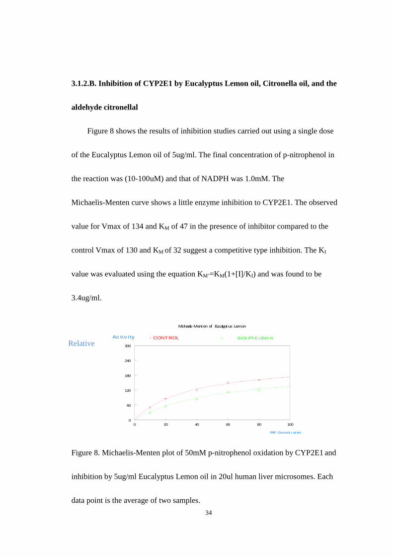
3.1.2.B. Inhibition of CYP2E1 by Eucalyptus Lemon oil, Citronella oil, and the
aldehyde citronellal
Figure 8 shows the results of inhibition studies carried out using a single dose
of the Eucalyptus Lemon oil of 5ug/ml. The final concentration of p-nitrophenol in
the reaction was (10-100uM) and that of NADPH was 1.0mM. The
Michaelis-Menten curve shows a little enzyme inhibition to CYP2E1. The observed
value for Vmax of 134 and KM of 47 in the presence of inhibitor compared to the
control Vmax of 130 and KM of 32 suggest a competitive type inhibition. The KI
value was evaluated using the equation KM’=KM(1+[I]/K I) and was found to be
3.4ug/ml.
0 20 40 60 80 1000
60
120
180
240
300
CONT ROL EUCALYPTUS LEMONAc tiv i ty
PNP Concent r at ion
Michaelis-Menten of Eucalyptus Lemon
Figure 8. Michaelis-Menten plot of 50mM p-nitrophenol oxidation by CYP2E1 and
inhibition by 5ug/ml Eucalyptus Lemon oil in 20ul human liver microsomes. Each
data point is the average of two samples.
Relative

35
Figure 9 shows the results of inhibition studies carried out using a single dose
of the Citronella oil of 5ug/ml. The final concentration of p-nitrophenol in the
reaction was (10-100uM) and that of NADPH was 1.0mM. The Michaelis-Menten
curve shows slight enzyme inhibition for the citronella oil. The observed value for
Vmax of 149 and KM of 55 in the presence of inhibitor compared to the control
Vmax of 149 and KM of 29 suggest a competitive type inhibition. The KI value was
evaluated using the equation KM’=KM(1+[I]/K I) and was found to be 5.6ug/L.
0 20 40 60 80 1000
40
80
120
160
200
CONTROL Citronel lar elat ive act ivit y
PNP Concent r at ion
Michael is -Menten of Citronel la
Figure 9. Michaelis-Menten plot of 50mM p-nitrophenol oxidation by CYP2E1 and
inhibition by 5ug/ml Citronella oil in the 20ul human liver microsomes. Each data
point is the average of two samples.
36
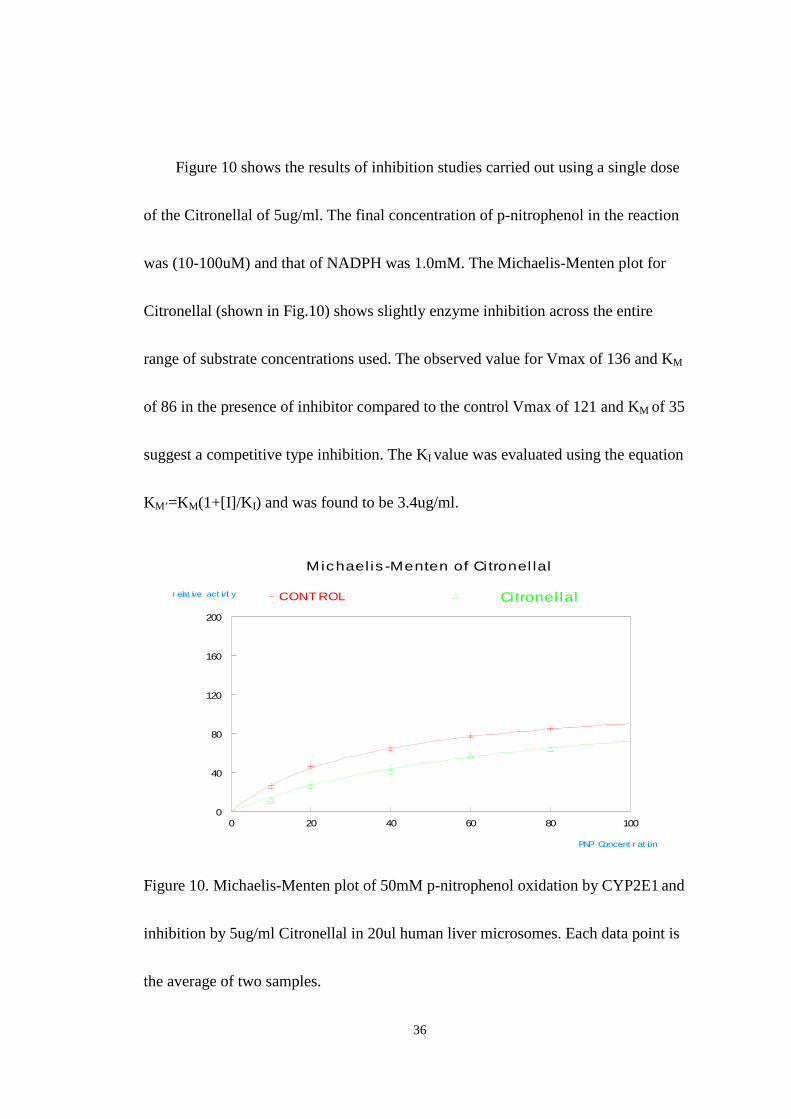
Figure 10 shows the results of inhibition studies carried out using a single dose
of the Citronellal of 5ug/ml. The final concentration of p-nitrophenol in the reaction
was (10-100uM) and that of NADPH was 1.0mM. The Michaelis-Menten plot for
Citronellal (shown in Fig.10) shows slightly enzyme inhibition across the entire
range of substrate concentrations used. The observed value for Vmax of 136 and KM
of 86 in the presence of inhibitor compared to the control Vmax of 121 and KM of 35
suggest a competitive type inhibition. The KI value was evaluated using the equation
KM’=KM(1+[I]/K I) and was found to be 3.4ug/ml.
0 20 40 60 80 1000
40
80
120
160
200
CONTROL Citronel lalr elat ive act ivit y
PNP Concent r at ion
Michael is -Menten of Citronel lal
Figure 10. Michaelis-Menten plot of 50mM p-nitrophenol oxidation by CYP2E1 and
inhibition by 5ug/ml Citronellal in 20ul human liver microsomes. Each data point is
the average of two samples.
37
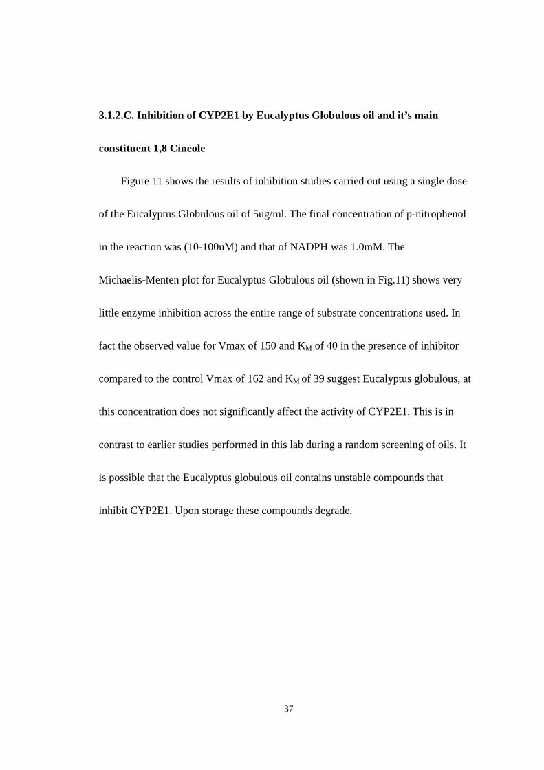
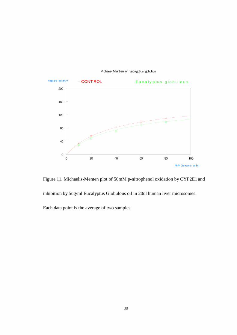
3.1.2.C. Inhibition of CYP2E1 by Eucalyptus Globulous oil and it’s main
constituent 1,8 Cineole
Figure 11 shows the results of inhibition studies carried out using a single dose
of the Eucalyptus Globulous oil of 5ug/ml. The final concentration of p-nitrophenol
in the reaction was (10-100uM) and that of NADPH was 1.0mM. The
Michaelis-Menten plot for Eucalyptus Globulous oil (shown in Fig.11) shows very
little enzyme inhibition across the entire range of substrate concentrations used. In
fact the observed value for Vmax of 150 and KM of 40 in the presence of inhibitor
compared to the control Vmax of 162 and KM of 39 suggest Eucalyptus globulous, at
this concentration does not significantly affect the activity of CYP2E1. This is in
contrast to earlier studies performed in this lab during a random screening of oils. It
is possible that the Eucalyptus globulous oil contains unstable compounds that
inhibit CYP2E1. Upon storage these compounds degrade.
38
0 20 40 60 80 1000
40
80
120
160
200
CONT ROL Eu c a l y p tu s g l o b u l o u sr elat ive act ivit y
PNP Concent r at ion
Michaelis-Ment en of Eucalypt us globulous
Figure 11. Michaelis-Menten plot of 50mM p-nitrophenol oxidation by CYP2E1 and
inhibition by 5ug/ml Eucalyptus Globulous oil in 20ul human liver microsomes.
Each data point is the average of two samples.
39
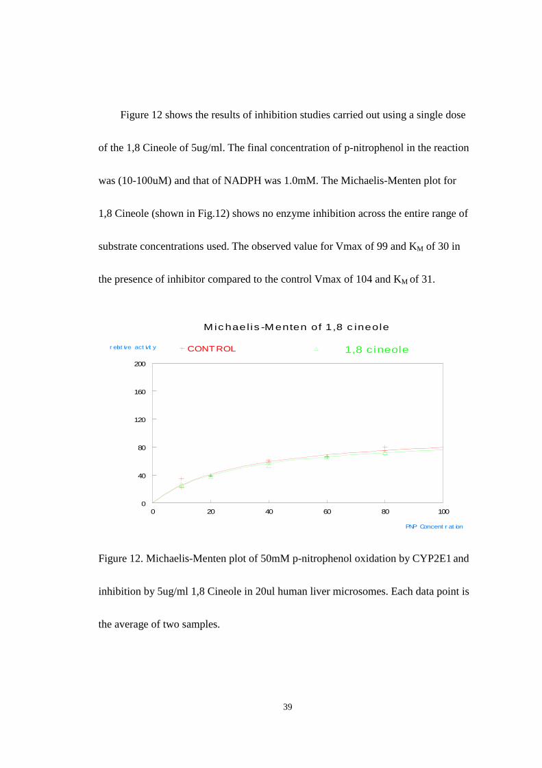
Figure 12 shows the results of inhibition studies carried out using a single dose
of the 1,8 Cineole of 5ug/ml. The final concentration of p-nitrophenol in the reaction
was (10-100uM) and that of NADPH was 1.0mM. The Michaelis-Menten plot for
1,8 Cineole (shown in Fig.12) shows no enzyme inhibition across the entire range of
substrate concentrations used. The observed value for Vmax of 99 and KM of 30 in
the presence of inhibitor compared to the control Vmax of 104 and KM of 31.
0 20 40 60 80 1000
40
80
120
160
200
CONTROL 1,8 c ineoler elat ive act ivit y
PNP Concent r at ion
Mic hael is -Menten of 1,8 c ineole
Figure 12. Michaelis-Menten plot of 50mM p-nitrophenol oxidation by CYP2E1 and
inhibition by 5ug/ml 1,8 Cineole in 20ul human liver microsomes. Each data point is
the average of two samples.
40
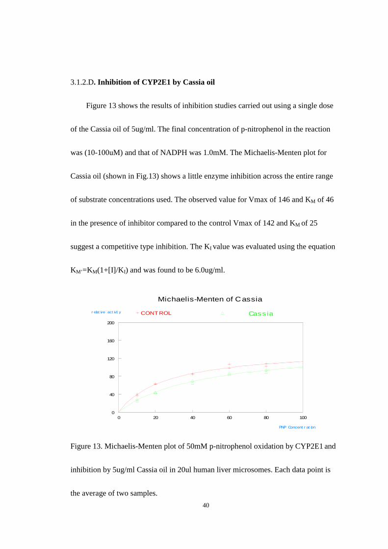
3.1.2.D. Inhibition of CYP2E1 by Cassia oil
Figure 13 shows the results of inhibition studies carried out using a single dose
of the Cassia oil of 5ug/ml. The final concentration of p-nitrophenol in the reaction
was (10-100uM) and that of NADPH was 1.0mM. The Michaelis-Menten plot for
Cassia oil (shown in Fig.13) shows a little enzyme inhibition across the entire range
of substrate concentrations used. The observed value for Vmax of 146 and KM of 46
in the presence of inhibitor compared to the control Vmax of 142 and KM of 25
suggest a competitive type inhibition. The KI value was evaluated using the equation
KM’=KM(1+[I]/K I) and was found to be 6.0ug/ml.
0 20 40 60 80 1000
40
80
120
160
200
CONT ROL Cassiar elat ive act ivit y
PNP Concent r at ion
Michaelis-Menten of Cassia
Figure 13. Michaelis-Menten plot of 50mM p-nitrophenol oxidation by CYP2E1 and
inhibition by 5ug/ml Cassia oil in 20ul human liver microsomes. Each data point is
the average of two samples.

41
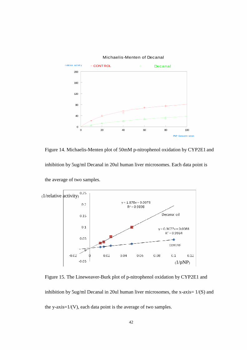
3.1.2.E. Inhibition of CYP2E1 by the aldehyde decanal
Figure 14 and figure 15 shows the results of inhibition studies carried out using
a single dose of Decanal of 5ug/ml. The final concentration of p-nitrophenol in the
reaction was (10-100uM) and that of NADPH was 1.0mM. The Michaelis-Menten
plot for Decanal (shown in Fig.14) shows that decanal is a very efficient inhibitor of
CYP2E1 at this dose. The observed value for Vmax of 81 and KM of 177 in the
presence of inhibitor compared to the control Vmax of 113 and KM of 40 suggest a
competitive type inhibition. The KI value was evaluated using the equation
KM’=KM(1+[I]/K I) and was found to be 2.6ug/ml. The lineweaver-Burk plot (Fig.15)
although the curves do not cross precisely at the Y axis is consistent with the
competitive model of inhibition.
42
0 20 40 60 80 1000
40
80
120
160
200
CONTROL Decanalr elat ive act ivit y
PNP Concent r at ion
Michaelis-Menten of Decanal
Figure 14. Michaelis-Menten plot of 50mM p-nitrophenol oxidation by CYP2E1 and
inhibition by 5ug/ml Decanal in 20ul human liver microsomes. Each data point is
the average of two samples.
Figure 15. The Lineweaver-Burk plot of p-nitrophenol oxidation by CYP2E1 and
inhibition by 5ug/ml Decanal in 20ul human liver microsomes, the x-axis= 1/(S) and
the y-axis=1/(V), each data point is the average of two samples.
(1/pNP)
(1/relative activity)
43
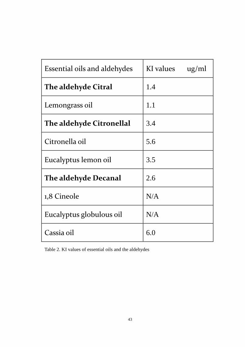
Essential oils and aldehydes KI values ug/ml
The aldehyde Citral 1.4
Lemongrass oil 1.1
The aldehyde Citronellal 3.4
Citronella oil 5.6
Eucalyptus lemon oil 3.5
The aldehyde Decanal 2.6
1,8 Cineole N/A
Eucalyptus globulous oil N/A
Cassia oil 6.0
Table 2. KI values of essential oils and the aldehydes
44
3.2 Growth and treatment of HepG2 cells
This experiment was to show Lemongrass oil,which contains the aldehyde
constituent Citral, can activate the antioxidant response elements found in a variety of
antioxidant/phase II drug metabolizing genes. In order to demonstrate the induction of
phase II genes, cultured human liver cells, HepG2 cells, were used to test the
induction of genes of interest and these cells were treated with Lemongrass oil and the
aldehyde citral. The RT-PCR and gel electrophoresis were used to monitor antioxidant
genes expression.
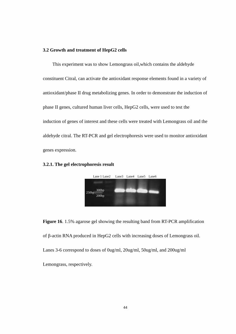
3.2.1. The gel electrophoresis result
Figure 16. 1.5% agarose gel showing the resulting band from RT-PCR amplification
of β-actin RNA produced in HepG2 cells with increasing doses of Lemongrass oil.
Lanes 3-6 correspond to doses of 0ug/ml, 20ug/ml, 50ug/ml, and 200ug/ml
Lemongrass, respectively.
Lane 1 Lane2 Lane3 Lane4 Lane5 Lane6
250bp 300bp
200bp
45
Figure 16 shows the transcriptional level of β-actin-1 with increasing doses of
the Lemongrass oil applied to the HepG2 cells. Based on visual inspection of the
bands, the β-actin levels appear to be similar, although lane6 may be slightly less
intense than the others, which may suggest a small change in expression of β-actin at
the very highest concentration of oil. Lane1 is the DNA ladder, lane 2 is also DNA
ladder, lane3 is the control without Lemongrass, lane4 is the sample of HepG2 cell
treated with 0.02 ug/ml of Lemongrass oil, lane5 is the sample of HepG2 cell treated
with 0.05 ug/ml of Lemongrass oil, lane 6 is the sample of HepG2 cell treated with
0.2 ug/ml of Lemongrass oil. β-actin in each sample was confirmed by the presence of
a PCR product at just under 300bp in length. Several experimental conclusions had to
be tested before consistent amplification of the β-actin gene was observed, more
importantly the most effective annealing temperature for the PCR was determined to
be 56 oC.
46
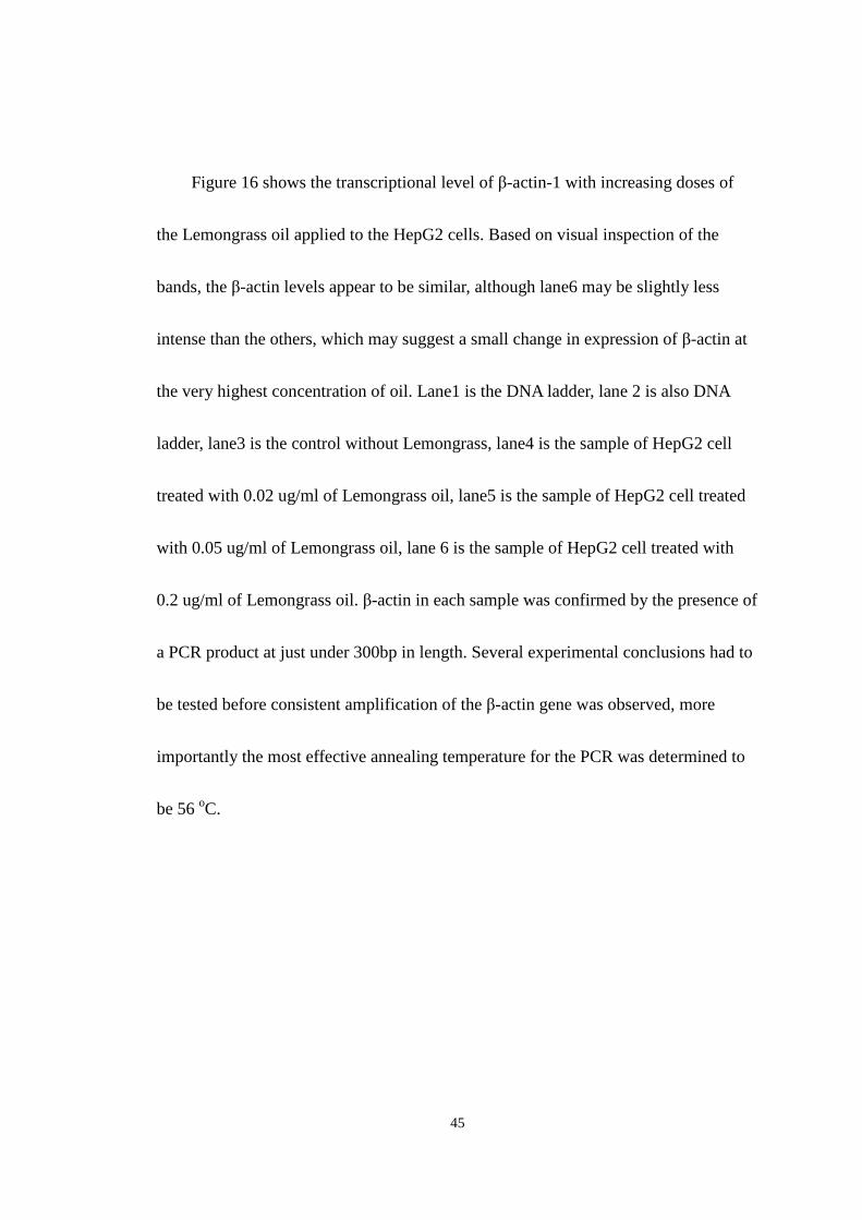
Figure 17. 1.5% agarose gel showing the resulting band from RT-PCR amplification
of β-actin RNA produced in HepG2 cells at 20ug/ml Lemongrass oil time-dependent
experiment. Lanes 2-5 correspond to the time of 0hr, 2hrs, 6hrs, and 24hrs
respectively.
Figure 17 shows the expression of β-actin-1 for time dependent when HepG2
cells were treated with 20ug/ml Lemongrass oil. In this experiment lane1 is the DNA
ladder, lane2 is the HepG2 cells treated with Lemongrass oil at 20ug/ml for 0hr
incubation, lane3 is the HepG2 cells treated with Lemongrass oil at 20ug/ml for 1hr
incubation, lane4 is the HepG2 cells treated with Lemongrass oil at 20ug/ml for 6hr
incubation, lane5 is the induction of HepG2 cells treated with Lemongrass oil at
20ug/ml for 24hr incubation post treatment. It appeared that the intensity of 4 bands
were uneven, however, lane 4 had lower intensity compared to the other lanes, which
may be the result of artifact, perhaps loading error. And lane 5 had stronger intensity
which could be the number of the cells were more than the other samples.
Lane 1 Lane2 Lane3 Lane4 Lane 5
250bp
47
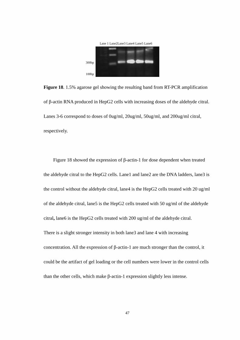
Figure 18. 1.5% agarose gel showing the resulting band from RT-PCR amplification
of β-actin RNA produced in HepG2 cells with increasing doses of the aldehyde citral.
Lanes 3-6 correspond to doses of 0ug/ml, 20ug/ml, 50ug/ml, and 200ug/ml citral,
respectively.
Figure 18 showed the expression of β-actin-1 for dose dependent when treated
the aldehyde citral to the HepG2 cells. Lane1 and lane2 are the DNA ladders, lane3 is
the control without the aldehyde citral, lane4 is the HepG2 cells treated with 20 ug/ml
of the aldehyde citral, lane5 is the HepG2 cells treated with 50 ug/ml of the aldehyde
citral, lane6 is the HepG2 cells treated with 200 ug/ml of the aldehyde citral.
There is a slight stronger intensity in both lane3 and lane 4 with increasing
concentration. All the expression of β-actin-1 are much stronger than the control, it
could be the artifact of gel loading or the cell numbers were lower in the control cells
than the other cells, which make β-actin-1 expression slightly less intense.
Lane 1 Lane2Lane3 Lane4 Lane5 Lane6
300bp
100bp

48
Figure 19. 1.5% agarose gel showing the resulting band from RT-PCR amplification
of β-actin RNA produced in HepG2 cells at 20ug/ml Citral time-dependent
experiment. Lanes 2-5 correspond to the time of 0hr, 2hrs, 6hrs, and 24hrs
respectively.
Figure 19 shows the expression of β-actin-1 for time dependent when HepG2
cells were treated with 20ug/ml Citral. In this experiment lane1 is the DNA ladder,
lane2 is the HepG2 cells treated with Citral at 20ug/ml for 0hr incubation, lane3 is the
HepG2 cells treated with Citral at 20ug/ml for 2hrs incubation, lane4 is the HepG2
cells treated with Citral at 20ug/ml for 6hrs incubation, lane5 is the induction of
HepG2 cells treated with Citral at 20ug/ml for 24hrs incubation post treatment. There
is a increasing trend of β-actin expression with increasing time of citral treated which
may be the numbers of cells were different and made the expression of β-actin
uneven.
Lane 1 Lane2 Lane3 Lane4 Lane5
250bp
49
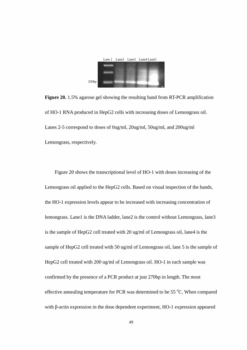
Figure 20. 1.5% agarose gel showing the resulting band from RT-PCR amplification
of HO-1 RNA produced in HepG2 cells with increasing doses of Lemongrass oil.
Lanes 2-5 correspond to doses of 0ug/ml, 20ug/ml, 50ug/ml, and 200ug/ml
Lemongrass, respectively.
Figure 20 shows the transcriptional level of HO-1 with doses increasing of the
Lemongrass oil applied to the HepG2 cells. Based on visual inspection of the bands,
the HO-1 expression levels appear to be increased with increasing concentration of
lemongrass. Lane1 is the DNA ladder, lane2 is the control without Lemongrass, lane3
is the sample of HepG2 cell treated with 20 ug/ml of Lemongrass oil, lane4 is the
sample of HepG2 cell treated with 50 ug/ml of Lemongrass oil, lane 5 is the sample of
HepG2 cell treated with 200 ug/ml of Lemongrass oil. HO-1 in each sample was
confirmed by the presence of a PCR product at just 270bp in length. The most
effective annealing temperature for PCR was determined to be 55 oC. When compared
with β-actin expression in the dose dependent experiment, HO-1 expression appeared
Lane 1 Lane2 Lane3 Lane4 Lane5
250bp

50
to increase with increasing dose of lemongrass. This may also correspond to
increasing antioxidant properties induced by lemongrass oil.
51
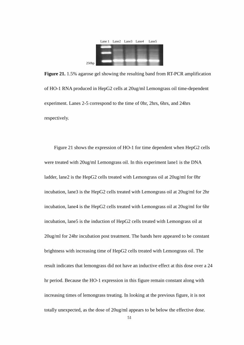
Figure 21. 1.5% agarose gel showing the resulting band from RT-PCR amplification
of HO-1 RNA produced in HepG2 cells at 20ug/ml Lemongrass oil time-dependent
experiment. Lanes 2-5 correspond to the time of 0hr, 2hrs, 6hrs, and 24hrs
respectively.
Figure 21 shows the expression of HO-1 for time dependent when HepG2 cells
were treated with 20ug/ml Lemongrass oil. In this experiment lane1 is the DNA
ladder, lane2 is the HepG2 cells treated with Lemongrass oil at 20ug/ml for 0hr
incubation, lane3 is the HepG2 cells treated with Lemongrass oil at 20ug/ml for 2hr
incubation, lane4 is the HepG2 cells treated with Lemongrass oil at 20ug/ml for 6hr
incubation, lane5 is the induction of HepG2 cells treated with Lemongrass oil at
20ug/ml for 24hr incubation post treatment. The bands here appeared to be constant
brightness with increasing time of HepG2 cells treated with Lemongrass oil. The
result indicates that lemongrass did not have an inductive effect at this dose over a 24
hr period. Because the HO-1 expression in this figure remain constant along with
increasing times of lemongrass treating. In looking at the previous figure, it is not
totally unexpected, as the dose of 20ug/ml appears to be below the effective dose.
Lane 1 Lane2 Lane3 Lane4 Lane5
250bp

52
Therefore, to examine the time dependence of this induction, future studies should be
carried out at 100ug/ml or higher.
53
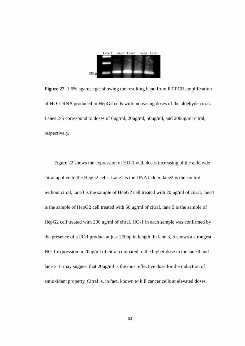
Figure 22. 1.5% agarose gel showing the resulting band from RT-PCR amplification
of HO-1 RNA produced in HepG2 cells with increasing doses of the aldehyde citral.
Lanes 2-5 correspond to doses of 0ug/ml, 20ug/ml, 50ug/ml, and 200ug/ml citral,
respectively.
Figure 22 shows the expression of HO-1 with doses increasing of the aldehyde
citral applied to the HepG2 cells. Lane1 is the DNA ladder, lane2 is the control
without citral, lane3 is the sample of HepG2 cell treated with 20 ug/ml of citral, lane4
is the sample of HepG2 cell treated with 50 ug/ml of citral, lane 5 is the sample of
HepG2 cell treated with 200 ug/ml of citral. HO-1 in each sample was confirmed by
the presence of a PCR product at just 270bp in length. In lane 3, it shows a strongest
HO-1 expression in 20ug/ml of citral compared to the higher dose in the lane 4 and
lane 5. It may suggest that 20ug/ml is the most effective dose for the induction of
antioxidant property. Citral is, in fact, known to kill cancer cells at elevated doses.
Lane 1 Lane2 Lane3 Lane4 Lane5
250bp
54
Figure 23. 1.5% agarose gel showing the resulting band from RT-PCR amplification
of HO-1 RNA produced in HepG2 cells at 20ug/ml the aldehyde citral time-dependent
experiment. Lanes 2-5 correspond to the time of 0hr, 2hrs, 6hrs, and 24hrs
respectively.
Figure 23 shows the expression of HO-1 for time dependent when HepG2 cells
were treated with 20ug/ml the aldehyde citral. In this experiment lane1 is the DNA
ladder, lane2 is the HepG2 cells treated with the aldehyde citral at 20ug/ml for 0hr
incubation, lane3 is the HepG2 cells treated with the aldehyde citral at 20ug/ml for
2hr incubation, lane4 is the HepG2 cells treated with the aldehyde citral at 20ug/ml
for 6hr incubation, lane5 is the induction of HepG2 cells treated with the aldehyde
citral at 20ug/ml for 24hr incubation post treatment. Both lane 3 and lane 4 have the
same intensity of HO-1 expression compare to lane 1 and lane 4, it shows that with 2
and 6 hours citral treating, there is a stronger antioxidant property. Moreover the
effect of antioxidant may be lesser when the cell treated with citral for 24 hours,
probably due to metabolism of the citral. Liver enzymes, including cytochrome
Lane 1 Lane2 Lane3 Lane4 Lane5
250bp
55
P450’s, are known to degrade this compound in a matter of hours.
3.3 Inhibition of CYP3A4 by nifedipine oxidation
The assay for monitoring CYP3A4 were developed utilizing the specific ability
of this enzyme to oxidize nifedipine and rat microsomes were used in the assay.
Experiments were carried out in the absence and presence of NADPH for identifying
the peak of oxidized nifedipine on the chromatogram. The experimental procedure
used for the evaluation of CYP3A4 inhibition was as follows: Along with 20ul of rat
microsomes, 50ul of 1M potassium phosphate buffer solution (pH 7.4), 40ul of 1mM
Nifedipine and deionized water were initially combined in a final volume of 0.5ml.
25ul of 1mM NADPH was added to initiate the reaction in a 37oC water bath for 10
minutes, then the reaction was quenched with the addition of 1ml of dichloromethane
and 100ul of 1M Na2CO3 buffer (pH 10.5) containing 2M NaCl. Samples were
extracted by liquid-liquid extraction then centrifuged at 3000 rpm for 10 minutes and
1ml of organic layer was transferred to centrifuge tube, reduced to dryness at 23oC
under vacuum, added 500ul of 45% methanol to centrifuge tube, transferred to HPLC
vials, and analyzed by HPLC(SHIMADZU) for product formation. The product of
Nifedipine oxidation, oxidized Nifedipine, was monitored using a Shimadzu LC 20A
Series HPLC system consisting of an SPD-20A UV/Vis detector, LC 20AT solvent
56
delivery, and a Sil 20A autosampler, all controlled using the Shimadzu EZStart
version 7.3 SP1 software. Absorbance detection was set to 254 nm with a mobile
phase consisting of 60% methanol, 40% H2O, 0.5% acetic acid at a flow rate of
0.6ml/min. The volume of injection was 40 ul for each sample and the column was a
RP-C18 HPLC column. All assays were carried out in duplicate.
3.3.1. The retention time of Nifedipine and Oxidized nifedipine was identified
Figure 24 shows the peaks of standard oxidized nifedipine and standard
nifedipine on the HPLC with UV detection. The peak at 4.0 minute was identified as
the retention time of standard oxidized nifedipine using an authentic standard
purchased for Oxford Biomedical and the peak at 6.2 minute was identified as the
retention time of nifedipine.
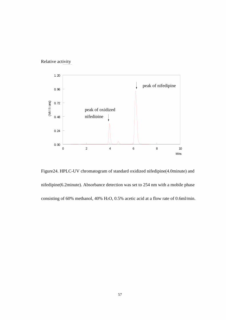
57
0 2 4 6 8 100. 00
0. 24
0. 48
0. 72
0. 96
1. 20
(Mil
lion
s)
Mins
Figure24. HPLC-UV chromatogram of standard oxidized nifedipine(4.0minute) and
nifedipine(6.2minute). Absorbance detection was set to 254 nm with a mobile phase
consisting of 60% methanol, 40% H2O, 0.5% acetic acid at a flow rate of 0.6ml/min.
peak of oxidized
nifedipine
peak of nifedipine
Relative activity
58
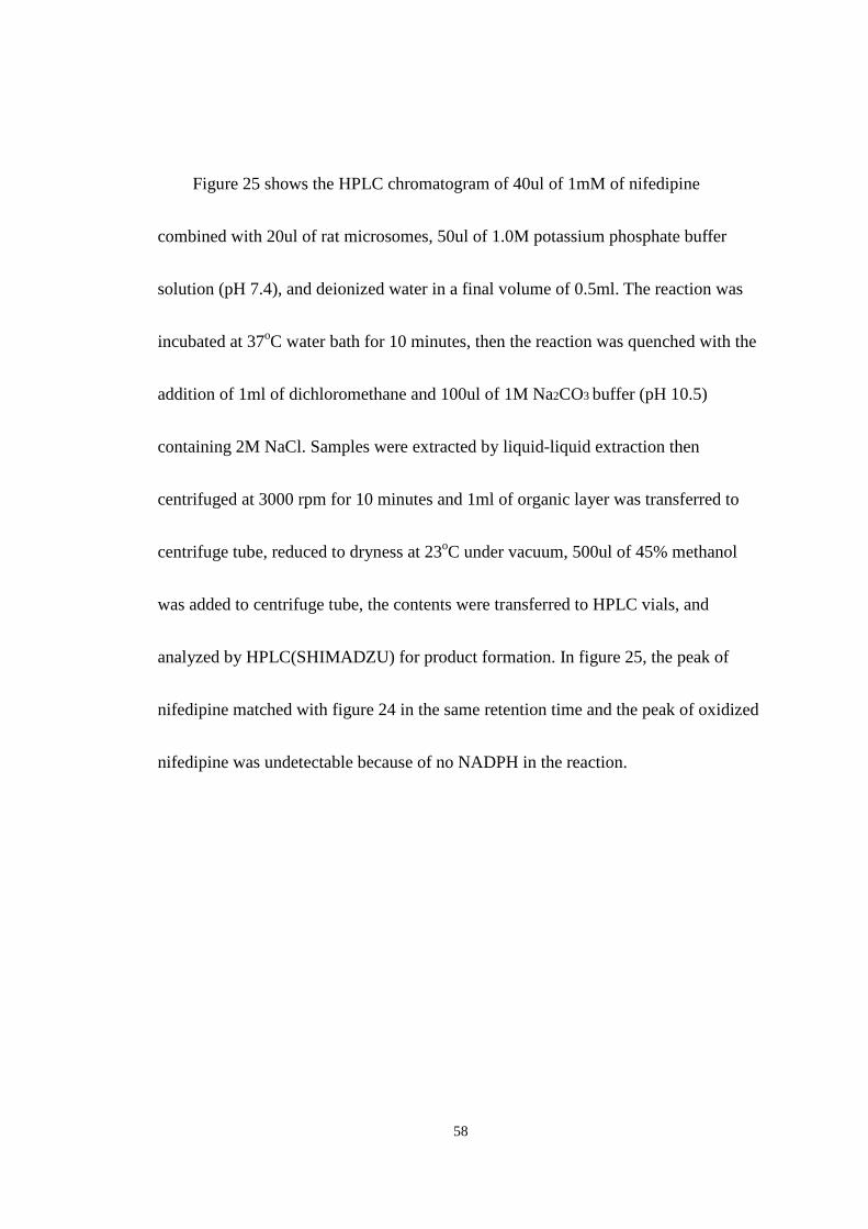
Figure 25 shows the HPLC chromatogram of 40ul of 1mM of nifedipine
combined with 20ul of rat microsomes, 50ul of 1.0M potassium phosphate buffer
solution (pH 7.4), and deionized water in a final volume of 0.5ml. The reaction was
incubated at 37oC water bath for 10 minutes, then the reaction was quenched with the
addition of 1ml of dichloromethane and 100ul of 1M Na2CO3 buffer (pH 10.5)
containing 2M NaCl. Samples were extracted by liquid-liquid extraction then
centrifuged at 3000 rpm for 10 minutes and 1ml of organic layer was transferred to
centrifuge tube, reduced to dryness at 23oC under vacuum, 500ul of 45% methanol
was added to centrifuge tube, the contents were transferred to HPLC vials, and
analyzed by HPLC(SHIMADZU) for product formation. In figure 25, the peak of
nifedipine matched with figure 24 in the same retention time and the peak of oxidized
nifedipine was undetectable because of no NADPH in the reaction.

59
0 2 4 6 8 100
2
4
6
8
10
(Tho
usan
ds)
Mins
Figure25. HPLC-UV chromatogram of nifedipine(6.2minute) when the reaction
incubated without the addition of NADPH. Absorbance detection was set to 254 nm
with a mobile phase consisting of 60% methanol, 40% H2O, 0.5% acetic acid at a
flow rate of 0.6ml/min.
Peak of nifedipine
Relative activity
60
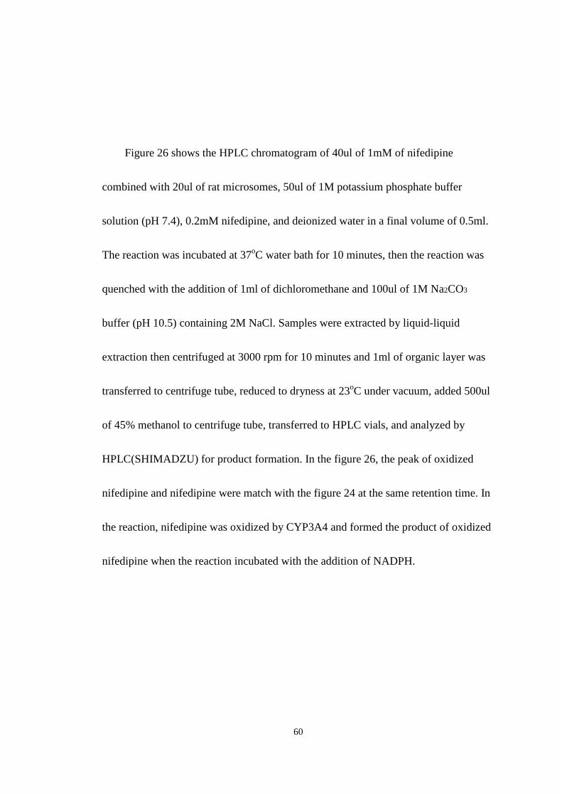
Figure 26 shows the HPLC chromatogram of 40ul of 1mM of nifedipine
combined with 20ul of rat microsomes, 50ul of 1M potassium phosphate buffer
solution (pH 7.4), 0.2mM nifedipine, and deionized water in a final volume of 0.5ml.
The reaction was incubated at 37oC water bath for 10 minutes, then the reaction was
quenched with the addition of 1ml of dichloromethane and 100ul of 1M Na2CO3
buffer (pH 10.5) containing 2M NaCl. Samples were extracted by liquid-liquid
extraction then centrifuged at 3000 rpm for 10 minutes and 1ml of organic layer was
transferred to centrifuge tube, reduced to dryness at 23oC under vacuum, added 500ul
of 45% methanol to centrifuge tube, transferred to HPLC vials, and analyzed by
HPLC(SHIMADZU) for product formation. In the figure 26, the peak of oxidized
nifedipine and nifedipine were match with the figure 24 at the same retention time. In
the reaction, nifedipine was oxidized by CYP3A4 and formed the product of oxidized
nifedipine when the reaction incubated with the addition of NADPH.
61
0 2 4 6 8 100. 00
2. 20
4. 40
6. 60
8. 80
11. 00
(Tho
usan
ds)
Mins
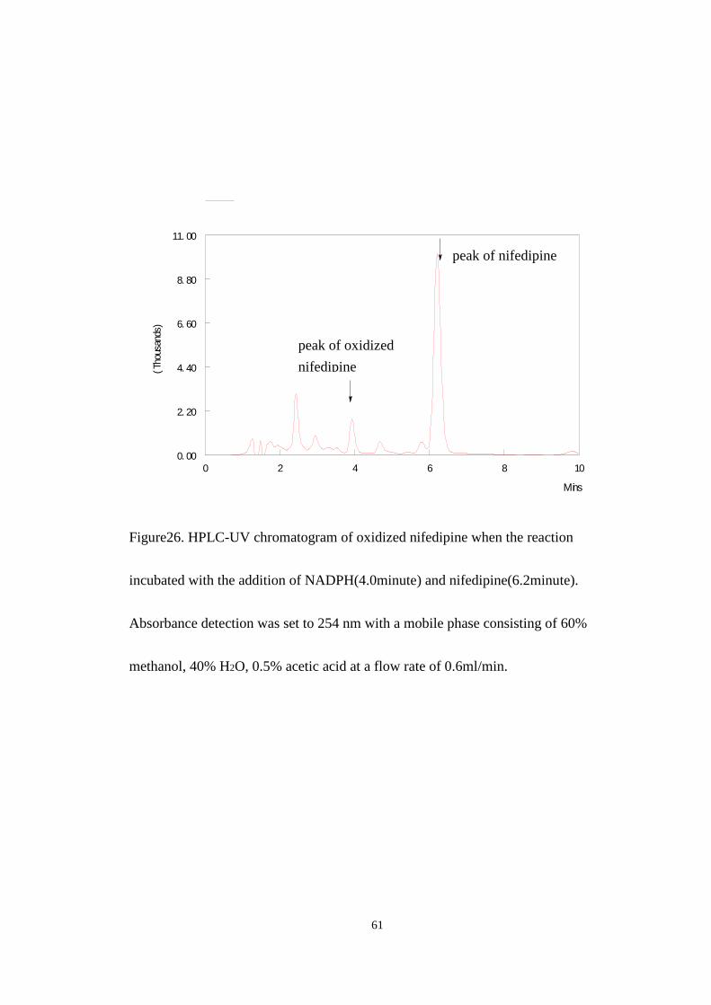
Figure26. HPLC-UV chromatogram of oxidized nifedipine when the reaction
incubated with the addition of NADPH(4.0minute) and nifedipine(6.2minute).
Absorbance detection was set to 254 nm with a mobile phase consisting of 60%
methanol, 40% H2O, 0.5% acetic acid at a flow rate of 0.6ml/min.
peak of nifedipine
peak of oxidized
nifedipine
62
3.3.2. The screen experiments of nifedipine oxidation by CYP3A4 with and
without Lemongrass oil and the aldehyde citral
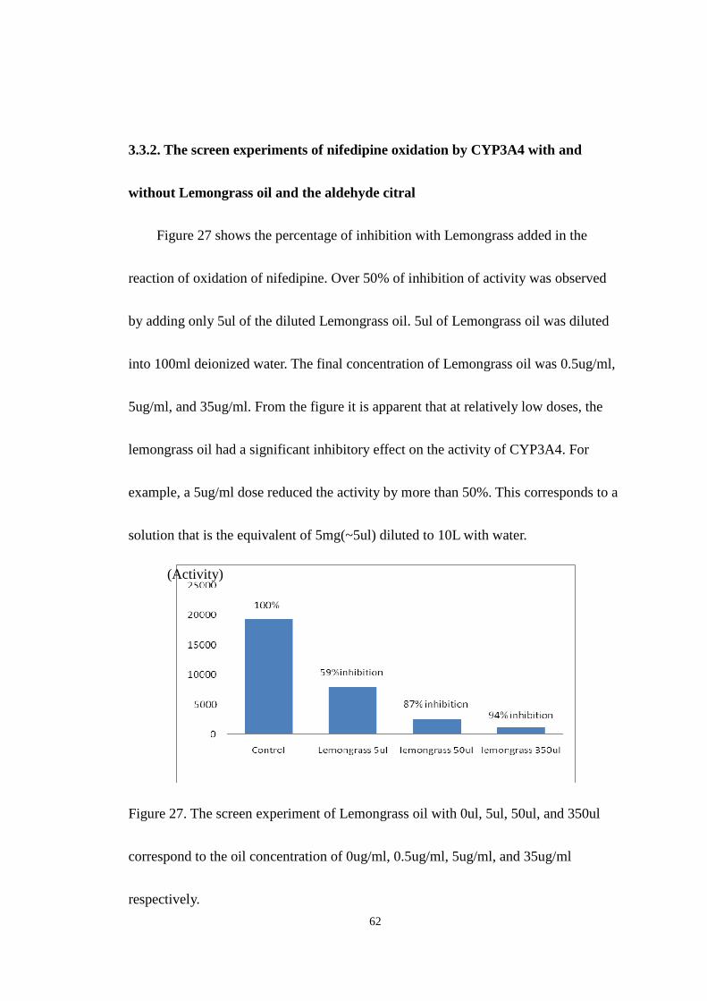
Figure 27 shows the percentage of inhibition with Lemongrass added in the
reaction of oxidation of nifedipine. Over 50% of inhibition of activity was observed
by adding only 5ul of the diluted Lemongrass oil. 5ul of Lemongrass oil was diluted
into 100ml deionized water. The final concentration of Lemongrass oil was 0.5ug/ml,
5ug/ml, and 35ug/ml. From the figure it is apparent that at relatively low doses, the
lemongrass oil had a significant inhibitory effect on the activity of CYP3A4. For
example, a 5ug/ml dose reduced the activity by more than 50%. This corresponds to a
solution that is the equivalent of 5mg(~5ul) diluted to 10L with water.
Figure 27. The screen experiment of Lemongrass oil with 0ul, 5ul, 50ul, and 350ul
correspond to the oil concentration of 0ug/ml, 0.5ug/ml, 5ug/ml, and 35ug/ml
respectively.
(Activity)
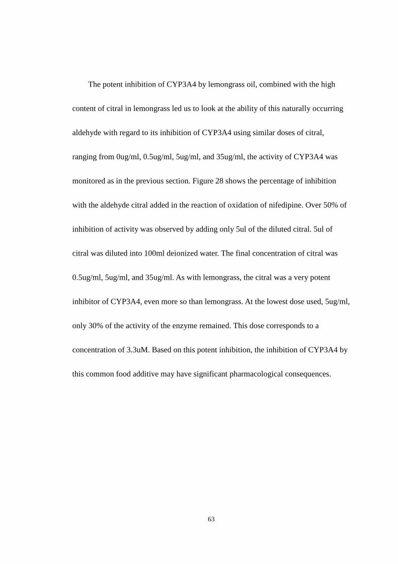
63
The potent inhibition of CYP3A4 by lemongrass oil, combined with the high
content of citral in lemongrass led us to look at the ability of this naturally occurring
aldehyde with regard to its inhibition of CYP3A4 using similar doses of citral,
ranging from 0ug/ml, 0.5ug/ml, 5ug/ml, and 35ug/ml, the activity of CYP3A4 was
monitored as in the previous section. Figure 28 shows the percentage of inhibition
with the aldehyde citral added in the reaction of oxidation of nifedipine. Over 50% of
inhibition of activity was observed by adding only 5ul of the diluted citral. 5ul of
citral was diluted into 100ml deionized water. The final concentration of citral was
0.5ug/ml, 5ug/ml, and 35ug/ml. As with lemongrass, the citral was a very potent
inhibitor of CYP3A4, even more so than lemongrass. At the lowest dose used, 5ug/ml,
only 30% of the activity of the enzyme remained. This dose corresponds to a
concentration of 3.3uM. Based on this potent inhibition, the inhibition of CYP3A4 by
this common food additive may have significant pharmacological consequences.
64
Figure28. The screen experiment of citral with 0ul, 5ul, 50ul, and 350ul correspond to
the oil concentration of 0ug/ml, 0.5ug/ml, 5ug/ml, and 35ug/ml respectively.
(Activity)
65
CHAPTER IV
DISCUSSION
From the Michaelis-Menten plots for inhibition of CYP2E1, there was a
correlation seen between the aldehyde content and the potency with regard to
cytochrome P4502E1 inhibition. Lemongrass, which contains the unsaturated aldehyde
citral, showed a significant level of cytochrome CYP2E1 inhibition. The essential oil
of lemongrass contains very high amounts of the aldehyde citral, which is the name
for the pair of cis, trans isomers neral and geranial. Citral is an α,β-unsaturated
aldehyde and has been shown to possess anti-inflammitory properties that may relate
to its ability to control redox balance within the cell. The anti-inflammitory response
results from suppression of iNOS expression and NF-kB activation. The relationship
of both of these aldehydes to a reduction in oxidative stress and their ability to inhibit
the human cytochrome P4502E1 isoform suggests that the two effects may be related,
given the involvement of this isoform in initiating lipid peroxidation.
According to my experimental data and gel electrophoresis experimental results, the
HO-1 showed significant increased expression in human liver cells in response to

66
increasing concentrations of both Lemongrass oil and the aldehyde citral. Furthermore,
the time-dependence experiment for both Lemongrass oil and the aldehyde citral also
showed the levels of mRNA for these genes increased in a time dependent manner up
to 8 hrs incubation. For the gene expression of HO-1, in the dose-dependence
experiment, it showed an increasing trend with respective to the increasing
concentration of Lemongrass oil and the aldehyde citral in the agarose gel
electrophoresis from the control sample to the highest concentration one, which
directly validated that the presence of Lemongrass oil induced phase II detoxyfying
genes in the HepG2 cells with increasing doses. It also assertively demonstrated the
hypothesis that Citral which is a component of Lemongrass oil plays the major role of
the induction of phase II detoxyfying genes. In the dose-dependence experiment,
Citral caused significant increases in the expression of HO-1 genes. This experimental
result suggests that the Citral is the main component of Lemongrass which induces
the expression of HO-1. From prior studies we know the activation of HO-1 is a
ubiquitous cellular response to oxidative stress, which produces ROS, such as
hydrogen peroxide and ultimately leads to lipid peroxidation. The expression of HO-1
gene is related to the production of biliverdin and its subsequent metabolite bilirubin,

67
both products have antioxidant properties against oxidative stress caused by
xenobiotics, oxidants, UV light, and radiation (33). In addition to the dose-dependent
experiment, I also performed time-dependent experiment to show the time manner of
the induction of HO-1. With the different period of time, the bands showed stronger
band with increasing time of oil treatment in both Lemongrass oil and Citral in the gel
electrophoresis up to 8hrs. The 24hr time resulted in a return to basal expression
levels, which may result from the metabolism of control over that time period. This
experiment results lead to the conclusion that with the same concentration of oil but
different time treating periods, the induction of HO-1 is time dependent.
In the method development of CYP3A4 inhibition assay, CYP3A4 is the enzyme to
metabolize Nifedipine to oxidized nifedipine. In order to identify the retention time of
Nifedipine and oxidized nifedipine, standard compounds of both Nifedipine and
oxidized nifedipine were acquired and injected onto HPLC and the retention time of
both compounds were determined using a variety of different mobile phases. After
identification of the peaks of Nifedipine and oxidized nifedipine and determination of
the exact retention time on the chromatogram, incubations were performed to
demonstrate the Nifedipine was oxidized to oxidized nifedipine by using 5 different
microsomes which included human liver microsomes, rabbit liver microsomes 1,

68
rabbit liver micorsomes 2, rat liver microsomes and supersomes which is enriched of
CYP3A4. The chromatograms showed that there was a significant absorbance for
oxidized nifedipine in the retention time of 3.8minutes with the presence of
supersomes and rat liver microsomes when the NADPH was added in the reaction. In
the human liver microsomes and rabbit liver microsomes 1, there was very little or no
significant difference for both with NADPH and without NADPH reaction. Finally, it
was shown that both lemongrass and citral inhibit CYP3A4 significantly at very low
micromolar concentrations. This may have very important implications in drug
metabolism as CYP3A4 is extremely important pharmacologically, in that it is
responsible for metabolism of nifedipine.
69
REFERENCES
1. Human Molecular Genetics 14, 18 2749–2755 (2005)
2. European Journal of Pharmacology 545, 39–50 (2006)
3. Toxicological Science 64, 1–3 (2001)
4. Proceedings of the National Academy of Sciences USA 91, 10771-10778 (1994)
5. Mutation Research 379, 201–210 (1997)
6. Human Reproduction 13, 6 1429–1436 (1998)
7. The Journal of Clinical Endocrinology & Metabolism 86, 6 2899-2902 (2001)
8. The Journal of Clinical Investigation 115, 3 (2005)
9. Asian Journal of Andrology 3, 135-138 (2001)
10. Toxicology and Applied Pharmacology 146, 180–188 (1997)
11. Environment Science Technology, 36, 364-372 (2002)
12. Analytical Chemistry 79, 5103-5106 (2007)
13. Free Radical Biology & Medicine 40, 1349–1361 (2006)
14. Free Radical Biology & Medicine 40, 1119-1130 (2006)
15. Free Radical Biology & Medicine 36, 3 307-318 (2004)
16. Nature Medicine 6, 422 – 428 (2000)

70
17. Journal of Experimental Medicine 192, 1015– 1026 (2000)
18. Proceedings of the National Academy of Sciences USA 94, 10925–10930 (1997)
19. Kidney International 53, 100–111 (1998)
20. Science 235, 1043–1046 (1987)
21. Biochimica et Biophysica Acta 1223, 9 – 14 (1994)
22. Biochemical and Biophysical Research Communications, 303 105–111 (2003)
23. Biochemical Pharmacology Lecture Notes by Michael Palmer, Department of
Chemistry, University of Waterloo, Canada 3rd edition (2007)
24. Biochemical and Biophysical Research Communications 298, 687–692 (2002)
25. International Journal of Pharmaceutics 337, 169–177 (2007)
26. Archives of Biochemistry and Biophysics 483, 66–74 (2009)
27. International Journal of Pharmaceutics 236, 111–123 (2002)
28. Toxicology in Vitro 21, 1592–1602 (2007)
29. Mutation Research 551, 153–166 (2004)
30. Journal of Pharmaceutical and Biomedical Analysis 22, 241–249 (2000)
31. Toxicology and Applied Pharmacology 216, 282–292 (2006)
32. Biochemical Pharmacology 50, 2 155-161 (1995)
33. Free Radical Biology & Medicine 28, 2 289–309 (2000)
71
34. Food and Chemical Toxicology 44, 1075–1081 (2006)
35. International Journal of Pharmaceutics 337, 169–177 (2007)
36. Food Chemistry 106, 698–705 (2008)
37. Toxicology 96, 105-113 (1995)
38. Reproductive Toxicology 24, 326–332 (2007)
39. Entomological Research 35(2), 117–120 (2005)
40. Food Chemistry 112, 120 (2008)
41. Journal of Inorganic Biochemistry 81, 153 –160 (2000)