Embed Size (px)
Citation preview
1
Chemistry 5.07SC Biological Chemistry I
Fall Semester, 2013
Lecture 14 An overview of glycolysis.
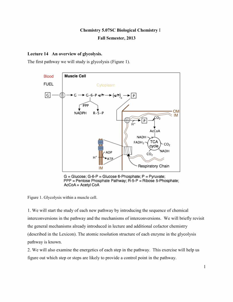
The first pathway we will study is glycolysis (Figure 1).
Figure 1. Glycolysis within a muscle cell.
1. We will start the study of each new pathway by introducing the sequence of chemical
interconversions in the pathway and the mechanisms of interconversions. We will briefly revisit
the general mechanisms already introduced in lecture and additional cofactor chemistry
(described in the Lexicon). The atomic resolution structure of each enzyme in the glycolysis
pathway is known.
2. We will also examine the energetics of each step in the pathway. This exercise will help us
figure out which step or steps are likely to provide a control point in the pathway.
2
3. After the TCA cycle and gluconeogenesis pathways are introduced, the big picture in terms of
REGULATION will be examined. How do cells decide whether to oxidize glucose or synthesize
glucose? How do cells prevent both processes from occurring simultaneously?
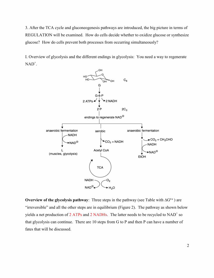
I. Overview of glycolysis and the different endings in glycolysis: You need a way to regenerate
NAD+.
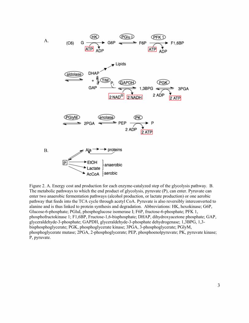
Overview of the glycolysis pathway: Three steps in the pathway (see Table with ∆Gº’) are
“irreversible” and all the other steps are in equilibrium (Figure 2). The pathway as shown below
yields a net production of 2 ATPs and 2 NADHs. The latter needs to be recycled to NAD+ so
that glycolysis can continue. There are 10 steps from G to P and then P can have a number of
fates that will be discussed.
3
A.
B.
Figure 2. A. Energy cost and production for each enzyme-catalyzed step of the glycolysis pathway. B. The metabolic pathways to which the end product of glycolysis, pyruvate (P), can enter. Pyruvate can enter two anaerobic fermentation pathways (alcohol production, or lactate production) or one aerobic pathway that feeds into the TCA cycle through acetyl CoA. Pyruvate is also reversibly interconverted to alanine and is thus linked to protein synthesis and degradation. Abbreviations: HK, hexokinase; G6P, Glucose-6-phosphate; PGluI, phosphoglucose isomerase I; F6P, fructose-6-phosphate; PFK 1, phosphofructokinase 1; F1,6BP, Fructose-1,6-bisphosphate; DHAP, dihydroxyacetone phosphate; GAP, glyceraldehyde-3-phosphate; GAPDH, glyceraldehyde-3-phosphate dehydrogenase; 1,3BPG, 1,3-bisphosphoglycerate; PGK, phosphoglycerate kinase; 3PGA, 3-phosphoglycerate; PGlyM, phosphoglycerate mutase; 2PGA, 2-phosphoglycerate; PEP, phosphoenolpyruvate; PK, pyruvate kinase; P, pyruvate.
4
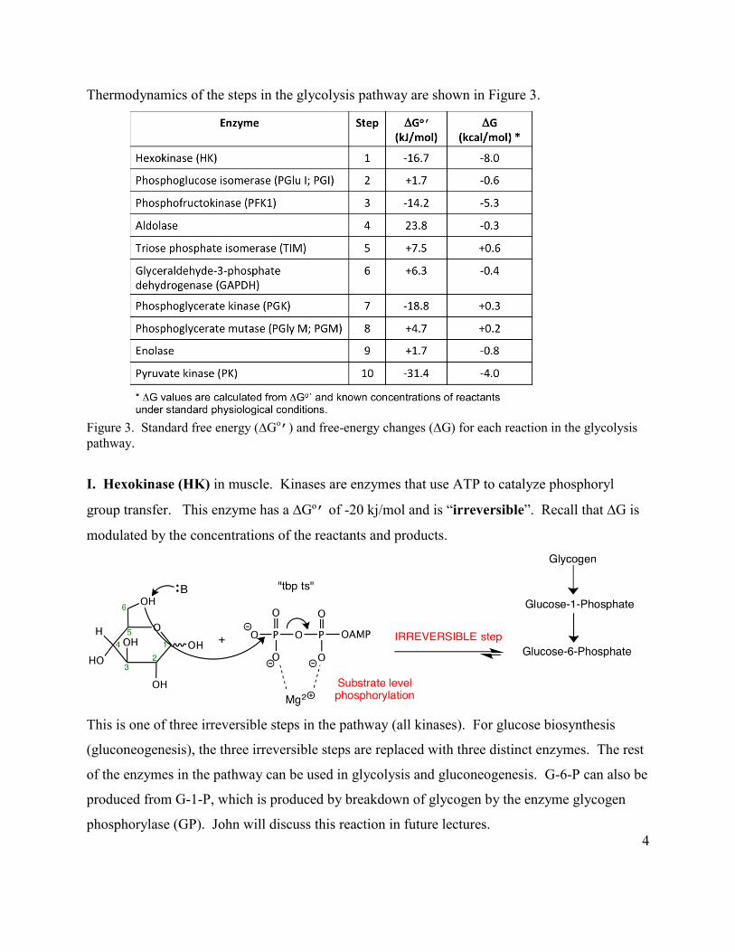
Thermodynamics of the steps in the glycolysis pathway are shown in Figure 3.
Figure 3. Standard free energy (∆Go
’) and free-energy changes (∆G) for each reaction in the glycolysis pathway.
I. Hexokinase (HK) in muscle. Kinases are enzymes that use ATP to catalyze phosphoryl
group transfer. This enzyme has a ∆Gº’ of -20 kj/mol and is “irreversible”. Recall that ∆G is
modulated by the concentrations of the reactants and products.
This is one of three irreversible steps in the pathway (all kinases). For glucose biosynthesis
(gluconeogenesis), the three irreversible steps are replaced with three distinct enzymes. The rest
of the enzymes in the pathway can be used in glycolysis and gluconeogenesis. G-6-P can also be
produced from G-1-P, which is produced by breakdown of glycogen by the enzyme glycogen
phosphorylase (GP). John will discuss this reaction in future lectures.
5
New general principle:
E1 E2 E3 Catabolic pathway A B C D E F G H I
F1 F2 F3 Anabolic pathway A B C D E F G H I
General principles of HK revisited:
1. Need to neutralize the negative charges on phosphate for the nucleophile to attack. As
previously discussed, Mg2+, K and R from the protein are key players in this role.
2. Water is phosphorylated at 1/40000 the rate of the C6 hydroxyl of glucose, despite similar pKa
and being smaller. This is another example of specificity that we discussed in the lecture on the
amazing properties of enzymes (see Lecture 7/8).
3. This was the first enzyme where the importance of the order of addition of substrates was
demonstrated crystallographically. Glucose must add before ATP. The structure was solved by
the Steitz group, the same group that won the Nobel Prize for the structure of the ribosome in
2009.
4. Kinases, as noted in the lecture on tertiary structure, have conserved ATP binding domains
and conserved amino acid motifs that allow them to be annotated as kinases in the data bases.
5. Phosphorylation keeps the metabolite (glucose) inside the cell. Phosphorylated species in
general cannot freely diffuse across any membrane.
Hexokinase was the first example of a protein where a huge conformational change was
observed on binding substrate (purple): Open (Figure 4A) and closed (Figure 4B) states.
6
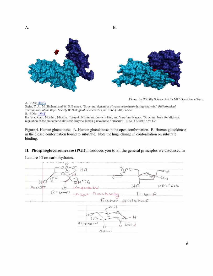
A. B.
Figure by O'Reilly Science Art for MIT OpenCourseWare.A. PDB: 1HKGSteitz, T. A., M. Shoham, and W. S. Bennett. "Structural dynamics of yeast hexokinase during catalysis." Philosophical Transactions of the Royal Society B: Biological Sciences 293, no. 1063 (1981): 43-52.B. PDB: 1V4TKamata, Kenji, Morihiro Mitsuya, Teruyuki Nishimura, Jun-ichi Eiki, and Yasufumi Nagata. "Structural basis for allosteric regulation of the monomeric allosteric enzyme human glucokinase." Structure 12, no. 3 (2004): 429-438.
Figure 4. Human glucokinase. A. Human glucokinase in the open conformation. B. Human glucokinase in the closed conformation bound to substrate. Note the huge change in conformation on substrate binding.
II. Phosphoglucoisomerase (PGI) introduces you to all the general principles we discussed in
Lecture 13 on carbohydrates.
7
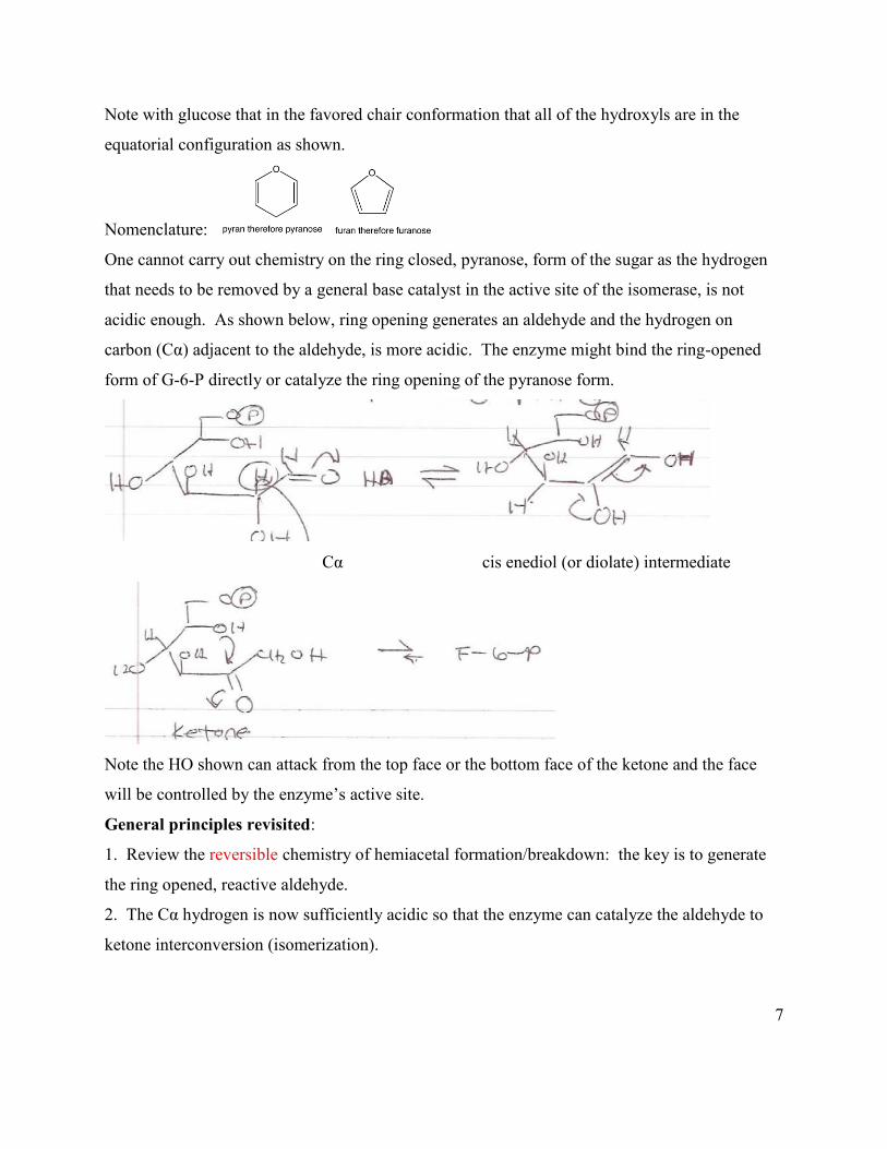
Note with glucose that in the favored chair conformation that all of the hydroxyls are in the
equatorial configuration as shown.
Nomenclature:
One cannot carry out chemistry on the ring closed, pyranose, form of the sugar as the hydrogen
that needs to be removed by a general base catalyst in the active site of the isomerase, is not
acidic enough. As shown below, ring opening generates an aldehyde and the hydrogen on
carbon (Cα) adjacent to the aldehyde, is more acidic. The enzyme might bind the ring-opened
form of G-6-P directly or catalyze the ring opening of the pyranose form.
Cα cis enediol (or diolate) intermediate
Note the HO shown can attack from the top face or the bottom face of the ketone and the face
will be controlled by the enzyme’s active site.
General principles revisited:
1. Review the reversible chemistry of hemiacetal formation/breakdown: the key is to generate
the ring opened, reactive aldehyde.
2. The Cα hydrogen is now sufficiently acidic so that the enzyme can catalyze the aldehyde to
ketone interconversion (isomerization).
8
III. PFK 1 (phosphofructokinase 1) is a key regulatory step in the pathway. It also has ∆Gº’
= -17 kj/mol, similar to HK.
Note that the regulation of this enzyme, which will be discussed after the introduction of
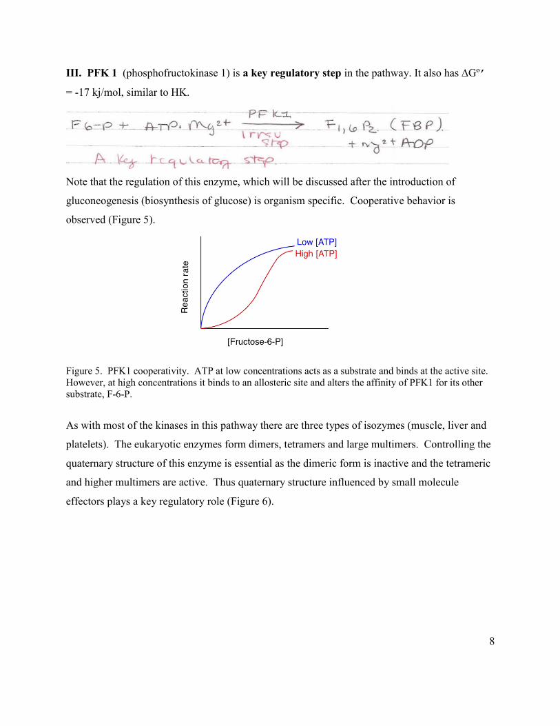
gluconeogenesis (biosynthesis of glucose) is organism specific. Cooperative behavior is
observed (Figure 5).
Figure 5. PFK1 cooperativity. ATP at low concentrations acts as a substrate and binds at the active site. However, at high concentrations it binds to an allosteric site and alters the affinity of PFK1 for its other substrate, F-6-P.
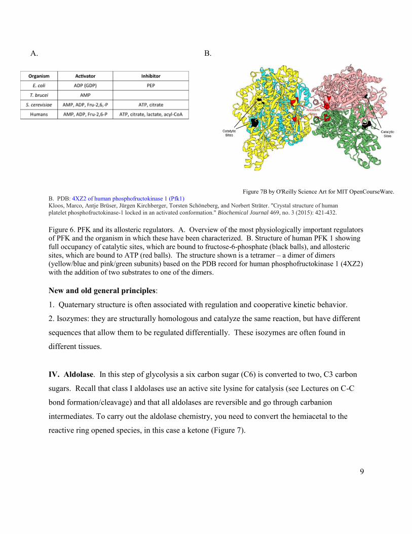
As with most of the kinases in this pathway there are three types of isozymes (muscle, liver and
platelets). The eukaryotic enzymes form dimers, tetramers and large multimers. Controlling the
quaternary structure of this enzyme is essential as the dimeric form is inactive and the tetrameric
and higher multimers are active. Thus quaternary structure influenced by small molecule
effectors plays a key regulatory role (Figure 6).
9
A. B.
Figure 7B by O'Reilly Science Art for MIT OpenCourseWare.
B. PDB: 4XZ2 of human phosphofructokinase 1 (Pfk1) Kloos, Marco, Antje Brüser, Jürgen Kirchberger, Torsten Schöneberg, and Norbert Sträter. "Crystal structure of human platelet phosphofructokinase-1 locked in an activated conformation." Biochemical Journal 469, no. 3 (2015): 421-432. Figure 6. PFK and its allosteric regulators. A. Overview of the most physiologically important regulators of PFK and the organism in which these have been characterized. B. Structure of human PFK 1 showing full occupancy of catalytic sites, which are bound to fructose-6-phosphate (black balls), and allosteric sites, which are bound to ATP (red balls). The structure shown is a tetramer – a dimer of dimers (yellow/blue and pink/green subunits) based on the PDB record for human phosphofructokinase 1 (4XZ2) with the addition of two substrates to one of the dimers. New and old general principles:
1. Quaternary structure is often associated with regulation and cooperative kinetic behavior.
2. Isozymes: they are structurally homologous and catalyze the same reaction, but have different
sequences that allow them to be regulated differentially. These isozymes are often found in
different tissues.
IV. Aldolase. In this step of glycolysis a six carbon sugar (C6) is converted to two, C3 carbon
sugars. Recall that class I aldolases use an active site lysine for catalysis (see Lectures on C-C
bond formation/cleavage) and that all aldolases are reversible and go through carbanion
intermediates. To carry out the aldolase chemistry, you need to convert the hemiacetal to the
reactive ring opened species, in this case a ketone (Figure 7).
10
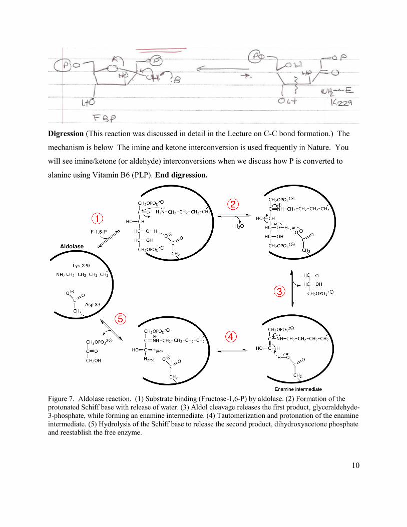
Digression (This reaction was discussed in detail in the Lecture on C-C bond formation.) The
mechanism is below The imine and ketone interconversion is used frequently in Nature. You
will see imine/ketone (or aldehyde) interconversions when we discuss how P is converted to
alanine using Vitamin B6 (PLP). End digression.
Figure 7. Aldolase reaction. (1) Substrate binding (Fructose-1,6-P) by aldolase. (2) Formation of the protonated Schiff base with release of water. (3) Aldol cleavage releases the first product, glyceraldehyde-3-phosphate, while forming an enamine intermediate. (4) Tautomerization and protonation of the enamine intermediate. (5) Hydrolysis of the Schiff base to release the second product, dihydroxyacetone phosphate and reestablish the free enzyme.
11
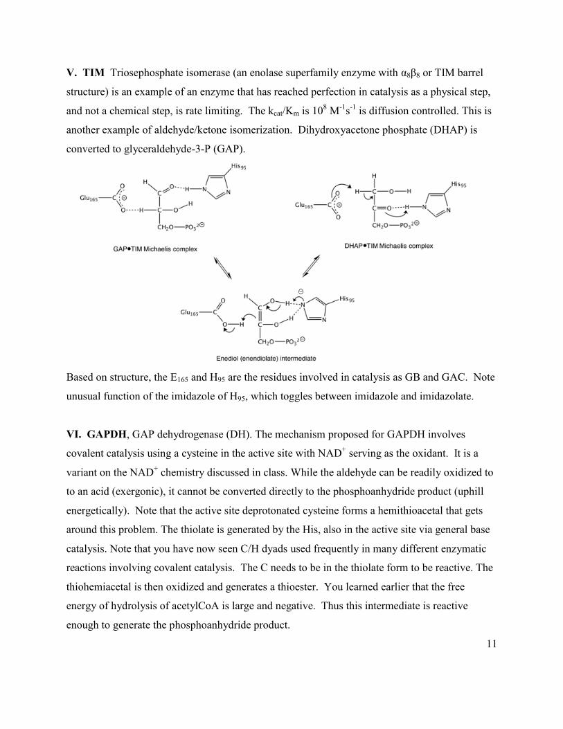
V. TIM Triosephosphate isomerase (an enolase superfamily enzyme with α8β8 or TIM barrel
structure) is an example of an enzyme that has reached perfection in catalysis as a physical step,
and not a chemical step, is rate limiting. The kcat/Km is 108 M-1s-1 is diffusion controlled. This is
another example of aldehyde/ketone isomerization. Dihydroxyacetone phosphate (DHAP) is
converted to glyceraldehyde-3-P (GAP).
Based on structure, the E165 and H95 are the residues involved in catalysis as GB and GAC. Note
unusual function of the imidazole of H95, which toggles between imidazole and imidazolate.
VI. GAPDH, GAP dehydrogenase (DH). The mechanism proposed for GAPDH involves
covalent catalysis using a cysteine in the active site with NAD+ serving as the oxidant. It is a
variant on the NAD+ chemistry discussed in class. While the aldehyde can be readily oxidized to
to an acid (exergonic), it cannot be converted directly to the phosphoanhydride product (uphill
energetically). Note that the active site deprotonated cysteine forms a hemithioacetal that gets
around this problem. The thiolate is generated by the His, also in the active site via general base
catalysis. Note that you have now seen C/H dyads used frequently in many different enzymatic
reactions involving covalent catalysis. The C needs to be in the thiolate form to be reactive. The
thiohemiacetal is then oxidized and generates a thioester. You learned earlier that the free
energy of hydrolysis of acetylCoA is large and negative. Thus this intermediate is reactive
enough to generate the phosphoanhydride product.
12
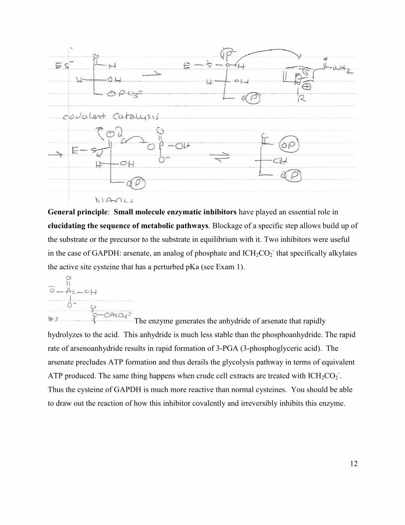
General principle: Small molecule enzymatic inhibitors have played an essential role in
elucidating the sequence of metabolic pathways. Blockage of a specific step allows build up of
the substrate or the precursor to the substrate in equilibrium with it. Two inhibitors were useful
in the case of GAPDH: arsenate, an analog of phosphate and ICH2CO2- that specifically alkylates
the active site cysteine that has a perturbed pKa (see Exam 1).
The enzyme generates the anhydride of arsenate that rapidly
hydrolyzes to the acid. This anhydride is much less stable than the phosphoanhydride. The rapid
rate of arsenoanhydride results in rapid formation of 3-PGA (3-phosphoglyceric acid). The
arsenate precludes ATP formation and thus derails the glycolysis pathway in terms of equivalent
ATP produced. The same thing happens when crude cell extracts are treated with ICH2CO2-.
Thus the cysteine of GAPDH is much more reactive than normal cysteines. You should be able
to draw out the reaction of how this inhibitor covalently and irreversibly inhibits this enzyme.
13
VII. Phosphoglycerate kinase (PGK) is the first step in the pathway where net ATP is
produced. There are two ATPs produced from one C6 sugar, as the two-C3 fragments undergo
this reaction.
GAPDH ΔG°’
GAP + Pi + NAD+ 1,3 PGA + NADH + 6.7 kj/mol
PGK
1, 3-PGA + Mg2+ADP Mg2+ATP + 3-PGA -18.8 kj/mol
The sum of the two steps is favorable ΔG°’ = -12.1 kj/mol. Think about why the free energy of
hydrolysis of 1,3-PGA is so large and negative.
General principle: The additivity of free energies, allows an unfavorable reaction to occur by
coupling it to a favorable reaction.
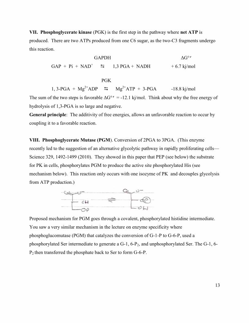
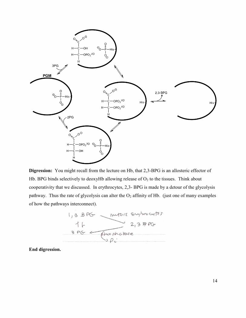
VIII. Phosphoglycerate Mutase (PGM). Conversion of 2PGA to 3PGA. (This enzyme
recently led to the suggestion of an alternative glycolytic pathway in rapidly proliferating cells—
Science 329, 1492-1499 (2010). They showed in this paper that PEP (see below) the substrate
for PK in cells, phosphorylates PGM to produce the active site phosphorylated His (see
mechanism below). This reaction only occurs with one isozyme of PK and decouples glycolysis
from ATP production.)
Proposed mechanism for PGM goes through a covalent, phosphorylated histidine intermediate.
You saw a very similar mechanism in the lecture on enzyme specificity where
phosphoglucomutase (PGM) that catalyzes the conversion of G-1-P to G-6-P, used a
phosphorylated Ser intermediate to generate a G-1, 6-P2, and unphosphorylated Ser. The G-1, 6-
P2 then transferred the phosphate back to Ser to form G-6-P.
14
Digression: You might recall from the lecture on Hb, that 2,3-BPG is an allosteric effector of
Hb. BPG binds selectively to deoxyHb allowing release of O2 to the tissues. Think about
cooperativity that we discussed. In erythrocytes, 2,3- BPG is made by a detour of the glycolysis
pathway. Thus the rate of glycolysis can alter the O2 affinity of Hb. (just one of many examples
of how the pathways interconnect).
End digression.
15
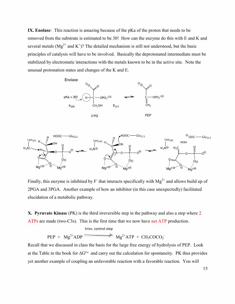
IX. Enolase: This reaction is amazing because of the pKa of the proton that needs to be
removed from the substrate is estimated to be 30! How can the enzyme do this with E and K and
several metals (Mg2+ and K+)? The detailed mechanism is still not understood, but the basic
principles of catalysis will have to be involved. Basically the deprotonated intermediate must be
stabilized by electrostatic interactions with the metals known to be in the active site. Note the
unusual protonation states and changes of the K and E.
Finally, this enzyme is inhibited by F- that interacts specifically with Mg2+ and allows build up of
2PGA and 3PGA. Another example of how an inhibitor (in this case unexpectedly) facilitated
elucidation of a metabolic pathway.
X. Pyruvate Kinase (PK) is the third irreversible step in the pathway and also a step where 2
ATPs are made (two-C3s). This is the first time that we now have net ATP production. Irrev. control step
PEP + Mg2+ADP Mg2+ATP + CH3COCO2-
Recall that we discussed in class the basis for the large free energy of hydrolysis of PEP. Look
at the Table in the book for ΔGº’ and carry out the calculation for spontaneity. PK thus provides
yet another example of coupling an unfavorable reaction with a favorable reaction. You will
16
return to this pathway and pyruvate kinase in John’s example of metabolism and cancer.
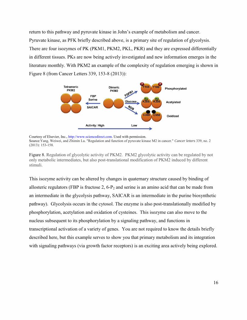
Pyruvate kinase, as PFK briefly described above, is a primary site of regulation of glycolysis.
There are four isozymes of PK (PKM1, PKM2, PKL, PKR) and they are expressed differentially
in different tissues. PKs are now being actively investigated and new information emerges in the
literature monthly. With PKM2 an example of the complexity of regulation emerging is shown in
Figure 8 (from Cancer Letters 339, 153-8 (2013)):
Courtesy of Elsevier, Inc., http://www.sciencedirect.com. Used with permission. Source:Yang, Weiwei, and Zhimin Lu. "Regulation and function of pyruvate kinase M2 in cancer." Cancer letters 339, no. 2 (2013): 153-158. Figure 8. Regulation of glycolytic activity of PKM2. PKM2 glycolytic activity can be regulated by not only metabolic intermediates, but also post-translational modification of PKM2 induced by different stimuli.
This isozyme activity can be altered by changes in quaternary structure caused by binding of
allosteric regulators (FBP is fructose 2, 6-P2 and serine is an amino acid that can be made from
an intermediate in the glycolysis pathway, SAICAR is an intermediate in the purine biosynthetic
pathway). Glycolysis occurs in the cytosol. The enzyme is also post-translationally modified by
phosphorylation, acetylation and oxidation of cysteines. This isozyme can also move to the
nucleus subsequent to its phosphorylation by a signaling pathway, and functions in
transcriptional activation of a variety of genes. You are not required to know the details briefly
described here, but this example serves to show you that primary metabolism and its integration
with signaling pathways (via growth factor receptors) is an exciting area actively being explored.

MIT OpenCourseWarehttps://ocw.mit.edu
5.07SC Biological Chemistry IFall 2013
For information about citing these materials or our Terms of Use, visit: https://ocw.mit.edu/terms.