Embed Size (px)
Citation preview
1
Chemistry 5.07SC Biological Chemistry I Fall Semester, 2013 Lecture 1. What is Biochemistry? Life at the molecular level.
Figure 1. Chemistry 5.07 focuses on the study of life at the molecular level.
A. What is Life? The ability to reproduce and to make order from chaos (Chapter 1 of course
textbook).
B. All known life forms share certain properties.
1. All organisms have the same morphological unit of life: the cell. Fleas and elephants have similar
sized cells.
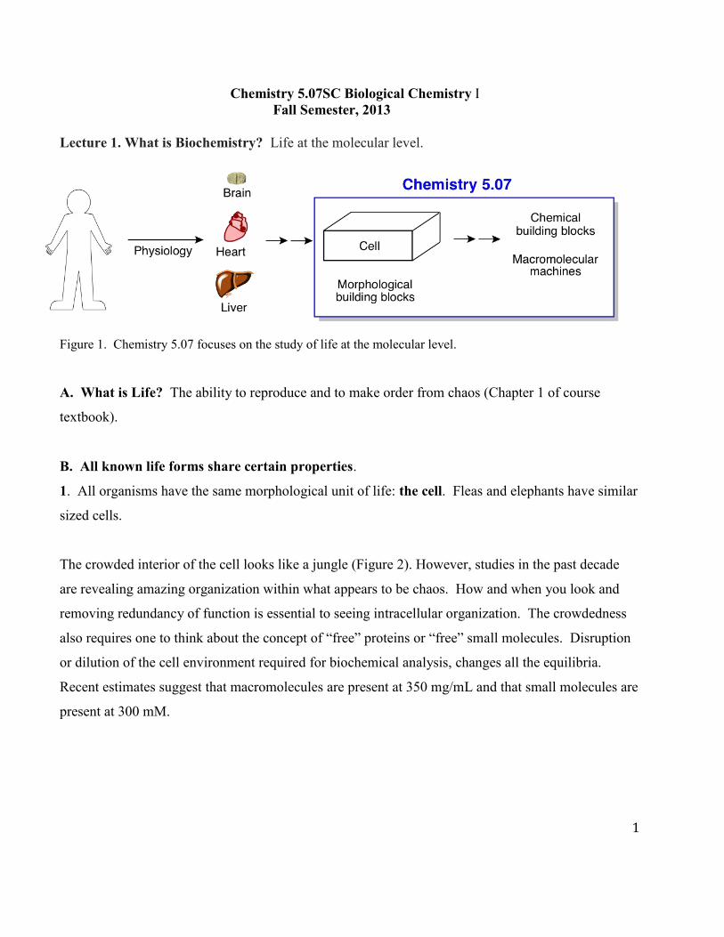
The crowded interior of the cell looks like a jungle (Figure 2). However, studies in the past decade
are revealing amazing organization within what appears to be chaos. How and when you look and
removing redundancy of function is essential to seeing intracellular organization. The crowdedness
also requires one to think about the concept of “free” proteins or “free” small molecules. Disruption
or dilution of the cell environment required for biochemical analysis, changes all the equilibria.
Recent estimates suggest that macromolecules are present at 350 mg/mL and that small molecules are
present at 300 mM.
2
Illustration courtesy of David S. Goodsell, the Scripps Research Institute.
Figure 2. The interior of the cell is crowded and appears chaotic as seen in this illustration of a bacterial cell magnified 106 fold. However, it is not simply a bag of enzymes and nucleic acids each occupying its own space. Cytoplasmic space is jammed full of proteins (dark yellow, pink, orange, light, medium and dark blue), tRNA (dark purple), mRNA (cream), ribosomes (light purple) and the DNA (yellow ribbons). The inner membrane is crammed with phospholipids (medium green) and various proteins and protein complexes (light green), while the periplasmic space contains the peptidoglycan (white), which is linked to the inner face of the outer membrane via lipoproteins (dark green). The outer membrane contains a variety of proteins and protein complexes interspersed with phospholipids, as well as lipopolysaccharides.
The crowded interior of the cell looks like a jungle. However, studies in the past decade are revealing
amazing organization within what appears to be chaos. How and when you look and removing
redundancy of function is essential to seeing intracellular organization. The crowdedness also
requires one to think about the concept of “free” proteins or “free” small molecules. Disruption or
dilution of the cell environment required for biochemical analysis, changes all the equilibria. Recent
estimates suggest that macromolecules are present at 350 mg/mL and that small molecules are
present at 300 mM.
2. The solvent of life is H2O due to its unusual properties. [Think about H bonding and buffers,
reviewed in the lexicon.]

3
3. All the building blocks in these organisms are the same: nucleotides, to make DNA
(deoxyribonucleic acids) and RNA (ribonucleic acids); acetylCoA, to make fatty acids and
triacylglycerols; sugars, to make polysaccharides; amino acids, to make proteins (polypeptides). The
chemistry of life is conserved. There are a limited number of chemical transformations that can
account for the vast majority of transformations in pathways (see the Lexicon).
4. Vitamins are conserved. Derivatives of vitamins extend the chemical repertoire of protein
catalysts and are essential for life. Look at the label on your vitamin bottle. You will understand the
functions of these molecules by the end of the semester.
5. All organisms share the same mechanism of information transfer: DNA is transcribed into RNA,
that is translated into protein. In 5.07 we will NOT focus on DNA replication, transcription, or
translation. This information and the macromolecular machines involved in these processes are the
focus, in part, of Chemistry 5.08. We will consequently not discuss nucleotide and nucleic acid
structure.
6. The energy currency, required to make order out of chaos, is the same in all organisms: ATP and
GTP. A 70 Kg person makes 45 Kg of ATP every 24 h. The machinery to make ATP is conserved.
7. Primary metabolic pathways, both biosynthetic (anabolic) and degradative (catabolic) pathways
are conserved. This semester we will investigate glycolysis/gluconeogenesis; fatty acid
biosynthesis/fatty acid degradation; the TCA cycle; the respiratory chain and how the energy
provided by reducing equivalents is used to make ATP; the pentose phosphate pathway.
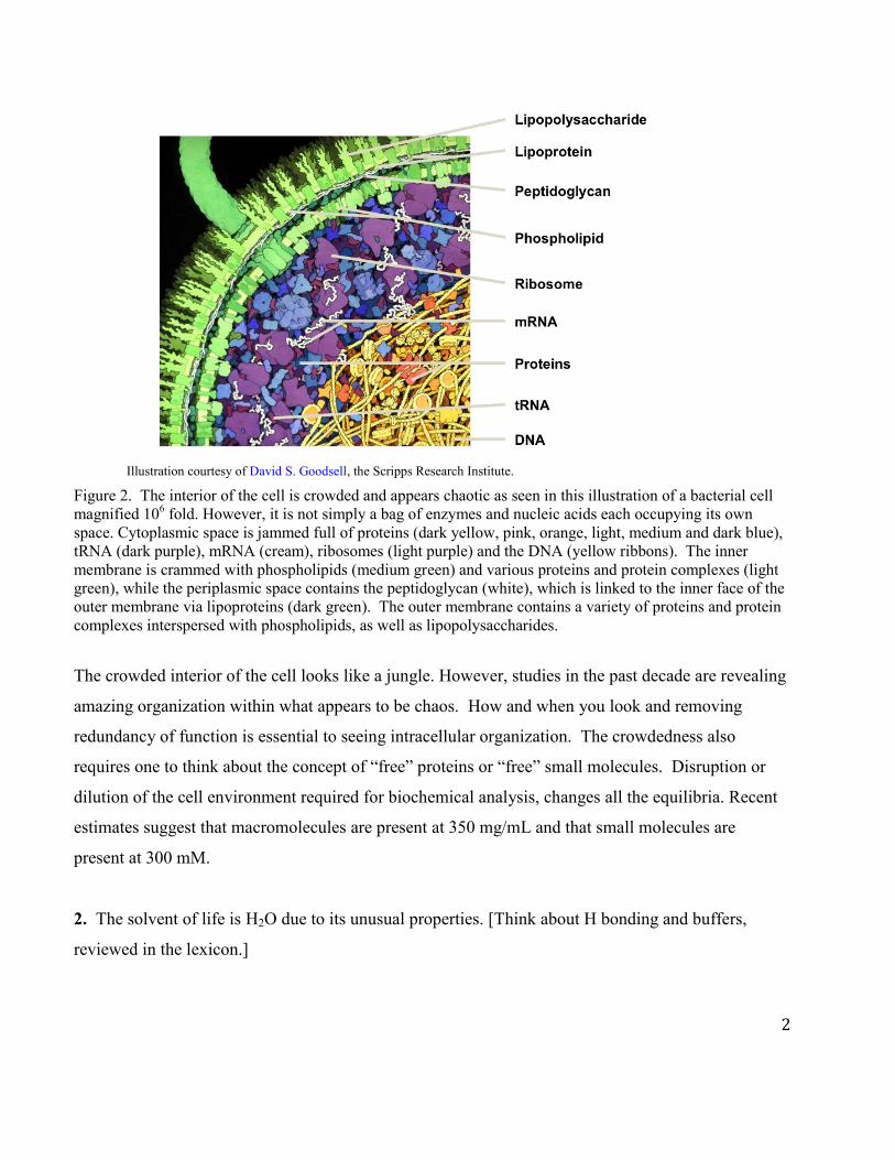
Environmental changes alter fluxes through the metabolic pathways in the chart below from the
course textbook. What are the general principles that control the changes required to meet and
maintain the organism’s needs? We will return to this chart over and over again. We hope to
convince you that metabolism is NOT the incredible jungle that it first appears. We will focus on
central metabolism, which is essential to almost all organisms: glycolysis, TCA, ET, and O/P to make
4
ATP. You are expected to know the structures of the central metabolites (glucose, acetyl coenzyme
A, pyruvate, etc).
Figure 3. Cellular metabolism is extremely complex and is the sum of its many substrates, products, macromolecular machines, biosynthetic and catabolic pathway, which provide life.
© John Wiley & Sons. All rights reserved. This content is excluded from our Creative
Commons license. For more information, see https://ocw.mit.edu/help/faq-fair-use/.
5
C. Pathways are tightly regulated. How do we control if fatty acids are synthesized or degraded or
whether sugars are synthesized or degraded? Regulatory mechanisms have also been conserved but,
in general, the more complex the organism, the more complex the regulatory mechanisms. You will
be introduced to the general mechanisms of regulation.
1. Allosteric regulation: small molecules bind outside the site where chemistry occurs in an
enzyme and affect the rate at which the enzyme converts a substrate to product.
2. Feedback inhibition: an end product of a pathway, may bind to the first enzyme in the
pathway to turn the pathway off when too much of the end product is present.
3. Post-translational modification: many activities of enzymes are modified by covalently
modifying the protein after it is made by the translational process. Proteins can be phosphorylated,
acetylated, hydroxylated, ubiquitinated, methylated, glycosylated etc.
4. Hormonal regulation: binding of hormones (extracellular) to receptors in membranes that
transmit information to the cellular interior. Examples encountered will be epinephrine, insulin, and
glucagon.
5. Transcriptional and translational regulation (not discussed in 5.07)
D. Biological relevance. Why should we care about biochemistry, which encompasses all of the
above topics? Understanding regulation of the “Fed” state and the “Fasting” state, and how an
organism switches its metabolism to accommodate these states is central to understanding chronic
disease mechanisms. Most diseases are associated with metabolic dysregulation. Thus,
understanding biochemistry plays a central role in dealing with and reversing diseased states.
E. Explicit GOALS of Chemistry 5.07
1. To introduce you to all of the chemical players of life: their structures and chemistry and,
consequently, function. Without this background, you cannot understand biochemistry.
2. To introduce you to the central pathways of metabolism. At first glance, metabolism can
be daunting, but we shall show you that all of biochemistry is made up of a surprisingly small number
of chemical transformations, which are used again and again. Understanding these transformations is

6
sufficient to allow you to predict most metabolic interconversions. See the Lexicon of reactions for
the course.
3. Once the pathways have been introduced, one needs to understand the regulation of the
pathways and their integration under different environmental conditions. This is the future.
F. The path ahead. ‘The more we learn about these complex systems, the more we realize that our
knowledge is dwarfed by our ignorance.’ This is a fascinating discipline with deep roots in both basic
and applied science … and there is still much to do.
Cellular components H2O and amino acids:
I. Water is essential for life and its properties are central to the function of all biological molecules.
H2O has two O-H bonds (0.95 Å) and has two pairs of non-bonded electrons that play a key role in
forming weak, non-covalent H (hydrogen) bonds.
There are four unique, physical properties of H2O:
A. H2O can form H bonds with itself and with many biological molecules.
B. H2O has dipolar character and plays an important role in the solvation of many molecules.
C. H2O minimizes interactions with non-polar species.
D. H2O is a neutral molecule with the ability to ionize. You have/will discuss pH and pKa.
Definition of an H bond: if an H is bonded to a very electronegative atom (in biology this atom is
usually O or N), it can form an additional weak bond with another electronegative atom that has a
pair of non-bonded electrons.

7
More generically, an H bond exists between a donor (D) and an acceptor (A) (that is, D-H---A), when
the distance between D and A is 2.7 to 3.1 Å. D and A are electronegative atoms O, N and
sometimes in Biology S. H bonds are directional, but not as directional as covalent bonds. A good H
bond does not need to be linear.
A.
B.
Figure 6. Types of bonds and bond energies. A. Bond energies for strong vs. weak non-covalent bonds (previous page). B. Examples of H bonds found in nature, including schematics of the donor and acceptor, as well as their bond lengths.
8
H bonds are on the order of 2 to 7 kcal/mol and fit into the category of weak non-covalent bonds.
These weak interactions (electrostatic, van der Waals, cation, dipole-dipole, dipole-induced dipole,
etc) play an essential role in the structure and function of all biological macromolecules in aggregate.
II. Amino acids, the building blocks of proteins.
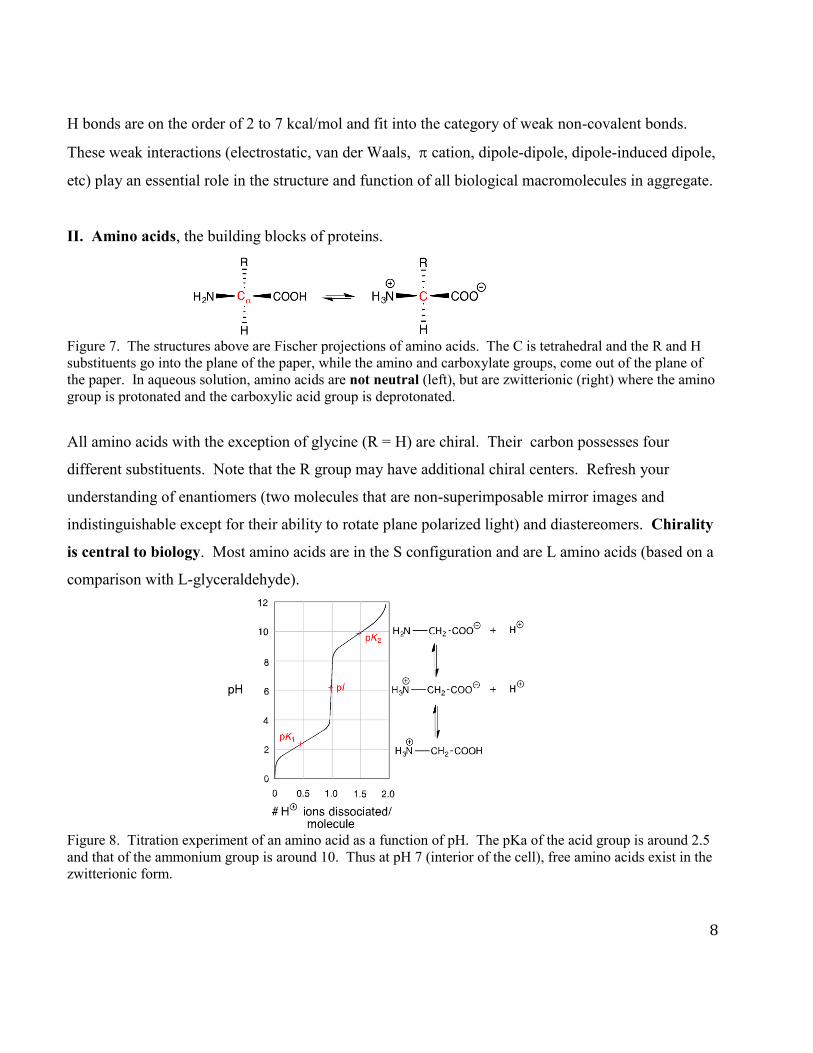
Figure 7. The structures above are Fischer projections of amino acids. The C is tetrahedral and the R and H substituents go into the plane of the paper, while the amino and carboxylate groups, come out of the plane of the paper. In aqueous solution, amino acids are not neutral (left), but are zwitterionic (right) where the amino group is protonated and the carboxylic acid group is deprotonated.
All amino acids with the exception of glycine (R = H) are chiral. Their carbon possesses four
different substituents. Note that the R group may have additional chiral centers. Refresh your
understanding of enantiomers (two molecules that are non-superimposable mirror images and
indistinguishable except for their ability to rotate plane polarized light) and diastereomers. Chirality
is central to biology. Most amino acids are in the S configuration and are L amino acids (based on a
comparison with L-glyceraldehyde).
Figure 8. Titration experiment of an amino acid as a function of pH. The pKa of the acid group is around 2.5 and that of the ammonium group is around 10. Thus at pH 7 (interior of the cell), free amino acids exist in the zwitterionic form.
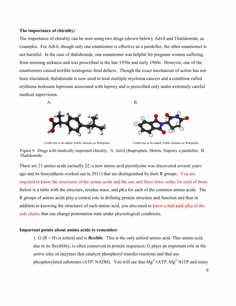
The importance of chirality:
The importance of chirality can be seen using two drugs (shown below), Advil and Thalidomide, as
examples. For Advil, though only one enantiomer is effective as a painkiller, the other enantiomer is
not harmful. In the case of thalidomide, one enantiomer was helpful for pregnant women suffering
from morning sickness and was prescribed in the late 1950s and early 1960s. However, one of the
enantiomers caused terrible teratogenic-fetal defects. Though the exact mechanism of action has not
been elucidated, thalidomide is now used to treat multiple myeloma cancers and a condition called
erythema nodosum leprosum associated with leprosy and is prescribed only under extremely careful
medical supervision.
A. B.
Credit line to be added: Public domain on Wikipedia. Credit line to be added: Public domain on Wikipedia.
Figure 9. Drugs with medically important chirality. A. Advil (Ibuprophen, Motrin, Nuprin), a painkiller. B. Thalidomide.
There are 21 amino acids (actually 22, a new amino acid pyrrolysine was discovered several years
ago and its biosynthesis worked out in 2011) that are distinguished by their R groups. You are
required to know the structures of the amino acids and the one and three letter codes for each of them.
Below is a table with the structure, residue mass, and pKa for each of the common amino acids. The
R groups of amino acids play a central role in defining protein structure and function and thus in
addition to knowing the structures of each amino acid, you also need to know a ball park pKa of the
side chains that can change protonation state under physiological conditions.
Important points about amino acids to remember:
1. G (R = H) is achiral and is flexible. This is the only achiral amino acid. This amino acid,
due to its flexibility, is often conserved in protein sequences. G plays an important role in the
active sites of enzymes that catalyze phosphoryl transfer reactions and that use
phosphorylated substrates (ATP, NADH). You will see that Mg2+•ATP, Mg2+•GTP and many
9
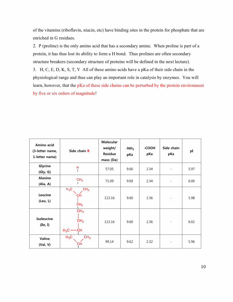
10
of the vitamins (riboflavin, niacin, etc) have binding sites in the protein for phosphate that are
enriched in G residues.
2. P (proline) is the only amino acid that has a secondary amine. When proline is part of a
protein, it has thus lost its ability to form a H bond. Thus prolines are often secondary
structure breakers (secondary structure of proteins will be defined in the next lecture).
3. H, C, E, D, K, S, T, Y All of these amino acids have a pKa of their side chain in the
physiological range and thus can play an important role in catalysis by enzymes. You will
learn, however, that the pKa of these side chains can be perturbed by the protein environment
by five or six orders of magnitude!
Amino acid
(3-letter name,
1-letter name)
Side chain R
Molecular
weight/
Residue
mass (Da)
-NH2
pKa
-COOH
pKa
Side chain
pKa pI
Glycine
(Gly, G) 57.05 9.60 2.34 - 5.97
Alanine
(Ala, A) 71.09 9.69 2.34 - 6.00
Leucine
(Leu, L)
113.16 9.60 2.36 - 5.98
Isoleucine
(Ile, I)
113.16 9.60 2.36 - 6.02
Valine
(Val, V)
99.14 9.62 2.32 - 5.96

11
Phenylalanine
(Phe, F)
147.18 9.21 1.83 - 5.48
Tryptophan
(Trp, W)
186.21 9.39 2.83 - 5.89
Tyrosine
(Tyr, Y)
163.18 9.11 2.20 10.07 5.66
Asparagine
(Asn, N)
114.11 8.80 2.02 - 5.41
Cysteine
(Cys, C)
103.15 10.28 1.96 8.18 5.07
Glutamine
(Gln, Q)
128.14 9.13 2.17 - 5.65
Methionine
(Met, M)
131.19 8.95 2.28 - 5.74
Serine 87.08 9.15 2.21 - 5.68
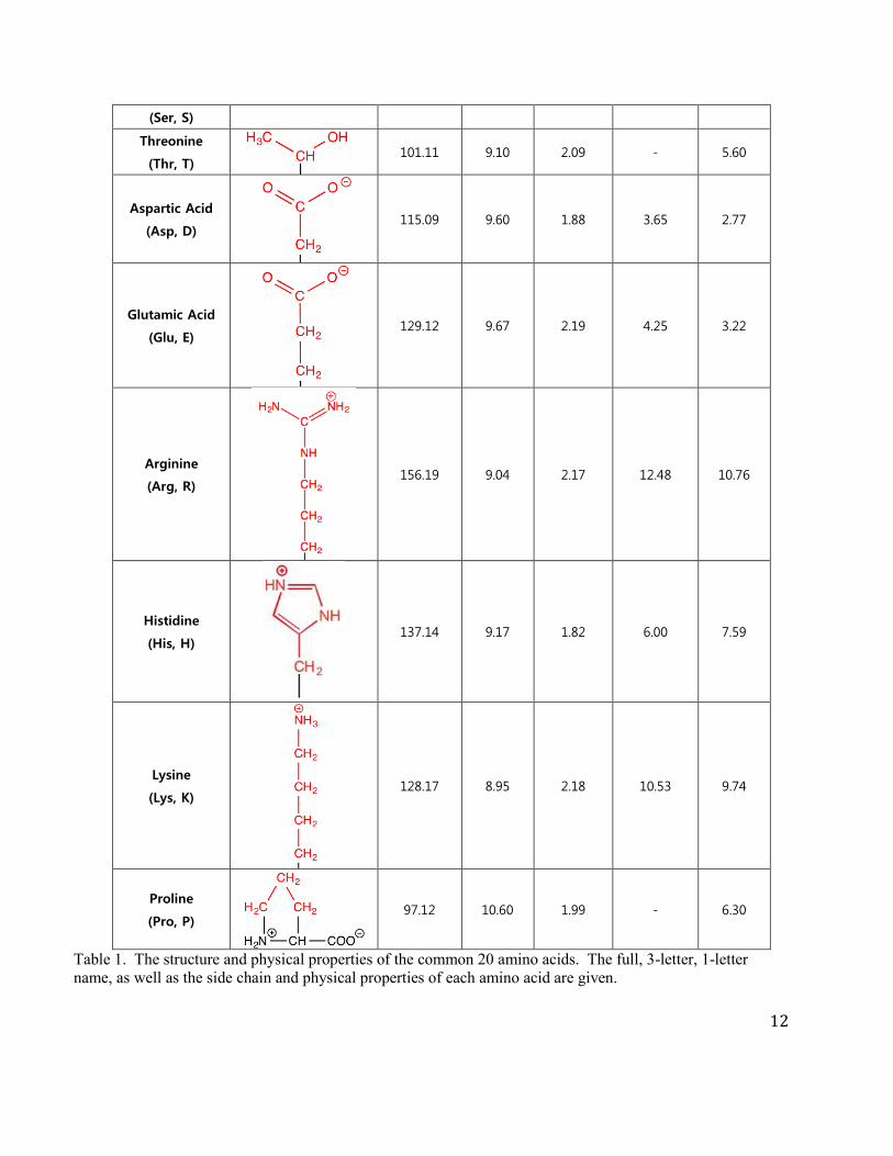
12
(Ser, S)
Threonine
(Thr, T) 101.11 9.10 2.09 - 5.60
Aspartic Acid
(Asp, D)
115.09 9.60 1.88 3.65 2.77
Glutamic Acid
(Glu, E)
129.12 9.67 2.19 4.25 3.22
Arginine
(Arg, R)
156.19 9.04 2.17 12.48 10.76
Histidine
(His, H)
137.14 9.17 1.82 6.00 7.59
Lysine
(Lys, K)
128.17 8.95 2.18 10.53 9.74
Proline
(Pro, P)
97.12 10.60 1.99 - 6.30
Table 1. The structure and physical properties of the common 20 amino acids. The full, 3-letter, 1-letter name, as well as the side chain and physical properties of each amino acid are given.
13
4. Amino acids are the building blocks of proteins. To study proteins one needs to know
their concentrations ([ ]). The [ ] can be determined from their extinction coefficient (ε).
Proteins have λmax at 280 nm. This absorption feature is distinct from nucleic acids (DNA,
RNA) that have λmax at 260 nm. The amino acid side chains that are largely responsible for
the A280nm are W and Y. From Beer’s law [A = εlc] where A is the absorbance, l is the path
length of light and c is the concentration of your protein, one can calculate the protein
concentration from the A280nm and the ε. There is a website, called Expasy, where one can
calculate the ε of any protein in the denatured (unfolded) state.
As discussed above, with the exception of R = H, all amino acids are chiral. Think about the
significance of the amino acid stereochemistry and how chirality affects protein conformation.
Proteins in the cell function in structural, catalytic, or receptor capacities and are built from amino
acids. In Chemistry 5.07, we will discuss proteins in each of these roles:
A. Collagen, which is the most abundant protein in vertebrates is extracellular and organized
into insoluble fibers of great tensile strength. Collagen is a major stress bearing component of
connective tissue.
B. Enzymes are proteins that catalyze all of the transformations in the metabolic pathways.
They can accelerate rates of reactions relative to non-enzyme catalyzed reactions by rates of 106 to
1015. We will discuss mechanisms of catalysis and you will be given a lexicon of catalytic
transformations that cover most reactions in the metabolic pathways we will examine.
C. Receptors, which are proteins (found in membranes) play a key role in signal transduction
and you will discuss G-protein receptors in insulin/glucagon (fed/starved states) regulation and in
adrenaline (fear) response.
In addition to the 22 amino acids, many amino acids are modified subsequent to their incorporation
into proteins (the translational process) as shown in Figure 10. This process is called post-
translational modification or PTM. These amino acids derivatives have unique properties that allow
them to play a central role in the chemistry of enzyme-catalyzed reactions or in the regulation of the
function of a protein. Mass spectrometric technology has allowed us to identify these modified
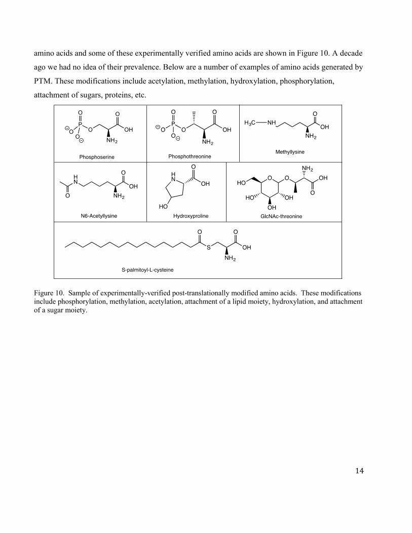
14
amino acids and some of these experimentally verified amino acids are shown in Figure 10. A decade
ago we had no idea of their prevalence. Below are a number of examples of amino acids generated by
PTM. These modifications include acetylation, methylation, hydroxylation, phosphorylation,
attachment of sugars, proteins, etc.
Figure 10. Sample of experimentally-verified post-translationally modified amino acids. These modifications include phosphorylation, methylation, acetylation, attachment of a lipid moiety, hydroxylation, and attachment of a sugar moiety.

MIT OpenCourseWarehttps://ocw.mit.edu
5.07SC Biological Chemistry IFall 2013
For information about citing these materials or our Terms of Use, visit: https://ocw.mit.edu/terms.