Embed Size (px)
DESCRIPTION
Manual de protocolo de extraccion de DNA..
Citation preview
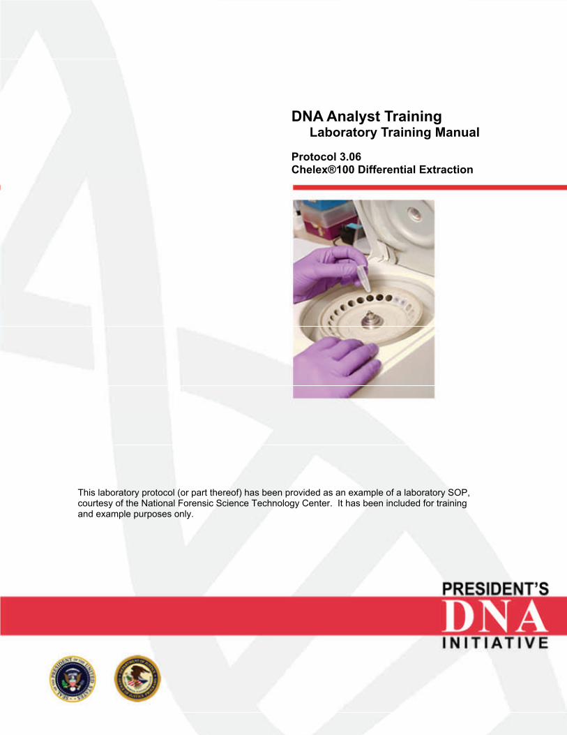
DNA Analyst Training Laboratory Training Manual Protocol 3.06 Chelex®100 Differential Extraction
This laboratory protocol (or part thereof) has been provided as an example of a laboratory SOP, courtesy of the National Forensic Science Technology Center. It has been included for training and example purposes only.
INTRODUCTION Chelex® 100 is an ion-exchange resin composed of styrene divinylbenzene copolymers with paired iminodiacetate ions that act as chelating groups in binding polyvalent metal ions. Removing magnesium from the sample inactivates nucleases and DNA destroying enzymes, thus protecting the DNA molecules. This Chelex® 100 extraction procedure provides a method for differential extraction of DNA from stains containing sperm. Epithelial cells are preferentially lysed by Proteinase K and the supernatant containing the lysed epithelial cells is removed from the sperm cell pellet after centrifugation. The remaining sperm cells are lysed with Proteinase K and dithiothreitol (DTT). Chelex® 100 is added to both the epithelial cell (female) fraction and the sperm cell (male) fractions. SAFETY CONSIDERATIONS Refer to the Laboratory Safety Manual(s) PREPARATIONS 5% Chelex® 100 Suspension
1. Add 50 ml of ultra pure or sterile deionized water to capped bottle. 2. Add a sterile stir bar to bottle. 3. Weigh out 2.5 grams of Chelex® 100 into bottle. 4. Properly label and date the container. Store at room temperature.
Expiration date is one year.
20% Chelex® 100 Suspension
1. Add 50 ml of ultra pure or sterile deionized water to capped bottle. 2. Add a sterile stir bar to bottle. 3. Weigh out 10.0 grams of Chelex® 100 into bottle. 4. Properly label and date the container. Store at room temperature.
Expiration date is one year. 0.39 M DTT
1. 60 mg DTT (Dithiothreitol C4H10O2S2) 2. 1 ml sterile DI water 3. Dissolve and store at -20°C for up to 2 weeks.
pdi_lab_pro_3.06.pdf President's DNA Initiative - DNA Analyst Training Page 1 of 5
Proteinase K (20 mg/ml)
1. 20 mg Proteinase K 2. 1 ml sterile DI water 3. Dissolve in a sterile microcentrifuge tube. Aliquot 100 μl into each of 10
sterile microcentrifuge tubes. Store at -20°C. Expiration date one year.
Stain Extraction Buffer Premix
1. 2.4 g Tris base 2. 9.26 g Na2EDTA•2H2O 3. 14.6 g NaCl 4. 1600 ml DI water 5. Dissolve and pH to 8.0. Adjust volume to 2 liters with DI water. Filter
sterilize or autoclave. Store at 2° to 8°C. Expiration date one year.
Stain Extraction Buffer Working Solution
1. 320 μl Stain Extraction Buffer Premix 2. 40 μl 20% SDS 3. 40 μl 0.39 M DTT 4. Store at -20°C for up to two weeks.
TE -4 (10 mM Tris-HCl, 0.1 mM EDTA, pH 8.0)
1. 10 ml 1 M Tris-HCl, pH 8.0 2. 0.2 ml 0.5 M EDTA 3. 990 ml DI water 4. Adjust pH to 8.0 ± 0.2 5. Autoclave. Store at 2° to 8°C.
Expiration date one year.
INSTRUMENTATION
• Centrifuge • Heat block and/or water bath • Pipettes • Top loading balance • Vortex
MINIMUM STANDARDS & CONTROLS
• Positive extraction control • Extraction blank – contains reagents only
pdi_lab_pro_3.06.pdf President's DNA Initiative - DNA Analyst Training Page 2 of 5
PROCEDURE OR ANALYSIS
General Method
1. Place portion of fabric or a swab into a sterile 1.5 ml microcentrifuge tube. The size of the cutting may be adjusted based upon the amount of biological material (i.e. sperm count) or the possible presence of inhibitors.
2. Pipette 800 –1000 µl of sterile deionized water into the microcentrifuge
tube. Vortex briefly.
3. Incubate for 30 minutes at room temperature. Mix occasionally by inversion or gentle vortexing.
4. Remove the cutting or swab and place into a spin basket filter unit.
5. Centrifuge at 10,000 to 15,000 x g for 1 to 3 minutes at room temperature.
6. Remove the spin basket and save the sample cutting.
7. Without disturbing the pellet, remove all but approximately 50 µl of the
supernatant. This supernatant may be saved for further analysis or discarded.
8. Re-suspend the pellet in the remaining liquid. A slide may be made at this
time using 3-5 µl to determine the efficiency of recovery of biological material. If the sample is found to contain a small amount of sperm or if it is suspected that the sperm has not been completely removed from the material, the cutting may be added back in during the digestion step.
9. To the sample tube add:
• 500 µl stain extraction buffer , digest buffer or TE-4 • 5 µl Proteinase K (20 µg/µl)
Note: The above volumes may be increased proportionally, as needed, to accommodate larger sample cuttings or swab.
10. Mix gently and incubate 30 minutes to two hours at 37 °C to lyse the non-sperm cells. Exceeding 2 hours may cause sperm cell lysis.
11. If the cutting was returned to the tube, remove the cutting and place into a
spin basket filter unit.
pdi_lab_pro_3.06.pdf President's DNA Initiative - DNA Analyst Training Page 3 of 5
12. Centrifuge for five minutes at approximately 10,000 x g at room temperature to pellet the sperm cells.
13. The substrate may be saved or discarded at this step. 14. Carefully remove the supernatant and place into a sterile microcentrifuge
tube. It is important that the sperm cell pellet not be disturbed and is left in approximately 50 µl of the supernatant. The removed supernatant primarily containing the lysed epithelial cells and is termed the “E fraction or female fraction”.
15. Add 50 µl of 20% Chelex® 100, for each 150 µl of supernatant from the E
fraction.
16. The remaining sperm cell pellet, termed the “S fraction or male fraction”, contains the sperm cells from the cutting or swab.
17. Wash the sperm cell fraction as follows:
a. Re-suspend the pellet in 1000 µl of digest buffer and vortex briefly. b. Centrifuge for approximately 5 minutes at 10,000 to 15,000 x g at
room temperature. c. Remove and discard all except 50 µl of the supernatant. Note: The purpose of the wash step is to remove any remaining epithelial cells. It may be necessary to repeat Steps 17a through 17c 2-3 times, depending on the amount of epithelial cells.
18. Wash the sperm cell pellet as follows: a. Re-suspend the pellet in 1000 µl of sterile distilled water and vortex
briefly. b. Centrifuge for approximately 5 minutes at 10,000 to 15,000 x g at
room temperature. c. Remove and discard all except 50 µl of the supernatant.
Note: This wash removes any remaining digest buffer. Digest buffer contains SDS, which can inhibit PCR.
19. Re-suspend the pellet in the 50 µl of supernatant and add:
• 150 µl of 5% Chelex® 100 • 2 µl of 10 mg/ml Proteinase K • 7 µl of 1M DTT
20. Vortex both the epithelial and sperm cell fractions at high speed for 5-10 seconds.
21. Pulse spin each fraction in a centrifuge.
pdi_lab_pro_3.06.pdf President's DNA Initiative - DNA Analyst Training Page 4 of 5
22. Incubate at 37-560 C for 30 – 60 minutes. 23. Vortex both the epithelial and sperm cell fractions at high speed for 5-10
seconds.
24. Heat each fraction for 8 minutes at 100°C in heating block or in a boiling water bath.
25. Vortex each fraction at high speed for 5-10 seconds.
26. Centrifuge each fraction for 3 minutes at approximately 10,000-15,000 x g.
27. The supernatant from each fraction may be transferred into sterile 1.5 ml
microcentrifuge tubes or centrifugal filter devices (i.e. Microcon® filter units) for concentration and purification.
28. Store DNA extract at 4°C or proceed to quantification. For long term
storage, samples should be frozen or freeze dried. Return to Laboratory Training Manual User Guide
pdi_lab_pro_3.06.pdf President's DNA Initiative - DNA Analyst Training Page 5 of 5