Embed Size (px)
Citation preview

Plant Physiol. (1970) 45, 348-358
Characterization of Rapidly Labeled Ribonucleic Acidfrom Dwarf Peas
Received for publication October 2, 1969
M. M. JOHRI1 AND J. E. VARNERMichigan State University-Atomic Energy Commission Plant Research Laboratory,Michigan State University, East Lansing, Michigan 48823
ABSTRACT
The ribonucleic acid synthesized by excised shoots ofdwarf pea (Pisum sativum L. cv. Progress No. 9) duringshort labeling periods has been characterized. Thirty percent of the total 32Pi incorporated in 1 hour is found in theribosomal fraction. This labeled RNA was polydisperse(6-18 Svedberg units) and after chromatography on amethylated albumin-kieselguhr column about 80% of theradioactivity appeared in two peaks. One of these appearedon the shoulder of heavy ribosomal RNA ("mRNA") whilethe other was tenaciously bound to the column (TB-RNA).In the presence of high NaCl concentration, about half ofthe polydisperse RNA interacted with ribosomal RNA andeluted as "mRNA" while the remainder eluted as TB-RNA.This interaction in the presence of salt seems to result inthe alteration of secondary structure because the "mRNA"fraction had a high sedimentation coefficient (45-50 Sved-berg units). The polydisperse RNA approaches DNA in lowcytidylate and guanylate content. After short periods oflabeling TB-RNA showed higher adenylate content than"mRNA." The radioactivity from the "mRNA" peak canbe chased, and these counts may represent a class of short-lived messenger RNA molecules with an average half-lifeof 10 to 15 minutes. The other component, TB-RNA, couldnot be chased and accumulated radioactivity during thechase period.
In bacteria and viruses the information for the sequence ofamino acids in a polypeptide chain is encoded into messengerRNA. Presence of a similar fraction has been suggested in somehigher plants (5, 6, 21, 26, 39). All of our knowledge about mes-senger RNA from higher plants is presumptive and based onanalogy to microorganisms; in no case has this fraction beenpurified or characterized, nor has its rate of synthesis or itsstability been studied.We chose a dwarf pea as the experimental material because
we have been investigating the RNA synthesized by nuclei iso-lated from shoots of this plant (19), and the knowledge obtainedfrom the system in vivo may be applicable to the results obtainedwith isolated nuclei.
1 Present address: Molecular Biology Unit, Tata Institute of Funda-mental Research, Homi Bhabha Road, Bombay 5, India.
MATERIALS AND METHODS
Uptake and Incorporation of Labeled Precursors. Dwarf peaseeds (Pisum sativum L., cv. Progress No. 9) were surface-sterilized for 7 to 10 min in 1% sodium hypochlorite, implantedin 500-ml Erlenmeyer flasks containing moist, sterile vermiculite,and allowed to germinate at 23 C in continuous light. At the endof 5 or 6 days, shoots 0.8 to 1 cm long were excised and 10 or 15shoots were incubated in 25-ml Erlenmeyer flasks under asepticconditions with labeled uridine or 32Pj (carrier-free H3u2PO4,neutralized) in 2 ml of water containing 100 ,ug of chlorampheni-col. Flasks were transferred to a shaker, and, after incubation forvarious times, the shoots were washed thoroughly in ice-cold0.05 M KH2PO4 and deionized water and were then extracted fornucleic acids.
In experiments designed to study the relationship between up-take and incorporation, the labeled shoots were homogenized inethanol, 80% (v/v,) and the nucleic acids were determined (33).The residue obtained after centrifuging at 1500g for 5 min wasextracted three times with 5% trichloroacetic acid (w/v) contain-ing KH2PO4 to remove the acid-soluble nucleotides. The pigmentsand lipid materials were removed by extracting the residue threetimes with ethanol-ether (3: 1). The supernatants at all steps weresaved and the radioactivity was determined to calculate the totaluptake of labeled precursors. The residue was hydrolyzed in 0.3 NKOH for 20 hr at 37 C, and after hydrolysis DNA was precipi-tated by the addition of perchloric acid. The radioactivity inperchloric acid-soluble and -insoluble fractions was determinedto measure the incorporation into RNA and DNA, respectively.The ribonucleotide fraction in experiments with labeling for lessthan 3 hr contained considerable amounts of radioactivity asinorganic phosphate (carried over from the soluble pools). Incor-poration into the RNA fraction in these experiments was takenas the total radioactivity present in 2'-3'-AMP,2 CMP, GMP, andUMP only.
Extraction of Nucleic Acids. To prepare total nucleic acids, theSAS and phenol method of Kirby (22) was used after slightmodification. The shoots (1-2 g) were homogenized in a porcelainmortar with 1 ml of bentonite (50 ,tg/ml, purified), 2 ml each ofSAS (pH 7.0, 6%, w/v) and phenol-cresol, and a little sand. Themortar was rinsed twice with 2 ml each of SAS and phenol-cresol,and the combined homogenate was vigorously mixed for 2 minfollowed by centrifugation at 3,500g for 10 min. The aqueous
2Abbreviations: AMP: adenylate or adenylic acid; CMP: cytidylicacid; DNA-RNA: DNA and the RNA associated with it; FU: 5-fluorouracil; GMP: guanylic acid; hrRNA: heavy ribosomal RNA;lrRNA: light ribosomal RNA; MAK: methylated albumin-kieselguhr;"mRNA": rapidly labeled RNA as described in the text; SAS: sodium4-amino-salicylate; SLS: sodium lauryl sulfate; sRNA: transfer RNA;TB-RNA: tenaciously bound RNA; UMP: uridylic acid or uridylate.
348 www.plantphysiol.orgon June 19, 2018 - Published by Downloaded from
Copyright © 1970 American Society of Plant Biologists. All rights reserved.

RAPIDLY LABELED RNA FROM DWARF PEAS
phase was removed carefully, and the phenol phase was extractedtwice with 3 ml of SAS. To the combined aqueous phase, solidNaCl (30 mg/ml) and 0.5 volume of phenol-cresol were added.The emulsion was mixed for 2 min. After centrifugation at10,OOOg for 10 min, the aqueous phase was removed, potassiumacetate was added to 1 %O final concentration, and nucleic acidswere precipitated overnight at -20 C by adding 2 to 3 volumes ofethanol-cresol. Usually, 75 to 90% of RNA and DNA present inthe tissue were recovered.
Isolation of Polyribosomes and Sucrose Density Gradient Cen-trifugation. Ribosomes were isolated from about 3 g of pea shoots(24). A sample containing 0.5 to 0.75 mg of ribosomes was lay-ered over a 24-ml linear gradient of sucrose (10-34%, w/v,sucrose, prepared in 0.05 M tris, pH 7.4; 0.015 M KC1 and 0.005 MMgCl2) and centrifuged at 24,000 rpm in the SW-25.1 Spincorotor for 2 hr at -2 C. The gradient tubes were punctured, andthe absorbance of the effluent was scanned at 260 m,u with a con-tinuous Gilford recorder and the ISCO model 180 density gradi-ent fractionator. When radioactivity determinations were done,the fractions (20 drops each, 60-70 fractions/24 ml) were col-lected from the top of the gradient.The ribosomal subunits and ribosomal RNA were character-
ized by zonal centrifugation with linear gradients of sucrose from4 to 20%, 4 to 40%, or 17 to 34% in 0.05 M NaCl and 0.05 Msodium acetate (pH 5.3). After centrifugation fractions werecollected either manually by piercing the tube from the bottomor mechanically using the ISCO density gradient fractionator asdescribed above. The concentration of monovalent cations waskept around 0.1 M and no further additions were made, in orderto avoid aggregation of RNA. In gradients containing less than0.1 M monovalent cations, the heavy ribosomal RNA was de-graded. The samples were precipitated by the addition of equalvolume of 10% trichloroacetic acid, and the precipitate wascollected by filtration on membrane filters that were washedwith five 5-ml portions of 5%O cold trichloroacetic acid. Thefilters were air-dried, and the radioactivity was determined aftersuspending filters in an appropriate scintillator. The sedimenta-tion coefficients of rapidly labeled RNA fractions were deter-mined as described by Martin and Ames (30), using the coeffi-cients of light and heavy ribosomal RNA as 18 and 25 S (27).
Fractionation of Nucleic Acids. The crude nucleic acid pelletwas washed three times with 66% ethanol containing 1 % potas-sium acetate, dissolved in 0.3 M NaCl in 0.05 M KH2PO4 (pH6.7), and fractionated on MAK columns (28). After addition ofthe sample, the column was washed with 200 ml of 0.3 M NaClin 0.05 M KH2PO4 (pH 6.7), and elution was done at 37 C witha 300- or 400-ml linear gradient of NaCl from 0.3 M to 1.6 M in0.05 M KH2PO4, pH 6.7. The fraction of RNA not eluted by thesalt (tenaciously bound RNA) was eluted with 1% SLS or with1.5 M NH40H after the termination of the salt elution (9). Mostof the TB-RNA was found to be eluted at 37 C. Fractions of 2.5to 5.0 ml each were collected, absorbance at 260 m, was meas-ured, and radioactivity was determined after filtering the acid-insoluble material on the membranes as described above.When TB-RNA was to be purified for rechromatography,
centrifuge tubes containing this fraction were cooled in ice for4 to 6 hr, and the precipitated SLS (also containing the methyl-ated albumin from column) was separated by centrifugation. TheRNA from the supernatant was recovered after addition ofNaCl (30 mg/ml), phenol deproteinization, and ethanol precipi-tation. A considerable amount of TB-RNA (about 40%) re-mained in the pellet which contained SLS and protein. Thesecounts were recovered by suspending the pellet in 5 ml of SAS,addition of NaCl, and phenol deproteinization. About 70% ofthe radioactivity was recovered with this procedure.
Determination of Nucleotide Composition. The nucleotidecomposition of 3nPi-labeled RNA samples was determined by
paper electrophoresis (34). The tubes containing the sample wereprecipitated with 2 volumes of ethanol at -20 C overnight inthe presence of 250 jig of carrier pea ribosomal RNA. The pelletwas dissolved in a small volume of water, and perchloric acidwas added to a final concentration of 0.6 N. After 4 hr in an icebath, the pellet was washed twice with 70%-1 ethanol to removethe residual perchloric acid. The pellets were hydrolyzed in 0.3 NKOH at 37 C for 18 to 20 hr, neutralized with perchloric acid,and subjected to paper electrophoresis. The distribution ofradioactivity in the 2'-3'-riboside monophosphates was deter-mined.
RESULTS
The present study deals with the characterization of rapidlylabeled RNA from the shoots of dwarf pea. We felt that it wasdesirable to have a detailed knowledge of: (a) the relationshipbetween the uptake and the incorporation of labeled precursors;(b) the half-time of formation of this rapidly labeled fraction;(c) its chromatographic behavior during MAK chromatography;and (d) its subcellular localization.
Kinetics of -"Pi Uptake Incorporation into Nucleic Acids. Asshown in Figure 1, 32Pi is taken up by pea shoots at a constantrate up to 10 to 12 hr when 19% of the input radioactivity wasfound in the shoots. After an initial lag of about 1 hr, the incor-poration into nucleic acids was linear up to 24 hr in spite of thefact that the uptake leveled off after 10 hr. At the end of 24 hrthe radioactivity incorporated into nucleic acids amounted toonly 12 to 15%l of that taken up by the shoots. During 1 hr ofincubation less than 3% of the radioactivity accumulated wasfound in the nucleic acids.The nucleic acids were resolved into seven fractions by MAK
chromatography (Fig. 2). The over-all pattern of elution ob-tained was similar to that described in bacteria and in higherplants (5, 16, 37, 41). Following the absorbance and radioactiv-ity profiles as a function of time, it was found that the two pat-perns did not coincide up to 6 hr of labeling. After shorterlabeling periods, the radioactivity in the light and heavy ribo-somal RNA region always appeared 1 or 2 tubes earlier than thebulk of the RNA. In these experiments (15-, 30-, and 60-minlabeling), 65 to 70% of the radioactivity was distributed intotwo prominent peaks. One of these peaks, "mRNA" is identi-fiable as a separate peak up to 60 min of labeling; with longertimes it becomes masked by the trailing edge of ribosomalRNA. After 12 hr of incorporation the radioactivity and absorb-ance profiles match one another.
w
IO
D
Hours of incubotion
FIG. 1. Kinetics of 32Pi uptake and incorporation into nucleic acids.Ten excised shoots were used for each treatment, and uptake and in-corporation into nucleic acids were determined as described in "Ma-terials and Methods" (1 mc/flask for 15-, 45-, and 60-min incorpora-tions; 500 uc/flask for 1.5-, 2-, 3-, and 6-hr incorporations; and 250suc/flask for 12- and 24-hr incorporations). All points represent theaverage of two separate incubations and have been corrected for thesame amount of input isotope.
ut
3 ,
/ ~~~3/ ~~~~~~~2°-
oou
12 1 6 20 24
Plant Physiol. Vol. 45, 1970 349
www.plantphysiol.orgon June 19, 2018 - Published by Downloaded from Copyright © 1970 American Society of Plant Biologists. All rights reserved.

JOHRI AND VARNER
E
C-)0
-
0
0.
0
-
l-O0-
Fraction number
Plant Physiol. Vol. 45, 1970
8
7
6
5
4
E0.
-6
0
3
2
.EU
0
IR
40 50 60
Fraction numberFIG. 2. MAK chromatography of nucleic acids synthesized by pea shoots labeled with 32Pj for 15 min (a), 90 min (b), 3 hr (c), and 12 hr (d).
Fifteen pea shoots were used for each treatment, and the amount of isotope used is the same as described in Figure 1. The radioactivity has beenexpressed as a percentage of total incorporation per tube. The numbers I through VII refer to transfer RNA, 5 S ribosomal RNA, DNA and theRNA associated with it, light ribosomal RNA, heavy ribosomal RNA, "mRNA," and tenaciously bound RNA.
- 80 A0
0o601nA
Z 40 R20
0 2 3 6 12 18 24Hours
FIG. 3. Distribution of radioactivity in the different nucleic acidfractions. Nucleic acids were extracted from pea shoots labeled with'tPi for various periods of time and were analyzed by chromatographyon MAK columns. The vertical bars represent the scatter, and thepoints are means of several determinations. 0: DNA-RNA; X: trans-fer and 5 S ribosomal RNA; A: light and heavy ribosomal RNA;*: "mRNA"; o: TB-RNA.
The relative distribution of '2Pi in the different fractions aftervarious periods of labeling is shown in Figure 3. The sRNA andDNA (also containing RNA associated with it) each containedabout 4 to 6% of the total counts, and this proportion remained
constant throughout the course of incorporation (15 min to 24hr). The fractions "mRNA" and TB-RNA accounted for morethan two-thirds of the radioactivity incorporated during 1 hrinto nucleic acids. The appearance of radioactivity in ribosomalRNA was slow during the 1st hr of incorporation, but later thepercentage of counts increased 4-fold. After a 2-hr labelingperiod steady state levels were observed, and under these condi-tions ribosomal RNA contained 75 to 80% of the total radio-activity incorporated into nucleic acids.The nucleotide compositions of various nucleic acid fractions
are shown in Table I. In the DNA peak isolated from shootslabeled for 16 hr, 40 to 60% of the total radioactivity was alkali-hydrolyzable, and on electrophoresis all four 2'-3'-ribosidemonophosphates were found to be labeled. This peak thus con-tains both RNA and DNA as indicated by alkali-hydrolyzableand -nonhydrolyzable counts. The composition of 1rRNA isdifferent from that of hrRNA, and both of these are rich inAMP + GMP (54-56%), not in GMP + CMP as is usuallythe case. The only fractions that approach DNA in low GMP +CMP are "mRNA" and TB-RNA. Since these two fractionsaccumulated the maximum radioactivity during 1-hr labeling,this incorporation period was used in all the subsequent experi-ments to characterize the rapidly labeled RNA in detail.
Sedimentation Characteristics and Purification of the TwoRapidly Labeled Fractions. The RNA eluting with the trailingedge of hrRNA has been variously described as D-RNA (DNA-
350
0C0N
0
40
C)
.cl
(b)II IV V VI VII
III 1n w v I
'4 II~~~~~~~4
hr Counts IIs
I lt_D.
I %I
www.plantphysiol.orgon June 19, 2018 - Published by Downloaded from Copyright © 1970 American Society of Plant Biologists. All rights reserved.
RAPIDLY LABELED RNA FROM DWARF PEAS
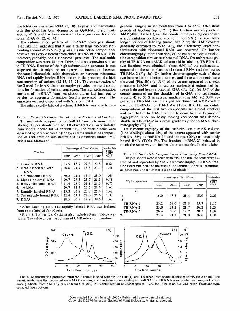
like RNA) or messenger RNA (5, 18). In yeast and mammaliancells this peak has been designated as Q1-RNA; it sedimentsaround 45 S and has been shown to be a precursor for ribo-somal RNA (9, 32, 40, 42).The sedimentation pattern of "mRNA" from pea shoots
(1-hr labeling) indicated that it was a fairly large molecule sedi-menting around 45 to 50 S (Fig. 4a). Its nucleotide composition,however, was very different from either lrRNA or hrRNA; it is,therefore, probably not a ribosomal precursor. The nucleotidecomposition was more like pea DNA and also somewhat similarto TB-RNA. Because of the high sedimentation constant it wassuspected that it might be an aggregate. Interaction betweenribosomal ribonucleic acids themselves or between ribosomalRNA and rapidly labeled RNA occurs in the presence of a highconcentration of cations (12-15, 17, 31). The concentration ofNaCl used for MAK chromatography provides the right condi-tions for formation of such an aggregate. The high sedimentationconstant of "mRNA" from pea shoots did in fact turn out tobe due to aggregate formation (results presented later). Theaggregate was not dissociated with SLS or EDTA.The other rapidly labeled fraction, TB-RNA, was very hetero-
Table I. Nucleotide Composition of Various Nucleic Acid FractionsThe nucleotide composition of "mRNA" was determined after
labeling the pea shoots for 1 hr. All other fractions were isolatedfrom shoots labeled for 24 hr with 32Pi. The nucleic acids wereseparated by MAK chromatography, and the nucleotide composi-tion of each fraction was determined as described under "Ma-terials and Methods."
Percentage of Total Counts NucleotideFraction Ratio.
CMP AMP GP UNMPI GMP
1. Transfer RNA 33.5 17.9 27.8 20.8 0.642. RNA associated with 26.8 25.9 18.3 27.4 1.41
DNA3. 5 S ribosomal RNA 30.2 24.2 14.6 28.0 1.654. Light ribosomal RNA 20.7 25.3 28.7 25.3 0.885. Heavy ribosomal RNA 21.4 25.0 32.1 21.5 0.776. "mRNA" 20.7 32.5 20.2 26.6 1.607. Rapidly labeled RNA' 23.3 30.8 20.7 25.4 1.488. Tenaciously bound RNA 22.4 28.2 21.0 26.6 1.349. DNA2 18.5 30.8 19.2 33.5 1.60
1 After Loening (26). The rapidly labeled RNA was isolatedfrom roots labeled for 10 min.
2 From J. Bonner (3). Cytidine also includes 5-methyldeoxycy-tidine. The value under the column of UMP refers to thymidine.
E
0
(0
N
0i
I~~~~~~~~~~~~~~Ic 68
I j~~~~~~~o
351
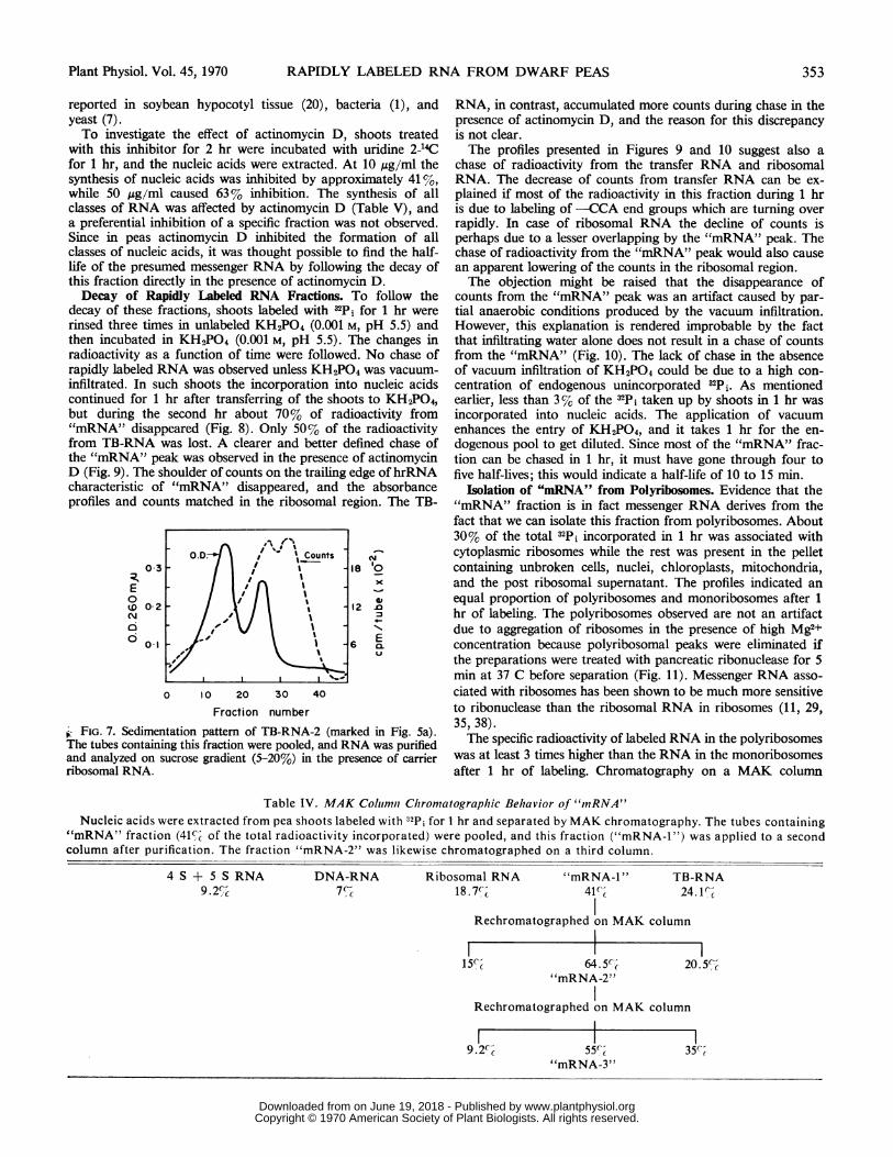
geneous, ranging in sedimentation from 4 to 32 S. After shortperiods of labeling (up to 2 hr) this fraction was very rich inAMP (48%, Table II), and the counts in the peak region showeda sedimentation coefficient around 13 to 15 S (Fig. 4b). Afterlonger periods of labeling (more than 2 hr) the AMP contentgradually decreased to 26 to 31%, and a relatively larger con-tamination with ribosomal RNA was observed. On furtherchromatography, more than 95 %7 of the counts showed a nucleo-tide composition similar to ribosomal RNA. On rechromatogra-phy of TB-RNA on a MAK column (16-hr labeling, TB-RNA-1),two fractions were obtained: about 65%C of the radioactivityappeared at the same place as ribosomal RNA and the rest asTB-RNA-2 (Fig. Sa). On further chromatography each of thesetwo behaved in an identical manner, and three components wereobserved (Fig. Sb): (a) 35%; of the counts appeared in a peakpreceding hrRNA, and in sucrose gradients it sedimented be-tween light and heavy ribosomal RNA (Fig. 6a); (b) 35% of thecounts appeared on the shoulder of hrRNA and sedimentedaround 45 to 50 S in sucrose gradient (Fig. 6b); (c) 30% ap-peared as TB-RNA-3 with a slight enrichment of AMP contentover the TB-RNA-1 or TB-RNA-2 (Table III). The nucleotidecompositions of the first two components are almost identicaland like that of hrRNA. Fraction b seems to arise because ofaggregation, since no heavy moving component was demon-strable in TB-RNA-2 in sucrose gradients prior to MAK chro-matography (Fig. 7).On rechromatography of the "mRNA" on a MAK column
(1-hr labeling), about 15% of the counts appeared with carrierhrRNA, 65%7 as "mRNA-2," and the rest (20%7c) as tenaciouslybound RNA (Table IV). The fraction "mRNA-2" behaved inmuch the same way on further chromatography. In short label-
Table II. Nucleotide Composition of Tenaciously Bound RNAThe pea shoots were labeled with l2Pi, and nucleic acids were ex-
tracted and separated by MAK chromatography. TB-RNA frac-tions were purified and the nucleotide composition was determinedas described under "Materials and Methods."
Percentage of Total Counts Nucleotide32Pi Incorporation Ratio,
CNIP ANIP GMP UMIP GMP
hr
16 16.0 47.8 21.4 18.9 2.2316TB-RNA-1 23.2 I 26.6 22.8 25.7 1.16TB-RNA-2 23.0 28.2 21.7 26.2 1.29TB-RNA-3 20.4 31.6 19.7 28.3 1.59
24 22.4 28.2 21.0 26.6 1.34
DO10)
.0
0E0.
0
'o~~~~~~~b 'C\--
_0
D,
EcLO
0 10 20 30 40 50 0 10 20 30 40
Fraction number Fraction number
FIG. 4. Sedimentation profiles of "mRNA," shoots labeled with 32Pi for 1 hr (a), and TB-RNA from shoots labeled with 32Pi for 2 hr (b). Thenucleic acids were first separated on a MAK column, and the tubes corresponding to "mRNA" or TB-RNA were pooled and analyzed on su-
crose gradients from S to 40%,c (a), or from S to 20% (b). Centrifugation at 23,000 rpm at -2 C for 18 hr in an SW 25.1 rotor. Fractions werecollected from bottom.
Plant Physiol. Vol. 45, 1970
0.3
0.2
0.1
www.plantphysiol.orgon June 19, 2018 - Published by Downloaded from Copyright © 1970 American Society of Plant Biologists. All rights reserved.

Plant Physiol. Vol. 45, 1970
ing periods the "mRNA" fraction was much less contaminatedby ribosomal RNA than was TB-RNA-1. This would be ex-pected because the steady state level of 32Pi in ribosomal RNAis reached only after 2 hr.
Effect of 5-Fluorouracil and Actinomycin D on RNA Synthesis.The purpose of these experiments was to determine whether it ispossible to inhibit selectively the synthesis of either the rapidlyturning over RNA with actinomycin D, or of ribosomal RNAand transfer RNA with FU, and then to use these inhibitors astools for characterizing the messenger RNA. FU inhibited the
0 8
E0
(D
020
E0
00
0It6a)
-
0.
I0n.o
0-CL
0 10 20 30 40 50 60 70 80 90
Fraction number
FIG. 5. Rechromatography of TB-RNA. Twenty pea shoots wereincubated with 500 juc of 32P1 for 16 hr, and nucleic acids were extractedand chromatographed on a MAK column. The TB-RNA-1 obtainedwas purified and applied to a second column. The transfer RNA, 5 Sribosomal RNA, and DNA were added as carrier (a). The TB-RNA-2was again purified and chromatographed on a third column togetherwith carrier pea nucleic acids (b).
synthesis of nucleic acids at relatively high concentrations (10-3to 10-2 M). Pretreating the shoots for 2 hr inhibited the incor-poration of 32Pi into nucleic acids about 35% (Table V). Theaccumulation of radioactivity was reduced in all classes ofRNA, and the inhibition of ribosomal RNA and DNA-RNAwas very marked at a concentration of 10-2 M. The decrease ofcounts in the "mRNA" and TB-RNA peaks may be due to theabsence of ribosomal RNA which is no longer contaminatingthese peaks. In pea shoots, the synthesis of "mRNA" and TB-RNA is affected by FU less than that of transfer RNA and ribo-somal RNA. Similar partially selective effects of FU have been
E O.D-. %
0O °3 16 -(.0 \.4.--Counts 02
N 1 % -o
o X _
0 10E2030 40'.~~~~C
(b) 0
0c5 Counts e t iI '
E /(m 03 0..d
0 0.,' 15 CE
0 10 20 30 40
Fraction number
FIG. 6. Sedimentation pattern of the fractions obtained on the re-chromatography of TB-RNA-2. The tubes containing peaks i and ii(marked in Fig. 5b) were pooled and analyzed on sucrose gradients(5-20% in a, 5-40% in b) after ethanol precipitation and removal ofNaCl by dialysis. Centrifugation at 23,000 rpm for 20 hr in an SW25.1 rotor. Ribosomal RNA was added as marker in each case.
Table III. MAK Coltiumn Clhromatographic Behavior of Teniaciouisly Bounild RNAPea shoots were incubated for 16 hr with "2Pi, and nucleic acids were extracted and separated by MAK chromatography. TB-RNA-1,
containing 6 to 8%c of total radioactivity, was purified and applied to a second column. The radioactivity eluting in the region of ribo-somal RNA and as TB-RNA-2 were again chromatographed on two separate columns. The nucleotide composition of various fractionswas determined as described in Table I.
CMP23.2
TB-RNA-1AMP GMP UMP26.6 22.8 25.7A_ A_I-_ _t
olRechromatographed on MAK column
Ribosomal RNA (65'%)CMP AMP GMPI1 A 1) A )fa
Rechromatographed on MAK column
CMP AMP GMP21.5 25.0 27.521.8 25.8 29.021.8 26.9 27.0
UMP22.2
UMP22.227.823.4
TB-RNA-2 (35%()CMP AMP GMP23.0 28.2 21.7
Rechromatographed on MAK column
CMP-i) 35(( 21.6ii) 40', 20.8iii) 25'% 20.4
AMP25.526.531.6
35' c35%c30%-C
UMP26.2
GMP31.327.119.7
TB-RNA-3
UMP20.924.428.3
352 JOHRI AND VARNER
.j. I
www.plantphysiol.orgon June 19, 2018 - Published by Downloaded from Copyright © 1970 American Society of Plant Biologists. All rights reserved.
RAPIDLY LABELED RNA FROM DWARF PEAS
reported in soybean hypocotyl tissue (20), bacteria (1), andyeast (7).To investigate the effect of actinomycin D, shoots treated
with this inhibitor for 2 hr were incubated with uridine 2-14Cfor 1 hr, and the nucleic acids were extracted. At 10 ,ug/ml thesynthesis of nucleic acids was inhibited by approximately 41 %,while 50 ,ug/ml caused 63% inhibition. The synthesis of allclasses of RNA was affected by actinomycin D (Table V), anda preferential inhibition of a specific fraction was not observed.Since in peas actinomycin D inhibited the formation of allclasses of nucleic acids, it was thought possible to find the half-life of the presumed messenger RNA by following the decay ofthis fraction directly in the presence of actinomycin D.Decay of Rapidly Labeled RNA Fractions. To follow the
decay of these fractions, shoots labeled with -"Pi for 1 hr wererinsed three times in unlabeled KH2PO4 (0.001 M, pH 5.5) andthen incubated in KH2PO4 (0.001 M, pH 5.5). The changes inradioactivity as a function of time were followed. No chase ofrapidly labeled RNA was observed unless KH2PO4 was vacuum-infiltrated. In such shoots the incorporation into nucleic acidscontinued for 1 hr after transferring of the shoots to KHYO4,but during the second hr about 70% of radioactivity from"mRNA" disappeared (Fig. 8). Only 50% of the radioactivityfrom TB-RNA was lost. A clearer and better defined chase ofthe "mRNA" peak was observed in the presence of actinomycinD (Fig. 9). The shoulder of counts on the trailing edge ofhrRNAcharacteristic of "mRNA" disappeared, and the absorbanceprofiles and counts matched in the ribosomal region. The TB-
0 3
E
0
(0
C\d
0 2
0*1
18
12
6
N
I0XI.
E
CL
0 10 20 30 40Fraction number
1, FIG. 7. Sedimentation pattern of TB-RNA-2 (marked in Fig. 5a).The tubes containing this fraction were pooled, and RNA was purifiedand analyzed on sucrose gradient (5-20%) in the presence of carrierribosomal RNA.
RNA, in contrast, accumulated more counts during chase in thepresence of actinomycin D, and the reason for this discrepancyis not clear.The profiles presented in Figures 9 and 10 suggest also a
chase of radioactivity from the transfer RNA and ribosomalRNA. The decrease of counts from transfer RNA can be ex-plained if most of the radioactivity in this fraction during 1 hris due to labeling of -CCA end groups which are turning overrapidly. In case of ribosomal RNA the decline of counts isperhaps due to a lesser overlapping by the "mRNA" peak. Thechase of radioactivity from the "mRNA" peak would also causean apparent lowering of the counts in the ribosomal region.The objection might be raised that the disappearance of
counts from the "mRNA" peak was an artifact caused by par-tial anaerobic conditions produced by the vacuum infiltration.However, this explanation is rendered improbable by the factthat infiltrating water alone does not result in a chase of countsfrom the "mRNA" (Fig. 10). The lack of chase in the absenceof vacuum infiltration of KH2PO4 could be due to a high con-centration of endogenous unincorporated 32Pi. As mentionedearlier, less than 3% of the 32Pi taken up by shoots in 1 hr wasincorporated into nucleic acids. The application of vacuumenhances the entry of KH2PO4, and it takes 1 hr for the en-dogenous pool to get diluted. Since most of the "mRNA" frac-tion can be chased in 1 hr, it must have gone through four tofive half-lives; this would indicate a half-life of 10 to 15 min.
Isolation of "mRNA" from Polyribosomes. Evidence that the"mRNA" fraction is in fact messenger RNA derives from thefact that we can isolate this fraction from polyribosomes. About30% of the total 4Pi incorporated in 1 hr was associated withcytoplasmic ribosomes while the rest was present in the pelletcontaining unbroken cells, nuclei, chloroplasts, mitochondria,and the post ribosomal supernatant. The profiles indicated an
equal proportion of polyribosomes and monoribosomes after 1
hr of labeling. The polyribosomes observed are not an artifactdue to aggregation of ribosomes in the presence of high Mg2+concentration because polyribosomal peaks were eliminated ifthe preparations were treated with pancreatic ribonuclease for 5min at 37 C before separation (Fig. 11). Messenger RNA asso-
ciated with ribosomes has been shown to be much more sensitiveto ribonuclease than the ribosomal RNA in ribosomes (11, 29,35, 38).The specific radioactivity of labeled RNA in the polyribosomes
was at least 3 times higher than the RNA in the monoribosomesafter 1 hr of labeling. Chromatography on a MAK column
Table IV. MAK Colhinii Chroniatographic Behavior of "mRNA"Nucleic acids were extracted from pea shoots labeled with "Pi for 1 hr and separated by MAK chromatography. The tubes containing
"mRNA" fraction (41%-C of the total radioactivity incorporated) were pooled, and this fraction ("mRNA-1") was applied to a secondcolumn after purification. The fraction "mRNA-2" was likewise chromatographed on a third column.
4 S + 5 S RNA9 .2%',c
DNA-RNA7 %,
Riboson18.
nal RNA "4mRNA-1 " TB-1.7%- 41%- 24
Rechromatographed on MAK column
15% 64.5%-C"mRNA-2"
I
-RNA,.1%r
20.5%'c
Rechromatographed on MAK column
9.2-c 55%R c
"mRNA-3"
35%c
0.D. / \_#%CountsI I
I~~~~~~~~~-- I
Plant Physiol. Vol. 45, 1970 353
www.plantphysiol.orgon June 19, 2018 - Published by Downloaded from Copyright © 1970 American Society of Plant Biologists. All rights reserved.

JOHRI AND VARNER Plant Physiol. Vol. 45, 1970
Table V. Inhibitionz of Nucleic Acid Synthesis by 5-Fluorouracil and Actinomycin D
Ten shoots were incubated aseptically in 2 ml of water containing FU or actinomycin D for 2 hr and were then allowed to incorpo-rate 500 Mc of 32Pi (FU experiment) or 5 ,uc of uridine 2-14C (actinomycin D experiment) for 1 hr. Nucleic acids were extracted and ana-lyzed by MAK chromatography.
Percentage Inhibition of Ribosomal RNA
Inhibitor
SRNA DNA-RNA 5S Light + heavy "mRNLA" TB-RNAA Total nucleicacids
5-Fluorouracil103M 20.5 54.7 11.3 47.9 24.7 33.9 33.4102Mm 30.9 62.7 28.9 71.6 17.7 32.4 37.3
Actinomycin D10 ,ug/ml 42.9 23.2 48.5 41.4 39.8 40.650,ug/ml 53.0 48.4 55.0 73.3 64.2 62.7c~~~~~~~~
8
%SI
6-I
1..-Counts Jo
.a--OD ~~~~~~~~II0.D.I I.
II
% II C
I
.I.*-Counts
I O.D.
It
II1
I II I
I I
I II II I
I II II ISII I
I
A.
0
to
a
8
66 -
.04 =
E2
0
SD8
6 °D
04
E2 X
54 SI~~~~~II
~~~~~II
S~ 545~~S~II
I X30 40 50 60 70 80
Fraction number
FIG. 8. Decay of "mRNA" fraction. Thirty pea shoots were incu-bated in a 50-ml Erlenmeyer flask (in 6 ml of sterile water containing 1
mc of 4Pi and 300 ,ug of chloroamphenicol) for 1 hr, and nucleic acidsfrom 10 shoots were washed three times with ice-cold 0.0001 KH2PO4(pH 5.5) and subjected to vacuum for 2 min to infiltrate KH2PO4. Tenshoots were removed after every hour, and nucleic acids were extractedand analyzed. Nucleic acids from shoots labeled for 1 hr immediatelyextracted (top), extracted 1 hr after transfer to KH2PO4 (middle), and2 hr after transfer to KH2PO4 (bottom).
indicated that the radioactivity associated with ribosomes repre-sented the same rapidly labeled RNA fractions as observed inthe total nucleic acids from shoots after a short labeling period.The labeled RNA associated with ribosomes was released
either by removing Mg2+ (by treating ribosomes with EDTA or
sodium pyrophosphate) (Fig. 12) or by treatment with SLS, andseparated on sucrose gradients. This labeled RNA was polydis-
2.0
1.6
1.2
0.41
90
2.0~
=k
0
SD40ca
ci
1.6 k
1.2
0.8~
0.4~
l
'I)
.0
"I,
Eo
x
12
10
8 °0
6 Jo
E4 XL
2
206
16
8
6
4
2
0 10 20 30 40 50 60 70 80 90
Fraction number
FIG. 9. Decay of "mRNA" fraction during chase with 0.001 M
KH2PO4 in the presence of actinomycin D. All experimental details as
described for Figure 8. During vacuum infiltration of KH2PO4, actino-mycin D (50,ug/ml) was also included.
354
17
A II~~~~~~~~~~~
It
I II ~~~~~~Ii
A II I
I~~ ~ ~~~~4 II
E
6
4
2
r
www.plantphysiol.orgon June 19, 2018 - Published by Downloaded from Copyright © 1970 American Society of Plant Biologists. All rights reserved.

RAPIDLY LABELED RNA FROM DWARF PEAS
-I
2. CDt
1.6 Counts jo 0' 2
ci~~~ ~ ~~~~~~~~'o lo 20 30 40 50 60 70 80 90E
0.4£I
g9
1.6
0
1.2
0
10
"mRN"frctio. Al exerimntaldetilsCons 2ecieo iue8
0.4~~~~~~~~~~~~~~.D
12
2.0 -M a
w-i Counts m Iet
0 3~~~~~~~~~~~~I1I2
tidecomposition each wasEdetermined(Fig. 13). The AMP
0t0 20 30 40 50 60 70 90
Fraction number
FIG. 10. Effect of infiltrating water under vacuum on the decay of
"mRNA" fraction. Al experimental details as described for Figure 8.
The shoots labeled for 1 hr with nPi were washed three times with ice-
cold 0.001 m KH2PO4, and water was infiltrated. Note the lack ofchase of "mRNA" fraction.
perse (4-18d) and about 30 to 40% of the counts were associatedwith a small ribosomal subunit; these counts may represent the
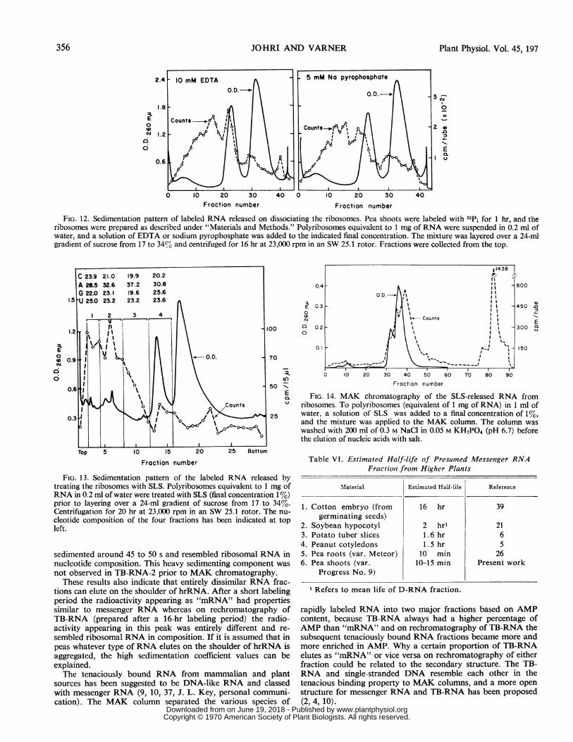
RNA still bound to it. The SLS-released RNA separated onsucrose gradient was divided into four fractions, and the nucleo-
tide composition of each was determined (Fig. 13). The AMP
content was different in each of these, but the over-all composi-tion of the first three fractions was the same as the "mRNA"peak. The RNA associated with lrRNA (fraction 4) had the
higher GMP content characteristic of ribosomal RNA. The
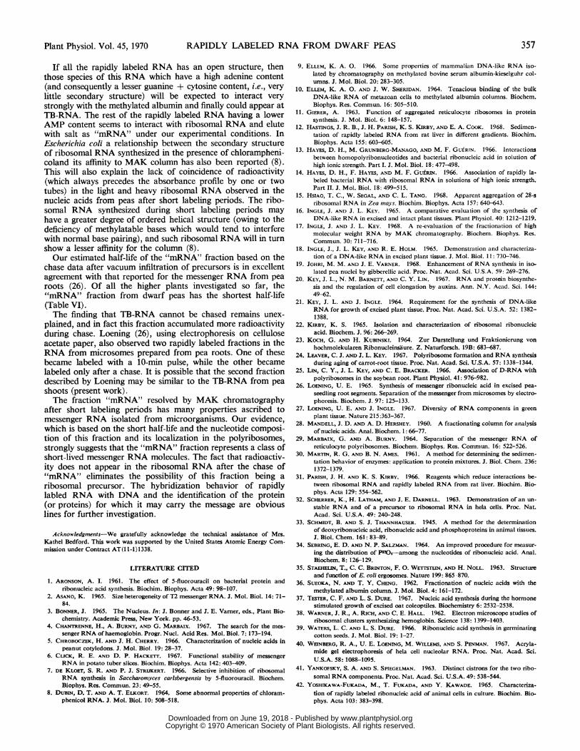
SLS-released RNA was also separated on a MAK column (Fig.14, without deproteinization to avoid degradation and also to
minim-ize loss). The fractions "mRNA" and TB-RNA each con-
tained 41% of the counts, the rest being in transfer RNA and
ribosomal RNA. The D-RNA (RNA eluting on the shoulder of
hrRNA) from soybean is also associated with ribosomes (25).
DIS3CUSSION
The rapidly labeled RNA after short labeling of pea shoots
has been characterized by MAK chromatography and sucrose
density gradient centrifugation. Whereas transfer RNA, DNA,and ribosomal RNA (all of which possess highly ordered second-
E
0
ID
0cli
3.8
3.4k
2.01
cs
E0
ID
6
1.6
1.2
0.8
0.4
0)
E
25.0
22.5
12.5 NYx
10.0.0
7.5 E0.
5.0
2.5
0 10 20 30 40 50 60 70
Fraction number
FIG. 11. Sedimentation profiles of polyribosomes from pea shootslabeled with 32Pi for 1 hr. Polyribosomes equivalent to 0.8 mg of RNAwere either layered directly (top) or were treated with 0.1 ,ug of pan-creatic ribonuclease for 5 min at 37 C and then layered (bottom) overa 24-ml gradient of sucrose from 10 to 34%. Centrifugation for 2 hr at-2 C at 23,000 rpm in an SW 25.1 rotor.
ary structure) are eluted on the basis of size, base composition,and extent of hydrogen bonding (36), the rapidly labeled RNAbehaves differently. In several cases it has been reported thatRNA aggregates because of high salt concentration and furtherthat a large percentage of rapidly labeled RNA cannot be elutedwith salt (9, 10, 23).When the "mRNA" fraction from pea shoots was analyzed on
sucrose gradients after separation by MAK chromatography, asedimentation coefficient of 45 to 50 S was noticed. An analysisof nucleotide composition eliminated the possibility of this frac-tion being the 45 S ribosomal precursor. No such heavy sedi-menting fraction was found if the rapidly labeled RNA releasedby SLS from ribosomes was analyzed. This RNA was polydis-perse (6-18 S), and on analysis on a MAK column 41 %0 of theradioactivity appeared in the "mRNA" peak. It is conceivablethat labeled RNA may have suffered some degradation during thepreparation of ribosomes, but our results argue against this anddegradation alone cannot account for the lack of a heavy movingcomponent from the rapidly labeled RNA prepared without theuse of high salt concentrations. On rechromatography on MAKcolumn of TB-RNA-2 the radioactivity eluting as "mRNA" also
_O.D._
II
I'I-
I'
0~~~~~~~~~~~~~
I^counh | | 1 1 A--I I
Plant Physiol. Vol. 45, 1970 355
www.plantphysiol.orgon June 19, 2018 - Published by Downloaded from Copyright © 1970 American Society of Plant Biologists. All rights reserved.
JOHRI AND VARNER Plant Physiol. Vol. 45, 197
E
0
N
C
x _
x
2
E1I
0 10 20 30 40 0 10 20 30 40Fraction number Fraction number
FIG. 12. Sedimentation pattern of labeled RNA released on dissociating the ribosomes. Pea shoots were labeled with 32Pi for 1 hr, and theribosomes were prepared as described under "Materials and Methods." Polyribosomes equivalent to 1 mg of RNA were suspended in 0.2 ml ofwater, and a solution of EDTA or sodium pyrophosphate was added to the indicated final concentration. The mixture was layered over a 24-mlgradient of sucrose from 17 to 34%-o and centrifuged for 16 hr at 23,000 rpm in an SW 25.1 rotor. Fractions were collected from the top.
E
0
4o
CX
I0
(D
C)c
"ICE
l.,
Fraction number
FIG. 13. Sedimentation pattern of the labeled RNA released bytreating the ribosomes with SLS. Polyribosomes equivalent to 1 mg ofRNA in 0.2 ml of water were treated with SLS (final concentration 1%)prior to layering over a 24-ml gradient of sucrose from 17 to 34%.Centrifugation for 20 hr at 23,000 rpm in an SW 25.1 rotor. The nu-cleotide composition of the four fractions has been indicated at topleft.
sedimented around 45 to 50 s and resembled ribosomal RNA innucleotide composition. This heavy sedimenting component wasnot observed in TB-RNA-2 prior to MAK chromatography.These results also indicate that entirely dissimilar RNA frac-
tions can elute on the shoulder of hrRNA. After a short labelingperiod the radioactivity appearing as "mRNA" had propertiessimilar to messenger RNA whereas on rechromatography ofTB-RNA (prepared after a 16-hr labeling period) the radio-activity appearing in this peak was entirely different and re-
sembled ribosomal RNA in composition. If it is assumed that inpeas whatever type of RNA elutes on the shoulder of hrRNA isaggregated, the high sedimentation coefficient values can beexplained.The tenaciously bound RNA from mammalian and plant
sources has been suggested to be DNA-like RNA and classedwith messenger RNA (9, 10, 37, J. L. Key, personal communi-cation). The MAK column separated the various species of
11438411
1 -600I I I
t I I - 450\ ~~~~II1- CountsI l I
isI - 300I
I
II% I %% L% %\ A.. ! 150
-I I
50 60 70 80 90
a
E(a.
Fraction number
FIG. 14. MAK chromatography of the SLS-released RNA fromribosomes. To polyribosomes (equivalent of 1 mg of RNA) in 1 ml ofwater, a solution of SLS was added to a final concentration of 1%,and the mixture was applied to the MAK column. The column waswashed with 200 ml of 0.3 M NaCl in 0.05 M KH2PO4 (pH 6.7) beforethe elution of nucleic acids with salt.
Table VI. Estimated Half-life of Presuimed Messenger RNAFractiont from Higher Plants
MIaterial Estimated Half-life Reference
1. Cotton embryo (from 16 hr 39germinating seeds)
2. Soybean hypocotyl 2 hrl 213. Potato tuber slices 1.6 hr 64. Peanut cotyledons 1.5 hr 55. Pea roots (var. Meteor) 10 min 266. Pea shoots (var. 10-15 min Present work
Progress No. 9)
1 Refers to mean life of D-RNA fraction.
rapidly labeled RNA into two major fractions based on AMPcontent, because TB-RNA always had a higher percentage ofAMP than "mRNA" and on rechromatography of TB-RNA thesubsequent tenaciously bound RNA fractions became more andmore enriched in AMP. Why a certain proportion of TB-RNAelutes as "mRNA" or vice versa on rechromatography of eitherfraction could be related to the secondary structure. The TB-RNA and single-stranded DNA resemble each other in thetenacious binding property to MAK columns, and a more openstructure for messenger RNA and TB-RNA has been proposed(2, 4,10).
356
www.plantphysiol.orgon June 19, 2018 - Published by Downloaded from Copyright © 1970 American Society of Plant Biologists. All rights reserved.
RAPIDLY LABELED RNA FROM DWARF PEAS
If all the rapidly labeled RNA has an open structure, thenthose species of this RNA which have a high adenine content(and consequently a lesser guanine + cytosine content, i.e., verylittle secondary structure) will be expected to interact verystrongly with the methylated albumin and finally could appear atTB-RNA. The rest of the rapidly labeled RNA having a lowerAMP content seems to interact with ribosomal RNA and elutewith salt as "mRNA" under our experimental conditions. InEscherichia coli a relationship between the secondary structureof ribosomal RNA synthesized in the presence of chlorampheni-coland its affinity to MAK column has also been reported (8).This will also explain the lack of coincidence of radioactivity(which always precedes the absorbance profile by one or twotubes) in the light and heavy ribosomal RNA observed in thenucleic acids from peas after short labeling periods. The ribo-somal RNA synthesized during short labeling periods mayhave a greater degree of ordered helical structure (owing to thedeficiency of methylatable bases which would tend to interferewith normal base pairing), and such ribosomal RNA will in turnshow a lesser affinity for the column (8).Our estimated half-life of the "mRNA" fraction based on the
chase data after vacuum infiltration of precursors is in excellentagreement with that reported for the messenger RNA from pearoots (26). Of all the higher plants investigated so far, the"mRNA" fraction from dwarf peas has the shortest half-life(Table VI).The finding that TB-RNA cannot be chased remains unex-
plained, and in fact this fraction accumulated more radioactivityduring chase. Loening (26), using electrophoresis on celluloseacetate paper, also observed two rapidly labeled fractions in theRNA from microsomes prepared from pea roots. One of thesebecame labeled with a 10-min pulse, while the other becamelabeled only after a chase. It is possible that the second fractiondescribed by Loening may be similar to the TB-RNA from peashoots (present work).The fraction "mRNA" resolved by MAK chromatography
after short labeling periods has many properties ascribed tomessenger RNA isolated from microorganisms. Our evidence,which is based on the short half-life and the nucleotide composi-tion of this fraction and its localization in the polyribosomes,strongly suggests that the "mRNA" fraction represents a class ofshort-lived messenger RNA molecules. The fact that radioactiv-ity does not appear in the ribosomal RNA after the chase of"mRNA" eliminates the possibility of this fraction being aribosomal precursor. The hybridization behavior of rapidlylabled RNA with DNA and the identification of the protein(or proteins) for which it may carry the message are obviouslines for further investigation.
Acknowledgments-We gratefully acknowledge the technical assistance of Mrs.Kathel Bedford. This work was supported by the United States Atomic Energy Com-mission under Contract AT (l -l) 1338.
LITERATURE CITED
1. ARONSON, A. I. 1961. The effect of 5-fluorouracil on bacterial protein andribonucleic acid synthesis. Biochim. Biophys. Acta 49: 98-107.
2. ASANO, K. 1965. Size heterogeneity of T2 messenger RNA. J. Mol. Biol. 14: 71-84.
3. BONNER, J. 1965. The Nucleus. In: J. Bonner and J. E. Varner, eds., Plant Bio-chemistry. Academic Press, New York. pp. 46-53.
4. CHANTRENNE, H., A. BURNY, AND G. MARBAIX. 1967. The search for the mes-senger RNA of haemoglobin. Progr. Nucl. Acid Res. Mol. Biol. 7:173-194.
5. CHROBOCZEK, H. AND J. H. CHERRY. 1966. Characterization of nucleic acids inpeanut cotyledons. J. Mol. Biol. 19: 28-37.
6. CLuCm, R. E. AND D. P. HACKETT. 1967. Functional stability of messengerRNA in potato tuber slices. Biochim. Biophys. Acta 142: 403-409.
7. DE KLoET, S. R. AND P. J. STRIIcERT. 1966. Selective inhibition of ribosomalRNA synthesis in Saccharomyces carlsbergensis by 5-fluorouracil. Biochem.Biophys. Res. Commun. 23: 49-55.
8. DuBIN, D. T. AND A. T. ELKORT. 1964. Some abnormal properties of chloram-phenicol RNA. J. Mol. Biol. 10: 508-518.
357
9. ELLEM, K. A. 0. 1966. Some properties of mammalian DNA-like RNA iso-lated by chromatography on methylated bovine serum albumin-kieselguhr col-umns. J. Mol. Biol. 20: 283-305.
10. ELLEM, K. A. 0. AND J. W. SHERIDAN. 1964. Tenacious binding of the bulkDNA-like RNA of metazoan cells to methylated albumin columns. Biochem.Biophys. Res. Commun. 16: 505-510.
11. GIERER, A. 1963. Function of aggregated reticulocyte ribosomes in proteinsynthesis. J. Mol. Biol. 6: 148-157.
12. HAsTINGS, J. R. B., J. H. PARISH, K. S. KIRBY, AND E. A. COOK. 1968. Sedimen-tation of rapidly labeled RNA from rat liver in different gradients. Biochim.Biophys. Acta 155: 603-605.
13. HAYES, D. H., M. GRUNBERG-MANAGO, AND M. F. GUERIN. 1966. Interactionsbetween homopolyribonucleotides and bacterial ribonucleic acid in solution ofhigh ionic strength. Part I. J. Mol. Biol. 18: 477-498.
14. HAYES, D. H., F. HAYES, AND M. F. GuRItm. 1966. Association of rapidly la-beled bacterial RNA with ribosomal RNA in solutions of high ionic strength.Part II. J. Mol. Biol. 18: 499-515.
15. HsIAo, T. C., W. SEGAL, AND C. L. TANG. 1968. Apparent aggregation of 28-sribosomal RNA in Zea mays. Biochim. Biophys. Acta 157: 640-643.
16. INGLE, J. AND J. L. KEY. 1965. A comparative evaluation of the synthesis ofDNA-like RNA in excised and intact plant tissues. Plant Physiol. 40: 1212-1219.
17. INGLE, J. AND J. L. KEY. 1968. A re-evaluation of the fractionation of highmolecular weight RNA by MAK chromatography. Biochem. Biophys. Res.Commun. 30: 711-716.
18. INGLE, J., J. L. KEY, AND R. E. HOLM. 1965. Demonstration and characteriza-tion of a DNA-like RNA in excised plant tissue. J. Mol. Biol. 11: 730-746.
19. JOHRI, M. M. AND J. E. VARNER. 1968. Enhancement of RNA synthesis in iso-lated pea nuclei by gibberellic acid. Proc. Nat. Acad. Sci. U.S.A. 59: 269-276.
20. KEY, J. L., N. M. BARNE1-r, AND C. Y. LIN. 1967. RNA and protein biosynthe-sis and the regulation of cell elongation by auxins. Ann. N.Y. Acad. Sci. 144:49-62.
21. KEY, J. L. AND J. INGLE. 1964. Requirement for the synthesis of DNA-likeRNA for growth of excised plant tissue. Proc. Nat. Acad. Sci. U.S.A. 52: 1382-1388.
22. KIRBY, K. S. 1965. Isolation and characterization of ribosomal ribonucleicacid. Biochem. J. 96: 266-269.
23. KOCH, G. AND H. KUBINSKl. 1964. Zur Darstellung und Fraktionierung vonhochmolekularen Ribonucleinsaure. Z. Naturforsch. 19B: 683-687.
24. LEAVER, C. J. AND J. L. KEY. 1967. Polyribosome formation and RNA synthesisduring aging of carrot-root tissue. Proc. Nat. Acad. Sci. U.S.A. 57: 1338-1344.
25. LIN, C. Y., J. L. KEY, AND C. E. BRAcKER. 1966. Association of D-RNA withpolyribosomes in the soybean root. Plant Physiol. 41: 976-982.
26. LOENING, U. E. 1965. Synthesis of messenger ribonucleic acid in excised pea-seedling root segments. Separation of the messenger from microsomes by electro-phoresis. Biochem. J. 97: 125-133.
27. LOENING, U. E. AND J. INGLE. 1967. Diversity of RNA components in greenplant tissue. Nature 215:363-367.
28. MANDELL, J. D. AND A. D. HERSHEY. 1960. A fractionating column for analysisof nucleic acids. Anal. Biochem. 1: 66-77.
29. MARBAIX, G. AND A. BURNY. 1964. Separation of the messenger RNA ofreticulocyte polyribosomes. Biochem. Biophys. Res. Commun. 16: 522-526.
30. MARTIN, R. G. AND B. N. AMEs. 1961. A method for deternining the sedimen-tation behavior of enzymes: application to protein mixtures. J. Biol. Chem. 236:1372-1379.
31. PARISH, J. H. AND K. S. KIRBY. 1966. Reagents which reduce interactions be-tween ribosomal RNA and rapidly labeled RNA from rat liver. Biochim. Bio-phys. Acta 129: 554-562.
32. SCHERRER, K., H. LATHAM, AND J. E. DARNELL. 1963. Demonstration of an un-stable RNA and of a precursor to ribosomal RNA in hela cells. Proc. Nat.Acad. Sci. U.S.A. 49: 240-248.
33. SCHMIDT, B. AND S. J. THANNHAUSER. 1945. A method for the determinationof deoxyribonucleic acid, ribonucleic acid and phosphoproteins in animal tissues.J. Biol. Chem. 161: 83-89.
34. SEBRING, E. D. AND N. P. SALZMAN. 1964. An improved procedure for measur-ing the distribution of P5504-among the nucleotides of ribonucleic acid. Anal.Biochem. 8: 126-129.
35. STAEHELuN, T., C. C. BRINTON, F. 0. WEMTEIN, AND H. NOLL. 1963. Structureand function of E. coli ergosomes. Nature 199: 865--870.
36. SuEoKA, N. AND T. Y. CHENG. 1962. Fractionation of nucleic acids with themethylated albumin column. J. Mol. Biol. 4: 161-172.
37. TESTER, C. F. AND L. S. DURE. 1967. Nucleic acid synthesis during the hormonestimulated growth of excised oat coleoptiles. Biochemistry 6: 2532-2538.
38. WARNER, J. R., A. RICH, AND C. E. HALL. 1962. Electron microscope studies ofribosomal clusters synthesizing hemoglobin. Science 138: 1399-1403.
39. WATERS, L. C. AND L. S. DURE. 1966. Ribonucleic acid synthesis in germinatingcotton seeds. J. Mol. Biol. 19: 1-27.
40. WEINBERG, R. A., U. E. LOENING, M. WILLEMS, AND S. PENMAN. 1967. Acryla-mide gel electrophoresis of hela cell nucleolar RNA. Proc. Nat. Acad. Sci.U.S.A. 58: 1088-1095.
41. YANKOFSKY, S. A. AND S. SPIEGELMAN. 1963. Distinct cistrons for the two ribo-somal RNA components. Proc. Nat. Acad. Sci. U.S.A. 49: 538-544.
42. YOSHIKAWA-FUKADA, M., T. FUKADA, AND Y. KAWADE. 1965. Characteriza-tion of rapidly labeled ribonucleic acid of animal cells in culture. Biochim. Bio-phys. Acta 103: 383-398.
Plant Physiol. Vol. 45, 1970
www.plantphysiol.orgon June 19, 2018 - Published by Downloaded from Copyright © 1970 American Society of Plant Biologists. All rights reserved.