Embed Size (px)
Citation preview
CHARACTERIZATION OF VACUOLE AQUAPORIN FUNCTION AND ITS
IMPLICATION IN MEMBRANE FISSION
Dipti Patel
A Thesis
in
The Department
of
Biology
Presented in Partial Fulfillment of the Requirements
for the degree of Master of Science (Biology) at
Concordia University
Montreal, Quebec, Canada
September 2013
© Dipti Patel, 2013
CONCORDIA UNIVERSITY
School of Graduate Studies
This is to certify that the thesis prepared
By: Dipti Patel
Entitled: Characterization of vacuole aquaporin function and its implication in
membrane fission
and submitted in partial fulfillment of the requirements for the degree of
Master of Science (Biology)
complies with the regulations of the University and meets the accepted standards
with respect to originality and quality.
Signed by the final Examining Committee:
___________________________________________________ Chair Dr. James Grant ___________________________________________________ Examiner Dr. Patrick Gulick ___________________________________________________ Examiner Dr. Michael Sacher
___________________________________________________ External Examiner
Dr. Catherine Bachewich ___________________________________________________ Supervisor Dr. Christopher Brett Approved by ___________________________________________________
Chair of Department or Graduate Program Director
_______________ 2013 ___________________________________________________
Dean of Faculty
iii
ABSTRACT
Characterization of vacuole aquaporin function and its implication in
membrane fission
Dipti Patel
All cells and organelles must regulate volume to control their size, shape and
copy number. Volume homeostasis, in turn, is dependent on osmosis facilitated by
aquaporins. Despite being critical for membrane fusion and fission events that
underlie organelle morphology and membrane trafficking, volume regulations in the
endocytic pathway and the aquaporins responsible have not been characterized.
Thus, the primary goals of my dissertation research were to better understand the
role of osmosis in membrane fusion or fission and identify the aquaporin(s)
responsible using the yeast vacuole as a model. First, I characterized osmosis across
isolated vacuole membranes and identified the aquaporins responsible, Aqy3 and
Fps1, using stopped-flow fluorometry. Fluorescence microscopy revealed that
knocking out AQY3 or both aquaporins disrupted vacuole morphology, indicative of
an impairment in membrane fission. Using a new cell-free vacuole fission assay, I
then determined that osmosis can drive fission in vitro and that this process
involves inactivation of Ytp7, a Rab-GTPase and key regulator of vacuole
morphology. These results support a model whereby water efflux by Aqy3 permits
membrane invagination while signaling the fission machinery necessary for
membrane fission.
iv
ACKNOWLEDGEMENTS
First and foremost, I would like to thank God almighty for his grace and
guidance. I also want to take this opportunity to thank everyone who helped me
complete my thesis research:
I cordially thank my supervisor Dr. Christopher Brett for his excellent
tutoring, and teaching me how to test ideas while paying attention to experimental
details. I am grateful to my co-supervisor Dr. Jean-Louis Schwartz for his guidance
and collaboration, and Vincent Vachon for teaching me how to perform stopped flow
fluorometry experiments at the Université de Montréal. I thank Mahmoud Karim for
the preparation of the protein reagents needed for my experiments and Gabriel
Lapointe for help with fluorescence microscopy. I thank Drs. Patrick Gulick, Michael
Sacher, and Selvadurai Dayanandan for serving on my research committee and
offering scientific guidance and advice. I thank my lab-mates Mahmoud Karim, Joel
Richard, and Mark Guterman for advice pertaining to science, school and life. I thank
members of the Sacher, Titorenko, and Gulick laboratories for reagents and
protocols. I thank NSERC for funding my research, and GEPROM for providing me
with a partial scholarship during my studies at Concordia.
I am deeply thankful to my husband and my son for their patience, love and
support, without which I wouldn’t have reached this present stage. I am thankful to
my family and friends who helped me through all my ups and downs, and encourage
me to always strive to be a better scientist.
TABLE OF CONTENTS
LIST OF FIGURES .................................................................................................................................................... vi
LIST OF TABLES ..................................................................................................................................................... vii
INTRODUCTION ....................................................................................................................................................... 1
Osmosis determines cell and organelle volume and morphology ........................................................... 1
Aquaporin function, phylogeny and structure ............................................................................................... 3
MATERIALS AND METHODS ............................................................................................................................... 8
Yeast strains ............................................................................................................................................................... 8
Phylogenetic analysis ............................................................................................................................................ 11
Yeast vacuole isolation ......................................................................................................................................... 11
Stopped flow spectrofluorometry ..................................................................................................................... 12
Fluorescence microscopy and morphometric analysis ............................................................................. 13
In vitro vacuole membrane fission assay ....................................................................................................... 14
In vitro vacuole lysis assay .................................................................................................................................. 15
Cell-free homotypic vacuole fusion assay ....................................................................................................... 16
Data analysis ............................................................................................................................................................ 17
Table 1. Yeast strains used in this study ...................................................................................................... 18
RESULTS .................................................................................................................................................................... 19
Fps1 and Aqy3 contribute to osmosis across vacuole membranes ....................................................... 19
Aqy3 facilitates vacuole lysis in the absence of membrane fusion ........................................................ 27
Fps1 and Aqy3 may localize to vacuole membranes.................................................................................. 31
Fps1 and Aqy3 contribute to vacuole morphology ..................................................................................... 31
Water efflux and Rab-GTPase inactivation facilitates vacuole membrane fission in response to
hypertonic shock ..................................................................................................................................................... 35
DISCUSSION ............................................................................................................................................................. 43
REFERENCES ........................................................................................................................................................... 49

LIST OF FIGURES
FIGURE 1. Aquaporin structure, phylogeny and function ..................................................................... 07
FIGURE 2. HgCl2 blocks vacuole osmosis caused by hypertonic shock ............................................... 24
FIGURE 3. Aquaporins AQY3 and FPS1 mediate water efflux from vacuole in response to
hypertonic shock ..................................................................................................................................................... 26
FIGURE 4. Aqy3 mediates water influx into vacuoles in response to hypotonic shock ................ 30
FIGURE 5. Knocking out AQY3 or FPS1 affects vacuole morphology in response to osmotic
shock ............................................................................................................................................................................ 34
FIGURE 6. Like KOAc, hyperosmotic shock prevents fusion and enhances fission of vacuoles in
vitro .............................................................................................................................................................................. 40
FIGURE 7. Effects of wortmannin and Gyp1-46 on vacuole fission in vitro ..................................... 42
FIGURE 8. Model of mechanisms responsible for vacuole fission ......................................................... 48

LIST OF TABLES
Table 1. Yeast strains used in this study ...................................................................................................... 18
1
INTRODUCTION
Osmosis determines cell and organelle volume and morphology
Osmosis is the net movement of water across a semipermeable membrane
from lower to higher concentration of solute (or osmolyte) to achieve equilibrium.
As water constitutes most of an organism’s mass, it is the key determinant of
volume. Cells increase their volume, or swell, when they are placed in hypotonic
solution or decrease their volume, or shrink, when placed in hypertonic solution due
to osmosis. Hence, cellular osmolarity is a critical determinant of cell volume, and
changes in osmolarity underlie morphological (size and shape) changes that permit
cell differentiation, migration, and division; events that underlie diverse physiology
(Monzani et al., 2009).
Like cells, organelles also change volume in response to osmosis to control
their size, shape and number required for function (Sugiya et al., 2008). One the
best-characterized examples is the yeast vacuole, the terminal compartment of the
endocytic pathway whose primary function is protein and lipid degradation (similar
to the metazoan lysosome), but it also acts as a store for cellular water to resist
dehydration (Klionsky et al., 1990)(Hicke et al, 1999). Normally S. cerevisiae has 2 or
3 spherical vacuoles, however in response to hypertonic stress, vacuoles shrink and
fragment within minutes, resulting in 5-10 smaller vacuoles (Denis and Cyert,
2002). In response to hypotonic stress, vacuoles within the cell rapidly swell and
fuse to form a single, large vacuole (Bone et al., 1998). Despite changes in size and
number, vacuoles retain their spherical shape, leading to a model based on
2
allometric scaling, whereby the cell maintains vacuole size, shape and number by
tight regulation of lumenal volume – determined by osmosis – with respect to
membrane surface area –determined by fission and fusion events (Brett and Merz,
2008). According to this model (see Figure 1A), membrane surface must decrease
when vacuoles fuse to retain their spherical shape, which is achieved by the
formation of an internalized membrane fragment as a byproduct of fusion (Wang et
al., 2002). Conversely, volume must decrease to accommodate membrane collapse
required for fission. Isolated vacuoles are capable of fusion and fission in vitro, and
isolated vacuoles fuse in response to hypotonic shock (Brett and Merz, 2008)
(Michaillat et al., 2012), suggesting that the mechanisms that control vacuole size,
shape do not require complex cellular signaling, as originally proposed (Schüller et
al., 1994)(Mager and Varela, 1993). The mechanisms that control homotypic
vacuole fusion and, to a lesser degree, fission are well characterized (Wickner et al.,
2002) (Michaillat et al., 2012), and a component of this machinery, the Rab-GTPase
Ypt7, is crucial for regulating homotypic vacuole fusion in response to osmotic
stress (Brett and Merz, 2008). Similarly, inactivation of Ypt7 is implicated in vacuole
fission in response to hypertonic stress, but whether it directly contributes to this
process is not known. Indeed, volume regulation at the vacuole is not understood in
detail: the mechanism(s) that mediate water transport across the vacuole
membrane are unknown, and how these mechanisms may contribute to vacuole
fission is not defined. Because water channel proteins (called aquaporins) regulate
osmosis at the plasma membrane to control cell volume and morphology, we
hypothesized that an aquaporin may be present on the vacuole where it likely
3
contributes to observed changes in vacuole morphology in response to osmotic
stress.
Aquaporin function, phylogeny and structure
First described by Agre and colleagues (Saboori et al., 1988), aquaporins are
protein channels that translocate water across membranes. Aquaporin function is
best characterized on the surface of cells where, in mammals for example, they
regulate cell volume to allow erythrocytes to squeeze into capillaries with narrow
diameters to facilitate gas exchange, or to change the volume of lens fiber cells to
focus light on the retina (Borgnia and Nielsen, 1999). Aquaporins on the apical
membranes of epithelial cells lining the collecting duct of the nephron are critical for
water reabsorption required for whole body volume homeostasis (Knepper et al.,
1996). Although most aquaporin genes in every eukaryotic genome are expressed
on the surface of cells, the remaining paralogs (e.g. human AQP8, AQP11, AQP12)
have been observed on organelles, including mitochondrial membrane, endosomes,
lysosomes and ER (Mizutani et al., 2006)(Amiry-Moghaddam et al.,
2005)(Marchissio et al., 2012) (Nozaki et al., 2008). Unfortunately, these
intracellular aquaporins are poorly characterized although it is hypothesized that
they also play a critical role in volume regulation underlying changes in organelle
morphology.
Besides cellular location, members of aquaporin family can be further
subdivided into two categories according to substrate specificity: Classical
aquaporins, which conduct water exclusively, and aquaglyceroporins, which
4
conduct water as well as other uncharged solutes such as glycerol (Borgnia and
Nielsen, 1999). Whereas humans have 13 aquaporin genes (Verkman, 2005) most
laboratory strains of Saccharomyces cerevisiae (e.g. S288c) have three (see Figure
1B): AQY1, an aquaporin exclusively found on the plasma membrane where it
contributes to cellular volume regulation (Carbrey et al., 2001); FPS1, an
intracellular aquaglyceroporin (Luyten et al., 1995); and YFL054C (which we have
named AQY3), an uncharacterized gene that shows sequence similarity (29%) to
FPS1 in yeast, but greater similarity (37%) to a classical human aquaporin AQP7,
thought to reside on endocytic organelles.
Classic aquaporins function as homo- or hetero-tetramers (Fetter et al.,
2004). Each has two tandem repeats that fold into a six transmembrane-helix
structure with two opposing hydrophobic loops that each contain an NPA (Asn-Pro-
Ala) sequence motif (Figure 1C; (Agre et al., 1993)). These NPA motifs meet near the
most narrow region of the central hourglass-shaped channel to form one of two
selectivity filters (Walz et al., 1997) (Murata et al., 2000). At this site, pore diameter
(3Å) and the lining residues restrict size and charge, respectively, to create a
narrow, electrostatic filter that only allows permeation of water molecules (Murata
et al., 2000; Sui et al., 2001; De Groot and Grubmüller, 2005). The classic water
channel-blocker Hg2+ occludes this region of the pore (Savage and Stroud, 2007).
Importantly, the NPA motifs and other critical residues necessary for water
transports are conserved in AQY3, suggesting that it is a bone fide aquaporin that is
sensitive to blockade by Hg2+ (Figure 1C). Thus, it is possible that AQY3 or FPS1
5
contribute to vacuole volume regulation and that inhibition of aquaporin function
by Hg2+ may reveal how they contribute to morphology.
All things considered, the central goals of my thesis research were to identify
the aquaporin(s) that mediate osmosis across the vacuole membrane, and to
demonstrate that this aquaporin alters GTPase signaling to drive vacuole membrane
fission in response to hypertonic stress. To achieve these goals, I first characterized
the osmolytes important for the osmotic gradient across the vacuole membrane
using stopped-flow spectrofluorometry. I then showed that the aquaporins Fps1 and
Aqy3 are required for osmosis at the vacuole, and they are important for vacuole
morphology within yeast cells. Finally, using a new cell-free assay to measure
vacuole fission, I demonstrate that hypertonic stress induces vacuole fission in vitro,
eliminating the need for complex cell signaling, and show that osmosis inactivates
Rab-GTPases to mediate this process.
6
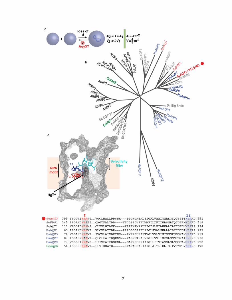
Figure 1. Aquaporin structure, phylogeny and function
(a) Cartoon illustrating that vacuole surface area (SA) and volume must change to
accommodate its spherical shape during fusion and fission events, respectively.
Equations used to calculate the area and volume of a sphere are shown for
reference. I hypothesize that AQY3 may contribute to the decrease in lumenal
volume required for fission. (b) Phylogenetic tree showing relationships between
aquaporin genes found in Ce, Caenorhabditis elegans (light grey); At, Arabidopsis
thaliana (black); Dm, Drosophila melanogaster (grey); Ec, Escherichia coli (green);
Sc, Saccharomyces cerevisiae (red); and Hs, Homo sapiens (blue). (c) Crystal
structure of mercury bound within AqpZ from E. coli, adapted from (Savage and
Stroud, 2007). The protein surface is highlighted with wire frame (grey), and
components of the central selectivity filter are shown: NPA I (red), NPA II (blue),
four amino acids in selectivity filter (dark cyan). Mercury atoms (dark grey spheres)
block the channel by occluding the part of the pore near these regions. For
reference, the membrane (orange line, width ~20 nm) is shown. A sequence
alignment between yeast, human and E. coli aquaporin genes is shown below. NPA
motif sequences are highlighted.
7
8
MATERIALS AND METHODS
Yeast strains
All strains of Saccharomyces cerevisiae used in this study are shown in Table
1. Most strains were obtained from the knock-out collection of 4,828 individual Mat-
haploid S. cerevisiae strains, each lacking a different non-essential gene
(Invitrogen, Carlsbad, CA): pep4Δ and pho8Δ were used to conduct cell-free
homotypic vacuole fusion assays, whereas wild type, aqy3Δ, and fps1Δ strains were
used for stopped-flow fluorometry experiments and to study vacuole morphology in
vivo by fluorescence microscopy. SEY6210 Pho8-GFP yeast (a kind gift from Dr.
Alexey Merz, University of Washington, Seattle, WA, USA) were used to examine
vacuole water influx, and SEY6210 pep4Δ Vph1-GFP yeast (a kind gift from Dr.
William Wickner, Dartmouth College, Hanover, NH, USA) were used to conduct cell-
free vacuole fission assays.
The Longtine method of genomic insertion by homologous recombination
was used to insert GFP and URA3 in-frame behind AQP3 in the genome, or to replace
genomic copies of AQY3 or FPS1 with HIS3-MX (Longtine et al., 1998). To knock out
FPS1, linear DNA fragments were fabricated using two-step PCR: First HIS3-MX was
amplified from pFA6a-His3MX6 using the forward primer
5′CCAAGTACGCTCGAGGGTACATTCTAATGCATTAAAAGACCGGATCCCCGGGTTAATT
AA-3’ and the reverse primer 5′- TACCGGCGGTAGTAAGCAGTATTTTTTTCTATCAGT
CTATGAATTCGAGCTCGTTTAAAC -3′, and then the homologous regions flanking the
FPS1 gene were extended to 80 nucleotides using the forward primer 5’-
9
TTGTCCCAATAAGCGTCGGTTGTTCTTCTTTATTATTTTACCAAGTACGCTCGAGGGTAC
– 3’ and reverse primer 5’ – ATGCAATCATCTATGTAAATATATATATATATATATATT
ATACCGGCGGTAGTAAGCAGT –3.’ The PCR product was then transformed into
BY4742 aqy3∆ to generate the aqy3∆fps1∆ strain (named DP1). The same approach
was used to knockout AQY3, using the forward primer 5′-
CAGTATAACGCTCCTCTGATATATGATCTAGACCCAAGTACGGATCCCCGGGTTAATTAA
-3’ and the reverse primer 5′- TATGTCGTCAAATAATAAGTTCGTGAAATTAATAATTA
GGGAATTCGAGCTCGTTTAAAC -3′ for the first PCR, and the forward primer 5’-
GTCTCCCCCTTATTCATATAAAAAGAAGCGTATAATCGCACAGTATAACGCTCCTCTGAT
– 3’ and reverse primer 5’ – AAGTAAGAGATGGTAACAAAGTCACGGCTCCCGGATGTA
GTATGTCGTCAAATAATAAGT– 3’ for the second extension PCR. The final PCR
product was transformed into SEY 6210 Pho8-GFP to create SEY 6210 Pho8-GFP
aqy3∆::HIS3MX (DPY2). GFP was inserted into the genome in-frame after AQY3
using the same method, with the first forward primer 5’- CACTGGCAGCAAGAAATCT
GTGCCTACTTCGTCAGGAGGACGGATCCCCGGGTTAATTAA -3’ and first reverse
primer 5’- TATGTCGTCAAATAATAAGTTCGTGAAATTAATAATTAGGGAATTCGAGCT
CGTTTAAAC –3’, and second forward primer 5’- ATGATGGCACTGTCTGAGATGAATC
TGGTGTTAACAGCAACAGCAACACTGGCAGCAAGA -3’ and second reverse primer 5’ –
AAGTAAGAGATGGTAACAAAGTCACGGCTCCCGGATGTAGTATGTCGTCAAATAATAAG
T – 3’. The final PCR product was transformed into BY4742 to generate a BY4742
AQY3::GFP strain (DPY3). All genomic deletions or insertions were confirmed by
PCR.
10
Yeast transformation was carried out in S. cerevisiae cells by using the
lithium acetate procedure developed by Gietz et al (Gietz et al., 1992). Unless
otherwise noted, yeast were grown in either YPD (1% yeast extract, 2% peptone,
2% dextrose) or in SC media (minimal medium containing 2% glucose, 0.5 %
ammonium sulfate, 0.17% yeast nitrogen base without amino acids, with or without
the addition of Histidine (30 µg/ml), Leucine (0.1 mg/ml), Uracil (30 µg/ml), and
Lysine (0.1 mg/ml)).
Reagents
All yeast and bacteria growth media was purchased from BIOSHOP (Bioshop,
Canada Inc., Burlington, ON). All other buffers and reagents (such as wortmannin)
were purchased from Sigma Aldrich (Spruce street, St. Louis, USA), with the
exception of Ficoll (GE Healthcare, Hyakunincho Shinjuku-ku, Tokyo, Japan); FM4-
64 (Invitrogen, Carlsbad, CA, USA); ATP (Roche, Indianapolis, IN, USA) and the
Bradford Assay Kit (Pierce, Merseyside Drive, Mississauga, ON). All primers were
purchased from Integrated DNA technologies (Coralville, Iowa, USA) and all
restriction enzymes, polymerases and ligases were purchased from New England
BioLabs Inc. (County Rd, Ipswich, MA, USA). Most consumables were purchased
from Fisher (Fair lawn, New Jersey, USA) or VWR (Radnor, Pennsylvania, USA).
Gyp1-46 6xHis and lyticase were expressed in E.coli, and purified by affinity
chromatography as described (Starai et al., 2007; Lo et al., 2011; Kreis et al., 2005;
Shen et al., 1991). E.coli (BL21, DE3) strains expressing Gyp1-46 6xHis were gifts
from Alexey Merz (University of Washington, Seattle, WA, USA); strains expressing
11
lyticase were gift from William Wickner (Dartmouth College Hanover, New
Hampshire, USA). All proteins and reagents added to fusion and fission reactions
were buffer exchanged into PS buffer (20mM PIPES + 200mM sorbitol), aliquoted,
flash frozen in liquid nitrogen and stored at -80 0C until use. All fusion reagent
stocks were prepared in PS buffer.
Phylogenetic analysis
Protein seqences for 48 aquaporin genes from worm (Caenorhabditis
elegans), plant (Arabidopsis thaliana), fly (Drosophila melanogaster), bacteria
(Escherichia coli), yeast (Saccharomyces cerevisiae) and human (Homo sapiens) were
obtained from NCBI. Amino acid sequence alignments were performed using
ClustalW (Thompson et al., 1994), and the final phylogenic tree shown was
generated using Nearest Neighbor Interchange software (Tamura et al., 2011),
whereby bootstrapping (100 replicates) and maximum likelihood mapping was
applied.
Yeast vacuole isolation
Yeast cell cultures were grown overnight in 1 L of YPD medium to final cell
density of 1.4 - 1.8 OD600nm/ml. Cells were then collected by centrifugation at 2,824 g
for 10 min at 4oC. The cell pellet was washed for 10 min at 30oC in 50 ml wash buffer
(100 µM DTT, 50 mM Tris-HCl pH 9.4), and then collected by centrifugation at 3,435
g for 5 minutes. Cells were then resuspended in 15 ml spheroplasting buffer (25 mM
potassium phosphate, pH 6.8, 200 mM sorbitol, in 1:20 diluted YPD medium) and 1-
12
2 µg/ml zymolyase (amount is strain dependent), and incubated for 30 minutes at
30˚C. Spheroplasts were collected by centrifugation at 1,237 g for 2 min at 4oC, and
then resuspended in 2 ml of ice-cold PS buffer (20 mM PIPES, 200mM sorbitol)
containing 15% ficoll. To gently disrupt the plasma membrane of cells, 0.2 – 0.4
µg/ml DEAE dextran was then added (strain dependent) to the spheroplasts prior to
incubation at 30oC for 3 min. The permeabilized spheroplasts were then transferred
to SW41-Ti Beckmen ultracentrifuge tube, and 8%, 4% and 0% ficoll layers were
added on the top to create a floatation gradient. Tubes were then centrifuged at
125,000 g for 90 minutes at 4˚C, and isolated vacuoles were collected at the 4-0 %
interphase (Haas et al., 1994). Final vacuole protein concentration was determined
by Bradford protein assay. This preparation of isolated vacuoles were then used to
study vacuole morphology in vitro, to perform cell-free vacuole fission and fusion
assays and to measure changes in organelle volume in response to osmotic stress.
Stopped flow spectrofluorometry
Changes in vacuole volume in response to hypertonic stress were recorded
over time using a light scattering assay previous employed to study mechanisms
responsible for volume regulation in vesicles reconstituted from epithelial cell
membranes(Kirouac et al., 2006). Vacuoles isolated from wild type, aqy3∆ or fps1∆
cells were diluted to a final concentration of 1 mg /ml in PS buffer, and rapidly
injected into a cuvette with equal amounts of PS buffer containing different
concentrations of osmolytes (e.g. 1.8 M glucose) using a stopped flow apparatus (Hi-
Tech Scientific, Salisbury, UK). During the injection, scattered light intensity at 450
13
nm was monitored with a photomultiplier tube located at an angle of 90° from the
incident beam using a PTI spectrofluorometer (Photon Technology International,
South Brunswick, NJ). Reduction in vacuole size is observed as an increase in
scattered light intensity, as the smaller vacuoles tumble more rapidly within
solution causing more incidents light to scatter. To block aquaporins, vacuoles were
pre-incubated with increasing concentrations of HgCl2 (0.01, 0.1 or 1 mM) prior to
exposure to hypertonic shock. To confirm that the osmolality of buffers containing
glucose and sorbitol were equal, PS buffer containing 400 mM of either osmolyte
was measured with a VAPRO vapor pressure osmometer.
Fluorescence microscopy and morphometric analysis
To study vacuole morphology in vivo, yeast cells were grown in SC medium
overnight at 30˚C and then diluted 1:10 in a 3 ml culture of SC medium containing 3
µM FM4-64, a vital dye that stains the vacuole membrane. After incubation at 30˚C
for one hour, cells were washed twice in SC medium without dye and then grown for
another hour in SC medium to chase FM4-64 to the vacuole. Cells were then pelleted
and resuspended in 0.5 M NaCl in SC medium to induce hypertonic shock, 2%
glucose in water to induce hypotonic shock, or SC medium representing isotonic
conditions. 4-6 μl of each yeast cell suspension was then transferred to a glass
coverslip for fluorescence imaging using a Nikon Eclipse TI-E inverted microscope
equipped with a 100 x 1.40 NA oil immersion objective lens, Photometric EMCCD
camera, super bright LED light source, custom filter set to image GFP and FM4-64,
and NIS Element AR V4.1 software (Nikon Canada, Mississauga, ON). Digital images
14
were saved as tiff files using Image/J v. 1.36b (J. Rasband, NIH) and processed using
Adobe Photoshop CS5 software (Adobe System, San Jose, CA, USA). Images shown
are the result of adjusting brightness and contrast levels, inverting the color and
applying an unsharpen filter. To quantify the effects of osmotic shock on vacuole
morphology, the number of vacuoles per cell were counted under each condition,
whereby data was binned into 4 groups (1, 2, 3 or ≥ 4 vacuoles per cell). An average
of 80 cells were analyzed for each strain under each condition.
In vitro vacuole membrane fission assay
To quantify vacuole fission in vitro, 6 µg of vacuoles isolated from SEY6210
pep4∆ strains expressing Vph1-GFP (a membrane-bound subunit of the V-ATPase
that is uniformly distributed with vacuole membranes; see Wang et al., 2002) were
added to standard fission buffer (PS buffer containing 5 mM MgCl2, 125 mM KoAc,
10 mM CoA, and 1mM ATP; (Michaillat et al., 2012) and incubated for 30 minutes at
27˚C to induce vacuole fission. Fission reactions (30 µl each) were then briefly
centrifuged at 3,000g for 3 minutes at 40C to separate smaller fragmented vacuoles
(present in the supernatant) from larger intact vacuoles (present in pellet). To
quantify the amount of vacuole fission that occurred in vitro, 20 µl of supernatant
was transferred to a 96-well black conical-bottom microplate. The remaining
supernatant in the reaction was then removed and the pellet was resuspended with
fission buffer on ice. 20 µl of the resuspended pellet was then transferred to the
same 96-well microplate and the amount of GFP fluorescence in each sample was
measured using a BioTek Synergy H1 multimode fluorescence microplate reader.
15
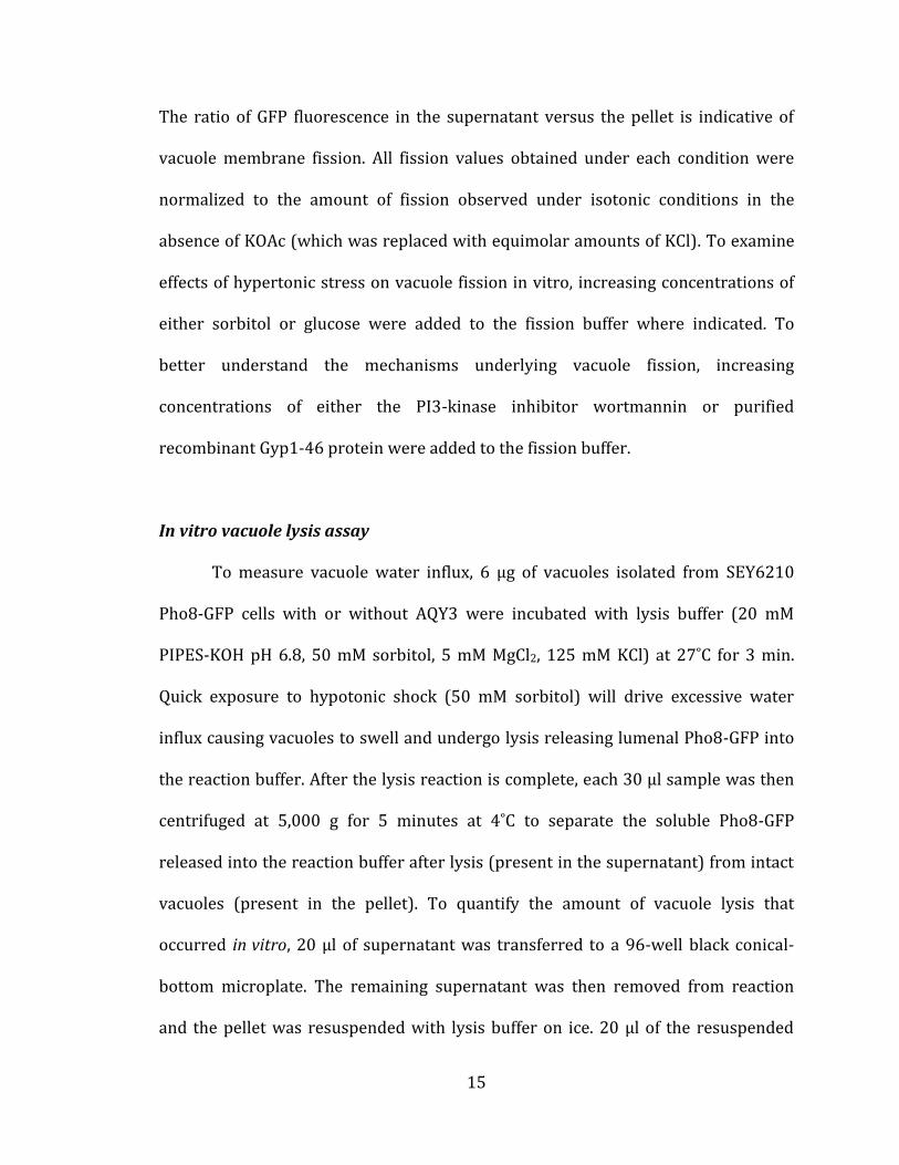
The ratio of GFP fluorescence in the supernatant versus the pellet is indicative of
vacuole membrane fission. All fission values obtained under each condition were
normalized to the amount of fission observed under isotonic conditions in the
absence of KOAc (which was replaced with equimolar amounts of KCl). To examine
effects of hypertonic stress on vacuole fission in vitro, increasing concentrations of
either sorbitol or glucose were added to the fission buffer where indicated. To
better understand the mechanisms underlying vacuole fission, increasing
concentrations of either the PI3-kinase inhibitor wortmannin or purified
recombinant Gyp1-46 protein were added to the fission buffer.
In vitro vacuole lysis assay
To measure vacuole water influx, 6 µg of vacuoles isolated from SEY6210
Pho8-GFP cells with or without AQY3 were incubated with lysis buffer (20 mM
PIPES-KOH pH 6.8, 50 mM sorbitol, 5 mM MgCl2, 125 mM KCl) at 27˚C for 3 min.
Quick exposure to hypotonic shock (50 mM sorbitol) will drive excessive water
influx causing vacuoles to swell and undergo lysis releasing lumenal Pho8-GFP into
the reaction buffer. After the lysis reaction is complete, each 30 µl sample was then
centrifuged at 5,000 g for 5 minutes at 4˚C to separate the soluble Pho8-GFP
released into the reaction buffer after lysis (present in the supernatant) from intact
vacuoles (present in the pellet). To quantify the amount of vacuole lysis that
occurred in vitro, 20 µl of supernatant was transferred to a 96-well black conical-
bottom microplate. The remaining supernatant was then removed from reaction
and the pellet was resuspended with lysis buffer on ice. 20 µl of the resuspended
16
pellet was then transferred to the same 96-well microplate and the amount of GFP
fluorescence in each sample was measured using a BioTek Synergy H1 multimode
fluorescence microplate reader. The ratio of GFP fluorescence in the supernatant
versus the pellet is indicative of vacuole lysis. As a control to measure complete
lysis, vacuoles isolated from wild type cells were treated with 1% triton X-100 to
dissolved membranes. Where indicated, increasing concentrations of sorbitol was
added to the reaction buffer to examine the effect of osmotic shock on lysis.
Cell-free homotypic vacuole fusion assay
In vitro homotypic vacuole fusion was measured using a simple colorimetric
assay based on the maturation of the lumenal alkaline phosphatase Pho8 (see (Haas
et al., 1994)). In brief, 3 μg of vacuoles isolated from pep4∆ and pho8∆ cells were
mixed and added to stand fusion reaction buffer (PS buffer containing 1 mM ATP, 40
mM creatine phosphate, 0.5 mg/ml creatine kinase, 125 mM KCl, 5 mM MgCl2, 10 μM
CoA) and then incubated for 90 minutes at 27˚C. Upon fusion, lumenal content
mixing will permit immature Pho8 to be cleaved by Pep4 to activate the enzyme.
Pho8 activity is then measured by the addition of 500 μl of development buffer
(250mM Tris-HCl pH 8.5, 10 mM MgCl2, 0.4 % triton X-100, 1 mM
paranitrophenolphosphate) and incubation for 5 min at 30˚C. The phosphatase
reaction is then stopped with 500 μl stop buffer (100 mM glycine pH 11) and
absorbance at 400 nm is measured using a NanoDrop spectrophotometer. Where
indicated, KCl was replaced with equimolar KOAc or increasing concentrations of
sorbitol were added to the reaction buffer. Fusion values shown represent A400nm
17
values obtained under each condition normalized to values obtained under normal
fusion conditions (e.g. 200 mM sorbitol, 125 mM KCl).
Data analysis
All quantitative data was processed using Microsoft Excel v.14.0.2 software
(Microsoft Cooperation, Redmond, Washington, USA), including calculation of
means and S.E.M. values. Final data sets were plotted using Kaleida Graph v.4.0
software (Synergy Software, Reading, PA, USA). All figures were prepared using
Adobe Illustrator CS5 software (Adobe Systems, San Jose, CA, USA). The cartoon of
the crystal structure of EcAqpZ (Savage and Stroud, 2007) (Benson et al., 2009) was
prepared using PyMOL software (Portland, OR, USA). The final thesis was written
and assembled in Microsoft Word V14.0.2 software (Microsoft Cooperation,
Redmond, Washington, USA), and references were prepared using Mendeley
software (Mendeley, New York, NY, USA).
18
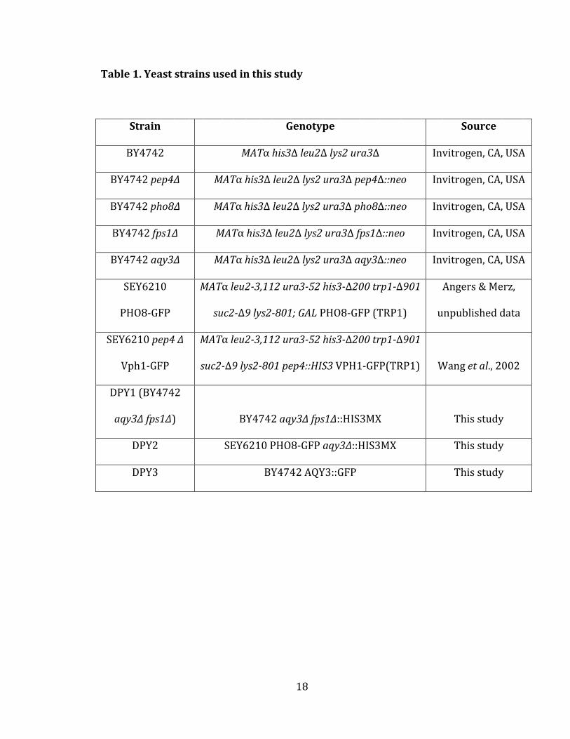
Table 1. Yeast strains used in this study
Strain Genotype Source
BY4742 MATα his3Δ leu2Δ lys2 ura3Δ Invitrogen, CA, USA
BY4742 pep4Δ MATα his3Δ leu2Δ lys2 ura3Δ pep4Δ::neo Invitrogen, CA, USA
BY4742 pho8Δ MATα his3Δ leu2Δ lys2 ura3Δ pho8Δ::neo Invitrogen, CA, USA
BY4742 fps1Δ MATα his3Δ leu2Δ lys2 ura3Δ fps1Δ::neo Invitrogen, CA, USA
BY4742 aqy3Δ MATα his3Δ leu2Δ lys2 ura3Δ aqy3Δ::neo Invitrogen, CA, USA
SEY6210
PHO8-GFP
MATα leu2-3,112 ura3-52 his3-Δ200 trp1-Δ901
suc2-Δ9 lys2-801; GAL PHO8-GFP (TRP1)
Angers & Merz,
unpublished data
SEY6210 pep4 Δ
Vph1-GFP
MATα leu2-3,112 ura3-52 his3-Δ200 trp1-Δ901
suc2-Δ9 lys2-801 pep4::HIS3 VPH1-GFP(TRP1) Wang et al., 2002
DPY1 (BY4742
aqy3Δ fps1∆) BY4742 aqy3∆ fps1∆::HIS3MX This study
DPY2 SEY6210 PHO8-GFP aqy3∆::HIS3MX This study
DPY3 BY4742 AQY3::GFP This study

19
RESULTS
Fps1 and Aqy3 contribute to osmosis across vacuole membranes
Despite playing a major role in cellular osmotolerance, the mechanisms that
transport water across vacuole membranes in S. cerevisiae have not been identified.
As a first step towards finding the vacuole water transporter, I devised a method to
accurately measure volume changes during osmosis across the vacuole membrane.
Water conductance through water channels is very rapid (0.5 sec), and vacuoles
fuse in response to swelling induced by hypotonic shock within seconds, suggesting
that water flux occurs very rapidly, i.e. within a second after stress is applied. Thus
to facilitate our studies, we collaborated with Drs. Vincent Vachon and Jean-Louis
Schwartz at the Université de Montréal. Drs. Vachon and Schwartz have decades of
experience measuring the rapid changes in membrane vesicle diameter in response
to different osmolytes using stopped flow spectrofluorometry (Kirouac et al., 2006).
Under hyperosmotic shock as water escaped from vacuoles, their volume decreased
rapidly, as evident by sharp increase in scattered light intensity (see Figure 2a).
After significant optimization of the methodology to accurately measure changes in
diameter of isolated vacuoles (which are 10 fold larger than the vesicles Drs. Vachon
and Schwartz typically use in their studies), I measured the effects of osmotic stress
on vacuole size using different osmolytes, whereby changes in vacuole diameter
reflect changes in volume in response to osmosis (see Figure 2). Experiments
employing hypotonic stress were quickly abandoned because our results suggested
that the isolated vacuoles were either undergoing lysis or the threshold of detection
20
by the apparatus was reached (data not shown). Instead, we focused on hypertonic
stress and decreases in vacuole size which were observed as increases in light
scattering at 450 nm by fluorometry. We avoided adding high concentrations of
salts, like KCl or KOAc, that are conventionally used to trigger vacuole fragmentation
within intact yeast cells, because the concentrations necessary to elicit a
hyperosmotic stress (e.g. 250 mM) completely abolished membrane fission or fusion
of isolated vacuoles. Instead, we chose to initially study the effects of adding the
osmolyte sorbitol on vacuole size, as it was reported to initiate changes in
morphology when added to isolated vacuoles (Brett and Merz, 2008). Surprisingly,
addition of sorbitol to isolated vacuoles did not cause a sustained decrease in
vacuole diameter (see Figure 2b). Rather, we observed a rapid decrease in size that
peaked at 500 ms, followed by a recovery to a size slightly smaller than observed
under isotonic conditions. This result is expected if the osmolyte is transported
across the membrane, as in the case of glycerol (see Fig 2b), which is known to cross
the vacuole membrane and does not change vacuole morphology. But to
comprehensively assess osmosis we decided to try other osmolytes thought to be
important for cytoplasmic osmolarity that may not be transported across the
vacuole membrane.
It was previously reported that addition of glucose to S. cerevisiae causes
vacuole fragmentation, unlike other yeast species (Vindeløv and Arneborg, 2002).
Thus, we applied it to the isolated vacuoles and observed rapid decreases in volume
that were sustained and increased in a concentration-dependent manner (see
Figure 3a), suggesting that it is an effective osmolyte that does not efficiently cross
21
the vacuole membrane. The change in volume occurred within 1 sec consistent with
the presence of fast-conducting water channels on the vacuole membrane. To
initially test this hypothesis, we pretreated isolated vacuoles with HgCl2, an
aquaporin blocker that lodges in the channel to prevent transmembrane water flow.
Treatment with increasing concentrations of HgCl2 cause progressive loss of the
observed change in volume induced by 900 mM glucose, and the signal was
abolished in the presence of 1 mM HgCl2 (see Figure 2c). This result proposed that
Hg2+-sensitive mechanism, likely an aquaporin, is mediating the observed rapid
change in organelle volume.
Thus, I hypothesized that one or more of the 3 aquaporin paralogs in S.
cerevisiae may mediate osmosis across the vacuole. Because Aqy1 is only found on
the yeast plasma membrane (Sidoux-Walter et al., 2004), I focused on Fps1, which is
present on intracellular membranes (Beese et al., 2009) and Aqy3, an
uncharacterized aquaporin protein. To study their contributions to vacuole osmosis,
I conducted stopped-flow fluorometry studies with vacuoles isolated from yeast
strains lacking each (fps1∆ or aqy3∆) or both (fps1∆aqy3∆) aquaporins. Deletion of
AQY3 or FPS1 diminishes the changes in vacuole size caused by addition of glucose
(see Figure 3b & 3c respectively), and deleting both aquaporin genes may
completely abolishes the effect, similar to the effect of pretreating vacuoles with 1
mM HgCl2. This is best illustrated by further analysis of the peak change and the
steady state change in relative vacuole size (see Figure 3d). Knocking out FPS1 has a
greater effect on the peak change than deleting AQY3; however the effect of
knocking out each single gene is not an additive. Together, these results proposed
22
that both Fps1 and Aqy3 contribute to osmosis across the vacuole membrane,
although they are not functionally redundant and may function synergistically (e.g.
as a heterodimer) to control organelle volume. When I have repeated this
experiment with isolated vacuoles from fps1∆aqy3∆ strain, it was unable to acquire
consistent result (data not shown).
23
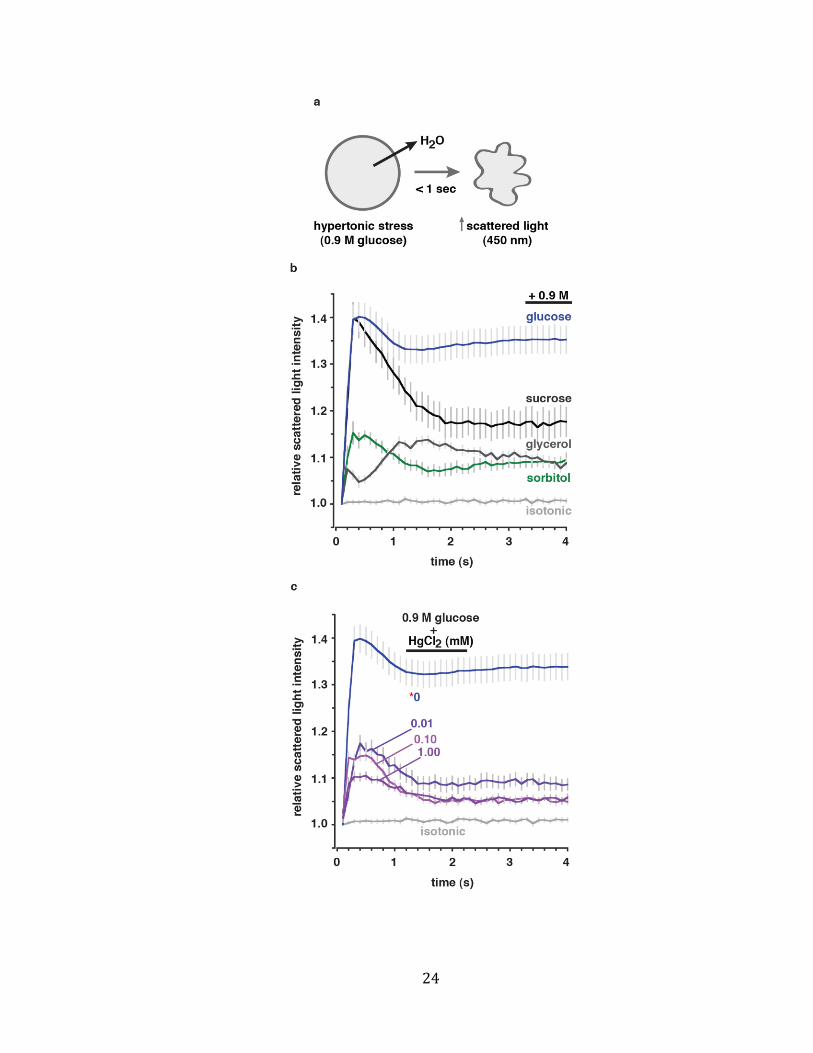
Figure 2. HgCl2 blocks vacuole osmosis caused by hypertonic shock
(a) Illustration of the water efflux assay. Isolated vacuoles are rapidly exposed to
hypertonic shock while their relative volume is monitored by spectrofluorometry.
(b) Changes in vacuole volume recorded in response to exposure of different
osmolytes as compared to isotonic conditions. Glucose was the only osmolyte tested
that caused a sustained decrease in vacuole volume. (c) Vacuole shrinking upon
hypertonic stress (+ 0.9 M glucose) was blocked by addition of increasing
concentrations of HgCl2. Means ± S.E.M. are shown for 17 – 22 independent
experiments depending on condition.
24

25
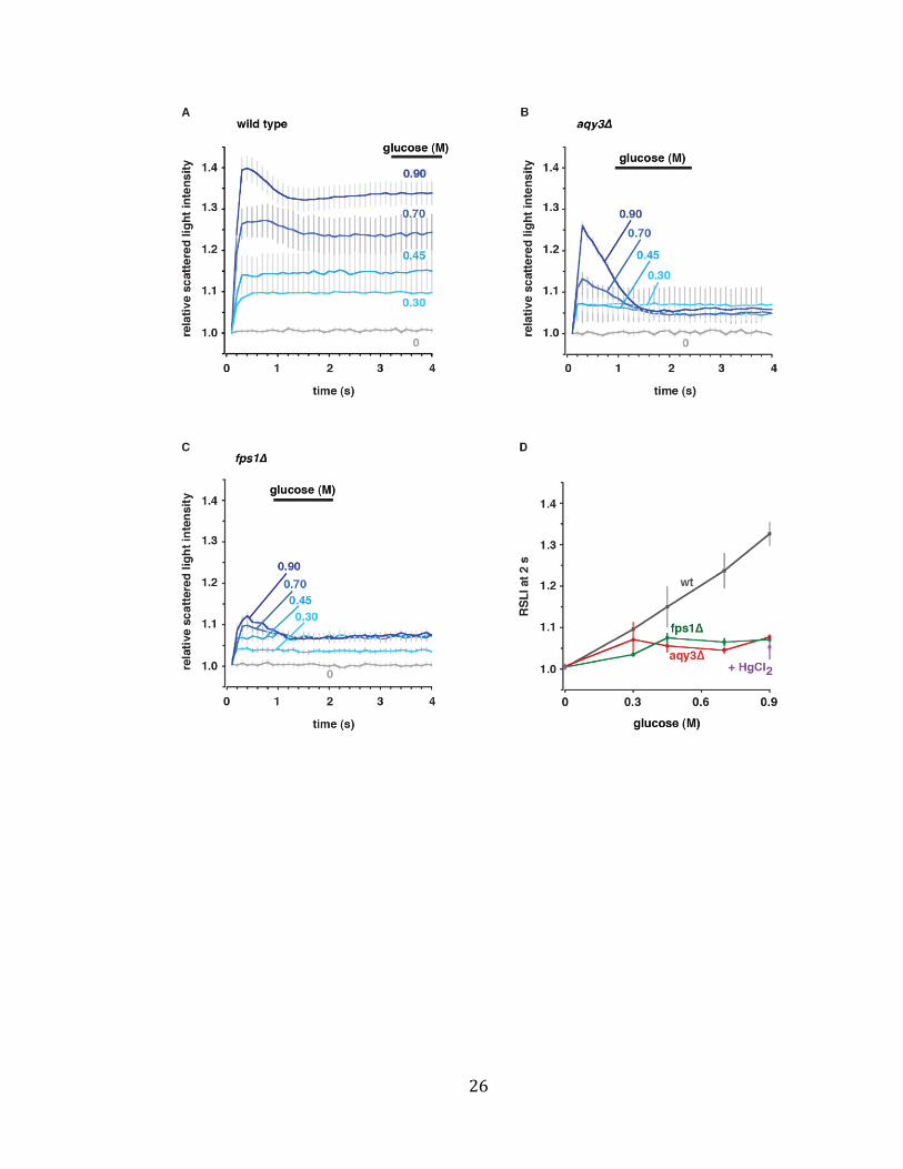
Figure 3. AQY3 and FPS1 mediate water efflux from vacuoles in response to
hypertonic shock
Isolated vacuoles from WT (a), aqy3∆ (b) or fps1∆ (c) cells were rapidly exposed to
increasing concentrations of glucose while volume was measured over time using
spectrofluorometry. (d) Steady state values of relative scattered light intensity
(indicative of relative volume) recorded at 2 second under conditions shown in
panels A-C were plotted against glucose concentration. Data was normalized to
values obtained in the absence of glucose and data shown represents the mean ±
S.E.M. from 4 – 67 independent experiments depending on condition.
26

27
Aqy3 facilitates vacuole lysis in the absence of membrane fusion
Using stopped-flow fluorometry, we were able to demonstrate that Fps1 and
Aqy3 contribute to water efflux from the vacuole lumen in response to hypertonic
stress. However, we were not able to examine water influx using this method. Thus,
we designed a second assay to measure osmosis across the vacuole membrane
based on the following reasoning: In response to hypotonic stress, water floods into
vacuoles causing them to rapidly swell and fuse (Brett and Merz, 2008). Fusion
effectively increases the total relative lumenal of the vacuole compartment within
the cell to accommodate the incoming water to prevent rupture. Thus, when the
membrane fusion machinery is impaired, vacuoles swell and rupture (Starai et al.,
2007). Under these conditions, the extent of rupture directly correlates with the
extent of water influx. To measure rupture, we isolated vacuoles from cells
expressing a GFP-tagged variant of Pho8, an alkaline phosphatase that is exclusively
found in the vacuole lumen, subjected them to hypotonic stress in the absence of
ATP (which is required for homotypic vacuole fusion in vitro), sedimented intact
organelles & membranes and measured the amount of Pho8-GFP that escaped from
lumen that was found in the supernatant (Figure 4a). Initially, I used this assay to
demonstrate that progressively stronger hypotonic shocks cause a gradual increase
in membrane rupture of vacuoles isolated from wild type cells (Figure 4b). Rupture
is blocked by the addition ATP, that promotes membrane fusion, but this effect is
reversed when vacuoles are pretreated with hypertonic stress (e.g. addition of 1 M
sorbitol), which stimulates water efflux and does not cause vacuole rupture.
28
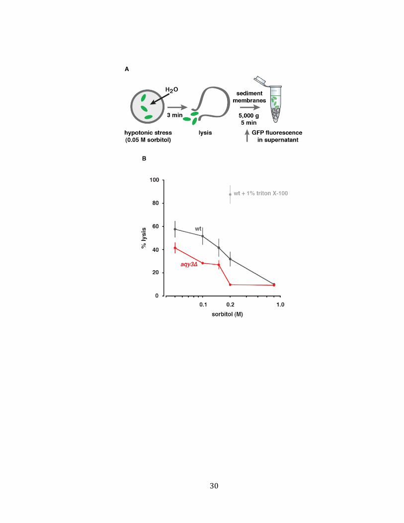
Next, I determined if the aquaporin Aqy3 contributes to water influx that
causes vacuole membrane rupture in response to hypotonic stress; a similar assay
used to first identify aquaporins by Peter Agre and colleagues (Preston et al., 1992).
Deleting AQY3 reduced rupture of isolated vacuoles in response to hypotonic shock
(Figure 4b). Thus, we conclude that Aqy3 facilitates bidirectional transmembrane
water flux across vacuole membranes.

29
Figure 4. Aqy3 mediates vacuole water influx into vacuoles in response to
hypotonic shock
(a) Illustration of the method used to measure vacuole water influx. Isolated
vacuoles were incubated at 270C for 3 min in 30 µl PS buffer with a range of sorbitol
concentrations between 50 and 600 mM. Excessive water influx at low sorbitol
concentrations causes lysis, evident by escape of lumenal GFP isolated in the
supernatant after sedimentation of intact vacuoles. (b) Percent vacuole lysis values
are shown for vacuoles isolated from wild type or aqy3∆ cells treated with reaction
buffer with decreasing concentrations of sorbitol. Lysis of WT vacuoles treated with
1% triton X-100 (light grey) to disrupt membranes is shown as a positive control.
Means ± S.E.M. values are shown from 4 independent experiments.
30
31
Fps1 and Aqy3 may localize to vacuole membranes
With the knowledge that deleting AQP3 gene diminishes water movement
across isolated vacuoles, we hypothesized that it must be present on vacuole
membrane to perform this function. In a previous study, Fps1 tagged with tdTomato
at the C-terminus was localized to the vacuole as well as plasma membrane
(Mollapour and Piper, 2007)(Beese et al., 2009) confirming its presence on the
vacuole membrane. However, the cellular location of Aqy3 is unknown. Thus, I
knocked-in GFP behind AQY3 in the yeast genome but acquire clear fluorescence
micrographs because it seems that endogenous expression levels of AQY3 are too
low for detection (data not shown). Thus, although our functional data shown in
Figure 3 and 4 strongly suggests the presence of Aqy3 on vacuole membranes,
additional experiments are necessary to confirm its presence on the vacuole.
Fps1 and Aqy3 contribute to vacuole morphology
Given that Fps1 and Aqy3 contribute to osmosis across the vacuole
membrane, we hypothesized that these aquaporins also contribute to vacuole
morphology, as osmotic stress is known to drive membrane fusion and fission
events (Weisman, 2003)(LaGrassa and Ungermann, 2005). Specifically, exposing
yeast cells to a hypotonic stress causes water influx into the vacuole, and homotypic
membrane fusion within seconds, whereas hypertonic stress causes water efflux
and vacuole fragmentation within minutes (Brett and Merz, 2008). To determine if
Fps1 or Aqy3 contribute to these observed changes in vacuole morphology, I stained
vacuoles with the vital dye FM4-64 (to visualized vacuole membrane) and imaged
32
them using epifluorescence microscopy (see Figure 5). Yeast cells devoid of FPS1,
AQY3 or both aquaporin genes were all tested. Deletion of both aquaporins causes a
severe defect in vacuole morphology – constitutive fragmentation – that also
abolished the response to osmotic shock, suggesting that vacuole biogenesis may be
impaired. Although no significant effects were observed when only FPS1 was
deleted, vacuoles within aqy3∆ cells failed to fragment in response to hyperosmotic
shock (see Figure 5a). Together these results suggest that Fps1 and Aqy3 are
important for maintaining vacuole morphology, and that Aqy3 may have a specific
function in vacuole fission in response to hypertonic stress.
33
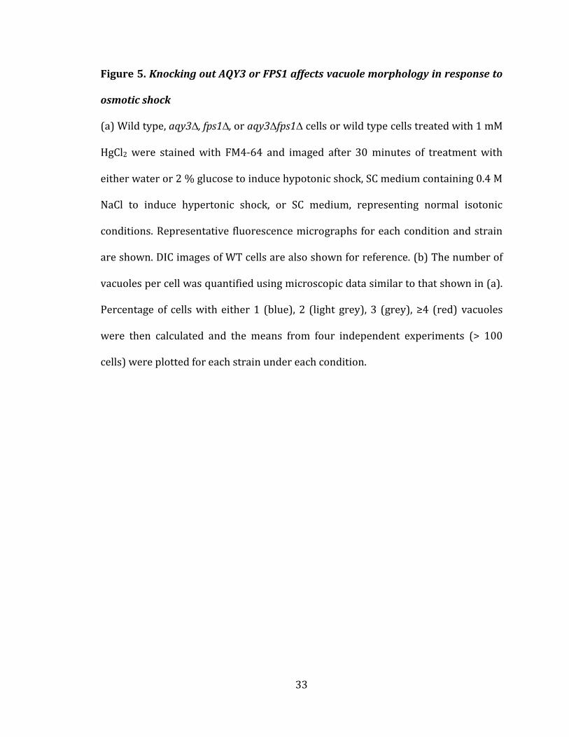
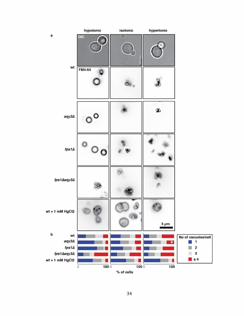
Figure 5. Knocking out AQY3 or FPS1 affects vacuole morphology in response to
osmotic shock
(a) Wild type, aqy3∆, fps1∆, or aqy3∆fps1∆ cells or wild type cells treated with 1 mM
HgCl2 were stained with FM4-64 and imaged after 30 minutes of treatment with
either water or 2 % glucose to induce hypotonic shock, SC medium containing 0.4 M
NaCl to induce hypertonic shock, or SC medium, representing normal isotonic
conditions. Representative fluorescence micrographs for each condition and strain
are shown. DIC images of WT cells are also shown for reference. (b) The number of
vacuoles per cell was quantified using microscopic data similar to that shown in (a).
Percentage of cells with either 1 (blue), 2 (light grey), 3 (grey), ≥4 (red) vacuoles
were then calculated and the means from four independent experiments (> 100
cells) were plotted for each strain under each condition.
34
35
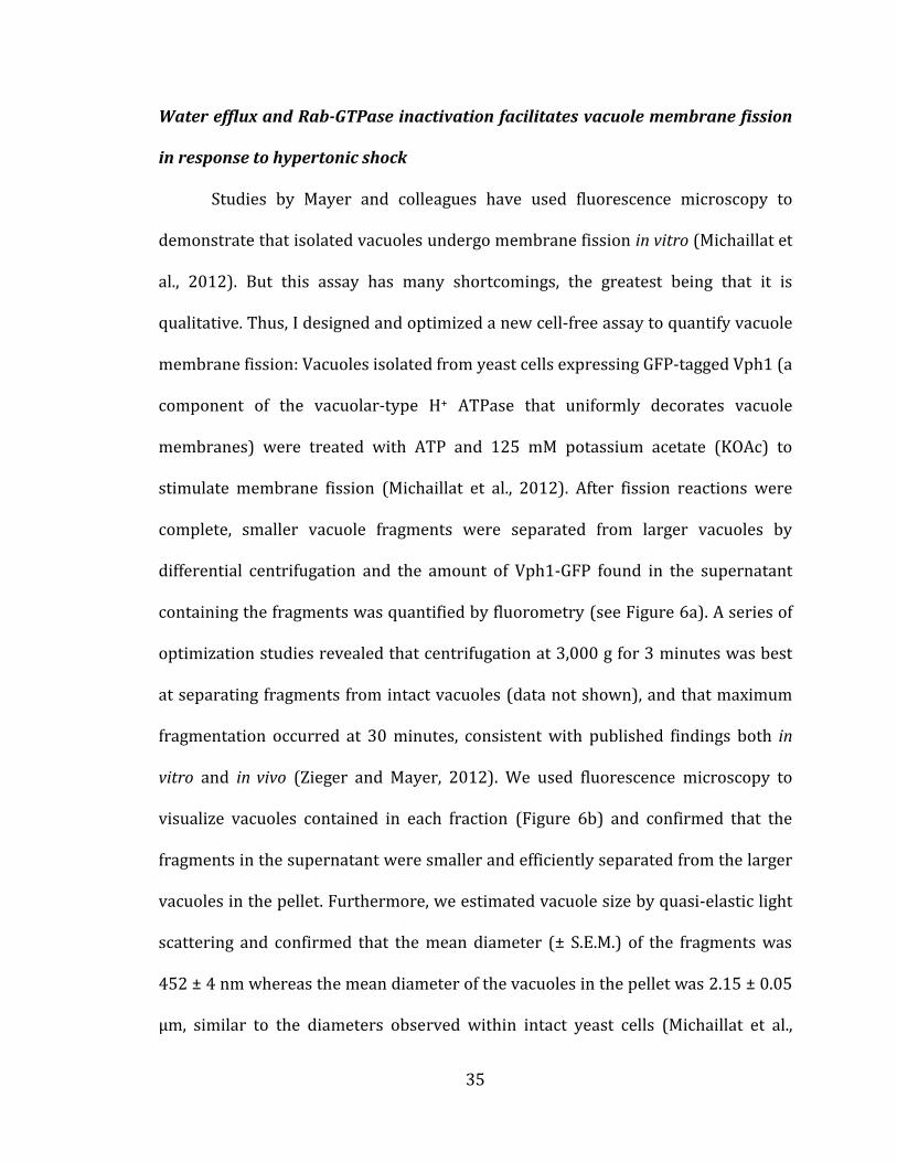
Water efflux and Rab-GTPase inactivation facilitates vacuole membrane fission
in response to hypertonic shock
Studies by Mayer and colleagues have used fluorescence microscopy to
demonstrate that isolated vacuoles undergo membrane fission in vitro (Michaillat et
al., 2012). But this assay has many shortcomings, the greatest being that it is
qualitative. Thus, I designed and optimized a new cell-free assay to quantify vacuole
membrane fission: Vacuoles isolated from yeast cells expressing GFP-tagged Vph1 (a
component of the vacuolar-type H+ ATPase that uniformly decorates vacuole
membranes) were treated with ATP and 125 mM potassium acetate (KOAc) to
stimulate membrane fission (Michaillat et al., 2012). After fission reactions were
complete, smaller vacuole fragments were separated from larger vacuoles by
differential centrifugation and the amount of Vph1-GFP found in the supernatant
containing the fragments was quantified by fluorometry (see Figure 6a). A series of
optimization studies revealed that centrifugation at 3,000 g for 3 minutes was best
at separating fragments from intact vacuoles (data not shown), and that maximum
fragmentation occurred at 30 minutes, consistent with published findings both in
vitro and in vivo (Zieger and Mayer, 2012). We used fluorescence microscopy to
visualize vacuoles contained in each fraction (Figure 6b) and confirmed that the
fragments in the supernatant were smaller and efficiently separated from the larger
vacuoles in the pellet. Furthermore, we estimated vacuole size by quasi-elastic light
scattering and confirmed that the mean diameter (± S.E.M.) of the fragments was
452 ± 4 nm whereas the mean diameter of the vacuoles in the pellet was 2.15 ± 0.05
µm, similar to the diameters observed within intact yeast cells (Michaillat et al.,
36
2012). Assuming all vacuoles are spherical in shape (as observed by many
independent research groups; (Wiemken et al., 1970)(Petekçakar et al.,
2000)(Zieger and Mayer, 2012)), we estimate that one vacuole gives rise to 17
fragments upon hypertonic stress, consistent with previous reports (Michaillat et al.,
2012). In addition to quantifying vacuole fission stimulated by KOAc, I also show
that KOAc has the opposite effect on homotypic vacuole fusion (see Figure 6d). Like
KOAc, I find that hypertonic stress, either by adding the osmolytes sorbitol (Brett
and Merz, 2008) or glucose, also induces vacuole fragmentation in vitro (see Figure
6c & 6e). How KOAc stimulates fission is not entirely understood, but it is known to
involve the V-ATPase (Michaillat et al., 2012), whereas hypertonic stress is thought
to involve aquaporin function and affects lateral membrane tension. In support of
this model, I find that effects of mild hyperosmotic stress (e.g. 0.4 M glucose) and
KOAc are additive (Figure 6e). However, excessive hypertonic stress (0.8 M glucose)
masks the effect of KOAc on fission (Figure 6e). Together these results suggest that
although KOAc and hypertonic stress initiate fission using different mechanisms,
these two independent signaling pathways ultimately converge on one pathway that
underlies the membrane fission process.
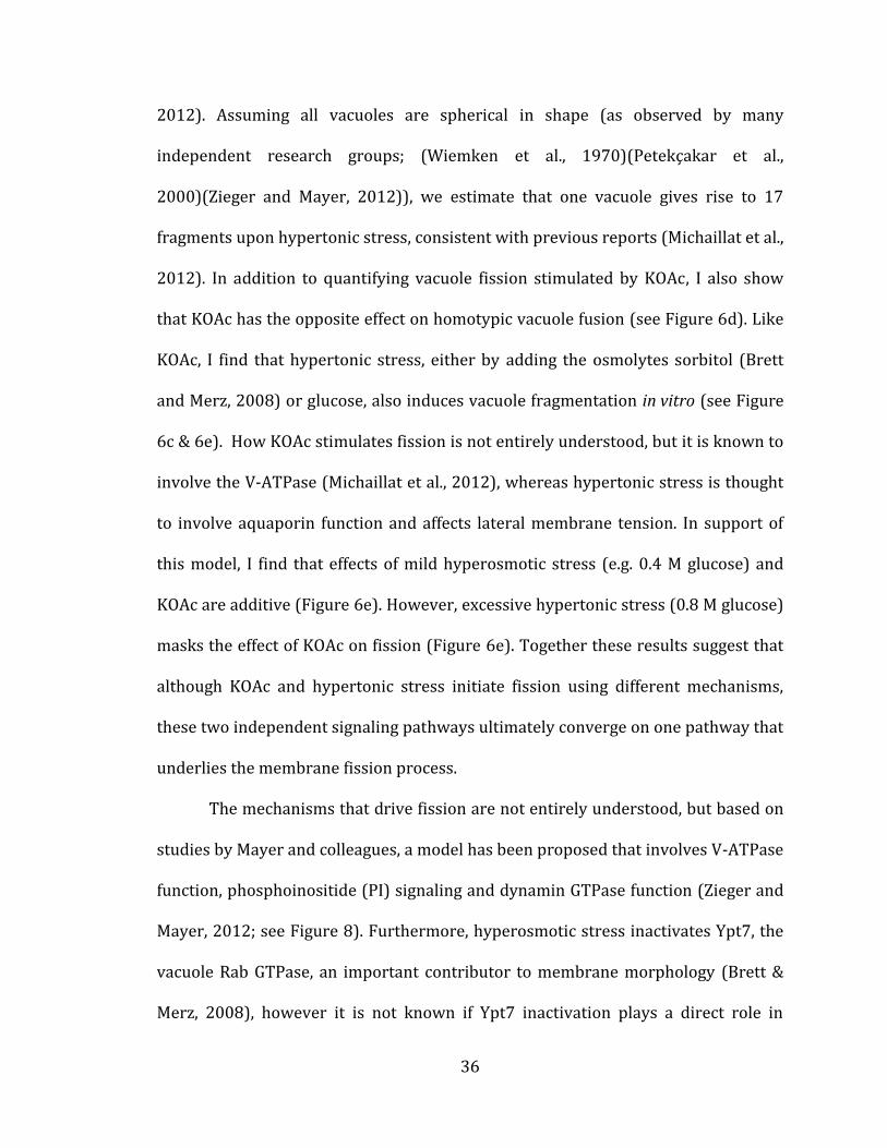
The mechanisms that drive fission are not entirely understood, but based on
studies by Mayer and colleagues, a model has been proposed that involves V-ATPase
function, phosphoinositide (PI) signaling and dynamin GTPase function (Zieger and
Mayer, 2012; see Figure 8). Furthermore, hyperosmotic stress inactivates Ypt7, the
vacuole Rab GTPase, an important contributor to membrane morphology (Brett &
Merz, 2008), however it is not known if Ypt7 inactivation plays a direct role in
37
vacuole membrane fission. Thus, I initially tested the effect of inactivating Ypt7 on
vacuole fission by treating vacuoles with Gyp1-46, a purified recombinant protein
consisting of the catalytic domain of Gyp1, a GTPase-activating protein that is
known to inactivate Ypt7 in vitro (Eitzen et al., 2000)(Albert et al., 1999). Treatment
with Gyp1-46 alone (without hypertonic shock or KOAc addition) is does not induce
fragmentation (Figure 7b), suggesting that Rab inactivation alone is not sufficient to
drive fission. However, addition of Gyp1-46 to vacuoles treated with KOAc further
enhanced fission, to the same extent as hyperosmotic shock (Figure 7b).
Interestingly, Gyp1-46 did not enhance fission induced by hypertonic shock, likely
because Ypt7 is already maximally inactivated under these conditions (Brett and
Merz, 2008). Together, these results suggested that hypertonic shock inactivates the
Rab-GTPase Ypt7, in part, to facilitate vacuole fission.
Conversion of PI(3)P to PI(3,5)P2 by the PI5-kinase Fab1 has been shown to
drive membrane fission both in vivo and in vitro (Zieger and Mayer, 2012). More
specifically the ratio of PI(3)P to PI(3,5)P2 on the vacuole membrane is thought to
underlie morphology, whereby PI(3)P < PI(3,5)P2 drives fission (Mayer et al.,
2000)(Michaillat et al., 2012). Treating vacuoles with the PI3-kinase (Vps34)
inhibitor wortmannin inhibits PI(3)P synthesis on membranes, causing the existing
pool to be rapidly converted to PI(3,5)P2 (Wymann et al., 1996). Thus, we next
examined if addition of wortmannin could stimulate vacuole fission in vitro. As
shown in Figure 7a, wortmannin does not affect fission induced by KOAc, suggesting
that KOAc maximally stimulates PI(3,5)P2 conversion to cause fission. Conversely,
wortmannin enhanced fission induced by hyperosmotic shock (with 0.8 M glucose;
38
Figure 7a), suggesting that PI signaling is not targeted by hypertonic stress to induce
vacuole fission.
It is worth noting that I attempted to inhibit aquaporin function with HgCl2 to
examine its effect on vacuole fission in vitro. However, Hg (II) quenches GFP
fluorescence (Bozkurt and Cavas, 2009) and thus is not compatible with this assay.
Furthermore, vacuoles isolated from fps1∆aqy3∆ cells are fragmented, and thus
differential centrifugation is not an effective way to quantify fission. Thus,
unfortunately, I was unable to directly test whether Aqy3 plays a direct role in
vacuole membrane fission using this in vitro assay.
39
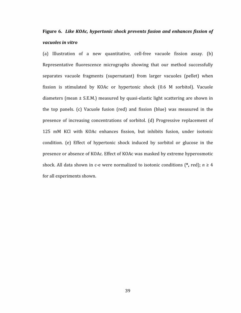
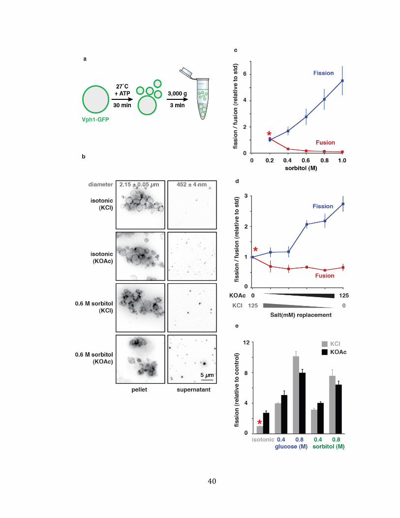
Figure 6. Like KOAc, hypertonic shock prevents fusion and enhances fission of
vacuoles in vitro
(a) Illustration of a new quantitative, cell-free vacuole fission assay. (b)
Representative fluorescence micrographs showing that our method successfully
separates vacuole fragments (supernatant) from larger vacuoles (pellet) when
fission is stimulated by KOAc or hypertonic shock (0.6 M sorbitol). Vacuole
diameters (mean ± S.E.M.) measured by quasi-elastic light scattering are shown in
the top panels. (c) Vacuole fusion (red) and fission (blue) was measured in the
presence of increasing concentrations of sorbitol. (d) Progressive replacement of
125 mM KCl with KOAc enhances fission, but inhibits fusion, under isotonic
condition. (e) Effect of hypertonic shock induced by sorbitol or glucose in the
presence or absence of KOAc. Effect of KOAc was masked by extreme hyperosmotic
shock. All data shown in c-e were normalized to isotonic conditions (*, red); n ≥ 4
for all experiments shown.
40
41
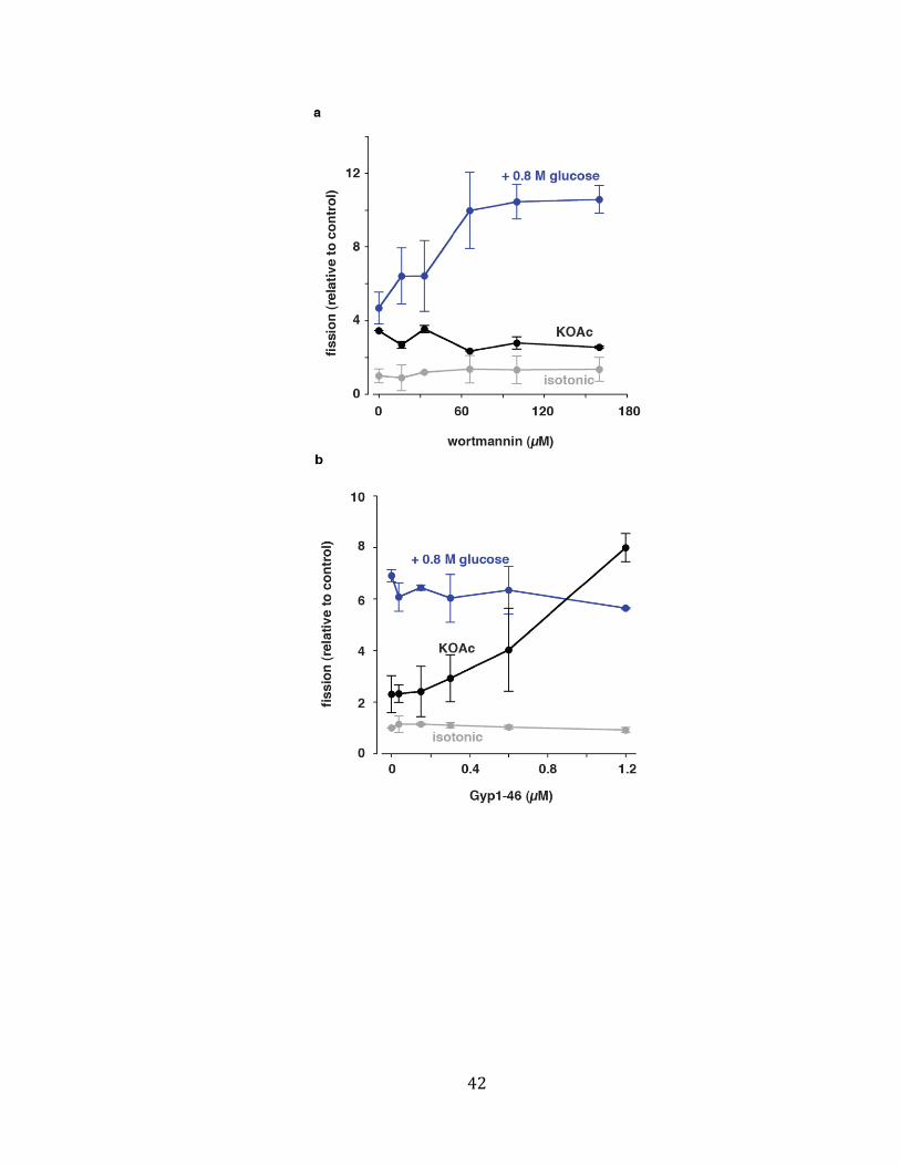
Figure 7. Effects of wortmannin and Gyp1-46 on vacuole fission in vitro
Vacuole fission stimulated by KOAc or hypertonic stress (0.8 M glucose) was
measured in the presence of increasing concentrations of (a) wortmannin or (b)
Gyp1-46. Fission under isotonic conditions is shown as a negative control. n ≥ 4 for
all experiments shown.
42
43
DISCUSSION
Under hypertonic stress, vacuoles lose water decreasing their volume to
permit invagination necessary for membrane fission (see Figure 1a). Herein I show
that the aquaporins Aqy3 and Fps1 mediate this observed osmosis across the
vacuole membrane (Figures 2 - 4), and contribute to changes in vacuole morphology
in response to osmotic stress (Figure 5). Using a new assay to measure vacuole
fission in vitro, I show for the first time that hyperosmotic shock drives vacuole
fission without the involvement of surface receptors and cytoplasmic signaling
factors (Figure 6). Furthermore, I demonstrate that hypertonic stress inactivates
Rab GTPase signaling to drive membrane fission, and this pathway is distinct from
KOAc stimulated fission, but converges at later stage during the fission reaction
(Figures 6 and 7), presumably where there is cross talk between Rab GTPase and
PIP signaling mechanisms prior to membrane scission by Vps1 (see Figure 8). These
findings reveal the mechanism(s) that control volume regulation at the vacuole, and
suggest that other intracellular aquaporins may perform a similar role in regulating
organelle size, shape and number in other organisms. However, one question arises
from my studies: (1) How does osmosis trigger Rab inactivation required for
vacuole fission? The answer to this question and a more detailed discussion of how
Fps1 and Aqy3 may mediate osmosis across the vacuole membrane are given below.
44
How Fps1 and Aqy3 mediate osmosis across the vacuole membrane
In response to the addition of 0.9 M glucose, isolated yeast vacuoles undergo
a rapid (< 0.5 second), sustained decrease in volume (Figure 2), suggesting that
water quickly exits the lumen through open water channels, rather than piggyback
solute transport (e.g. through the requirement of hydration required for ion
translocation) by other mechanisms (Agre et al., 1993). Furthermore, of the
osmolytes tested, only glucose showed a sustained decrease in volume, suggesting
that the other osmolytes are transported across the vacuole membrane, depleting
the osmolyte gradient required for osmosis. This was a particularly surprising result
for sorbitol, as it has been shown to effectively prevent homotypic vacuole fusion
and stimulate vacuole fission (Figure 7;(Brett and Merz, 2008)), but shows anemic
osmosis when compared to equimolar addition of glucose (Figure 2b). However,
using glucose to induce hyperosmotic shock has a greater effect on vacuole fission
(Figure 7e) consistent with this observation. Furthermore, addition of glycerol does
not significantly change vacuole volume (Figure 2b), consistent with previous
reports that Fps1 is an aquaglyceroporin and is found on the vacuole membrane,
suggesting that it may permit glycerol entry preventing rapid osmosis, and
consistent with observation that glycerol is stored in the vacuole to maintain
cytoplasmic osmolarity.
Also surprising was the observation that deleting only FPS1 or AQY3 nearly
abolished osmosis, to the same extent as adding HgCl2 (Figure 3d), i.e. deletion of
either aquaporin prevent vacuole osmosis even when the other is present. One
interpretation of this result is that Aqy3 and Fps1 may function as obligate hetero-
45
tetramers to transport water across the membrane, like many surface aquaporins
studied in mammalian cells (Fetter et al., 2004). However, changes in vacuole
morphology in response to osmotic stress are not impaired when FPS1 is absent,
and only vacuole fission in response to hyperosmotic stress is impaired in aqy3∆
cells. These somewhat conflicting results, and the observation that osmosis
triggered by the addition of low concentrations of sorbitol are sufficient to induce
vacuole fission, have lead us to speculate that (1) only a small, rapid change in
vacuole volume (observed within the first 500 ms of hypertonic shock, see Figures 2
and 3) may be sufficient to drive changes in vacuole morphology, and (2) perhaps
Aqy3, and not Fps1, may directly interact the fission machinery to initiate vacuole
fission.
How does osmosis trigger Rab inactivation required for vacuole fission?
Consistent with our hypothesized roles for Aqy3 and Fps1 in osmosis, we
speculate that a small rapid change in volume is sufficient to rapidly decrease lateral
surface tension of the vacuole membrane. Although we have not measured this
change in tension, it may be sufficient to activate the TrpY1, a mechanosensitive
cation channel that opens in response to changes in membrane tension. Although it
is unclear whether TrpY1 plays a direct role in the membrane fission process, it
does open in response to hyperosmotic stress to mediate calcium efflux from the
vacuole (Denis and Cyert, 2002). Furthermore, Ca2+ signaling through calmodulin
and calcineurin have been shown to regulate vacuole morphology, and preliminary
results from Alexey Merz’s group suggest that endosomal Rab-GAP proteins may
46
respond to Ca2+ signaling (personal communication). Thus, we speculate that TrpY1
may act immediately downstream of Aqy3 in the fission pathway and that Ca2+ efflux
by TrpY1 may trigger a Rab-GAP to inactivate Ytp7 to drive vacuole membrane
fission.
To understand aquaporin function, we impose experimental conditions to
drive water movement across the vacuole membrane. However, cellular osmolarity
is normally tightly regulated, and vacuoles are rarely subjected to such extreme
conditions in nature. Thus, to put our findings into greater biological context, we
speculate that vacuole aquaporin function may be coupled to osmolyte transport in
response to secondary messenger signaling that underlies changes in vacuole
morphology necessary for normal yeast cell physiology. For example, yeast vacuoles
must undergo fission to donate organelles to daughter cells, which is critical for
organelle inheritance during cell division. As vacuoles fuse or fragment in response
to changes in kinase activity, e.g. Yck3 and TorC1 (LaGrassa and Ungermann, 2005)
(Michaillat et al., 2012), the possibility exists that Aqy3 (or osmolyte transporters
that would stimulate osmosis through Aqy3) may respond to kinases known to
orchestrate cell division, which in turn would drive water efflux required for
vacuole fragmentation necessary for vacuole inheritance.
47
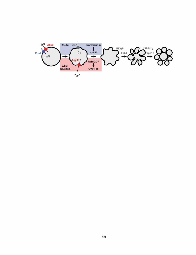
Figure 8. Model of mechanisms responsible for vacuole fission
Cartoon illustrating how Aqy3 or Fps1 mediate osmosis to stimulate vacuole
fragmentation upon hyperosmotic shock. Unlike, KOAc that is thought to stimulate
PI-signaling through V-ATPase function, water efflux caused by hyperosmotic shock
likely decreases lateral membrane tension to inactivate Ypt7 to signal downstream
mechanisms responsible for membrane fission. I speculate that pathways
stimulated by KOAc or hyperosmotic stress converge prior to Fab1 activity required
for membrane invagination and membrane scission by the dynamin ortholog Vps1.
48
49
REFERENCES
Agre, P., G.M. Preston, B.L. Smith, J.S. Jung, S. Raina, C. Moon, W.B. Guggino, and S. Nielsen. 1993. Aquaporin CHIP: the archetypal molecular water channel. American Journal of Physiology. 265:F463–F476.
Albert, Š., E. Will, and D. Gallwitz. 1999. Identification of the catalytic domains and their functionally critical arginine residues of two yeast GTPase-activating proteins specific for Ypt/Rab transport GTPases. The EMBO journal. 18:5216–25.
Amiry-Moghaddam, M., H. Lindland, S. Zelenin, B.A. Roberg, B.B. Gundersen, P. Petersen, E. Rinvik, I.A. Torgner, and O.P. Ottersen. 2005. Brain mitochondria contain aquaporin water channels: evidence for the expression of a short AQP9 isoform in the inner mitochondrial membrane. The FASEB journal official publication of the Federation of American Societies for Experimental Biology. 19:1459–1467.
Beese, S.E., T. Negishi, and D.E. Levin. 2009. Identification of positive regulators of the yeast fps1 glycerol channel. PLoS genetics. 5:13.
Benson, D. a, I. Karsch-Mizrachi, D.J. Lipman, J. Ostell, and E.W. Sayers. 2009. GenBank. Nucleic acids research. 37:D26–31.
Bone, N., J.B. Millar, T. Toda, and J. Armstrong. 1998. Regulated vacuole fusion and fission in Schizosaccharomyces pombe: an osmotic response dependent on MAP kinases. Current biology : CB. 8:135–44.
Borgnia, M., and S. Nielsen. 1999. Cellular and molecular biology of the aquaporin water channels. Annual review of biochemistry. 68:425–458.
Bozkurt, S.S., and L. Cavas. 2009. Can Hg(II) be determined via quenching of the emission of green fluorescent protein from Anemonia sulcata var. smaragdina? Applied biochemistry and biotechnology. 158:51–8.
Brett, C.L., and A.J. Merz. 2008. Osmotic regulation of Rab-mediated organelle docking. Current biology : CB. 18:1072–7.
Carbrey, J.M., M. Bonhivers, J.D. Boeke, and P. Agre. 2001. Aquaporins in Saccharomyces: Characterization of a second functional water channel protein. Proceedings of the National Academy of Sciences of the United States of America. 98:1000–5.
50
Denis, V., and M.S. Cyert. 2002. Internal Ca(2+) release in yeast is triggered by hypertonic shock and mediated by a TRP channel homologue. The Journal of cell biology. 156:29–34.
Eitzen, G., E. Will, D. Gallwitz, A. Haas, and W. Wickner. 2000. Sequential action of two GTPases to promote vacuole docking and fusion. The EMBO journal. 19:6713–20.
Fetter, K., V. Van Wilder, M. Moshelion, and F. Chaumont. 2004. Interactions between Plasma Membrane Aquaporins Modulate Their Water Channel Activity. The Plant Cell. 16:215–228.
Gietz, D., a St Jean, R. a Woods, and R.H. Schiestl. 1992. Improved method for high efficiency transformation of intact yeast cells. Nucleic acids research. 20:1425.
De Groot, B.L., and H. Grubmüller. 2005. The dynamics and energetics of water permeation and proton exclusion in aquaporins. Current Opinion in Structural Biology. 15:176–183.
Haas, a, B. Conradt, and W. Wickner. 1994. G-protein ligands inhibit in vitro reactions of vacuole inheritance. The Journal of cell biology. 126:87–97.
Hicke, L. 1999. Gettin’ down with ubiquitin: turning off cell-surface receptors, transporters and channels. Trends in Cell Biology. 9:107–12.
Kirouac, M., V. Vachon, M. Fortier, M.-C. Trudel, A. Berteloot, J.-L. Schwartz, and R. Laprade. 2006. A mechanical force contributes to the “osmotic swelling” of brush-border membrane vesicles. Biophysical journal. 91:3301–12.
Klionsky, D.J., P.K. Herman, and S.D. Emr. 1990. The fungal vacuole: composition, function, and biogenesis. Microbiological reviews. 54:266–92.
Knepper, M.A., J.B. Wade, J. Terris, C.A. Ecelbarger, D. Marples, B. Mandon, C.L. Chou, B.K. Kishore, and S. Nielsen. 1996. Renal aquaporins. Kidney International. 49:1712–1717.
Kreis, S., H.-J. Schönfeld, C. Melchior, B. Steiner, and N. Kieffer. 2005. The intermediate filament protein vimentin binds specifically to a recombinant integrin alpha2/beta1 cytoplasmic tail complex and co-localizes with native alpha2/beta1 in endothelial cell focal adhesions. Experimental cell research. 305:110–21.
LaGrassa, T.J., and C. Ungermann. 2005. The vacuolar kinase Yck3 maintains organelle fragmentation by regulating the HOPS tethering complex. The Journal of Cell Biology. 168:401–414.
51
Lo, S., C. Brett, and R. Plemel. 2011. Intrinsic tethering activity of endosomal Rab proteins. Nature structural & molecular biology. 19:40–7.
Longtine, M.S., A. McKenzie, D.J. Demarini, N.G. Shah, A. Wach, A. Brachat, P. Philippsen, and J.R. Pringle. 1998. Additional modules for versatile and economical PCR-based gene deletion and modification in Saccharomyces cerevisiae. Yeast (Chichester, England). 14:953–61.
Luyten, K., J. Albertyn, W.F. Skibbe, B. a Prior, J. Ramos, J.M. Thevelein, and S. Hohmann. 1995. Fps1, a yeast member of the MIP family of channel proteins, is a facilitator for glycerol uptake and efflux and is inactive under osmotic stress. The EMBO journal. 14:1360–71.
Mager, W.H., and J.C. Varela. 1993. Osmostress response of the yeast Saccharomyces. Molecular Microbiology. 10:253–258.
Marchissio, M.J., D.E.A. Francés, C.E. Carnovale, and R.A. Marinelli. 2012. Mitochondrial aquaporin-8 knockdown in human hepatoma HepG2 cells causes ROS-induced mitochondrial depolarization and loss of viability. Toxicology and applied pharmacology. 264:246–54.
Mayer, a, D. Scheglmann, and S. Dove. 2000. Phosphatidylinositol 4, 5-bisphosphate regulates two steps of homotypic vacuole fusion. Molecular biology of the cell. 11:807–17.
Michaillat, L., T.L. Baars, and A. Mayer. 2012. Cell-free reconstitution of vacuole membrane fragmentation reveals regulation of vacuole size and number by TORC1. Molecular biology of the cell. 23:881–95.
Mizutani, M., S. Watanabe, T. Nakagawa, and M. Maeshima. 2006. Aquaporin NIP2;1 is mainly localized to the ER membrane and shows root-specific accumulation in Arabidopsis thaliana. Plant cell physiology. 47:1420–1426.
Mollapour, M., and P.W. Piper. 2007. Hog1 mitogen-activated protein kinase phosphorylation targets the yeast Fps1 aquaglyceroporin for endocytosis, thereby rendering cells resistant to acetic acid. Molecular and cellular biology. 27:6446–56.
Monzani, E., R. Bazzotti, C. Perego, and C. a M. La Porta. 2009. AQP1 is not only a water channel: it contributes to cell migration through Lin7/beta-catenin. PloS one. 4:e6167.
Murata, K., K. Mitsuoka, T. Hirai, T. Walz, P. Agre, J.B. Heymann, A. Engel, and Y. Fujiyoshi. 2000. Structural determinants of water permeation through aquaporin-1. Nature. 407:599–605.
52
Nozaki, K., D. Ishii, and K. Ishibashi. 2008. Intracellular aquaporins: clues for intracellular water transport? Pflügers Archiv : European journal of physiology. 456:701–7.
Petekçakar, Z., U. Sauer, and J. Bailey. 2000. Vacuolar morphology and cell cycle distribution are modified by leucine limitation in auxotrophic Saccharomyces cerevisiae. Biology of the cell / under the auspices of the European Cell Biology Organization of the Cell. 92:629–637.
Preston, G.M., T.P. Carroll, W.B. Guggino, and P. Agre. 1992. Appearance of water channels in Xenopus oocytes expressing red cell CHIP28 protein. Science. 256:385–387.
Saboori, a M., B.L. Smith, and P. Agre. 1988. Polymorphism in the Mr 32,000 Rh protein purified from Rh(D)-positive and -negative erythrocytes. Proceedings of the National Academy of Sciences of the United States of America. 85:4042–5.
Savage, D.F., and R.M. Stroud. 2007. Structural basis of aquaporin inhibition by mercury. Journal of Molecular Biology. 368:607–17.
Schüller, C., J.L. Brewster, M.R. Alexander, M.C. Gustin, and H. Ruis. 1994. The HOG pathway controls osmotic regulation of transcription via the stress response element (STRE) of the Saccharomyces cerevisiae CTT1 gene. the The European Molecular Biology Organization Journal. 13:4382–4389.
Shen, S., P. Chretien, L. Bastien, and S. Slilaty. 1991. Primary sequence of the glucanase gene from Oerskovia xanthineolytica. Expression and purification of the enzyme from Escherichia coli. Journal of Biological Chemistry. 266:1058–1063.
Sidoux-Walter, F., N. Pettersson, and S. Hohmann. 2004. The Saccharomyces cerevisiae aquaporin Aqy1 is involved in sporulation. Proceedings of the National Academy of Sciences of the United States of America. 101:17422–17427.
Starai, V.J., Y. Jun, and W. Wickner. 2007. Excess vacuolar SNAREs drive lysis and Rab bypass fusion. Proceedings of the National Academy of Sciences of the United States of America. 104:13551–13558.
Sugiya, H., M. Matsuki-Fukushima, and S. Hashimoto. 2008. Role of aquaporins and regulation of secretory vesicle volume in cell secretion. Journal of cellular and molecular medicine. 12:1486–94.
Sui, H., B.G. Han, J.K. Lee, P. Walian, and B.K. Jap. 2001. Structural basis of water-specific transport through the AQP1 water channel. Nature. 414:872–878.
Tamura, K., D. Peterson, N. Peterson, G. Stecher, M. Nei, and S. Kumar. 2011. MEGA5: molecular evolutionary genetics analysis using maximum likelihood, evolutionary
53
distance, and maximum parsimony methods. Molecular biology and evolution. 28:2731–9.
Thompson, J.D., D.G. Higgins, and T.J. Gibson. 1994. CLUSTAL W: improving the sensitivity of progressive multiple sequence alignment through sequence weighting, position-specific gap penalties and weight matrix choice. Nucleic acids research. 22:4673–80.
Verkman, A.S. 2005. More than just water channels: unexpected cellular roles of aquaporins. Journal of Cell Science. 118:3225–3232.
Vindeløv, J., and N. Arneborg. 2002. Saccharomyces cerevisiae and Zygosaccharomyces mellis exhibit different hyperosmotic shock responses. Yeast (Chichester, England). 19:429–39.
Walz, T., T. Hirai, K. Murata, J.B. Heymann, K. Mitsuoka, Y. Fujiyoshi, B.L. Smith, P. Agre, and A. Engel. 1997. The three-dimensional structure of aquaporin-1. Nature. 387:624–627.
Wang, L., E.S. Seeley, W. Wickner, and A.J. Merz. 2002. Vacuole fusion at a ring of vertex docking sites leaves membrane fragments within the organelle. Cell. 108:357–369.
Weisman, L.S. 2003. Yeast vacuole inheritance and dynamics. Annual Review of Genetics. 37:435–460.
Wickner, W. 2002. Yeast vacuoles and membrane fusion pathways. the The European Molecular Biology Organization Journal. 21:1241–1247.
Wiemken, a, P. Matile, and H. Moor. 1970. Vacuolar dynamics in synchronously budding yeast. Archiv für Mikrobiologie. 70:89–103.
Wymann, M.P., G. Bulgarelli-Leva, M.J. Zvelebil, L. Pirola, B. Vanhaesebroeck, M.D. Waterfield, and G. Panayotou. 1996. Wortmannin inactivates phosphoinositide 3-kinase by covalent modification of Lys-802, a residue involved in the phosphate transfer reaction. Molecular and Cellular Biology. 16:1722–1733.
Zieger, M., and A. Mayer. 2012. Yeast vacuoles fragment in an asymmetrical two-phase process with distinct protein requirements. Molecular Biology of the Cell. 23:3438–49.