Embed Size (px)
Citation preview
JOURNAL OF BACTERIOLOGY, May 2002, p. 2755–2766 Vol. 184, No. 100021-9193/02/$04.00�0 DOI: 10.1128/JB.184.10.2755–2766.2002Copyright © 2002, American Society for Microbiology. All Rights Reserved.
Characterization of Two Cryptic Helicobacter pylori Plasmids: aPutative Source for Horizontal Gene Transfer and Gene Shuffling
Dirk Hofreuter† and Rainer Haas*
Max von Pettenkofer Institut für Hygiene und Medizinische Mikrobiologie, Ludwig-MaximiliansUniversität München, D-80336 Munich, Germany
Received 29 November 2001/Accepted 20 February 2002
Many Helicobacter pylori isolates carry cryptic plasmids of extremely variable size. In this study we analyzedtwo H. pylori plasmids, pHel4 and pHel5, from H. pylori strains P8 and P29, respectively. Plasmid pHel4consists of 10,970 bp, constituting 15 putative open reading frames (ORFs), whereas pHel5 consists of 18,291bp, constituting 17 ORFs. The findings that both plasmids encode a conserved RepA protein and that both havean origin of replication containing an iteron place them in the group of theta plasmids. In pHel4, the productsof the overlapping orf4C, orf4D, orf4E, and orf4F sequences are homologous to MobA, MobB, MobC, andMobD, encoded by colicinogenic plasmids, suggesting that pHel4 might be mobilizable. A further putativeoperon consists of orf4B and orf4A, the products of which are homologous to microcin C7 (MccC7) biosynthesisand secretion proteins MccB and MccC, respectively. Plasmid pHel5 carries putative genes encoding proteinswith homology to an endonuclease and gene products of an H. pylori chromosomal plasticity zone. Bothplasmids contain repeat sequences, such as the previously identified R2 repeat, which are considered preferredrecombination sites. In pHel4, a new repeat sequence (R4 repeat), which seems to act as a hot spot forsite-specific recombination, was identified. All H. pylori plasmids characterized so far have a modular struc-ture. We suggest a model that explains the existing plasmids by insertions and deletions of genetic elementsat the repeat sequences. A genetic exchange between plasmids and the bacterial chromosome, combined withplasmid mobilization, might add a novel mechanism to explain the high genetic macrodiversity within the H.pylori population.
Helicobacter pylori is a highly motile, microaerophilic, gram-negative bacterium colonizing the human stomach mucosa(44). H. pylori is well recognized as a major cause of severalgastroduodenal pathologies, including chronic active gastritis,peptic ulcer disease, mucosa-associated lymphoid tissue lym-phoma, and gastric adenocarcinoma (6). Several bacterial fac-tors are known to be essential for colonization in animal mod-els, including a bundle of sheathed flagella and a potent urease(13, 14). Other bacterial virulence factors only present in asubset of H. pylori strains, the type I strains, include the vacu-olating cytotoxin (VacA) and the cytotoxin-associated antigenA (CagA). Genes such as vacA and cagA and 32 genes encod-ing putative outer membrane proteins (OMPs) show charac-teristics of mosaic genes (4), resulting in the existence of sev-eral alleles in different H. pylori strains (microdiversity). Genesinvolved in lipopolysaccharide biosynthesis or restriction-mod-ification systems are prone to frameshift mutations by slipped-strand mispairing during replication (38), a means by whichoverproduction or nonproduction of the corresponding pro-teins is determined (16).
Comparison of the two available genome sequences of H.pylori 26695 (42) and J99 (1) reveals that differences betweenthe strains are the result of intragenomic rearrangements, re-
sulting in deletion, inversion, or translocation of larger genomefragments (macrodiversity) (1). Many H. pylori strains possessseveral copies of the insertion sequences IS605 and IS606.Furthermore, H. pylori 26695 contains five so-called plasticityzones with G�C contents of 33% (zone 1), 35% (zone 2), 33%(zone 3), 43% (zone 4), and 33% (zone 5), which differ fromthe chromosomal G�C content of 39% (42). Strain H. pyloriJ99 (1) carries several different plasticity zones which are notpresent in H. pylori 26695. It is speculated that H. pylori re-ceived the plasticity zones by horizontal gene transfer.
Transduction, conjugation, and natural transformation arethe three common mechanisms of horizontal gene transfer inbacteria. Natural transformation competence was first de-scribed for H. pylori by Nedenskov-Sorensen et al. (32). Severalgenes that are involved in the transformation process of H.pylori have been identified (2, 24, 39, 40). We demonstratedrecently that natural transformation of H. pylori is mediated bybasic components of a type IV secretion system (23). Plasmidconjugation has not been proven for H. pylori, but there issome evidence that horizontal transfer of genes in H. pyloricould take place via a DNase-resistant, conjugation-like mech-anism (3, 28).
Although about 50% of H. pylori strains carry cryptic plas-mids ranging in size from 2 to about 100 kb (35), the role ofthese plasmids is not well understood. Several H. pylori plas-mids of up to 6 kb have been analyzed in detail in the last fewyears (20, 27, 30). They can be grouped into at least twoseparate classes. Plasmid pHPK255 (27) reveals homology toplasmids of gram-positive bacteria replicating via the “rolling-circle” mechanism, whereas other plasmids (pHPM180, pHel1,
* Corresponding author. Mailing address: Max von Pettenkofer In-stitut, LMU Munich, Pettenkoferstr. 9A, D-80336 Munich, Germany.Phone: (0049)-89-5160 5255. Fax: (0049)-89-5160 5223. E-mail:[email protected].
† Present address: Boyer Center for Molecular Medicine, Yale Uni-versity School of Medicine, New Haven, CT 06536-0812.
2755
on March 4, 2020 by guest
http://jb.asm.org/
Dow
nloaded from

and pHPS1) fall into the group of iteron-containing plasmidsand replicate via the theta mechanism (10).
In this study, we investigated which genetic information isencoded by larger H. pylori plasmids (6 to 20 kb) and whetherplasmids might be a source for horizontal gene transfer and forgeneration of macrodiversity in H. pylori. The sequencing andcomparison of two H. pylori plasmids, pHel4 and pHel5, iden-tified both common and unique open reading frames (ORFs),suggesting that individual plasmids express distinct features.From database comparisons we identified putative genes in-volved in conjugative transfer (mob) as well as gene sequenceshomologous to that of an operon present in certain Escherichiacoli strains responsible for microcin production. Furthermore,the H. pylori plasmids described in this study show a modularstructure, which apparently enables them to integrate or deletecomplete functional modules, thus making them ideal candi-dates for an efficient gene-shuffling mechanism.
MATERIALS AND METHODS
Bacterial strains and culture conditions. H. pylori strains were grown on GCagar plates (Difco) supplemented with horse serum (8%), vancomycin (10 mgliter�1), trimethoprim (5 mg liter�1), and nystatin (1 mg liter�1) (serum plates)and incubated for 2 to 3 days in a microaerobic atmosphere (85% N2, 10% CO2,5% O2) at 37°C. For H. pylori liquid cultures Brucella medium (Becton Dickin-son) supplemented with 10% fetal calf serum was used; the medium was inoc-ulated with a bacterial suspension with an optical density at 550 nm of 0.1.
For selection of H. pylori mutant strains, serum plates supplemented withchloramphenicol (6 mg liter�1) were used. E. coli strains HB101 (8) and DH5�(Bethesda Research Laboratories) were grown on Luria-Bertani (LB) agarplates or in LB liquid medium (37) supplemented with ampicillin (100 mgliter�1), chloramphenicol (30 mg liter�1), or tetracycline (15 mg liter�1), asappropriate. The H. pylori strains were isolated from patients undergoing endos-copy in different university gastroenterology units in Germany (Munich, Ham-burg, and Erlangen). Thus, most of the analyzed strains were from Germany orfrom other European countries.
DNA manipulations and plasmid constructions. Cloning and DNA analysisprocedures were performed as described by Sambrook et al. (37). H. pylorichromosomal DNA was isolated with the QIAamp tissue kit (Qiagen). PlasmidDNA was purified from E. coli by the boiling procedure, and electroporation-competent E. coli cells were prepared according to the protocol recommendedfor the Gene Pulser (Bio-Rad).
Plasmid preparation and plasmid library construction for DNA sequencing.Plasmid DNA of different Helicobacter strains was extracted from cells growingon plates or from cell pellets after cultivation in liquid media. The latter methodresulted in general in plasmid DNA of better quality. Plasmid DNA was isolatedby using either the Wizard Plus SV Minipreps purification system (Promega)according to the manufacturers protocol or the XSP buffer extraction methoddescribed by De Ungria et al. (11). In brief, cell pellets were resuspended in 100�l of TES (10 mM Tris-HCl [pH 7.4], 1 mM EDTA [pH 8.0], 15 mM NaCl) byadding 1.5 ml of prewarmed (65°C) XSP buffer. XSP buffer contains XS buffer(1% potassium ethyl xanthogenate, 100 mM Tris-HCl [pH 7.4], 20 mM EDTA[pH 8.0], 1% sodium dodecyl sulfate [SDS], and 800 mM ammonium acetate)with an equal volume of phenol. The resuspended pellets were incubated for 30min at 65°C. After a short vortexing, incubation on ice for 5 min, and centrifu-gation at 14,000 � g for 15 min the aqueous phase was transferred to a freshEppendorf tube. The samples were purified with two phenol-chloroform-isoamylalcohol (25:24:1) extractions, and RNA was digested with RNase A for 30 min at37°C. One volume of isopropanol and 0.1 volume of 3 M sodium acetate, pH 4.8,were used to precipitate the DNA, which was washed with 70% ethanol once.After being dried the plasmid DNA was resuspended in 50 �l of distilled water.
The isolation of plasmid DNA from H. pylori by the XSP method resulted inminor contamination with chromosomal DNA, which was removed by digestionwith an exonuclease (Plasmid-Safe ATP-dependent DNase; Epicentre). Purifiedplasmids pHel4 and pHel5 were mechanically dissected into 1.2- to 1.4-kb frag-ments, and the plasmid fragments were cloned into the Topo-blunt-II vector(Invitrogen) for DNA sequencing of 96 subclones. Every section of the plasmidswas covered with at least two contigs. The analysis of the sequences with MAP-
SORT resulted in HindIII fragments corresponding in size to the restrictionpatterns, verifying the sequence assembly.
Tetracycline susceptibility determination of H. pylori. Tetracycline resistanceof H. pylori strains was determined by a plate diffusion test. Approximately 108 H.pylori cells were plated on serum plates, and an antibiotic disk with a tetracyclineconcentration of 30 �g/ml was placed in the middle of the plates. After incuba-tion under microaerobic conditions for 5 days at 37°C the size of the zone ofresistance around the antibiotic disk was measured. For quantitative tetracyclineresistance determination an E-test strip with a tetracycline gradient of 0.016 to256 mg/ml was used.
Southern hybridization for detection of mob genes in H. pylori plasmids.Southern blotting and hybridizations with DNA fragments were performed usingthe ECL labeling and detection system according to the manufacturer’s protocol(Amersham). For hybridization 0.5 M NaCl was used; the washing buffer con-tained 0.5� SSPE (180 mM NaCl, 10 mM sodium phosphate [pH 7.5], 1 mMEDTA), 6 M urea, and 0.4% SDS at 42°C. Filters were washed with 2� SSPE–0.05% SDS at 42°C.
Computer analyses. Predictions of ORFs were performed by the programsMAP of Genetics Computer Group (GCG) software (12), ARTEMIS (http://www.sanger.ac.uk/Software/Artemis), with a cutoff of 100 bp, and BLASTX(http://www.ncbi.nlm.nih.gov/BLAST). The GC contents were determined byCOMPOSITION (GCG), the GC profiles were created by ARTEMIS, and theterminator structures were identified with TERMINATOR (GCG). Physicalproperties of proteins (Mr, pI, and net charge) were calculated with PEPTIDE-SORT (GCG), and structural properties were determined with PREDICTPRO-TEIN (http://www.embl-heidelberg.de/predictprotein/). Membrane associationswere determined by the programs PEPTIDESTRUCTURE (GCG), PSORT(http://psort.nibb.ac.jp), TMPRED (http://www.ch.embnet.org/software/TMPRED),and TMHMM (http://www.cbs.dtu.dk/services/TMHMM). The program COILS(http://www.ch.embnet.org/software/COILS) was used for finding �-helical coiled-coil domains, SIGNALP (http://www.cbs.dtu.dk/services/SignalP) was used foridentification of putative s-dependent signal sequences, and SMART (http://smart.embl-heidelberg.de) was used for determining additional structural prop-erties and domains. Protein motifs were searched with the programs SCAN-PROSITE (http://www.expasy.org/tools/scnpsit1.html) and MOTIF (http://motif.genome.ad.jp). Homology searches were performed with BLASTP/N (http://www.ncbi.nlm.nih.gov/BLAST) and FASTA3 (http://www2.ebi.ak.uk/fasta3),and identities were calculated with BESTFIT (GCG). Sequence alignments wereperformed with PILEUP (GCG) and BOXSHADE (http://www.ch.embnet.org/software/BOX_form.html).
Nucleotide sequence accession numbers. The complete plasmid sequences ofpHel4 and pHel5 have been submitted to GenBank, and the following accessionnumbers have been provided: pHel4, AF469112; pHel5, AF469113.
RESULTS AND DISCUSSION
Nucleotide sequence and gene organization of plasmidspHel4 and pHel5. Agarose gel electrophoresis of total H. pyloriDNA isolated from a set of 10 H. pylori strains revealed, inaddition to the chromosomal DNA, the presence of plasmidsof different sizes in five H. pylori strains (P3, P8, P12, P26, andP29) (data not shown). Of these strains only plasmid pHel1 ofstrain P3 has been studied in detail before (20). Plasmid pHel4from strain P8 was estimated to be approximately 11 kb in size,and plasmid pHel5 from P29 was estimated to be about 19 kb(Fig. 1). The xanthogenate-SDS-phenol method of plasmidextraction (11) produced better-quality plasmids and less con-tamination with chromosomal DNA than the Promega WizardPlus SV Minipreps method (Fig. 1), which was employed inearlier studies (20, 21). Plasmid pHel6, isolated from strainP12, was difficult to isolate by either method, probably due toa high DNase activity in this strain (data not shown).
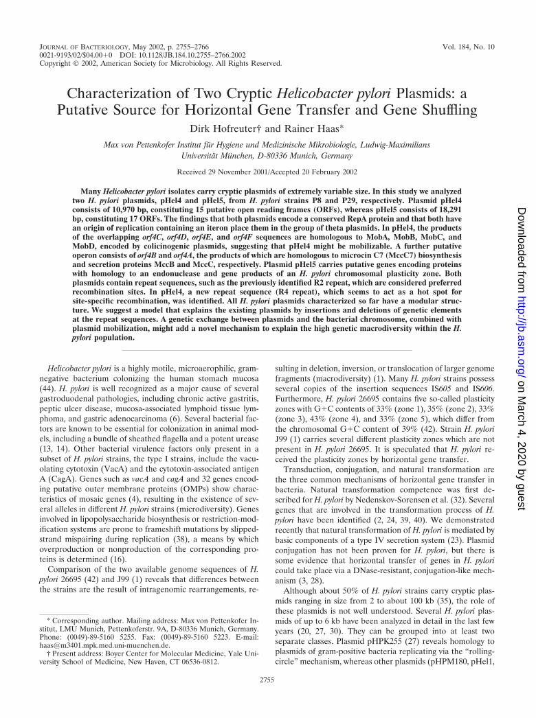
A shotgun approach was used to clone random gene seg-ments for sequencing pHel4 and pHel5. Plasmid pHel4 con-sists of a total of 10,970 bp, whereas pHel5 consists of 18,291bp and represents the largest H. pylori plasmid sequence de-termined to date. The annotation revealed a total of 15 puta-tive ORFs in plasmid pHel4 and 17 in plasmid pHel5 (Fig. 2A
2756 HOFREUTER AND HAAS J. BACTERIOL.
on March 4, 2020 by guest
http://jb.asm.org/
Dow
nloaded from

and 3A). In addition to ATG, GTG and TTG were identifiedas putative translation start codons (Tables 1 and 2), a featurethat is also described for chromosomal H. pylori genes (1, 42).The overall G�C contents of 34.5 (pHel4) and 34.4% (pHel5)(Fig. 2B and 3B) are significantly lower than that of H. pylorichromosomes of strains 26695 and J99 (39%) (1, 42), thoughthey are comparable to those of other H. pylori plasmids.
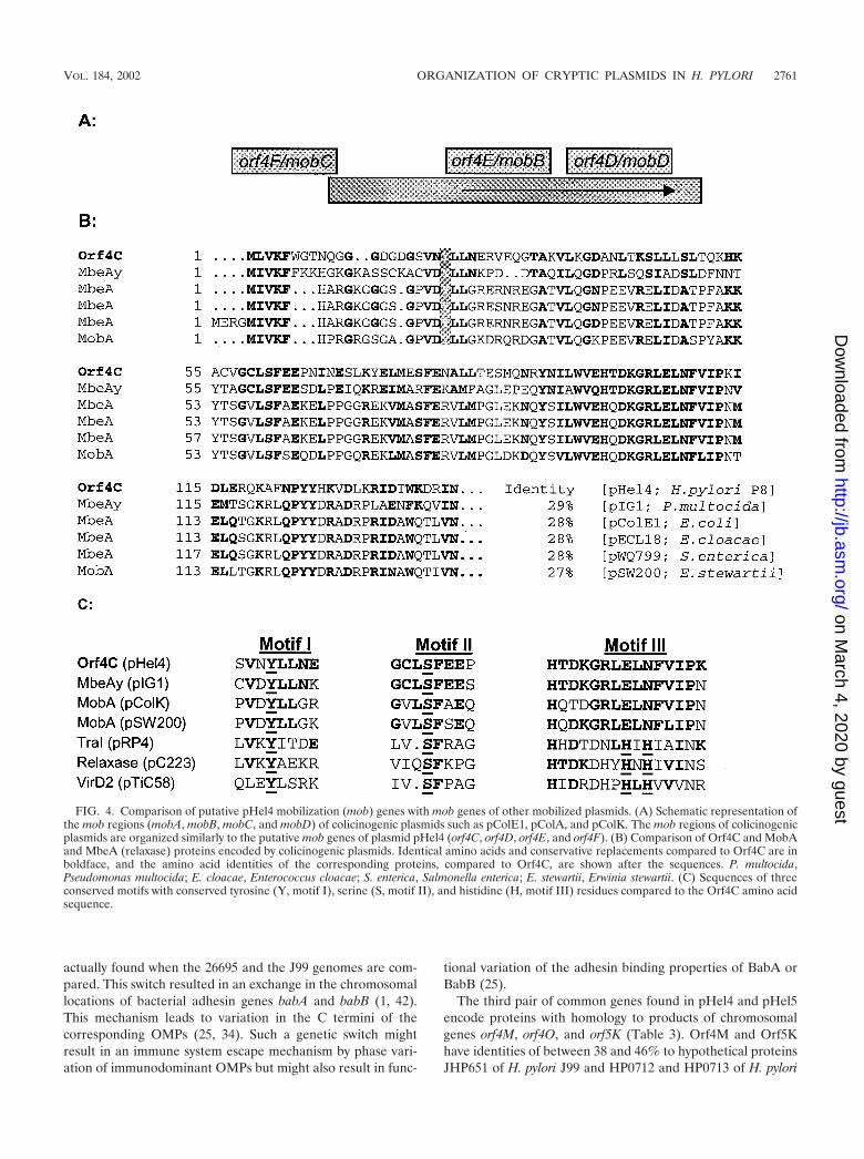
The best homologies of the putative ORFs to database en-tries and some important features of the deduced proteins aresummarized in Tables 1 and 2. Based on these searches itappears that plasmids pHel4 and pHel5 consist of genes forplasmid maintenance, bacteriocin production, and conjugaltransfer and chromosomal homologues of H. pylori, organizedin distinct regions. Whether these regions are functional or nothas to be demonstrated in the future. Moreover, pHel4 andpHel5 harbor border sequences of IS elements and conservedrepeat sequences also found in other plasmids of H. pylori,estimated to act as hot spots for DNA recombination.
Plasmid maintenance regions of pHel4 and pHel5. Orf4I,encoded by pHel4, and Orf5A, encoded by pHel5, were iden-tified as replication initiation proteins (Rep) showing signifi-cant sequence identity to the corresponding RepA proteinsencoded by different H. pylori plasmids (Table 3). This group ofH. pylori Rep proteins supports replication according to thetheta replication mechanism (10). The upstream region oforf4I is highly conserved compared to the pHel1 repA gene,indicating that the same transcriptional start site as that exper-imentally determined for pHel1 (20) might be used in pHel4.The upstream region of orf5A (pHel5) differs significantly fromthe corresponding regions of pHel1 and pHel4 but is identicalto the upstream region of repA of pHPS1, suggesting that therepA genes of pHel5 and pHPS1 use the same transcriptionalstart site (data not shown).
As described for pHel1 and pHPM180, we find a region of97 bp, consisting of 22-bp iterons repeated 41⁄2 times and lo-cated about 350 bp upstream of the start codon of the repAgene (data not shown). The sequences of the iterons of pHel4and pHel1 are identical, whereas the iteron region of pHel5shows best homology to the iteron sequence of pHPS1 (84%).The iteron regions of pHel4 and pHel5 are 77% identical.
For pHel1 we demonstrated a low copy number of about 4to 10 plasmid copies per cell in H. pylori (21). In addition,pHel4 and pHel5 replicate stably in H. pylori growing in vitroon agar plates without any selection pressure. The putativegene product of Orf5L has significant homology to a group ofParA proteins involved in the correct partitioning of plasmidsto the bacterial daughter cells during cell division, which isnecessary for the maintenance of low-copy-number plasmids(17). A protein identical to Orf5L, encoded by plasmidpHPM186 (the unpublished plasmid sequence is available inthe database under accession no. AF077006), and one encodedby plasticity zone 3 in H. pylori 26695 (HP1000; 49.5% identity)were described. We also identified JHP0935, homologous toParA and encoded by a plasticity zone of H. pylori J99; thisprotein is 45.5% identical to Orf5L. HP1000 was recentlygrouped in the new ParF subgroup of the ParA superfamily(19). Further studies need to be conducted to show whether ornot plasmids without parA or -F homologues, for example,pHel1, pHel4, pHPM180, and pHS1, use the chromosomallyencoded plasmid partitioning systems for plasmid maintenance.
Microcin MccC7 homology region. Orf4A (pHel4) revealedthe best sequence homology to a putative TetA(P) tetracyclineefflux membrane transporter protein of H. pylori strains 26695(HP1165) (42) and J99 (JHP1092) (1) (Table 1). Since H. pyloriP8 did not tolerate higher concentrations of tetracycline thanH. pylori strains 26695, J99, P1, P12, and P29 (0.05 to 0.6 �g/ml;data not shown), it was concluded that Orf4A and chromo-somally encoded proteins HP1165 and JHP1092 are probablynot involved in tetracycline resistance.
Orf4A displayed a lower homology to several transporterproteins involved in protein secretion, especially E. coli mic-rocin secretion protein MccC (23% identity). Interestingly, theorf4B sequence of pHel4, located upstream of orf4A, seems tobe organized with orf4A in an operon, and its product revealsin addition a sequence identity to the MccB protein (28%identity), encoded by the E. coli microcin operon (18). In theE. coli system, MccB is involved in modification of MccA(MccC7) (J. E. Gonzalez-Pastor, J. L. San Millan, and F.Moreno, Letter, Nature 369:281, 1994) and microcin is ex-ported by MccC. The modified secreted MccA peptide is takenup by related bacteria and acts there as an inhibitor of trans-lation if no corresponding immunity protein is expressed. Inplasmid pHel4 a candidate microcin structural gene upstreamof mccB was also identified (D. Hofreuter and R. Haas, un-published data).
Conjugation-like ORFs. Between two regions (bp 2380 to2454 and bp 5043 to 5245) of pHel4 with very low G�Ccontents (21 and 25%, respectively) a gene cluster with homol-ogy to a conjugal mobilization (mob) region of colicinogenicplasmids was identified. The observed overlap of ORFs orf4Cto orf4F (Fig. 4A) is very reminiscent of the structural organi-zation of mobA, mobB, mobC, and mobD of colicin-encodingplasmids pColA (31), pColE1 (7), pColD157 (22), and
FIG. 1. Purification and analysis of H. pylori plasmids by restrictionenzyme digestion. Plasmids pHel4 and pHel5 were isolated from H.pylori strains P8 and P29, respectively, by the Wizard Plus SV Mini-preps (Promega) and the xanthogenate-SDS-phenol methods. Plas-mids were digested with restriction endonucleases BglII (lane 1),EcoRI (lane 2), HindIII (lane 3), and SacI (lane 4) and separated ona 0.8% agarose gel. �, partially digested EcoRI fragment. Lane M,marker. kb, kilobases.
VOL. 184, 2002 ORGANIZATION OF CRYPTIC PLASMIDS IN H. PYLORI 2757
on March 4, 2020 by guest
http://jb.asm.org/
Dow
nloaded from
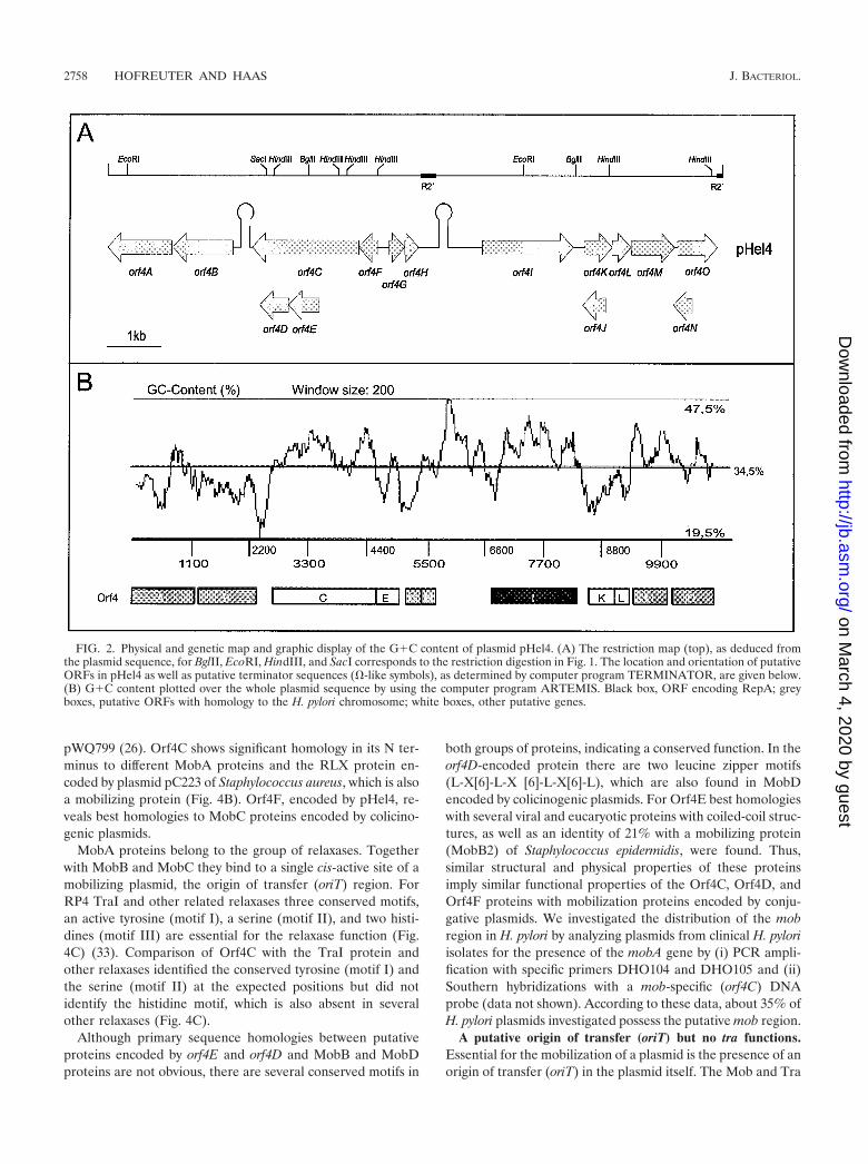
pWQ799 (26). Orf4C shows significant homology in its N ter-minus to different MobA proteins and the RLX protein en-coded by plasmid pC223 of Staphylococcus aureus, which is alsoa mobilizing protein (Fig. 4B). Orf4F, encoded by pHel4, re-veals best homologies to MobC proteins encoded by colicino-genic plasmids.
MobA proteins belong to the group of relaxases. Togetherwith MobB and MobC they bind to a single cis-active site of amobilizing plasmid, the origin of transfer (oriT) region. ForRP4 TraI and other related relaxases three conserved motifs,an active tyrosine (motif I), a serine (motif II), and two histi-dines (motif III) are essential for the relaxase function (Fig.4C) (33). Comparison of Orf4C with the TraI protein andother relaxases identified the conserved tyrosine (motif I) andthe serine (motif II) at the expected positions but did notidentify the histidine motif, which is also absent in severalother relaxases (Fig. 4C).
Although primary sequence homologies between putativeproteins encoded by orf4E and orf4D and MobB and MobDproteins are not obvious, there are several conserved motifs in
both groups of proteins, indicating a conserved function. In theorf4D-encoded protein there are two leucine zipper motifs(L-X[6]-L-X [6]-L-X[6]-L), which are also found in MobDencoded by colicinogenic plasmids. For Orf4E best homologieswith several viral and eucaryotic proteins with coiled-coil struc-tures, as well as an identity of 21% with a mobilizing protein(MobB2) of Staphylococcus epidermidis, were found. Thus,similar structural and physical properties of these proteinsimply similar functional properties of the Orf4C, Orf4D, andOrf4F proteins with mobilization proteins encoded by conju-gative plasmids. We investigated the distribution of the mobregion in H. pylori by analyzing plasmids from clinical H. pyloriisolates for the presence of the mobA gene by (i) PCR ampli-fication with specific primers DHO104 and DHO105 and (ii)Southern hybridizations with a mob-specific (orf4C) DNAprobe (data not shown). According to these data, about 35% ofH. pylori plasmids investigated possess the putative mob region.
A putative origin of transfer (oriT) but no tra functions.Essential for the mobilization of a plasmid is the presence of anorigin of transfer (oriT) in the plasmid itself. The Mob and Tra
FIG. 2. Physical and genetic map and graphic display of the G�C content of plasmid pHel4. (A) The restriction map (top), as deduced fromthe plasmid sequence, for BglII, EcoRI, HindIII, and SacI corresponds to the restriction digestion in Fig. 1. The location and orientation of putativeORFs in pHel4 as well as putative terminator sequences (�-like symbols), as determined by computer program TERMINATOR, are given below.(B) G�C content plotted over the whole plasmid sequence by using the computer program ARTEMIS. Black box, ORF encoding RepA; greyboxes, putative ORFs with homology to the H. pylori chromosome; white boxes, other putative genes.
2758 HOFREUTER AND HAAS J. BACTERIOL.
on March 4, 2020 by guest
http://jb.asm.org/
Dow
nloaded from

proteins are usually active in trans and might be encoded byother conjugative plasmids or the bacterial chromosome. TheoriT sequences in colicinogenic plasmids are usually locatedupstream of the mob regions, and the nic sites for plasmidspColE1 and pColA are identical. A corresponding sequencefor pHel4 was not found at this position. A putative nic se-quence identical to the well-characterized nic sequence of IncPplasmid RP4 was found in orf4M of pHel4 (TATCCTG/C[consensus sequence in bold type]); this sequence might act asa functional nic site of the plasmid. P-type nic sites in conju-gative colicinogenic plasmids have not been described yet (29).
A further question relates to the tra functions for a conju-gative transfer of pHel4, which are missing in the plasmid.Several putative proteins, which might be involved in DNAconjugation, are encoded by the genome sequence, especiallyin plasticity zones of H. pylori 26695 (42) and J99 (1). Theyshow significant homology to VirB and Trb proteins (Fig. 5).The identification of putative IncP type relaxases encoded byhp0996 and hp1004 (5, 41) also supports the conjugative trans-fer of plasmids in H. pylori.
ORFs in pHel4 and pHel5 whose products are homologousto H. pylori plasmid-encoded and chromosomally encoded geneproducts. Five ORFs of pHel4 and eight of pHel5 show besthomologies to chromosomal genes of strains 26695 and J99.Three ORFs of this group are common to both plasmids, andthe deduced pairs of proteins (Orf4G and Orf5G, Orf4H and
Orf5F, and Orf4M and Orf5K) show a remarkable sequenceidentity, ranging between 91.3 and 96.6% (Table 3). We alsoidentified in pHel1, pHPM186, and pHPM8 homologues toorf4H and orf5F; however, in these plasmids the genes appearto be truncated and possess only the conserved 3� ends, prob-ably as a result of recombination or integration events. Thesefragments are flanked at the 5� end by IS elements or bordersequences of IS elements (Fig. 6).
The deduced Orf4G and Orf5G proteins reveal best homol-ogies to proteins JHP0828 and HP0316 of H. pylori J99 (1) and26695, respectively (42), both of which have unknown func-tions (Tables 1 to 3). The homology between orf4G and orf5Gand the chromosomal region of jhp0828 is not restricted to thecoding region but extends into a 43-bp upstream region (93%identity). The N-terminal regions of Orf4G and Orf5G show anearly 100% identity to that of JHP0828, whereas their Ctermini are most homologous to that of HP0316.
The second pair of homologous proteins, Orf4H and Orf5F,show best homologies to HP0892 and HP0894 of H. pylori26695, as well as to JHP0825 and JHP0831 of H. pylori J99(Tables 1 to 3). These hypothetical proteins have well-con-served C termini. The BLASTP search also identified signifi-cant homology among Orf4H and Orf5F and gene products ofother bacterial species, encoded either on plasmids or in thechromosome (Tables 1 and 2).
Most unexpected, orf4G and orf4H of plasmid pHel4 and
FIG. 3. Physical and genetic map and graphic display of the G�C content of plasmid pHel5. (A) Restriction map (top), as deduced from theplasmid sequence, for HindIII, which corresponds to the restriction digestion in Fig. 1. The location and orientation of putative ORFs in pHel5as well as putative terminator sequences (�-like symbols), as determined by the computer program TERMINATOR, are given below. Half-squaresmark shifts in the reading frame for putative genes orf5B and orf5Q, which result in premature termination of the putative proteins. (B) G�Ccontent plotted over the whole plasmid sequence with ARTEMIS. Black box, ORF encoding RepA; grey boxes, putative ORFs with homology tothe H. pylori chromosome; white boxes, other putative genes.
VOL. 184, 2002 ORGANIZATION OF CRYPTIC PLASMIDS IN H. PYLORI 2759
on March 4, 2020 by guest
http://jb.asm.org/
Dow
nloaded from

orf5F and orf5G of pHel5 have the same organization as thecorresponding chromosomal genes of H. pylori 26695 and H.pylori J99, suggesting an exchange of the gene clusters betweenchromosome and plasmid. For the chromosomal genes it wasshown that the members of these two families interact witheach other in a two-hybrid screen (36). Homologies byBLASTP are as follows: HP0316 and HP0895, 1e-57; HP0894and HP0895, 1e-71; HP0895 and HP0895, 1e-204. ORFs in thechromosomes of H. pylori 26695 and J99 homologous to orf4Gand orf4H and orf5F and orf5G are clustered in regions withhigh genetic diversity (9).
A switch-inducing repeat 1 sequence located in plasmidspHel4 and pHel5. Interestingly, genes homologous to orf4Gand orf5G are part of the repeat 1 sequence (42), which islocated at different positions in the chromosome and which iscoupled with the 3� regions of different OMPs. The repeat 1sequence can be considered a switch-inducing sequence, whichmight be supported by the concerted activity of Orf4G andOrf5G or Orf4H and Orf5F, acting as recombination-inducingproteins.
Such a switch mechanism, which is related to an intrachro-mosomal recombination event at the repeat 1 sequence, is
TABLE 1. List of identified ORFs of plasmid pHel4
ORF Codon position(start–stop) Sequencea Molecular
mass (kDa) pI Netcharge Propertiesb Homology (BLASTP)c
orf4A 1166–3 AATTTATAGAGTTTCTTAATATG 43.9 9.3 9 TM; SP; �-� HP1165, 1e-92; JHP1092, 2e-92; BB126, 7e-34;TetA(P), 2e-06; MccC, 5e-5
orf4B 2254–1178 AAAGTTTTATCGGAGTAGAGTTG 41.0 6.6 �2 �-� MccB, 1e-18orf4C 4488–2506 TTAGAACAACTAAGAGCTAAATG 78.1 10.4 26 Mix; cc MbeAy, 7e-30; MbeA, 23–27; MobA, 5e-24;
BdrC3, 0.026orf4D 3245–2739 CCAAGAAATAAGGGGAAAACATG 19.6 5.3 �4 Leucine zipper; �orf4E 3785–3255 ATTAAAAGGATTTTACTACCATG 20.4 10.1 8 TM; cc; � MobB2, 0.65orf4F 4822–4478 TAAGGGATAGCCAAAAAAATATG 13.2 10.6 8 Mix MbeCy, 0.021orf4G 5020–5298 GATTGAGTTAAGGAGATAAGATG 10.8 10.1 5 cc; � JHP0828, 2e-16; HP0316, 0.002orf4H 5285–5551 ATGAGTTAAAAAGAGAGTATATG 10.7 8.7 2 Mix HP0894, 4e-26; HP0892, 1e-21; JHP0831, 4e-21;
JHP0825, 2e-18; ORF10, 1e-10; YafQ, 6e-09orf4I 6671–8296 AAGCATTAAAAGGTGCTTAAATG 64.1 10.2 19 cc; mix RepA, 0.0orf4J 8854–8483 GAAAAACAAGGGGATCACTAATG 13.6 8.5 4 TM; �-�orf4K 8490–8990 TTGTAATAGGAGTTTAAAAAATG 20.0 9.8 6 � (cc)orf4L 8990–9322 AGTTATGTAAAGAGCATGTAGTG 13.4 9.6 3 TM; cc; mixorf4M 9326–10099 TCAATCGTTTGGAGTAGCATGTG 29.6 6.5 �5 Mix Orf6-pHPM8, 4e-57; JHP0651, 5e-33; HP0712,
7e-22; pHPM180-Orf2, 1e-08; HP0713, 5e-07orf4N 10406–10086 TATTCTAAACTCAAAGGTTCTTG 12.2 9.2 1 TM; �-�orf4O 10149–10856 ACAGGTGAAAAGACAGATGCATG 27.6 7.9 1 Mix pHPM180-ORF2, e-115; JHP0651, 6e-11;
HP0713, 7e-09; ZK593.8, 5e-08; Tou1, 5e-06;ORF56-TP901-1, 0.20; ORF178-wss virus, 0.23
a The putative Shine-Dalgarno sequence is underlined; the start codon is in boldface.b TM, transmembrane domain; SP, signal peptide; �, �-�, and mix, secondary structure elements as predicted by PREDICTPROTEIN; cc, coiled-coil structure.c BB126, AAC66191; TetA(P), BAA19230; MccC, CAA40810; MccB, CAA40809; MbeAy, AAB05464; MbeA, CAA33883; MobA, AAA69498; BdrC3, AAF19116;
BobB2, AAD02406; MbeCy, AAB05463; ORF10, AAF05106; YafQ, Q47149; ZK593.8, T27927; Tou1, AAF08816; ORF56-TP901-1, AAK38073; ORF178-white spotsyndrome (wss) virus, AAK77847.
TABLE 2. List of ORFs of plasmid pHel5
ORF Codon positions(start–stop) Sequencea Molecular
mass (kDa) pI Netcharge Propertiesb Homology
(BLASTP)
orf5A 865–2481 CCAAAAGATAAGGAGTATAGAGTG 47.0 10.25 19 cc; mix RepA, 0.0orf5B 3352–2706 GCAACTTACACAGGAAAAACAATG 25.2 10.36 13 SP; mix JHP1295, 2e-37; HP1382, 2e-05orf5C 3761–4279 AAAGGAAAGGAAAATGCAACCATG 19.9 7.81 1 �orf5D 4898–5749 CAACACAAGGAGATTTTAAAAATG 34.1 9.73 7 cc; �orf5E 5862–6929 CACAAGCAAGAAGGACCAAAAATG 41.6 9.28 7 Mixorf5F 7387–7121 ATGAACTAAAAAGAGAGATACATG 10.8 9.24 3 Mix HP0894, 4e-22; JHP831, 2e-17; HP0892,
3e-17; JHP0825, 5e-13orf5G 7652–7374 AGCTTAATACAAGGAAATGAGATG 10.8 10.19 5 cc; JHP0828, 4e-11; HP0316, 0.030orf5H 8271–8783 ACTATTTTTATCAATGTCGCAATG 19.8 8.49 3 cc; �orf5I 8795–9196 AGAAGAATAGGAACAAAAAGAATG 15.0 8.50 3 SP; cc; �-�orf5J 9193–9405 GAAAGAACAATTGGATGACTTATG 8.1 9.99 4 Mixorf5K 9440–10048 TTAGACACTAGGAACAAAGTGATG 23.4 5.60 �10 Mix JHP0651, 3e-31; HP0712, 2e-22orf5L 10800–11474 CATTAAATATAAAGGAACAGAATG 25.5 5.96 �2 Mix HP1000, 4e-53; JHP0935, 1e-36orf5M 11471–12037 ACCCAACGAAGAAAGGAAAGTATG 21.7 9.76 3 cc; mixorf5N 12898–13491 CACATAAAAAGGATAAAAATTATG 23.2 9.18 5 Mix HP0879, 7e-08; JHP812, 8e-08; HP1334,
2e-06orf5O 13571–14731 ATTTTTCCACAAAGGATCGCAATG 46.7 8.98 5 cc; mixorf5P 14834–15085 AAATTTACTAGGAATAGTAAAATG 9.8 4.67 �4 Mix HP0993, 7e-30orf5Q 15057–15769 TCCCTATGGAAAATATTCAGTATG 27.8 6.52 �4 Mix HP0994, 2e-94
a The putative Shine-Dalgarno sequence is underlined; the start codon is in boldface.b TM, transmembrane domain; SP, signal peptide; �, �-�, and mix, secondary structure elements as predicted by PREDICTPROTEIN; cc, coiled-coil structure.
2760 HOFREUTER AND HAAS J. BACTERIOL.
on March 4, 2020 by guest
http://jb.asm.org/
Dow
nloaded from
actually found when the 26695 and the J99 genomes are com-pared. This switch resulted in an exchange in the chromosomallocations of bacterial adhesin genes babA and babB (1, 42).This mechanism leads to variation in the C termini of thecorresponding OMPs (25, 34). Such a genetic switch mightresult in an immune system escape mechanism by phase vari-ation of immunodominant OMPs but might also result in func-
tional variation of the adhesin binding properties of BabA orBabB (25).
The third pair of common genes found in pHel4 and pHel5encode proteins with homology to products of chromosomalgenes orf4M, orf4O, and orf5K (Table 3). Orf4M and Orf5Khave identities of between 38 and 46% to hypothetical proteinsJHP651 of H. pylori J99 and HP0712 and HP0713 of H. pylori
FIG. 4. Comparison of putative pHel4 mobilization (mob) genes with mob genes of other mobilized plasmids. (A) Schematic representation ofthe mob regions (mobA, mobB, mobC, and mobD) of colicinogenic plasmids such as pColE1, pColA, and pColK. The mob regions of colicinogenicplasmids are organized similarly to the putative mob genes of plasmid pHel4 (orf4C, orf4D, orf4E, and orf4F). (B) Comparison of Orf4C and MobAand MbeA (relaxase) proteins encoded by colicinogenic plasmids. Identical amino acids and conservative replacements compared to Orf4C are inboldface, and the amino acid identities of the corresponding proteins, compared to Orf4C, are shown after the sequences. P. multocida,Pseudomonas multocida; E. cloacae, Enterococcus cloacae; S. enterica, Salmonella enterica; E. stewartii, Erwinia stewartii. (C) Sequences of threeconserved motifs with conserved tyrosine (Y, motif I), serine (S, motif II), and histidine (H, motif III) residues compared to the Orf4C amino acidsequence.
VOL. 184, 2002 ORGANIZATION OF CRYPTIC PLASMIDS IN H. PYLORI 2761
on March 4, 2020 by guest
http://jb.asm.org/
Dow
nloaded from

26695. JHP0651 might be a fusion between HP0712 (N termi-nus) and HP713 (C terminus). The BLASTP analysis alsoidentified homology to eucaryotic proteins, for instance,ZK593.8 of Caenorhabditis elegans and human huntingtin in-teracting protein E, an approximately 350-kDa protein of un-
known function involved in the neuropathology of Hunting-ton’s disease (15). Orf4M and Orf5K also possess homology tothe Orf2 protein, described first as a product of H. pyloriplasmid pHPM180 (30), and to Orf-2, encoded by the NBU1element (nonreplicating Bacteroides units) (43). The homology
FIG. 5. Distribution of genes encoding putative Mob- and Tra-like proteins in the genome of H. pylori 26695 and J99. The tra-like genes arealso homologues to the Agrobacterium tumefaciens virB genes, encoding the type IV secretion system for Ti plasmid mobilization and transferred-DNA transfer. In the chromosomes of H. pylori 26695 (HP) and J99 (JHP) virB-like genes are located either in plasticity zones (PR1 to PR3) asoperons, such as the comB operon, or as single genes, such as hp1421 and hp17. cag-PAI, cag pathogenicity island.
TABLE 3. Comparison of products of ORFs in plasmids pHel4 and pHel5 with products of the H. pylori chromosome
Chromosome-encoded protein,% identity to pHel4-
encoded protein
pHel4-encodedprotein
% Identity between pHel4-and pHel5-encoded
proteinsa
pHel5-encodedprotein
Chromosome-encoded protein,% identity to pHel5-
encoded protein
Orf41 (RepA) 78.3 Orf5A (RepA)HP1165, 53.8 Orf4AJHP1092, 52.3
Orf5B JHP1295, 50.3JHP0828, 67.8 Orf4G 91.3 Orf5G JHP0828, 73.5HP0316, 35.6 HP0316, 35.6HP0892, 61.4 Orf4H 96.6 Orf5F HP0892, 59.1HP0894, 60.3 HP0894, 58.0JHP0831, 60.2 JHP0831, 59.1JHP0825, 55.7 JHP0825, 53.4HP0712, 46.0 Orf4M 95.0 Orf5K HP712, 46.0JHP0651, 40.6 JHP0651, 44.0HP0713, 38.1 HP0713, 44.3JHP0651, 30.1 Orf4O 30.7 Orf5K JHP0651, 44.0
Orf5L HP1000, 49.5JHP0935, 45.5
Orf5O HP0879, 26.7JHP0812, 33.5HP1334-36.2
Orf5P HP0993, 85.5Orf5Q HP0944, 79.7
a pHel4-encoded RepA identities to RepA of plasmids pHel1, pHPM180, pHPS1: 98.0, 89.3, and 74.8, respectively; corresponding values for pHel5-encoded RepA:78.7, 81.8, and 76.3%, respectively.
2762 HOFREUTER AND HAAS J. BACTERIOL.
on March 4, 2020 by guest
http://jb.asm.org/
Dow
nloaded from
is clustered mainly in the N-terminal parts of Orf4M andOrf5K, which contain the conserved amino acid sequencePFSDGNGRTGRALMF (data not shown). Thus, Orf4M,Orf4O, and Orf5K belong to a group of proteins whose codingsequences are widespread in bacteriophages, plasmids, andNBU-1 elements and also in eubacteria and some eucaryoticorganisms.
Further plasmid-encoded proteins also encoded by the H.pylori chromosome. As observed in pHPM186, plasmid pHel5encodes proteins with homology to products of genes clusteredin plasticity zone 3 of H. pylori 26695. As described above,Orf5L belongs to the group of ParA proteins, as do the ho-mologous proteins HP1000 and JHP0935. Additionally, orf5Pand orf5Q encode proteins with high identity to HP0993 andHP0994, hypothetical proteins encoded by genes in the H.pylori plasticity zones with unknown functions (Table 3). Thesequence of orf5Q in pHel5 carries a frameshift, resulting in atruncated HP0994 gene product. Further proteins encoded bypHel4 and pHel5 with homology to products of genes wide-spread in the chromosomes of H. pylori 26695 and J99 areOrf5B (similar to endonuclease HP1295) and Orf5O (besthomologue to hypothetical protein HP1334).
Repeat sequences and site-specific recombination events de-termine size variation and the modular structure of H. pylori
plasmids. The comparison of independent H. pylori plasmidsequences identified several sequence repeats termed R1, R2,and R3 (10). The R1 and R3 repeats correspond to iteronsequences, located upstream of genes encoding replication ini-tiation proteins RepA and RepB. It has been suggested thatthe R2 repeat, which consists, in pHP180, of two copies of 232noncoding nucleotides, might act as a target sequence for re-combination events (10, 30). Our sequence analysis of pHel4and pHel5 revealed for both plasmids incomplete sequences ofthe 232-bp R2 repeat, designated R2� (Fig. 6). For pHel4 wefound one nearly complete 232-bp sequence (bp 5665 to 5842)between orf4H and the putative origin of replication. Thisregion also contains an intact 36-bp stretch (bp 5578 to 5613),which was first found between the two R2 repeats inpHPM180. A second incomplete copy of the R2 repeat inpHel4 is between orf4O and orf4A (bp 10845 to 10954). ForpHel5, one identical stretch to the 232-bp sequence was found(bp 18120 to 18291). The comparison of the locations of repeatsequences in H. pylori plasmids suggests a modular structure ofH. pylori plasmids with insertion and deletion of sequencemodules at different repeat sequences, which might act as hotspots for recombination or site-specific integration events (Fig. 6).
A novel R4 repeat in H. pylori plasmids is associated with
FIG. 6. Schematic comparison of the modules represented on individual H. pylori plasmids. Each plasmid carries a repA or a repB gene or both.The R1, R2, and R3 repeats and the novel R4 repeat are located at the borders of sequence modules. Complete IS605 elements are only presentin pHPM186; pHel4 and pHel5 carry border sequences of IS elements, such as IS606 and IS608. Genes with homology to the H. pylori chromosomehave the corresponding hp or jhp ORF number, and homologues have the same color. All putative elements involved in recombination ortransposition events are red or orange. R1 to R4, repeats R1 to R4; IS, IS element; LB, left border sequence; RB, right border sequence.
VOL. 184, 2002 ORGANIZATION OF CRYPTIC PLASMIDS IN H. PYLORI 2763
on March 4, 2020 by guest
http://jb.asm.org/
Dow
nloaded from
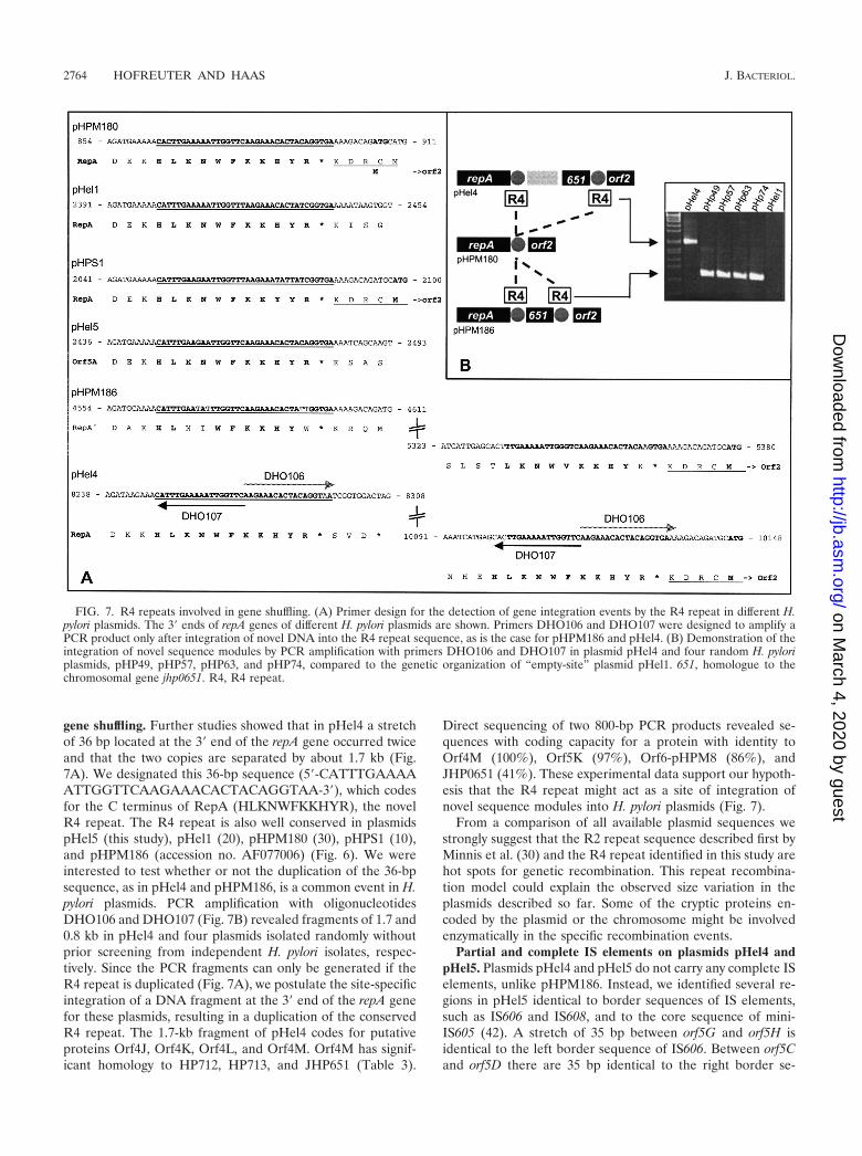
gene shuffling. Further studies showed that in pHel4 a stretchof 36 bp located at the 3� end of the repA gene occurred twiceand that the two copies are separated by about 1.7 kb (Fig.7A). We designated this 36-bp sequence (5�-CATTTGAAAAATTGGTTCAAGAAACACTACAGGTAA-3�), which codesfor the C terminus of RepA (HLKNWFKKHYR), the novelR4 repeat. The R4 repeat is also well conserved in plasmidspHel5 (this study), pHel1 (20), pHPM180 (30), pHPS1 (10),and pHPM186 (accession no. AF077006) (Fig. 6). We wereinterested to test whether or not the duplication of the 36-bpsequence, as in pHel4 and pHPM186, is a common event in H.pylori plasmids. PCR amplification with oligonucleotidesDHO106 and DHO107 (Fig. 7B) revealed fragments of 1.7 and0.8 kb in pHel4 and four plasmids isolated randomly withoutprior screening from independent H. pylori isolates, respec-tively. Since the PCR fragments can only be generated if theR4 repeat is duplicated (Fig. 7A), we postulate the site-specificintegration of a DNA fragment at the 3� end of the repA genefor these plasmids, resulting in a duplication of the conservedR4 repeat. The 1.7-kb fragment of pHel4 codes for putativeproteins Orf4J, Orf4K, Orf4L, and Orf4M. Orf4M has signif-icant homology to HP712, HP713, and JHP651 (Table 3).
Direct sequencing of two 800-bp PCR products revealed se-quences with coding capacity for a protein with identity toOrf4M (100%), Orf5K (97%), Orf6-pHPM8 (86%), andJHP0651 (41%). These experimental data support our hypoth-esis that the R4 repeat might act as a site of integration ofnovel sequence modules into H. pylori plasmids (Fig. 7).
From a comparison of all available plasmid sequences westrongly suggest that the R2 repeat sequence described first byMinnis et al. (30) and the R4 repeat identified in this study arehot spots for genetic recombination. This repeat recombina-tion model could explain the observed size variation in theplasmids described so far. Some of the cryptic proteins en-coded by the plasmid or the chromosome might be involvedenzymatically in the specific recombination events.
Partial and complete IS elements on plasmids pHel4 andpHel5. Plasmids pHel4 and pHel5 do not carry any complete ISelements, unlike pHPM186. Instead, we identified several re-gions in pHel5 identical to border sequences of IS elements,such as IS606 and IS608, and to the core sequence of mini-IS605 (42). A stretch of 35 bp between orf5G and orf5H isidentical to the left border sequence of IS606. Between orf5Cand orf5D there are 35 bp identical to the right border se-
FIG. 7. R4 repeats involved in gene shuffling. (A) Primer design for the detection of gene integration events by the R4 repeat in different H.pylori plasmids. The 3� ends of repA genes of different H. pylori plasmids are shown. Primers DHO106 and DHO107 were designed to amplify aPCR product only after integration of novel DNA into the R4 repeat sequence, as is the case for pHPM186 and pHel4. (B) Demonstration of theintegration of novel sequence modules by PCR amplification with primers DHO106 and DHO107 in plasmid pHel4 and four random H. pyloriplasmids, pHP49, pHP57, pHP63, and pHP74, compared to the genetic organization of “empty-site” plasmid pHel1. 651, homologue to thechromosomal gene jhp0651. R4, R4 repeat.
2764 HOFREUTER AND HAAS J. BACTERIOL.
on March 4, 2020 by guest
http://jb.asm.org/
Dow
nloaded from
quence of IS606. This region also has homology to mini-IS605.A second smaller stretch of this IS606 inverted repeat right(IRR) is located between orf5M and orf5N. This sequence ispart of a sequence duplication (5�-TTTTGACATACTCCCCATAGCTAAAGCTAGAGACTTTGCGG-3�) in pHel5 (bp6997 to 7037 and 12353 to 12380) since a second copy of thatsequence was identified between orf5E and orf5F (Fig. 6). A
single copy of this 41-bp sequence could also be found in pHel4between the R4 repeat and orf4J.
Again, the repeat and IS border sequences are always lo-cated at defined positions, flanking certain genes or groups ofgenes, such as repA (R2 and R4 repeats), the mcc-mob regionin pHel4 and pHP186 (R2 repeat), and the orf4J-orf4M regionof plasmid pHel4 (R4 repeat). The advantage of such a mod-
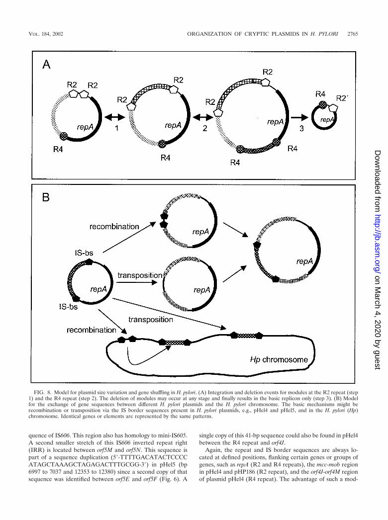
FIG. 8. Model for plasmid size variation and gene shuffling in H. pylori. (A) Integration and deletion events for modules at the R2 repeat (step1) and the R4 repeat (step 2). The deletion of modules may occur at any stage and finally results in the basic replicon only (step 3). (B) Modelfor the exchange of gene sequences between different H. pylori plasmids and the H. pylori chromosome. The basic mechanisms might berecombination or transposition via the IS border sequences present in H. pylori plasmids, e.g., pHel4 and pHel5, and in the H. pylori (Hp)chromosome. Identical genes or elements are represented by the same patterns.
VOL. 184, 2002 ORGANIZATION OF CRYPTIC PLASMIDS IN H. PYLORI 2765
on March 4, 2020 by guest
http://jb.asm.org/
Dow
nloaded from

ular organization is rather obvious (Fig. 8). H. pylori can dis-tribute a high number of diverse genetic modules in the pop-ulation. Different combinations of modules might be createdby recombination (deletion and insertion) and selected for bythe needs of the bacteria in their individual hosts. Due to themodular structure, plasmids might either pick up chromosomalgenes of H. pylori or integrate sequence modules from foreignplasmids, which are taken up by the bacteria during its naturaltransformation competence (gene shuffling). For conjugativeplasmids, as we postulate for pHel4, the novel sequences couldbe rapidly distributed within the H. pylori population and ex-change novel plasmid sequences with the bacterial chromo-some. Such events might help to explain the development ofmacrodiversity among H. pylori strains and the rapid genera-tion of substrains.
ACKNOWLEDGMENTS
We thank E. Weiss for excellent technical assistance and B. P. Burnsfor critical reading of the manuscript.
This work was supported by the Deutsche Forschungsgemeinschaft(HA2697/3-1).
REFERENCES
1. Alm, R. A., L. S. Ling, D. T. Moir, B. L. King, E. D. Brown, P. C. Doig, D. R.Smith, B. Noonan, B. C. Guild, et al. 1999. Genomic-sequence comparisonof two unrelated isolates of the human gastric pathogen Helicobacter pylori.Nature 397:176–180.
2. Ando, T., D. A. Israel, K. Kusugami, and M. J. Blaser. 1999. HP0333, amember of the dprA family, is involved in natural transformation in Helico-bacter pylori. J. Bacteriol. 181:5572–5580.
3. Ando, T., Q. Xu, M. Torres, K. Kusugami, D. A. Israel, and M. J. Blaser.2000. Restriction-modification system differences in Helicobacter pylori are abarrier to interstrain plasmid transfer. Mol. Microbiol. 37:1052–1065.
4. Atherton, J. C., P. Cao, R. M. Peek, Jr., M. K. R. Tummuru, M. J. Blaser,and T. L. Cover. 1995. Mosaicism in vacuolating cytotoxin alleles of Helico-bacter pylori. J. Biol. Chem. 270:17771–17777.
5. Backert, S., E. Von Nickisch-Rosenegk, and T. F. Meyer. 1998. Potential roleof two Helicobacter pylori relaxases in DNA transfer? Mol. Microbiol. 30:673–674.
6. Blaser, M. J. 1996. The bacteria behind ulcers. Sci. Am. 274:104–107.7. Boyd, A. C., J. A. Archer, and D. J. Sherratt. 1989. Characterization of the
ColE1 mobilization region and its protein products. Mol. Gen. Genet. 217:488–498.
8. Boyer, H. W., and D. Roulland-Dussoix. 1969. A complementation analysisof the restriction and modification of DNA in Escherichia coli. J. Mol. Biol.41:459–472.
9. Cao, P., and T. L. Cover. 1997. High-level genetic diversity in the vapDchromosomal region of Helicobacter pylori. J. Bacteriol. 179:2852–2856.
10. De Ungria, M. C., T. Kolesnikow, P. T. Cox, and A. Lee. 1999. Molecularcharacterization and interstrain variability of pHPS1, a plasmid isolated fromthe Sydney strain (SS1) of Helicobacter pylori. Plasmid 41:97–109.
11. De Ungria, M. C., D. Tillett, B. A. Neilan, P. T. Cox, and A. Lee. 1998. Anovel method of extracting plasmid DNA from Helicobacter species. Heli-cobacter 3:269–277.
12. Devereux, J., P. Haeberli, and O. Smithies. 1984. A comprehensive set ofsequence analysis programs for the VAX. Nucleic Acids Res. 12:387–395.
13. Eaton, K. A., C. L. Brooks, D. R. Morgan, and S. Krakowka. 1991. Essentialrole of urease in pathogenesis of gastritis induced by Helicobacter pylori ingnotobiotic piglets. Infect. Immun. 59:2470–2475.
14. Eaton, K. A., S. Suerbaum, C. Josenhans, and S. Krakowka. 1996. Coloni-zation of gnotobiotic piglets by Helicobacter pylori deficient in two flagellingenes. Infect. Immun. 64:2445–2448.
15. Faber, P. W., G. T. Barnes, J. Srinidhi, J. Chen, J. F. Gusella, and M. E.MacDonald. 1998. Huntingtin interacts with a family of WW domain pro-teins. Hum. Mol. Genet. 7:1463–1474.
16. Ge, Z., and D. E. Taylor. 1999. Contributions of genome sequencing tounderstanding the biology of Helicobacter pylori. Annu. Rev. Microbiol. 53:353–387.
17. Gerdes, K., J. Moller-Jensen, and J. R. Bugge. 2000. Plasmid and chromo-some partitioning: surprises from phylogeny. Mol. Microbiol. 37:455–466.
18. Gonzalez-Pastor, J. E., J. L. San Millan, M. A. Castilla, and F. Moreno.1995. Structure and organization of plasmid genes required to produce thetranslation inhibitor microcin C7. J. Bacteriol. 177:7131–7140.
19. Hayes, F. 2000. The partition system of multidrug resistance plasmid TP228includes a novel protein that epitomizes an evolutionarily distinct subgroupof the ParA superfamily. Mol. Microbiol. 37:528–541.
20. Heuermann, D., and R. Haas. 1995. Genetic organization of a small crypticplasmid of Helicobacter pylori. Gene 165:17–24.
21. Heuermann, D., and R. Haas. 1998. A stable shuttle vector system forefficient genetic complementation of Helicobacter pylori strains by transfor-mation and conjugation. Mol. Gen. Genet. 257:519–528.
22. Hofinger, C., H. Karch, and H. Schmidt. 1998. Structure and function ofplasmid pColD157 of enterohemorrhagic Escherichia coli O157 and its dis-tribution among strains from patients with diarrhea and hemolytic-uremicsyndrome. J. Clin. Microbiol. 36:24–29.
23. Hofreuter, D., S. Odenbreit, and R. Haas. 2001. Natural transformationcompetence in Helicobacter pylori is mediated by the basic components of atype IV secretion system. Mol. Microbiol. 41:379–391.
24. Hofreuter, D., S. Odenbreit, G. Henke, and R. Haas. 1998. Natural compe-tence for DNA transformation in Helicobacter pylori: identification and ge-netic characterization of the comB locus. Mol. Microbiol. 28:1027–1038.
25. Ilver, D., A. Arnqvist, J. Ogren, I. M. Frick, D. Kersulyte, E. T. Incecik, D. E.Berg, A. Covacci, L. Engstrand, and T. Borén. 1998. Helicobacter pyloriadhesin binding fucosylated histo-blood group antigens revealed by retag-ging. Science 279:373–377.
26. Keenleyside, W. J., and C. Whitfield. 1995. Lateral transfer of rfb genes: amobilizable ColE1-type plasmid carries the rfbO:54 (O:54 antigen biosyn-thesis) gene cluster from Salmonella enterica serovar Borreze. J. Bacteriol.177:5247–5253.
27. Kleanthous, H., C. L. Clayton, and S. Tabaqchali. 1991. Characterization ofa plasmid from Helicobacter pylori encoding a replication protein common toplasmids in gram-positive bacteria. Mol. Microbiol. 5:2377–2389.
28. Kuipers, E. J., D. A. Israel, J. G. Kusters, and M. J. Blaser. 1998. Evidencefor a conjugation-like mechanism of DNA transfer in Helicobacter pylori. J.Bacteriol. 180:2901–2905.
29. Lanka, E., and B. M. Wilkins. 1995. DNA processing reactions in bacterialconjugation. Annu. Rev. Biochem. 64:141–169.
30. Minnis, J. A., T. E. Taylor, J. E. Knesek, W. L. Peterson, and S. A. McIntire.1995. Characterization of a 3.5-kbp plasmid from Helicobacter pylori. Plasmid34:22–36.
31. Morlon, J., M. Chartier, M. Bidaud, and C. Lazdunski. 1988. The completenucleotide sequence of the colicinogenic plasmid ColA. High extent ofhomology with ColE1. Mol. Gen. Genet. 211:231–243.
32. Nedenskov-Sorensen, P., G. Bukholm, and K. Bovre. 1990. Natural compe-tence for genetic transformation in Campylobacter pylori. J. Infect. Dis. 161:365–366.
33. Pansegrau, W., W. Schroder, and E. Lanka. 1994. Concerted action of threedistinct domains in the DNA cleaving-joining reaction catalyzed by relaxase(TraI) of conjugative plasmid RP4. J. Biol. Chem. 269:2782–2789.
34. Peck, B., M. Ortkamp, K. D. Diehl, E. Hundt, and B. Knapp. 1999. Conser-vation, localization and expression of HopZ, a protein involved in adhesionof Helicobacter pylori. Nucleic Acids Res. 27:3325–3333.
35. Penfold, S. S., A. J. Lastovica, and B. G. Elisha. 1988. Demonstration ofplasmids in Campylobacter pylori. J. Infect. Dis. 157:850–851.
36. Rain, J. C., L. Selig, H. De Reuse, V. Battaglia, C. Reverdy, S. Simon, G.Lenzen, F. Petel, J. Wojcik, V. Schachter, Y. Chemama, A. Labigne, and P.Legrain. 2001. The protein-protein interaction map of Helicobacter pylori.Nature 409:211–215.
37. Sambrook, J., E. F. Fritsch, and T. Maniatis. 1989. Molecular cloning: alaboratory manual. Cold Spring Harbor Laboratory Press, Cold Spring Har-bor, N.Y.
38. Saunders, N. J., J. F. Peden, D. W. Hood, and E. R. Moxon. 1998. Simplesequence repeats in the Helicobacter pylori genome. Mol. Microbiol. 27:1091–1098.
39. Schmitt, W., S. Odenbreit, D. Heuermann, and R. Haas. 1995. Cloning of theHelicobacter pylori recA gene and functional characterization of its product.Mol. Gen. Genet. 248:563–572.
40. Smeets, L. C., J. J. Bijlsma, S. Y. Boomkens, C. M. Vandenbroucke-Grauls,and J. G. Kusters. 2000. comH, a novel gene essential for natural transfor-mation of Helicobacter pylori. J. Bacteriol. 182:3948–3954.
41. Thorsted, P. B., D. P. Macartney, P. Akhtar, A. S. Haines, N. Ali, P. David-son, T. Stafford, M. J. Pocklington, W. Pansegrau, B. M. Wilkins, E. Lanka,and C. M. Thomas. 1998. Complete sequence of the IncPbeta plasmid R751:implications for evolution and organisation of the IncP backbone. J. Mol.Biol. 282:969–990.
42. Tomb, J.-F., O. White, A. R. Kerlavage, R. A. Clayton, G. G. Sutton, R. D.Fleischmann, K. A. Ketchum, H. P. Klenk, S. Gill, et al. 1997. The completegenome sequence of the gastric pathogen Helicobacter pylori. Nature 388:539–547.
43. Wang, J., N. B. Shoemaker, G. R. Wang, and A. A. Salyers. 2000. Charac-terization of a Bacteroides mobilizable transposon, NBU2, which carries afunctional lincomycin resistance gene. J. Bacteriol. 182:3559–3571.
44. Warren, J. R., and B. Marshall. 1983. Unidentified curved bacilli on gastricepithelium in active chronic gastritis. Lancet i:1273–1275.
2766 HOFREUTER AND HAAS J. BACTERIOL.
on March 4, 2020 by guest
http://jb.asm.org/
Dow
nloaded from