Embed Size (px)
Citation preview

University of Montana University of Montana
ScholarWorks at University of Montana ScholarWorks at University of Montana
Graduate Student Theses, Dissertations, & Professional Papers Graduate School
2016
CHARACTERIZATION OF THE ASSEMBLY OF HUMAN CHARACTERIZATION OF THE ASSEMBLY OF HUMAN
CYTOMEGALOVIRUS gH/gL/gO and gH/gL/UL128-131 CYTOMEGALOVIRUS gH/gL/gO and gH/gL/UL128-131
COMPLEXES AND STUDIES ON THEIR FUNCTIONS DURING COMPLEXES AND STUDIES ON THEIR FUNCTIONS DURING
VIRUS ENTRY AND TROPISM VIRUS ENTRY AND TROPISM
Momei Zhou
Follow this and additional works at: https://scholarworks.umt.edu/etd
Let us know how access to this document benefits you.
Recommended Citation Recommended Citation Zhou, Momei, "CHARACTERIZATION OF THE ASSEMBLY OF HUMAN CYTOMEGALOVIRUS gH/gL/gO and gH/gL/UL128-131 COMPLEXES AND STUDIES ON THEIR FUNCTIONS DURING VIRUS ENTRY AND TROPISM" (2016). Graduate Student Theses, Dissertations, & Professional Papers. 10909. https://scholarworks.umt.edu/etd/10909
This Dissertation is brought to you for free and open access by the Graduate School at ScholarWorks at University of Montana. It has been accepted for inclusion in Graduate Student Theses, Dissertations, & Professional Papers by an authorized administrator of ScholarWorks at University of Montana. For more information, please contact [email protected].

CHARACTERIZATION OF THE ASSEMBLY OF HUMAN CYTOMEGALOVIRUS
gH/gL/gO and gH/gL/UL128-131 COMPLEXES AND STUDIES ON THEIR
FUNCTIONS DURING VIRUS ENTRY AND TROPISM
By
Momei Zhou
B.S., Sichuan Agricultural University, Ya’an, Sichuan, China, 2009
Dissertation
presented in partial fulfillment of the requirements for the degree of
Ph.D.
in Cellular, Molecular and Microbial Biology
The University of Montana Missoula, MT
May 2016
Approved by:
Scott Whittenburg, Dean of The Graduate School
Graduate School
Dr. Brent Ryckman, Research Advisor Division of Biological Sciences
Dr. Jesse Hay, Committee Chair Division of Biological Sciences
Dr. J. Stephen Lodmell
Division of Biological Sciences
Dr. Jack Nunberg Division of Biological Sciences
Dr. Bruce Bowler
Department of Chemistry and Biochemistry

ii
Zhou, Momei, Ph.D., May 2016 Cellular, Molecular and Microbial Biology Abstract Title: Characterization of the assembly of Human Cytomegalovirus gH/gL/gO and gH/gL/UL128-‐131 complexes and studies on their functions during virus entry and tropism Chairperson: Dr. Jesse Hay Research Advisor: Dr. Brent J Ryckman
Human cytomegalovirus (HCMV) is a human pathogen that can cause severe diseases in immunocompromised individuals, and also is a leading cause for congenital infection, making it a major public health concern. Currently, there is no effective vaccine available and antiviral treatment is often associated with problems, like drug toxicity, and drug resistance. Intervention in the virus entry process during the replication cycle could serve as a useful therapeutic strategy. The overall aims of the research in this dissertation are to characterize the roles of HCMV two gH/gL glycoprotein complexes during virus entry and tropism, and to study the molecular basis for the regulation of the assembly of those two complexes. The work has revealed that gH/gL/gO complex promotes virus fusion into all cell types whereas gH/gL/UL128-‐131 complex provides a non-‐fusion but necessary function for virus entry into select cell types. Importantly, the work also demonstrated that different HCMV strains vary dramatically in the relative abundance of those two gH/gL complexes on the virion envelope, and that could have a fundamental impact on virus efficiency of entry. The regulation of the assembly of those two complexes is likely influenced by multiple viral factors. This work will help us better understand the molecular biology of how HCMV initiates infection of different cell types, and will aid in the development of antiviral strategies in the future.

iii
TABLE OF CONTENTS TABLE OF CONTENTS ............................................................................................................ iii
LIST OF FIGURES ....................................................................................................................... v
LIST OF TABLES ...................................................................................................................... vii
ACKNOWLEDGEMENTS ....................................................................................................... viii
CHAPTER 1. INTRODUCTION ............................................................................................ 1
Overview of HCMV pathogenesis .............................................................................................. 2 HCMV epidemiology ........................................................................................................................ 2 HCMV pathogenesis in immunocompromised individuals ............................................ 5 HCMV congenital infection ........................................................................................................... 6 HCMV pathogenesis in immunocompetent individuals .................................................. 7
Overview of HCMV biology .......................................................................................................... 8 HCMV virion structure ................................................................................................................... 8 HCMV replication cycle ............................................................................................................... 15 HCMV latent infection ................................................................................................................. 23 HCMV genetic diversity .............................................................................................................. 25
HCMV cell tropism and entry ................................................................................................... 28 HCMV in vivo cell tropism and viral dissemination ........................................................ 28 HCMV in vitro cell tropism and associated adaptive genetic changes .................... 30 HCMV entry and membrane fusion event .......................................................................... 33 HCMV core fusion machinery and accessory glycoproteins ....................................... 36 HCMV gH/gL/UL128-‐131 and gH/gL/gO complexes in entry and tropism ........ 39
Focus of the dissertation ............................................................................................................ 44
CHAPTER 2. COMPARATIVE ANALYSIS OF gO ISOFORMS REVEALS THAT STRAINS OF HUMAN CYTOMEGELOVIRUS DIFFER IN THE RATIO OF gH/gL/gO AND gH/gL/UL128-‐131 IN THE VIRION ENVELOPE ................................................... 47
INTRODUCTION ............................................................................................................................. 48 MATERIALS AND METHODS .................................................................................................... 52 RESULTS ............................................................................................................................................ 57 DISCUSSION ..................................................................................................................................... 75
CHAPTER 3. CHARACTERIZATION OF HUMAN CYTOMEGALOVIRUS gH/gL COMPLEXES FUNCTION DURING VIRUS ENTRY AND TROPISM ............................. 82
INTRODUCTION ............................................................................................................................. 83 MATERIALS AND METHODS .................................................................................................... 86 RESULTS ............................................................................................................................................ 90

iv
DISCUSSION .................................................................................................................................. 105
CHAPTER 4. CHARACTERIZATION OF THE VIRAL FACTORS THAT INFLUENCE HUMAN CYTOMEGALOVIRUS gH/gL COMPLEXES ASSEMBLY ............................. 113
INTRODUCTION .......................................................................................................................... 114 MATERIALS AND METHODS ................................................................................................. 117 RESULTS ......................................................................................................................................... 121 DISCUSSION .................................................................................................................................. 130
CHAPTER 5. GENERAL DISCUSSION AND FUTURE DIRECTIONS ...................... 136
REFERENCES ......................................................................................................................... 149

v
LIST OF FIGURES Figure 1-‐1 Model for the relationship between socioeconomic status and the
cumulative HCMV seroprevalence by age. ............................................................................. 5
Figure 1-‐2 HCMV virion structure. ..................................................................................................... 9
Figure 1-‐3 Schematic genome organization of HCMV. ........................................................... 10
Figure 1-‐4 Structure of HCMV capsid in a T-‐16 symmetry. .................................................. 11
Figure 1-‐5 Schematic of HCMV replication cycle. ...................................................................... 17
Figure 1-‐6 Schematic of gH/gL/gO and gH/gL/UL128-‐131 complexes. ........................ 44
Figure 1-‐7 HCMV distinct-‐function tropism model. ................................................................. 46
Figure 2-‐1 Phylogenetic relationships of predicted gO amino acid sequences
encoded by lab strains and clinical isolates of HCMV. ................................................... 58
Figure 2-‐2 Comparisons of amino acid sequences of gO encoded by laboratory
strains, and clinical isolates of HCMV. .................................................................................. 60
Figure 2-‐3 Expression of HCMV gO isoforms by replication-‐defective adenovirus
(Ad) vectors. .................................................................................................................................... 61
Figure 2-‐4 Western blot detection of gO isoforms by anti-‐peptide rabbit sera. .......... 63
Figure 2-‐5 Effects of distinct gO isoforms on the intracellular trafficking of a
common isoform of gH/gL. ....................................................................................................... 66
Figure 2-‐6 Detection of Golgi-‐associated glycoforms of gO isoforms during
coexpression with gH/gL. ........................................................................................................ 67
Figure 2-‐7 Analysis of disulfide bonds in the interactions between gH/gL and gO
isoforms. ............................................................................................................................................ 69

vi
Figure 2-‐8 Comparative analysis of gH/gL/gO complexes from different strains of
HCMV. ................................................................................................................................................. 71
Figure 2-‐9 Suppression of UL128-‐131 expression enhances incorporation of
gH/gL/gO into the Merlin virion envelope. ........................................................................ 74
Figure 3-‐1 Comparison of glycoprotein concentrations in the virion envelope of
different HCMV strains. ............................................................................................................... 91
Figure 3-‐2 Comparison of gH/gL complexes in the virion envelope of different HCMV
strains. ................................................................................................................................................ 93
Figure 3-‐3 Comparison of gH/gL complexes in the virion envelope of Merlin (ME) or
Merlin-‐Trimer (ME-‐T). ................................................................................................................ 94
Figure 3-‐4 Particle-‐to-‐PFU analysis of HCMV. ............................................................................ 95
Figure 3-‐5 HCMV plaque formation from infectious centers. .............................................. 99
Figure 3-‐6 Neutralization of HCMV by UL130, and UL131 specific antibodies. ....... 101
Figure 3-‐7 Effects of polyethylene glycol (PEG) on infection by gH/gL/UL128-‐131-‐
rich, and gH/gL/gO-‐rich HCMV. ........................................................................................... 103
Figure 3-‐8 Effects of polyethylene glycol (PEG) on HCMV plaque formation on
epithelial cells. ............................................................................................................................. 104
Figure 4-‐1 Comparison of glycoproteins expression level in the fibroblasts cells
infected with strains of HCMV TR and ME. ...................................................................... 122
Figure 4-‐2 Western blotting analysis of the maturation of HCMV gH/gL in the
infected fibroblast cells. ........................................................................................................... 124
Figure 4-‐3 Characterizations of HCMV gO inter-‐strain swap mutants. ........................ 127
Figure 4-‐4 Characterizations of HCMV gH N-‐term inter-‐strain swap mutants. ........ 129

vii
LIST OF TABLES Table 1-‐1. Pathogenesis comparison of human herpesviruses. ............................................. 3
Table 1-‐2. Comparison of host range, cell tropism and site of latency among human
herpesviruses. ................................................................................................................................. 25

viii
ACKNOWLEDGEMENTS
I would like to express my most sincere thanks to my research mentor Dr. Brent Ryckman. I feel extremely grateful for having the opportunity to learn from such a talented and brilliant scientist. During the seven years of study in Ryckman lab, Brent has always been patient and dedicated in mentoring and training of me. The tremendous amount of time that he spent helping me in the lab has made me into the scientist who I am today. He is the person who helped me realized my dream and raised me up to more than I can be. His enthusiasm in science and persistent pursuit of ignorance will always be an inspiration that I would look up to in the future.
I also have a lot of thanks to my committee members Dr. J. Stephen Lodmell, Dr.
Jesse Hay, Dr. Bruce Bowler, and Dr. Jack Nunberg for their guidance and advice regarding my projects and dissertation. Their effort and time are deeply appreciated. I feel fortunate to complete my Ph.D. study in such a positive environment that they contributed to create.
I want to thank my lab members for their support as well. Being thousands of
miles away from my families and friends in China and studying abroad by myself sometimes can be hard, luckily enough I have such a “lab-‐family” in Missoula and I am grateful for the care and happiness they gave to me. Here I would like to specially thank Dr. Jean-‐Marc Lanchy for his generosity of sharing his scientific knowledge and expertise whenever I am seeking for a good source of discussion or help. He also taught me lab techniques that contributed greatly to the research work in this dissertation.
Last but not least, my deepest gratitude goes to my parents Xianghua Zhou and
Xiumei Li for their unconditional love and support. There are not enough words to describe how thankful I am to both of them. Dad and Mom, thank you for always being there for me, no matter laughter and tears, ups and downs. Thank you for working so hard to provide me with the equal education opportunity as other children, which I know was not easy during the time I grew up in China and sometimes even meant you had to make sacrifice. Thank you for everything and I would work hard to make you proud.

1
CHAPTER 1. INTRODUCTION

2
Human cytomegalovirus (HCMV) is an enveloped, double-‐stranded DNA virus, a
prototypic member of beta-‐herpesvirus family. Like all the other herpesviruses,
HCMV is widely spread in human population. Although primary infection in healthy
individuals is normally subclinical, persistent and latent infection will be established
for a lifetime. In immunocompromised individuals, like AIDS patients and solid-‐
organ or bone marrow transplant recipients, HCMV infection or reactivation can
lead to severe diseases, causing high morbidity and mortality. HCMV is also a
leading cause for congenital birth defects and can lead to serious neurological
sequelae in children. The different disease manifestations correlate well with the
virus ability to establish infection in a wide range of cells in human body. This
dissertation is focused on the molecular mechanisms of HCMV entry into different
cell types. The work has improved our understanding of the mechanisms about
HCMV entry and tropism, which may help direct vaccine design that can be applied
to HCMV susceptible individuals in the future.
Overview of HCMV pathogenesis
HCMV epidemiology
HCMV is also known as human herpes virus 5 (HHV5). There are eight human
herpesviruses, which are herpes simplex virus 1 (HSV-‐1, HHV1), herpes simplex
virus 2 (HSV-‐2, HHV2), varicella zoster virus (VZV, HHV3), Epstein-‐Barr virus (EBV,
HHV-‐4), HHV6, HHV7 and Kaposi’s Sarcoma virus (KSHV, HHV8). HSV-‐1, HSV-‐2 and
VZV belong to alpha-‐herpesvirus subfamily; HCMV and HHV6/7 belong to beta-‐

3
herpesvirus subfamily; EBV and KSHV belong to gamma-‐herpesvirus subfamily. The
pathogenesis features of all the human herpesviruses are compared in Table 1-‐1.
Table 1-‐1. Pathogenesis comparison of human herpesviruses.
Transmission Prevalence Diseases Vaccine
Alpha-‐
HSV-‐1 (HHV-‐1)
Intimate oral contact
80%
Oral lesion N/A
HSV-‐2 (HHV-‐2)
Intimate genital contact
80%
Genital lesion N/A
VZV
(HHV-‐3)
Aerosol; contact with virus in varicella or zoster lesions
95%
Chicken pox (primary infection); Shingles (reactivation).
Licensed
Beta-‐
HCMV (HHV-‐5)
Contact with body fluids (saliva, tears, breast milk, semen, blood, urine, cervical secretions)
95%
Retinitis, encephalitis, hepatitis, transplant rejection in immunocompromised individuals; congenital disease
N/A
HHV6A/6B
Oral contact (saliva)
90%
Exanthema; transplant rejection
N/A
HHV-‐7
Oral contact (saliva)
90%
Exanthema; transplant rejection
N/A
Gamma-‐
EBV (HHV-‐4)
Generally by oral contact (saliva); could also be genital transmitted by blood transfusion and organ/bone marrow transplantation.
90%
Mononucleosis; B, T, NK-‐cell tumor; epithelial tumors
N/A
KSHV (HHV-‐8)
Sexual contact; Salivary transmission is also likely
5-‐50% (Africa;
Mediterranean sea)
B cell tumors; endothelial tumors
N/A
*Data and information shown in the table above is summarized from (1).

4
HCMV primary infection is usually not associated with overt symptoms,
although some adults may experience mild mononucleosis-‐like symptoms including
fever, fatigue or malaise. Following primary infection, HCMV, like all the other
herpesviruses, establishes a life-‐long persistent or latent infection. Reactivation
happens periodically and reinfection with multiple HCMV strains may also occur (2–
7). HCMV is shed in various body fluids, particularly in saliva and urine. The person-‐
to-‐person transmission usually happens via close contact with body fluids, blood
transfusion or organ transplantation. HCMV is widespread, ranging from 20% to 95%
in the population, depending on the geographic locations, ethnicity and
socioeconomic status (SES). Primary infection can be acquired as a fetus, a neonate,
a child or an adult. It was estimated that the seroprevalence (as measured by the
presence of HCMV-‐specific IgG in the serum) of HCMV was 36.3% among 6-‐11 years
old, 49.3% among 20-‐29 years old and 90.8% in those above 80 years old in the
United States (8, 9). Interestingly, several publications have showed that the uptake
of HCMV was much faster and occurred in a much younger group in the low SES
groups than in the high SES groups (Figure. 1-‐1)(10, 11). The reason for this
differential is less clear. It is possible that factors, such as over-‐crowded living place,
inadequate sanitation and different maternal-‐infant feeding habits, could influence
the chance of HCMV acquisition in the young age.

5
Figure 1-‐1 Model for the relationship between socioeconomic status and the cumulative HCMV seroprevalence by age. (Modified from (10)).
HCMV pathogenesis in immunocompromised individuals
HCMV is often referred to as an “opportunistic pathogen”, which mainly reflects
the fact that this virus causes overt disease manifestations when host immune
system is weakened or immature. In HIV/AIDS patients, whose immune system
suffer severe infliction from loss of CD4 T cells, reactivation or reinfection by HCMV
can cause serious diseases, including retinitis, pneumonitis, gastrointestinal
diseases, and encephalitis, with retinitis accounting for 85% of the cases (12).
Antiretroviral drugs that maintain the blood CD4 T cells counts above 100
cells/mm3 exhibit efficient prophylaxis treatment for HCMV retinitis in AIDS
patients, confirming the notion that sufficient immune response is an absolute
requirement for controlling HCMV infection.
6
For solid-‐organ or bone marrow transplant recipients, who have been on
immunosuppression drugs, HCMV reactivation or reinfection is common, ranging
from 20~70% during the first year post transplantation. The infection is often
associated with diseases including pneumonia, retinitis, hepatitis and encephalitis
(13). Moreover, HCMV likely contributes to the graft failure in hematopoietic stem
cell transplantation by inducing bone marrow hypoplasia (14). Also, in the case of
solid-‐organ transplantation, HCMV infection is associated with both acute and
chronic graft rejections (15). It was thought that HCMV-‐mediated injury and
inflammation response can lead to the state of transplant vascular sclerosis, which
eventually results in graft failure due to ischemia. Antiviral drugs that inhibit HCMV
DNA replication, like ganciclovir or its analogs, can delay the graft rejection and
improve the patient outcomes related to HCMV infection (16).
HCMV congenital infection
HCMV is the leading cause for congenital infections and birth defects in the
United States, affecting from 0.2% to as high as 6% of all live births (17, 18). It was
estimated that each year in the United States, about 40,000 children are born with
congenital HCMV infection, resulting in an estimated 400 deaths and leaving 8,000
children with permanent neurological sequelae, such as hearing loss, vision loss,
mental retardation or movement disabilities (19–21). Annual economic costs of
caring for those affected children were estimated at $1-‐$2 billion in the United
States (19). Moreover, while HCMV-‐associated diseases in AIDS or transplant
patients can be prevented, controlled or cured by antiviral drugs, HCMV-‐associated

7
congenital injuries are usually irreversible even with antiviral therapy. These huge
economic burdens and severe health problems highlight the priority of HCMV
vaccine development (22, 23).
Congenital HCMV infection is caused by vertical transmission of the virus from
mother to fetus through placenta, as a result of the following situations: maternal
primary infection, reinfection with different strains, or reactivation of latent virus
(24–28). Women who experience primary infection during pregnancy have a
transmission rate about 30~40%, which is higher compared to women who
experience non-‐primary infection (i.e., reinfection or reactivation) (18, 29).
Moreover, maternal primary infection is more likely to result in severe sequelae in
children born with HCMV congenital infections (9, 18). These observations indicate
that the preexisting maternal immunity provides some, although incomplete
protection to fetus from HCMV. This “incomplete maternal protection” might be due
to the limit of the preexisting immunity to control the reinfection conducted by new
strains of the virus or the reactivation of the existing virus.
HCMV pathogenesis in immunocompetent individuals
As aforementioned, HCMV infection in healthy individuals is generally
asymptomatic, due to a competent immune system that controls the virus
replication efficiently. And it may also reflect the fact that HCMV is an ancient virus
evolved from a progenitor more than 100 million year ago (30, 31), and this long-‐
time coevolution with human hosts leads to a fine-‐balanced coexistence status.
Therefore, HCMV is generally thought to be an innocuous human pathogen.

8
However, recent studies have raised the concern for HCMV pathogenesis even in
healthy individuals. First, for healthy seropositive individuals who have been
critically ill because of burns, septic shock or heart disease, reactivation of HCMV
happens at 30% rate, leading to viremia and in some cases, increasing mortality
(32). In addition, HCMV persistent infection seems to contribute to the dysfunction
and down-‐regulation of immune system exhibited in elderly individuals (12, 33).
Moreover, there is evidence suggesting that HCMV infection might be associated
with some human autoimmune diseases, although the causative linkage is still
lacking to date (34). HCMV may also be involved in the development and
acceleration of atherosclerosis through a chronic inflammatory process (35, 36).
Although there are still a lot of controversies in the literature about the negative
role of HCMV in affecting healthy individuals, this virus might be an underestimated
pathogen and may associate with more diseases than what is previously
acknowledged.
Overview of HCMV biology
HCMV virion structure
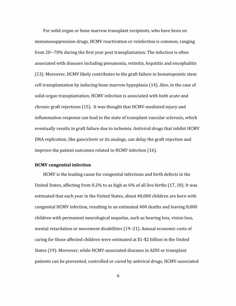
HCMV double-‐stranded linear DNA genome is encased inside of an icosahedral
capsid, which is surrounded by a less structured layer of viral proteins, called
tegument. These components are enclosed in a lipid bilayer envelope that is studded
by a number of viral glycoproteins (Figure. 1-‐2). Mature virion size is about
200~300 nanometer (nm) in diameter.
9
Figure 1-‐2 HCMV virion structure. a) Electron microscopy picture (37). b) Schematic picture (38).
1) Genome
Compared to other human herpesviruses, HCMV contains the biggest genome
of >230Kb (39). Due to high packing density, HCMV genome is estimated to be at a
liquid-‐crystalline state inside of the capsid (40). The coding capacity of HCMV is big
compared to other herpesviruses, with more than 200 predicted open reading
frames (ORFs) (39, 41–43). The genome is composed of the unique long (UL) and
unique short (US) segments, which are flanked by terminal repeated sequences TRL
(ab) and TRS (ca) on one end and internal repeated sequences IRL (b’a’) (lab strain
contains IRL while clinical strain does not) and IRS (a’c’) on the other end, resulting
in a total TRL-‐UL-‐(IRL)-‐IRS-‐US-‐TRS configuration (Figure. 1-‐3). HCMV genes are
named with a prefix based on the segments where they are located and are
numbered sequentially (39), for instance, ORF 74 in UL region is named UL74, and
the protein encoded by this ORF is named pUL74.

10
Figure 1-‐3 Schematic genome organization of HCMV. The letters within each segment depicts the following features: terminal repeat long (TRL), unique long (UL), internal repeat long (IRL), internal repeat short (IRS), unique short (US), and terminal repeat short (TRS) (modified from (41)).
2) Capsid
HCMV capsid is about 130 nm in diameter (44). There are four main component
proteins: major capsid protein (MCP; pUL86), minor capsid protein (mCP; pUL85),
minor capsid binding protein (mC-‐BP; pUL46), and smallest capsid protein (SCP;
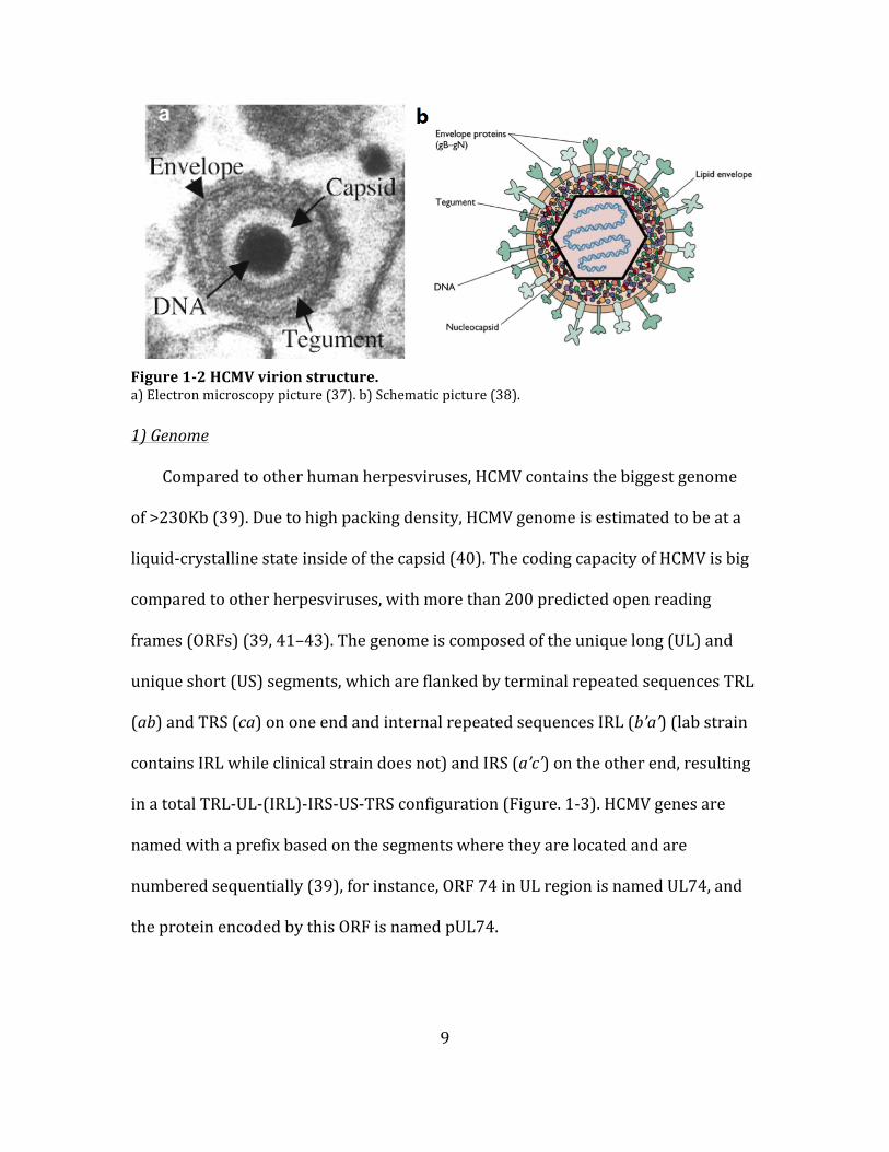
pUL48.5). MCP is the most abundant component in capsid (45–49). The 162
capsomeres (150 hexons plus 12 pentons) are formed by MCP, which are located at
the vertices of the T=16 icosahedral symmetry. Heterotrimers formed by two mCP
and one mC-‐BP comprise of the 320 triplexes located in between the capsomeres
(Figure. 1-‐4) (44, 50–52). SCP decorates each MCP hexon in the mature capsid and
stabilizes the capsid structure through interaction with tegument proteins (53). One
of the penton is shared with or occupied by a portal complex (formed by pUL104),
through which the genome enters or leaves the capsid (54).
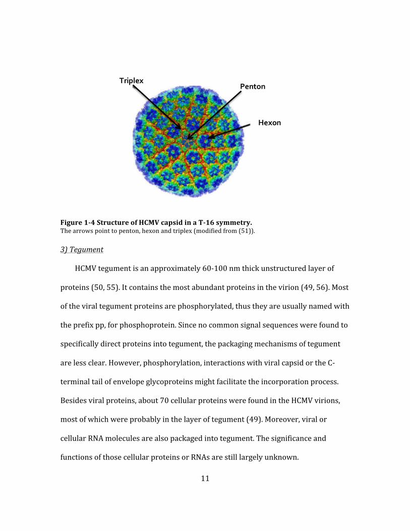
11
Figure 1-‐4 Structure of HCMV capsid in a T-‐16 symmetry. The arrows point to penton, hexon and triplex (modified from (51)).
3) Tegument
HCMV tegument is an approximately 60-‐100 nm thick unstructured layer of
proteins (50, 55). It contains the most abundant proteins in the virion (49, 56). Most
of the viral tegument proteins are phosphorylated, thus they are usually named with
the prefix pp, for phosphoprotein. Since no common signal sequences were found to
specifically direct proteins into tegument, the packaging mechanisms of tegument
are less clear. However, phosphorylation, interactions with viral capsid or the C-‐
terminal tail of envelope glycoproteins might facilitate the incorporation process.
Besides viral proteins, about 70 cellular proteins were found in the HCMV virions,
most of which were probably in the layer of tegument (49). Moreover, viral or
cellular RNA molecules are also packaged into tegument. The significance and
functions of those cellular proteins or RNAs are still largely unknown.
Triplex Penton
Hexon

12
It has been appreciated for a long time that HCMV tegument proteins could
involve in the early stages of the infection, thus they are packaged within the virion
as a “tool box” to ensure their presence upon a new round of infection before the de
novo proteins synthesis. For example, upon entry, viral tegument protein pUL47
facilitate genome-‐containing capsid moving along microtubules to the nuclear pore
through interactions with the capsid protein, and might also help the subsequential
DNA release into the nucleus (57). In addition, viral tegument proteins could also
play a critical role in gene expression. For example, after viral DNA is released into
nucleus, a cellular protein Daxx is recruited to major immediate early promoters to
repress viral gene transcription. Tegument protein pp71, encoded by UL82, gets
inside of the nucleus independently from the capsid and induces the proteasome
degradation of Daxx, thus activating viral immediate-‐early (IE) gene expression (58,
59). If this process occurs successfully, HCMV proceeds into lytic infection, however,
if pp71 gets trapped in the cytoplasm and Daxx represses IE gene expression, latent
infection would be established (56). Furthermore, viral tegument protein also
influences late stages of the infection, e.g. assembly and egress. pp150, encoded by
UL32, likely directs capsid to the assembly compartments for its packaging into the
mature viral particle (60).
Last but not least, viral tegument proteins could modulate host cell antiviral
immune response. pp65, gene product of UL83, is the most abundant protein in the
virion (49). While the gene is totally dispensable for virus replication in tissue
culture, the product of it is very important for virus immune evasion. pp65 achieves
this goal through several mechanisms, including: 1) it mediates the phosphorylation

13
of viral IE proteins to block the recognition of IE proteins by MHC class I molecules
(61); 2) it downregulates the surface presentation of MHC class II molecules by
targeting them to lysosomal degradation (62); and 3) it attenuates the interferon
response (63). Since pp65 plays such an important role in counteracting host
immune response, it is probably not surprising that it is a major antigen target for
cytotoxic T lymphocytes (64).
In conclusion, HCMV viral proteins residing in the tegument layer are involved
in a wide variety of events during viral replication cycle. Thus a better
understanding of their functions will benefit the intervention strategies against
HCMV in the future.
4) Envelope
Herpesvirus acquires its viral envelope through an envelopment-‐
deenvelopment-‐reenvelopment process. Viral capsid acquires the primary envelope
through budding into the inner nuclear membrane, loses that envelope while exiting
the outer nuclear membrane. Once getting into the cytoplasm, viral capsid acquires
the secondary envelope (which is the final envelope) through budding into the
Golgi-‐derived assembly apparatus membrane. HCMV potentially encodes 75
membrane-‐associated proteins, at least 25 of which are found on the envelope (41,
49). Most of the envelope proteins are glycosylated. Thus, they are often named with
a prefix g, for glycoprotein. The functions of most of those glycoproteins are still
elusive, but the hypothesis is that they mainly participate in the initiation of an
infection.

14
Early studies have identified three families of glycoprotein complexes on HCMV
envelope (designated as gC-‐I, gC-‐II, and gC-‐III) (65, 66). The functions and
compositions of those complexes are becoming clearer now: gC-‐I is the
homotrimeric complexes of gB. gB, encoded by UL55, is the fusion protein that
mediates the membrane fusion between viral envelope and cellular membrane (67).
Besides acting as a fusogen, gB is also thought to initiate HCMV absorption onto the
target cell through interactions with cell surface proteins, like heparan sulfate
proteoglycans (HSPGs) (68, 69). gC-‐II is the heterodimeric complex formed by gM
disulfide-‐linked to gN (70). gM/gN, encoded by UL100 and UL73 respectively, are
the most abundant component on the virion envelope (49). gM/gN are essential for
virus replication, and studies suggest that the complex likely participate in both
viral initial attachment and also assembly and egress processes (71, 72). gC-‐III is the
disulfide-‐linked gH/gL/gO complex (73–76). Recently, another gH/gL complex was
identified, gH/gL/UL128-‐131 (UL128-‐131 are small proteins encoded by UL128-‐
131 genes) (77–80). Both gH/gL complexes are very important for HCMV entry. The
characterization of their functions is one of the research focuses of this dissertation,
thus more detail will be described or discussed in the following chapters.
Viral envelope glycoproteins are the main immunogens. Anti-‐gB, anti-‐gM, anti-‐
gN, anti-‐gH, anti-‐gL and anti-‐UL128-‐131 neutralizing antibodies have all been
described (81–85). Among all the glycoproteins listed above, gB, gH/gL, gM and
UL128-‐131 are highly conserved among all strains of HCMV, while gN and gO are
highly variable in amino acid sequences (86–89). The significance of this sequence
diversity is less clear, but might reflect an immune evasion role of these proteins.

15
Besides viral glycoproteins, HCMV envelope also contains numbers of cellular
proteins, such as CD46, CD55, CD59 and annexin II (90–93). CD46, CD55, and CD59
are the host-‐encoded complement regulatory proteins. The incorporation of these
proteins into virion envelope might be a smart way of the virus to prevent
complement-‐mediated lysis (91). Annexin II is a ubiquitous host protein distributed
on cell surface or on the intracellular membranes. It is involved in a lot of cell
processes, including cell mobility, endocytosis, and exocytosis. Annexin II might
enhance virus binding to phospholipid membrane, but it is not found to influence
HCMV entry into fibroblast cells (94, 95). Another possibility is that annexin II, as
part of the virion, might play a role in modulating host cell responses during viral
replication.
HCMV replication cycle
1) Attachment and entry
Virus replication cycle begins by attachment and entry process, which is
initiated by the random collision between virus particle and the target cell, followed
by tethering of viral particle to cell surface and then entering of the virus into the
cell. Studies have shown that HCMV attachment is initiated by low-‐affinity binding of
viral gB and gM/gN with cell surface heparan sulfate proteoglycans (HSPGs) (96,
97), this step helps to concentrate viral particles on cell surface, thus may increase
the chance that viral particles could interact with downstream receptors exhibiting
lower avidity. A subsequent quick switch to a more stable docking occurs by
interaction between gB and the epidermal growth factor receptor (EGFR), platelet

16
derived growth factor receptor (PDGFR-‐alpha) or integrin α2β1, α6β1, αVβ3 (98–
100). Although more recent studies didn’t support their roles as HCMV entry
receptors, the interaction with them probably further facilitates virus attachment
(101, 102).
Depending on the cell type that HCMV encounters, entry could happen directly
at plasma membrane or from within the endosome (Figure. 1-‐5). Regardless of
either entry pathway, the critical event is viral envelope fusion with cellular
membranes, which is mediated by the “core fusion machinery” comprised of gB and
gH/gL. After membrane fusion, HCMV deposits its viral components into the
cytoplasm. Genome-‐containing capsid, facilitated by some tegument proteins, is
transported to nuclear pores along microtubules and DNA genome is released into
nucleus afterwards (Figure. 1-‐5). At the same time, other tegument proteins diffuse
to nucleus independently or stay in different subcellular locations to either regulate
viral gene expression or inhibit host cell anti-‐viral immune response.

17
Figure 1-‐5 Schematic of HCMV replication cycle. (Modified from (103)). Detailed description of the diagram is given in the text.
2) Gene expression
The expression of HCMV viral proteins is finely regulated, and falls into a
cascade of events, which are divided into three main temporal classes: immediate
early (IE), delayed early (DE) and late (L) (Figure. 1-‐5). IE genes are immediately
expressed following nuclear entry, in absence of any de novo protein synthesis.
Their expression can be detected within a few hours in HCMV infected cells (104). IE
genes are transcribed by RNA polymerase II of the host cell under the control of
major immediate early promoter (MIEP). The most abundant IE proteins are IE1-‐72
kDa (pUL123), and IE2-‐86 kDa (pUL122) (39, 104). IE1-‐72 kDa is necessary for
efficient viral replication after low MOI infection (105). IE2-‐86 kDa is critical for
viral DE genes expression and it can negatively regulate the transcription of IE1 and
IE2 genes (106, 107). Together they act as trans-‐activators to stimulate the
transcription of various viral and host genes, which are important for the efficiency
18
of viral replication (108). The progression of IE gene expression is very important in
deciding virus fate of productive infection versus latent infection.
Expression of DE genes depends on IE gene expression. The products of DE
genes are often directly involved in viral DNA replication (e.g., DNA polymerase) or
priming the cellular environment ready for viral DNA replication (e.g., up-‐regulation
of cellular genes exhibited in S-‐phase). And the kinetics of DE gene expression varies
from as early as after IE gene expression to as late as before viral DNA synthesis.
Expression of L genes is largely dependent on viral DNA replication. There are
two groups of L genes: leaky-‐late and true-‐late genes. Leaky-‐late genes are
expressed at very low level at early times and the levels are tremendously increased
at late times. True-‐late genes are exclusively after, and dependent on viral DNA
replication. L genes are expressed about 24-‐48 hours after infection, the products of
which are often structure proteins that are required for viral assembly and egress.
3) Gene replication
Soon after deposited into the nucleus, HCMV linear DNA genome is rapidly
circularized and serves as the template for transcription and replication. The
initiation position of DNA replication resides at a defined region on the genome, the
so-‐called lytic origins of replication, oriLyt. The oriLyt for HCMV is identified in the
middle of the UL segment, upstream of the gene (UL57) that encodes the single-‐
stranded DNA binding protein (109, 110). Approximately at 16 hours post infection,
DNA synthesis takes place in the vicinity of oriLyt as soon as essential viral proteins
are present. The core DNA replication proteins include helicase-‐primase complex
(formed by pUL105, pUL70 and pUL102), DNA polymerase (pUL54), polymerase

19
accessory protein (pUL44) and single-‐stranded DNA binding protein (pUL57).
Another protein, gene product of UL84, is also essential. pUL84 is thought to trans-‐
activate a responsive promoter within the oriLyt through interaction with IE2-‐
86KDa, the process of which might partly initiate DNA replication (111). Moreover,
study also showed that pUL84 has UTPase activity, which may help generate energy
needed for helicase activity in the initiation of DNA replication (112).
The initiation of DNA replication for alpha-‐herpesviruses is thought to involve
an origin-‐binding protein (OBP), which binds to specific origin sequences to provide
the initial unwinding spot with its intrinsic helicase activity. Since HCMV lacks the
homologue to OBP, the mechanism of initiation is less clear. One interesting model is
that RNA-‐DNA hybrid formed during transcription may yield an open region that
could be used as the initial spot. This hypothesis is in concert with the trans-‐
activator property of the replication essential protein pUL84. An alternative but not
mutually exclusive possibility is that HCMV could make use of cellular initiation
machinery.
While alpha-‐herpesvirus DNA replication is believed to begin with a
bidirectional theta-‐like mechanism, followed by a unidirectional rolling-‐circle
mechanism at later stage (1), HCMV likely exclusively relies on the rolling-‐circle
model, producing several thousands copies of concatemeric genomic DNA per cell
(1).

20
4) Assembly and egress
a) Assembly and egress in nucleus
The first step in HCMV assembly and egress involves capsid assembly and
genome packaging in the nucleus (Figure. 1-‐5). Capsid assembly initiates with the
formation of a scaffold that could “crystalize” the capsid. There are two scaffolding
proteins identified for HCMV, pUL80 and pUL80.5 (113, 114). Both proteins can
interact with MCP through carboxyl terminus and lead to the translocation of MCP
from cytoplasm to nucleus. The amino terminus of pUL80 and pUL80.5 promotes
self-‐interactions, resulting in the generation of MCP multimers and formation of
hexons and pentons (115, 116). At the same time, mCP and mc-‐BP translocate into
nucleus to form triplexes. Hexons and pentons, together with triplexes, form an
immature capsid shell. Next step is genome packaging. Two conserved sequence
motifs, pac-‐1 and pac-‐2, are located at each end of the concatemeric HCMV DNA
sequence. Viral terminase complex comprised of pUL56 and pUL89 recognizes pac-‐1
and pac-‐2 and cleaves DNA into genome-‐length units (117, 118). Before DNA
packaging, scaffolding protein pUL80 and pUL80.5 is disassociated from MCP by a
protelytic cleavage at the carboxyl terminus. Then, insertion of DNA genome into
capsid occurs through the portal formed by pUL104, coupled with the extrusion of
the scaffolding protein remainders. It is worth noting that capsid formation and
DNA packaging do not necessarily accompany each other. In fact, there are three
types of viral capsid that could form: 1) A-‐capsid is an empty capsid lacking genome,
which is a result of failure in DNA packaging; 2) B-‐capsid is an immature, scaffold
containing capsids that lacks genome, probably due to unsuccessful proteolytic

21
digestion of scaffolding proteins; and 3) C-‐capsid is the mature, genome-‐containing
capsid. Only C capsid will generate infectious viral particles. Although A-‐, B-‐capsids
are not infectious, they might affect viral replication cycle somehow.
Nucleocapsid will then go through a process known as nuclear egress or
primary envelopment. Nuclear lamina, a network of proteins underlying the
nucleoplasmic side of the inner nuclear membrane (INM) is the first obstacle.
Disruption of the nuclear lamina is essential to gain access to the inner nuclear
membrane (INM). HCMV cleverly finds a way to achieve that by mimicking the
phosphorylation-‐mediated depolymerizaion of nuclear lamina occurring during
cellular mitosis. Virus encodes a nuclear egress complex (NEC), comprised of pUL50
and pUL53. pUL50 is a INM protein while pUL53 is a nucleoplasmic protein, which is
brought to INM by binding with pUL50 (42, 119). HCMV NEC recruits cellular kinase
PKC, as well as a viral kinase pUL97, to phosphorylate and dissolve nuclear lamina
(120–122). The disruption of nuclear lamina allows the nucleocapsid budding into
INM, resulting in an enveloped nucleocapsid in the lumen of nuclear membrane. A
subsequent deenvelopment process occurs by fusion with the outer nuclear
membrane (ONM) and leads to the delivery of the nucleocapsid into cytoplasm. It
has been suggested that there is a quality control during nuclear egress to
preferentially allow the genome-‐containing capsid budding through INM (123, 124).
To support this hypothesis, C-‐capsid is found to be the predominant type of capsid
present in the cytoplasm. Some tegument proteins are acquired during primary
envelopment,, probably due to interactions with capsid proteins.

22
b) Assembly and egress in cytoplasm
Nucleocapsid acquires its final tegument and envelope by budding through the
cytoplasmic assembly compartment (AC), the process of which is also called
secondary envelopment (Figure. 1-‐5). AC is a juxtanuclear body formed by
extensively reorganization of cellular secretory organelles (e.g., endoplasmic
reticulum (ER), Golgi, trans-‐Golgi and endosomes). The formation of AC is a unique
characteristic of HCMV infected cells during the late stage of infection. It is not well
understood how nucleocapsid is directed to AC after nuclear egress, but tegument
proteins are suggested to play an important role in it. Moreover, the putative
intimate connection between nucleus and AC, achieved by HCMV-‐mediated
remodeling of nucleus shape and formation of membranous channel on the nucleus-‐
AC interface, might also facilitate nucleocapsid trafficking to AC (125). Some of the
tegument proteins and almost all envelope glycoproteins are located in AC, which
are finally incorporated into mature viral particle when nucleocapsid buds into the
endosomal and/or trans-‐Golgi derived membranes. Enveloped viral particle is
secreted to the extracellular environment by cellular exocytic-‐associated pathways.
Together with mature viral particle, infected cells also produce noninfectious
enveloped particles (NIEPs) and dense bodies (DBs) (126). NIEPs are viral particles
that lack genome (i.e., formed by A-‐ and B-‐ capsid). DBs are viral particles that
contain tegument proteins (mainly pp65) instead of capsid (therefore also lacking
genome), thus they are noninfectious as well. The relative proportions of NIEPs and
DBs to infectious virus particles may vary due to differences in genetic background
of the virus and/or in cell types where progenies are produced. Although being

23
replication defective, it is possible that NIEPs and DBs could act like “decoy” to wear
out host immune response (e.g., the humoral antibody attack), thus giving infectious
particles higher chance to survive and establish infection in a new cell.
HCMV latent infection
HCMV infection of a host for the first time is considered as the “primary
infection”. Primary infection generally has two outcomes: 1) the virus enters the
lytic replication cycle, where viral gene expression and replication successfully
proceed, leading to production and release of infectious progeny virions; 2)
Alternatively, under some conditions or in certain cell types, the virus enters a latent
infection state, where the viral gene expression is largely limited and progeny
production is shut off. The current consensus is that HCMV is never cleared away
after primary infection and it coexists with the host through latency for a lifetime,
with reactivation happening periodically. While shedding of the virions from body
fluids peaks at a few months after primary infection, persistent virus shedding at
low levels could be detected for years, and intermittently virus shedding due to
reactivation continues for the rest of the life, all of which could contribute to the
horizontal transmission of the virus. Surprisingly, a large portion (up to 40%) of an
individual’s T cell repertoire is directed against epitopes on HCMV viral proteins
that are expressed at all stages of replication cycle (64), suggesting a continual
exposure of host immunity to HCMV antigen. This observation implies there is a
relatively huge immune burden added from HCMV due to persistent infection
and/or frequent reactivation from latency.

24
Latency is a common feature of herpesvirus family. While alpha-‐herpesviruses
and gamma-‐herpesviruses establish latency mainly in neurons and lymphocytes,
respectively, beta-‐herpesviruses establish latency in myeloid lineage cells (Table. 1-‐
2). CD34+ myeloid progenitor cells and CD14+ monocytes are the main latency sites
for HCMV (127–131). The prevailing view for establishment of latency and
triggering of reactivation is that HCMV major immediate early promoter (MIEP) is
silenced by cellular transcriptional repressor in those myeloid cells, and the
repression of MIEP hampers the IE gene expression thus prevents lytic infection,
leading to the establishment of latency. During latency, HCMV genome exists in the
nucleus like an episome, coupled with limited transcription of specific viral genes,
which are thought to be important for latency maintenance. Following
differentiation of myeloid cells into macrophages or dendritic cells, the cellular
environment changes in a way that the repression on MIEP is relieved, which
induces IE gene expression and reactivation of the virus (reviewed in (132, 133)).

25
Table 1-‐2. Comparison of host range, cell tropism and site of latency among human herpesviruses.
Host range Cell tropism Site of latency
Alpha-‐
HSV-‐1 (HHV-‐1)
Human (In lab: mice, rabbits, guinea pigs, zebrafish)
Epithelial cells; Neurons
Neurons (Sensory ganglia, mainly in head and neck)
HSV-‐2 (HHV-‐2)
Human (In lab: mice, rabbits, guinea pigs, zebrafish)
Epithelial cells; Neurons
Neurons (Sensory ganglia)
VZV (HHV-‐3)
Human Epithelial cells; Neurons; Lymphocytes (T cells)
Neurons (Sensory ganglia, all over the body)
Beta-‐
HCMV (HHV-‐5)
Human All cell types Monocyte lineage
HHV6A/6B
Human Lymphocytes (T cells); Neurons Monocyte lineage
HHV-‐7
Human Lymphocytes (T cells) Monocyte lineage
Gamma-‐
EBV (HHV-‐4)
Human Epithelial cells; Lymphocytes (B cells)
Lymphocytes (Memory B cells)
KSHV (HHV-‐8)
Human Lymphocytes (B cells and T cells); Monocytes; Endothelial cells
Lymphocytes (T cells)
HCMV genetic diversity
When considering about HCMV genetic diversity, cautions need to be taken with
some important terminologies. An “isolate” refers to HCMV recovered from a human
specimen and passaged in tissue culture for a limited number of times. Generally an
isolate is not plaque purified, suggesting that several genomically different viruses
might present in one isolate. A “strain” refers to a passaged derivative of an isolate

26
acquired by plaque purification. Strains of HCMV differ in the pattern of viral DNA
fragments generated by restriction enzyme digestion, which is later confirmed with
genome-‐wide sequencing techniques. A “variant” refers to a derivative of a strain
that expresses a phenotypic or genotypic characteristic different from the parental
strain (134).
About third-‐quarter of HCMV genome shows polymorphisms, while one-‐quarter
is highly conserved. The most conserved regions of the genome are localized to
genes encoding DNA-‐processing enzymes, capsid, and tegument proteins, while the
variable regions are genes encoding envelope glycoproteins and immune evasion
proteins (135, 136). Several variable regions of the genome have been selected to
define distinct genotype based on the clustering of polymorphisms, including genes
that encode for glycoprotein gB (5 genotypes), gH (2 genotypes), gN (7 genotypes),
and gO (8 genotypes) (87, 89, 137, 138).
It has been appreciated for decades that HCMV exhibits significant levels of
genetic diversity between individuals (inter-‐host diversity) (139–141). However,
more recent research work has showed that this virus also exhibited high level of
genetic diversity within a single individual (intra-‐host diversity) (reviewed in (142)).
Quite surprisingly, this intra-‐host diversity could be comparable to what has been
observed for RNA viruses, a benchmark of highly diverse viral populations (7). Then
the intriguing question is what source contributes to the high levels of HCMV intra-‐
host diversity. Currently, there is no clear answer to explain the observed diversity,
though several hypotheses could be proposed: 1) High levels of replication during
primary infection could contribute to the generation of de novo mutations in each

27
host, albeit the total numbers of mutations may be limited by the proof reading of
HCMV DNA polymerase. 2) Reinfections may offer a way for introducing diversity
into virus intra-‐host population. Indeed, mixed infection of multiple strains of HCMV
happens in a wide variety of hosts, including immunocompetent, immunocomprised
and congenital infected individuals (2–7). 3) Recombination and natural selections
within a host could also alter the pattern of HCMV intra-‐host diversity.
The high levels of HCMV intra-‐host diversity probably would help us rethink the
evolution of the virus within a host. Given the high level of genetic diversity, HCMV
should be considered more as a “swarm”-‐heterogeneous population. In line with
this notion, recent work also showed that different genotypes were detected from
different compartments within the body (e.g., plasma and urine) (143), suggesting
that during virus dissemination to distal compartments, the viral population can
rapidly evolve, either due to natural selection or stochastic mutations, and can lead
to populations of virus colonized in distal organs or tissues genetically different
from the ones present in peripheral blood. Meanwhile, research also suggests that
HCMV is genetically stable over time, as shown by that while mixed genotypes were
detected in patients, the compositions of the mixed populations remains nearly
constant over time (144, 145). Thus, HCMV can be diverse but yet stable within its
host. The intra-‐host diversity may provide a mechanism for the virus to gain overall
fitness by rapidly adapting to the changing environment, like new host organ/tissue,
immune surveillance, and antiviral therapy. However, once settled down, the virus
population tends to remain stable probably due to its low mutation rate.

28
HCMV cell tropism and entry
HCMV in vivo cell tropism and viral dissemination
One of the fundamental questions in virology has been that why a given virus
will particularly infect one host species but not the other, or at a cellular level, why a
given virus will specifically infect one cell type while failing to infect others. The
above-‐mentioned phenomena have been referred to as viral “host range” or “cell
tropism”. Natural infections with most herpesviruses are restricted to a single
species, although some can infect other species experimentally. In general, alpha-‐
herpesvirues have a relatively broader host range (excepting VZV) but restricted
cell tropism. Beta-‐herpesviruses have a restricted host range but a wide cell tropism.
Gamma-‐herpesviruses have both restricted host range and cell tropism (reviewed in
(146)) (Table. 1-‐2). HCMV, a human-‐specific virus, is able to virtually infect all cell
types in human body, including endothelial cells, epithelial cells, fibroblasts, smooth
muscle cells, placental trophoblasts, hepatocyte, polymorphonuclear leukocyte
(PMNL), monocyte, dendritic cells, macrophages, lymphocyte and neuronal cells
(147–157). HCMV infection in PMNL, monocyte and lymphocyte is blocked at the IE
gene expression stage, thus those cells are considered to be susceptible but not fully
permissive to HCMV infection. It is worthwhile to point out that monocyte will turn
into full permissiveness to HCMV when it is differentiated into macrophages or
dendritic cells (154, 158). With this extremely wide range of cell tropism in vivo,
HCMV can acquire systemic dissemination and cause pathology in multiple organs,
e.g., lung, kidney, liver, spleen, intestine, heart, brain and bone marrow (159).
Several cell types, of significant roles in viral dissemination, will be described below.

29
Epithelial cell
Epithelial cell, lining the major cavities of the body, could be the first cell that
HCMV infects if the transmission route is through oral or genital contact. It is the
main target of HCMV in lung, gastrointestinal tract, kidney, and secretory glands
(147, 160). Since epithelial cell is permissive for HCMV infection, it mainly involves
in virus shedding into body fluids. In addition, infected epithelial cells have been
reported to detach from the basal membrane, thus might contribute to infectivity in
body fluids as well (159).
Endothelial cell
Endothelial cell, lining the inner wall of blood and lymphatic vessel, could be the
first cell that HCMV infects if transmission happens by blood transfusion. This cell
type is believed to be involved in HCMV haematogenous dissemination for the
following two reasons: 1) endothelial cell is permissive for HCMV infection and 2)
infected endothelial cells may detach from vessel wall and circulate in peripheral
blood (160, 161). Transmission of the virus from infected endothelial cells to
leukocytes has been reported in tissue culture model (162). Moreover, evidence also
suggests that endothelial cell can also be a site for HCMV latent infection (reviewed
in (163)).
Monocyte/Macrophage
Monocyte is also found to be the key contributor for HCMV haematogenous
dissemination. Although complete replication cycle is blocked, HCMV could promote
monocyte migration into organ tissues and ensuing its differentiation into the
permissive macrophage cell. Infected macrophage could then migrate into bone

30
marrow and infect myeloid progenitor cells, where latency is usually established
(164).
Taken together, a putative model for HCMV in vivo dissemination is that
epithelial cells or endothelial cells are first infected by HCMV at mucosal epithelium
or in the blood vessel. HCMV then replicates and spreads to monocytes in the
peripheral blood by some unresolved mechanisms, where HCMV enters a temporary
latent state. Infection by HCMV increases monocyte migration into different organs,
and differentiation into permissive macrophages. Infected macrophages could either
enter bone marrow to establish latency in CD34+ myeloid progenitor cells or keep
migrating and infect mucosal epithelial cells at a different site, where virus shedding
and transmission take place.
HCMV in vitro cell tropism and associated adaptive genetic changes
Historically, HCMV in vitro isolation and propagation has been heavily
dependent on primary human fibroblast cultures (e.g., primary human foreskin
fibroblasts, HFF) because those cells supported high titer replications of HCMV.
Based on the extent of passage on fibroblast cultures, strains of HCMV were
conventionally divided into two categories: “lab strain” (high-‐passaged) and “clinical
strain” (low-‐passaged). Two prototypic lab strains are AD169 and Towne. Strain
AD169, isolated from the adenoid culture of a 7-‐year-‐old girl, was developed as an
attenuated vaccine candidate by extensively propagation on fibroblasts and was
then used by laboratories worldwide for over 50 years (165); Strain Towne, isolated
from the urine of a congenitally infected infant, was also developed as an attenuated

31
vaccine candidate by passaging on fibroblasts for over 128 times (166). In contrast,
strains like Toledo, isolated from the urine of a congenitally infected child (167), and
TR, an ocular isolate from an AIDS patient with retinitis (168) are considered
clinical strains due to their more limited propagations on fibroblast cells.
Lab strains and clinical strains exhibit different competencies in tropism. Lab
strains are able to replicate on fibroblasts with high efficiency, but show restricted
tropisms by failing to infect epithelial cells, endothelial cell, and some types of
leukocytes (134, 169, 170). Clinical strains show broader cell tropism and replicate
in cells types in addition to fibroblast cells (171), although unexpectedly, the clinical
strain Toledo was later found to be negative in endothelial cells tropism (170, 172).
Accordingly, the tissue culture tropism model for HCMV involves fibroblasts versus
endothelial/epithelial cells, and other cell types in the body tend to fall into this
dichotomy.
Initial genome comparative studies of lab strains AD169, Towne, and clinical
strain Toledo found that both lab strains A169 and Towne had suffered dramatic
genetic deletions, missing 15, and 13 Kb sequence from the right end of the UL
region respectively (173). Perhaps in order to keep intact genome length, both
AD169 and Towne also exhibit genetic rearrangement by replacing the missing
region with an inverted version (b’a’ or also called IRL) of the terminal repeated
block (ab or also called TRL), which is otherwise lacking in clinical strains (Figure. 1-‐
3) (41). Thus the missing region is designated the UL/b’, which is predicted to
encode 19 ORFs (UL133-‐151) or 14 ORFs (UL133-‐146) in AD169 or Towne
respectively and many of those ORFs are predicted to be glycoproteins (41, 173).

32
Some of the ORFs missing in lab strains are displayed in an inverted orientation in
clinical strain Toledo as compared to other clinical strains (41, 134). UL128-‐131
gene locus on the UL/b’ was later shown to be indispensable for virus infection in
epithelial/endothelial cells and transmission to leukocytes (78). This was confirmed
by genome-‐wide sequencing showing that AD169 contains a single nucleotide
insertion in UL131; Towne contains a frameshift mutation in UL130, and Toledo
contains an inverted site disrupting UL128 (41, 78, 174, 175). Therefore the so-‐
called clinical strain Toledo behaves more like lab strain with respect to the
mutations acquired in the UL/b’.
Recently, Dargan et al. demonstrated that genetic changes inevitably occurred
when clinical viruses were serially passaged in tissue cultures, including epithelial
and endothelial cells (176). These authors passaged HCMV on fibroblasts, epithelial
and endothelial cells in parallel and showed that in all three cultures, mutations
were first selected in gene RL13, and then in genes UL128-‐131 (specifically
associated with fibroblasts as described above for AD169, Towne and Toledo), and
in some cases, eventually in UL/b’ region (as what happened to AD169 and Towne).
RL13 encodes a glycoprotein that tracks to the virion assembly site, suggesting it
might be a virion envelope protein (177). Although the function of the RL13 protein
is currently unknown, the rapid negative selection against it suggests that it is likely
a potent inhibitor for HCMV in vitro replication and the loss of function in RL13 may
also contribute, at least in part, to the less cell-‐associated phenotype observed in lab
strains (177–180).

33
The overall picture is that HCMV strains that have been propagated in vitro tend
to harbor mutation(s) resulted from selection pressure in all cell culture systems.
And the appearance of the mutants is not only inevitable but also prone to emerge at
early stages of the passage (43, 176). Therefore, even the less propagated “clinical
strain” could contain mutations that we are not aware of, thus might not truly
represent its clinical ancestor. Recently Wilkinson et al. suggested a more adequate
terminology system, where HCMV strains are divided into “low-‐passage” and “high-‐
passage” groups, with low-‐passage strains being more genome intact than high-‐
passage strains (181).
Bacterial Artificial Chromosome (BAC) (E. coli. fertility plasmid) cloning
technology was introduced to preserve HCMV genome from further mutations and
also allowed for intentional mutagenesis manipulation of the virus genome (182,
183). Virus can be recovered from transfection of permissive cells with BAC. High-‐
passage strains AD169 and Towne as well as low-‐passage strains TR, TB40/E,
Merlin (ME), PH all have been cloned into BACs (41, 177, 184), and are used for
research in this dissertation.
HCMV entry and membrane fusion event
Viral tropism can be determined at any replication step, beginning with the
entry into cells and ending with the production of progeny. It has been well
appreciated that viral tropism can be greatly influenced by virus entry. Different cell
types vary a lot in their physical morphologies as well as surface protein,
carbohydrate and lipid compositions. Thus virus entry is largely governed by how

34
the virus particle overcomes the barriers on cell surface and accomplishes
interactions with their receptors. The broad cell tropism of HCMV reflects its
flexibility at entry, suggesting that the virus might either bind to a ubiquitous
receptor shared with all cell types or a number of specific ones on different cell
types. On the other hand, HCMV broad tropism correlates well with its envelope
complexity. This virus is remarkable because it contains more than 20 different
kinds of glycoproteins on the envelope. It’s almost like HCMV has a chain of “keys” to
readily open different “locks”.
As an enveloped virus, entry has to happen through membrane fusion event
between viral envelope and cellular membranes. Depending on the cell types that
the virus encounters, the site of fusion can be at either the plasma membrane or
from within the endosomes. Moreover, the types of endosomes can vary depending
on what kind of endocytosis that the virus triggers, thus the requirement for low-‐pH
can vary as well. For HCMV, it has been reported that the virus enters fibroblasts
through pH-‐independent fusion at the plasma membrane. Although more recent
research suggested that the virus utilizes macropinocytosis pathway to enter
fibroblasts in a pH-‐independent manner. Meanwhile, entry into
epithelial/endothelial cells requires endocytosis and depends on low-‐pH (79, 185,
186).
Although thermodynamically favorable, fusion of two membranes must
overcome a repulsive “hydration force”, which increases steeply as the two
membranes come closer than 20 angstrom (187). Because of that, membrane fusion
requires a source of free energy to bring them together, for example, a chemical

35
called polyethylene glycol (PEG) could induce membrane fusion by dehydration of
the inter-‐membrane-‐space to overcome the repulsive “hydration force” and bring
apposing membranes closer than 20 angstrom. Essentially, enveloped viruses
encode a “fusogen” to achieve this end, which is defined as viral fusion protein. Viral
fusion protein is held in a “prefusion conformation” on the virion envelope by
constraints that either come from another part of the same protein or from another
viral protein. Usually two events have to happen before fusion protein
conformational change: 1) “priming” of fusion protein to make the transition
possible, e.g., proteolytic cleavage; 2) “triggering” of fusion protein to initiate the
transition, e.g., ligand binding. The ligand can be a receptor (e.g., receptor-‐mediated
fusion of HIV virus) or a proton (e.g., low-‐pH induced fusion of influenza virus).
Upon the triggering, the constraints that hold the fusion protein in the “prefusion
conformation” is released, leading to a dramatic conformational change and
exposure of the hydrophobic “fusion peptide” or “fusion loop”, which then inserts
into the apposing cellular membrane. This state of the fusion protein is “metastable”,
and favors to fold back to the more energetic stable “postfusion conformation”. Thus,
the collapse of the intermediate conformation draws two membranes together
(review in (188)).
Fusion proteins fall into three categories. (1) Class I fusion proteins,
represented by influenza virus hemagglutinin (HA) or HIV-‐1 gp160, are mainly
composed of alpha-‐helices and contain a “fusion peptide” located closed to the N-‐
terminus. The postfusion structure of it is a trimeric alpha-‐helical coiled coil. (2)
Class II fusion proteins, represented by flavivirus envelope protein E, are mostly

36
comprised of beta-‐sheets. Unlike class I fusion proteins being perpendicular to viral
envelope, class II fusion proteins form a parallel lattice on the envelope. Upon
triggering, the proteins flip up to insert internal “fusion loop” to cellular membrane.
(3) Class III fusion proteins, represented by vesicular stomatitis virus glycoprotein G
(VSV-‐G), combine the feature of both class I and class II. They contain a central
trimeric coiled coil domain just like class I fusion proteins in their postfusion state,
but other domains are mostly made up by beta-‐sheets, and the “fusion loop” is
buried internally, typical feature of class II fusion proteins (reviewed in (188)).
HCMV core fusion machinery and accessory glycoproteins
All herpesviruses share conserved core fusion machinery comprised of gB and
gH/gL complexes (146, 189, 190). All three glycoproteins are essential for virus
entry, for example, HCMV gB-‐null, gH-‐null and gL-‐null mutants all fail to infect cells
unless treated with the chemical fusogen PEG, suggesting that the defect is at fusion
(191–193).
HCMV gB (pUL55) is a 160~170 kDa transmembrane protein. It contains a
cleavage motif R-‐X-‐K/R-‐R recognized by intracellular endoprotease furin and is
cleaved into a 116 kDa N-‐terminal fragment and a 55 kDa C-‐terminal fragment,
which are hold together by disulfide bonds. The proteolytic cleavage probably
occurs in the TGN-‐derived assembly compartment during virus maturation, thus gB
incorporated into virion envelope is a disulfide-‐linked complex of the two fragments
(194, 195). Several gB homologues across the three herpesvirus families share this
cleavage feature, including gB of pseudorabies virus (PRV), VZV, EBV, KSHV, and

37
other betaherpesviruses (e.g., HHV-‐6 and HHV-‐7) (196–201). In contrast, gB of some
members of herpesviruses do not harbor the cleavage site, including HSV-‐1 and
HSV-‐2 (202, 203). The influence of gB cleavage on virus replication has been
variable depending on different viruses. There are reports showing that for HCMV,
PRV and VZV, the cleavage site of gB is dispensable for virus growth in tissue culture,
while for EBV, the proteolytic cleavage seems to be required for gB function in cell-‐
cell fusion (197, 204–206). The fact that cleavage of gB in HCMV is not necessarily
needed during replication suggests that this furin cleavage is not required for the
activation of fusion potential, as in the case of influenza virus HA or HIV gp160.
Moreover, posttranslational glycosylation adds both N-‐linked and O-‐linked
polysaccharides to gB, contributing to one third of the total molecular weight (122,
207, 208). Recent research found that antigenic sites that elicit neutralizing
antibodies are more glycosylated than those eliciting non-‐neutralizing antibodies,
suggesting that the glycans might help shielding the neutralizing epitopes on gB
(67). Crystal structure of HCMV gB ectodomain has been resolved and has revealed
structural homology to HSV-‐1 and EBV gB, all of which resemble the structure of
class III fusion protein VSV-‐G (67, 209, 210). However, unlike VSV-‐G being necessary
and sufficient in mediating membrane fusion, herpesvirus gB is not sufficient on its
own and it needs a partner gH/gL.
HCMV gH (pUL75) is a 97-‐kDa transmembrane protein, and gL (pUL115) is a
37-‐kDa soluble protein. Together they form a disulfide-‐linked heterodimer (211,
212). Crystal structures have revealed the overall similar but slightly different
structures between HSV and EBV gH/gL. HSV gH/gL adopts a boot-‐like

38
configuration whereas EBV gH/gL is more linear and elongated. In both cases,
gH/gL heterodimer is intimately integrated with each other, with gL binding to the
N-‐terminal of gH, suggesting the two proteins require one another for proper
folding (213, 214). Although HCMV gH/gL crystal structure has not been resolved
yet, cryo-‐EM analysis suggested a more similar structure to HSV gH/gL (215). HCMV
gB and gH/gL together are necessary and sufficient to mediate cell-‐cell fusion in a
transient expression system (216), with gH/gL provides an essential regulatory role
for gB’s fusion function. However, the mechanisms by which gH/gL regulates gB-‐
fusion are still largely unknown.
Besides the core fusion machinery, each herpesvirus employs accessory
proteins to regulate the action of the gB and gH/gL. Accessory proteins usually
interact with gH/gL transiently or stably, and influence viral tropism through
receptor binding. HSV encodes gD to bind to cell surface receptors like herpesvirus
entry mediator (HVEM), nectin, or heparan sulfate (217). Upon receptor binding, gD
experiences conformational changes, which leads to transient interaction with
gH/gL, followed by gB-‐mediated fusion (218–220). EBV encodes gp42, which forms
a trimeric complex with gH/gL. Thus EBV gH/gL exists in two forms, gH/gL/gp42
or unbound gH/gL. gH/gL/gp42 promotes virus entry into B cells through
interaction between gp42 and MHC II molecules on B cell surface. gH/gL promotes
virus entry into epithelial cells through interaction between gH/gL and specific
integrins on epithelial cell surface. Binding of gp42 blocks gH/gL interaction with
integrins, thus gp42 acts like a tropism switch adaptor (74, 221–224). HCMV gH/gL
can be bound by gO or the small set of proteins UL128-‐131, forming gH/gL/gO or

39
gH/gL/UL128-‐131 complex. Both complexes could influence HCMV entry and
tropism.
HCMV gH/gL/UL128-‐131 and gH/gL/gO complexes in entry and tropism
gH/gL/UL128-‐131
The gene locus of UL128, UL130, and UL131 (collectively designated the UL128
locus, UL128L) encodes three small soluble proteins UL128 (15 kDa), UL130 (35
kDa), and UL131 (18 kDa). It has been shown that an upstream promoter drives a
long transcript that expresses UL131, UL130 and UL128, at the same time, there
might be a UL128 specific promoter overlapping the UL131-‐UL130 region to
independently express UL128 (78, 175). Both UL130 and UL131 are modified by N-‐
linked glycosylations, while UL128 are not (80, 225, 226). UL128 and UL130 have
also been predicted to contain chemokine domains. UL128 potentially has a CC
chemokine domain near the N terminus, while UL130 might contain a central CXC
chemokine domain (78, 175, 226). Whether those chemokine domains functions in
vivo has not been convincingly determined yet.
UL128 is covalently bound to gH/gL through disulfide linkage to gL, while
UL130/UL131 are disulfide linked to each other but are noncovalently bound to
gH/gL (Figure. 1-‐6). Together UL128-‐131 proteins bind to gH/gL, forming a
pentameric gH/gL/UL128-‐131 complex. Incomplete complexes lacking any of the
UL128-‐131 proteins would not be transported to assembly compartment (AC) to be
incorporated into the mature virion envelope (77–80, 215, 225, 227). UL128-‐131
sequences from different HCMV strains are highly conserved (>92.3% at nucleotide

40
level and >91.1% at amino acid level) (11, 228), suggesting the pentameric complex
likely provides an important function for HCMV replication.
The consensus now is that gH/gL/UL128-‐131 is necessary for HCMV entry into
endothelial/epithelial cells, but is dispensable on fibroblasts. Several lines of
evidences support this conclusion: (1) Natural selection on fibroblasts leads to
spontaneous loss-‐of-‐function mutations within UL128L. Historically, HCMV was
mainly propagated on fibroblasts, and as a result, the high-‐passage viruses were
associated with restricted tropism on epithelial and endothelial cells. The high-‐
passage strains AD169 and Towne were found to contain a single nucleotide
insertion in UL131, and a frameshift mutation in UL130 respectively. Relatively less
passaged strain Toledo contains an inverted UL/b’ region, with the site of inversion
within UL128 (41, 43, 78, 173–175). More recently Dargan et al. took isolates of
HCMV from clinical samples and intentionally passaged them on various tissue
cultures, and found that mutations in UL128L inevitably happened and were
exclusively limited to fibroblasts (176). (2) Reversions of the natural
epithelial/endothelial loss-‐of–tropism mutants are all associated with reversal in
UL128L. Similarly, when UL131 is intentionally repaired, AD169 recovers infectivity
on endothelial/epithelial cells. (3) Introductions of targeted deletions into UL128L
result in loss of endothelial/epithelial tropism. (4) Trans-‐complementation with
individual UL128, UL130, and UL131 could also partially rescue
endothelial/epithelial tropism (41, 77–79, 225). Furthermore, antibodies against
UL128, UL130, or UL131 efficiently blocks virus entry into endothelial/epithelial

41
cells, but not fibroblasts (77), confirming that gH/gL/UL128-‐131 is required for
HCMV entry into endothelial/epithelial cells but not fibroblasts.
Trans-‐expression of gH, gL, UL128-‐131 renders epithelial cell resistant to HCMV
infection, indicating gH/gL/UL128-‐131 facilitates HCMV entry into epithelial cells
through interactions with a specific, yet unknown, receptor (229). This putative
receptor interaction likely triggers a signaling cascade (e.g., Src/paxillin pathway),
which leads to internalization of viral particle through endocytosis (230).
Subsequent virus entry happens by fusion from within the endosome in a low-‐pH
dependent manner (79). Interestingly, viruses lacking gH/gL/UL128-‐131 can still be
efficiently internalized into endothelial/epithelial cells, although productive
infection is not observed (79, 231), suggesting that subsequent fusion from within
the endosome is not allowed. This implies two possibilities: 1) gH/gL/UL128-‐131 is
also required for the subsequent fusion event, or 2) when lacking the “right”
gH/gL/UL128-‐131-‐specific endocytosis, virus gets internalized through a “wrong”
signaling pathway, from which escape into cytoplasm via membrane fusion is simply
impossible. If the first possibility were true, it would suggest that HCMV
internalization on epithelial cells is promiscuous and not entirely dependent on
gH/gL/UL128-‐131. And gH/gL/UL128-‐131 is required specifically for the fusion
events, indicating that gH/gL/UL128-‐131 is sufficient for entry into
epithelial/endothelial cells. However, this view is challenged by gO-‐null mutant
phenotype (descrided later in gH/gL/gO section), thus favors the second possibility.
In line with this, Vanarsdall et al. reported that while wild type HCMV can be
internalized into epithelial cells through endocytosis (i.e., macropinocytosis)

42
followed by low-‐pH fusion with endosome, gH/gL/UL128-‐131-‐null virus can be
internalized as well, although via a different pathway (i.e., dynamin-‐dependent
endocytosis), however virus particles were not capable of fusion with endosome,
and were probably degraded in lysosome instead (102).
gH/gL/gO
gO is an approximately 55 kDa protein encoded by UL74 gene. Posttranslational
modifications, including N-‐link glycosylation, result in gO glycoforms ranging from
100~130 kDa. gO is among the most diverse proteins encoded by HCMV.
Phylogenetic analysis showed that there are at least eight families of gO in nature,
varying by 10~30% at amino acid level (89). gO forms a complex with gH/gL
through a disulfide linkage to gL (Figure. 1-‐ 6) (73, 74, 76, 215, 232). The binding of
gO promotes gH/gL trafficking from ER to Golgi-‐derived assembly compartment,
resulting in the incorporation of gH/gL/gO complex into virion envelope (232, 233).
The functions of gH/gL/gO have been mostly studied by deletion mutants. gO-‐
null mutants on the background of several strains have exhibited a “small plaque”
phenotype on fibroblasts. Similar phenotype is also observed with the deletion
mutants of gO homologues in murine cytomegalovirus and guinea pig
cytomegalovirus (42, 234–237). It has been suggested that the “small plaque”
phenotype is due to less production of cell-‐free progeny virions, as a result of
deficiency in secondary envelopment (235, 236). However, a more recent study by
Wille et al. demonstrated that the gO-‐null virus released comparable amount of
progeny virions as wild type virus, arguing that gO-‐null mutant was not defective in
secondary envelopment and egress, instead the “small plaque” phenotype was

43
probably due to poor infectivity of the progeny. In line with this hypothesis, cell-‐free
gO-‐null viruses fail to enter fibroblasts (238), indicating gH/gL/gO is required for
entry into fibroblasts. Trans-‐expression of gH/gL/gO renders fibroblasts resistant to
virus infection (239), further suggesting that gH/gL/gO probably engages specific
receptor on fibroblasts to mediate virus entry. Phenotypes of gO-‐null virus on
epithelial/endothelial cells have not been looked carefully until a more recent
analysis of gO-‐null virus published by Wille et al. Compared to wild type virus, the
mutant virus contained drastically less total gH/gL, probably due to loss of gO, but
had increasing amount of gH/gL/UL128-‐131. However the mutant virus failed to
enter endothelial/epithelial cells in the presence of elevated level of gH/gL/UL128-‐
131 (238). Thus, it seems unlikely that gH/gL/UL128-‐131 is sufficient for virus
entry into endothelial/epithelial cells; instead infection of epithelial/endothelial
cells requires both gH/gL/gO and gH/gL/UL128-‐131 complexes. Taken together,
gO-‐null mutagenesis studies seem to suggest that gH/gL/gO is required for HCMV
entry into both fibroblasts and epithelial/endothelial cells.

44
Figure 1-‐6 Schematic of gH/gL/gO and gH/gL/UL128-‐131 complexes. S-‐S represents disulfide linkage. Most of the disulfide bonds are not resolved, except recently characterized gO-‐gL-‐cys144 in gH/gL/gO, and UL128-‐gL-‐cys144 in gH/gL/UL128-‐131.
Focus of the dissertation
The central focus of this dissertation is to study the entry mechanisms of HCMV
into different cell types. Interaction between gH/gL and the fusion protein gB is a
conserved feature of the entry mechanism for all herpesviruses. HCMV gH/gL can be
bound by gO, or by the set of proteins UL128, UL130, and UL131, forming gH/gL/gO
and gH/gL/UL128-‐131 complexes in the virion envelope.
Studies in Chapter 2 showed that gO and UL128-‐131 competed for binding to
gH/gL, and that the ratio of the two complexes in the virion envelope can vary
dramatically among strains. This result was quite surprising and provided an
opportunity to address the functions and mechanisms of the gH/gL complexes. The
prevailing view has been that gH/gL/gO promotes gB-‐fusion on fibroblast cells
while gH/gL/UL128-‐131 does the same thing on epithelial/endothelial cells. This
model is supported by the phenotype of UL128-‐131 deletion mutants, where the
45
mutants fail to infect epithelial/endothelial cells but can infect fibroblast cells as
efficiently as wild type viruses. However, analysis of gO deletion mutants seems to
be inconsistent with this model. gO mutants fail to enter any cell type, suggesting
that gH/gL/UL128-‐131 is not sufficient on epithelial/endothelial cells, and entry
into that cell type requires both complexes. However, further analysis is largely
limited due to the severe growth defect of the gO mutants and the lack of a
complementation system. Studies in Chapter 3 bypassed this problem by utilizing
viruses that contain different amount of gH/gL/gO and gH/gL/UL128-‐131 on the
virion envelope, and showed that entry into of both fibroblasts and epithelial cells
was positively correlated with the abundance of gH/gL/gO, but not with
gH/gL/UL128-‐131, indicating a new model where gH/gL/gO provides the
conserved herpesvirus “gH/gL entry function” of promoting gB fusion for entry into
all cell types, whereas gH/gL/UL128-‐131 provides a distinct, yet necessary function
on select cell types (i.e. epithelial/endothelial cells) (Figure. 1-‐7). Studies in Chapter
4 underpinned multiple viral factors involving in the regulation of HCMV gH/gL
complexes assembly.
In conclusion, these findings stressed the importance of until now
underappreciated HCMV strain variation in the level of gH/gL glycoprotein
complexes. The resultant distinct function model highlighted that the fusion-‐
competent complex gH/gL/gO could serve as a potential vaccine target, and the
non-‐fusion complex gH/gL/UL128-‐131 may provide an unique opportunity to
characterize the fundamental mechanisms by which gH/gL regulates gB-‐fusion.

46
Figure 1-‐7 HCMV distinct-‐function tropism model. gH/gL/gO provides the core-‐fusion function by promoting gB-‐fusion on all cell types, whereas gH/gL/UL128-‐131 broadens virus tropism by providing a non-‐fusion, yet necessary function for entry into select cell types, like endothelial/ epithelial cells.

47
CHAPTER 2. COMPARATIVE ANALYSIS OF
gO ISOFORMS REVEALS THAT STRAINS OF
HUMAN CYTOMEGELOVIRUS DIFFER IN THE
RATIO OF gH/gL/gO AND gH/gL/UL128-‐131
IN THE VIRION ENVELOPE
This chapter is a modified version of the manuscript published in Journal of Virology
in September 2013; 87(17), 9680-‐9690

48
INTRODUCTION
Human cytomegalovirus (HCMV) is a ubiquitous human pathogen that
contributes to significant disease throughout the world (16, 240–242). Infections of
immunocompetent adults are generally mild, but like other herpesviruses, HCMV
establishes a persistent, or latent infection. Immunosuppression resulting from HIV
infection or post-‐transplantation chemotherapy can allow reactivation of latent
HCMV, leading to a wide range of pathologies, including gastroenteritis, encephalitis,
retinitis, pneumonitis, and in the case of transplant recipients, graft rejection. In
pregnant women, HCMV can cross the placental barrier, and is associated with
several types of birth defects. The pleomorphic manifestation of HCMV-‐associated
disease likely relates to the ability of the virus to infect a diverse spectrum of cell
types in the human body, including epithelial and endothelial cells, fibroblasts,
monocyte/macrophages, dendritic cells, hepatocytes, neurons and leukocytes (147,
148). The broad cell tropism of HCMV may reflect the abundance of distinct
glycoprotein complexes in the virion envelope. Computer predictions and mass
spectrometry analysis of purified virions have suggested that HCMV envelope may
contain more than 20 different glycoproteins (41, 49).
Homologues of gB and gH/gL are found in all herpesviruses, and they represent
the core entry machinery for the family. Extensive studies of herpes simplex virus
(HSV) and Epstein -‐ Barr virus (EBV) have shown that gB is the fusion protein, and
that the gH/gL provides a regulatory role, likely requiring direct interaction with gB
(209, 210, 213, 214, 219). In addition, each of the different herpesviruses encodes
glycoproteins that appear to modify, or regulate the activity of the gB-‐gH/gL core

49
fusion machinery. For example HSV, EBV, and human herpesvirus 6 (HHV-‐6)
encode gD, gp42 and gQ1/gQ2 respectively. Each of these glycoproteins interacts
transiently, or stably with gH/gL, and each engages cell surface proteins as entry-‐
mediating receptors (reviewed in (189, 243)) (244). For EBV, the accessory protein
gp42 has a well-‐characterized effect on tropism. Unmodified gH/gL can bind to
integrin molecules to mediate entry into epithelial cells (222, 223). When bound to
gH/gL, gp42 likely blocks engagement of integrin by gH/gL, and instead gp42
engages MHC II molecules on the surfaces of B cells, thus acting like an adaptor to
switch viral tropism from epithelial cells to B cells (214, 245, 246). Importantly,
entry of EBV into epithelial or B cells requires either gH/gL (epithelial cells) or
gH/gL/gp42 (B cells), not both (221, 247). This indicates that, given the proper cell
receptors, either complex can make the necessary interactions with gB to mediate
fusion.
Because entry into all cell types requires membrane fusion mediared by gB, the
regulation of gB through direct interactions may be considered the core entry
function of the herpesvirus gH/gL. In a reduced system involving expression of
HCMV proteins by replication-‐defective adenovirus (Ad) vectors, gH/gL and gB
were sufficient to induce cell-‐cell fusion, indicating that the HCMV gH/gL provides
the direct interaction surfaces involved in the regulation of gB fusion activity (216).
However, like EBV, HCMV encodes a set of proteins that bind to gH/gL, i.e., gO and
the set of UL128, UL130, and UL131 proteins (73, 74, 77, 227). Trafficking of HCMV
gH/gL from the endoplasmic reticulum (ER) to the Golgi-‐derived viral envelopment
compartment depends on interactions with either the UL128-‐131 or gO, but the

50
specific effects of these accessory proteins on the entry-‐mediating function(s) of
gH/gL are not well understood (80, 233).
UL128 binds to gH/gL through disulfide bond, while UL130/UL131 are not
(215). The binding of UL128-‐131 proteins promotes gH/gL trafficking to trans-‐Golgi
derived assembly compartment, leading to a pentameric complex gH/gL/UL128-‐
131 present in virion envelope (80). UL128-‐131 deleion mutants failed to enter
epithelial or endothelial cells, but replicated well in primary fibroblasts (77–79,
248), and epithelial cells expressing gH/gL/UL128-‐131 from replication-‐defective
adenovirus (Ad) vectors were resistant to infection with HCMV (229). These data
suggest that the gH/gL/UL128-‐131 facilitates entry by engaging molecules on the
surfaces of specific cell types.
The role of gO as an interaction partner of HCMV gH/gL is complicated by the
fact that it is among the most diverse proteins encoded by the virus. Rasmussen et
al. performed a phylogenetic analysis of the predicted gO sequences of more than 40
clinical isolates of HCMV, and found eight groups, or families (89). Within each
group, the gO amino acid sequences were 98-‐100% identical, whereas differences
between groups ranged from about 10-‐30%. Stanton et al. analyzed HCMV from
clinical specimens collected from donors over 10 years, and found gO sequences
that fit within the families described by Rassmussen et al. Remarkably, the gO
sequences within individual donors were stable over time (145). Similar results
were also reported by Gorzer et al. (249). The tight grouping of gO sequences into
families, and their relative stability suggests that they represent alleles of the same

51
gene, fixed in nature. Thus, the gO proteins encoded by different strains or isolates
of HCMV may be considered different gO isoforms.
Only limited isoforms of gO have been studied in detail. Huber et al. first
characterized gO as an approximately 55 kDa protein encoded by the UL74 gene of
the fibroblast-‐adapted strain AD 169 (AD) (75). Posttranslational modifications,
including extensive N-‐linked glycosylation, result in gO glycoforms that range from
about 100 to 130 kDa, and interactions with gH/gL involve disulfide bonds such that
a trimer of gH/gL/gO is present in the AD virion envelope (73, 74, 76). More recent
analyses of the low-‐passage clinical strain TR failed to detect gO in the virion
envelope, and several lines of evidence supported the conclusion that TRgO might
act as a true chaperone, promoting trafficking of gH/gL lacking UL128-‐131 from the
ER to the Golgi-‐derived envelopment compartment, and then disassociating from
the complex, leaving unmodified gH/gL in the virion envelope (233). Because HCMV
strains encoding other gO isoforms were not analyzed, it was not clear if the
observed difference between AD, and TR reflected the diversity of gO, or the
fibroblast-‐adaptation of AD. Regardless, gO mutants on the background of several
strains have exhibited extensive replication defects in a wider range of cell types
than were observed for UL128-‐131 mutants (42, 234, 235, 238). Wille et al. found
that a gO null mutant TR, released normal numbers of DNA-‐containing viral
particles to the culture supernatants, but these particles were defective for entry
into fibroblasts. Surprisingly, gO-‐null TR was also unable to enter epithelial or
endothelial cells, despite elevated levels of the pentameric gH/gL/UL128-‐131
complex in the virion envelope. In sum, these observations suggest a model in

52
which there are different requirements for entry of HCMV into different cells types.
Entry into every cell type likely requires interactions between a gH/gL molecule and
the fusion protein gB. The failure of gO null TR to enter any cell type suggests that
this function is performed by gH/gL/gO or gH/gL, and not the pentameric
gH/gL/UL128-‐131. This is markedly different from the model of EBV tropism, in
which either gH/gL or gH/gL/gp42 is sufficient as a “core gH/gL complex” for entry
in epithelial or B cells, respectively.
The aim of the studies reported this chapter was to further explore gO
function(s) through comparative analysis of a wider set of gO isoforms.
Experiments involving reconstruction of gH/gL – gO interactions by replication-‐
defective adenovirus (Ad) vector expression suggested that despite the sequence
diversity, all isoforms of gO form stable disulfide-‐linked trimers with gH/gL.
Analyses of extracellular HCMV virions indicated that unlike previously reported, all
HCMV strains, including TR, likely contain gH/gL/gO and gH/gL/UL128-‐131, and
little if any unbound gH/gL. Furthermore, strains of HCMV differ dramatically in the
ratio of gH/gL/gO to gH/gL/UL128-‐131 incorporated into the virion envelope,
which may relate to the pleomorphic tropism, and clinical pathology observed in
HCMV infections.
MATERIALS AND METHODS
Cell lines. Primary human foreskin fibroblasts (HFF; LifeTechnologies), and
U373-‐MG human microglial cells (American Type Culture Collection; Manassas, VA,
USA) were grown in Dulbecco’s modified Eagle’s medium (DMEM; LifeTechnologies)

53
supplemented with 12%, heat-‐inactivated fetal bovine serum (FBS; HyClone), or
10% bovine growth serum (BGS; HyClone). 239(IQ) cells (Microbix, Toronto,
Canada) were grown in minimum essential medium (MEM; LifeTechnologies) plus
10% FBS. HFFtet cells, which express the tetracycline repressor protein, were
provided by Richard Stanton (Cardiff Univ., Cardiff, UK), and were propagated in
DMEM supplemented with 12% FBS (177).
Human cytomegalovirus (HCMV). All HCMV were derived from bacterial
artificial chromosome (BAC) clones. BAC clone TR was provided by Jay Nelson
(Oregon Health and Sciences Univ., Portland OR, USA.) (41). BAC clone TB40/e
(BAC-‐4;TB) was provided by Christian Sinzger (Univ. of Tübingen, Tübingen,
Germany) (184). BAC clones PH, and AD169 (AD) were provided by Tom Shenk
(Princeton Univ., Princeton, NJ, USA) (41). BAC clone Merlin (ME) (pAL1393), which
harbors tetracycline operator sequences within the transcriptional promoter of
UL130, and UL131, was provided by Richard Stanton (Cardiff Univ., Cardiff, UK)
(177). Infectious HCMV was recovered by electroporation of BAC DNA into MRC-‐5
fibroblasts as described by Wille et al. (238). HCMV stocks were produced by
infecting HFF (or HFFtet for ME) using 0.1 plaque-‐forming units (PFU) per cell for
10-‐16 days. Cell-‐associated virus was prepared by sonic disruption of infected cells,
and clearing large cellular debris by centrifugation at 1500 x g for 10 min.
Extracellular virus was concentrated from culture supernatants by centrifugation at
50,000 x g for 1 h, and pellets were resuspended in DMEM plus 10% FBS. PFU were
determined by plaque assay, or by TCID50 on replicate HFF cultures.

54
Phylogenetic and sequence alignment analyses. DNA sequences of the UL74
genes of clinical HCMV isolates, and HCMV BAC clones have been reported along
with Genbank accession numbers (41, 43, 89). Alignment, and phylogenetic
analyses were performed using the default parameters of the online MAFFT (v.7)
tool offered by the Computational Biology Research Center (CBRC), National
Institute of Advanced Industrial Science and Technology (AIST), Japan.
Antibodies. Monoclonal antibodies (MAb) specific for HCMV gH (14-‐4b) were
provided by Bill Britt (Univ. of Alabama, Birmingham, AL) (250). Anti-‐HA MAb were
obtained from Sigma Aldrich (St Louis, MO, USA). Anti-‐UL128 MAb, 4B10 was
provided by Tom Shenk (77). Rabbit, polyclonal anti-‐peptide antibodies directed
against HCMV gH, gL, TRgO, UL130 and UL131 were provided by David Johnson
(Oregon Health and Sciences Univ., Portland OR, USA) (80, 233). Additional rabbit
polyclonal antisera were raised against synthetic peptides corresponding to
residues 258-‐277 of MEgO, 250-‐269 of TBgO, 252-‐271 of ADgO, and 248-‐267 of
TNgO. Peptide synthesis, rabbit immunization, and serum collection was performed
by GenScript (Piscataway, NJ, USA.).
Replication defective Ad vectors. Nonreplicating (E1-‐) Ad vectors for
expression of HCMV TR gH, gL, and gO, MEgO, TBgO, ADgO and TNgO were
generating using a commercial (Microbix) modification of the method of Matthews
et al. (251). Briefly, HCMV genes (gH: UL75, gL: UL115, gO: UL74) were synthesized,
and codon optimized by GeneArt/Invitrogen (Regensburg, Germany). Coding
sequence corresponding to the influenza HA epitope was added, in frame with the C-‐
terminus of UL74 constructs. HCMV genes were ligated into shuttle plasmid

55
pDC316 (io) (Microbix), and shuttle plasmids were transfected into 293IQ cells
along with the Ad genomic plasmid, pBHGloxΔE1, 3Cre (Microbix). Cre-‐lox
recombination resulted in Ad vectors that were subsequently propagated on 293IQ
cells, which express the lac repressor protein. The lac repressor protein binds to lac
operator sequences between the promoter, and the HCMV transgene, reducing
expression of the transgene in 293IQ cells. Ad vector PFU counts were determined
using 293IQ cells. Multiplicities of infection (MOI) for experiments on U373 or HFF
cells were determined empirically for each Ad vector to give appropriate
expression, and ranged from 3 -‐ 30 PFU/cell. There was little production of Ad
proteins, and little or no cytopathic effect was observed.
Radiolabeling of Ad vector transduced cells, and immunoprecipitation (IP)
analysis of expressed proteins. Cells were transduced with Ad vectors for 12-‐24
hours, washed extensively in labeling medium (DMEM lacking methionine and
cysteine) then incubated in this medium for 45 min at 37 C. Cells were then
incubated for 5 min in labeling medium supplemented with [35S]-‐
methionine/cysteine (500-‐1000 µCi/ml; Perkin Elmer). Radioactivity was chased
by incubating cells in DMEM containing 10-‐fold excess nonradioactive methionine
and cysteine for indicated times. Cell proteins were extracted with 1% Triton X-‐
100, 0.5% sodium deoxycholate in 20mM tris (pH 6.8), 100 mM NaCl; (TBS-‐
TX/DOC), supplemented with 0.5% bovine serum albumin (BSA), and 1mM
phenylmethylsulfonyl fluoride (PMSF). Extracts were clarified by centrifugation at
16000 x g for 30 min, and incubation with protein A-‐agarose beads (Invitrogen) for

56
1 – 2 h. Proteins were IP’ed by the addition of MAb, or rabbit polyclonal antiserum
as indicated for 2 h followed by protein A-‐agarose for an additional 2 h. Agarose
beads were collected by centrifugation, and washed 3 times in TBS-‐TX/DOC. For
endo H and PNGase F treatment, IP’ed proteins bound to protein A-‐agarose were
incubated with endo H or PNGase F in the buffer specified by the enzyme
manufacturer (New England Biolabs). Proteins were eluted from protein A-‐agarose
by boiling in 2% SDS, and 2% 2-‐mercaptoethanol, and separated by electrophoresis
using SDS-‐polyacrylamide gels. For sequential IP experiments, proteins were eluted
from protein A-‐agarose beads by incubation in 2% SDS, 30mM dithiothreitol (DTT),
in 100 mM tris (pH 7.4), 100mM NaCl at 95 C for 5min. The samples were diluted
35-‐fold with solution of 50 mM tris (pH 7.4), 300mM NaCl, 10mM iodoacetamide,
1%TX100, 0.5% DOC, 5mM EDTA, 0.02% sodium azide. Then, the secondary IP
antibody was added, and samples analyzed as described above.
Western blot analysis of Ad vector -‐ or HCMV-‐ infected cells, and HCMV
extracellular virus particles. HFF cells were transduced with Ad vectors for 12-‐24
days, and proteins were extracted in TBS-‐TX/DOC. For HCMV experiments, HFF or
HFFtet cells were infected with an MOI of 3 of different strains of HCMV. At 5-‐10
days post infection (depending on the strain) proteins were extracted from infected
cells, and extracellular virus collected from the culture supernatant by
centrifugation as described in (233) in the same volume of TBS-‐TX/DOC. Extracts
were clarified by centrifugation at 16000 x g for 30 min, and adjusted to 2% SDS
with, or without 2% 2-‐ME, and boiled for 10 min. Proteins were separated by SDS-‐
PAGE and transferred to polyvinylidine fluoride membranes (Whatman) in a buffer

57
containing 10mM NaHCO3 and 3mM Na2CO3 (pH 9.9), plus 10% methanol.
Transferred proteins were probed with rabbit polyclonal antibodies, or MAbs
specific for HCMV proteins, or the HA epitope, followed by horseradish peroxidase-‐
conjugated secondary antibodies, and chemiluminescence was detected using a
Fujifilm LAS-‐3000 luminescent image analyzer.
RESULTS
Diversity of HCMV gO among laboratory strains, and clinical isolates of
HCMV. A meta-‐analysis of the gO sequences reported by Rasmussen et al. was
performed using modern phylogeny software, and included several commonly
studied low-‐passage, “clinical” BAC-‐cloned strains of HCMV that were not available
at the time of the prior studies (89) (Figure. 2-‐1). The gO isoforms encoded by
HCMV TR, Merlin (ME), TB40/e (TB) and PH fit well into the groups described by
Rasmussen et al. TRgO, MEgO, and TBgO were each identical to at least one of the
other sequences from the same group, whereas PHgO differs from its closest family
members, (540, 1176, and 1736) by two amino acid substitutions (G280V and
S398P). Strains AD169 (AD), and Towne (TN) were extensively propagated in
cultured fibroblasts, and are known to harbor many genetic mutations, deletions
and rearrangements compared to the low-‐passage strains, such as TR, ME, TB and
PH (41, 43, 173).
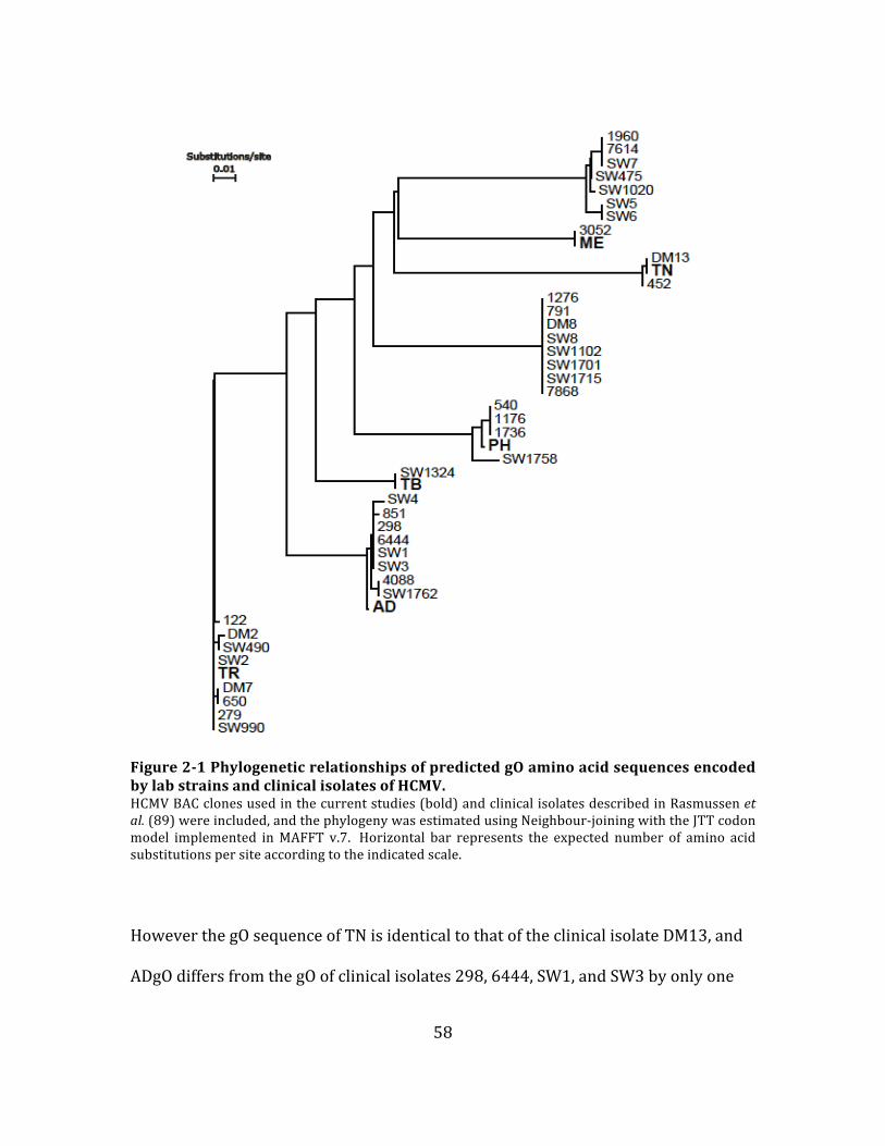
58
Figure 2-‐1 Phylogenetic relationships of predicted gO amino acid sequences encoded by lab strains and clinical isolates of HCMV. HCMV BAC clones used in the current studies (bold) and clinical isolates described in Rasmussen et al. (89) were included, and the phylogeny was estimated using Neighbour-‐joining with the JTT codon model implemented in MAFFT v.7. Horizontal bar represents the expected number of amino acid substitutions per site according to the indicated scale.
However the gO sequence of TN is identical to that of the clinical isolate DM13, and
ADgO differs from the gO of clinical isolates 298, 6444, SW1, and SW3 by only one

59
amino acid located within the predicted signal peptide. Thus, the gO isoforms
encoded by these lab and clinical strains are reflective of the diversity of this protein
in nature. It is important to note that given the limited number of sequences
available, and the lack of understanding of gO function(s), no conclusions can be
made concerning potential adaptive evolution of gO isoforms along lineages from a
common ancestor. Despite this, there clearly exists extensive diversity in gO
encoded by strains of HCMV in nature, and it is a compelling hypothesis that these
differences may affect the function(s) of gO, and the biology of the virus.
Sequence alignments of representative gO isoforms revealed regions of high
diversity, and regions of conservation (Figure. 2-‐2). Sequence diversity was found
in the N-‐terminal 100 residues, of which approximately 70 are predicted to remain
following cleavage of the signal peptide. The region corresponding to
approximately 270-‐340 of the alignment also harbored notable diversity. Six
conserved cysteine (cys) residues were identified at aligned positions 31, 152, 160,
178, 229, and 354. Cys 31 is located within the predicted signal peptide, suggesting
that it is not relevant to the function of gO. The remaining five cys are also found in
the gO homologues encoded by other beta-‐herpesviruses such as HHV-‐6, rhesus
CMV, and murine CMV, suggesting the importance of disulfide bonds for the
structure, and function of the gO interactions with gH/gL complex (252–254).
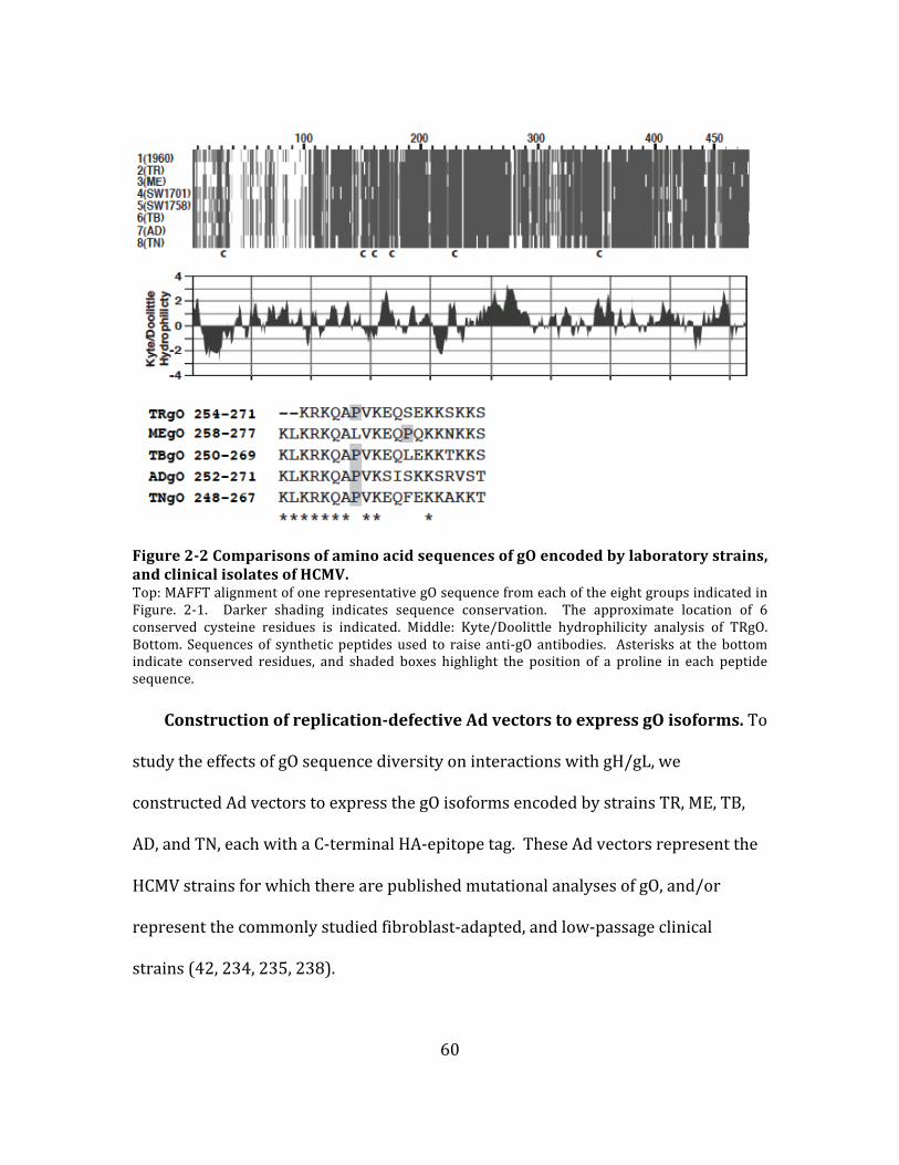
60
Figure 2-‐2 Comparisons of amino acid sequences of gO encoded by laboratory strains, and clinical isolates of HCMV. Top: MAFFT alignment of one representative gO sequence from each of the eight groups indicated in Figure. 2-‐1. Darker shading indicates sequence conservation. The approximate location of 6 conserved cysteine residues is indicated. Middle: Kyte/Doolittle hydrophilicity analysis of TRgO. Bottom. Sequences of synthetic peptides used to raise anti-‐gO antibodies. Asterisks at the bottom indicate conserved residues, and shaded boxes highlight the position of a proline in each peptide sequence.
Construction of replication-‐defective Ad vectors to express gO isoforms. To
study the effects of gO sequence diversity on interactions with gH/gL, we
constructed Ad vectors to express the gO isoforms encoded by strains TR, ME, TB,
AD, and TN, each with a C-‐terminal HA-‐epitope tag. These Ad vectors represent the
HCMV strains for which there are published mutational analyses of gO, and/or
represent the commonly studied fibroblast-‐adapted, and low-‐passage clinical
strains (42, 234, 235, 238).

61
Figure 2-‐3 Expression of HCMV gO isoforms by replication-‐defective adenovirus (Ad) vectors. U373 cells were transduced with Ad vectors expressing gO isoforms (HA-‐tagged) derived from HCMV TR (TRgO), Merlin (MEgO), TB40 (TBgO), AD169 (ADgO), and Towne (TNgO). Cells were labeled for 5 min with [35S] methionine/cysteine, and then the label was chased for 0 or 180 min. Proteins were immunoprecipitated with anti-‐HA antibodies, left untreated (-‐), or treated with endo H (H) or PNGaseF (F), and separated by SDS-‐PAGE (8%) under reducing conditions. Arrows, and arrowheads mark untreated, and deglycosylated forms of gO, respectively.
For initial characterization of gO isoforms, cells were transduced with Ad
vectors, and proteins were pulse-‐radiolabeled, and then chased for 0 or 180 min.
Cell extracts were then analyzed by immunoprecipitation (IP) with anti-‐HA

62
antibodies followed by treatment with endoglycosidase H (endo H), which removes
the high-‐mannose N-‐linked glycans added to proteins in ER, or PNGase F, which can
remove all types of N-‐linked glycans, including those that have been modified by
enzymes present only in the Golgi complex. Proteins were then separated by SDS-‐
PAGE under reducing conditions (Figure. 2-‐3). Each gO isoform migrated with an
apparent molecular weight of approximately 100 kDa. Slight differences in mobility
likely related to differences in polypeptide length among these isoforms (predicted
gO amino acids, including signal peptide: TR; 462, ME; 472, TB; 464, AD; 466, and
TN; 456), as well as differences in N-‐linked glycosylation, or other post translation
modifications. Faster migrating bands were also observed in the untreated lanes of
the 0 min chase samples. Since these bands were absent following glycosidase
treatment, and were not apparent after 3 hours of chase, they were likely
glycosylation intermediates of gO present in the cells after the pulse labeling. The
N-‐linked glycans on each gO isoform remained sensitive to endo H treatment for at
least 3 h of chase time, as indicated by the decrease in apparent molecular weight to
approximately 55 kDa. These data indicate that gO isoforms share the
characteristics of being highly modified with N-‐linked glycans, and being retained in
the ER when expressed in the absence of other HCMV proteins.
Cross-‐reactivity of antibodies raised against distinct gO isoforms.
Kyte/Doolittle analysis of TRgO indicated one prominent hydrophilic stretch

63
Figure 2-‐4 Western blot detection of gO isoforms by anti-‐peptide rabbit sera. Primary human fibroblasts (HFF) were transduced with Ad vectors expressing the indicated gO isoforms (HA-‐tagged). Cell extracts were analyzed by western blot using anti-‐HA antibodies (A), or antibodies raised against peptides derived from gO of HCMV TR (A), Merlin (B), TB40 (C), or AD169 (E). For detection with anti-‐peptide antibodies (A – E), long, and short chemiluminescent detections (top and bottom, respectively) were performed in parallel, allowing comparison of signal intensities.
including amino acids 245-‐271 (Figure. 2-‐2). A synthetic peptide corresponding to
these residues was used to raise anti-‐TRgO antibodies in rabbits that reacted well
with TRgO, but not other gO isoforms (233). Kyte/Doolittle analysis of other gO
isoforms indicated the presence of a similar hydrophilic region at the aligned
location (data not shown). However, there are some amino acid differences in this

64
region that may explain the lack of cross-‐reactivity of the TRgO antiserum with
other gO isoforms.
To allow more comprehensive comparative analyses of gO isoforms, antibodies
were raised against peptides corresponding to the aligned hydrophilic region of the
gO isoforms encoded by strains ME, TB, AD, and TN, and including conserved lysine,
and leucine residues (Figure. 2-‐2). These antibodies were characterized by western
blot analysis using extracts from cells transduced with Ad vectors expressing the
HA-‐tagged gO isoforms (Figure. 2-‐4A). The previously characterized anti-‐TRgO
antibodies cross-‐reacted with TBgO, but failed to detect MEgO or ADgO (Figure. 2-‐
4B). Both the anti-‐TBgO, and anti-‐ADgO antibodies showed overall stronger
reactivity, and unexpected cross-‐reactivity with other isoforms of gO, detecting
TRgO better than the TRgO-‐specific antiserum (Figure. 2-‐4 compare panel B, with D
and E). In contrast, anti-‐MEgO antibodies were highly specific for MEgO (Figure. 2-‐
4C). The specificity of the anti-‐MEgO antibodies may be related to the unique
position a proline residue in the aligned peptide sequences (Figure. 2-‐2). The TNgO
peptide sequence is less hydrophilic than the others (data not shown). Consistent
with this, antibodies raised against TNgO peptides failed to react with any isoform of
gO, and other antibodies failed to detect TNgO (data not shown). Thus, strain TN
was not included in the comparative analyses of gO in the HCMV envelope described
below. However, the TBgO and ADgO antibodies detected the gO isoform expressed
by the low-‐passage strain, PH (data not shown).
Reconstruction of gH/gL/gO complexes by Ad vector expression. Unlike
gO, gH/gL is highly conserved among strains of HCMV, differing by <5% of amino

65
acids among strains, but it is possible that gO diversity reflects coevolution with
specific gH/gL isoforms. To test whether or not distinct gO isoforms could bind to a
common isoform of gH/gL, Ad vectors were used to express TRgH/gL along with the
different gO isoforms. Cells were pulse-‐chase labeled, and analyzed by IP with anti-‐
gH antibodies followed by treatment with endo H or PNGaseF, and separation by
reducing SDS-‐PAGE (Figure. 2-‐5). In the absence of gO, gH/gL remained endo H
sensitive for at least 3 h, indicative of ER retention. This was consistent with
previous results (233). Coexpression of any of the gO isoforms resulted in
conversion of the majority of the labeled gH/gL to an endo H resistant form,
indicating transit from the ER to the trans-‐Golgi network (TGN). It was also of
interest to determine whether gO isoforms remain bound to gH/gL during transit to
the TGN. Previous analyses of extracellular AD virions demonstrated that some of
the N-‐linked glycans on virion-‐associated gO remain (partially) endo H sensitive
(i.e., high mannose content) despite transit through the TGN during virion
morphogenesis (233, 255). This would result in comigration of post-‐Golgi gO with
ER-‐associated gH, precluding direct observation of gO glycoforms in the experiment
described in Figure. 2-‐5. Thus, a modified version of the experiment was performed
in which pulse-‐chase labeled proteins were IP’ed with anti-‐gH antibodies, and heat
denatured in the presence of SDS, and reducing agents. The SDS, and reducing
agents in the samples were then diluted, and gO isoforms were analyzed by IP using
anti-‐HA antibodies, treatment with endo H or PNGaseF, and separation by reducing
SDS-‐PAGE (Figure. 2-‐6).
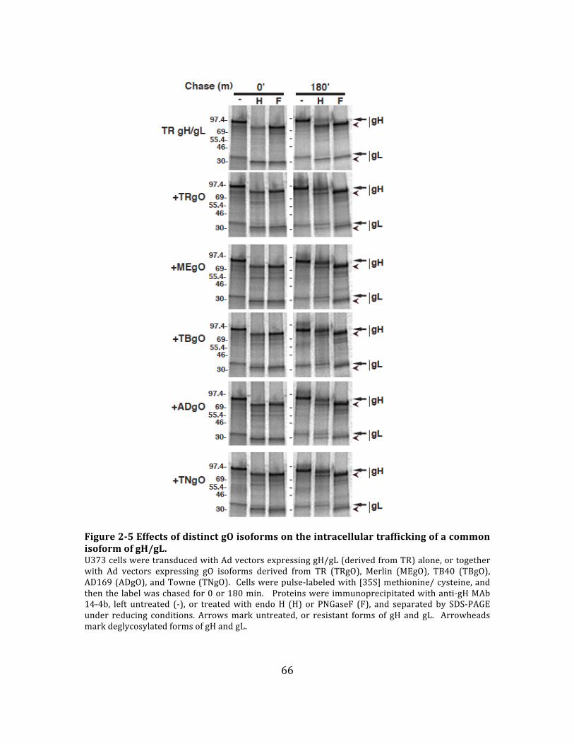
66
Figure 2-‐5 Effects of distinct gO isoforms on the intracellular trafficking of a common isoform of gH/gL. U373 cells were transduced with Ad vectors expressing gH/gL (derived from TR) alone, or together with Ad vectors expressing gO isoforms derived from TR (TRgO), Merlin (MEgO), TB40 (TBgO), AD169 (ADgO), and Towne (TNgO). Cells were pulse-‐labeled with [35S] methionine/ cysteine, and then the label was chased for 0 or 180 min. Proteins were immunoprecipitated with anti-‐gH MAb 14-‐4b, left untreated (-‐), or treated with endo H (H) or PNGaseF (F), and separated by SDS-‐PAGE under reducing conditions. Arrows mark untreated, or resistant forms of gH and gL. Arrowheads mark deglycosylated forms of gH and gL.
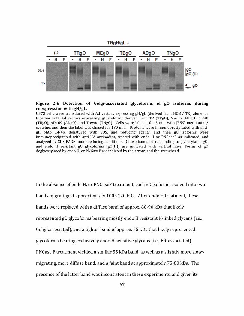
67
Figure 2-‐6 Detection of Golgi-‐associated glycoforms of gO isoforms during coexpression with gH/gL. U373 cells were transduced with Ad vectors expressing gH/gL (derived from HCMV TR) alone, or together with Ad vectors expressing gO isoforms derived from TR (TRgO), Merlin (MEgO), TB40 (TBgO), AD169 (ADgO), and Towne (TNgO). Cells were labeled for 5 min with [35S] methionine/ cysteine, and then the label was chased for 180 min. Proteins were immunoprecipitated with anti-‐gH MAb 14-‐4b, denatured with SDS, and reducing agents, and then gO isoforms were immunoprecipitated with anti-‐HA antibodies, treated with endo H or PNGaseF as indicated, and analyzed by SDS-‐PAGE under reducing conditions. Diffuse bands corresponding to glycosylated gO, and endo H resistant gO glycoforms (gO(H)) are indicated with vertical lines. Forms of gO deglycosylated by endo H, or PNGaseF are indicted by the arrow, and the arrowhead.
In the absence of endo H, or PNGaseF treatment, each gO isoform resolved into two
bands migrating at approximately 100~120 kDa. After endo H treatment, these
bands were replaced with a diffuse band of approx. 80-‐90 kDa that likely
represented gO glycoforms bearing mostly endo H resistant N-‐linked glycans (i.e.,
Golgi-‐associated), and a tighter band of approx. 55 kDa that likely represented
glycoforms bearing exclusively endo H sensitive glycans (i.e., ER-‐associated).
PNGase F treatment yielded a similar 55 kDa band, as well as a slightly more slowy
migrating, more diffuse band, and a faint band at approximately 75-‐80 kDa. The
presence of the latter band was inconsistent in these experiments, and given its
68
apparently molecular weight, it likely represented residual gH–gO interactions
recaptured in the second IP reaction. The 55 kDa band produced by PNGase F likely
represented the same fraction of gO that resolved at 55 kDa following endo H
treatment (i.e. ER-‐associated), whereas the slower migrating species produced by
PNGase F likely corresponded to the fraction of gO that was endo H resistant (Golgi-‐
associated). This suggests the acquisition of modifications, in addition to N-‐linked
oligosaccharides, such as O-‐linked glycans (76). Also, extracts from cells
coexpressing gH/gL and any of the gO isoforms contained a proteins species that
migrated at approximately 240-‐260 kDa under non-‐reducing conditions that were
detected by both anti-‐gL antibodies (Figure. 2-‐7A), anti-‐HA (gO) antibodies (Figure.
2-‐7B) and anti-‐gH antibodies (data not shown). Together, these data strongly
suggest that the assembly of all gH/gL/gO complexes involves disulfide interactions,
and the conserved sequences of gO. However, it is important to note that these Ad
vector expression experiments were not well suited to compare how efficiently each
gO isoform interacts with gH/gL (i.e., binding affinity).

69
Figure 2-‐7 Analysis of disulfide bonds in the interactions between gH/gL and gO isoforms. U373 cells were transduced with Ad vectors expressing gH/gL (derived from TR) alone, or together with Ad vectors expressing gO isoforms derived from TR (TRgO), Merlin (MEgO), TB40 (TBgO), AD169 (ADgO), and Towne (TNgO). Cell extracts were separated under non-‐reducing conditions and, analyzed by western blot using anti-‐gL (A), or HA (B) antibodies. The predicted migration of gH/gL/gO trimers, gH/gL dimers, and gO monomers are indicated to the right of both panels.
HCMV strains differ in the ratio of gH/gL/gO, and gH/gL/UL128-‐131 in the
virion envelope. The results of the Ad vector expression experiments described
above indicated that all isoforms of gO can form a disulfide-‐linked complex with
gH/gL. Previous comparative analysis of gO from strains TR and AD did not address
disulfide interactions (233). Thus, using the new gO antibodies described in Figure.
2-‐4, we analyzed cell-‐free TR, ME, TB, AD and PH virions, and infected cells by
reducing, and non-‐reducing western blot (Figure. 2-‐8). Under reducing conditions,

70
and probing for gO, two bands were detected in TR-‐infected cell extracts, a
prominent 100 kDa band, and a fainter, more diffuse band of approximately 120 kDa
(Figure. 2-‐8A). These bands likely corresponded to different glycoforms of gO
reflecting maturation of N-‐linked glycans, and other modifications acquired during
transit though the Golgi-‐derived, virion assembly compartment. Consistent with the
previous report, gO was not detected in extracts of extracellular TR virions under
reducing conditions (Figure. 2-‐8A) (233). However, when analyzed under non-‐
reducing conditions, a band was detected in both cell, and virion extracts that
migrated slower than the 250 kDa marker, consistent with a gH/gL/gO complex
(Figure. 2-‐8A). This species was also detected with anti-‐gL (Figure. 2-‐8B) and anti-‐
gH (not shown) antibodies, confirming its identity as gH/gL/gO. Analysis of 4 other
strains of HCMV, each encoding a different gO isoform, yielded similar results
(Figure. 2-‐8A and B). Since these experiments involved detection of gO isoforms
using different antibodies, and separate chemiluminescent detections, only the
fraction of gO detected as gH/gL/gO versus gO monomer can be compared between
strains. Note that on non-‐reducing gels, a greater fraction of the total gO migrated at
the expected monomeric size in ME infected cells than in cells infected with other
strains (Figure. 2-‐8A).

71
Figure 2-‐8 Comparative analysis of gH/gL/gO complexes from different strains of HCMV. Extracts of HFF cells infected with BAC-‐derived HCMV TR, Merlin, TB40/e, AD169, or PH, or virions collected from the culture supernatant were separated by non-‐reducing or reducing SDS-‐PAGE transferred to PVDF membranes and probed with anti-‐gO (A) or anti-‐gL (B) antibodies. For gO blots, anti-‐TBgO antibodies were used for TR, TB and PH samples, anti-‐MEgO antibodies used for ME samples, and anti-‐ADgO antibodies were used for AD samples. Arrowheads mark bands corresponding to the disulfide-‐linked gH/gL/gO trimer, and vertical lines indicate bands that correspond to gO, gL or the disulfide-‐linked gH/gL/UL128.
gH/gL/UL128
72
The detection of gO in TR virions when bound to gH/gL, but not when liberated
from the complex by reduction of disulfide bonds is difficult to explain since the
anti-‐peptide epitopes are generally more accessible following reduction of disulfide
bonds. It is notable that similar results were observed for MEgO, ADgO (Figure. 2-‐
8A) and gL (Figure. 2-‐8B), but not TBgO and PHgO (Figure. 2-‐8A). While we cannot
fully explain this phenomenon, the results of these experiments clearly indicate that
the disulfide-‐linked gH/gL/gO complex is a conserved feature of the HCMV
envelope.
Since gO and UL128 bind to gH/gL through disulfide bonds whereas UL130/131
associate with gH/gL through non-‐covalent interactions, (and non-‐covalent
interactions will be disrupted on SDS-‐PAGE gel), under non-‐reducing condition,
gH/gL/gO would still stay intact while UL130/UL131 dissociate from the the
gH/gL/UL128-‐131 complex, leaving gH/gL/UL128 behind, which can be
distinguished by different molecular weitght from gH/gL/gO. The analyses of virion,
and infected cell extracts with anti-‐gL antibodies revealed a striking difference
between ME, and other strains (Figure. 2-‐8B). For each of the strains TR, TB, AD,
and PH, the majority of the gL detected in virions extracts under non-‐reducing
conditions was associated with gH/gL/gO whereas for ME, most of the gL was
detected at an apparent molecular weight consistent with gH/gL/UL128
heterodimers; approx. 130 kDa (Figure. 2-‐8B, top row). Similar results were
obtained using anti-‐gH antibodies (not shown). In this context, these results suggest
that TR, TB, AD and PH virions contain vastly more gH/gL/gO compared to
gH/gL/UL128-‐131, whereas ME virions contain more gH/gL/UL128-‐131.

73
The ratio of gH/gL/gO and gH/gL/UL128-‐131 in the HCMV virion envelope
is influenced by competition between gO, and the set of UL128-‐131 proteins
for binding to gH/gL. To confirm that the apparent gH/gL/UL128 detected in the
ME virion envelope was used to be gH/gL/UL128-‐131 complex, and to test the
hypotheses that the ratio of gH/gL complexes in the virion envelope is influenced by
competition between gO and UL128-‐131 proteins, the tetracycline (Tet) expression-‐
repression system described by Stanton et al. was used (177). Briefly, ME is highly
sensitive to mutational inactivation of the UL128-‐131 genes during serial
propagation in cultured fibroblasts, making it difficult to maintain wild-‐type ME in
culture (175, 176). To overcome this, Stanton et al. engineered a ME BAC with Tet
operator sequences inserted within the promoter that regulates expression of
UL130, and UL131, and a fibroblast cell line expressing the Tet repressor protein
(HFFtet). During replication in HFFtet cells, transcription from the UL130/131
promoter, and consequently, assembly of the gH/gL/UL128-‐131 complex is
suppressed, reducing selective pressure against UL128-‐131.
Extracts of ME virions produced in HFF, and HFFtet cells were analyzed by
reducing and non-‐reducing western blots (Figure. 2-‐9). Reducing blots indicated
that virions produced by both cell types contained comparable amounts of total gL
(Figure. 2-‐9A), and gB (not shown). On non-‐reducing gels, the majority of gL
detected in ME virions produced by HFF cells was in the form of gH/gL/UL128,
consistent with the results of the previous experiment (compare Figure. 2-‐8B and 2-‐
9A). Strikingly, in virions produced under conditions of suppressed UL128-‐131
expression (HFFtet cells), the majority of the gL corresponded to the slower
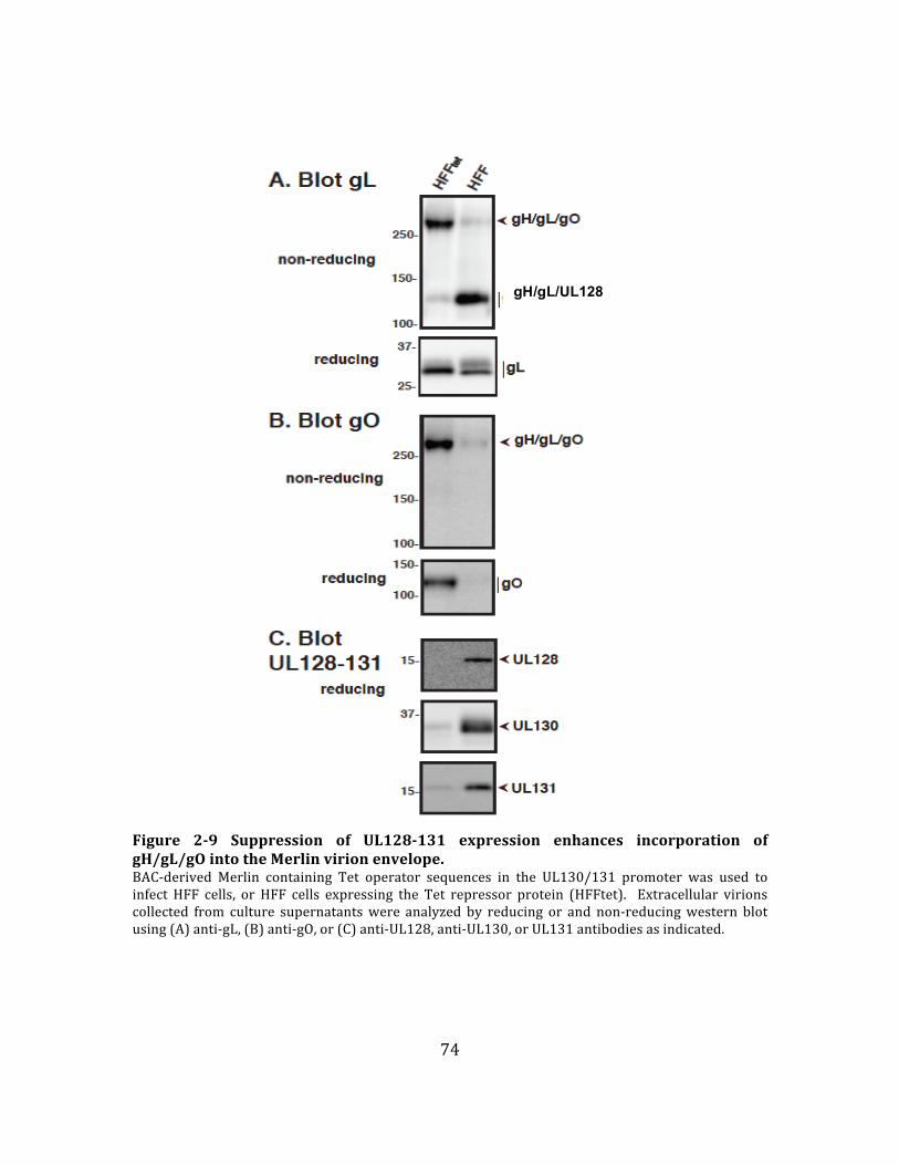
74
Figure 2-‐9 Suppression of UL128-‐131 expression enhances incorporation of gH/gL/gO into the Merlin virion envelope. BAC-‐derived Merlin containing Tet operator sequences in the UL130/131 promoter was used to infect HFF cells, or HFF cells expressing the Tet repressor protein (HFFtet). Extracellular virions collected from culture supernatants were analyzed by reducing or and non-‐reducing western blot using (A) anti-‐gL, (B) anti-‐gO, or (C) anti-‐UL128, anti-‐UL130, or UL131 antibodies as indicated.
gH/gL/UL128

75
migrating gH/gL/gO complex (Figure. 2-‐9A). Consistent with this, ME virions
produced from HFFtet cells contained more gO (Figure. 2-‐9B), and less UL128-‐131
proteins (Figure. 2-‐9C). Note that due to the increased virion-‐associated MEgO
under conditions of UL128-‐131 suppression, much shorter chemiluminescent
exposures were needed for detection of MEgO in these experiments than for those
described in Figure. 2-‐8A. Since unbound gH/gL dimers are poorly transported out
of the ER, and the presence of UL128-‐131 in the virion exactly correlates with the
detection of gH/gL/UL128, these data strongly support the conclusion that the
gH/gL/UL128 remainder detected in ME virions represented the gH/gL/UL128-‐131
complex, and the ratio of the two gH/gL complexes in the virion envelope is
influenced by competition between gO and the UL128-‐131 proteins for binding to
gH/gL.
DISCUSSION
Previous analyses suggested that some gO isoforms remain bound to gH/gL in
the virion envelope, and others behave more like chaperones to promote assembly
of unbound gH/gL dimers into the virion envelope (233). The chaperone model was
based largely on the failure to detect gO in TR virions by reducing western blots.
Here, we used non-‐reducing gel systems to compare several strains of HCMV. In all
cases, gO clearly was detected as part of a disulfide-‐linked complex with gH/gL in
the virion envelope.
The discrepancy between non-‐reducing and reducing western blots is puzzling.
As noted, anti-‐peptide antibodies are generally thought to react better with proteins
76
that have been separated under denaturing-‐reducing conditions because continuous
epitopes should be more accessible. It is notable that virion-‐associated ADgO was
clearly detected in the reducing western blot experiments reported in Ryckman et
al. (233), but not in the similar experiments reported here. This difference might be
explained by the fact that in the previous studies, ADgO was detected with the
polyclonal serum described by Huber et al. (75), which was raised against full length
gO, whereas in the current studies, anti-‐peptide antibodies were used for detection
of all gO isoforms. This observation points towards specific modification of gO, such
as proteolysis, at or near the anti-‐peptide antibody target site (the conserved
hydrophilic domain shown in Figure. 2-‐2). If this were the case, then perhaps the
disulfide nature of the gH/gL/gO complex might preserve epitope recognition in
western blot experiments. Potential modifications are likely coincident with virion
morphogenesis, and release because intracellular gO was easily detected with the
anti-‐peptide antibodies on reducing western blots. Also, since detection of some gO
isoforms was less affected by disulfide reduction, sequence differences might affect
these potential modifications. It is also important to note that a similar
phenomenon was observed during analysis of gL, and it is interesting that the anti-‐
gL antibodies used were also raised against synthetic peptides. In order to
efficiently elicit antibodies, synthetic peptides are conjugated to a carrier protein
(such as keyhole limpet hemocyanin; KLH) via a terminal cys residue that either
represents a cys in the protein of interest, or one that is added to the synthetic
peptide for the sole purpose of conjugation to the carrier. Thus, it is tempting to
suggest that the presence of a cys in the peptides used to generate the antibodies

77
might contribute to the observed phenomenon. However, for all antibodies
described, the cys used for conjugation was extraneous, and not present in the
actual protein. Finally, it is possible that the observed effects relate to differences
between cell-‐associated, and virion-‐associated proteins in the transfer to blotting
membranes, and that these differences are less apparent during transfer of the
intact gH/gL/gO complex. Again, this explanation would suggest posttranslational
modifications of proteins coincident with virion release. Regardless, the analyses of
TR, and other HCMV strains reported here clearly demonstrate that disulfide-‐linked
gH/gL/gO complexes are a conserved feature of the HCMV virion envelope.
Regulation of gB can be considered the “core entry function” of the herpesvirus
gH/gL. HCMV gH/gL, without UL128-‐131 or gO, can regulate gB-‐mediated cell-‐cell
fusion, indicating that the necessary surfaces for gB interaction are comprised of
amino acids contained within gH/gL alone (216). However, data presented here
indicate that the HCMV envelope contains a mixture of gH/gL/UL128-‐131 and
gH/gL/gO, and little or no unbound gH/gL. Thus, for HCMV, the core function
involving interactions with gB must be provided by either the pentameric
gH/gL/UL128-‐131, or the trimeric gH/gL/gO. EBV also has different types of gH/gL,
unmodified gH/gL or a trimeric gH/gL/gp42, and either can perform this function
depending on the cell type involved (221, 247). In contrast, the phenotypes of
HCMV gO, and UL128-‐131 mutants strongly suggest that gH/gL/gO performs the
core gB-‐interaction function during entry into all cell types, whereas gH/gL/UL128-‐
131 serves a distinct, receptor-‐binding function that is an additional necessity for
entry into specific cell types such as epithelial, endothelial, and dendritic cells, but

78
not others, such as fibroblasts or neurons (42, 77, 79, 156, 227, 234, 235, 238, 248,
256). The receptors for gH/gL/UL128-‐131 are unknown, but engagement of these
molecules might trigger endocytosis, actin rearrangement or other cell surface
functions that then allow for the universally required gH/gL/gO -‐ gB interactions
that result membrane fusion and entry. An important prediction of this model is
that the surfaces on gH/gL that interact with gB are inaccessible on gH/gL/UL128-‐
131, but are accessible on gH/gL/gO, and studies to test aspects of this model are in
progress.
The significance of gO sequence diversity on the composition of the HCMV
envelope, and the function of the gH/gL/gO complex adds a layer of complexity to
models of HCMV entry and tropism that has not previously been addressed. In the
analyses presented here, all isoforms of gO bound to a common isoform of gH/gL to
form a disulfide-‐linked trimer that was transported from the ER to the TGN. This
suggests that gH/gL/gO complexes share a similar structure involving the
conserved sequences of gO. This conclusion is consistent with the model of
gH/gL/gO providing a core entry function since all gO isoforms must leave critical
surfaces on gH/gL available for interaction with gB. These results are also
consistent with those of Rasmussen et al., who investigated genetic linkages of
polymorphic loci in clinical HCMV isolates, and found evidence of several
combinations of gH/gL and gO isoforms (257).
The finding that virions of TR, TB, AD and PH virions contained vastly more
gH/gL/gO than gH/gL/UL128-‐131, whereas ME contain more gH/gL/UL128-‐131
than gH/gL/gO was unexpected, and has a number of important implications. First,

79
the observation that UL128-‐131 suppression during ME replication dramatically
shifted the ratio of gH/gL complexes in the virion envelope towards gH/gL/gO
indicates that gO, and UL128-‐131 compete for binding of gH/gL to form the two
complexes, and the relative abundance of unbound gO in ME infected cells suggests
that MEgO may be less able to compete than other gO isoforms. Thus, the variable
sequences of gO might affect the ratio of the core entry factor gH/gL/gO to the
tropism factor gH/gL/UL128-‐131. However, these results do not exclude the
possibility that differences in protein expression levels, or small sequence
differences in gH/gL, or UL128-‐131 also contribute to strain variation. Wille et al.
showed that a TR mutant that could not express gO produced virions that contained
elevated amounts of gH/gL/UL128-‐131, but drastically lower total amounts of
gH/gL, indicating that UL128-‐131 was unable to bind a significant fraction of the
remaining gH/gL (238). This might indicate that TR expresses more gH/gL, or less
UL128-‐131 than ME. Second, it is well appreciated that there is a selective pressure
against UL128-‐131 during replication in fibroblast cultures, and that ME is
especially sensitive to this selection (175–177). If the assembly of gH/gL/gO and
gH/gL/UL128-‐131 come at the expense of one another, and if the gH/gL/UL128-‐
131 cannot participate in the interactions with gB that are necessary for fusion, then
mutation of UL128-‐131 would be expected to result in a greater fibroblast-‐
replication fitness advantage for a virus like ME than for a virus like TR. This model
predicts an evolutionary balancing act requiring the virus to express enough
gH/gL/UL128-‐131 for broad tropism, while retaining enough gH/gL/gO to promote
fusion on all cell types. Clearly, the amounts of each complex in the ME virion are
80
sufficient to allow infection of both fibroblasts, and epithelial/endothelial cells, but
the relative efficiency (i.e. per virion) compared to other HCMV strains with
different amounts of the complexes is difficult to directly measure. As noted above,
it is possible that HCMV strains differ in the total amount of gH/gL, and this could
either lessen, or magnify the importance of the ratio of the two complexes. Clearly
suppression of UL128-‐131 enhances the replication of ME, but the effects of
manipulation the expression of gO or gH/gL has not been addressed. Finally, It is
tempting to speculate that adaptive evolution of HCMV tropism observed in culture
may also occur in vivo. Infection of neuronal cells is likely independent of
gH/gL/UL128-‐131 (156). Thus, it is conceivable that the ratio of the core entry
factor (gH/gL/gO), and the tropism factor (gH/gL/UL128-‐131) might be subject to
similar selective pressures in neuronal or, other tissues as has been observed in
fibroblast cultures. If so, the gO genotype of HCMV might influence viral pathology,
and the ability of the virus to spread to other hosts, which likely involves replication
in epithelial and endothelial tissues.
Host cell characteristics may also influence the protein composition of the
HCMV envelope. Wang et al. found that HCMV produced by epithelial cells entered
epithelial cells though fusion at the plasma membrane rather than via fusion
following endocytosis, as had been described for fibroblast-‐produced virus. They
suggested that this correlated with an approximately 2-‐fold increase in the ratio of
gH/gL/UL128-‐131 vs. gH/gL/gO in epithelial cell-‐produced virions (258). Scrivano
et al. similarly analyzed HCMV virions produced by fibroblasts with those produced
by endothelial cells, and suggested that both cell types produce virion populations

81
that are heterogeneous in the relative levels of gH/gL/gO and gH/gL/UL128-‐131
(259). The notion of heterogeneity in the amounts of gH/gL complexes
incorporated into the HCMV envelope had been previously suggested by Li et al.
(260). Our results linking gO diversity with the ratio of the two gH/gL complexes
raises the question of whether all strains display similar envelope heterogeneity, or
are equally sensitive to producer cell.
In summary, the data presented in this report clearly indicate that the
gH/gL/gO complex is an envelope component of all strains of HCMV, and suggest
that the sequence differences between gO isoforms may affect the ratio of gH/gL
complexes incorporated in the virion envelope. The diverse sequences of gO may
also affect other functions, and aspects of the gH/gL/gO complex. For example,
Vanarsdall et al. provided evidence from receptor interference experiments that
suggested gH/gL/gO might act as a receptor-‐binding complex, and the gO isoform
might participate in these interactions or modulate the activity (239). Also, Jiang et
al. recently suggested that the extensive array of N-‐linked glycans on gO might act to
shield the virus from the effects of neutralizing antibodies (261). Research work in
Chapter 4 studied the possible significances of gO diversity by analyzing inter-‐strain
of gO recombinant HCMVs.
82
CHAPTER 3. CHARACTERIZATION OF
HUMAN CYTOMEGALOVIRUS gH/gL
COMPLEXES FUNCTION DURING VIRUS
ENTRY AND TROPISM
This chapter is a modified version of the manuscript published in Journal of
Virology in September 2015; 89(17), 8999-‐9009

83
INTRODUCTION
Primary infection of healthy adults by human cytomegalovirus (HCMV) is
usually subclinical or mildly symptomatic, but leads to lifelong persistent or latent
infection. Primary infection or reactivation of HCMV in immunocompromised hosts,
such those infected with HIV and transplant recipients on anti-‐rejection
chemotherapies, is associated with significant morbidity and mortality, and
maternal transmission of HCMV to the developing fetus across the placenta can
result in severe congenital birth defects (16, 21, 240). The diverse nature of HCMV-‐
associated disease likely relates to the ability of the virus to infect many cell types in
vivo, including epithelial and endothelial cells, fibroblasts, monocyte-‐macrophages,
dendritic cells, hepatocytes, leukocytes and neurons (147, 148, 156). Much research
effort has been focused on understanding the mechanisms by which HCMV initiates
infection of different cell types (reviewed in (190)). Most studies have described an
entry mechanism dichotomy between fibroblasts and epithelial/endothelial cells.
Other relevant cell types, though more difficult to work with in the laboratory, likely
fit to one side or the other of the fibroblasts – epithelial/endothelial entry
dichotomy.
Entry of all herpesviruses requires membrane fusion, which is mediated by a
conserved core fusion apparatus comprised of glycoprotein B (gB) and gH/gL.
Extensive studies of herpes simplex virus (HSV) and Epstein-‐Barr virus (EBV) have
shown that gB has characteristics of a class III viral fusion protein (209, 210, 213,
214). The necessary role of gH/gL in herpesvirus membrane fusion is unclear, but
likely involves direct interaction between gH/gL and gB (218, 223, 262–265).

84
Herpesviruses also encode accessory glycoproteins such as HSV gD and EBV gp42,
which regulate the core fusion apparatus through receptor-‐binding, and stable or
transient interactions with gH/gL (reviewed in (189)).
HCMV shares the conserved features of herpesviruses entry, but there are some
important unique aspects, especially related to cell tropism. Structural analyses of
HCMV gB suggest general mechanistic similarities with other herpesvirus gB
homologues (122) and the combination of HCMV gB and gH/gL is necessary, and
sufficient for membrane fusion in cell-‐cell fusion experiments (216). The
ectodomain of HCMV gH/gL can be bound by either gO, or proteins encoded by
UL128, UL130 and UL131 (UL128-‐131), forming the gH/gL/gO, and gH/gL/UL128-‐
131 complexes on the virion envelope (73, 74, 77, 227). Experiments reported in
Chapter 2 indicated the following: 1) extracellular virions contain little, if any
unbound gH/gL, 2) the ratio of gH/gL/gO to gH/gL/UL128-‐131 can vary
dramatically between different HCMV strains, and 3) gO and UL128-‐131 compete
for binding of gH/gL (232). The mutually exclusive nature of gH/gL/gO and
gH/gL/UL128-‐131 formation is consistent with a recent structural analysis showing
that gO, and the UL128 protein make a disulfide bond with the same cysteine
residue of the gL polypeptide (215). The gH/gL/gO and gH/gL/UL128-‐131
complexes influence cell tropism at the level of entry, although the mechanisms are
not well understood.
The roles of HCMV gH/gL complexes have mostly been studied with deletion
mutant approaches. In general, mutants that lack gH/gL/UL128-‐131 replicate as
well, or better than wild type strains on fibroblasts (77–79, 227). This indicates that

85
gB-‐mediated fusion with fibroblast membranes can be facilitated by gH/gL/gO. In
contrast gH/gL/UL128-‐131 mutants fail to infect cell types such as epithelial,
endothelial and monocyte/macrophages (77, 79, 227, 248, 266). These observations
have generally been interpreted to mean that gB-‐mediated fusion with membranes
of epithelial cells (and other cell types for which gH/gL/UL128-‐131 is necessary for
HCMV entry) is facilitated by gH/gL/UL128-‐131. The resulting model suggests that
both gH/gL/gO, and gH/gL/UL128-‐131 provide essentially the same function for
entry into different cell types, i.e., promoting gB-‐mediated membrane fusion. This
model was attractive due to analogy with the well-‐described model of EBV tropism
(reviewed in (189)). EBV gH/gL can exist in the virion envelope as unmodified
gH/gL, or bound by gp42. For entry into epithelial cells, gH/gL binds specific
integrins, and promotes gB-‐mediated membrane fusion. The presence of gp42 on
gH/gL blocks integrin binding, and instead gp42 binds MHC II, which facilitates
entry into B-‐cell. Presumably, both gH/gL and gH/gL/gp42 promote gB-‐mediated
membrane fusion through interactions involving “pro-‐fusion” surfaces of gH/gL,
with gp42 acting as a receptor-‐adapter to switch cell tropism. While HCMV mutants
lacking gH/gL/UL128-‐131 replicate well on fibroblasts, mutants lacking gH/gL/gO
are severely replication impaired on all cell types (42, 234, 235, 238). In particular,
Wille et al. showed that HCMV virions lacking gH/gL/gO were impaired at the fusion
step of entry into fibroblasts, epithelial, and endothelial cells, despite containing
elevated amounts of gH/gL/UL128-‐131 (238). Together, the results of deletion
mutant approaches seem to suggest that gH/gL/gO is sufficient for entry into
fibroblast, but that entry into epithelial/endothelial cells requires both gH/gL/gO

86
and, gH/gL/UL128-‐131.
Characterization of the mechanisms by which HCMV gH/gL/gO and
gH/gL/UL128-‐131 facilitate cell type dependent entry using deletion mutants has
been limited largely due to the severe replication defects associated with gO
deletions, and the lack of an adequate complementation system. The studies
described herein, capitalize on our previously reported observations that the
amounts of gH/gL complexes in the HCMV virion envelope can vary dramatically
among strains, and that the ratio of the two gH/gL complexes can be manipulated
through suppression of the UL128-‐131 proteins. Here, we report further
characterizations of strain variability in the abundance of gH/gL complexes in the
virion envelope, and relate these differences to the efficiency of entry on different
cell types. Comparisons of genetically identical HCMV preparations either enriched
in gH/gL/gO, or gH/gL/UL128-‐131 suggest a model in which gH/gL/gO provides
the herpesvirus “core-‐fusion gH/gL” function of promoting gB-‐mediated membrane
fusion for entry into all cell types, whereas gH/gL/UL128-‐131 facilitates entry into
select cell types, such as epithelial cells, through a distinct, yet necessary
mechanism.
MATERIALS AND METHODS
Cell lines. Primary human foreskin fibroblasts (HFF; Life Technologies), MRC-‐
5 fibroblasts (ATCC CCL-‐171), and HFFtet cells (which express the tetracycline
(Tet) repressor protein (177); provided by Richard Stanton (Cardiff University,
Cardiff, United Kingdom)) were grown in Dulbecco’s modified Eagle’s medium
87
(DMEM; Life Technologies) supplemented with 6% heat-‐inactivated fetal bovine
serum (FBS; Rocky Mountain Biologicals, Inc., Missoula, Montana, USA.) and 6%
bovine growth serum (BGS; Rocky Mountain Biologicals, Inc., Missoula, Montana,
USA.). Retinal pigment epithelial cell line, ARPE-‐19 (American Type Culture
Collection; Manassas, VA, USA) was grown in a 1:1 dilution mix of DMEM and Ham’s
F-‐12 medium (DMEM:F-‐12; Life Technologies) supplemented with 10% FBS.
HCMV. All HCMV were derived from bacterial artificial chromosome (BAC)
clones. BAC clone TR was provided by Jay Nelson (Oregon Health and Sciences
University, Portland OR, USA) (41). BAC clone TB40/e (BAC-‐4) (TB) was provided
by Christian Sinzger (University of Ulm, Germany) (184). BAC clone Merlin (ME)
(pAL1393), which contains tetracycline operator sequences within the
transcriptional promoter of UL130 and UL131, was provided by Richard Stanton
(Cardiff University, Cardiff, United Kingdom) (177). Infectious HCMV was recovered
by electroporation of BAC DNA into MRC-‐5 fibroblasts as described by Wille et al.
(238). Cell-‐free HCMV stocks were produced by infecting HFF or HFFtet at 2 plaque-‐
forming unit (PFU) per cell. At 8-‐10 days post infection (when cells were still
visually intact), culture supernatants were harvested, and cellular contaminants
were removed by centrifugation at 1,000 X g for 10min, and again at 6,000 X g for
10min. Stocks were judged cell-‐free by the lack of calnexin, and actin in western
blot analyses, stored at -‐80°C. The number of PFU was determined by plaque assay
on triplicate HFF or ARPE-‐19 cultures. Freeze/thaw cycles were avoided.
Antibodies. Monoclonal antibodies (MAbs) specific for HCMV major capsid
protein (MCP) 28-‐4 and gB 27-‐156 were provided by Bill Britt (University of

88
Alabama, Birmingham, AL, USA) (45, 267). Anti-‐UL128 MAb 4B10 was provided by
Tom Shenk (Princeton University, Princeton, NJ, USA) (77). Rabbit polyclonal
antipeptide antibodies directed against HCMV gH/gL, UL130, and UL131 were
provided by David Johnson (Oregon Health and Sciences University, Portland, OR,
USA) (80). Rabbit polyclonal antipeptide antibodies directed against MEgO were
described in (232).
Western blot. Cell-‐free virions from culture supernatants (as described above)
were concentrated by centrifugation at 50,000 X g for 1 h, and resuspended in 2%
SDS in 20mM tris-‐buffered saline (TBS; pH 6.8). Insoluble material was removed by
centrifugation at 16,000 X g for 30min, cleared extracts were heated to 95 °C for
10min. For reducing blots, extracts were adjusted to 2% DTT. Proteins were
separated by SDS-‐PAGE and transferred to PVDF membranes (Whatman) in a buffer
containing 10mM NaHCO3 and 3mM Na2CO3 (PH9.9), plus 10% methanol.
Transferred proteins were probed with MAbs or rabbit polyclonal antibodies
specific for HCMV proteins, followed by horseradish peroxidase-‐conjugated
secondary antibodies; chemi luminescence was detected using a Bio-‐Rad ChemiDoc
MP imaging system. Band densities were quantified using Bio-‐Rad Image lab v 5.1 or
ImageJ v 1.48 software.
Quantitative PCR (qPCR). 200 μl cell-‐free HCMV stock was first treated with
DNase I to remove DNA not protected within viral capsids. Capsids were disrupted
by using the viral lysis buffer, and proteinase K provided in the PureLink Viral
RNA/DNA mini kit (Life Technologies). Viral DNA was eluted in 50μl RNAse-‐free
water. A conserved region within UL83 among ME, TR and TB was chosen as

89
amplicon, and genomes were quantified by real-‐time qPCR using SYBR Green dye
(Bio-‐Rad): forward primer, TGGTCACCTATCACCTGCAT; reverse primer,
GAAAGAGCCCGACGTCTACT. PCR samples were made to a total well volume of 20μl
with 1X SYBR Green master mix (Bio-‐Rad), 1:100 dilution of viral DNA and 500nM
forward and reverse primers. PCR products were detected using MyiQ real-‐time
PCR detection system (Bio-‐Rad). QPCR results were compared to a plasmid
pTRUL83 standard and then calculated into genome/ml. Efficiency of DNase I
treatment were tested in independent experiments by spiking plasmid DNA. For
each experiment, two independent viral DNA extractions and three independent
QPCR reactions were performed.
Determination of plaque forming units (PFU). Cell monolayers were
infected with cell free HCMV, or HCMV infected cells (infectious centers), and cell
culture medium was replaced with a 1:1 mixture of 2XDMEM and 1.2% SeaPlaque
agarose to prevent spread of progeny virus by diffusion. Plaques were counted by
light microscopy 3 weeks later.
Antibody neutralization of HCMV. 1:100 diluted (vol/vol) anti-‐UL130 and
anti-‐UL131 rabbit serum were added to culture medium containing cell-‐free HCMV.
The mixture was incubated for at room temperature for 1 h, and then added to HFF
or ARPE-‐19 cultures.
Polyethylene glycol (PEG) treatment. Cell-‐free HCMV was adsorbed to cells
for 2 h at 10°C, under 800 X g centrifugation or at 37°C without centrifugation.
Unbound virus was removed by two cold PBS washes, and cells were subsequently
treated with 44% PEG (pre-‐warmed to 37°C) for 30s followed by 10 washes with
90
warm culture medium to remove PEG. Cultures were incubated 37°C until
immunofluorescence analysis was performed. PEG 6000 (Fluka) was prepared as a
60% (wt/wt) solution in PBS, and diluted with PBS to 44%.
Immunofluorescence. HCMV infected cells were fixed with 2%
formaldehyde in PBS for 15 min followed by three PBS washes. Fixed cells were
incubated, for 1hr in immunofluorescence (IF) buffer (0.5% Triton X-‐100, 0.5%
deoxycholate, 1% bovine serum albumin [BSA], 0.05% sodium azide in PBS). To
visualize HCMV infected cells, anti-‐IE1/IE2 MAb P63-‐27 was used at 1:50 in IF
buffer, and Alexa 488-‐conjugated goat-‐anti-‐mouse antibodies 1:1000 diluted in IF
buffer. 4’, 6’-‐diamidino-‐2-‐phenylindole dihydrochloride (DAPI) was used to
visualize total cells.
RESULTS
HCMV strains vary in both the ratio of gH/gL/gO to gH/gL/UL128-‐131,
and the total amount of gH/gL complexes. In Chapter 2, gH/gL complexes of
different HCMV strains were analyzed by non-‐reducing western blot. Since the main
goal of those experiments was to compare the ratio of gH/gL/gO to gH/gL/UL128-‐
131 among strains, gels loads were adjusted to equalize the total amount of gH/gL.
TR and TB40/e (TB) virions were found to contain gH/gL mostly in the form of
gH/gL/gO, whereas Merlin (ME) had mostly gH/gL/UL128-‐131 (232). Those
experiments made use of recombinant ME in which the assembly of gH/gL/UL128-‐
131 could be reduced by the tetracycline-‐repressor protein (TetR), which resulted
in ME virions containing mostly gH/gL/gO (denoted Merlin-‐trimer; ME-‐T) (177,

91
232). To further characterize the amounts of gH/gL complexes among HCMV
strains, extracellular virions of TB, TR, ME and ME-‐T (ME virions produced in TetR
Figure 3-‐1 Comparison of glycoprotein concentrations in the virion envelope of different HCMV strains. Cell-‐free virions of HCMV TB40/e (TB), TR, Merlin (ME) or Merlin-‐Trimer (ME-‐T) were separated by reducing SDS-‐PAGE, and analyzed by western blot with antibodies specific for major capsid protein (MCP), the 55-‐kDa portion of gB, gH, gL, or UL131. Mass markers (kDa) are shown on the left of each panel. Gel loads were normalized to equal MCP (A), or to 5-‐fold less MCP for ME (B). Numbers below blots indicate band density analysis, normalized to the most intense band on the same blot. nd: not detected.

92
expressing cells) were analyzed by western blot under reducing conditions, and gel
loads were normalized to major capsid protein to allow for comparison of the
amounts of the different glycoproteins per virion (Figure. 3-‐1). All strains contained
comparable amounts of gB, but the amounts of gH/gL varied dramatically. TB
contained the most total gH/gL, followed by TR, ME and ME-‐T virions contained the
least total gH/gL (Figure. 3-‐1A). Despite the low amount of gH/gL, ME virions
contained dramatically more UL131 (a marker of the gH/gL/UL128-‐131 complex)
(Figure. 3-‐1B). Note that in Figure. 3-‐1B, gel loads were adjusted to allow
visualization of the lower amounts of UL131 in TB and TR virions, suggesting that
the difference in the level of UL131 between ME and the other viruses was greater
then 10-‐fold. Also note that UL128, UL130 and gO were not directly analyzed in
these experiments because amino acid polymorphisms between strains affect cross
reactivity of available antibodies (232).
HCMV gH/gL complexes were also analyzed by non-‐reducing western blot
(loads normalized to MCP) (Figure. 3-‐2). Consistent with analyses reported in
Chapter 2, the vast majority of gH/gL in TB, TR and ME-‐T virions migrated at a size
indicative of disulfide-‐linked gH/gL/gO, whereas most of the gH/gL in ME virions
was at the size of disulfide-‐linked gH/gL/UL128 (Figure. 3-‐2). Note that UL130 and
UL131 are not disulfide-‐linked to gH/gL/UL128-‐131, and are thus removed by SDS-‐
PAGE (77, 80). Unbound, or free gH/gL comprised a very small fraction of the total
gH/gL in all strains. Whether this represents spontaneous reduction of disulfide
bonds in the extract samples, or is indicative of small amounts of free gH/gL in
virions is not clear. Band density analyses of the three gH/gL species on non-‐

93
reducing blots corresponded well with the quantitation of total gH and gL on
reducing blots (compare Figure. 3-‐1A and 3-‐2).
Figure 3-‐2 Comparison of gH/gL complexes in the virion envelope of different HCMV strains. Cell-‐free virions of HCMV TB40/e (TB), TR, Merlin (ME) or Merlin-‐Trimer (ME-‐T) were separated by non-‐reducing SDS-‐PAGE, and analyzed by western blot with antibodies specific for gL. Mass markers (kDa) are shown on the left. Gel loads were normalized to equal MCP as in Figure. 3-‐1A (not shown). Numbers below blot indicate analysis of band density for each species of gH/gL. nd: not detected.
The low amounts of gH/gL complexes in ME and ME-‐T virions made
quantitation in these viruses difficult to perform on the same blots as TB and TR.
Thus, ME and ME-‐T virions were also analyzed on separate non-‐reducing gels with
increased gel loads to allow detection in a quantifiable range (Figure. 3-‐3).
Furthermore, since ME and ME-‐T are genetically identical viruses, these virions
could be directly compared by western blots using anti-‐MEgO and anti-‐UL128
antibodies (232). As in the previous analyses, ME virions contained more total
gH/gL than ME-‐T virions, although at these increased loads, the difference was less
pronounced. This likely reflected the limitations of band density quantitation on the

94
lower end of the spectrum in Figures. 3-‐1 and 3-‐2. Comparison of the amount of
gH/gL/gO and gH/gL/UL128-‐131 in ME and ME-‐T virions by probing blots for
either the common antigen (gL), or the unique antigen, (gO or UL128) confirmed
that ME-‐T virions contained more gH/gL/gO than ME virions, whereas ME virions
had more gH/gL/UL128-‐131 than ME-‐T virions (compare analogous bands in
Figure. 3-‐3A, B, and C). Together, these analyses indicated that strains of HCMV vary
widely in both the total amount of gH/gL complexes in the virion envelope, and also
vary in whether that gH/gL is in the form of gH/gL/gO or gH/gL/UL128-‐131.
Figure 3-‐3 Comparison of gH/gL complexes in the virion envelope of Merlin (ME) or Merlin-‐Trimer (ME-‐T). Cell-‐free virions were separated by non-‐reducing SDS-‐PAGE, and analyzed by western blot with antibodies specific for (A) gL, (B), MEgO, or (C) UL128. Mass markers (kDa) are shown on the far left. Gel loads were normalized to equal MCP (not shown). Numbers below blots indicate analysis of band density analysis, normalized to the more intense band within the same blot. nd: not detected.

95
Figure 3-‐4 Particle-‐to-‐PFU analysis of HCMV. Cell free stocks of TB40/e (TB), TR, Merlin (ME), or Merlin-‐trimer (ME-‐T) virions were analyzed by qPCR to quantitate genome-‐containing virions, and plaque-‐forming units (PFU) were determined on fibroblast (A) or epithelial (B) cell cultures. Specific infectivity was calculated as the ratio of average genomes per mL to average PFU per mL. Averages and standard deviations of three experiments are plotted. Brackets above columns indicate results of 2-‐tailed, unpaired Student’s T-‐test. P values: * = 0.04 -‐ 0.05; ** < 0.04.

96
Particle-‐to-‐PFU analysis of strains of HCMV that contain different amounts
of gH/gL complexes. One model of HCMV tropism suggests that gH/gL/gO and
gH/gL/UL128-‐131 serve mechanistically analogous roles for entry into fibroblasts
and epithelial cells, respectively. However, that model is inconsistent with
observations that a gO-‐null mutant TR was impaired for entry into fibroblasts and
epithelial cells (238). To help resolve this issue, we reasoned that specific infectivity
(particle-‐to-‐PFU ratio) of HCMV on both fibroblasts and epithelial cells should be
influenced by the amounts of the gH/gL complexes in the virion envelope. Thus, cell
free virions of TB, TR, ME, and ME-‐T were quantitated by real-‐time quantitative PCR
(qPCR) for viral genomes, titered by plaque assay on both fibroblasts and epithelial
cells and the specific infectivity was calculated (Figure. 3-‐4).
Particle-‐to-‐PFU measurements on fibroblasts varied widely between strains,
and were statistically significant in all comparisons (Figure. 3-‐4A). The extremes
were TB and ME at 11, and 5825 particles-‐per-‐PFU respectively, which correlated
with the respectively high and low amounts of gH/gL/gO detected in these strains
(Figure. 3-‐2). Values for TR and ME-‐T were intermediate (217 and 83 respectively),
which generally correlated with their intermediate amounts of gH/gL/gO relative to
TB and ME. Notably however, TR contained considerably more gH/gL/gO than ME-‐
T, but was 2.6-‐fold less infectious than ME-‐T. It is possible that this relatively
modest difference might indicate that the level of gH/gL/gO in ME-‐T represents a
minimum threshold. Additionally, this might reflect other genetic differences
between the strains. Thus, the comparison of ME with ME-‐T is highly informative
97
since these are genetically identical viruses that likely differ mostly, if not
exclusively, in the amounts of gH/gL/gO and gH/gL/UL128-‐131.
Particle-‐to-‐PFU ratios were generally higher on epithelial cells, consistent
with previous observations that these cells are more difficult to infect with HCMV
than are fibroblasts, perhaps reflecting the more complicated route of entry which
involves internalization followed by fusion from within endosomes (79). TB was
the most infectious on epithelial cells (419 particles/PFU), followed by ME-‐T (1250
particles/PFU), ME (10,529 particles/PFU), and TR (325,330 particles/PFU) (Figure.
3-‐4B). All comparisons were statistically significant. Given that ME virions
contained dramatically more gH/gL/UL128-‐131 than the other viruses, the
observation that ME was nearly 10-‐fold less infectious than ME-‐T, and 25-‐fold less
infectious than TB on epithelial cells was inconsistent with the model that
gH/gL/UL128-‐131 performs the conserved herpesvirus gH/gL function of
promoting gB mediated fusion on these cells. Rather, the comparatively poor
infectivity of ME on both fibroblast and epithelial cells is more consistent with the
hypothesis that gH/gL/gO provides the core fusion function for entry into all cell
types, and that gH/gL/UL128-‐131 provides a mechanistically distinct, additional
function necessary for entry into epithelial cells. However, the particle-‐to-‐PFU ratio
of TR on epithelial cells was log-‐folds higher than any of the other viruses, despite
being relatively rich in gH/gL/gO. This discrepancy was likely due in part to the fact
that plaque formation is not only a function of initial entry of cell free virions, but
also of replication and spread.

98
To address post-‐entry differences in plaque formation between these viruses
(i.e., replication and spread), an infectious center experiment was performed.
Fibroblast or epithelial cells were infected with TB, TR, or ME, and at 2 dpi. cells
were detached, and re-‐plated onto monolayers of the homologous cell type, and
resultant plaques were counted (Figure. 3-‐5). Note that since ME-‐T virions were
produced by propagating ME in cells expressing TetR to repress the transcription
from the UL128 locus (177, 232), they are genetically identical to ME virions. Thus,
there was no logical distinction to be made between ME and ME-‐T in these
infectious center experiments. On fibroblasts, TB, TR, and ME were all comparably
efficient at forming plaques from infectious centers (40-‐60 plaques per 100
infectious centers seeded) (Figure. 3-‐5A). On epithelial cells, TB and ME formed
plaques from infectious centers as efficiently as they did on fibroblasts, (approx.
50%), but TR was notably less efficient, yielding only 10 plaques per 100 infectious
centers (Figure. 3-‐5B). In interpreting these results, it is important to note that gO-‐
null TR was severely impaired for entry, but more efficient at cell-‐cell spread in
these epithelial cells compared to the parental wild type TR (238), suggesting a
mechanistic distinction between entry and spread on these cells. Thus, the poor
ability of TR to form plaques on epithelial cells from infectious centers helps to
explain the apparently high particle-‐to-‐PFU ratio of TR on these cells, and is also
consistent with reports indicating that HCMV tropism is not solely determined at
the level of entry (268, 269).

99
Figure 3-‐5 HCMV plaque formation from infectious centers. Replicate cultures of fibroblasts (A), or epithelial cells (B) were infected were infected with TB, TR or ME. At 2 dpi., one set of cultures was fixed and the fraction of infected cells was quantified for each infection by immunofluorescence detection of immediate early gene expression. Cells in replicate cultures were detached, counted and an amount of cells corresponding to 100 IE-‐positive cells were plated onto cultures of the homologous cell type. Plaques were allowed to develop and then counted. Averages and standard deviations from three experiments are plotted.

100
Perhaps the most surprising result of the particle-‐to-‐PFU analysis presented in
Figure. 3-‐4 was that ME-‐T was approximately 10-‐fold more efficient than ME on
epithelial cells (Figure. 3-‐4B). Western blot analysis clearly demonstrated that ME-‐
T virions contain mostly gH/gL/gO, and very little gH/gL/UL128-‐131, whereas ME
contains abundant gH/gL/UL128-‐131, and smaller amounts of gH/gL/gO (Figure. 3-‐
1~3-‐3). It was possible that these differences in gH/gL complexes affected the
fundamental mechanism, or route of entry for ME and ME-‐T into these cells. To test
this, antibody neutralization experiments were performed using anti-‐UL130 and
anti-‐UL131, which have been shown previously to block HCMV infection of
epithelial cells, but not fibroblasts (270) (Figure. 3-‐6). Anti-‐UL130/131 antibodies
blocked infection of both ME and ME-‐T on epithelial but not on fibroblasts,
indicating that for both viruses, infection of epithelial cells but not fibroblasts was
gH/gL/UL128-‐131-‐dependent. These results indicate that 1) ME-‐T virions were not
completely devoid of gH/gL/UL128-‐131, and 2) the enhanced infectivity of ME-‐T on
epithelial cells was not due to an aberrant, gH/gL/UL128-‐131-‐independent process.
Taken together, these analyses strongly contradict the model that gH/gL/gO and
gH/gL/UL128-‐131 serve mechanistically analogous roles for entry of HCMV into
fibroblasts and epithelial cells, respectively. Instead, it seems more likely that the
two complexes act through distinct mechanisms to facilitate entry into these cell
types.
The specific infectivity difference between ME and ME-‐T virions can be
overcome with polyethylene glycol. Since ME and ME-‐T are genetically identical
viruses, their post entry replication and spread characteristics are identical in cells

101
that do not express TetR (177, 232). Thus, the observed differences in particle-‐to-‐
PFU ratios (Figure. 3-‐4) were likely indicative of differences in the entry process as a
direct result of the amounts of gH/gL/gO and gH/gL/UL128-‐131 in the virion
envelope, likely acting on the ability of fusion. To test this hypothesis, equivalent
amounts of ME or ME-‐T were adsorbed onto fibroblasts, or epithelial cells, and then
both viruses were provided with an equivalent, surrogate fusion mechanism,
polyethylene glycol (PEG) treatment (Figure. 3-‐7) (79).
Figure 3-‐6 Neutralization of HCMV by UL130, and UL131 specific antibodies. Cell free Merlin (ME) or Merlin-‐trimer (ME-‐T) were plated on fibroblast or epithelial cell cultures in the presence of anti-‐UL130 and anti-‐UL131 antibodies, and the number of resultant plaques was determined. The average number of plaques as percent of control (no antibody) and standard deviations from three experiments are plotted. For each experiment, the number of plaque under control conditions ranged from 80-‐120.
ME-‐T efficiently infected fibroblast cells, but ME did not (66% and 2.5%,
respectively; Figure. 3-‐7A). PEG treatment had no significant effect on the number
of cells infected with ME-‐T suggesting that most ME-‐T virions were capable of

102
efficient entry without PEG. This was consistent with the relatively low particle-‐to-‐
PFU ratio of ME-‐T on these cells (Figure. 3-‐4A). In contrast, PEG resulted in an
approximately 20-‐fold increase in the numbers of cells infected with ME, bringing
the percent of cells infected with ME very near the values for ME-‐T. These results
confirmed that ME virions were poorly infectious on fibroblasts largely due to an
inefficient fusion mechanism.
At the multiplicity used in these experiments (150 particles per cell), neither
ME nor ME-‐T infected measurable numbers of epithelial cells (Figure. 3-‐7B). This
was consistent with the much higher particle-‐to-‐PFU ratio measured on these cells
(Figure. 3-‐4B). PEG treatment resulted in a similar number of infected cells for both
ME and ME-‐T, consistent with the notion that the high particle-‐to-‐PFU ratio of ME
compared to ME-‐T (Figure. 3-‐4B) reflected a reduced ability of ME virus to
accomplish the fusion step of entry, not later stages of the replication cycle.
However, because of the relatively high particle-‐to-‐PFU ratios for both ME and ME-‐T
on these cells (1250 and 10,529, respectively; Figure. 3-‐4B), it was possible that the
150 particles per cell used in these experiments did not reflect the replication-‐
competent virions in each population. To address this caveat, another set of
experiments were performed in which 10, or 30 PFU were plated on epithelial cells,
virus entry was induced with PEG, and the number of resultant plaques was
determined (Figure. 3-‐8). For ME, PEG treatment resulted in plaque numbers 4-‐5
fold greater than the inputs, but PEG had no significant effect on the numbers of ME-‐
T plaques. The lack of effect on ME-‐T plaque formation suggests that a large portion

103
of the 1250 particle-‐to-‐PFU ratio may represent virions with post-‐entry defects.
Indeed, infectious center experiments suggested that approximately 50% of virus
Figure 3-‐7 Effects of polyethylene glycol (PEG) on infection by gH/gL/UL128-‐131-‐rich, and gH/gL/gO-‐rich HCMV. Cell free Merlin (ME) or Merlin-‐trimer (ME-‐T) virions were plated on fibroblast (A) or epithelial cell (B) cultures at 150 particles per cell, and then treated with either PBS (-‐) or PEG (+). Cells were fixed 48h later, and analyzed by immunofluorescence to detect HCMV IE gene expression. Representative microscopic fields are shown (left), and the average percent of cells infected among 6 microscope fields (> 80 cells each) are plotted (right). Error bars represent standard deviations.

104
Figure 3-‐8 Effects of polyethylene glycol (PEG) on HCMV plaque formation on epithelial cells. 10 PFU (A), or 30 PFU (B) of Merlin (ME) or Merlin-‐trimer (ME-‐T) were plated on epithelial cell cultures, treated with PEG, and the number of resultant plaques was determined. The average number of plaques and standard deviations from three experiments are plotted.
entry events that progress to IE expression fail to result in a plaque on these cells
(Figure. 3-‐5B.) However, it may also reflect limitations in PEG-‐induction of virus
entry. The observed enhancement of ME plaque formation by PEG strongly suggests
that ME virions were less infectious on epithelial cells due to an inability to
accomplish the fusion stage of entry, despite their elevated levels of gH/gL/UL128-‐
131.

105
DISCUSSION
Specific infectivity (also referred to as “plating efficiency”) was among the
earliest established parameters of virus characterization, and has been generally
expressed as some unit of physical quantitation of virus (e.g., grams of protein or
nucleic acid, number of capsids, number of genomes, etc.) per unit of virus
infectivity (e.g., plaque forming units (PFU), lethal/infectious dose (LD/ID) 50,
tissue culture infectious dose (TCID) 50, etc.) (271–273). High particle-‐to-‐PFU
ratios are common, and reflect the fact that some viral particles in a given
preparation are defective, and incapable of initiating or completing a replication
cycle. However, irrespective of bona fide defective virions, high specific infectivity
ratios also reflect the generally low probability that any given replication-‐competent
virion within a population will successfully accomplish a measurable replication
event in an inherently time-‐limited assay. Indeed, Thomas et al. reported that high
particle-‐to-‐PFU ratios of HIV in part reflect the poor odds that any given virion will
even attach to a host cell (274). It has been suggested that the envelope
composition of HCMV virions within a population is heterogeneous (259, 260), and
it is plausible that this phenomenon contributes to high and variable particle-‐to-‐PFU
ratios, through stochastic probability effects.
Early analyses of the specific infectivity of HCMV involved counting particles
by electron microscopy, and particle-‐to-‐PFU ratios ranging from approximately 102
to 108 were reported (275–277). The wide ranges, and high values were likely due
in part to variable numbers of dense bodies (DB) and non-‐infectious enveloped
particles (NIEPs), which lack genomes, and therefore cannot be PFUs (49, 126, 278).

106
Our use of quantitative PCR avoided particles that lack genomes, and consequently,
values obtained were log-‐orders lower, more consistent between independent
samples of the same virus, and consistent with previous uses of this approach (279).
While the specific infectivity measurements reported herein were highly
reproducible for any given strain of HCMV, there were large differences between
strains, and between cell types. Values on epithelial cells were considerably higher
than on fibroblasts for all strains analyzed. This indicates that the susceptibility and
permissiveness of different cell types to HCMV is not a simple “yes-‐no” binary, but
rather a continuum that likely reflects complex differences including receptor
availabilities, and the mechanism of entry and spread on different cell types.
Indeed, entry of HCMV into fibroblasts likely occurs through fusion at the cell
surface, whereas entry into other cell types, such as epithelial cells, likely involves
internalization of the virus by endocytosis-‐like processes, and subsequent fusion
from within endosomes (79, 102, 280). Additionally, the large differences between
strains of HCMV observed likely have multi-‐locus genetic underpinnings, since
plaque formation is a function of every stage of the replication cycle.
Comparative western blot analyses indicated considerable variation in the
amounts of gH/gL complexes in the virion envelope between strains of HCMV. TB
and TR were found to contain gH/gL mostly in the form of gH/gL/gO, and
comparatively less gH/gL/UL128-‐131, whereas ME virions contained much less
total gH/gL, and that was mostly in the form of gH/gL/UL128-‐131. These results
were consistent with the previous data reported in Chapter 2 (232). Importantly,
despite the overall low amount of the amount of gH/gL in ME, the amount of
107
gH/gL/UL128-‐131 was dramatically more than was detected in TB or TR virions.
While the HCMV virions analyzed here were produced in fibroblasts, Murrell et al.
produced virions in epithelial cells and found that TR contained amounts of
gH/gL/UL128-‐131 comparable to ME, and TB had noticeably less (281). These
discrepancies suggest that factors of the host cell type can influence the assembly of
HCMV gH/gL complexes. Furthermore, our analyses suggest that the gH/gL deficit
in ME represents a deficit of gH/gL/gO. This was reversed by Tet-‐repression of
gH/gL/UL128-‐131, resulting in ME virions contained similar amounts of total
gH/gL, but it was mostly in the form of gH/gL/gO ((232) and Figure. 3-‐3). This virus
was denoted ME-‐trimer (ME-‐T). These characterizations of gH/gL complexes
among HCMV strains allowed for studies into the mechanisms by which gH/gL/gO
and gH/gL/UL128-‐131 facilitate entry into different cell types.
Characterization of gH/gL/gO and gH/gL/UL128-‐131 mechanisms has relied
largely on the analysis of UL128-‐131, and gO deletion mutants. (77–79, 227) (42,
234, 235, 238). UL128-‐131 mutants replicate well on fibroblasts, allowing them to
be easily propagated, and clearly characterized for their defective(s) on select cells
such as epithelial, and endothelial cells. In contrast, gO mutants are severely
impaired on all cell types. This suggests a fundamental role for gH/gL/gO in entry,
but without a complementation system, further characterization of mechanism has
been difficult since the rare infection events observed likely represent non-‐
physiologic mechanisms. Thus, there has been no clear data to distinguish two
mutually exclusive models for how HCMV gH/gL complexes facilitate entry into
different cell types. A commonly assumed model is one in which gH/gL/gO and

108
gH/gL/UL128-‐131 each performs a mechanistically analogous function for entry
into different cell types. Under this model, that analogous function is likely the
“core” herpesvirus gH/gL function of promoting gB-‐mediated membrane fusion
during entry, and the basis of cell type selectivity is alternate receptor binding. An
alternative model however, is that gH/gL/gO promotes gB-‐mediated fusion for
entry into all cell types, where as gH/gL/UL128-‐131 facilitates infection of select
cell type, including epithelial cells, though a distinct mechanism that does not
functionally replace the need for gH/gL/gO.
Our use of ME virions enriched for either gH/gL/UL128-‐131 or gH/gL/gO
(ME and ME-‐T, respectively) avoided many of the caveats associated with null
mutants. Since the viruses are not devoid of either gH/gL/gO, or gH/gL/UL128-‐131,
they are theoretically capable of physiologically relevant viral entry mechanisms on
all cell types, albeit with different efficiencies. Furthermore, since they are
genetically identical viruses, after the initial entry event, replication and plaque
spread should be identical. Indeed, plaque size and morphologies were
indistinguishable on all cells lacking TetR expression (177). The higher particle-‐to-‐
PFU ratio of ME compared to ME-‐T on both fibroblasts and epithelial cells indicated
that a greater fraction of virions in ME stocks failed to form plaques. Experiments
involving PEG-‐induced fusion indicated that these ME virions failed largely because
they could not accomplish the fusion event of entry. The fate of non-‐fusing virions is
unclear but may include endocytosis, and destruction within the lysosome.
Regardless, these results are inconsistent with the model that gH/gL/gO and
gH/gL/UL128-‐131 are each sufficient as a “herpesvirus core-‐fusion gH/gL” for entry

109
into fibroblasts and epithelial cells, respectively. Rather, it is more likely that only
gH/gL/gO performs the conserved “herpesvirus gH/gL function” of promoting gB-‐
mediated membrane fusion during entry into all cell types, and gH/gL/UL128-‐131
provides a distinct, yet necessary function for entry into select cell types, such as
epithelial cells.
The exceedingly high particle-‐to-‐PFU ratio of TR on epithelial cells was
seemingly incongruent with the model of distinct functions for gH/gL complexes
since TR virions contained comparable amounts of gH/gL/gO and gH/gL/gUL128-‐
131 as TB, and contained more of both gH/gL complexes than ME-‐T virions, yet both
TB and ME-‐T were vastly more efficient at plaque formation than TR. However, as
noted above, plaque formation reflects the entire entry-‐replication-‐spread cycle.
Analysis of the efficiency of post-‐entry replication and spread indicated that TR was
much less likely to progress from entry to form an observable plaque on epithelial
cells than were TB or ME (Figure. 3-‐5). Furthermore, plaques on epithelial cells that
were observed for TR were notably smaller than those for TB and ME. Thus, it
seems likely that the poor plaque formation of TR on epithelial cells reflects genetic
differences between strains loci other than gO and UL128-‐131, and underscores
observations that HCMV tropism is not only determined by entry mechanisms (268,
269).
The model of distinct mechanisms for HCMV gH/gL complexes fits well with
other recent observations of CMV entry and spread. First, HCMV entry into epithelial
cells likely involves membrane fusion from within low-‐pH endosomes (79, 190,
280). Nogalski et al. characterized the interaction of HCMV with monocytes, a cell

110
type for which infection is also gH/gL/UL128-‐131 dependent (230, 266). They
found positive correlation between the level of gH/gL/UL128-‐131 in the virion
envelope, and the intensity of Src kinase activation following virion attachment
(230). Thus, one possibility is that signaling through gH/gL/UL128-‐131 receptors
might affect the nature of the endosome in a way that fosters subsequent membrane
fusion mediated by gB and gH/gL/gO. Second, while data presented here indicate
that gH/gL/gO is important for infection of all cell types by cell-‐free HCMV, analyses
of gO mutants suggested that cell-‐to-‐cell spread on epithelial or endothelial cells and
monolayers does not require gH/gL/gO (235, 238). Furthermore, a recent in vivo
study involving murine CMV suggesting that gH/gL/gO is important for initial
infection of mice, but less important for subsequence spread to distal organs and
tissues (282).
The proposed mechanistic distinction between gH/gL/gO and
gH/gL/UL128-‐131 may provide a unique opportunity to study the general
principles of how the herpesvirus gH/gL promotes fusion by gB (i.e., the “core
fusion” function of gH/gL). Herpesvirus fusion has been most extensively studied for
HSV, which has only one form of gH/gL, and EBV, which has two, gH/gL, and
gH/gL/gp42 (reviewed in (189)). In the case of EBV, both complexes appear to
perform the conserved function of promoting gB-‐mediated membrane fusion for
entry into either epithelial, or B-‐cells, indicating that gp42 does not cover, or
otherwise alter the profusion surfaces of gH/gL. Fusion by HCMV also likely
involves interactions between gB and surfaces of gH/gL (122, 216). However, data
presented here suggest that gH/gL cannot promote gB fusion when the UL128-‐131

111
proteins are bound. This could be due to differential allosteric effects on gH/gL
conformation as the result of gO versus UL128-‐131 binding. Alternatively, it could
be that gO and UL128-‐131 physically cover different surfaces of gH/gL. Recent
cryo-‐EM analyses reported by Ciferri et al. indicated that gO and UL128-‐131 bind
the same face of gH/gL, but the data provided neither support nor contradiction to
either aforementioned possibility (215). It is also noteworthy that Ciferri et al.
suggested that gH/gL/UL128-‐131 was capable of facilitating gB-‐mediated
membrane fusion in cell-‐cell fusion experiments. However, these data were difficult
to interpret because in the same experiments, unbound gH/gL (lacking UL128-‐131
or gO) failed to promote gB-‐mediated fusion. This was an important discrepancy
with the results of Vanarsdall et al. showing that gH/gL alone was capable of
promoting gB fusion in the same type of cell-‐cell fusion experiment, though Ciferri et
al. did not comment on this (215, 216).
Several recent studies have characterized the antibody response to the
pentameric gH/gL/UL128-‐131 with an aim towards vaccine development (283–
285). Pentamer vaccines clearly elicit antibodies that are specific to epitopes that
are contained on the UL128-‐131 proteins surfaces themselves, or present on the
gH/gL surfaces that are allosterically dependent on UL128-‐131 (i.e., pentamer-‐
specific antibodies), as well as antibodies that react to surfaces of gH/gL that are
independent of the UL128-‐131 proteins (i.e., gH/gL-‐specific antibodies). Pentamer-‐
specific antibodies powerfully neutralize HCMV infection of epithelial cells, but
show little to no neutralizing potency on fibroblasts. In contrast, the gH/gL-‐specific
antibodies have significantly lower neutralizing potency, but block infection of both

112
fibroblasts and epithelial cells with comparable potency. In the context of the model
of distinct mechanisms proposed here, it seems likely that the pentamer-‐specific
antibodies interfere with the binding of the pentamer to cell-‐type specific receptors,
and thus block the subsequent signaling cascades that enhance infection of select
cell types, whereas the gH/gL-‐specific antibodies block the role of gH/gL/gO in
promoting gB fusion on all cell types. The disparity in potency between pentamer-‐
specific and gH/gL-‐specific antibodies may be due to any of several, non-‐mutually
exclusive factors including; 1) the differential abundance of gH/gL/gO and
gH/gL/UL128-‐131 in the virion envelope, 2) the apparent fact that infection of
epithelial cells can likely be inhibited at either of two independent mechanisms,
internalization or fusion, compared to only fusion on fibroblasts, 3) the relevant
gH/gL-‐specific epitopes may be differentially displayed due to different allosteric
effects of UL128-‐131 binding compared to gO binding, or by the presence of the
extensive gO glycan network. The latter possibility seems especially plausible given
that gH/gL-‐specific antibodies described have been raised either against gH/gL
alone, or gH/gL/UL128-‐131 (i.e., in the absence of gO).
In summary, the observations reported here and elsewhere strongly argue
that the fusion event of entry of HCMV into all cell types is mediated by gB and
gH/gL/gO. On some cell types, such as epithelial cells, efficient fusion must be
preceded by a process mediated by the pentameric gH/gL/UL128-‐131 complex.
This model of entry and tropism may have important implications for the
development of HCMV vaccines as well as broader implications for understanding
the principles of that the herpesvirus gH/gL does to promote gB-‐fusion.

113
CHAPTER 4. CHARACTERIZATION OF THE
VIRAL FACTORS THAT INFLUENCE HUMAN
CYTOMEGALOVIRUS gH/gL COMPLEXES
ASSEMBLY

114
INTRODUCTION
Human cytomegalovirus (HCMV) is a member of beta-‐herpesvirus family, which
is widespread and can infect up to 95% of the population in the United States (9).
The primary infection of HCMV in healthy host is normally subclinical, followed by
life-‐long persistent and latent infection. However, with immunocompromised (e.g.,
HIV/AIDS patients) or immunosuppressed (e.g., transplant recipients) individuals,
HCMV primary infection, reinfection by another strain, or reactivation from latency
can cause life-‐threatening diseases (reviewed in (240)). The virus is also a leading
cause for congenital birth defects. Each year about 0.2% of all live births are infected
by HCMV, resulting in approximately 1% death and 20% with permanent
neurological impairments (18). HCMV can infect a wide spectrum of cell types in the
human body, including epithelial/endothelial cells, fibroblasts, monocytes, placental
trophoblasts, smooth muscle cells, hepatocyte and neuronal cells (147, 149–157).
The broad cell tropism reflects that this virus is capable of entering numerous cell
types. No matter which cell type the virus encounters, as an enveloped virus, the key
event is to fuse viral envelope with cellular membranes, which is mediated by the
viral envelope glycoproteins.
All herpesviruses share conserved fusion machinery comprised of gB and
heterodimeric complex gH/gL. gB is the fusion protein, while gH/gL provides
essential regulatory role, probably involving in direct interaction with gB (67, 189,
198, 209, 210, 213, 214, 219, 286). Besides gB and gH/gL, each herpesvirus also
encodes accessory proteins that can regulate the action of the core fusion
machinery, for instance, herpes simplex virus (HSV) gD, and Epstein-‐Barr virus

115
(EBV) gp42 (214, 218, 221–223, 247, 262). The accessory proteins could either
transiently or stably interact with gH/gL, and are often involved in receptor binding
and can act as viral tropism determinants. For both HSV and EBV, their unbound
gH/gL or gH/gL complex could provide essential regulation for gB’s fusion activity,
which is referred to as the “core fusion function”.
HCMV gH/gL has two sets of binding partners, gO and UL128-‐131, forming
gH/gL/gO and gH/gL/UL128-‐131 complexes on the virion envelope (73, 74, 77, 80,
227). Both complexes could influence HCMV cell tropism, but the mechanisms are
poorly understood. In vitro cell-‐cell fusion assay demonstrated that gB and gH/gL
formed the minimal set of HCMV glycoproteins that are required for membrane
fusion, while adding UL128-‐131 or gO didn’t inhibit or enhance cell-‐cell fusion
efficiency (216, 239). However, there is little, if any, unbound gH/gL dimer on the
virion envelope (data reported in Chapter 2), thus it is possible that either one of
HCMV gH/gL complexes or both of them could provide the “core fusion function”.
Data reported in Chapter 3 supported a model where gH/gL/gO provides the
conserved herpesvirus “core fusion function” of promoting gB fusion for entry into
all cell types, whereas gH/gL/UL128-‐131 provides a non-‐fusion, yet necessary
function on select cell types (i.e. epithelial/endothelial cells). This model is
consistent with the phenotype of UL128-‐131, and gO deletion mutants: the UL128-‐
131 mutants fail to infect epithelial/endothelial cells but can still infect fibroblast
cells as efficiently as wild type viruses while the gO mutants fail to enter any cell
type (77–79, 227, 238).

116
The other important finding in both Chapter 2 and Chapter 3 was that strain
Merlin (ME) contained less total amount of gH/gL than strains TB40/e (TB) or TR,
and the majority of ME gH/gL was in the form of gH/gL/UL128-‐131 while TB and
TR contained mostly gH/gL/gO. The aim of the research in this chapter was to
characterize the mechanism(s) that regulates the differential assembly of gH/gL
complexes among strains of HCMV. Strain TR and ME were chosen to represent
gH/gL/gO-‐rich and gH/gL/UL128-‐131-‐rich viruses respectively for studies carried
out in this Chapter.
Herpesvirus glycoproteins are synthesized in the endoplasmic reticulum (ER),
and have to be transported to trans-‐Golgi-‐derived viral assembly compartment in
order to be incorporated into the virion envelope. So either, or both of the
glycoprotein expression level and trafficking efficiency from ER to Golgi
compartment could affect the assembly of glycoprotein complexes. Moreover,
UL148, an ER-‐chaperone protein, was recently reported to favor the formation of
gH/gL/gO (287). Comparison of the glycoprotein levels in the infected cells showed
that ME expressed much more of UL128-‐131 proteins, less of UL148, and
comparable amount of gH/gL as compared to TR. However while TR gH/gL were
efficiently transported to the trans-‐Golgi derived virus assembly compartment, ME
gH/gL remained mostly in the ER. Moreover, the transport of ME gH/gL out of ER
was even worse in fibroblast cells where UL128-‐131 expression was repressed,
suggesting that ME gH/gL/gO assembly was inherently inefficient. It is likely that
both high expression of UL128-‐131 and low expression of UL148 contributed to the
poor assembly of ME gH/gL/gO in ER, which dampened the transport of gH/gL to

117
trans-‐Golgi derived assembly compartment, and thus resulted in the less total
gH/gL, and dearth of gH/gL/gO in ME virion envelope. Further characterization
showed that neither gO sequence diversity nor gH N-‐term polymorphisms had great
affect in regulating the gH/gL complexes assembly. Taken together, report here
demonstrated that multiple viral factors are involved in the regulation of gH/gL
complexes assembly, and helped us better understand how HCMV adjusts tropism
and navigates through the host.
MATERIALS AND METHODS
Cell lines. Primary human foreskin fibroblasts (HFF) (Life Technologies), HFF
tet cells (which express the tetracycline repressor protein (177)), and MRC-‐5
fibroblasts (ATCC CCL-‐171) were grown in Dulbecco’s modified Eagle’s medium
(DMEM) (Life Technologies) supplemented with 6% heat-‐inactivated fetal bovine
serum (FBS) and 6% bovine growth serum (Rocky Mountain Biologicals, Inc.). The
retinal pigment epithelial cell line ARPE-‐19 (American Type Culture Collection,
Manassas, VA, USA) was grown in a 1:1 dilution mixture of DMEM and Ham’s F-‐12
medium (DMEM: F12) (Life Technologies) supplemented with 10% FBS.
HCMV. The HCMV strains were derived from bacterial artificial chromosome
(BAC) clones. BAC clone TR was provided by Jay Nelson (Oregon Health and Science
University, Portland, OR, USA) (41). BAC clone Merlin (ME) (pAL1393), which
contains tetracycline operator sequences inserted in front of the promoter of UL130
and UL131, was provided by Richard Stanton (Cardiff University, Cardiff, United
Kingdom) (177). Infectious HCMV was recovered by electroporation of BAC DNA

118
into MRC-‐5 fibroblasts, as described by Wille et al. (238). Cell-‐free TR and ME stocks
were produced by infecting HFF and HFFtet at 0.1 PFU per cell respectively. At 8 to
14 days postinfection (when 90% of the cells were still visually intact), culture
supernatants were harvested, and cellular contaminants were removed by
centrifugation at 1,000X g for 10 min and again at 6,000Xg for 10 min. Stocks were
judged cell free by the lack of calnexin in western blotting analyses, and were
aliquoted in adequate amounts and stored at -‐80°C. The number of PFU was
determined by plaque assay on triplicate HFF.
Recombineering. The gO inter-‐strain swap mutants were constructed by
Richard Stanton (Cardiff University, Cardiff, United Kingdom) using two-‐step
galactokinase (GalK) selection as described previously (281). The gH N-‐term inter-‐
strain swap mutants were generated in our lab by en passant mutagenesis (288,
289). A selectable kanamycin (Kan)-‐resistance cassette with endonuclease I-‐SceI
restriction site, duplications of the target gene sequences and short homologous
sequences flanking at either end was PCR amplified. Insertion of the cassette into
the region to be modified on the BAC DNA that contained chloamphenicol (Cm)
resistance gene was allowed by recombination at the short homologous sequences,
followed by positive selection for Cm and Kan resistance. Next, expression of I-‐SceI
was induced and the BAC DNA was cleaved into linear dsDNA. The resulting dsDNA
ends recombined at the duplicated sequences of target gene, resulting in the
completely removal of all foreign sequences including the selection marker. The
recombined BAC was selected by Cm resistance.

119
Antibodies. Monoclonal antibodies (MAbs) specific for HCMV major capsid
protein (MCP) 28-‐4 and gB 27-‐156 were provided by Bill Britt (University of
Alabama, Birmingham, AL, USA) (45, 267). Anti-‐UL128 MAb 4B10 was provided by
Tom Shenk (Princeton University, Princeton, NJ, USA) (77). Rabbit polyclonal
antipeptide antibodies directed against HCMV gH, gL, UL130, and Ul131 were
provided by David Johnson (Oregon Health and Sciences University, Portland, OR,
USA) (80). Rabbit polyclonal antipeptide antibody directed against HCMV UL148
was provided by Jeremy Kamil (Louisiana State University, Health Sciences Center,
Shreveport, LA, USA) (287). Anti-‐IE1/2 MAb was commercially purchased from
abcam.
Western blotting analysis. Cell-‐free virions from culture supernatants were
concentrated by centrifugation at 50,000Xg for 1 h and resuspended in 2% SDS,
20mM Tris-‐buffered saline (TBS) (pH 6.8), followed by heating at 95°C for 10 min.
Insoluble material was removed by centrifugation at 16,000Xg for 30 min, and the
cleared supernatants were saved as virion extract and stored at -‐80°C. For cell
extract, HCMV infected cells were scraped into medium at either 2 dpi or 5 dpi, and
cell fractions were separated by centrifugation at 1,000Xg for 10 min. Cell pellets
were resuspended in 2% SDS, 20 mM TBS, followed by heating at 95°C for 10 min.
Insoluble material was removed by centrifugation at 16,000Xg for 30 min, and the
cleared supernatants were saved as cell extract and stored at -‐80°C. For
endoglycosidase H (endo H) and peptide N-‐glycosidase F (PNGase F) treatment, cell
extract were incubated with endo H or PNGase F for 3~5 h at 37°C in the buffer
specified by the enzyme manufacturer (New England Biolabs). For reducing blots,

120
extracts were then adjusted to 25mM dithiothreitol (DTT), and proteins were
separated by SDS-‐PAGE and transferred to polyvinylidene difluoride (PVDF)
membranes (Whatman) in a buffer containing 10 mM NaHCO3 and 3 mM Na2CO3
(pH 9.9) plus 10% Methanol. The transferred proteins were probed with MAbs or
rabbit polyclonal antibodies specific for HCMV proteins, followed by horseradish
peroxidase-‐conjugated secondary antibodies; chemiluminescence was detected
using a Bio-‐Rad ChemiDoc MP imaging system.
qPCR. Cell-‐free HCMV stock (20 μl) was first treated with DNase I to remove
DNA not protected within viral capsids. Capsids were disrupted by using the viral
lysis buffer and proteinase K provided in the PureLink Viral RNA/DNA minikit (Life
Technologies). Viral DNA was eluted in 50 μl RNAase-‐free water. A region within
UL83 conserved among TR and ME was chosen as the amplicon (as described in
Chapter 3), and genomes were quantified by real-‐time quantitative PCR (qPCR)
using SYBR green dye (Bio-‐Rad) and the forward primer
TGGTCACCTATCACCTGCAT and the reverse primer GAAAGAGCCCGACGTCTACT.
PCR samples were made to a total well volume of 20 μl with 1X SYBR green master
mix (Bio-‐Rad), a 1:100 dilution of viral DNA, and 500 nM forward and reverse
primers. PCR products were detected using MyiQ real-‐time PCR detection system
(Bio-‐Rad). The qPCR results were compared to a plasmid pTRUL83 standard and
then calculated as genomes per ml. For each experiment, two independent viral DNA
extractions and three independent qPCRs were performed.
Plaque assay. Cell-‐free HCMV were serially 10 fold diluted in DMEM medium
and subsequently used to infect monolayers of HFF or ARPE-‐19 cells. After 2 h

121
incubation, cells were overlaid by 1:1 mixture of 2X DMEM, and 1.2% Sea-‐Plaque
agarose to prevent the spread of progeny virus by diffusion. Plaques were counted
by light microscopy three weeks later and titer was calculated as plaque forming
unit (PFU) per ml. For each experiment, three independent plaque assays were
performed.
Multi-‐step growth curve. HFF cells were infected by HCMV at 0.1 PFU per cell,
and cell-‐free supernatant viruses were harvested at various time points (4, 8, 14, 21
dpi) as described above. Quantitation of cell-‐free virus infectivity was performed by
plaque assay on HFF cells.
RESULTS
Analysis of viral glycoprotein expression levels and trafficking efficiency
in fibroblast cells infected by different HCMV strains. To compare the
glycoprotein expression level, fibroblast HFF cells were infected with either TR or
ME at MOI=1. Cell lysate were prepared at different time points post infection and
analyzed by western blotting under reducing conditions. Gel loads were normalized
to viral immediate-‐early (IE) gene products (IE1 and IE2) at 2 days post infection (2
dpi) to normalize the input of viral gene expression (Figure. 4-‐1A). Note that only
IE1 was detected at 2 dpi while all the other viral glycoproteins of interest were not
(data not shown), which was consistent with the kinetic profiles of those proteins.

122
Figure 4-‐1 Comparison of glycoproteins expression level in the fibroblasts cells infected with strains of HCMV TR and ME. Cell lysate harvested from 2 dpi (A) or 5 dpi (B) were separated by reducing SDS-‐PAGE and analyzed by western blotting with antibodies specific for IE1/2, MCP, the 55-‐kDa fragment of gB, gH, gL, UL128-‐131 respectively, and UL148. The gel loads were normalized to equal amount IE1 at 2 dpi. Mass markers (kilodaltons) are shown on the left.
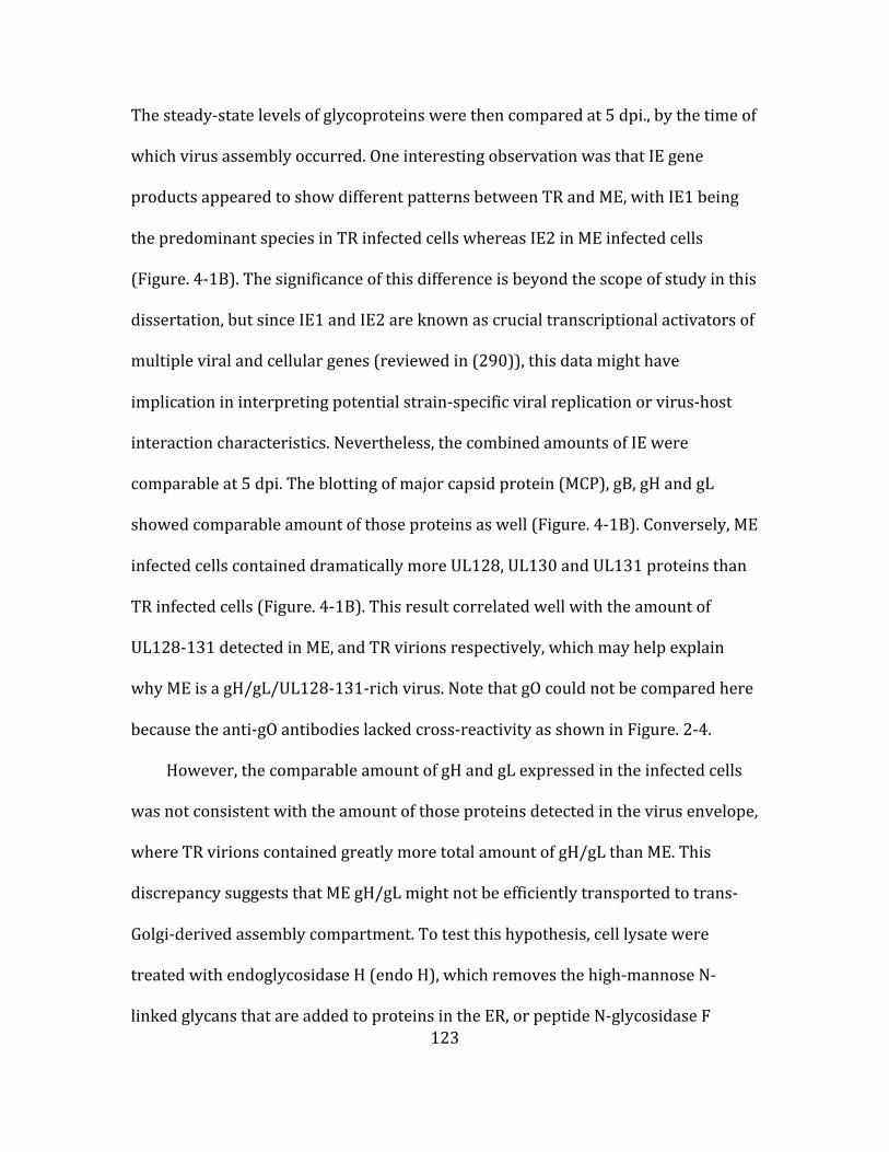
123
The steady-‐state levels of glycoproteins were then compared at 5 dpi., by the time of
which virus assembly occurred. One interesting observation was that IE gene
products appeared to show different patterns between TR and ME, with IE1 being
the predominant species in TR infected cells whereas IE2 in ME infected cells
(Figure. 4-‐1B). The significance of this difference is beyond the scope of study in this
dissertation, but since IE1 and IE2 are known as crucial transcriptional activators of
multiple viral and cellular genes (reviewed in (290)), this data might have
implication in interpreting potential strain-‐specific viral replication or virus-‐host
interaction characteristics. Nevertheless, the combined amounts of IE were
comparable at 5 dpi. The blotting of major capsid protein (MCP), gB, gH and gL
showed comparable amount of those proteins as well (Figure. 4-‐1B). Conversely, ME
infected cells contained dramatically more UL128, UL130 and UL131 proteins than
TR infected cells (Figure. 4-‐1B). This result correlated well with the amount of
UL128-‐131 detected in ME, and TR virions respectively, which may help explain
why ME is a gH/gL/UL128-‐131-‐rich virus. Note that gO could not be compared here
because the anti-‐gO antibodies lacked cross-‐reactivity as shown in Figure. 2-‐4.
However, the comparable amount of gH and gL expressed in the infected cells
was not consistent with the amount of those proteins detected in the virus envelope,
where TR virions contained greatly more total amount of gH/gL than ME. This
discrepancy suggests that ME gH/gL might not be efficiently transported to trans-‐
Golgi-‐derived assembly compartment. To test this hypothesis, cell lysate were
treated with endoglycosidase H (endo H), which removes the high-‐mannose N-‐
linked glycans that are added to proteins in the ER, or peptide N-‐glycosidase F

124
Figure 4-‐2 Western blotting analysis of the maturation of HCMV gH/gL in the infected fibroblast cells. TR-‐ and ME-‐infected HFF cell lysate (A), or ME-‐infected HFFtet cell lysate (B) were either left untreated (-‐), or treated with endo H (H) or PNGase F (F), and then were separated by reducing SDS-‐PAGE and analyzed by western blotting with antibodies specific to gH or gL. Bands corresponding to glycosylated form, or endo H resistant form of the protein were pointed by arrow; and the deglycosylated forms of the protein either by endo H or PNGase F was pointed by arrowhead. Mass markers (kilodaltons) are shown on the left.
(PNGase F), which removes all types of N-‐linked glycans, including those ones that
have been modified by enzymes in the Golgi compartments. Thus, glycoproteins
retained in the ER should stay endo H sensitive, resulting in a faster migrating band
on SDS-‐PAGE gels, while the ones present in Golgi should be endo H resistant. And
glycoproteins in either ER or Golgi compartments should be sensitive to PNGase F.
The treated samples were analyzed by western blotting under reducing conditions
and assessed for protein mobility. As expected, gH and gL in both TR and ME
infected cells were fully sensitive to PNGase F. However, the majority of gH and gL
were in the faster-‐migrating, endo H sensitive form for ME, while the reverse
pattern was true for TR, with larger proportion of gH and gL remaining endo H
resistant (Figure. 4-‐2A). This result confirmed the hypothesis that most of ME gH/gL

125
are not efficiently transported out of ER, which correlated with the significant lower
amount of total gH/gL present in ME virion envelope.
ER to trans-‐Golgi transport of gH/gL is largely dependent on binding of gO or
the UL128-‐131 proteins (80). Analysis of the cell lysate from ME infected fibroblasts
where the expression of UL128-‐131was repressed (HFF-‐tet) showed that there was
even a smaller proportion of endo H resistant gH and gL detected (Figure. 4-‐2B), as
compared to that in regular fibroblast cells as shown in Figure. 4-‐2A. When the
assembly of gH/gL/UL128-‐131was suppressed, the transport of gH/gL should be
mainly dependent on the assembly of gH/gL/gO, thus this result indicated that ME
gH/gL/gO assembly was inherently not efficient even without the competitor
UL128-‐131. This finding was also consistent with previous reports that ME virions
produced from HFF-‐tet cells contained elevated level of gH/gL/gO, but that amount
of gH/gL/gO didn’t compensate the amount of gH/gL that was used to be in the form
of gH/gL/UL128-‐131. In summary, these results suggested that for ME, the endo H
resistant gH/gL observed in Figure. 4-‐2A was largely due to the formation of
gH/gL/UL128-‐131, and that the deficit of total gH/gL in the virion represents an
inefficient assembly of gH/gL/gO.
Recently, Li et al. reported that a viral protein, UL148, likely acted as an ER-‐
chaperone that favors the formation of gH/gL/gO (287). Analysis of TR and ME
infected fibroblast cell lysate showed that ME expressed quite less of UL148 protein
(Figure. 4-‐1B). This result was consistent with the model that UL148 promotes the
assembly of gH/gL/gO, and suggested that the dearth of gH/gL/gO in ME virions
was likely due to both high expression of UL128-‐131 and low expression of UL148.

126
Characterizations of HCMV gO inter-‐strain swap mutants. Compared to the
more conserved gH, gL and Ul128-‐131 proteins, gO is one of the most diverse
proteins encoded by the virus, with 10 to 30% diversity at amino acid sequence
level among the eight families that are categorized based on phylogenetic analysis of
more than 40 clinical isolates (reported in (89) and Chapter 2). Because gH/gL/gO
and gH/gL/UL128-‐131 are formed through competitive interactions, one
compelling hypothesis was that gO sequence diversity might influence the assembly
of the two gH/gL complexes through differential binding affinity to gH/gL. To test
this hypothesis, gO inter-‐strain mutants were constructed by swapping the UL74
ORFs between strain ME and TR. ME virus with TRgO was designated as
MEdeltaTRgO, and TR virus with MEgO was designated as TRdeltaMEgO. Both
mutant viruses were viable, and further characterizations were performed. HFF
cells were infected with MEdeltaTRgO, ME, TR and TRdeltaMEgO at MOI=1, and
extracellular virions were analyzed by western blotting under non-‐reducing
conditions. Gel loads were normalized to total amount of gH/gL for better
comparison of the ratio. Probing with common antigen gL showed that
MEdeltaTRgO was a gH/gL/UL128-‐131-‐rich virus as wild type ME, and
TRdeltaMEgO was a gH/gL/gO-‐rich virus as wild type TR. There might be slightly
more gH/gL/gO in MEdeltaTRgO virus than ME, but this clearly did not fully explain
why ME virion is deficient in gH/gL/gO (Figure. 4-‐3A). Therefore, swapping gO did
not drastically affect the assembly of gH/gL complexes, suggesting that the sequence
diversity of gO did not greatly influence the binding affinity of gO to gH/gL.

127
Figure 4-‐3 Characterizations of HCMV gO inter-‐strain swap mutants. (A) Cell-‐free virions of MEdeltaTRgO, ME, TR and TRdeltaMEgO were analyzed by western blotting under non-‐reducing condition. Gel loads were normalized to equal amount of total gL and blot was probed with antibody specific to gL. Mass markers (kilodaltons) are shown on the left. (B) HFF cells were infected with the indicated viruses at MOI=0.1, and cell-‐free supernatants were retained at different time points after infection and then titrated on HFF to measure the infectivity of cell-‐free progeny viruses. (Note: titer of ME and TRdeltaMEgO were zero at 4 dpi, which cannot be plotted on a log scale axis).
To test whether the virus infectivity is altered in the presence of different gO
isoform, multi-‐step growth curve analysis was performed. HFF cells were infected
by the indicated viruses at MOI=0.1. Extracellular viruses were harvested at various
time points post infection, and the infectivity was measured by plaque assay on HFF
cells. TR produced higher peak titers of cell-‐free virus than those of ME by more
than 40-‐fold, which is consistent with previous report in Chapter 3 that TR had a
better specific infectivity on fibroblasts than ME (291). Mutant viruses exhibited
roughly similar pattern as their corresponding wild type viruses. However, in both
genetic backgrounds, TRgO seemed to correlate with a more robust replication of

128
the virus (Figure. 4-‐3B). Since this experiment was only performed once, no
definitive conclusion can be drawn so far.
Characterizations of HCMV gH N-‐term inter-‐strain swap mutants. There are
small amino acids polymorphisms residing on the N-‐term of gH. It seems possible
that these polymorphisms might influence the assembly of gH/gL complexes. To test
this hypothesis, gH N-‐term inter-‐strain mutants were constructed by swapping
between the N-‐term of 10~36 amino acids sequence of ME gH and 10~37 amino
acid sequence of TR gH. Designations of the mutant viruses mimic gO swap mutants
for the purpose of consistency, with MEdeltaTRgH and TRdeltaMEgH respectively.
To characterize the level of gH/gL/gO and gH/gL/UL128-‐131 on virion
envelope, fibroblast HFF cells were infected by TR, TRdeltaMEgH, ME and
MEdeltaTRgH at MOI=1. Extracellular virions were harvested and analyzed by
western blotting under reducing conditions (Figure. 4-‐4A). When gel loads were
normalized to equal amount of MCP, all viruses contained comparable amount of gB,
while TR contained more total amount of gH/gL than ME (consistent with the report
in Chapter 3). Swapping the N-‐term of gH didn’t dramatically change this pattern.
Analysis of gH/gL complexes ratio by non-‐reducing western blotting showed that
the vast majority of gH/gL was in the form of gH/gL/gO in TR while most of gH/gL
in ME was in the form of gH/gL/UL128-‐131 (consistent with report in Chapter 3).
The corresponding mutant viruses didn’t change the ratio pattern either (Figure. 4-‐
4B). Thus, N-‐term polymorphisms in gH didn’t dramatically influence the assembly
of gH/gL complexes.
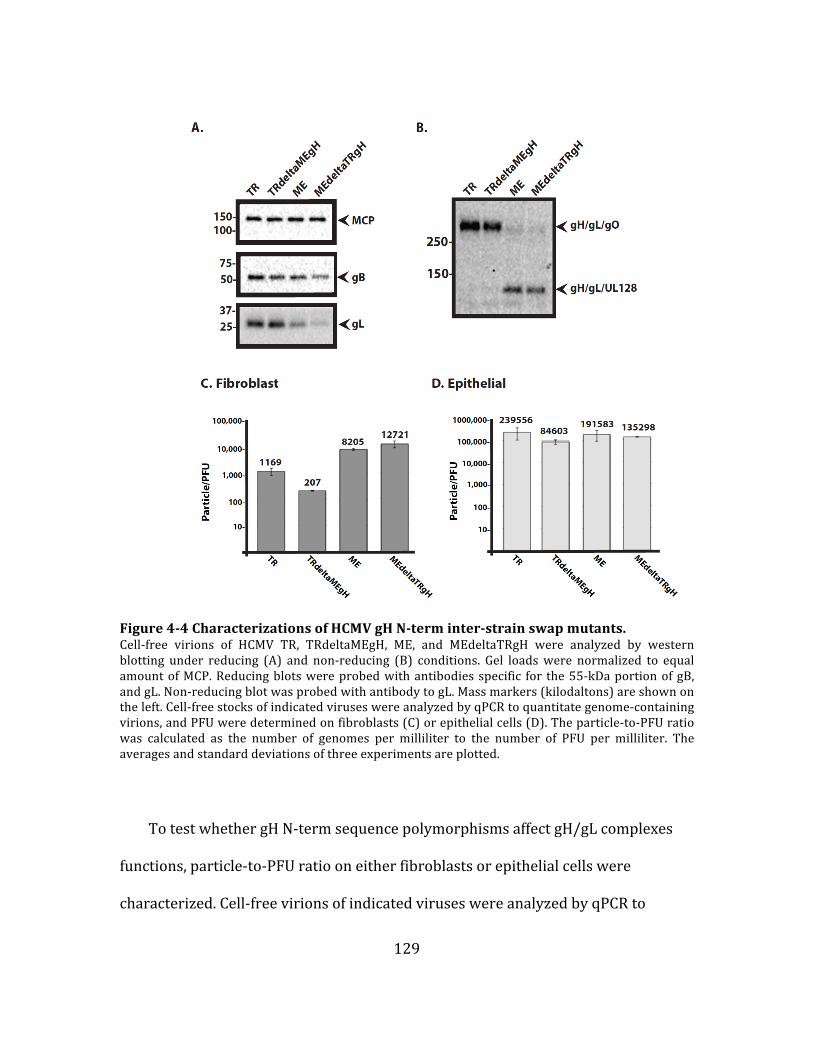
129
Figure 4-‐4 Characterizations of HCMV gH N-‐term inter-‐strain swap mutants. Cell-‐free virions of HCMV TR, TRdeltaMEgH, ME, and MEdeltaTRgH were analyzed by western blotting under reducing (A) and non-‐reducing (B) conditions. Gel loads were normalized to equal amount of MCP. Reducing blots were probed with antibodies specific for the 55-‐kDa portion of gB, and gL. Non-‐reducing blot was probed with antibody to gL. Mass markers (kilodaltons) are shown on the left. Cell-‐free stocks of indicated viruses were analyzed by qPCR to quantitate genome-‐containing virions, and PFU were determined on fibroblasts (C) or epithelial cells (D). The particle-‐to-‐PFU ratio was calculated as the number of genomes per milliliter to the number of PFU per milliliter. The averages and standard deviations of three experiments are plotted.
To test whether gH N-‐term sequence polymorphisms affect gH/gL complexes
functions, particle-‐to-‐PFU ratio on either fibroblasts or epithelial cells were
characterized. Cell-‐free virions of indicated viruses were analyzed by qPCR to

130
measure genome-‐containing virions and plaque assay on both cells type to
determine titers, and particle-‐to-‐PFU ratio was calculated. Consistent with the
report in Chapter 3, on fibroblasts TR was more infectious than ME. Interestingly,
TRdeltaMEgH was about 5-‐fold more infectious than TR while MEdeltaTRgH was
reciprocally less infectious than ME, although with less than 2-‐fold difference
(Figure. 4-‐4C). Since the amount of gH/gL/gO was not greatly changed, it is possible
that the gained replication efficiency of TRdeltaMEgH on fibroblast cells is due to the
qualitative differences in gH/gL/gO complex exerted by ME N-‐term of gH. Particle-‐
to-‐PFU measurements were generally higher on epithelial cells. Extremes were TR
and TRdeltaMEgH, again with TRdeltaMEgH slightly more infectious than TR;
MEdeltaTRgH and ME were comparable to each other at intermediate values
(Figure. 4-‐4D). Therefore, the specific infectivity on epithelial cells was roughly
unaffected by swapping the N-‐term of gH.
DISCUSSION
During initial infection, dissemination, establishment of latent/persistent
infection, and reactivation/shedding, HCMV requires a “entry machinery” that
enables itself to enter a wide array of distinct cell types in the host. HCMV employs
two gH/gL complexes that could influence cell tropism. Our current model suggests
that gH/gL/gO provides the “core-‐fusion function” for entry into all cell types,
probably through interaction with the fusion protein gB. Meanwhile, gH/gL/UL128-‐
131 acts through a distinct function for virus entry into select cell types, like
epithelial/endothelial cells, which broadens viral tropism. The fact that different

131
HCMV strains vary in the relative amount of gH/gL/gO and gH/gL/UL128-‐131 on
the virion envelope indicates that the amount of the two gH/gL complexes could be
adjusted by the virus differentially. However, the mechanisms that regulate the
assembly of the two gH/gL complexes have remained mostly elusive.
Comparison of glycoproteins steady-‐state levels in the infected cells showed
that strain ME expressed considerably more UL128-‐131 proteins, which correlated
well with the high level of gH/gL/Ul128-‐131 in its virion. And this result is
consistent with the observation reported in Murrell et al., where polymorphisms in
the UL128-‐131 locus of strains TB40/E and FIX, both of which contained little
gH/gL/UL128-‐131 in the virion, when introduced into ME, were found to reduce the
incorporation of gH/gL/UL128-‐131 (281). However, ME is not only gH/gL/UL128-‐
131-‐rich, but also contains less total amount of gH/gL, and apparently the UL128-‐
131 locus cannot be the sole reason to explain why ME behaves the way it does.
Intracellular trafficking studies showed that ME gH/gL was poorly transported from
ER to trans-‐Golgi apparatus, albeit the similar expression levels of gH/gL compared
to strain TR, which contains a lot more total amount of gH/gL in the virion. The
finding that even less efficient transport of ME gH/gL from ER to trans-‐Golgi
apparatus in fibroblast cells where the expression of UL128-‐131 were repressed
suggested that the inefficient transport of ME gH/gL was likely due to the poor
assembly of gH/gL/gO.
Recently, Li et al. reported that another viral glycoprotein UL148 could increase
the virion incorporation of gH/gL/gO, and this effect may or may not comes at the
expense of gH/gL/UL128-‐131 (287). Given the positive role of UL148 in favoring

132
the formation of gH/gL/gO, it is possible that the expression level of UL148 could
affect the assembly of the gH/gL complexes. Data here showed that ME expressed
considerably less UL148, which correlated with its poor assembly efficiency of
gH/gL/gO. In the future, it would be important to characterize whether boost of ME
UL148 level by trans-‐expression would influence the abundance of gH/gL/gO in ME
virion, and ask why different strains of HCMV differ in the expression level of UL148.
Disruption of UL148 in TB decreased the level of gH/gL/gO in virion envelope
without largely increasing the amount of gH/gL/UL128-‐131. The resulting UL148-‐
null virus had similar infectivity on fibroblast cells, but improved replication
efficiency on epithelial cells as compared to wild type TB (287). On the other hand,
disruption of UL148 in ME resulted in a decreased infectivity on epithelial cells
(Brent’s communication with Richard Stanton), although the influence on the level
of gH/gL complexes hasn’t been looked carefully to date. Thus, the effect of UL148
on HCMV infectivity seems to vary depending on the genetic background of the
virus. And this discrepancy draws attention to the unaddressed question, what is
the minimum amount of either gH/gL/gO or gH/gL/UL128-‐131 required for their
function respectively. It is likely that the amount of gH/gL/gO in TB virus is in large
excess above the “threshold” of gH/gL/gO required to promote gB-‐fusion, such that
even though the amount of gH/gL/gO was reduced in UL148-‐null virus, that amount
is still ample to keep gH/gL/gO at comparable efficiency. The little amount of
gH/gL/UL128-‐131 in TB is probably close to the “threshold” of gH/gL/UL128-‐131
required for its function on select cell types, and the increased infectivity of TB
UL148-‐null virus on epithelial cells could be due to the possibility of slightly

133
increased gH/gL/UL128-‐131, which was not detected in Li et al. (287). However, for
ME, a gH/gL/gO-‐deficit virus, the amount of gH/gL/gO it contains might be closer to,
or even below the “threshold”, such that decreasing the amount of gH/gL/gO might
result in a “sharp” drop in infectivity. Disruption of UL148 in ME might result in
reduced amount of gH/gL/gO, which decreases the virus infectivity, like on
epithelial cells, as mentioned above. Therefore, the different amount of gH/gL
complexes that strains of HCMV contain indicates where they are relative to the
minimum amount of either gH/gL/gO or gH/gL/UL128-‐131 required, and that
would be important reference to the interpretation of mutant phenotypes, when the
abundances of gH/gL complexes are changed.
The mechanisms by which UL148 works remain largely uncharacterized at this
time. Interestingly, what Li et al. found is that UL148, an ER retained protein,
exclusively interacted with gH/gL complexes containing UL130 and UL131, but not
with UL128 or gO (287), suggesting that UL148 might compete with either gO or
UL128 for binding onto gH/gL, but the binding of UL148 apparently doesn’t hurt the
loading of UL130/UL131. Therefore, UL148 seems to temporally “reserve” a
reservoir of gH/gL in ER from forming either gH/gL/gO or gH/gL/UL128-‐131
complexes. But how does UL148 eventually favor the assembly of gH/gL/gO? One
possibility is that UL148 passively promote gH/gL/gO formation by occluding
gH/gL/UL128-‐131. Because gO and UL128 each covalently binds to gH/gL through
the disulfide-‐bond with the same cysteine residue on gL, whereas UL130 and UL131
do not (76, 77, 80, 215), binding of UL148 with gH/gL may prevent UL128 from
forming a disulfide-‐bond with gL, which could be an irreversible process required
134
for gH/gL/UL128-‐131 assembly. On the other hand, the resulting intermediate
complex “gH/gL/UL148/U130/UL131” might be less stable given the noncovalent
linkage between UL130/UL131 and gH/gL. Thus, at some point this complex would
dissociate, providing the opportunity for gO to load to gH/gL, which then allows the
assembly of gH/gL/gO. Alternatively, another non-‐mutually exclusive possibility is
that binding of UL148 to gH/gL may actively help gH/gL fold into a favorable
conformation for gO to bind. Since whether UL148 specifically binds to the
immature form of gH/gL/UL130/UL131 or also binds to gH/gL dimer in the absence
of UL128-‐131 is currently not known, it is hard to distinguish the abovementioned
“passive” versus “active” mechanisms. But if UL148 could bind to gH/gL dimer in the
absence of UL128-‐131, and still promote the formation of gH/gL/gO, it would
suggest UL148 could act actively for gH/gL/gO assembly. Nevertheless, in either
scenario, the loading of gO would likely displace UL148 in some manner in order to
form gH/gL/gO. Characterization of the working mechanism of UL148 would be an
interesting spot of research in the future.
Although neither gO or gH N-‐term inter-‐strain swap mutants showed obvious
changes on the viral composition of gH/gL complexes, there were slightly better
replication efficiency associated with TRgO, and the N-‐term of MEgH on fibroblast
cells respectively. It is not clear yet if the increased infectivity is the result of slight
changes in the amount of gH/gL/gO, or is indicative of functional differences of
TRgO and N-‐term of MEgH. Future work is needed to clarify those possibilities in
detail, and if the growth difference is not due to the alteration in the abundance of
gH/gL/gO, these results would be the first piece of evidence suggesting that the
135
sequence diversity of gO or the polymorphisms of gH N-‐term could influence the
function of gH/gL/gO.
In summary, studies in this chapter helped to sort out some viral factors that
underpin the differential regulation of the gH/gL complexes assembly, that was
expression levels of UL128-‐131 and UL148 likely contribute to the regulation
through the influence on the competitive assembly between gH/gL/gO and
gH/gL/UL128-‐131.

136
CHAPTER 5. GENERAL DISCUSSION AND
FUTURE DIRECTIONS

137
HCMV is a significant public health concern due to the severe diseases that it
can cause in immunocompromised and/or immunoimmature individuals. The
toxicity and drug-‐resistance associated with antiviral drugs and the lack of licensed
vaccine have highlighted the need for a better understanding of the molecular
biology of the virus, which will, in turn, aid the development of effective vaccines
against this virus, as well as the design of antiviral therapeutic treatment in the
future.
The overall focus of this dissertation is on the mechanisms by which HCMV
manages to enter different cell types. The current consensus is that the interaction
between gH/gL and the fusion protein gB serves as the conserved entry mechanism
among all herpesviruses (reviewed in (189)). Additionally, each herpesvirus
encodes accessory proteins that bind to gH/gL, and can regulate the action of the
core fusion machinery. HCMV gH/gL can be bound by gO, or by the set of proteins
UL128, UL130, and UL131, forming gH/gL/gO and gH/gL/UL128-‐131 complexes on
the virion envelope (73, 74, 76, 77, 80, 227). There has been a lot of interest in these
complexes as targets for vaccine development. However, the mechanisms by which
these complexes facilitate virus entry are still poorly understood. Moreover, the
specific function of gO in gH/gL/gO complex has not been addressed in detail yet.
gO is among the most diverse proteins encoded by the virus. There are at least 8
isoforms of gO in nature, varying by 10~30% at amino acid sequence level (89). It is
reasonable to hypothesize that the amino acid differences of gO might affect the
properties of gO isoforms. Thus, the research work in this dissertation was started
by addressing the function of gO through comparative analysis of different gO

138
isoforms. The results of these studies, as detailed in Chapter 2, indicated that gO
and UL128-‐131 competed for binding to gH/gL, and that the ratio of the two
complexes on the virion envelope can vary dramatically among strains. The finding
that some strains contain mostly gH/gL/gO, whereas others contain mostly
gH/gL/UL128-‐131 was quite surprising. While these studies did not shed light on
the function of gO, they did provide an opportunity to address the important roles
that the two gH/gL complexes play in virus entry into different cell types.
gH/gL and gB comprised the minimum set of requirement for HCMV fusion in
cell-‐cell fusion experiment, suggesting HCMV gH/gL is able to promote gB-‐mediated
fusion (216). This function of gH/gL is referred to as the “core fusion function”.
However, in virus scenario, there is little, if any, unbound gH/gL. Most of the gH/gL
is in the form of gH/gL/gO or gH/gL/UL128-‐131 complexes, thus it is possible that
either one of those two complexes or both of them can perform the core fusion
function.
The functions of HCMV gH/gL complexes have been mostly studied with
deletion mutants. UL128-‐131 deletion mutants replicate well on fibroblasts but fail
to enter epithelial/endothelial cells, suggesting that gH/gL/UL128-‐131 is required
for entry into epithelial/endothelial cells but is dispensable on fibroblasts. This
phenotype is often interpreted to mean that entry into epithelial/endothelial cells is
facilitated by gH/gL/UL128-‐131, whereas entry into fibroblasts is facilitated by
gH/gL/gO. The resulting model suggests that both gH/gL/UL128-‐131, and
gH/gL/gO provide essentially the same function for entry into different cell types,
i.e., promoting gB-‐mediated membrane fusion. This was an attractive model due to

139
the analogy with the well-‐studied Epstein-‐Barr virus (EBV) tropism model. EBV’s
gH/gL exists in two forms as well, naked gH/gL or gH/gL/gp42 complex. Entry into
epithelial cells is facilitated by gH/gL, through interactions with specific integrin on
the cell surface. Binding with gp42 blocks the interaction of gH/gL with integrin,
instead, gH/gL/gp42 interacts with MHCII molecule on B cell surface, thus facilitates
virus entry into B cells. Therefore, EBV gH/gL and gH/gL/gp42 can both promote
gB-‐mediated fusion into different cell types, with gp42 acting like a tropism switch
adapter. While UL128-‐131 deletion mutants replicate well on fibroblasts, gO
deletion mutants fail to enter any cell type, suggesting that gH/gL/UL128-‐131 is not
sufficient to promote gB-‐mediated fusion into epithelial/endothelial cells, and entry
into that cell type requires both complexes. However, further analysis is largely
limited due to the severe growth defect of the gO mutants and the lack of a
complementation system.
The work in Chapter 3 avoided the caveats of gO-‐null mutants by employing a
panel of HCMV strains that contained different amounts of gH/gL/gO and
gH/gL/UL128-‐131 complexes. Since none of the viruses were absolutely lacking
either complex, they did not exhibit severe growth defect as observed in gO-‐null
mutants, thus the infectivity measured were more biologically relevant. Cell-‐type
specific particle-‐to-‐PFU ratios were determined and showed that infection of both
fibroblasts and epithelial cells was positively correlated with the abundance of
gH/gL/gO, but not with gH/gL/UL128-‐131. And the low infectivity of the gH/gL/gO-‐
deficit virus on both cell types could be recovered by treatment with chemical
fusogen PEG, indicating that the deficiency of infection was due to less efficient
140
fusion. These results confirmed that infection of fibroblasts requires gH/gL/gO, and
further showed that infection of epithelial cells requires both gH/gL/gO and
gH/gL/UL128-‐131. The sufficient role of gH/gL/gO in promoting gB-‐mediated
fusion on fibroblasts demonstrated that gH/gL/gO is clearly fusion competent. The
necessary role of gH/gL/gO on epithelial cells suggested that gH/gL/gO likely acts
as the fusion competent complex on that cell type as well. This speculation supports
the data described above better than either of the other two explanations: 1)
gH/gL/gO is fusion competent on fibroblasts but not on epithelial cells, instead it
performs an alternate function. 2) The two different gH/gL complexes perform the
same (redundant) core fusion function on epithelial cells. Therefore, we proposed a
new model where gH/gL/gO provides the conserved herpesvirus gH/gL “core fusion
function” of promoting gB-‐mediated fusion for entry into all cell types, whereas
gH/gL/UL128-‐131 provides a non-‐fusion, yet necessary mechanism to allow
infection of select cell types (i.e. epithelial/endothelial cells). This is a lot different
from the EBV tropism model, and to our knowledge, is also the first report of a
fusion-‐incompetent gH/gL complexes in the herpesvirus field, which might provide
an important opportunity to study the fundamental mechanism by which gH/gL
regulates gB fusion. These results also help explained the cell type-‐dependent virus
neutralization exhibited by anti-‐gH/gL/UL128-‐131 antibodies, and underscore the
importance of gH/gL/gO as a potential HCMV vaccine or therapeutic target.
Several important questions regarding to the model are still remaining to be
addressed: 1) what underpins the distinct functions exhibited in gH/gL/gO and
gH/gL/UL128-‐131? , 2) what is the non-‐fusion function of gH/gL/UL128-‐131? , and
141
3) What is gO’s role in the fusion competent complex gH/gL/gO? Some speculations
regarding those questions will be discussed below.
What underpins the distinct functions exhibited in gH/gL/gO and gH/gL/UL128-‐
131? Studies of HSV and EBV suggest that the surfaces of gH/gL could directly
contact with gB to promote membrane fusion (reviewed in (189)). For HCMV, cell-‐
cell fusion assay demonstrated that gH/gL and gB comprised the minimum
requirement for membrane fusion, indicating that HCMV gH/gL is capable of
triggering gB-‐fusion as well (216). The distinct function model raises the possibility
that the “profusion” surfaces of HCMV gH/gL may be differentially formed or
exposed on gH/gL/gO versus gH/gL/UL128-‐UL131. This discrepancy might be due
to the direct binding with gO versus UL128-‐131, or allosteric conformation changes
of gH/gL as a result of the binding with gO versus UL128-‐131. Although Cryo EM
revealed overall similar architecture between the two complexes (215, 215),
detailed structure differences were not resolved in those studies. Our lab have
created a library of gH and gL charge-‐cluster-‐to-‐alanine mutants, which might allow
more detailed structure differences between gH/gL/gO and gH/gL/UL128-‐131 to be
elucidated in the future.
The other possibility is that gH/gL/gO and gH/gL/UL128-‐131 might not be
uniformly distributed on the virion envelope; in other words, they might cluster in
different “landscape”. One speculation could be that the fusion competent gH/gL/gO
complex might either be spatially closer to gB or be more flexible in mobility on the
viral membrane, such that upon triggering, it could interact with gB with proximity
or “swim” around and have higher probability to interact with gB. In contrast, fusion

142
incompetent gH/gL/UL128-‐131 complex might be more spatially sequestered or
restricted in mobility. Precedents have been reported in herpesvirus that
glycoproteins could be associated with the less fluid lipid raft on membranes (292–
294). It could be possible that lipid raft might play a role in the distinct functions
model of gH/gL/gO and gH/gL/UL128-‐131.
What is the non-‐fusion function of gH/gL/UL128-‐131? gH/gL/UL128-‐131 null
virus cannot replicate on epithelial/endothelial cells, but with the aid of chemical
fusogen PEG, the mutant virus could establish infection as well as wild type virus
(79), suggesting that the deficiency is at pre-‐fusion but not post-‐fusion steps.
Infection of HCMV on epithelial/endothelial cells has been showed to happen
through endocytosis in a pH-‐dependent manner (79). Trans-‐expressed
gH/gL/UL128-‐131 was showed to exert interference on epithelial cells, indicating
that the complex is involved in receptor interaction (229). Moreover, recent study
showed that gH/gL/UL128-‐131 triggered specific signaling transduction pathway
for virus internalization into monocytes, the cell type where entry requires
gH/gL/UL128-‐131 as epithelial/endothelial cells does (230). Taken the above
evidences together, one compelling model would be that gH/gL/UL128-‐131
interacts with its receptors on epithelial/endothelial cell surface and triggers
specific signaling/endocytosis pathway, the process of which is required for
subsequent gH/gL/gO-‐promoted core fusion event to occur.
However, it is worth to note that gH/gL/UL128-‐131-‐null viruses are still
efficiently internalized into epithelial/endothelial cells (79, 231, 258, 295),
suggesting gH/gL/UL128-‐131 is dispensable for the internalization of the virus.

143
Perhaps, it is not surprising that non-‐specific endocytosis could be triggered even
without gH/gL/UL128-‐131 given that the virus contains numerous different
glycoproteins on the virion envelop. However, the non-‐specific endocytosis would
not lead to productive infection, indicating that efficient fusion can only occur in
specific endosome compartments triggered by gH/gL/UL128-‐131-‐dependent
endocytosis.
What is gO’s role in the fusion-‐competent complex gH/gL/gO? It seems quite
clear now that at least one conserved function of gO is to facilitate the incorporation
of the fusion-‐competent gH/gL/gO into the virion envelope. But does gO perform
more function other than that?
gH/gL/gO expressed in trans system exerted interference on fibroblasts but not
on epithelial cells (239), suggesting that the complex engages some currently
uncharacterized receptor on the surface of fibroblasts. This receptor-‐gH/gL/gO
interaction likely triggers gB-‐mediated fusion, resulting in virus entry into
fibroblasts through either direct fusion with plasma membrane or from within
endosomes followed by macropinocytosis at neutral pH (185, 186). The negative
data of interference on epithelial cells could be explained at least by the following
two possibilities: 1) in a trans-‐expressed system, gH/gL/gO cannot reach to a
compartment where its receptor resides. In fact, trans-‐expressed gH/gL/gO was
showed to be mostly retained in Golgi-‐like compartments (239). It is possible that
receptor of gH/gL/gO in epithelial cells is sequestered in different compartments,
such as early endosomes or multivesicular bodies, thus making gH/gL/gO-‐receptor
interaction spatially impossible. 2) Or simply the receptor for gH/gL/gO is not
144
available on epithelial cells, suggesting that the conformational change of the
complex on epithelial cells does not require a receptor; instead, the ligand could be a
proton, which is acquired in low-‐pH endosomes after endocytosis. Recently, there is
evidence suggesting that gH/gL/gO can bind to PDGFRα, which is selectively
expressed on fibroblasts but not on epithelial cells (Manuscript reviewed by Brent).
This seems to favor the latter possibility, and also help to explain that the
requirement for gH/gL/UL128-‐131 on epithelial cells is probably to overcome the
lack of gH/gL/gO receptor, and that gH/gL/UL128-‐131-‐dependent endocytosis
somehow provides an alternative way for gH/gL/gO-‐mediated gB fusion to occur.
gO might directly involve in gH/gL/gO’s function (i.e. receptor interaction), and
it is conceivable that gO sequence diversity might affect the “efficiency” of gH/gL/gO
in performing its job. In line with this hypothesis, ME-‐TR inter-‐strain gO swap
mutants displayed slight growth difference as compared to wild type virus (data in
Chapter 4). However, more detailed studies in the future are warranted to verify
whether the growth difference is due to entry, and also whether this observation
applies to other gO isoforms. Moreover, since gO is heavily glycosylated, it is
possible that it could also act like a glycan shield, protecting gH/gL from
neutralizing antibodies. No research has been carried out in testing this hypothesis
in the context of gO sequence diversity. Our lab has created a library of anti-‐peptide
antibodies raised against gH and gL and there are also anti-‐gH mAbs library created
by Brent’s colleague Bill Britt. So future research could include the screening of the
strain-‐specific neutralizing antibodies from the above antibody resources and
testing the correlation of neutralization specificity with gO isoforms.

145
In summary, the work in Chapter 2 and Chapter 3 showed that different strains
of HCMV varied dramatically in the relative amount of gH/gL/gO and gH/gL/UL128-‐
131, and this could influence virus infectivity on different cell types due to the
distinct functions of the two gH/gL complexes during entry and tropism. The next
question was that why strains differ in the abundance of the gH/gL /gO and
gH/gL/UL128-‐131. Recently, Li et al. reported a viral protein, UL148, which might
act as an ER-‐chaperone to promote the formation of gH/gL/gO over gH/gL/UL128-‐
131 (287). The research work in Chapter 4 found that strains of HCMV varied in the
protein expression level in the infected cells. ME expressed much more of UL128-‐
131 proteins, but less of UL148 protein as compared to TR. gH/gL levels were more
comparable, however while TR gH/gL were efficiently transported to the trans-‐
Golgi derived virus assembly compartment, ME gH/gL remained mostly in the ER.
These results suggested that both high expression of UL128-‐131 and the low
expression of UL148 probably contributed to the poor assembly of ME gH/gL/gO in
ER, which likely dampened the transport of gH/gL to trans-‐Golgi derived assembly
compartment, and thus resulted in the less total gH/gL, and dearth of gH/gL/gO in
ME virion envelope. Therefore, the assembly of gH/gL/gO and gH/gL/UL128-‐131 is
probably regulated through interstrain variability at multiple loci. In the future, a
further characterization of how the virus adjusts the level of those viral factors (i.e.,
Ul128-‐131 and UL148) would help us better understand how HCMV determines
tropism and navigates through the host.
The abovementioned distinct function model is focused on the contribution of
gH/gL complexes to cell-‐free entry of HCMV. Another way of virus infection is via

146
cell-‐to-‐cell spread. While cell-‐free entry is apparently important for HCMV to
establish infection in a new host, cell-‐to-‐cell spread is probably essential for virus
dissemination within a host and can be one of the strategies that the virus utilize to
evade immune surveillance. Cell-‐free infection and cell-‐to-‐cell spread could be
mechanistically distinguished by using neutralizing antibodies: cell-‐free entry is
sensitive to neutralizing antibodies whereas cell-‐to-‐cell spread is largely resistant.
How HCMV spreads cell-‐to-‐cell is currently unknown. The fact that double deletions
of gO and UL128-‐131 proteins are lethal to the virus indicates that viral spread
absolutely depends on either of, or both of gH/gL/gO and gH/gL/UL128-‐131, but
not on gH/gL dimer (180). Single deletion of either gO or UL128-‐131 proteins
results in a phenotype with similar spreading efficiency on fibroblast cells,
suggesting that gH/gL/gO and gH/gL/UL128-‐131 probably provide redundant
function for viral spread on fibroblast cells (79, 180). On epithelial cells, single
deletion mutants of UL128-‐131 proteins cannot spread, while single deletion
mutants of gO spread slightly better, suggesting that gH/gL/UL128-‐131 is necessary
and sufficient for cell-‐to-‐cell spread on epithelial cells, but gH/gL/gO is dispensable
(79, 238). Note how gH/gL/gO and gH/gL/UL128-‐131 are differentially required in
cell-‐free entry versus cell-‐to-‐cell spread, and this discrepancy can be important
consideration to take when it comes to studies on the molecular biology of HCMV
transmission and dissemination. An intriguing difference between virus cell-‐to-‐cell
spread and transiently cell-‐cell fusion is that gH/gL dimer is sufficient to trigger gB-‐
mediated cell-‐cell fusion but it cannot do it in the context of virus cell-‐to-‐cell spread.
This fits so well with the observation that HCMV infection usually would not result
147
in the formation syncytium (massive cell-‐cell fusion), while gH/gL and gB expressed
in trans exclusively generates syncytium, indicating that the cell-‐to-‐cell spread
during virus infection is somehow less “disruptive” and mechanistically different
from gH/gL and gB promoted cell-‐cell fusion in trans. One speculation could be that
during HCMV infection, gH/gL dimer is prevented to reach to the cell surface or cell-‐
cell junction where it can interact with gB to trigger cell-‐cell fusion. If so, UL148
might be a potential candidate since it is an ER-‐chaperone that can bind to gH/gL
(Kamil’s new abstract), and probably holds off the immature gH/gL in ER, thus
negatively controls the extent of cell-‐cell fusion during virus infection.
HCMV is a virus that has been successfully adapted to the human host, which
exhibits significant levels of genetic diversity between individuals (interhost
diversity) and also within a single individual (intrahost diversity) (reviewed in
(142)). The intrahost diversity of HCMV has been largely underappreciated
previously in the field, and it might have important influence on virus pathogenesis.
This diversity may provide a mechanism by which the virus could rapidly adapt to
the changing host environments through influences on virus ability of dissemination.
The possible role in dissemination is likely based on viral glycoproteins, which are
involved in virus entry and tropism. Even with the glycoproteins that this
dissertation studies, there was a dramatic variation that different HCMV strains
exhibit in the relative amount of gH/gL/gO and gH/gL/UL128-‐131 complexes on the
virion envelope and this variation significantly influenced virus infectivity on
different cell types. It is conceivable that there are genetic bases that attribute to
this trait. One possibility is that during infection in human host, HCMV exists as a

148
diverse population that may harbor different genetic variations affecting the relative
abundance of gH/gL/gO and gH/gL/UL128-‐131, such that upon encountering
distinct host tissue/cell, subset of the population could replicate better than the
others, resulting in overall efficient dissemination. Although no clear conclusion can
be drawn to date on the correlation between the variation of HCMV genotypes and
the risk of virus transmission, or the outcome and severity of diseases, HCMV
intrahost diversity is an important aspect that needs more appreciation and might
help with future vaccine or antiviral drug design.

149
REFERENCES 1. 2007. Human Herpesviruses, Arvin A, Campadelli-Flume G, Mocarski E, Moore
PS, Roizman B, Whitley R, Yamanishi K (ed), Cambridge University Press, Cambridge.
2. Görzer I, Kerschner H, Jaksch P, Bauer C, Seebacher G, Klepetko W, Puchhammer-Stöckl E. 2008. Virus load dynamics of individual CMV-genotypes in lung transplant recipients with mixed-genotype infections. J Med Virol 80:1405–1414.
3. Novak Z, Ross SA, Patro RK, Pati SK, Kumbla RA, Brice S, Boppana SB. 2008. Cytomegalovirus strain diversity in seropositive women. J Clin Microbiol 46:882–886.
4. Murphy E, Shenk T. 2008. Human cytomegalovirus genome. Curr Top Microbiol Immunol 325:1–19.
5. Ross SA, Novak Z, Pati S, Patro RK, Blumenthal J, Danthuluri VR, Ahmed A, Michaels MG, Sánchez PJ, Bernstein DI, Tolan RW, Palmer AL, Britt WJ, Fowler KB, Boppana SB. 2011. Mixed infection and strain diversity in congenital cytomegalovirus infection. J Infect Dis 204:1003–1007.
6. Dieamant DC, Bonon SH, Peres RM, Costa CR, Albuquerque DM, Miranda EC, Aranha FJ, Oliveira-Duarte G, Fernandes VC, De Souza CA, Costa SC, Vigorito AC. 2013. Cytomegalovirus (CMV) genotype in allogeneic hematopoietic stem cell transplantation. BMC Infect Dis 13:310.
7. Renzette N, Bhattacharjee B, Jensen JD, Gibson L, Kowalik TF. 2011. Extensive genome-wide variability of human cytomegalovirus in congenitally infected infants. PLoS Pathog 7:e1001344.
8. Staras SA, Dollard SC, Radford KW, Flanders WD, Pass RF, Cannon MJ. 2006. Seroprevalence of cytomegalovirus infection in the United States, 1988-1994. Clin Infect Dis 43:1143–1151.
9. Bate SL, Dollard SC, Cannon MJ. 2010. Cytomegalovirus seroprevalence in the United States: the national health and nutrition examination surveys, 1988-2004. Clin Infect Dis 50:1439–1447.
10. Dowd JB, Aiello AE, Alley DE. 2009. Socioeconomic disparities in the seroprevalence of cytomegalovirus infection in the US population: NHANES III. Epidemiol Infect 137:58–65.
11. Sun X, Liu Z, Wang B, Shi L, Liang R, Li L, Qian D, Song X. 2013. Sero-epidemiological survey of human cytomegalovirus-infected children in Weifang (Eastern China) between 2009 and 2012. Virol J 10:42.
12. Griffiths P, Baraniak I, Reeves M. 2015. The pathogenesis of human cytomegalovirus. J Pathol 235:288–297.
13. Beam E, Razonable RR. 2012. Cytomegalovirus in solid organ transplantation: epidemiology, prevention, and treatment. Curr Infect Dis Rep 14:633–641.
14. Ariza-Heredia EJ, Nesher L, Chemaly RF. 2014. Cytomegalovirus diseases after hematopoietic stem cell transplantation: a mini-review. Cancer Lett 342:1–8.
15. Opelz G, Döhler B, Ruhenstroth A. 2004. Cytomegalovirus prophylaxis and graft outcome in solid organ transplantation: a collaborative transplant study report. Am J

150
Transplant 4:928–936. 16. Streblow DN, Orloff SL, Nelson JA. 2007. Acceleration of allograft failure by
cytomegalovirus. Curr Opin Immunol 19:577–582. 17. Gaytant MA, Steegers EA, Semmekrot BA, Merkus HM, Galama JM. 2002.
Congenital cytomegalovirus infection: review of the epidemiology and outcome. Obstet Gynecol Surv 57:245–256.
18. Britt W. 2015. Controversies in the natural history of congenital human cytomegalovirus infection: the paradox of infection and disease in offspring of women with immunity prior to pregnancy. Med Microbiol Immunol 204:263–271.
19. Cannon MJ, Davis KF. 2005. Washing our hands of the congenital cytomegalovirus disease epidemic. BMC Public Health 5:70.
20. Cheeran MC, Lokensgard JR, Schleiss MR. 2009. Neuropathogenesis of congenital cytomegalovirus infection: disease mechanisms and prospects for intervention. Clin Microbiol Rev 22:99–126, Table of Contents.
21. Boppana SB, Fowler KB, Pass RF, Rivera LB, Bradford RD, Lakeman FD, Britt WJ. 2005. Congenital cytomegalovirus infection: association between virus burden in infancy and hearing loss. J Pediatr 146:817–823.
22. Arvin AM, Fast P, Myers M, Plotkin S, Rabinovich R, National VAC. 2004. Vaccine development to prevent cytomegalovirus disease: report from the National Vaccine Advisory Committee. Clin Infect Dis 39:233–239.
23. Krause PR, Bialek SR, Boppana SB, Griffiths PD, Laughlin CA, Ljungman P, Mocarski ES, Pass RF, Read JS, Schleiss MR, Plotkin SA. 2013. Priorities for CMV vaccine development. Vaccine 32:4–10.
24. Stagno S, Pass RF, Cloud G, Britt WJ, Henderson RE, Walton PD, Veren DA, Page F, Alford CA. 1986. Primary cytomegalovirus infection in pregnancy. Incidence, transmission to fetus, and clinical outcome. JAMA 256:1904–1908.
25. Boppana SB, Fowler KB, Britt WJ, Stagno S, Pass RF. 1999. Symptomatic congenital cytomegalovirus infection in infants born to mothers with preexisting immunity to cytomegalovirus. Pediatrics 104:55–60.
26. Fisher S, Genbacev O, Maidji E, Pereira L. 2000. Human cytomegalovirus infection of placental cytotrophoblasts in vitro and in utero: implications for transmission and pathogenesis. J Virol 74:6808–6820.
27. Boppana SB, Rivera LB, Fowler KB, Mach M, Britt WJ. 2001. Intrauterine transmission of cytomegalovirus to infants of women with preconceptional immunity. N Engl J Med 344:1366–1371.
28. Ross SA, Arora N, Novak Z, Fowler KB, Britt WJ, Boppana SB. 2010. Cytomegalovirus reinfections in healthy seroimmune women. J Infect Dis 201:386–389.
29. Revello MG, Gerna G. 2002. Diagnosis and management of human cytomegalovirus infection in the mother, fetus, and newborn infant. Clin Microbiol Rev 15:680–715.
30. McGeoch DJ, Dolan A, Ralph AC. 2000. Toward a comprehensive phylogeny for mammalian and avian herpesviruses. J Virol 74:10401–10406.
31. McGeoch DJ, Rixon FJ, Davison AJ. 2006. Topics in herpesvirus genomics and evolution. Virus Res 117:90–104.

151
32. Limaye AP, Boeckh M. 2010. CMV in critically ill patients: pathogen or bystander. Rev Med Virol 20:372–379.
33. Sansoni P, Vescovini R, Fagnoni FF, Akbar A, Arens R, Chiu YL, Cičin-Šain L, Dechanet-Merville J, Derhovanessian E, Ferrando-Martinez S, Franceschi C, Frasca D, Fulöp T, Furman D, Gkrania-Klotsas E, Goodrum F, Grubeck-Loebenstein B, Hurme M, Kern F, Lilleri D, López-Botet M, Maier AB, Marandu T, Marchant A, Matheï C, Moss P, Muntasell A, Remmerswaal EB, Riddell NE, Rothe K, Sauce D, Shin EC, Simanek AM, Smithey MJ, Söderberg-Nauclér C, Solana R, Thomas PG, van Lier R, Pawelec G, Nikolich-Zugich J. 2014. New advances in CMV and immunosenescence. Exp Gerontol 55:54–62.
34. Halenius A, Hengel H. 2014. Human cytomegalovirus and autoimmune disease. Biomed Res Int 2014:472978.
35. Horváth R, Cerný J, Benedík J, Hökl J, Jelínková I, Benedík J. 2000. The possible role of human cytomegalovirus (HCMV) in the origin of atherosclerosis. J Clin Virol 16:17–24.
36. DuRose JB, Li J, Chien S, Spector DH. 2012. Infection of vascular endothelial cells with human cytomegalovirus under fluid shear stress reveals preferential entry and spread of virus in flow conditions simulating atheroprone regions of the artery. J Virol 86:13745–13755.
37. 2008. Human Cytomegalovirus, TES, Stinski MF (ed), 325, Springer, Berlin Heideberg.
38. Flint SJ, Enquist LW, Racaniello VR, Skalka AM. 2008. Principles of Virology, (ed), I, ASM press, Washington DC.
39. Chee MS, Bankier AT, Beck S, Bohni R, Brown CM, Cerny R, Horsnell T, Hutchison CA, Kouzarides T, Martignetti JA. 1990. Analysis of the protein-coding content of the sequence of human cytomegalovirus strain AD169. Curr Top Microbiol Immunol 154:125–169.
40. Bhella D, Rixon FJ, Dargan DJ. 2000. Cryomicroscopy of human cytomegalovirus virions reveals more densely packed genomic DNA than in herpes simplex virus type 1. J Mol Biol 295:155–161.
41. Murphy E, Yu D, Grimwood J, Schmutz J, Dickson M, Jarvis MA, Hahn G, Nelson JA, Myers RM, Shenk TE. 2003. Coding potential of laboratory and clinical strains of human cytomegalovirus. Proc Natl Acad Sci U S A 100:14976–14981.
42. Dunn W, Chou C, Li H, Hai R, Patterson D, Stolc V, Zhu H, Liu F. 2003. Functional profiling of a human cytomegalovirus genome. Proc Natl Acad Sci U S A 100:14223–14228.
43. Dolan A, Cunningham C, Hector RD, Hassan-Walker AF, Lee L, Addison C, Dargan DJ, McGeoch DJ, Gatherer D, Emery VC, Griffiths PD, Sinzger C, McSharry BP, Wilkinson GW, Davison AJ. 2004. Genetic content of wild-type human cytomegalovirus. J Gen Virol 85:1301–1312.
44. Butcher SJ, Aitken J, Mitchell J, Gowen B, Dargan DJ. 1998. Structure of the human cytomegalovirus B capsid by electron cryomicroscopy and image reconstruction. J Struct Biol 124:70–76.

152
45. Chee M, Rudolph SA, Plachter B, Barrell B, Jahn G. 1989. Identification of the major capsid protein gene of human cytomegalovirus. J Virol 63:1345–1353.
46. Gibson W, Baxter MK, Clopper KS. 1996. Cytomegalovirus “missing” capsid protein identified as heat-aggregable product of human cytomegalovirus UL46. J Virol 70:7454–7461.
47. Baldick CJ, Shenk T. 1996. Proteins associated with purified human cytomegalovirus particles. J Virol 70:6097–6105.
48. Lai L, Britt WJ. 2003. The interaction between the major capsid protein and the smallest capsid protein of human cytomegalovirus is dependent on two linear sequences in the smallest capsid protein. J Virol 77:2730–2735.
49. Varnum SM, Streblow DN, Monroe ME, Smith P, Auberry KJ, Pasa-Tolic L, Wang D, Camp DG, Rodland K, Wiley S, Britt W, Shenk T, Smith RD, Nelson JA. 2004. Identification of proteins in human cytomegalovirus (HCMV) particles: the HCMV proteome. J Virol 78:10960–10966.
50. Trus BL, Gibson W, Cheng N, Steven AC. 1999. Capsid structure of simian cytomegalovirus from cryoelectron microscopy: evidence for tegument attachment sites. J Virol 73:2181–2192.
51. Zhou ZH, Dougherty M, Jakana J, He J, Rixon FJ, Chiu W. 2000. Seeing the herpesvirus capsid at 8.5 A. Science 288:877–880.
52. Yu X, Shah S, Atanasov I, Lo P, Liu F, Britt WJ, Zhou ZH. 2005. Three-dimensional localization of the smallest capsid protein in the human cytomegalovirus capsid. J Virol 79:1327–1332.
53. Dai X, Yu X, Gong H, Jiang X, Abenes G, Liu H, Shivakoti S, Britt WJ, Zhu H, Liu F, Zhou ZH. 2013. The smallest capsid protein mediates binding of the essential tegument protein pp150 to stabilize DNA-containing capsids in human cytomegalovirus. PLoS Pathog 9:e1003525.
54. Komazin G, Townsend LB, Drach JC. 2004. Role of a mutation in human cytomegalovirus gene UL104 in resistance to benzimidazole ribonucleosides. J Virol 78:710–715.
55. Chen DH, Jiang H, Lee M, Liu F, Zhou ZH. 1999. Three-dimensional visualization of tegument/capsid interactions in the intact human cytomegalovirus. Virology 260:10–16.
56. Kalejta RF. 2008. Tegument proteins of human cytomegalovirus. Microbiol Mol Biol Rev 72:249–65, table of contents.
57. Bechtel JT, Shenk T. 2002. Human cytomegalovirus UL47 tegument protein functions after entry and before immediate-early gene expression. J Virol 76:1043–1050.
58. Baldick CJ, Marchini A, Patterson CE, Shenk T. 1997. Human cytomegalovirus tegument protein pp71 (ppUL82) enhances the infectivity of viral DNA and accelerates the infectious cycle. J Virol 71:4400–4408.
59. Cantrell SR, Bresnahan WA. 2005. Interaction between the human cytomegalovirus UL82 gene product (pp71) and hDaxx regulates immediate-early gene expression and viral replication. J Virol 79:7792–7802.
60. AuCoin DP, Smith GB, Meiering CD, Mocarski ES. 2006. Betaherpesvirus-conserved cytomegalovirus tegument protein ppUL32 (pp150) controls cytoplasmic
153
events during virion maturation. J Virol 80:8199–8210. 61. Gilbert MJ, Riddell SR, Plachter B, Greenberg PD. 1996. Cytomegalovirus
selectively blocks antigen processing and presentation of its immediate-early gene product. Nature 383:720–722.
62. Odeberg J, Plachter B, Brandén L, Söderberg-Nauclér C. 2003. Human cytomegalovirus protein pp65 mediates accumulation of HLA-DR in lysosomes and destruction of the HLA-DR alpha-chain. Blood 101:4870–4877.
63. Abate DA, Watanabe S, Mocarski ES. 2004. Major human cytomegalovirus structural protein pp65 (ppUL83) prevents interferon response factor 3 activation in the interferon response. J Virol 78:10995–11006.
64. Sylwester AW, Mitchell BL, Edgar JB, Taormina C, Pelte C, Ruchti F, Sleath PR, Grabstein KH, Hosken NA, Kern F, Nelson JA, Picker LJ. 2005. Broadly targeted human cytomegalovirus-specific CD4+ and CD8+ T cells dominate the memory compartments of exposed subjects. J Exp Med 202:673–685.
65. Gretch DR, Kari B, Rasmussen L, Gehrz RC, Stinski MF. 1988. Identification and characterization of three distinct families of glycoprotein complexes in the envelopes of human cytomegalovirus. J Virol 62:875–881.
66. Kari B, Lussenhop N, Goertz R, Wabuke-Bunoti M, Radeke R, Gehrz R. 1986. Characterization of monoclonal antibodies reactive to several biochemically distinct human cytomegalovirus glycoprotein complexes. J Virol 60:345–352.
67. Burke HG, Heldwein EE. 2015. Crystal Structure of the Human Cytomegalovirus Glycoprotein B. PLoS Pathog 11:e1005227.
68. Carlson C, Britt WJ, Compton T. 1997. Expression, purification, and characterization of a soluble form of human cytomegalovirus glycoprotein B. Virology 239:198–205.
69. Boyle KA, Compton T. 1998. Receptor-binding properties of a soluble form of human cytomegalovirus glycoprotein B. J Virol 72:1826–1833.
70. Mach M, Kropff B, Dal Monte P, Britt W. 2000. Complex formation by human cytomegalovirus glycoproteins M (gpUL100) and N (gpUL73). J Virol 74:11881–11892.
71. Kari B, Gehrz R. 1993. Structure, composition and heparin binding properties of a human cytomegalovirus glycoprotein complex designated gC-II. J Gen Virol 74:255–264.
72. Krzyzaniak M, Mach M, Britt WJ. 2007. The cytoplasmic tail of glycoprotein M (gpUL100) expresses trafficking signals required for human cytomegalovirus assembly and replication. J Virol 81:10316–10328.
73. Huber MT, Compton T. 1997. Characterization of a novel third member of the human cytomegalovirus glycoprotein H-glycoprotein L complex. J Virol 71:5391–5398.
74. Li L, Nelson JA, Britt WJ. 1997. Glycoprotein H-related complexes of human cytomegalovirus: identification of a third protein in the gCIII complex. J Virol 71:3090–3097.
75. Huber MT, Compton T. 1998. The human cytomegalovirus UL74 gene encodes the third component of the glycoprotein H-glycoprotein L-containing envelope complex. J Virol 72:8191–8197.

154
76. Huber MT, Compton T. 1999. Intracellular formation and processing of the heterotrimeric gH-gL-gO (gCIII) glycoprotein envelope complex of human cytomegalovirus. J Virol 73:3886–3892.
77. Wang D, Shenk T. 2005. Human cytomegalovirus UL131 open reading frame is required for epithelial cell tropism. J Virol 79:10330–10338.
78. Hahn G, Revello MG, Patrone M, Percivalle E, Campanini G, Sarasini A, Wagner M, Gallina A, Milanesi G, Koszinowski U, Baldanti F, Gerna G. 2004. Human cytomegalovirus UL131-128 genes are indispensable for virus growth in endothelial cells and virus transfer to leukocytes. J Virol 78:10023–10033.
79. Ryckman BJ, Jarvis MA, Drummond DD, Nelson JA, Johnson DC. 2006. Human cytomegalovirus entry into epithelial and endothelial cells depends on genes UL128 to UL150 and occurs by endocytosis and low-pH fusion. J Virol 80:710–722.
80. Ryckman BJ, Rainish BL, Chase MC, Borton JA, Nelson JA, Jarvis MA, Johnson DC. 2008. Characterization of the human cytomegalovirus gH/gL/UL128-131 complex that mediates entry into epithelial and endothelial cells. J Virol 82:60–70.
81. Rasmussen L, Matkin C, Spaete R, Pachl C, Merigan TC. 1991. Antibody response to human cytomegalovirus glycoproteins gB and gH after natural infection in humans. J Infect Dis 164:835–842.
82. Urban M, Klein M, Britt WJ, Hassfurther E, Mach M. 1996. Glycoprotein H of human cytomegalovirus is a major antigen for the neutralizing humoral immune response. J Gen Virol 77:1537–1547.
83. Shimamura M, Mach M, Britt WJ. 2006. Human cytomegalovirus infection elicits a glycoprotein M (gM)/gN-specific virus-neutralizing antibody response. J Virol 80:4591–4600.
84. Gerna G, Sarasini A, Patrone M, Percivalle E, Fiorina L, Campanini G, Gallina A, Baldanti F, Revello MG. 2008. Human cytomegalovirus serum neutralizing antibodies block virus infection of endothelial/epithelial cells, but not fibroblasts, early during primary infection. J Gen Virol 89:853–865.
85. Fouts AE, Chan P, Stephan JP, Vandlen R, Feierbach B. 2012. Antibodies against the gH/gL/UL128/UL130/UL131 complex comprise the majority of the anti-cytomegalovirus (anti-CMV) neutralizing antibody response in CMV hyperimmune globulin. J Virol 86:7444–7447.
86. Dal Monte P, Pignatelli S, Mach M, Landini MP. 2001. The product of human cytomegalovirus UL73 is a new polymorphic structural glycoprotein (gpUL73). J Hum Virol 4:26–34.
87. Pignatelli S, Dal Monte P, Landini MP. 2001. gpUL73 (gN) genomic variants of human cytomegalovirus isolates are clustered into four distinct genotypes. J Gen Virol 82:2777–2784.
88. Paterson DA, Dyer AP, Milne RS, Sevilla-Reyes E, Gompels UA. 2002. A role for human cytomegalovirus glycoprotein O (gO) in cell fusion and a new hypervariable locus. Virology 293:281–294.
89. Rasmussen L, Geissler A, Cowan C, Chase A, Winters M. 2002. The genes encoding the gCIII complex of human cytomegalovirus exist in highly diverse

155
combinations in clinical isolates. J Virol 76:10841–10848. 90. Spear GT, Lurain NS, Parker CJ, Ghassemi M, Payne GH, Saifuddin M. 1995.
Host cell-derived complement control proteins CD55 and CD59 are incorporated into the virions of two unrelated enveloped viruses. Human T cell leukemia/lymphoma virus type I (HTLV-I) and human cytomegalovirus (HCMV). J Immunol 155:4376–4381.
91. Spiller OB, Hanna SM, Devine DV, Tufaro F. 1997. Neutralization of cytomegalovirus virions: the role of complement. J Infect Dis 176:339–347.
92. Wright JF, Kurosky A, Wasi S. 1994. An endothelial cell-surface form of annexin II binds human cytomegalovirus. Biochem Biophys Res Commun 198:983–989.
93. Wright JF, Kurosky A, Pryzdial EL, Wasi S. 1995. Host cellular annexin II is associated with cytomegalovirus particles isolated from cultured human fibroblasts. J Virol 69:4784–4791.
94. Raynor CM, Wright JF, Waisman DM, Pryzdial EL. 1999. Annexin II enhances cytomegalovirus binding and fusion to phospholipid membranes. Biochemistry 38:5089–5095.
95. Pietropaolo R, Compton T. 1999. Interference with annexin II has no effect on entry of human cytomegalovirus into fibroblast cells. J Gen Virol 80:1807–1816.
96. Kari B, Gehrz R. 1992. A human cytomegalovirus glycoprotein complex designated gC-II is a major heparin-binding component of the envelope. J Virol 66:1761–1764.
97. Compton T, Nowlin DM, Cooper NR. 1993. Initiation of human cytomegalovirus infection requires initial interaction with cell surface heparan sulfate. Virology 193:834–841.
98. Wang X, Huong SM, Chiu ML, Raab-Traub N, Huang ES. 2003. Epidermal growth factor receptor is a cellular receptor for human cytomegalovirus. Nature 424:456–461.
99. Soroceanu L, Akhavan A, Cobbs CS. 2008. Platelet-derived growth factor-alpha receptor activation is required for human cytomegalovirus infection. Nature 455:391–395.
100. Feire AL, Koss H, Compton T. 2004. Cellular integrins function as entry receptors for human cytomegalovirus via a highly conserved disintegrin-like domain. Proc Natl Acad Sci U S A 101:15470–15475.
101. Isaacson MK, Feire AL, Compton T. 2007. Epidermal growth factor receptor is not required for human cytomegalovirus entry or signaling. J Virol 81:6241–6247.
102. Vanarsdall AL, Wisner TW, Lei H, Kazlauskas A, Johnson DC. 2012. PDGF receptor-α does not promote HCMV entry into epithelial and endothelial cells but increased quantities stimulate entry by an abnormal pathway. PLoS Pathog 8:e1002905.
103. Jean Beltran PM, Cristea IM. 2014. The life cycle and pathogenesis of human cytomegalovirus infection: lessons from proteomics. Expert Rev Proteomics 11:697–711.
104. Stinski MF, Thomsen DR, Stenberg RM, Goldstein LC. 1983. Organization and expression of the immediate early genes of human cytomegalovirus. J Virol 46:1–14.

156
105. Gawn JM, Greaves RF. 2002. Absence of IE1 p72 protein function during low-multiplicity infection by human cytomegalovirus results in a broad block to viral delayed-early gene expression. J Virol 76:4441–4455.
106. Pizzorno MC, O’Hare P, Sha L, LaFemina RL, Hayward GS. 1988. trans-activation and autoregulation of gene expression by the immediate-early region 2 gene products of human cytomegalovirus. J Virol 62:1167–1179.
107. Pizzorno MC, Hayward GS. 1990. The IE2 gene products of human cytomegalovirus specifically down-regulate expression from the major immediate-early promoter through a target sequence located near the cap site. J Virol 64:6154–6165.
108. Meier JL, Stinski MF. 1997. Effect of a modulator deletion on transcription of the human cytomegalovirus major immediate-early genes in infected undifferentiated and differentiated cells. J Virol 71:1246–1255.
109. Anders DG, Kacica MA, Pari G, Punturieri SM. 1992. Boundaries and structure of human cytomegalovirus oriLyt, a complex origin for lytic-phase DNA replication. J Virol 66:3373–3384.
110. Masse MJ, Karlin S, Schachtel GA, Mocarski ES. 1992. Human cytomegalovirus origin of DNA replication (oriLyt) resides within a highly complex repetitive region. Proc Natl Acad Sci U S A 89:5246–5250.
111. Xu Y, Cei SA, Rodriguez Huete A, Colletti KS, Pari GS. 2004. Human cytomegalovirus DNA replication requires transcriptional activation via an IE2- and UL84-responsive bidirectional promoter element within oriLyt. J Virol 78:11664–11677.
112. Colletti KS, Xu Y, Yamboliev I, Pari GS. 2005. Human cytomegalovirus UL84 is a phosphoprotein that exhibits UTPase activity and is a putative member of the DExD/H box family of proteins. J Biol Chem 280:11955–11960.
113. Welch AR, McNally LM, Gibson W. 1991. Cytomegalovirus assembly protein nested gene family: four 3’-coterminal transcripts encode four in-frame, overlapping proteins. J Virol 65:4091–4100.
114. Welch AR, Woods AS, McNally LM, Cotter RJ, Gibson W. 1991. A herpesvirus maturational proteinase, assemblin: identification of its gene, putative active site domain, and cleavage site. Proc Natl Acad Sci U S A 88:10792–10796.
115. Beaudet-Miller M, Zhang R, Durkin J, Gibson W, Kwong AD, Hong Z. 1996. Virus-specific interaction between the human cytomegalovirus major capsid protein and the C terminus of the assembly protein precursor. J Virol 70:8081–8088.
116. Wood LJ, Baxter MK, Plafker SM, Gibson W. 1997. Human cytomegalovirus capsid assembly protein precursor (pUL80.5) interacts with itself and with the major capsid protein (pUL86) through two different domains. J Virol 71:179–190.
117. Bogner E, Radsak K, Stinski MF. 1998. The gene product of human cytomegalovirus open reading frame UL56 binds the pac motif and has specific nuclease activity. J Virol 72:2259–2264.
118. Scheffczik H, Savva CG, Holzenburg A, Kolesnikova L, Bogner E. 2002. The terminase subunits pUL56 and pUL89 of human cytomegalovirus are DNA-metabolizing proteins with toroidal structure. Nucleic Acids Res 30:1695–1703.
119. Yu D, Silva MC, Shenk T. 2003. Functional map of human cytomegalovirus

157
AD169 defined by global mutational analysis. Proc Natl Acad Sci U S A 100:12396–12401.
120. Milbradt J, Auerochs S, Marschall M. 2007. Cytomegaloviral proteins pUL50 and pUL53 are associated with the nuclear lamina and interact with cellular protein kinase C. J Gen Virol 88:2642–2650.
121. Hamirally S, Kamil JP, Ndassa-Colday YM, Lin AJ, Jahng WJ, Baek MC, Noton S, Silva LA, Simpson-Holley M, Knipe DM, Golan DE, Marto JA, Coen DM. 2009. Viral mimicry of Cdc2/cyclin-dependent kinase 1 mediates disruption of nuclear lamina during human cytomegalovirus nuclear egress. PLoS Pathog 5:e1000275.
122. Sharma S, Wisner TW, Johnson DC, Heldwein EE. 2013. HCMV gB shares structural and functional properties with gB proteins from other herpesviruses. Virology 435:239–249.
123. Toropova K, Huffman JB, Homa FL, Conway JF. 2011. The herpes simplex virus 1 UL17 protein is the second constituent of the capsid vertex-specific component required for DNA packaging and retention. J Virol 85:7513–7522.
124. Tandon R, Mocarski ES, Conway JF. 2015. The A, B, Cs of herpesvirus capsids. Viruses 7:899–914.
125. Alwine JC. 2012. The human cytomegalovirus assembly compartment: a masterpiece of viral manipulation of cellular processes that facilitates assembly and egress. PLoS Pathog 8:e1002878.
126. Irmiere A, Gibson W. 1983. Isolation and characterization of a noninfectious virion-like particle released from cells infected with human strains of cytomegalovirus. Virology 130:118–133.
127. Schrier RD, Nelson JA, Oldstone MB. 1985. Detection of human cytomegalovirus in peripheral blood lymphocytes in a natural infection. Science 230:1048–1051.
128. Taylor-Wiedeman J, Sissons JG, Borysiewicz LK, Sinclair JH. 1991. Monocytes are a major site of persistence of human cytomegalovirus in peripheral blood mononuclear cells. J Gen Virol 72:2059–2064.
129. Hahn G, Jores R, Mocarski ES. 1998. Cytomegalovirus remains latent in a common precursor of dendritic and myeloid cells. Proc Natl Acad Sci U S A 95:3937–3942.
130. Mendelson M, Monard S, Sissons P, Sinclair J. 1996. Detection of endogenous human cytomegalovirus in CD34+ bone marrow progenitors. J Gen Virol 77:3099–3102.
131. Reeves MB, MacAry PA, Lehner PJ, Sissons JG, Sinclair JH. 2005. Latency, chromatin remodeling, and reactivation of human cytomegalovirus in the dendritic cells of healthy carriers. Proc Natl Acad Sci U S A 102:4140–4145.
132. Poole E, Wills M, Sinclair J. 2014. Human cytomegalovirus latency:targeting differences in the latenly infected cell with a view to clearing latent infection. Hindawi publishing corporation
New Journal of Science 2014 133. Goodrum F, Caviness K, Zagallo P. 2012. Human cytomegalovirus persistence.
Cell Microbiol 14:644–655.
158
134. Prichard MN, Penfold ME, Duke GM, Spaete RR, Kemble GW. 2001. A review of genetic differences between limited and extensively passaged human cytomegalovirus strains. Rev Med Virol 11:191–200.
135. Renzette N, Kowalik TF, Jensen JD. 2015. On the relative roles of background selection and genetic hitchhiking in shaping human cytomegalovirus genetic diversity. Mol Ecol
136. Sijmons S, Thys K, Mbong Ngwese M, Van Damme E, Dvorak J, Van Loock M, Li G, Tachezy R, Busson L, Aerssens J, Van Ranst M, Maes P. 2015. High-throughput analysis of human cytomegalovirus genome diversity highlights the widespread occurrence of gene-disrupting mutations and pervasive recombination. J Virol
137. Chou SW, Dennison KM. 1991. Analysis of interstrain variation in cytomegalovirus glycoprotein B sequences encoding neutralization-related epitopes. J Infect Dis 163:1229–1234.
138. Chou S. 1992. Molecular epidemiology of envelope glycoprotein H of human cytomegalovirus. J Infect Dis 166:604–607.
139. Huang ES, Huong SM, Tegtmeier GE, Alford C. 1980. Cytomegalovirus: genetic variation of viral genomes. Ann N Y Acad Sci 354:332–346.
140. Bradley AJ, Lurain NS, Ghazal P, Trivedi U, Cunningham C, Baluchova K, Gatherer D, Wilkinson GW, Dargan DJ, Davison AJ. 2009. High-throughput sequence analysis of variants of human cytomegalovirus strains Towne and AD169. J Gen Virol 90:2375–2380.
141. Cunningham C, Gatherer D, Hilfrich B, Baluchova K, Dargan DJ, Thomson M, Griffiths PD, Wilkinson GW, Schulz TF, Davison AJ. 2010. Sequences of complete human cytomegalovirus genomes from infected cell cultures and clinical specimens. J Gen Virol 91:605–615.
142. Renzette N, Gibson L, Jensen JD, Kowalik TF. 2014. Human cytomegalovirus intrahost evolution-a new avenue for understanding and controlling herpesvirus infections. Curr Opin Virol 8:109–115.
143. Renzette N, Gibson L, Bhattacharjee B, Fisher D, Schleiss MR, Jensen JD, Kowalik TF. 2013. Rapid intrahost evolution of human cytomegalovirus is shaped by demography and positive selection. PLoS Genet 9:e1003735.
144. Garrigue I, Faure-Della Corte M, Magnin N, Recordon-Pinson P, Couzi L, Lebrette ME, Schrive MH, Roncin L, Taupin JL, Déchanet-Merville J, Fleury H, Lafon ME. 2008. UL40 human cytomegalovirus variability evolution patterns over time in renal transplant recipients. Transplantation 86:826–835.
145. Stanton R, Westmoreland D, Fox JD, Davison AJ, Wilkinson GW. 2005. Stability of human cytomegalovirus genotypes in persistently infected renal transplant recipients. J Med Virol 75:42–46.
146. Spear PG, Longnecker R. 2003. Herpesvirus entry: an update. J Virol 77:10179–10185.
147. Sinzger C, Grefte A, Plachter B, Gouw AS, The TH, Jahn G. 1995. Fibroblasts, epithelial cells, endothelial cells and smooth muscle cells are major targets of human cytomegalovirus infection in lung and gastrointestinal tissues. J Gen Virol 76:741–750.

159
148. Plachter B, Sinzger C, Jahn G. 1996. Cell types involved in replication and distribution of human cytomegalovirus. Adv Virus Res 46:195–261.
149. Gerna G, Zipeto D, Percivalle E, Parea M, Revello MG, Maccario R, Peri G, Milanesi G. 1992. Human cytomegalovirus infection of the major leukocyte subpopulations and evidence for initial viral replication in polymorphonuclear leukocytes from viremic patients. J Infect Dis 166:1236–1244.
150. Halwachs-Baumann G, Wilders-Truschnig M, Desoye G, Hahn T, Kiesel L, Klingel K, Rieger P, Jahn G, Sinzger C. 1998. Human trophoblast cells are permissive to the complete replicative cycle of human cytomegalovirus. J Virol 72:7598–7602.
151. Sinzger C, Bissinger AL, Viebahn R, Oettle H, Radke C, Schmidt CA, Jahn G. 1999. Hepatocytes are permissive for human cytomegalovirus infection in human liver cell culture and In vivo. J Infect Dis 180:976–986.
152. van Den Pol AN, Mocarski E, Saederup N, Vieira J, Meier TJ. 1999. Cytomegalovirus cell tropism, replication, and gene transfer in brain. J Neurosci 19:10948–10965.
153. Odeberg J, Wolmer N, Falci S, Westgren M, Seiger A, Söderberg-Nauclér C. 2006. Human cytomegalovirus inhibits neuronal differentiation and induces apoptosis in human neural precursor cells. J Virol 80:8929–8939.
154. Rice GP, Schrier RD, Oldstone MB. 1984. Cytomegalovirus infects human lymphocytes and monocytes: virus expression is restricted to immediate-early gene products. Proc Natl Acad Sci U S A 81:6134–6138.
155. Gnann JW, Ahlmén J, Svalander C, Olding L, Oldstone MB, Nelson JA. 1988. Inflammatory cells in transplanted kidneys are infected by human cytomegalovirus. Am J Pathol 132:239–248.
156. Luo MH, Schwartz PH, Fortunato EA. 2008. Neonatal neural progenitor cells and their neuronal and glial cell derivatives are fully permissive for human cytomegalovirus infection. J Virol 82:9994–10007.
157. Riegler S, Hebart H, Einsele H, Brossart P, Jahn G, Sinzger C. 2000. Monocyte-derived dendritic cells are permissive to the complete replicative cycle of human cytomegalovirus. J Gen Virol 81:393–399.
158. Gerna G, Percivalle E, Baldanti F, Sozzani S, Lanzarini P, Genini E, Lilleri D, Revello MG. 2000. Human cytomegalovirus replicates abortively in polymorphonuclear leukocytes after transfer from infected endothelial cells via transient microfusion events. J Virol 74:5629–5638.
159. Bissinger AL, Sinzger C, Kaiserling E, Jahn G. 2002. Human cytomegalovirus as a direct pathogen: correlation of multiorgan involvement and cell distribution with clinical and pathological findings in a case of congenital inclusion disease. J Med Virol 67:200–206.
160. Revello MG, Gerna G. 2010. Human cytomegalovirus tropism for endothelial/epithelial cells: scientific background and clinical implications. Rev Med Virol 20:136–155.
161. Percivalle E, Revello MG, Vago L, Morini F, Gerna G. 1993. Circulating endothelial giant cells permissive for human cytomegalovirus (HCMV) are detected in disseminated HCMV infections with organ involvement. J Clin Invest 92:663–

160
670. 162. Grundy JE, Lawson KM, MacCormac LP, Fletcher JM, Yong KL. 1998.
Cytomegalovirus-infected endothelial cells recruit neutrophils by the secretion of C-X-C chemokines and transmit virus by direct neutrophil-endothelial cell contact and during neutrophil transendothelial migration. J Infect Dis 177:1465–1474.
163. Jarvis MA, Nelson JA. 2007. Human cytomegalovirus tropism for endothelial cells: not all endothelial cells are created equal. J Virol 81:2095–2101.
164. Chan G, Nogalski MT, Stevenson EV, Yurochko AD. 2012. Human cytomegalovirus induction of a unique signalsome during viral entry into monocytes mediates distinct functional changes: a strategy for viral dissemination. J Leukoc Biol 92:743–752.
165. ROWE WP, HARTLEY JW, WATERMAN S, TURNER HC, HUEBNER RJ. 1956. Cytopathogenic agent resembling human salivary gland virus recovered from tissue cultures of human adenoids. Proc Soc Exp Biol Med 92:418–424.
166. Plotkin SA, Furukawa T, Zygraich N, Huygelen C. 1975. Candidate cytomegalovirus strain for human vaccination. Infect Immun 12:521–527.
167. Quinnan GV, Delery M, Rook AH, Frederick WR, Epstein JS, Manischewitz JF, Jackson L, Ramsey KM, Mittal K, Plotkin SA. 1984. Comparative virulence and immunogenicity of the Towne strain and a nonattenuated strain of cytomegalovirus. Ann Intern Med 101:478–483.
168. Smith IL, Taskintuna I, Rahhal FM, Powell HC, Ai E, Mueller AJ, Spector SA, Freeman WR. 1998. Clinical failure of CMV retinitis with intravitreal cidofovir is associated with antiviral resistance. Arch Ophthalmol 116:178–185.
169. Revello MG, Percivalle E, Arbustini E, Pardi R, Sozzani S, Gerna G. 1998. In vitro generation of human cytomegalovirus pp65 antigenemia, viremia, and leukoDNAemia. J Clin Invest 101:2686–2692.
170. Gerna G, Percivalle E, Baldanti F, Revello MG. 2002. Lack of transmission to polymorphonuclear leukocytes and human umbilical vein endothelial cells as a marker of attenuation of human cytomegalovirus. J Med Virol 66:335–339.
171. Brown JM, Kaneshima H, Mocarski ES. 1995. Dramatic interstrain differences in the replication of human cytomegalovirus in SCID-hu mice. J Infect Dis 171:1599–1603.
172. Baldanti F, Revello MG, Percivalle E, Labò N, Gerna G. 2003. Genomes of the endothelial cell-tropic variant and the parental Toledo strain of human cytomegalovirus are highly divergent. J Med Virol 69:76–81.
173. Cha TA, Tom E, Kemble GW, Duke GM, Mocarski ES, Spaete RR. 1996. Human cytomegalovirus clinical isolates carry at least 19 genes not found in laboratory strains. J Virol 70:78–83.
174. Davison AJ, Dolan A, Akter P, Addison C, Dargan DJ, Alcendor DJ, McGeoch DJ, Hayward GS. 2003. The human cytomegalovirus genome revisited: comparison with the chimpanzee cytomegalovirus genome. J Gen Virol 84:17–28.
175. Akter P, Cunningham C, McSharry BP, Dolan A, Addison C, Dargan DJ, Hassan-Walker AF, Emery VC, Griffiths PD, Wilkinson GW, Davison AJ. 2003. Two novel spliced genes in human cytomegalovirus. J Gen Virol 84:1117–1122.

161
176. Dargan DJ, Douglas E, Cunningham C, Jamieson F, Stanton RJ, Baluchova K, McSharry BP, Tomasec P, Emery VC, Percivalle E, Sarasini A, Gerna G, Wilkinson GW, Davison AJ. 2010. Sequential mutations associated with adaptation of human cytomegalovirus to growth in cell culture. J Gen Virol 91:1535–1546.
177. Stanton RJ, Baluchova K, Dargan DJ, Cunningham C, Sheehy O, Seirafian S, McSharry BP, Neale ML, Davies JA, Tomasec P, Davison AJ, Wilkinson GW. 2010. Reconstruction of the complete human cytomegalovirus genome in a BAC reveals RL13 to be a potent inhibitor of replication. J Clin Invest 120:3191–3208.
178. Waldman WJ, Sneddon JM, Stephens RE, Roberts WH. 1989. Enhanced endothelial cytopathogenicity induced by a cytomegalovirus strain propagated in endothelial cells. J Med Virol 28:223–230.
179. Waldman WJ, Roberts WH, Davis DH, Williams MV, Sedmak DD, Stephens RE. 1991. Preservation of natural endothelial cytopathogenicity of cytomegalovirus by propagation in endothelial cells. Arch Virol 117:143–164.
180. Laib Sampaio K, Stegmann C, Brizic I, Adler B, Stanton RJ, Sinzger C. 2016. The contribution of pUL74 to growth of human cytomegalovirus is masked in the presence of RL13 and UL128 expression. J Gen Virol
181. Wilkinson GW, Davison AJ, Tomasec P, Fielding CA, Aicheler R, Murrell I, Seirafian S, Wang EC, Weekes M, Lehner PJ, Wilkie GS, Stanton RJ. 2015. Human cytomegalovirus: taking the strain. Med Microbiol Immunol 204:273–284.
182. Borst EM, Hahn G, Koszinowski UH, Messerle M. 1999. Cloning of the human cytomegalovirus (HCMV) genome as an infectious bacterial artificial chromosome in Escherichia coli: a new approach for construction of HCMV mutants. J Virol 73:8320–8329.
183. Messerle M, Crnkovic I, Hammerschmidt W, Ziegler H, Koszinowski UH. 1997. Cloning and mutagenesis of a herpesvirus genome as an infectious bacterial artificial chromosome. Proc Natl Acad Sci U S A 94:14759–14763.
184. Sinzger C, Hahn G, Digel M, Katona R, Sampaio KL, Messerle M, Hengel H, Koszinowski U, Brune W, Adler B. 2008. Cloning and sequencing of a highly productive, endotheliotropic virus strain derived from human cytomegalovirus TB40/E. J Gen Virol 89:359–368.
185. Compton T, Nepomuceno RR, Nowlin DM. 1992. Human cytomegalovirus penetrates host cells by pH-independent fusion at the cell surface. Virology 191:387–395.
186. Hetzenecker S, Helenius A, Krzyzaniak MA. 2015. HCMV induces macropinocytosis for host cell entry in fibroblasts. Traffic
187. Rand RP, Parsegian VA. 1984. Physical force considerations in model and biological membranes. Can J Biochem Cell Biol 62:752–759.
188. Harrison SC. 2015. Viral membrane fusion. Virology 479-480:498–507. 189. Connolly SA, Jackson JO, Jardetzky TS, Longnecker R. 2011. Fusing structure
and function: a structural view of the herpesvirus entry machinery. Nat Rev Microbiol 9:369–381.
190. Vanarsdall AL, Johnson DC. 2012. Human cytomegalovirus entry into cells. Curr Opin Virol 2:37–42.

162
191. Isaacson MK, Compton T. 2009. Human cytomegalovirus glycoprotein B is required for virus entry and cell-to-cell spread but not for virion attachment, assembly, or egress. J Virol 83:3891–3903.
192. Bowman JJ, Lacayo JC, Burbelo P, Fischer ER, Cohen JI. 2011. Rhesus and human cytomegalovirus glycoprotein L are required for infection and cell-to-cell spread of virus but cannot complement each other. J Virol 85:2089–2099.
193. Wille PT, Wisner TW, Ryckman B, Johnson DC. 2013. Human cytomegalovirus (HCMV) glycoprotein gB promotes virus entry in trans acting as the viral fusion protein rather than as a receptor-binding protein. MBio 4:e00332–13.
194. Spaete RR, Saxena A, Scott PI, Song GJ, Probert WS, Britt WJ, Gibson W, Rasmussen L, Pachl C. 1990. Sequence requirements for proteolytic processing of glycoprotein B of human cytomegalovirus strain Towne. J Virol 64:2922–2931.
195. Spaete RR, Thayer RM, Probert WS, Masiarz FR, Chamberlain SH, Rasmussen L, Merigan TC, Pachl C. 1988. Human cytomegalovirus strain Towne glycoprotein B is processed by proteolytic cleavage. Virology 167:207–225.
196. Hampl H, Ben-Porat T, Ehrlicher L, Habermehl KO, Kaplan AS. 1984. Characterization of the envelope proteins of pseudorabies virus. J Virol 52:583–590.
197. Oliver SL, Sommer M, Zerboni L, Rajamani J, Grose C, Arvin AM. 2009. Mutagenesis of varicella-zoster virus glycoprotein B: putative fusion loop residues are essential for viral replication, and the furin cleavage motif contributes to pathogenesis in skin tissue in vivo. J Virol 83:7495–7506.
198. Backovic M, Leser GP, Lamb RA, Longnecker R, Jardetzky TS. 2007. Characterization of EBV gB indicates properties of both class I and class II viral fusion proteins. Virology 368:102–113.
199. Baghian A, Luftig M, Black JB, Meng YX, Pau CP, Voss T, Pellett PE, Kousoulas KG. 2000. Glycoprotein B of human herpesvirus 8 is a component of the virion in a cleaved form composed of amino- and carboxyl-terminal fragments. Virology 269:18–25.
200. Ellinger K, Neipel F, Foà-Tomasi L, Campadelli-Fiume G, Fleckenstein B. 1993. The glycoprotein B homologue of human herpesvirus 6. J Gen Virol 74:495–500.
201. Nicholas J. 1996. Determination and analysis of the complete nucleotide sequence of human herpesvirus. J Virol 70:5975–5989.
202. Claesson-Welsh L, Spear PG. 1986. Oligomerization of herpes simplex virus glycoprotein B. J Virol 60:803–806.
203. Eberle R, Courtney RJ. 1982. Multimeric forms of herpes simplex virus type 2 glycoproteins. J Virol 41:348–351.
204. Strive T, Borst E, Messerle M, Radsak K. 2002. Proteolytic processing of human cytomegalovirus glycoprotein B is dispensable for viral growth in culture. J Virol 76:1252–1264.
205. Kopp A, Blewett E, Misra V, Mettenleiter TC. 1994. Proteolytic cleavage of bovine herpesvirus 1 (BHV-1) glycoprotein gB is not necessary for its function in BHV-1 or pseudorabies virus. J Virol 68:1667–1674.
206. Sorem J, Longnecker R. 2009. Cleavage of Epstein-Barr virus glycoprotein B is

163
required for full function in cell-cell fusion with both epithelial and B cells. J Gen Virol 90:591–595.
207. Britt WJ, Vugler LG. 1989. Processing of the gp55-116 envelope glycoprotein complex (gB) of human cytomegalovirus. J Virol 63:403–410.
208. Vey M, Schäfer W, Reis B, Ohuchi R, Britt W, Garten W, Klenk HD, Radsak K. 1995. Proteolytic processing of human cytomegalovirus glycoprotein B (gpUL55) is mediated by the human endoprotease furin. Virology 206:746–749.
209. Heldwein EE, Lou H, Bender FC, Cohen GH, Eisenberg RJ, Harrison SC. 2006. Crystal structure of glycoprotein B from herpes simplex virus 1. Science 313:217–220.
210. Backovic M, Longnecker R, Jardetzky TS. 2009. Structure of a trimeric variant of the Epstein-Barr virus glycoprotein B. Proc Natl Acad Sci U S A 106:2880–2885.
211. Kaye JF, Gompels UA, Minson AC. 1992. Glycoprotein H of human cytomegalovirus (HCMV) forms a stable complex with the HCMV UL115 gene product. J Gen Virol 73:2693–2698.
212. Spaete RR, Perot K, Scott PI, Nelson JA, Stinski MF, Pachl C. 1993. Coexpression of truncated human cytomegalovirus gH with the UL115 gene product or the truncated human fibroblast growth factor receptor results in transport of gH to the cell surface. Virology 193:853–861.
213. Chowdary TK, Cairns TM, Atanasiu D, Cohen GH, Eisenberg RJ, Heldwein EE. 2010. Crystal structure of the conserved herpesvirus fusion regulator complex gH-gL. Nat Struct Mol Biol 17:882–888.
214. Matsuura H, Kirschner AN, Longnecker R, Jardetzky TS. 2010. Crystal structure of the Epstein-Barr virus (EBV) glycoprotein H/glycoprotein L (gH/gL) complex. Proc Natl Acad Sci U S A 107:22641–22646.
215. Ciferri C, Chandramouli S, Donnarumma D, Nikitin PA, Cianfrocco MA, Gerrein R, Feire AL, Barnett SW, Lilja AE, Rappuoli R, Norais N, Settembre EC, Carfi A. 2015. Structural and biochemical studies of HCMV gH/gL/gO and Pentamer reveal mutually exclusive cell entry complexes. Proc Natl Acad Sci U S A
216. Vanarsdall AL, Ryckman BJ, Chase MC, Johnson DC. 2008. Human cytomegalovirus glycoproteins gB and gH/gL mediate epithelial cell-cell fusion when expressed either in cis or in trans. J Virol 82:11837–11850.
217. Spear PG, Eisenberg RJ, Cohen GH. 2000. Three classes of cell surface receptors for alphaherpesvirus entry. Virology 275:1–8.
218. Atanasiu D, Whitbeck JC, Cairns TM, Reilly B, Cohen GH, Eisenberg RJ. 2007. Bimolecular complementation reveals that glycoproteins gB and gH/gL of herpes simplex virus interact with each other during cell fusion. Proc Natl Acad Sci U S A 104:18718–18723.
219. Atanasiu D, Saw WT, Cohen GH, Eisenberg RJ. 2010. Cascade of events governing cell-cell fusion induced by herpes simplex virus glycoproteins gD, gH/gL, and gB. J Virol 84:12292–12299.
220. Eisenberg RJ, Atanasiu D, Cairns TM, Gallagher JR, Krummenacher C, Cohen GH. 2012. Herpes virus fusion and entry: a story with many characters.

164
Viruses 4:800–832. 221. Wang X, Kenyon WJ, Li Q, Müllberg J, Hutt-Fletcher LM. 1998. Epstein-Barr
virus uses different complexes of glycoproteins gH and gL to infect B lymphocytes and epithelial cells. J Virol 72:5552–5558.
222. Chesnokova LS, Nishimura SL, Hutt-Fletcher LM. 2009. Fusion of epithelial cells by Epstein-Barr virus proteins is triggered by binding of viral glycoproteins gHgL to integrins alphavbeta6 or alphavbeta8. Proc Natl Acad Sci U S A 106:20464–20469.
223. Chesnokova LS, Hutt-Fletcher LM. 2011. Fusion of Epstein-Barr virus with epithelial cells can be triggered by αvβ5 in addition to αvβ6 and αvβ8, and integrin binding triggers a conformational change in glycoproteins gHgL. J Virol 85:13214–13223.
224. Chen J, Rowe CL, Jardetzky TS, Longnecker R. 2012. The KGD motif of Epstein-Barr virus gH/gL is bifunctional, orchestrating infection of B cells and epithelial cells. MBio 3
225. Patrone M, Secchi M, Fiorina L, Ierardi M, Milanesi G, Gallina A. 2005. Human cytomegalovirus UL130 protein promotes endothelial cell infection through a producer cell modification of the virion. J Virol 79:8361–8373.
226. Novotny J, Rigoutsos I, Coleman D, Shenk T. 2001. In silico structural and functional analysis of the human cytomegalovirus (HHV5) genome. J Mol Biol 310:1151–1166.
227. Adler B, Scrivano L, Ruzcics Z, Rupp B, Sinzger C, Koszinowski U. 2006. Role of human cytomegalovirus UL131A in cell type-specific virus entry and release. J Gen Virol 87:2451–2460.
228. Baldanti F, Paolucci S, Campanini G, Sarasini A, Percivalle E, Revello MG, Gerna G. 2006. Human cytomegalovirus UL131A, UL130 and UL128 genes are highly conserved among field isolates. Arch Virol 151:1225–1233.
229. Ryckman BJ, Chase MC, Johnson DC. 2008. HCMV gH/gL/UL128-131 interferes with virus entry into epithelial cells: evidence for cell type-specific receptors. Proc Natl Acad Sci U S A 105:14118–14123.
230. Nogalski MT, Chan GC, Stevenson EV, Collins-McMillen DK, Yurochko AD. 2013. The HCMV gH/gL/UL128-131 complex triggers the specific cellular activation required for efficient viral internalization into target monocytes. PLoS Pathog 9:e1003463.
231. Sinzger C, Kahl M, Laib K, Klingel K, Rieger P, Plachter B, Jahn G. 2000. Tropism of human cytomegalovirus for endothelial cells is determined by a post-entry step dependent on efficient translocation to the nucleus. J Gen Virol 81:3021–3035.
232. Zhou M, Yu Q, Wechsler A, Ryckman BJ. 2013. Comparative analysis of gO isoforms reveals that strains of human cytomegalovirus differ in the ratio of gH/gL/gO and gH/gL/UL128-131 in the virion envelope. J Virol 87:9680–9690.
233. Ryckman BJ, Chase MC, Johnson DC. 2010. Human cytomegalovirus TR strain glycoprotein O acts as a chaperone promoting gH/gL incorporation into virions but is not present in virions. J Virol 84:2597–2609.
234. Hobom U, Brune W, Messerle M, Hahn G, Koszinowski UH. 2000. Fast

165
screening procedures for random transposon libraries of cloned herpesvirus genomes: mutational analysis of human cytomegalovirus envelope glycoprotein genes. J Virol 74:7720–7729.
235. Jiang XJ, Adler B, Sampaio KL, Digel M, Jahn G, Ettischer N, Stierhof YD, Scrivano L, Koszinowski U, Mach M, Sinzger C. 2008. UL74 of human cytomegalovirus contributes to virus release by promoting secondary envelopment of virions. J Virol 82:2802–2812.
236. Scrivano L, Esterlechner J, Mühlbach H, Ettischer N, Hagen C, Grünewald K, Mohr CA, Ruzsics Z, Koszinowski U, Adler B. 2010. The m74 gene product of murine cytomegalovirus (MCMV) is a functional homolog of human CMV gO and determines the entry pathway of MCMV. J Virol 84:4469–4480.
237. Coleman S, Hornig J, Maddux S, Choi KY, McGregor A. 2015. Viral Glycoprotein Complex Formation, Essential Function and Immunogenicity in the Guinea Pig Model for Cytomegalovirus. PLoS One 10:e0135567.
238. Wille PT, Knoche AJ, Nelson JA, Jarvis MA, Johnson DC. 2010. A human cytomegalovirus gO-null mutant fails to incorporate gH/gL into the virion envelope and is unable to enter fibroblasts and epithelial and endothelial cells. J Virol 84:2585–2596.
239. Vanarsdall AL, Chase MC, Johnson DC. 2011. Human cytomegalovirus glycoprotein gO complexes with gH/gL, promoting interference with viral entry into human fibroblasts but not entry into epithelial cells. J Virol 85:11638–11645.
240. Britt W. 2008. Manifestations of human cytomegalovirus infection: proposed mechanisms of acute and chronic disease. Curr Top Microbiol Immunol 325:417–470.
241. Landolfo S, Gariglio M, Gribaudo G, Lembo D. 2003. The human cytomegalovirus. Pharmacol Ther 98:269–297.
242. Pass RF, Fowler KB, Boppana SB, Britt WJ, Stagno S. 2006. Congenital cytomegalovirus infection following first trimester maternal infection: symptoms at birth and outcome. J Clin Virol 35:216–220.
243. Hutt-Fletcher LM. 2007. Epstein-Barr virus entry. J Virol 81:7825–7832. 244. Mori Y, Akkapaiboon P, Yonemoto S, Koike M, Takemoto M, Sadaoka T,
Sasamoto Y, Konishi S, Uchiyama Y, Yamanishi K. 2004. Discovery of a second form of tripartite complex containing gH-gL of human herpesvirus 6 and observations on CD46. J Virol 78:4609–4616.
245. Borza CM, Morgan AJ, Turk SM, Hutt-Fletcher LM. 2004. Use of gHgL for attachment of Epstein-Barr virus to epithelial cells compromises infection. J Virol 78:5007–5014.
246. Kirschner AN, Lowrey AS, Longnecker R, Jardetzky TS. 2007. Binding-site interactions between Epstein-Barr virus fusion proteins gp42 and gH/gL reveal a peptide that inhibits both epithelial and B-cell membrane fusion. J Virol 81:9216–9229.
247. Borza CM, Hutt-Fletcher LM. 2002. Alternate replication in B cells and epithelial cells switches tropism of Epstein-Barr virus. Nat Med 8:594–599.
248. Gerna G, Percivalle E, Lilleri D, Lozza L, Fornara C, Hahn G, Baldanti F, Revello MG. 2005. Dendritic-cell infection by human cytomegalovirus is restricted

166
to strains carrying functional UL131-128 genes and mediates efficient viral antigen presentation to CD8+ T cells. J Gen Virol 86:275–284.
249. Görzer I, Guelly C, Trajanoski S, Puchhammer-Stöckl E. 2010. Deep sequencing reveals highly complex dynamics of human cytomegalovirus genotypes in transplant patients over time. J Virol 84:7195–7203.
250. Bogner E, Reschke M, Reis B, Reis E, Britt W, Radsak K. 1992. Recognition of compartmentalized intracellular analogs of glycoprotein H of human cytomegalovirus. Arch Virol 126:67–80.
251. Matthews DA, Cummings D, Evelegh C, Graham FL, Prevec L. 1999. Development and use of a 293 cell line expressing lac repressor for the rescue of recombinant adenoviruses expressing high levels of rabies virus glycoprotein. J Gen Virol 80:345–353.
252. Gompels UA, Nicholas J, Lawrence G, Jones M, Thomson BJ, Martin ME, Efstathiou S, Craxton M, Macaulay HA. 1995. The DNA sequence of human herpesvirus-6: structure, coding content, and genome evolution. Virology 209:29–51.
253. Rivailler P, Kaur A, Johnson RP, Wang F. 2006. Genomic sequence of rhesus cytomegalovirus 180.92: insights into the coding potential of rhesus cytomegalovirus. J Virol 80:4179–4182.
254. Smith LM, McWhorter AR, Masters LL, Shellam GR, Redwood AJ. 2008. Laboratory strains of murine cytomegalovirus are genetically similar to but phenotypically distinct from wild strains of virus. J Virol 82:6689–6696.
255. Theiler RN, Compton T. 2002. Distinct glycoprotein O complexes arise in a post-Golgi compartment of cytomegalovirus-infected cells. J Virol 76:2890–2898.
256. Hahn G, Rose D, Wagner M, Rhiel S, McVoy MA. 2003. Cloning of the genomes of human cytomegalovirus strains Toledo, TownevarRIT3, and Towne long as BACs and site-directed mutagenesis using a PCR-based technique. Virology 307:164–177.
257. Rasmussen L, Geissler A, Winters M. 2003. Inter- and intragenic variations complicate the molecular epidemiology of human cytomegalovirus. J Infect Dis 187:809–819.
258. Wang D, Yu QC, Schroer J, Murphy E, Shenk T. 2007. Human cytomegalovirus uses two distinct pathways to enter retinal pigmented epithelial cells. Proc Natl Acad Sci U S A 104:20037–20042.
259. Scrivano L, Sinzger C, Nitschko H, Koszinowski UH, Adler B. 2011. HCMV spread and cell tropism are determined by distinct virus populations. PLoS Pathog 7:e1001256.
260. Li L, Coelingh KL, Britt WJ. 1995. Human cytomegalovirus neutralizing antibody-resistant phenotype is associated with reduced expression of glycoprotein H. J Virol 69:6047–6053.
261. Jiang XJ, Sampaio KL, Ettischer N, Stierhof YD, Jahn G, Kropff B, Mach M, Sinzger C. 2011. UL74 of human cytomegalovirus reduces the inhibitory effect of gH-specific and gB-specific antibodies. Arch Virol 156:2145–2155.
262. Atanasiu D, Whitbeck JC, de Leon MP, Lou H, Hannah BP, Cohen GH, Eisenberg RJ. 2010. Bimolecular complementation defines functional regions of
167
Herpes simplex virus gB that are involved with gH/gL as a necessary step leading to cell fusion. J Virol 84:3825–3834.
263. Atanasiu D, Saw WT, Gallagher JR, Hannah BP, Matsuda Z, Whitbeck JC, Cohen GH, Eisenberg RJ. 2013. Dual split protein-based fusion assay reveals that mutations to herpes simplex virus (HSV) glycoprotein gB alter the kinetics of cell-cell fusion induced by HSV entry glycoproteins. J Virol 87:11332–11345.
264. Cairns TM, Whitbeck JC, Lou H, Heldwein EE, Chowdary TK, Eisenberg RJ, Cohen GH. 2011. Capturing the herpes simplex virus core fusion complex (gB-gH/gL) in an acidic environment. J Virol 85:6175–6184.
265. Plate AE, Smajlović J, Jardetzky TS, Longnecker R. 2009. Functional analysis of glycoprotein L (gL) from rhesus lymphocryptovirus in Epstein-Barr virus-mediated cell fusion indicates a direct role of gL in gB-induced membrane fusion. J Virol 83:7678–7689.
266. Straschewski S, Patrone M, Walther P, Gallina A, Mertens T, Frascaroli G. 2011. Protein pUL128 of human cytomegalovirus is necessary for monocyte infection and blocking of migration. J Virol 85:5150–5158.
267. Schoppel K, Hassfurther E, Britt W, Ohlin M, Borrebaeck CA, Mach M. 1996. Antibodies specific for the antigenic domain 1 of glycoprotein B (gpUL55) of human cytomegalovirus bind to different substructures. Virology 216:133–145.
268. Bughio F, Elliott DA, Goodrum F. 2013. An endothelial cell-specific requirement for the UL133-UL138 locus of human cytomegalovirus for efficient virus maturation. J Virol 87:3062–3075.
269. Umashankar M, Petrucelli A, Cicchini L, Caposio P, Kreklywich CN, Rak M, Bughio F, Goldman DC, Hamlin KL, Nelson JA, Fleming WH, Streblow DN, Goodrum F. 2011. A novel human cytomegalovirus locus modulates cell type-specific outcomes of infection. PLoS Pathog 7:e1002444.
270. Saccoccio FM, Sauer AL, Cui X, Armstrong AE, Habib E-SE, Johnson DC, Ryckman BJ, Klingelhutz AJ, Adler SP, McVoy MA. 2011. Peptides from cytomegalovirus UL130 and UL131 proteins induce high titer antibodies that block viral entry into mucosal epithelial cells. Vaccine 29:2705–2711.
271. KUHN CW, BANCROFT JB. 1961. Concentration and specific infectivity changes of alfalfa mosaic virus during systemic infection. Virology 15:281–288.
272. BACHRACH HL, SCHWERDT CE. 1954. Purification studies on Lansing poliomyelitis virus. II. Analytical electron microscopic identification of the infectious particle in preparations of high specific infectivity. J Immunol 72:30–38.
273. BANG FB. 1948. Studies on Newcastle disease virus; characters of the virus itself with particular reference to electron microscopy. J Exp Med 88:251–266.
274. Thomas JA, Ott DE, Gorelick RJ. 2007. Efficiency of human immunodeficiency virus type 1 postentry infection processes: evidence against disproportionate numbers of defective virions. J Virol 81:4367–4370.
275. Benyesh-Melnick M, Probstmeyer F, McCombs R, Brunschwig JP, Vonka V. 1966. Correlation between infectivity and physical virus particles in human cytomegalovirus. J Bacteriol 92:1555–1561.
276. SMITH KO, RASMUSSEN L. 1963. MORPHOLOGY OF CYTOMEGALOVIRUS (SALIVARY GLAND VIRUS). J Bacteriol 85:1319–

168
1325. 277. Stinski MF, Mocarski ES, Thomsen DR. 1979. DNA of human cytomegalovirus:
size heterogeneity and defectiveness resulting from serial undiluted passage. J Virol 31:231–239.
278. Ahlqvist J, Mocarski E. 2011. Cytomegalovirus UL103 controls virion and dense body egress. J Virol 85:5125–5135.
279. Heider JA, Bresnahan WA, Shenk TE. 2002. Construction of a rationally designed human cytomegalovirus variant encoding a temperature-sensitive immediate-early 2 protein. Proc Natl Acad Sci U S A 99:3141–3146.
280. Haspot F, Lavault A, Sinzger C, Laib Sampaio K, Stierhof YD, Pilet P, Bressolette-Bodin C, Halary F. 2012. Human cytomegalovirus entry into dendritic cells occurs via a macropinocytosis-like pathway in a pH-independent and cholesterol-dependent manner. PLoS One 7:e34795.
281. Murrell I, Tomasec P, Wilkie GS, Dargan DJ, Davison AJ, Stanton RJ. 2013. Impact of sequence variation in the UL128 locus on production of human cytomegalovirus in fibroblast and epithelial cells. J Virol 87:10489–10500.
282. Lemmermann NA, Krmpotic A, Podlech J, Brizic I, Prager A, Adler H, Karbach A, Wu Y, Jonjic S, Reddehase MJ, Adler B. 2015. Non-redundant and Redundant Roles of Cytomegalovirus gH/gL Complexes in Host Organ Entry and Intra-tissue Spread. PLoS Pathog 11:e1004640.
283. Wussow F, Yue Y, Martinez J, Deere JD, Longmate J, Herrmann A, Barry PA, Diamond DJ. 2013. A vaccine based on the rhesus cytomegalovirus UL128 complex induces broadly neutralizing antibodies in rhesus macaques. J Virol 87:1322–1332.
284. Kabanova A, Perez L, Lilleri D, Marcandalli J, Agatic G, Becattini S, Preite S, Fuschillo D, Percivalle E, Sallusto F, Gerna G, Corti D, Lanzavecchia A. 2014. Antibody-driven design of a human cytomegalovirus gHgLpUL128L subunit vaccine that selectively elicits potent neutralizing antibodies. Proc Natl Acad Sci U S A 111:17965–17970.
285. Wen Y, Monroe J, Linton C, Archer J, Beard CW, Barnett SW, Palladino G, Mason PW, Carfi A, Lilja AE. 2014. Human cytomegalovirus gH/gL/UL128/UL130/UL131A complex elicits potently neutralizing antibodies in mice. Vaccine 32:3796–3804.
286. Hannah BP, Cairns TM, Bender FC, Whitbeck JC, Lou H, Eisenberg RJ, Cohen GH. 2009. Herpes simplex virus glycoprotein B associates with target membranes via its fusion loops. J Virol 83:6825–6836.
287. Li G, Nguyen CC, Ryckman BJ, Britt WJ, Kamil JP. 2015. A viral regulator of glycoprotein complexes contributes to human cytomegalovirus cell tropism. Proc Natl Acad Sci U S A 112:4471–4476.
288. Tischer BK, von Einem J, Kaufer B, Osterrieder N. 2006. Two-step red-mediated recombination for versatile high-efficiency markerless DNA manipulation in Escherichia coli. Biotechniques 40:191–197.
289. Tischer BK, Smith GA, Osterrieder N. 2010. En passant mutagenesis: a two step markerless red recombination system. Methods Mol Biol 634:421–430.
290. Castillo JP, Kowalik TF. 2002. Human cytomegalovirus immediate early proteins
169
and cell growth control. Gene 290:19–34. 291. Zhou M, Lanchy JM, Ryckman BJ. 2015. Human Cytomegalovirus gH/gL/gO
Promotes the Fusion Step of Entry into All Cell Types, whereas gH/gL/UL128-131 Broadens Virus Tropism through a Distinct Mechanism. J Virol 89:8999–9009.
292. Bender FC, Whitbeck JC, Ponce de Leon M, Lou H, Eisenberg RJ, Cohen GH. 2003. Specific association of glycoprotein B with lipid rafts during herpes simplex virus entry. J Virol 77:9542–9552.
293. Favoreel HW, Mettenleiter TC, Nauwynck HJ. 2004. Copatching and lipid raft association of different viral glycoproteins expressed on the surfaces of pseudorabies virus-infected cells. J Virol 78:5279–5287.
294. Kawabata A, Tang H, Huang H, Yamanishi K, Mori Y. 2009. y Human herpesvirus 6 envelope components enriched in lipid rafts: evidence for virion-associated lipid rafts. Virol J 6:127.
295. Bodaghi B, Slobbe-van Drunen ME, Topilko A, Perret E, Vossen RC, van Dam-Mieras MC, Zipeto D, Virelizier JL, LeHoang P, Bruggeman CA, Michelson S. 1999. Entry of human cytomegalovirus into retinal pigment epithelial and endothelial cells by endocytosis. Invest Ophthalmol Vis Sci 40:2598–2607.