Embed Size (px)
Citation preview
1
Characterization of SGN-CD123A, a potent CD123-directed antibody-drug conjugate for
acute myeloid leukemia
Fu Li1, May Kung Sutherland
1, Changpu Yu
1, Roland B. Walter
2,3, Lori Westendorf
1, John
Valliere-Douglass4, Lucy Pan
4, Ashley Cronkite
1, Django Sussman
1, Kerry Klussman
1, Michelle
Ulrich1, Martha E. Anderson
1, Ivan J. Stone
1, Weiping Zeng
1, Mechthild Jonas
1, Timothy S.
Lewis1, Maitrayee Goswami
5, Sa A. Wang
5, Peter D. Senter
1, Che-Leung Law
1,*, Eric J.
Feldman1 and Dennis R. Benjamin
1
1. Translational Research, Seattle Genetics, Inc. Bothell WA; 2. Fred Hutchinson Cancer
Research Center, Seattle WA; 3. Division of Hematology and Department of Medicine,
University of Washington, Seattle, WA. 4. Analytical Science, Seattle Genetics, Inc. Bothell
WA; 5.Department of Hematopathology, The University of Texas MD Anderson Cancer Center,
Houston, Texas.
*Current address, Harpoon Therapeutics, South San Francisco, CA.
Running title: Preclinical evaluation of SGN-CD123A
For correspondence
Fu Li, PhD
Seattle Genetics, Inc.
21823 30th
DR SE, Bothell WA 98021
Email: [email protected]
Phone: 425-527-4626
on May 15, 2018. © 2017 American Association for Cancer Research. mct.aacrjournals.org Downloaded from
Author manuscripts have been peer reviewed and accepted for publication but have not yet been edited. Author Manuscript Published OnlineFirst on November 15, 2017; DOI: 10.1158/1535-7163.MCT-17-0742
2
Abstract
Treatment choices for acute myeloid leukemia (AML) patients resistant to conventional
chemotherapies are limited and novel therapeutic agents are needed. Interleukin-3 receptor alpha
(IL-3Rα, or CD123) is expressed on the majority of AML blasts and there is evidence that its
expression is increased on leukemic relative to normal hematopoietic stem cells, which makes it
an attractive target for antibody-based therapy. Here we report the generation and preclinical
characterization of SGN-CD123A, an antibody-drug conjugate utilizing the
pyrrolobenzodiazepine dimer (PBD) linker and a humanized CD123 antibody with engineered
cysteines for site-specific conjugation. Mechanistically, SGN-CD123A induces activation of
DNA damage response pathways, cell cycle changes, and apoptosis in AML cells. In vitro,
SGN-CD123A mediated potent cytotoxicity of 11/12 CD123+ AML cell lines and 20/23 primary
samples from AML patients, including those with unfavorable cytogenetic profiles or FLT3
mutations. In vivo, SGN-CD123A treatment led to AML eradication in a disseminated disease
model, remission in a subcutaneous xenograft model, and significant growth delay in a multidrug
resistance xenograft model. Moreover, SGN-CD123A also resulted in durable complete
remission of a patient-derived xenograft AML model. When combined with a FLT3 inhibitor
quizartinib, SGN-CD123A enhanced the activity of quizartinib against two FLT3-mutated
xenograft models. Overall, these data demonstrate that SGN-CD123A is a potent anti-leukemic
agent, supporting an ongoing trial to evaluate its safety and efficacy in AML patients
(NCT02848248).
on May 15, 2018. © 2017 American Association for Cancer Research. mct.aacrjournals.org Downloaded from
Author manuscripts have been peer reviewed and accepted for publication but have not yet been edited. Author Manuscript Published OnlineFirst on November 15, 2017; DOI: 10.1158/1535-7163.MCT-17-0742

3
Introduction
Acute myeloid leukemia (AML) is the most common form of adult leukemia and patients with
AML need new therapies. The older patients who are unable to receive intensive chemotherapy
have an extremely poor prognosis, with a median overall survival between 5 to 10 months (1, 2).
For younger and fit patients, the intensive treatment options include multi-agent chemotherapy
with or without allogeneic hematopoietic stem cell transplant. However, most AML patients will
experience disease recurrence within 3 years after diagnosis, and will thus require alternative
treatment options (2).
One of the approaches is to use monoclonal antibodies that bind to leukemia cells. The alpha
chain of interleukin receptor 3, CD123, is an important antigen for AML. Upon binding of IL-3,
CD123 forms a heterodimer with the beta subunit of the IL-3 receptor leading to the transduction
of intracellular signals associated with cell proliferation, differentiation, and survival(3).
Multiple studies have demonstrated that CD123 is expressed on the surface of blasts in the
majority of patients with AML(4, 5), with expression levels detected at significantly higher
levels compared to those seen in normal CD34+ hematopoietic progenitors (6, 7). Expression
levels of CD123 on AML cells vary depending on genetic and molecular subtypes (5). It has
been reported that high CD123 levels on blasts is associated with increased resistance to
apoptotic cell death and activation of the signal transducer and activator of transcription 5
pathway (8). In addition, clinical studies showed that increased CD123 expression was
associated with higher blast counts at presentation and reduced responses to chemotherapy(9).
In addition to myeloblast cells from patients with AML, CD123 has been found selectively
expressed on the leukemic stem cell (LSC) population as measured by both flow cytometry and
functional assays (6, 10). The LSC population is of particular interest in AML, since it has been
on May 15, 2018. © 2017 American Association for Cancer Research. mct.aacrjournals.org Downloaded from
Author manuscripts have been peer reviewed and accepted for publication but have not yet been edited. Author Manuscript Published OnlineFirst on November 15, 2017; DOI: 10.1158/1535-7163.MCT-17-0742
4
linked to chemotherapy-resistance and potentially responsible for the persistence of minimal
residual disease in patients in hematologic complete remission, which may ultimately lead to
relapse (11, 12). Clinical data have demonstrated that patients with elevated LSC populations at
baseline (as measured by CD34+/CD38
-/CD123
+ cells) have significantly worse outcomes when
compared to patients with minimal LSC populations (13). Importantly, CD123 expression is very
low or absent from normal hematopoietic stem cells, suggesting a therapeutic window of
opportunity for CD123-directed therapies (14). Preclinical studies with anti-CD123 antibodies
have suggested that targeting these cells is associated with a reduction in LSCs and increased
survival in xenograft models (10). These studies provide a strong rationale to develop CD123-
directed therapy in patients with AML.
In the present study, we report the preclinical development of SGN-CD123A, a
pyrrolobenzodiazepine dimer (PBD)-based antibody-drug conjugate targeting CD123 on AML
cells. In vitro, SGN-CD123A specifically binds to and is internalized into CD123+ cells. The
ADC was found to kill AML cell lines and primary AML samples at concentrations between
0.02 ng/mL and 2.5 ng/mL. Moreover, neither cytogenetic profiles nor multiple drug resistance
(MDR) status affected the activity of SGN-CD123A. In both MDR+ and MDR
- xenograft
models, treatment with single dose of SGN-CD123A yielded tumor remissions. Furthermore,
SGN-CD123A also mediated complete remissions of a patient-derived AML model. Finally, we
also demonstrated that SGN-CD123A combines effectively with a FLT3 inhibitor, quizartinib
(15), in FLT3-mutated AML models.
Materials and Methods
Cell culture
on May 15, 2018. © 2017 American Association for Cancer Research. mct.aacrjournals.org Downloaded from
Author manuscripts have been peer reviewed and accepted for publication but have not yet been edited. Author Manuscript Published OnlineFirst on November 15, 2017; DOI: 10.1158/1535-7163.MCT-17-0742
5
AML cell lines THP-1(ATCC®
TIB-20™), MV4-11(ATCC®
CRL-9591™), KG-1 (ATCC®
CCL-246™), TF-1α(ATCC®
CRL-2451™), and HEL92.1.7(ATCC®
TIB-180™) were obtained
from American Type Culture Collection (Manassas, VA). The cell lines HNT-34(ACC-600),
MOLM-13(ACC-554), EOL-1(ACC-386), NOMO-1(ACC-542), SKM-1(ACC-547), GDM-
1(ACC-87) and Kasumi-1(ACC-220) were obtained from German Collection of Microorganisms
and Cell Cultures GmbH (Braunschweig, Germany). All cells were authenticated and confirmed
free of mycoplasma using CellCheck 16®
analysis and IMPACT®
testing (IDEXX BioResearch,
Columbia, MO). RPMI-1640 medium (Thermo Fisher Scientific, Waltham, MA) supplemented
with 10% heat inactivated fetal bovine serum (FBS) (Gibco, Carlsbad, CA) was used to grow
TF1-α, HEL92.1.7, EOL-1, NOMO-1, THP-1, and HNT-34 cells. RPMI-1640 medium
supplemented with 20% FBS was used for MOLM-13, SKM-1, GDM-1, and Kasumi-1 cells.
IMDM medium (Thermo Fisher Scientific, Waltham, MA) supplemented with 20% FBS was
used to culture MV-4-11 and KG-1 cells. All cells were maintained in a humidified 37°C
incubator with 5% CO2.
Antibody production and conjugation preparation
h7G3ec antibody was produced in CHO cells and purified prior to conjugation (Seattle Genetics,
Inc). The full amino acid sequence is shown in Supplementary Figure 4. The conjugation of
PBD dimers to antibodies with engineered cysteines (S239C) has been described previously (16).
The non-binding control ADC is composed of a recombinant humanized IgG1 with S239C
engineered cysteine mutations and PBD dimer drug linker.
Flow cytometry
Quantitation of CD123 antigen on cell surface was performed using QIFIKit following the
manufacturer’s directions (Agilent Technologies, Santa Clara, CA). Briefly, cells were washed
on May 15, 2018. © 2017 American Association for Cancer Research. mct.aacrjournals.org Downloaded from
Author manuscripts have been peer reviewed and accepted for publication but have not yet been edited. Author Manuscript Published OnlineFirst on November 15, 2017; DOI: 10.1158/1535-7163.MCT-17-0742
6
and stained with primary anti-CD123 antibody (BD Bioscience) or mouse IgG2a (BD
Bioscience) on ice for 30 minutes. The cells were then washed and stained with fluorescein
isothiocyanate (FITC) labeled secondary antibody. The calibration beads of the QIFIKit were
also stained with FITC conjugate using the same condition. After flow cytometry analysis on
FACSCalibur (BD Bioscience), antigen number was determined using the geometric means of
the samples stained with CD123 antibody, isotype control, and calibration beads.
To determine the binding affinity on CD123 cells, h7G3ec, SGN-CD123A, and hIgG1 were
conjugated with Alexa Fluor-647 (ThermoFisher) and incubated with pre-washed MV4-11 cells
for 60 minutes on ice at various concentrations (between 0 to 996nM. 1nM=151ng/mL)). The
stained cells were analyzed on Attune NxT flow cytometer (Thermo Fisher), and the geometric
means of the data were fitted in GraphPad to dissociation constant.
Western Blot
Western blot analyses of phosphorylated H2AX, total H2AX, and phosphorylated-ATM were
performed with protein extracts from HNT-34 cells that had been treated with 0, 1, 5, or 10
ng/mL of SGN-CD123A for 48 hours and either lysed using cell lysis buffer (Cell Signaling
Technologies, Danvers, MA) or extracted for histone using EpiSeeker kit (Abcam, Cambridge,
MA). These following primary antibodies were used for western blot: rabbit anti-H2AX
(Abcam, Cambridge, MA), mouse anti-phospho-γH2AXS139 (Millipore, Billerica, MA), rabbit
anti-cleaved PARP (Cell Signaling Technologies, Danvers, MA), rabbit anti-P-ATM S1981 (Cell
Signaling Technologies, Danvers, MA), rabbit anti-phospho-P53S15(Cell Signaling Technologies,
Danvers, MA) and mouse anti-beta actin (Cell Signaling Technologies, Danvers, MA). For
detection, membranes were than incubated with secondary antibodies IRDye800CW goat anti-
on May 15, 2018. © 2017 American Association for Cancer Research. mct.aacrjournals.org Downloaded from
Author manuscripts have been peer reviewed and accepted for publication but have not yet been edited. Author Manuscript Published OnlineFirst on November 15, 2017; DOI: 10.1158/1535-7163.MCT-17-0742

7
rabbit IgG (LI-COR, Lincoln, NE) or IRDye680RD goat anti-mouse IgG (LI-COR, Lincoln,
NE), and imaged on an Odyssey LX imager (Li-COR, Lincoln, NE).
Internalization assay
Cells were incubated on ice with 1µg/mL SGN-CD123 for 30 minutes before returning to 37°C
incubator for 0, 4 or 24 hours. Cells were then washed, fixed and permeabilized using
Cytofix/Cytoperm buffer (BD Biosciences, San Jose, CA) before stained with anti-human IgG Fc
antibody (BD Biosciences) and anti-lysosomal-associated membrane protein1 (LAMP-1, BD
Biosciences). Nuclei were stained with 4',6-Diamidino-2-Phenylindole, Dihydrochloride (DAPI).
Images were acquired with an Olympus IX83 microscope equipped with a Hamamatsu digital
camera (C11440).
In vitro cytotoxicity assays
Cells were grown at densities between 5,000 to 10,000 per well in 96-well plates in their
respective growth medium, supplemented with 10% heat-inactivated human serum (Gemini
Bioproducts, Broderick, CA) to block non-specific uptake of ADCs by FcγR. Cells were exposed
to ADCs for 96 hours at 37C° incubator with 5% CO2. Cell viability was determined using Cell-
Titer Glo (Promega, Madison, WI) following manufacturer’s instructions. The IC50 value of each
drug was determined using Prism 6 (GraphPad, La Jolla, CA).
The effect of SGN-CD123A on primary patient isolates was determined by incubating samples
with increasing concentrations of SGN-CD123A for 96 hours before flow cytometric analyses of
live/dead cells. Data are expressed as IC50, the concentration of ADC required to give a 50%
reduction in cell viability. CD123 expression on bulk tumor cells and the CD34+ subset was
determined by flow cytometry and expressed as median fluorescence intensity (MFI).
Caspase measurement
on May 15, 2018. © 2017 American Association for Cancer Research. mct.aacrjournals.org Downloaded from
Author manuscripts have been peer reviewed and accepted for publication but have not yet been edited. Author Manuscript Published OnlineFirst on November 15, 2017; DOI: 10.1158/1535-7163.MCT-17-0742
8
Caspase activity was measured using Caspas-3/7 Glo®
kit according to the manufactures’ guide
(Promega. Madison WI). Briefly, AML cells were cultured in the presence of various
concentrations of SGN-CD123A or control ADCs in 96-well plates for 48 hour, prior to the
addition of Caspas-3/7 Glo®
reagent. The luminescence was then measured on an Envision plate
reader (Perkin Elmer, Billerica, MA).
Animal studies
All animal experiments were performed according to Institutional Animal Care and Use
Committee guidelines. Five million MOLM-13 cells were injected intravenously into severe
combined immune deficiency mice (SCID, Harlan Laboratories, Indianapolis, IN) to establish
disseminated disease. All animals received 10 mg/kg human immune globulin (hIVIg, Grifols
Therapeutics Inc. Clayton, NC) to block non-specific uptake from the Fc receptors on blast cells
one day prior ADC administration. ADCs were injected intraperitoneally post hIVIg
administration. Animals were monitored for signs of disease including weight loss and hind limb
paralysis as the termination signal. KG-1 cells (ATCC CCL-246) were implanted subcutaneously
into SCID mice, and ADCs were administered intraperitoneally (ip) when the average tumor
volume reached 100mm3. Tumor growth was measured using a digital caliper and the volume
was calculated using the formula V=0.5X length X width2. The establishment and
characterization of the AML-patient derived xenograft model has been described previously (17).
Disease burden was determined using flow cytometry of human CD45 in bone marrow. ADCs
were given at 300µg/kg twice ip in this model. For combination studies, quizartinib (LC
Laboratories, Woburn, MA) was given orally daily for 21 days, while ADCs were given once ip
at the doses specified in each figure.
Statistical analysis
on May 15, 2018. © 2017 American Association for Cancer Research. mct.aacrjournals.org Downloaded from
Author manuscripts have been peer reviewed and accepted for publication but have not yet been edited. Author Manuscript Published OnlineFirst on November 15, 2017; DOI: 10.1158/1535-7163.MCT-17-0742
9
Error bars in xenograft tumor volume measurement represent standard error of means. Two-way
ANOVA test was used to compare tumor volume. Survival analysis of xenograft was performed
using the time for tumor volume to quadruple for subcutaneous xenograft models. Log-rank test
was used to determine statistical significance of survival analysis.
Analysis of patient samples
The design of studies using patient samples were reviewed and approved by institutional review
boards of either Texas MD Anderson Cancer Center or Fred Hutchinson Cancer Center. Patients
were consented for the research use of the samples. CD123 expression on AML cell lines and
AML patients was determined using phycoerythrin conjugated mouse anti-CD123 antibody (BD
Biosciences, San Jose, CA). Blasts of AML patients were first characterized by a panel of
antibodies (CD45, CD34, CD38, CD3, CD7, CD19, and CD33) that are routinely used for
clinical work-up of AML. The blast gating was primarily based on CD45 and side scatter. If the
blasts were uniformly positive for CD34, the CD34 gate was also applied for better delineation
of the blast population (Supplementary Figure 1).
Results
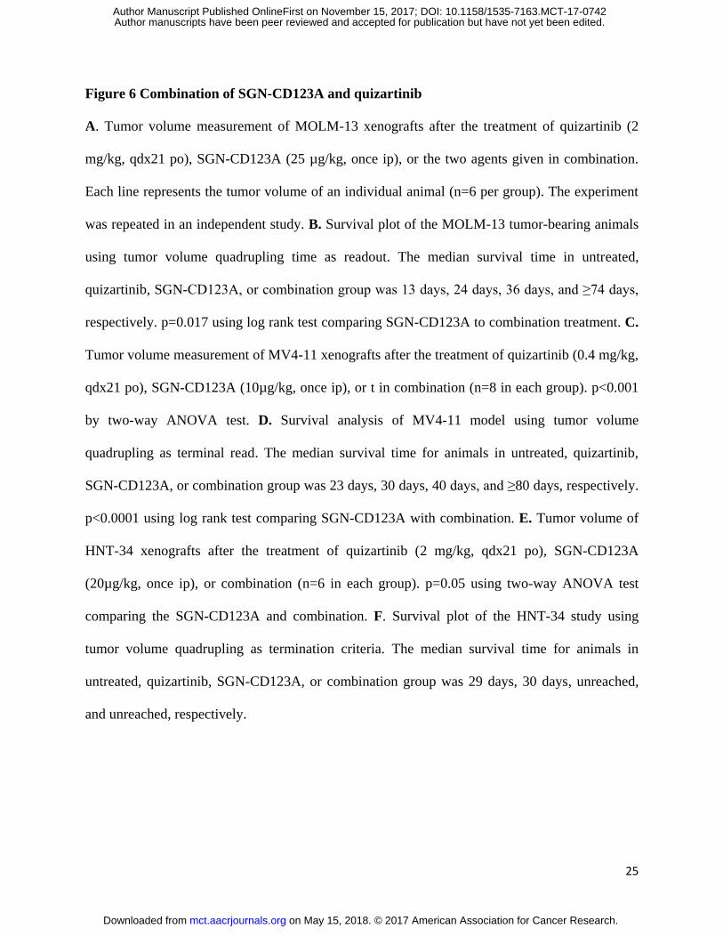
Expression of CD123 on AML patients
It has been reported that CD123 is expressed on the surface of blasts in the majority of patients
with AML(4, 5). To confirm the prevalence of CD123 expression, we surveyed a cohort of 52
AML patients using multicolor flow cytometry. Fifteen of these patients had newly diagnosed
AML, while 37 patients had recurrent or persistent disease. These cases included various
morphological subtypes and AML with high-risk molecular genetic abnormalities. In line with
previous studies, CD123 expression was detected on more than 50% leukemic blasts (CD34+)
among 48 (92%) patients (e.g. Figure 1A). Furthermore, 45 of these 48 patients (94%) had
on May 15, 2018. © 2017 American Association for Cancer Research. mct.aacrjournals.org Downloaded from
Author manuscripts have been peer reviewed and accepted for publication but have not yet been edited. Author Manuscript Published OnlineFirst on November 15, 2017; DOI: 10.1158/1535-7163.MCT-17-0742
10
CD123 expression on that blast that was 10-fold higher than the background (Figure 1B). These
data confirm the potential of CD123 as a target for antibody-based therapies for the treatment of
AML.
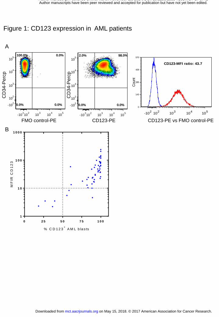
Conjugation and characterization of SGN-CD123A
To produce a CD123-targeted ADC, we first humanized a murine anti-CD123 antibody by
replacing the complementarity determining regions and select framework residues in a human
germline antibody with the corresponding murine sequence to retain binding affinity and
maximize homology to the human acceptor sequence. To enable site-specific conjugation, we
further introduced an engineered cysteine at amino acid position 239 in each heavy chain,
yielding humanized antibody h7G3ec (17, 18). SGN-CD123A is generated by conjugation of
SGD-1910, a chemical intermediate that contains both the pyrrolobenzodiazepine dimer (PBD)
payload and the valine-alanine dipeptide linker, to the two engineered cysteine residues of
h7G3ec(16) (Figure 2A). SGN-CD123A incorporates approximately two SGD-1910 molecules
per antibody molecule.
We next evaluated the binding of h7G3ec and SGN-CD123A to CD123 using a flow cytometry-
based cell-binding assay. On CD123+ MV4-11 cells, both unconjugated antibody and ADC
showed high affinity binding to CD123 (Figure 2B). The equilibrium dissociation constant
values of h7G3ec and SGN-CD123A were 7.6 nM (1.15 µg/mL) and 6 nM (0.92µg/mL),
respectively.
Once bound to surface antigens, ADCs are internalized through endosomes and trafficked to
lysosomes for drug release (19). Consistent with this, binding of SGN-CD123A to AML cells
resulted in rapid internalization of the ADC-CD123 complex as detected by fluorescence
microscopy (Figure 2C). SGN-CD123A and lysosomes were stained using antibodies
on May 15, 2018. © 2017 American Association for Cancer Research. mct.aacrjournals.org Downloaded from
Author manuscripts have been peer reviewed and accepted for publication but have not yet been edited. Author Manuscript Published OnlineFirst on November 15, 2017; DOI: 10.1158/1535-7163.MCT-17-0742
11
recognizing human Fc (red), or the lysosomal marker LAMP-1(green), respectively. SGN-
CD123A was detected on surface and the lysosomes were distinct and punctate at time zero
(T=0). Within 4 to 24 hours, SGN-CD123A was readily detected inside cells and co-localized
with LAMP-1(yellow), showing rapid internalization and trafficking to the lysosomes.
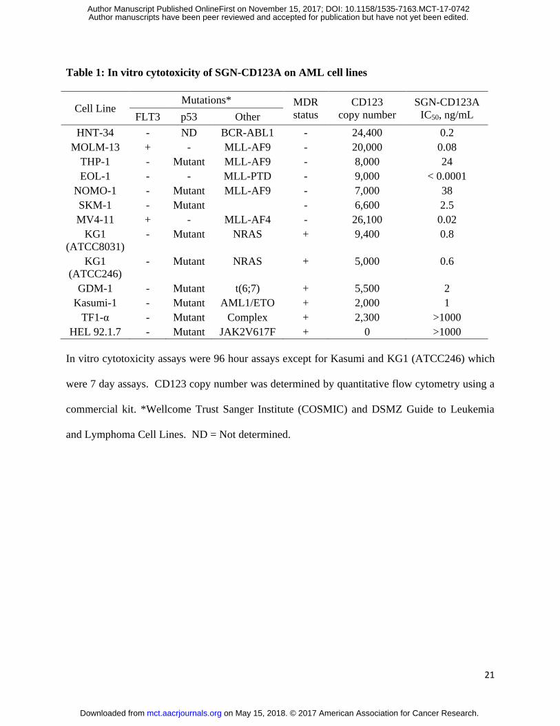
In vitro cytotoxic activity on AML cell lines
We then evaluated the cytotoxic potential of SGN-CD123A in 13 AML cell lines, of which 12
expressed between 2,000 and 26,100 copies of CD123 on the cell surface. SGN-CD123A was
highly active in 11 of 12 CD123+ AML cell lines tested (mean IC50, 6 ng/mL; range of 0.02 to
38 ng/mL). For example, SGN-CD123A killed HNT-34 cells with an IC50 of 0.2 ng/m, while
neither the control ADC nor unconjugated h7G3ec were cytotoxic (Figure 3A). The PBD dimer-
based payload has been reported to overcome multiple-drug resistance (MDR) (17). We
evaluated the activity of SGN-CD123A in five MDR+ cell lines (KG-1-ATCC8031 and KG-1-
CCL246, GDM-1, Kasumi-1 and TF1-α). SGN-CD123A was highly active against four of the
five cell lines (Table 1). For instance, KG-1 cells are MDR+, but SGN-CD123A mediated cell
killing with an IC50 of 0.8 ng/mL Figure 3B). Importantly, the cytotoxic activity was
immunologically specific, as the non-binding control ADC showed minimal activity (Figures 3A
and 3B). Moreover, SGN-CD123A was not cytotoxic against the CD123-negative HEL92.1.7
AML cell line, further supporting target-specificity (Table 1).
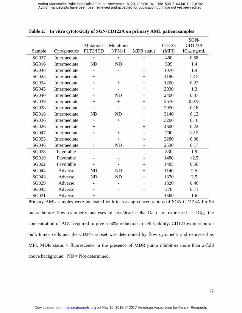
In vitro activity of SGN-CD123A in primary AML patient specimens
We also evaluated the cytotoxic potential of SGN-CD123A on blast samples isolated from 23
AML patients (Table 2). These patients had favorable (n=3), intermediate (n=15), or adverse
(n=5) cytogenetic profiles. Fourteen of these patients also had FLT3 internal tandem duplication
(FLT3/ITD), a mutation that correlates with poor prognosis in AML (20). Twenty six percent
on May 15, 2018. © 2017 American Association for Cancer Research. mct.aacrjournals.org Downloaded from
Author manuscripts have been peer reviewed and accepted for publication but have not yet been edited. Author Manuscript Published OnlineFirst on November 15, 2017; DOI: 10.1158/1535-7163.MCT-17-0742

12
(6/23) of patients had mutations in nucleophosmin gene (NPM1), which is associated with good
prognosis in AML patients (21). Consistent with the cell line data shown in Table 1, the
cytogenetic profiles did not affect the cytotoxicity of SGN-CD123A. In fact, 20 of 23 primary
samples showed an IC50 of 0.8 ng/mL (Table 2 and Supplementary Figure 2 A and B). Also in
agreement with our cell line data, 14 out of the 15 MDR+ AML samples had EC50 values of
SGN-CD123A of 0.06 to 2.5 ng/mL, indicating that SGN-CD123A can also overcome MDR in
primary AML samples.
Cell death kinetics induced by SGN-CD123A
The mechanism of SGN-CD123A-mediated cytotoxicity starts with the release of PBD dimers
upon internalization. Upon release, the PBD dimer can crosslink DNA and induce a DNA
damage response (17). One of the early markers of DNA damage is phosphorylated histone 2AX
(pγH2AX) (22). SGN-CD123A induced pγH2AX in a dose-dependent manner in HNT-34 cells
(Figure 4A). Induction of pγH2AX was also confirmed using flow cytometry, where we found
that 5 ng/mL SGN-CD123A induced a 6-fold increase of pH2AX in HNT-34 cells within 48
hours post ADC treatment (Supplementary Figure S3A). A similar magnitude of pγH2AX
increase was also observed in KG-1 anTHP-1 cells after the treatment of SGN-CD123A
(Supplementary Figure S3B and C). Accompanying the pH2AX was the phosphorylation and,
hence, the activation of the ataxia telangiectasia mutated (ATM) kinase (p-ATM, Figure 4A),
which is known to trigger the a cascade of events leading to cell cycle arrest as the cells attempt
DNA repair(23). Sensing DNA damage response, P53 will be phosphorylated and trigger cell
cycle arrest (24). Increased level of phosphorylated P53 protein and cell cycle arrest were
observed in SGN-CD123A-treated cells (Figure 4B). Indeed, cell cycle distribution analysis of
SGN-CD123A treated cells confirmed arrest in the G2/M phase in a dose-dependent manner
on May 15, 2018. © 2017 American Association for Cancer Research. mct.aacrjournals.org Downloaded from
Author manuscripts have been peer reviewed and accepted for publication but have not yet been edited. Author Manuscript Published OnlineFirst on November 15, 2017; DOI: 10.1158/1535-7163.MCT-17-0742

13
(Figure 4C). Concomitant with the pileup in the G2/M phase were marked decreases in the
fraction of cells found in the G1 and S phases.
When DNA repair mechanisms fail, the pathway to apoptosis and cell death is initiated.
Activation of cysteinyl-aspartic acid proteases (caspases) is an important early step leading to
cleavage of cellular proteins such as poly ADP-ribose polymerase (PARP) and apoptosis (Figure
4B). Caspase-3 activity was measured by flow cytometry. SGN-CD123A increased caspase-3
activity in HNT-34 cells in the low picomolar range, more potent than the free PBD molecule. In
contrast, it took 25 µM cisplatin to induce the same level of caspase activity, while the non-
binding control ADC treatment had no caspase induction (Figure 4D). SGN-CD123A also
induced caspase-3, in a dose-dependent manner, in the GDM-1 cells that express 5500 CD123 on
cell surface (Supplementary Figure 3D).
Anti-leukemic activity on xenograft models
Next we evaluated the activity of SGN-CD123A against a panel of leukemia cell line-based
xenograft models. First, in the disseminated disease model established from MOLM-13 cells, a
single dose of SGN-CD123A at 10 or 30 µg/kg yielded a significant survival advantage over a
non-binding control ADC. All eight mice that received the non-binding control died within 32
days. In contrast, none of the SGN-CD123A treated animals showed any signs of disease by the
end of study (Figure 5A).
In a subcutaneous xenograft model established from HNT-34 cells, a single injection of 25 µg/kg
of SGN-CD123A led to 50 days of tumor stasis. On the other hand, a single dose of 75 µg/kg of
SGN-CD123A treatment resulted in remission of tumors in all of the 8 mice. The control ADC
(75 µg/kg) led to an initial tumor growth delay out to day 40 post-implantation, followed by
on May 15, 2018. © 2017 American Association for Cancer Research. mct.aacrjournals.org Downloaded from
Author manuscripts have been peer reviewed and accepted for publication but have not yet been edited. Author Manuscript Published OnlineFirst on November 15, 2017; DOI: 10.1158/1535-7163.MCT-17-0742
14
rapid tumor regrowth. These data indicate potent, antigen-specific activity of SGN-CD123A in
vivo (Figure 5B).
To assess the activity of SGN-CD123A in the MDR+ xenograft models, we implanted KG-1-
CCL246 cells subcutaneously in SCID mice. In this model, a single dose of 300 µg/kg SGN-
CD123A significantly decreased tumor growth, including four durable remissions (p =0.008
using log-rank test). This is in contrast to treatment with a non-binding control ADC, which did
not slow the growth rate (Figure 5C).
Patient-derived xenograft models are thought to better mimic the physiology of AML patients
and thus better predict the clinical performance of drugs (25). The anti-leukemic activity of
SGN-CD123A was also evaluated in a primary AML model, AML06-227(17). Flow cytometric
analysis showed that that approximately 47% of the bone marrow consisted of CD45+ human
blasts at 53 days post implant. Moreover, 93% of the blasts expressed CD123 (Figure 5D).
Remarkably, two injections of SGN-CD123A eliminated the blasts cells in the bone marrow until
the end of study (124 days post implant). In contrast, the non-binding control ADC had no
impact on the disease burden during the course of study (Figure 5E).
Combination of SGN-CD123A and quizartinib
Approximately 23% of AML patients carry a mutation in the FLT3 gene (20, 26).
Coincidentally, AML patients carrying FLT3 mutations have higher level of CD123 expression
(27, 28). Since it is possible that FLT3 mutated AML patients will receive both a FLT3
inhibitor and SGN-CD123A during clinical studies, we evaluated whether the combination of
quizartinib, a FLT3-ITD specific inhibitor, and SGN-CD123A provides additional efficacy in
vivo.
on May 15, 2018. © 2017 American Association for Cancer Research. mct.aacrjournals.org Downloaded from
Author manuscripts have been peer reviewed and accepted for publication but have not yet been edited. Author Manuscript Published OnlineFirst on November 15, 2017; DOI: 10.1158/1535-7163.MCT-17-0742
15
In the FLT3/ITD, subcutaneous MOLM-13 xenograft model, 2mg/kg daily dosing of quizartinib
led to a modest growth delay. Injection of 25µg/kg SGN-CD123A led to growth delay in five
animals and remission in another animal. Strikingly, combination of SGN-CD123A and
quizartinib led to durable remissions in five out the six tumor-bearing animals (Figure 6A).
Using tumor-quadrupling time as a measurement of survival, the combination group showed
significant extension, with a median survival longer than 70 days. In contrast, the median
survival for quizartinib and SGN-CD123A was 24 and 36 days, respectively (Figure 6B). In
addition, we observed a similar combination benefit between SGN-CD123A and quizartinib in
another FLT3/ITD xenograft model MV4-11. While SGN-CD123A and quizartinib led to growth
delays, the combination resulted in remissions lasting longer than 80 days (Figure 6C and D).
Furthermore, the quizartinib and SGN-CD123A were specific to FLT3 mutant tumor models, as
in FLT-3 wild type HNT-34 tumor model, quizartinib has no activity on its own and it did not
influence the activity of SGN-CD123A (Figure 6E and F).
Discussion
Every year about 38,000 patients are diagnosed with AML in the United States and European
Union(29). Although induction chemotherapy can be effective in some patients, between 10%
and 40% of newly diagnosed AML patients are refractory or resistant to the regimen. Even
among patients who achieve remission, the majority will relapse between several months and
several years after initial treatment(29). These refractory and relapsed AML patients are in need
of novel therapeutic approaches.
One hurdle in these patients is multidrug resistance (MDR), which confers resistance to
gemtuzumab ozogamicin, as calicheamicin is a substrate of MDR proteins (30). PBD dimers-
based ADCs, have demonstrated antitumor activity in MDR+ xenograft models and show activity
on May 15, 2018. © 2017 American Association for Cancer Research. mct.aacrjournals.org Downloaded from
Author manuscripts have been peer reviewed and accepted for publication but have not yet been edited. Author Manuscript Published OnlineFirst on November 15, 2017; DOI: 10.1158/1535-7163.MCT-17-0742
16
in AML patients (16, 31). Moreover, other PBD-based ADCs, including rovalpituzumab tesirine
and ADC-301, have demonstrated clinical activities in small cell lung cancer as well as non-
Hodgkin’s lymphoma (32, 33). Our preclinical results demonstrate that PBD dimers conjugated
to a CD123 binding antibody are active in AML cell lines, patient isolates, and xenograft models.
Together with the clinical trial results of other PBD-based ADCs, these data support further
evaluation of these ADCs for cancer treatment.
Another challenge is the persistence of leukemic stem cells. Multiple studies have demonstrated
that there is an hierarchical organization of AML(34), which is thought to be responsible for the
relapse of AML. Importantly, CD123 was discovered as a marker of LSC and high CD123
expression on the CD34+CD38
- population is associated with poor prognosis (6, 9). CSL362, a
CD123 antibody, was shown to reduce the LSC population in xenograft studies (10). In line with
this hypothesis, we also observed long-term remission of patient-derived xenografts after SGN-
CD123A treatment, suggesting that the drug eradicated the LSC in this model. On the other
hand, CD123 was also detected on low percentage of hematopoietic progenitor cells, which may
lead to on-target but off-disease side effects of CD123-targeted therapies(7). Because the h7G3ec
antibody does not bind to murine CD123, we could not study the potential impact on
hematopoietic cells in this study.
A third challenge for AML drug development is the resistance to targeted small molecule
inhibitors(2). For example, FLT3 inhibitors represent a major class of the drugs in development
for FLT3 mutated AML patients(26). However, while patients can respond to treatments with
FLT3 inhibitors, they almost invariably develop resistance(26). The rationale to combine FLT3
inhibitors with CD123 targeted drugs include: 1) FLT3 mutated patients, especially those
carrying the FLT3/ITD mutations, often have higher CD123 than those without FLT3
on May 15, 2018. © 2017 American Association for Cancer Research. mct.aacrjournals.org Downloaded from
Author manuscripts have been peer reviewed and accepted for publication but have not yet been edited. Author Manuscript Published OnlineFirst on November 15, 2017; DOI: 10.1158/1535-7163.MCT-17-0742
17
mutations(5); and 2) FLT3 mutation and CD123 expression may overlap on the leukemic stem
cells(35). Remarkably, our study shows that a low dose of SGN-CD123A significantly improved
the activity of quizartinib in two xenograft models. These data may support clinical evaluation of
SGN-CD123A in combination with a FLT3 inhibitor.
The overexpression of CD123 on putative LSCs raised major interest in developing CD123-
targeting therapeutics as AML therapies. Examples include unconjugated monoclonal antibodies,
T-cell engaging bispecific antibodies, immunotoxins, and chimeric antigen receptor (CAR)-
modified T cells(36-41). While each of the modality has its own advantages, our study show that
SGN-CD123A is highly active against AML models regardless of cytogenetics profiles and
MDR status, and can be effectively combined with quizartinib. These data support the ongoing
clinical trial to evaluate its utility in AML treatment.
on May 15, 2018. © 2017 American Association for Cancer Research. mct.aacrjournals.org Downloaded from
Author manuscripts have been peer reviewed and accepted for publication but have not yet been edited. Author Manuscript Published OnlineFirst on November 15, 2017; DOI: 10.1158/1535-7163.MCT-17-0742
18
References
1. Burnett A, Wetzler M, Lowenberg B. Therapeutic advances in acute myeloid leukemia. Journal of clinical oncology : official journal of the American Society of Clinical Oncology. 2011;29:487-94. 2. Dohner H, Weisdorf DJ, Bloomfield CD. Acute Myeloid Leukemia. The New England journal of medicine. 2015;373:1136-52. 3. Guthridge MA, Stomski FC, Thomas D, Woodcock JM, Bagley CJ, Berndt MC, et al. Mechanism of activation of the GM-CSF, IL-3, and IL-5 family of receptors. Stem cells. 1998;16:301-13. 4. Munoz L, Nomdedeu JF, Lopez O, Carnicer MJ, Bellido M, Aventin A, et al. Interleukin-3 receptor alpha chain (CD123) is widely expressed in hematologic malignancies. Haematologica. 2001;86:1261-9. 5. Ehninger A, Kramer M, Rollig C, Thiede C, Bornhauser M, von Bonin M, et al. Distribution and levels of cell surface expression of CD33 and CD123 in acute myeloid leukemia. Blood cancer journal. 2014;4:e218. 6. Jordan CT, Upchurch D, Szilvassy SJ, Guzman ML, Howard DS, Pettigrew AL, et al. The interleukin-3 receptor alpha chain is a unique marker for human acute myelogenous leukemia stem cells. Leukemia. 2000;14:1777-84. 7. Taussig DC, Pearce DJ, Simpson C, Rohatiner AZ, Lister TA, Kelly G, et al. Hematopoietic stem cells express multiple myeloid markers: implications for the origin and targeted therapy of acute myeloid leukemia. Blood. 2005;106:4086-92. 8. Reddy EP, Korapati A, Chaturvedi P, Rane S. IL-3 signaling and the role of Src kinases, JAKs and STATs: a covert liaison unveiled. Oncogene. 2000;19:2532-47. 9. Testa U, Pelosi E, Frankel A. CD 123 is a membrane biomarker and a therapeutic target in hematologic malignancies. Biomarker research. 2014;2:4. 10. Jin L, Lee EM, Ramshaw HS, Busfield SJ, Peoppl AG, Wilkinson L, et al. Monoclonal antibody-mediated targeting of CD123, IL-3 receptor alpha chain, eliminates human acute myeloid leukemic stem cells. Cell stem cell. 2009;5:31-42. 11. Guan Y, Gerhard B, Hogge DE. Detection, isolation, and stimulation of quiescent primitive leukemic progenitor cells from patients with acute myeloid leukemia (AML). Blood. 2003;101:3142-9. 12. Konopleva MY, Jordan CT. Leukemia stem cells and microenvironment: biology and therapeutic targeting. Journal of clinical oncology : official journal of the American Society of Clinical Oncology. 2011;29:591-9. 13. Vergez F, Green AS, Tamburini J, Sarry JE, Gaillard B, Cornillet-Lefebvre P, et al. High levels of CD34+CD38low/-CD123+ blasts are predictive of an adverse outcome in acute myeloid leukemia: a Groupe Ouest-Est des Leucemies Aigues et Maladies du Sang (GOELAMS) study. Haematologica. 2011;96:1792-8. 14. Huang S, Chen Z, Yu JF, Young D, Bashey A, Ho AD, et al. Correlation between IL-3 receptor expression and growth potential of human CD34+ hematopoietic cells from different tissues. Stem cells. 1999;17:265-72. 15. Zarrinkar PP, Gunawardane RN, Cramer MD, Gardner MF, Brigham D, Belli B, et al. AC220 is a uniquely potent and selective inhibitor of FLT3 for the treatment of acute myeloid leukemia (AML). Blood. 2009;114:2984-92. 16. Jeffrey SC, Burke PJ, Lyon RP, Meyer DW, Sussman D, Anderson M, et al. A potent anti-CD70 antibody-drug conjugate combining a dimeric pyrrolobenzodiazepine drug with site-specific conjugation technology. Bioconjugate chemistry. 2013;24:1256-63. 17. Kung Sutherland MS, Walter RB, Jeffrey SC, Burke PJ, Yu C, Kostner H, et al. SGN-CD33A: a novel CD33-targeting antibody-drug conjugate using a pyrrolobenzodiazepine dimer is active in models of drug-resistant AML. Blood. 2013;122:1455-63.
on May 15, 2018. © 2017 American Association for Cancer Research. mct.aacrjournals.org Downloaded from
Author manuscripts have been peer reviewed and accepted for publication but have not yet been edited. Author Manuscript Published OnlineFirst on November 15, 2017; DOI: 10.1158/1535-7163.MCT-17-0742
19
18. Sussman D, Torrey L, Westendorf L, Miyamoto JB, Meyer DW, Lyon R, et al. Engineered cysteine drug conjugates show potency and improved safety. Cancer Research. 2012;72:Abstract 4634. 19. Francisco JA, Cerveny CG, Meyer DL, Mixan BJ, Klussman K, Chace DF, et al. cAC10-vcMMAE, an anti-CD30-monomethyl auristatin E conjugate with potent and selective antitumor activity. Blood. 2003;102:1458-65. 20. Swords R, Freeman C, Giles F. Targeting the FMS-like tyrosine kinase 3 in acute myeloid leukemia. Leukemia. 2012;26:2176-85. 21. Port M, Bottcher M, Thol F, Ganser A, Schlenk R, Wasem J, et al. Prognostic significance of FLT3 internal tandem duplication, nucleophosmin 1, and CEBPA gene mutations for acute myeloid leukemia patients with normal karyotype and younger than 60 years: a systematic review and meta-analysis. Annals of hematology. 2014;93:1279-86. 22. Rogakou EP, Nieves-Neira W, Boon C, Pommier Y, Bonner WM. Initiation of DNA fragmentation during apoptosis induces phosphorylation of H2AX histone at serine 139. J Biol Chem. 2000;275:9390-5. 23. Marechal A, Zou L. DNA damage sensing by the ATM and ATR kinases. Cold Spring Harbor perspectives in biology. 2013;5. 24. Taylor WR, Stark GR. Regulation of the G2/M transition by p53. Oncogene. 2001;20:1803-15. 25. Townsend EC, Murakami MA, Christodoulou A, Christie AL, Koster J, DeSouza TA, et al. The Public Repository of Xenografts Enables Discovery and Randomized Phase II-like Trials in Mice. Cancer cell. 2016;29:574-86. 26. Konig H, Levis M. Targeting FLT3 to treat leukemia. Expert opinion on therapeutic targets. 2015;19:37-54. 27. Angelini DF, Ottone T, Guerrera G, Lavorgna S, Cittadini M, Buccisano F, et al. A Leukemia-Associated CD34/CD123/CD25/CD99+ Immunophenotype Identifies FLT3-Mutated Clones in Acute Myeloid Leukemia. Clinical cancer research : an official journal of the American Association for Cancer Research. 2015;21:3977-85. 28. Riccioni R, Pelosi E, Riti V, Castelli G, Lo-Coco F, Testa U. Immunophenotypic features of acute myeloid leukaemia patients exhibiting high FLT3 expression not associated with mutations. British journal of haematology. 2011;153:33-42. 29. Kell J. Considerations and challenges for patients with refractory and relapsed acute myeloid leukaemia. Leukemia research. 2016;47:149-60. 30. Linenberger ML, Hong T, Flowers D, Sievers EL, Gooley TA, Bennett JM, et al. Multidrug-resistance phenotype and clinical responses to gemtuzumab ozogamicin. Blood. 2001;98:988-94. 31. Stein EM, Stein A, Walter RB, Fathi AT, Lancet JE, Kovacsovics TJ, et al. Interim Analysis of a Phase 1 Trial of SGN-CD33A in Patients with CD33-Positive Acute Myeloid Leukemia (AML). Blood. 2014;124:623-. 32. Wolska-Washer A, Robak P, Smolewski P, Robak T. Emerging antibody-drug conjugates for treating lymphoid malignancies. Expert Opin Emerg Drugs. 2017:1-15. 33. Rudin CM, Pietanza MC, Bauer TM, Ready N, Morgensztern D, Glisson BS, et al. Rovalpituzumab tesirine, a DLL3-targeted antibody-drug conjugate, in recurrent small-cell lung cancer: a first-in-human, first-in-class, open-label, phase 1 study. Lancet Oncol. 2017;18:42-51. 34. Dick JE. Acute myeloid leukemia stem cells. Annals of the New York Academy of Sciences. 2005;1044:1-5. 35. Al-Mawali A, Gillis D, Lewis I. Immunoprofiling of leukemic stem cells CD34+/CD38-/CD123+ delineate FLT3/ITD-positive clones. Journal of hematology & oncology. 2016;9:61. 36. He SZ, Busfield S, Ritchie DS, Hertzberg MS, Durrant S, Lewis ID, et al. A Phase 1 study of the safety, pharmacokinetics and anti-leukemic activity of the anti-CD123 monoclonal antibody CSL360 in relapsed, refractory or high-risk acute myeloid leukemia. Leukemia & lymphoma. 2015;56:1406-15.
on May 15, 2018. © 2017 American Association for Cancer Research. mct.aacrjournals.org Downloaded from
Author manuscripts have been peer reviewed and accepted for publication but have not yet been edited. Author Manuscript Published OnlineFirst on November 15, 2017; DOI: 10.1158/1535-7163.MCT-17-0742
20
37. Smith BD, Roboz GJ, Walter RB, Altman JK, Ferguson A, Curcio TJ, et al. First-in Man, Phase 1 Study of CSL362 (Anti-IL3Rα / Anti-CD123 Monoclonal Antibody) in Patients with CD123+ Acute Myeloid Leukemia (AML) in CR at High Risk for Early Relapse. Blood. 2014;124:120-. 38. Al-Hussaini M, Rettig MP, Ritchey JK, Karpova D, Uy GL, Eissenberg LG, et al. Targeting CD123 in acute myeloid leukemia using a T-cell-directed dual-affinity retargeting platform. Blood. 2016;127:122-31. 39. Chu SY, Pong E, Chen H, Phung S, Chan EW, Endo NA, et al. Immunotherapy with Long-Lived Anti-CD123 × Anti-CD3 Bispecific Antibodies Stimulates Potent T Cell-Mediated Killing of Human AML Cell Lines and of CD123+ Cells in Monkeys: A Potential Therapy for Acute Myelogenous Leukemia. Blood. 2014;124:2316-. 40. Luo Y, Chang L-J, Hu Y, Dong L, Wei G, Huang H. First-in-Man CD123-Specific Chimeric Antigen Receptor-Modified T Cells for the Treatment of Refractory Acute Myeloid Leukemia. Blood. 2015;126:3778-. 41. Frankel AE, Woo JH, Ahn C, Pemmaraju N, Medeiros BC, Carraway HE, et al. Activity of SL-401, a targeted therapy directed to interleukin-3 receptor, in blastic plasmacytoid dendritic cell neoplasm patients. Blood. 2014;124:385-92.
on May 15, 2018. © 2017 American Association for Cancer Research. mct.aacrjournals.org Downloaded from
Author manuscripts have been peer reviewed and accepted for publication but have not yet been edited. Author Manuscript Published OnlineFirst on November 15, 2017; DOI: 10.1158/1535-7163.MCT-17-0742
21
Table 1: In vitro cytotoxicity of SGN-CD123A on AML cell lines
Cell Line Mutations* MDR
status
CD123
copy number
SGN-CD123A
IC50, ng/mL FLT3 p53 Other
HNT-34 - ND BCR-ABL1 - 24,400 0.2
MOLM-13 + - MLL-AF9 - 20,000 0.08
THP-1 - Mutant MLL-AF9 - 8,000 24
EOL-1 - - MLL-PTD - 9,000 < 0.0001
NOMO-1 - Mutant MLL-AF9 - 7,000 38
SKM-1 - Mutant - 6,600 2.5
MV4-11 + - MLL-AF4 - 26,100 0.02
KG1
(ATCC8031)
- Mutant NRAS + 9,400 0.8
KG1
(ATCC246)
- Mutant NRAS + 5,000 0.6
GDM-1 - Mutant t(6;7) + 5,500 2
Kasumi-1 - Mutant AML1/ETO + 2,000 1
TF1-α - Mutant Complex + 2,300 >1000
HEL 92.1.7 - Mutant JAK2V617F + 0 >1000
In vitro cytotoxicity assays were 96 hour assays except for Kasumi and KG1 (ATCC246) which
were 7 day assays. CD123 copy number was determined by quantitative flow cytometry using a
commercial kit. *Wellcome Trust Sanger Institute (COSMIC) and DSMZ Guide to Leukemia
and Lymphoma Cell Lines. ND = Not determined.
on May 15, 2018. © 2017 American Association for Cancer Research. mct.aacrjournals.org Downloaded from
Author manuscripts have been peer reviewed and accepted for publication but have not yet been edited. Author Manuscript Published OnlineFirst on November 15, 2017; DOI: 10.1158/1535-7163.MCT-17-0742
22
Table 2. In vitro cytotoxicity of SGN-CD123A on primary AML patient samples
Sample Cytogenetics
Mutations
FLT3/ITD
Mutations
NPM-1 MDR status
CD123
(MFI)
SGN-
CD123A
IC50, ng/mL
SG037 Intermediate + + + 480 0.68
SG016 Intermediate ND ND + 595 1.4
SG048 Intermediate + - + 1070 1.9
SG025 Intermediate + - + 1190 >2.5
SG034 Intermediate + + + 1200 0.22
SG045 Intermediate + - + 2030 1.2
SG040 Intermediate + ND + 2400 0.37
SG039 Intermediate + + + 2670 0.075
SG038 Intermediate - - + 2950 0.18
SG018 Intermediate ND ND + 3140 0.12
SG036 Intermediate + + + 3260 0.16
SG026 Intermediate + - + 4600 0.22
SG047 Intermediate + + - 790 >2.5
SG023 Intermediate + + - 2280 0.06
SG046 Intermediate + ND - 2530 0.17
SG028 Favorable - - - 830 1.9
SG019 Favorable - - - 1480 >2.5
SG022 Favorable - - - 1485 0.56
SG044 Adverse ND ND + 1140 2.5
SG043 Adverse ND ND + 1370 2.5
SG029 Adverse - - + 1820 0.46
SG042 Adverse + - - 270 0.11
SG021 Adverse + - - 1560 1.6
Primary AML samples were incubated with increasing concentrations of SGN-CD123A for 96
hours before flow cytometry analyses of live/dead cells. Data are expressed as IC50, the
concentration of ADC required to give a 50% reduction in cell viability. CD123 expression on
bulk tumor cells and the CD34+ subset was determined by flow cytometry and expressed as
MFI. MDR status = fluorescence in the presence of MDR pump inhibitors more than 2-fold
above background. ND = Not determined.
on May 15, 2018. © 2017 American Association for Cancer Research. mct.aacrjournals.org Downloaded from
Author manuscripts have been peer reviewed and accepted for publication but have not yet been edited. Author Manuscript Published OnlineFirst on November 15, 2017; DOI: 10.1158/1535-7163.MCT-17-0742
23
Figure Legend
Figure 1 Expression of CD123 in AML patients
A. Flow cytometry staining of CD123 of CD34+ blasts of one AML patient. FMO: Fluorescence
minus one control, which contains all other fluorochromes except CD123. B. CD123 expression
in 52 AML patients was determined using flow cytometry. MFIR: median fluorescence intensity
ratio calculated by the CD123-PE intensity over FMO control.
Figure 2 Structure, binding and internalization of SGN-CD123A
A. Chemical structure of SGN-CD123A, comprised of the monoclonal antibody h7G3ec and
drug linker (SGD-1910). B. Binding kinetics of SGN-CD123A and h7G3ec to CD123+ MV4-11
cells, as measured by flow cytometry. The highest median fluorescence intensity was set to 100
percent for normalization. C. Immunofluorescence staining of SGN-CD123A (red, staining
human IgG Fc kappa chain), lysosome (green, staining LAMP-1), and nucleus (blue, staining
4',6-diamidino-2-phenylindole). Images from 0, 4hr, and 24 hrs post-ADC incubation are
represented. When ADC trafficks to the lysosomes and co-localizes with LAMP-1, the resulting
signal is projected as yellow in the merged images (arrows).
Figure 3 In vitro cytotoxicity of SGN-CD123A on leukemia cell lines
A. In vitro cytotoxicity of SGN-CD123A,a non-binding control ADC, and h7G3ec on HNT-34
cells. ADCs were titrated in growth media and cells were incubated with ADC for 96 hours. Cell
viability was normalized to untreated cells. B. In vitro cytotoxicity of SGN-CD123A and non-
binding control ADC on MDR+ KG-1 cells.
Figure 4 DNA damage responses and cell cycle changes induced by SGN-CD123A
A. Western blot of phosphorylated H2AX, total H2AX, and phosphorylated-ATM in protein
extracts from HNT-34 cells treated with 0, 1, 2, or 5 ng/mL of SGN-CD123A for 48 hours. B.
on May 15, 2018. © 2017 American Association for Cancer Research. mct.aacrjournals.org Downloaded from
Author manuscripts have been peer reviewed and accepted for publication but have not yet been edited. Author Manuscript Published OnlineFirst on November 15, 2017; DOI: 10.1158/1535-7163.MCT-17-0742
24
Western blot staining of phosphorylated P53, cleaved PARP, and beta-actin of HNT-34 cells
treated with SGN-CD123A at indicated concentrations for 48 hours. C. Cell cycle analysis of
HNT-34 cells after treatment of SGN-CD123A at indicated concentrations. The cell cycle phase
was determined using propidium iodide staining. D. Flow cytometry analysis of cleaved caspase-
3 of HNT-34 cells after the treatment with SGN-CD123A, control ADC, free PBD drug, or
cisplatin. The X axis represents the molar concentrations of each reagents and Y axis represents
the fold difference of mean fluorescence intensity between treated cells and untreated cells. Each
data point is the average of two replicates and the experiment has been repeated twice.
Figure 5 In vivo anti-leukemic activity of SGN-CD123A
A. Survival plot of disseminated leukemia model MOLM-13 after SGN-CD123A treatment.
MOLM-13 cells were intravenously implanted in SCID mice and animals received a dose of
SGN-CD123A (10 µg/kg or 30 µg/kg) or control ADC (30 µg/kg). All animals who received
SGN-CD123A remained alive until the end of study (day 80). N=8 mice per group. B. Tumor
volume measurement of HNT-34 xenografts. HNT-34 tumors were subcutaneously implanted on
SCID mice and ADCs were given once when average tumor volume reached 100 mm3 (n=8 per
group, p<0.0001 using two-way ANOVA test). C. Tumor volume measurement of MDR+ KG-1
xenograft implanted subcutaneously in SCID mice. Animals were dosed once with either SGN-
CD123A or control ADC at 300 µg/kg (n=8 each group, p=0.0003 using two-way ANOVA test).
D. Flow cytometry of staining for CD123 and human CD45 of leukemic blasts from patient-
derived xenograft model AML06227 grown in NSG mice. E. Measurement of CD45+ blasts in
bone marrow of AML06227 PDX mice that were treated with SGN-CD123A or control ADC.
Mice were randomized and treated when the average blast in the bone marrow cells reached
50%. Two mice were sacrificed and analyzed for blast percentage at each time point.
on May 15, 2018. © 2017 American Association for Cancer Research. mct.aacrjournals.org Downloaded from
Author manuscripts have been peer reviewed and accepted for publication but have not yet been edited. Author Manuscript Published OnlineFirst on November 15, 2017; DOI: 10.1158/1535-7163.MCT-17-0742
25
Figure 6 Combination of SGN-CD123A and quizartinib
A. Tumor volume measurement of MOLM-13 xenografts after the treatment of quizartinib (2
mg/kg, qdx21 po), SGN-CD123A (25 µg/kg, once ip), or the two agents given in combination.
Each line represents the tumor volume of an individual animal (n=6 per group). The experiment
was repeated in an independent study. B. Survival plot of the MOLM-13 tumor-bearing animals
using tumor volume quadrupling time as readout. The median survival time in untreated,
quizartinib, SGN-CD123A, or combination group was 13 days, 24 days, 36 days, and ≥74 days,
respectively. p=0.017 using log rank test comparing SGN-CD123A to combination treatment. C.
Tumor volume measurement of MV4-11 xenografts after the treatment of quizartinib (0.4 mg/kg,
qdx21 po), SGN-CD123A (10µg/kg, once ip), or t in combination (n=8 in each group). p<0.001
by two-way ANOVA test. D. Survival analysis of MV4-11 model using tumor volume
quadrupling as terminal read. The median survival time for animals in untreated, quizartinib,
SGN-CD123A, or combination group was 23 days, 30 days, 40 days, and ≥80 days, respectively.
p<0.0001 using log rank test comparing SGN-CD123A with combination. E. Tumor volume of
HNT-34 xenografts after the treatment of quizartinib (2 mg/kg, qdx21 po), SGN-CD123A
(20µg/kg, once ip), or combination (n=6 in each group). p=0.05 using two-way ANOVA test
comparing the SGN-CD123A and combination. F. Survival plot of the HNT-34 study using
tumor volume quadrupling as termination criteria. The median survival time for animals in
untreated, quizartinib, SGN-CD123A, or combination group was 29 days, 30 days, unreached,
and unreached, respectively.
on May 15, 2018. © 2017 American Association for Cancer Research. mct.aacrjournals.org Downloaded from
Author manuscripts have been peer reviewed and accepted for publication but have not yet been edited. Author Manuscript Published OnlineFirst on November 15, 2017; DOI: 10.1158/1535-7163.MCT-17-0742
0 2 5 5 0 7 5 1 0 0
1
1 0
1 0 0
1 0 0 0
% C D 1 2 3+
A M L b la s ts
MF
IR C
D1
23
Figure 1: CD123 expression in AML patients
Count
-102
102
103
104
105
0
143
285
428
570
CD123-MFI ratio: 43.7C
D34 P
erC
P-C
y5-5
-A
-10210
210
310
410
5
-102
102
103
104
105
0.0%0.0%
98.0%2.0%
CD
34 P
erC
P-C
y5-5
-A
-10210
210
310
410
5
-102
102
103
104
105
0.0%0.0%
0.0%100.0%
FMO control-PE CD123-PE CD123-PE vs FMO control-PE
CD
34-P
erc
p
CD
34-P
erc
p
A
B
on May 15, 2018. © 2017 American Association for Cancer Research. mct.aacrjournals.org Downloaded from
Author manuscripts have been peer reviewed and accepted for publication but have not yet been edited. Author Manuscript Published OnlineFirst on November 15, 2017; DOI: 10.1158/1535-7163.MCT-17-0742
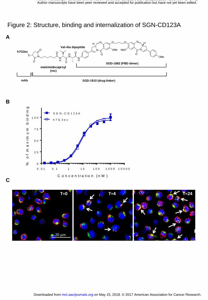
Figure 2: Structure, binding and internalization of SGN-CD123A
O O
M e O O M e N
N N
N O O
H H
N H
O M e H N
O
N H
O
O N
O
O S G D - 1 8 8 2 ( P B D d i m e r )
S G D - 1 9 1 0 ( d r u g - l i n k e r )
S h7G3ec
m a l e i m i d o c a p r o y l ( m c )
Val-Ala dipeptide
m A b
20 µm
T=0 T=4 T=24
A
B
C
0 . 0 1 0 . 1 1 1 0 1 0 0 1 0 0 0 1 0 0 0 0
0
2 5
5 0
7 5
1 0 0
C o n c e n t r a t i o n ( n M )
% o
f m
ax
imu
m b
ind
ing
S G N - C D 1 2 3 A
h 7 G 3 e c
on May 15, 2018. © 2017 American Association for Cancer Research. mct.aacrjournals.org Downloaded from
Author manuscripts have been peer reviewed and accepted for publication but have not yet been edited. Author Manuscript Published OnlineFirst on November 15, 2017; DOI: 10.1158/1535-7163.MCT-17-0742
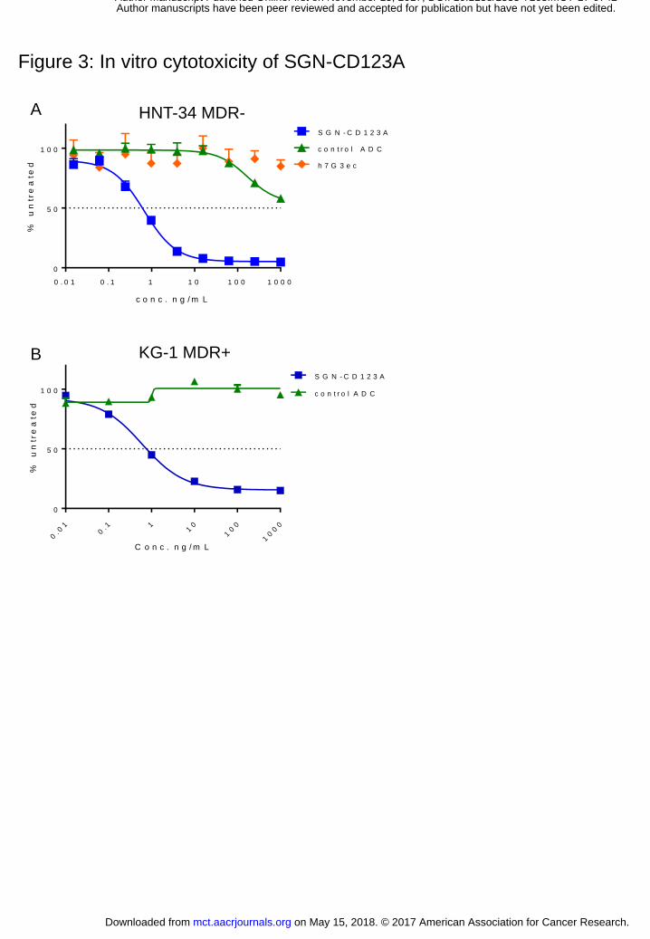
Figure 3: In vitro cytotoxicity of SGN-CD123A
A
B
0 . 0 1 0 . 1 1 1 0 1 0 0 1 0 0 0
0
5 0
1 0 0
c o n c . n g / m L
% u
ntr
ea
ted
S G N - C D 1 2 3 A
c o n t r o l A D C
h 7 G 3 e c
HNT-34 MDR-
0. 0
10
. 1 11
0
10
0
10
00
0
5 0
1 0 0
S G N - C D 1 2 3 A
c o n t r o l A D C
C o n c . n g / m L
% u
ntr
ea
ted
KG-1 MDR+
on May 15, 2018. © 2017 American Association for Cancer Research. mct.aacrjournals.org Downloaded from
Author manuscripts have been peer reviewed and accepted for publication but have not yet been edited. Author Manuscript Published OnlineFirst on November 15, 2017; DOI: 10.1158/1535-7163.MCT-17-0742
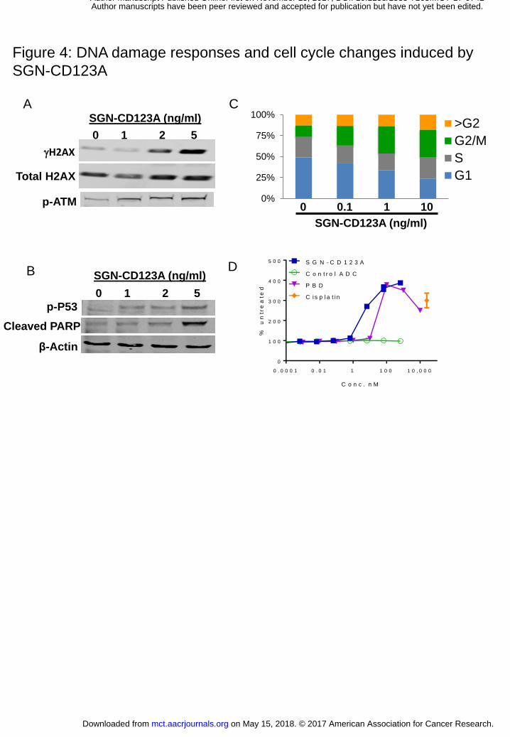
Figure 4: DNA damage responses and cell cycle changes induced by
SGN-CD123A
SGN-CD123A (ng/ml)
gH2AX
Total H2AX
0 1 2 5
p-ATM
Cleaved PARP
β-Actin
p-P53
SGN-CD123A (ng/ml)
0 1 2 5
B D
SGN-CD123A (ng/ml)
0%
25%
50%
75%
100%>G2
G2/M
S
G1
0 0.1 1 10
0 . 0 0 0 1 0 . 0 1 1 1 0 0 1 0 , 0 0 0
0
1 0 0
2 0 0
3 0 0
4 0 0
5 0 0
C o n c . n M
% u
ntr
ea
ted
S G N - C D 1 2 3 A
C o n t r o l A D C
P B D
C is p la t in
A C
on May 15, 2018. © 2017 American Association for Cancer Research. mct.aacrjournals.org Downloaded from
Author manuscripts have been peer reviewed and accepted for publication but have not yet been edited. Author Manuscript Published OnlineFirst on November 15, 2017; DOI: 10.1158/1535-7163.MCT-17-0742
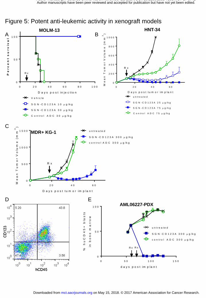
Figure 5: Potent anti-leukemic activity in xenograft models
0 2 0 4 0 6 0 8 0 1 0 0
0
5 0
1 0 0
D a y s p o s t i n j e c t i o n
Pe
rc
en
t s
ur
viv
al
V e h i c le
S G N - C D 1 2 3 A 1 0 g / k g
S G N - C D 1 2 3 A 3 0 g / k g
C o n t r o l A D C 3 0 g / k g
R x
MOLM-13
MDR+ KG-1
0 2 0 4 0 6 0
0
5 0 0
1 0 0 0
1 5 0 0 u n t r e a t e d
S G N - C D 1 2 3 A 3 0 0 g / k g
c o n t r o l A D C 3 0 0 g / k g
D a y s p o s t t u m o r im p la n t
Me
an
Tu
mo
r V
olu
me
(m
m3
)
R x
0 5 0 1 0 0 1 5 0
0
5 0
1 0 0
d a y s p o s t im p la n t
% h
uC
D4
5+
bla
sts
in b
on
e m
arro
w
u n t r e a t e d
S G N - C D 1 2 3 A 3 0 0 g / k g
c o n t r o l A D C 3 0 0 g / k g
R x R x
A B
C
0 2 0 4 0 6 0
0
2 0 0
4 0 0
6 0 0
8 0 0
1 0 0 0
u n t r e a t e d
S G N - C D 1 2 3 A 2 5 g / k g
S G N - C D 1 2 3 A 7 5 g / k g
C o n t r o l A D C 7 5 g / k g
D a y s p o s t t u m o r im p la n t
Me
an
Tu
mo
r V
olu
me
(m
m3
)
R x
HNT-34
D E AML06227-PDX
on May 15, 2018. © 2017 American Association for Cancer Research. mct.aacrjournals.org Downloaded from
Author manuscripts have been peer reviewed and accepted for publication but have not yet been edited. Author Manuscript Published OnlineFirst on November 15, 2017; DOI: 10.1158/1535-7163.MCT-17-0742

Figure 6: SGN-CD123A combination with quizartinib
02
04
06
08
0
0
5 0 0
1 0 0 0
1 5 0 0
d a y s p o s t im p la n t
tum
or V
olu
me
(m
m3
)
u n t r e a t e d
q u iz a r t i n ib
S G N - C D 1 2 3 A
c o m b in a t io n
R x
A
0 2 0 4 0 6 0 8 0
0
5 0
1 0 0
D a y s t o r e a c h 4 f o l d t u m o r v o l u m e
pe
rc
en
t r
em
ain
ing
u n t r e a t e d
q u iz a r t i n ib
S G N - C D 1 2 3 A
c o m b in a t io n
P = 0 . 0 1 7
B
C D
0 2 0 4 0 6 0
0
5 0 0
1 0 0 0
1 5 0 0
d a y s p o s t im p la n t
me
an
tu
mo
r v
olu
me
(m
m3
)
u n t r e a t e d
S G N - C D 1 2 3 A
q u iz a r t i n ib
c o m b in a t io n
p < 0 . 0 0 1R x
0 2 0 4 0 6 0
0
5 0 0
1 0 0 0
1 5 0 0
d a y s p o s t im p la n t
me
an
tu
mo
r v
olu
me
(m
m3
)
u n t r e a t e d
S G N - C D 1 2 3 A
q u iz a r t i n ib
c o m b in a t io n
N S
MOLM-13 MOLM-13
MV4-11
HNT-34
0 2 0 4 0 6 0 8 0 1 0 0
0
5 0
1 0 0
d a y s t o r e a c h 4 f o l d t u m o r v o l u m e
pe
rc
en
t re
ma
inin
g
u n t r e a t e d
S G N - C D 1 2 3 A
Q u iz a r t i n ib
C o m b in a t io n
MV4-11
E F
0 2 0 4 0 6 0
0
5 0
1 0 0
d a y s t o r e a c h 4 f o l d t u m o r v o l u m e
pe
rc
en
t re
ma
inin
g
u n t r e a t e d
S G N - C D 1 2 3 A
Q u iz a r t i n ib
c o m b in a t io n
HNT-34
on May 15, 2018. © 2017 American Association for Cancer Research. mct.aacrjournals.org Downloaded from
Author manuscripts have been peer reviewed and accepted for publication but have not yet been edited. Author Manuscript Published OnlineFirst on November 15, 2017; DOI: 10.1158/1535-7163.MCT-17-0742
Published OnlineFirst November 15, 2017.Mol Cancer Ther Fu Li, May Kung Sutherland, Changpu Yu, et al. antibody-drug conjugate for acute myeloid leukemiaCharacterization of SGN-CD123A, a potent CD123-directed
Updated version
10.1158/1535-7163.MCT-17-0742doi:
Access the most recent version of this article at:
Material
Supplementary
http://mct.aacrjournals.org/content/suppl/2017/11/15/1535-7163.MCT-17-0742.DC1
Access the most recent supplemental material at:
Manuscript
Authoredited. Author manuscripts have been peer reviewed and accepted for publication but have not yet been
E-mail alerts related to this article or journal.Sign up to receive free email-alerts
Subscriptions
Reprints and
To order reprints of this article or to subscribe to the journal, contact the AACR Publications
Permissions
Rightslink site. Click on "Request Permissions" which will take you to the Copyright Clearance Center's (CCC)
.http://mct.aacrjournals.org/content/early/2017/11/15/1535-7163.MCT-17-0742To request permission to re-use all or part of this article, use this link
on May 15, 2018. © 2017 American Association for Cancer Research. mct.aacrjournals.org Downloaded from
Author manuscripts have been peer reviewed and accepted for publication but have not yet been edited. Author Manuscript Published OnlineFirst on November 15, 2017; DOI: 10.1158/1535-7163.MCT-17-0742