Embed Size (px)
Citation preview
CHARACTERIZATION OF MULTIMETHYLATION IN RICKETTSIA
OUTER MEMBRANE PROTEIN B (OMPB) CATALYZED BY
LYSINE METHYLTRANSFERASES
A Dissertation
submitted to the Faculty of the
Graduate School of Arts and Sciences
of Georgetown University
in partial fulfillment of the requirements for the
degree of
Doctor of Philosophy
in Chemistry
By
Amila Abeykoon, M.S.
Washington, DC
August 21, 2014

ii
Copyright 2014 by Amila Abeykoon
All Rights Reserved
iii
CHARACTERIZATION OF MULTIMETHYLATION IN RICKETTSIA OUTER
MEMBRANE PROTEIN B (OMPB) CATALYZED BY LYSINE METHYLTRANSFERASES
Amila Abeykoon, M.S.
Thesis Advisor: David C.H. Yang, Ph.D.
ABSTRACT
Rickettsiae are bacteria responsible for typhus and spotted fever, but no early detection
reagents or vaccines are available at present. The observation of correlation between methylation
of lysine residues in Rickettsial Outer membrane protein B (OmpB) and bacterial virulence
suggested the importance of an enzymatic system for OmpB methylation. However no Rickettsial
lysine methyltransferase has been functionally or structurally characterized. Bioinformatic
analysis of genomic DNA sequences of Rickettsia provided sequences of putative protein lysine
methyltransferases. Five genes of the potential methyltransferases were synthesized, expressed in
E. coli, and purified. Two distinct types of protein lysine methyltransferases of OmpB were found:
PKMT1 and PKMT2. PKMT1 catalyzes primarily monomethylation and PKMT2 functions as
trimethyltransferase as characterized using radioactivity assay, immunoblotting and mass
spectrometry. RP789 from R. prowazekii, RT0776 from R. typhi were found to be PKMT1 and
RP027-028 from R prowazekii, RT0101 from R. typhi as PKMT2. Semiquantitative integrated
liquid chromatography-tandem mass spectrometry was used to characterize the location, state and
level of methylation of enzymatically methylated rOmpB fragments and native OmpB purified
from Rickettsia. In vitro trimethylation occurs at relatively specific locations in OmpB with
iv
consensus motifs, KX(G/A/V/I)N and KT(I/L/F), while monomethylation is pervasive in OmpB.
Methylation at multiple sites of a protein by methyltransferases has not been previously reported.
Native OmpB from R. typhi contains mono- and trimethyllysines at locations well correlated with
those catalyzed by PKMT1 and PKMT2. Clusters of highly trimethylated lysines were found in
OmpB from virulent strains but not avirulent strain, and the number of clusters of highly
trimethylated lysines in OmpB correlates with the rickettsial virulence. The three dimensional
structures of RP789 and RT0101, and their complexes with S-adenosylmethionine or S-
adenosylhomocysteine were determined using protein X-ray crystallography. Both enzymes are
dimeric, and each monomer has a large open cleft. Mutation of selected amino acid residues
followed by steady state kinetic analysis supports that the large open cleft is the putative OmpB
binding pocket. The unique protein fold may provide the structural basis for the unusual enzymatic
methylation. This study provides the first in-depth characterization of methylation of an OMP at
the molecular level and may lead to uncover the links between OmpB methylation and rickettsial
virulence, improve OmpB-based diagnostic reagents, and develop vaccines against
methyltransferases.
v
Dedication
To my inspiration, my loving husband Suneth,
For being patient with me when I’m frustrated, for celebrating with me
when even the littlest things go right, for being there whenever I need you
to just listen
To my parents, Upatissa and Asoka Abeykoon and Suneth’s parents,
Amarawansa and Subadhra Wijetunga
Who have always loved me
unconditionally and whose good examples have taught me to work hard for
the things that I aspire to achieve
To our darling Dinil, who blesses our home with love and gives our lives so much meaning
.

vi
Acknowledgements
I am very fortunate to have performed my graduate work at a university as collaborative as
the Georgetown; therefore, there are many people to thank for their part in my success. I would
first like to thank my advisor, Dr. David C.H. Yang, for giving me a home in his lab and support
over the years. I am grateful for his guidance and the opportunities he has afforded me. He is
incredibly patient and a great problem solver andoth of these qualities were immensely helpful in
moving my project forward. Under his mentorship, I have learned so many skills in both research
and life, which will be invaluable to have as my career moves forward. He is also exceptionally
generous and would frequently take his students on outings to let us know our work is appreciated.
I will remember my time in the lab and these outings very fondly.
I would also like to thank my thesis committee members, Dr. P. Boon Chock, Dr.
Radhakrishnan Padmanabhan, and Dr.Jong-In Hahm, for their contributions to this work. Over the
years, each has given me superb scientific guidance, many insightful suggestions and demonstrated
a sincere interest in my work. I am fortunate to have such a group of intelligent scientists to guide
me. Also, to my collaborators at Naval Medical Research Center, Dr.Wei Mei Ching and Dr.
Chien-Chung Chao who have been instrumental to the initiation of this project and their continuous
mentorship throughout the project and also for financial support. I would like to acknowledge the
support from the National Institute of Health, especially Dr. Nicholas Noinaj and Dr. Guanghui
Wang for their invaluable time to teach me and to aid in experiments and Dr. Susan Buchanan and
Dr. Marjan Gucek for their advice and generosity. In addition, I would like to thank the staffs at

vii
the SER-CAT and GM/CA-CAT beamlines at the Advanced Photon Source at Argonne National
Laboratory for use of their resources to collect the data used to determine the crystal structures
presented here.
I would like to recognize members of the Yang lab who have all contributed to the progress
I have made. Dr. Minghao Feng, Christiana Addei-Maanu, Bok-Eum Choi, and Basma Raggab
have given their time to teach me and help my experiments. Also thanks for Bok-Eum Choi for
sharing her enthusiasm for science with me.
Finally, I would like to thank my friends and family for their continued support and
encouragement. The individuals I have met in graduate school that I consider friends are too
numerous to name. There are a few, however, that cannot go unmentioned. I would specifically
like to thank Nick for being the best of friends more than a mentor and for being there for me when
the challenges of graduate school seemed too great to overcome and also to Bok-eum, Sonia,
Daniel, Vidumin and Amila for filling the not - so – good days in the lab with their laughter.I
would like to express the deepest gratitude to my family, Mom, Dad, and my two brothers. You
have all provided support, encouragement and interest in my thesis work. Also to Suneth’s Mom,
Dad and his two brothers for making me a part of their own. Thanks for listening to my problems
and providing perspective. I would not be who am I today without you all. Finally, I would like to
thank my husband, Suneth. You have been my motivation, inspiration, and my strength. I am truly
thankful for having you in my life.
viii
TABLE OF CONTENTS
Chapter 1 Introduction ..................................................................................................................1
1.1: Definition of Rickettsia ..............................................................................................1
1.2: Rickettsial diseases ....................................................................................................2
1.2.1: Epidemic typhus and Rickettsia prowazekii ...............................................3
1.2.2: Endemic typhus and Rickettsia typhi ..........................................................5
1.3: Pathogenic mechanism and Potential virulence factors .............................................6
1.3.1: The Rickettsial cell surface .........................................................................7
1.3.2: Outer Membrane protein B (OmpB) ...........................................................9
1.3.3: The passenger domain ..............................................................................10
1.4: OmpB as vaccine candidates ...................................................................................11
1.5: Insight into virulence of Rickettsia ..........................................................................14
1.5.1: Different R.prowazekii phenotypes...........................................................14
1.6: Relationship between lysine methylation and virulence of Rickettsia ....................18
1.6.1: Post translational modifications on proteins .............................................18
1.6.2: Post translational modifications on lysines ...............................................19
1.6.3: Methylation of lysine ................................................................................20
1.7: Different OmpB lysine methylation profiles ...........................................................23
1.7.1: Multiple methylation of Rickettsia OmpB ................................................23
1.7.2: Genetic differences between virulent and avirulent strains ......................26
ix
1.8: Structural Insights of Outer Membrane Protein Methyltransferases
from Rickettsia ................................................................................................................30
1.9: Hypothesis and Objective ........................................................................................31
Chapter 2 Materials and Methods ...............................................................................................33
2.1: Materials ..................................................................................................................33
2.1.1: Bacterial strains.........................................................................................33
2.1.2: Plasmid vectors .........................................................................................34
2.1.3: Baterial cDNA clones ...............................................................................34
2.1.4: Oligonucleotides .......................................................................................35
2.1.5: Buffers, media and solutions ....................................................................35
2.1.6: Antibodies .................................................................................................36
2.1.7: Chemical reagents and other materials .....................................................36
2.2: Methods ...................................................................................................................37
2.2.1: Construction of plasmids ..........................................................................37
2.2.2: Site directed mutagenesis ..........................................................................38
2.2.3: OmpB fragment preparation .....................................................................39
2.2.4: Bioinformatics analysis.............................................................................39
2.2.5: Synthesis and cloning of genes encoding putative methyltransferases from
R. prowazekii and R. typhi ..................................................................................40
2.2.6: Deletions of N-terminal sequences of RP789 and RT0776 ......................41
2.2.7: Expression and purification of putative methyltransferases
or R. prowazekii and R. typhi ..............................................................................42
x
2.2.8: Protein lysine methyltransferase activity assay by incorporation of 3H-Me ..................................................................................................................42
2.2.9: Quantifying radioactivity in proteins extracted from
polyacrylamide gels ............................................................................................43
2.2.10: Western blot analysis of putative lysine methyltransferase ....................44
2.2.11: Steady-state kinetic studies .....................................................................45
2.2.12: Preparation of proteins for LC-MS/MS analysis ....................................45
2.2.13: In-gel digestion .......................................................................................46
2.2.14: LC-MS/MS .............................................................................................46
2.2.15: Data analysis ...........................................................................................50
2.2.16: Molecular weight characterization using SEC-MALS ...........................50
2.2.17: Protein expression, purification, and crystallization ...............................51
2.2.18: Crystallization conditions .......................................................................52
2.2.19: Data collection and structure determination ...........................................54
2.2.20: Modeling the putative substrate binding .................................................54
Chapter 3 Two protein lysine methyltransferases methylate outer membrane protein B from
Rickettsia .....................................................................................................................................56
3.1: Introduction ..............................................................................................................56
3.2: Results ......................................................................................................................58
3.2.1: Genes encode putative protein meythyltransferase in R. prowazekii
genomes ..............................................................................................................58
3.2.2: Expression and purification of RP789 and RP027-028 ............................65
xi
3.2.3: RP789 and RP027-028 catalyzed methylation of OmpB fragments ........67
3.2.4: RP789 and RP027-028 catalyzed trimethylation of OmpB ......................72
3.2.5: Enzyme characterization of RP789...........................................................76
3.2.6: Absence of cofactors of RP027-028 .........................................................80
3.2.7: No proteins other than OmpB were methylated by
RP789 and RP027-028 ........................................................................................85
3.2.8: RP789 catalyzed mono-, di-, and tri-methylation of OmpB(AN) ............87
3.2.9: RP027-028 catalyzed trimethylation of recombinant OmpB(AN) ...........88
3.2.10: Methylated OmpB(K) showed similar patterns ......................................89
3.3: Discussion ................................................................................................................91
3.4: Conclusion ...............................................................................................................96
Chapter 4 Multimethylation in Rickettsia OmpB catalyzed by lysine methyltransferases .........99
4.1: Introduction ..............................................................................................................99
4.2: Results ....................................................................................................................102
4.2.1: Kinetics of methylation by rickettsial methyltransferases ......................102
4.2.2: R. typhi RT0101 and RT0776 catalyzed methylation of OmpB .............103
4.2.3: R. prowazekii RP789 and RP027-028 catalyzed methylation of recombinant
OmpB ................................................................................................................111
4.2.4: N-terminal sequences in RT0776 and RP789 contribute to catalysis and
substrate recognition .........................................................................................115
4.2.5: Native OmpB from virulent R. typhi contains a cluster of highly methylated
lysine residues ...................................................................................................120
xii
4.2.6: Methylation in native OmpBs from virulent and avirulent strains of
R. prowazekii.....................................................................................................122
4.2.7: OmpB purified from the avirulent strain Madrid E is minimally methylated
by RT0101 and RP027-028 128
4.3: Discussion ..............................................................................................................130
4.4: Conclusion .............................................................................................................134
Chapter 5 Structural insight into substrate recognition and catalysis in the methyltransferases
RP789 and RT0101 in Rickittsia...............................................................................................137
5.1: Introduction ............................................................................................................137
5.2: Results ....................................................................................................................140
5.2.1: Overall structures of RP789 and RT0101 ...............................................140
5.2.2: Comparison of RP789 and RT0101........................................................145
5.2.3: Structures of RP789 and RT0101 in complex with AdoMet and
AdoHcy .............................................................................................................147
5.2.4: Site directed mutagenesis of the AdoMet/AdoHcy binding site .............150
5.2.5: Site directed mutagenesis of the extra loop in RT0101 ..........................152
5.2.6: Model for substrate binding to RP789 and RT0101 ...............................156
5.2.7: Substrate binding site mutagenesis and kinetics assays .........................159
5.3: Discussion ..............................................................................................................161
5.4: Conclusions ............................................................................................................166
Appendix 1 Nucleotide and amino acid sequences...................................................................169
A 1.1: cDNA sequences for Rickettsial methyltransferases .........................................169
xiii
A1.1.1: RP789 ...................................................................................................169
A1.1.2: RP027-028 ...........................................................................................170
A1.1.3: RP027 ...................................................................................................171
A1.1.4: RP028 ...................................................................................................172
A1.1.5: RP545 ...................................................................................................173
A1.1.6: RP527 ...................................................................................................174
A1.1.7: RT0101 ................................................................................................175
A1.1.8: RT0776 ................................................................................................176
A 1.2: Primer sequences for RP789 mutants ................................................................178
A 1.2.1: AdoMet binding mutants ....................................................................178
A 1.2.2: N-terminal deletion .............................................................................179
A 1.2.3: Loop insertion .....................................................................................179
A 1.3: Primer sequences for RT0101 mutants ..............................................................180
A 1.3.1: AdoMet binding mutants ....................................................................180
A 1.3.2: Substrate binding mutants ...................................................................181
A 1.3.3: Mutation affecting dimerization .........................................................182
A 1.3.4: Loop insertion and single amino acid mutants on the loop ................183
A 1.4: Identity, accession number and species/strain of RP789 and RP027-028
homologous proteins .....................................................................................................184
A 1.5: Amino acid sequence of R. typhi OmpB passenger domain ..............................186
xiv
Appendix 2 LC-MS/MS data ....................................................................................................187
PSM in peptides from OmpB(AN) and (K) that were methylated by RT0101
and RP027-028 followed by chymotrypsin digestion as analyzed by LC-MS/MS .......187
PSM in peptides from recombinant OmpB (AN) and rOmpB (K) that were
methylated using RT0776 and RP789 ...........................................................................189
PSM of peptides from methylated OmpB(AN) and (K) catalyzed by
RT0776ΔNand RP789 ..................................................................................................190
Methylations in native OmpB from R. typhi .................................................................191
Methylations in purified OmpB from R. prowazekii .....................................................192
Bibliography .............................................................................................................................194
xv
LIST OF FIGURES
Figure 1.1: Schematic diagram of the OMP structure and secretion ............................................ 8
Figure 1.2: Schematic diagram showing the recombinant fragments of R.tyhi OmpB .............. 13
Figure 1.3: Representative scheme of R. prowazekii strains showing the origin and evolution
of different phenotypes with varying degrees of virulence ........................................................ 15
Figure 1.4: Methylation reaction scheme ................................................................................... 21
Figure 1.5: The gene encoding the methyltransferase RP027-028 ............................................. 27
Figure 1.6: Schematic diagram showing the alignment between the conserved domains of
putative methyltransferase in RP22 and RP028 and RP027 from Madrid ................................. 29
Figure 2.1: The overall scheme of LC-MS/MS .......................................................................... 49
Figure 3.1: Schematic diagram of the conserved domains of selected putative
meyhyltransferases ..................................................................................................................... 61
Figure 3.2: Multiple amino acid sequence alignment between RP789 and RP027-028 ............ 62
Figure 3.3: Molecular models of the AdoMet binding domains of R. prowazekii Madrid E
RP789 and R. prowazekii RP22 RP027-028 .............................................................................. 64
Figure 3.4: The expression of putative methyltransferases using Ni-NTA ................................ 66
Figure 3.5: SDS-PAGE of OmpB fragments ............................................................................ 67
Figure 3.6: Time courses of methylation of recombinant OmpB(AN) and OmpB(K)
by RP789 .................................................................................................................................... 69
xvi
Figure 3.7: Time courses of methylation of recombinant OmpB(AN) and OmpB(K)
by RP027-028 ............................................................................................................................. 70
Figure 3.8: Western blot analysis of RP789 and RP027-028 catalyzed methylation of
OmpB(AN) and OmpB(K) ......................................................................................................... 74
Figure 3.9: Effects of KCl on the RP789 methylation of (A) OmpB(K)
and (B) OmpB(AN) .................................................................................................................... 78
Figure 3.10: Effects of pH on the RP789 catalyzed methylation of OmpB(AN) ....................... 79
Figure 3.11: Effects of divalent cations on the rRP027-028 catalyzed methylation
of OmpB(AN) ............................................................................................................................. 81
Figure 3.12: Effects of DTT on the RP027-028 catalyzed methylation of OmpB(AN) ............ 82
Figure 3.13: Possible synergistic effect between RP789 and RP027-028 using OmpB(K) ....... 84
Figure 3.14: Substrate specificities of RP789 and RP027-028 .................................................. 86
Figure 4.1. Time courses and normalized fractions of methylation in OmpB(AN)
and OmpB(K) catalyzed by RT010 .......................................................................................... 104
Figure 4.2. Time courses and normalized fractions of methylation in OmpB(AN)
and OmpB(K) catalyzed by RT0776 ....................................................................................... 105
Figure 4.3: The normalized fractions of trimethylation at indicated lysine residues in
OmpB(AN) and (K) that are catalyzed by RT0101 .................................................................. 107
Figure 4.4: The normalized fractions of trimethylation at indicated lysine residues in
OmpB(AN) and (K) that are catalyzed by RT0776 .................................................................. 110
Figure 4.5: The normalized fractions of trimethylation at indicated lysine residues in
OmpB(AN) and (K) that are catalyzed by RT0101 and RP027-028 ........................................ 112
xvii
Figure 4.6: The normalized fractions of trimethylation at indicated lysine residues in
OmpB(AN) and (K) that are catalyzed by RT0776 and RP789 ............................................... 114
Figure 4.7: Generation of RT776ΔN mutant by deleting the first 27 amino acid residues
from N-terminal of RT0776 ..................................................................................................... 115
Figure 4.8: The normalized fractions of monomethylaton at indicated Lys residues in
OmpB(AN) and (K) that are catalyzed by RT0776ΔN and full-length RT0776 ..................... 117
Figure 4.9: The normalized fractions of monomethylaton at indicated Lys residues in
OmpB(AN) and (K) that are catalyzed by RP789ΔN and full-length RP789 .......................... 118
Figure 4.10. Normalized fractions of mono-, di- and trimethylation at indicated lysine
residues in native OmpB from R. typhi .................................................................................... 121
Figure 4.11: The locations and normalized fractions of trimethylation in native OmpB
purified from R. typhi are compared with those of the trimethylation in OmpB(AN)
and (K) catalyzed by RT0101 ................................................................................................... 123
Figure 4.12. Methylation in native OmpBs purified from R. prowazekii strains Breinl,
RP22 and Madrid E .................................................................................................................. 125
Figure 4.13: Trimethylation in native OmpB of R. prowazekii strain Madrid E catalyzed
by trimethyltransferases ............................................................................................................ 129
Figure 5.1: Structure determination of RP789 and RT0101 ..................................................... 141
Figure 5.2: The overall structure of RP789 and RT0101 ......................................................... 144
Figure 5.3: Ribbon representation showing the short β-strands and two 310 helices
in the middle domains of RP789 and RT0101 ......................................................................... 146
Figure 5.4: The AdoMet and AdoHcy binding site .................................................................. 148
xviii
Figure 5.5: Interaction of RP789 with AdoHcy ....................................................................... 149
Figure 5.6: Methyltransferase activity of RP789 mutant with the loop and the RT0101
mutant with the loop removed .................................................................................................. 153
Figure 5.7: The normalized fractions of mono-, di- and trimethylation at indicated lysine residues
in OmpB(AN) and (K) catalyzed by RP789 wild type and RP789 with loop .......................... 154
Figure 5.8: Sequence alignment of OmpB methyltransferases from Rickettsia ....................... 157
Figure 5.9: Putative OmpB binding site in RP789 and RT0101 .............................................. 158
Figure 5.10: Comparison of wild-type and mutant RT0101 activities ..................................... 160
Figure 5.11: Sequence and structural alignments of RP789 and RT0101 ................................ 164

xix
LIST OF TABLES
Table 1.1: Summarized results of the four R. prowazekii phenotypes ....................................... 17
Table 1.2: Amino acid composition of OmpB from different strains of R.prowazekii .............. 25
Table 2.1: E.coli strains used in the study .................................................................................. 33
Table 2.2: Plasmid vectors .......................................................................................................... 34
Table 2.3: Putative methyltransferases ....................................................................................... 41
Table 2.4: Crystallization conditions .......................................................................................... 53
Table 3.1: Potential protein lysine methyltransferases ............................................................... 60
Table 3.2: Specific activities of RP789 and RP027-028 ............................................................ 71
Table 3.3: Molar ratios of incorporated methyl groups per substrate molecule ......................... 75
Table 3.4: Catalytic parameters of RP789 .................................................................................. 76
Table 3.5: Methylation sites and types in recombinant OmpB(AN) by P789 ............................ 88
Table 3.6: Methylation sites and types in recombinant OmpB(AN) by RP027-028 .................. 89
Table 3.7 (a): Methylation sites and types in recombinant OmpB(K) by RP789 ...................... 90
Table 3.7 (b): Methylation sites and types in recombinant OmpB(K) by RP027-028 ............... 90
Table 4.1: Kinetic parameters of Rickettsial methyltransferases ............................................. 103
Table 4.2: Normalized fractions of methylated peptides in OmpB(AN) and (K)
catalyzed by RT0776, RT0776ΔN, RP789, and RP789ΔN ..................................................... 120
Table 5.1: Crystallographic data collection and refinement summary ..................................... 142
Table 5.2: Kinetic parameters for RP789 residues affecting AdoMet/AdoHcy binding site 151
xx
Table 5.3: Kinetic parameters for RT0101 residues affecting AdoMet/AdoHcy binding site . 151
Table 5.4: Kinetic parameters for alanine scanning of RT0101 loop mutants ......................... 156
Table S1: PSM in peptides from OmpB(AN) and (K) that were methylated by RT0101
and RP027-028 followed by chymotrypsin digestion as analyzed by LC-MS/MS ................... 187
Table S2: PSM in peptides from recombinant OmpB (AN) and rOmpB (K) that were
methylated using RT0776 and RP789 ...................................................................................... 189
Table S3. PSM of peptides from methylated OmpB(AN) and (K) catalyzed by
RT0776ΔNand RP789 .............................................................................................................. 190
Table S4. Methylations in native OmpB from R. typhi ............................................................ 191
Table S5. Methylations in purified OmpB from R. prowazekii ................................................ 192
xxi
LIST OF ABBREVIATIONS
AdoMet S-adenosyl methionine
AdoHcy S-adenosyl homocysteine
BSA Bovine Serum Albiumin
CD Circular Dichroism
cDNA Complementary DNA
DMSO Dimethyl sulfoxide
dNTP Deoxy nucleotide triphosphates
DTT Dithiothreitol
EDTA ethylenediaminetetraacetic acid
ICP-MS Inductively coupled plasma mass spectrometry
IPTG Isopropyl--D-1-thiogalactopyranoside
kcat Catalytic rate constant
Kd Dissociation constant
Km Michaelis constant
LC-MS Liquid chromatography–mass spectrometry
MALDI Matrix assisted laser desorption ionization
Me Methyl
MT Methyltransferase
NTA Nitrilotriacetic acid
OmpB Outer membrane protein B
PBS Phosphate buffered saline
PCR polymerase chain reaction
PKMT Protein lysine methyltransferase
PMSF phenylmethylsulfonyl fluoride
PSM peptide spectrum matches
RT Rickettsia typhi
RP Rickettsia prowazekii
xxii
SAM S-adenosyl methionine
SAH S-adenosyl homocysteine
SDS-PAGE Sodium dodecyl sulfate-polyacrylamide gel elctrophpresis
SPA Surface protein antigen
TCA Trichloroacetic acid
TCEP Tris(2-carboxyethyl)phosphine
Tm Oligonucleotide melting temperature
Tris-HCl 2-amino-2-(hydroxymethyl)-1,3-proanediol hydrochloride
WT wild type
1
Chapter 1
Introduction
1.1: Definition of Rickettsia
The genus Rickettsia comprise a group of small (0.3 - 0.5 x 0.8 -1.0 µm), obligately
intracellular bacteria which are genetically closely related. They are Gram-negative, aerobic,
coccobacilli with typical Gram – negative cell walls but with no flagella [Walker D, 2007]. They
normally have a close relationship with Arthropod vectors that may transmit the bacteria to
mammalian hosts. They may reside in the cytoplasm or within the nucleus of the mammalian host
cells and in an arthropod cells as at least part of their ecologic cycle [Walker D, 2009].
Rickettsiae have undergone genome reduction throughout their evolution and as a result have
very small genomes about 1.0 – 1.5 million bases. The genomes are highly conserved, with similar
gene content but with many pseudogenes and a high proportion of noncoding DNA [McLeod et al.,
2004]. The part of their life cycle spent in cytosol, which is rich in nutrients, amino acids, and
nucleotides has allowed rickettsiae to lose the genes encoding enzymes for sugar metabolism and
synthesis of lipid, nucleotide and amino acids. They divide by binary fission and utilize host-derived
glutamate via aerobic respiration and the citric acid cycle [Walker et al., 2005]. For these reasons it
is not possible to cultivate the bacteria in cell free medium and adopt an obligate relationship with
the eukaryotic cells.
2
1.2: Rickettsial diseases
The genus Rickettsia contains etiologic agents of spotted fever and typhus, and based on
their antigenicity and intracellular actin – based mobility1 [Walker D, 2007] they can be categorized
into two major groups, the spotted fever group (SFG) and the typhus group (TG). The SFG
rickettsiae include R. rickettsi (Rocky Mountain spotted fever) and R. conorii (Mediterranean
Spotted fever) and are pathogenic organisms transmitted to humans through tick saliva contents
during the blood meal [Uchiyama et al. 2006]. This bacterium infects human vascular endothelial
cells, producing an inflammatory response [Walker, 1989]. The SF group bacteria utilize an actin
based mechanism to invade the host cells [Walker D, 2007]
The TG rickettsiae include R. prowazekii (Epidemic typhus) and R. typhi (Endemic typhus)
and normally transmitted to humans through the excrement of human body louse and flea
respectively, and are inoculated into abraded skin by scratching. Human vascular endothelial cells
are infected producing widespread vasculitis. In contrast to SFG, typhus is more common under
particular weather conditions. Infection usually transmitted from person to person by the body louse
or flea and therefore tends to occur under conditions of crowding and poor hygiene [Bise et al.,
1997]. The TG bacteria do not stimulate actin-based mobility, and they accumulate to massive
quantities intracellularly until the endothelial cell bursts, releasing rickettsiae into the blood
[Uchiyama, 2006]
R. rickettsi, R. prowazekii, and R. typhi have been classified as Category B and C Priority
Pathogens by the National Institute of Allergy and Infectious Diseases (NIAID) and select agents
3
(R. rickettsi and R. prowazekii) by the Center for Disease control and Prevention (CDC) for their
potential use as tools for biological terrorism [Walker D, 2003].
1.2.1: Epidemic typhus and Rickettsia prowazekii
Typhus is primarily a disease of humans. The name ‘typhus’ adopted from the Greek term
typhos meaning smoky or hazy, referring to the state of mind of those affected with typhus [Walker D,
1988] . R. prowazekii is the causative agent of epidemic typhus. Henrique da Rocha Lima in 1916
proved that the bacterium Rickettsia prowazekii was the agent responsible for epidemic typhus; he
named it after H. T. Ricketts and Stanislaus von Prowazek, two zoologists who died investigating a
typhus epidemic in a prison camp in 1915 [Anderson et al., 2000]. The human body louse is responsible
for transmission of the agent from human to human [Walker D, 2003]. The louse gets infected by
feeding on a human who carries the bacillus. Once inside the host louse typhus group rickettsiae grows
in the louse's gut and is excreted in its feces. The widespread bacteria normally cause infection when
the crushed body or feces of a host louse is rubbed into a bite or skin abrasions of an uninfected human
[Chao et al., 2004]. The bacterium then disseminated through the blood stream and invades the human
vascular endothelial cells producing widespread vasculitis [Baxter, 1996]. Bacterial replication within
the endothelial tissues and subsequent damage to the vasculature leads to complications such as
encephalitis, interstitial pneumonia, hypotensive shock and acute renal failure [Walker T, 1984].
This mode of louse infestation is common in cold climates where people live in overcrowded
and unsanitary conditions with few opportunities to change their clothes or bathe. Such conditions often
occur during war and natural disasters [Carpenter et al., 1999]. During World War I and II typhus
4
infected and killed several millions of people. Recent outbreaks have occurred in places like Uganda,
Nigeria, Rwanda and Burundi and also prevalent in mountainous regions of Guatemala and Himalayan
countries such as Afghanistan and Pakistan providing evidence that epidemic typhus is reemerging as
an infectious disease [Raoult et al., 2004].
Another mode of infection occurs through inhalation or mucosal inoculation of the bacteria,
allowing R. prowazekii to be deployed in an aerosol form [Gonder et al., 1980] which can bring the
disease to a whole new dimension. R. prowazekii has the potential to be selected as a biological weapon
because of its stability as a small particle aerosol and its high infectivity by low dose aerosol
transmission [Kelly et al., 2002]. It was the first biological weapon developed by the Soviet Union in
1930. Among the other properties which make R. prowazekii a potential biological weapon are,
virulence in causing severe disease, difficulty in establishing a timely diagnosis, ineffectiveness of
usual antibiotic treatments for the undiagnosed clinical manifestations, potential to develop antibiotic
resistant strains, and low level of immunity in the population [Walker D, 2003]. In 1975 the World
Health Organization estimated the hypothetical situation; if 50 kg of typhus agent is deployed by air it
will kill 19,000 people and incapacitate 85,000 [Moe et al., 1980]. This was documented before the
drug resistant strains had been developed. A real concern is, with the advancement of biotechnology,
the misuse of molecular techniques to develop highly virulent or antibiotic resistant pathogenic agents.
Therefore with drug resistant strains the death toll could be much higher. R. prowazekii is resistant to
all but two classes of antibiotics, tetracyclines and chloramphenicol [Ogarkova et al., 1985]. But recent
reports show that it can be made resistant to rifampicin and erythromycin by electroporation of a
plasmid containing the resistant gene [Rachek et al., 1998, Rachek et al., 2000]. It signals the
development of tetracycline and chloramphenicol resistant strains are not far from success and an attack
5
with Rickettsia that is universally resistant to antimicrobial treatment is a real possibility [Anderson et
al., 1998]. Therefore R. prowazekii is listed as a select biothreat agent by the Center for Disease Control
and Prevention (CDC) in the United States.
Infection with R. prowazekii causes severe headache, a sustained high fever, cough, rash, severe
muscle pain, chills, falling blood pressure, sensitivity to light, and delirium. Headache and fever
appears one to two weeks after exposure. A rash begins on the chest about five days after the fever
appears, and spreads to the trunk and extremities. As the disease progresses, significant alterations of
mental status from stupor to coma are observed. Epidemic typhus is a life threatening illness even for
young, previously healthy persons. When untreated, the fatal outcome can go up to about 40 percent
[Brenner et al., 1993].
Establishing a timely diagnosis of this disease is difficult because signs and symptoms such as
fever, headache, myalgia and nausea could represent a myriad of diseases and could be nonspecific and
not uniform. The lack of specific diagnostic tools during the acute stage of the disease could lead to
misdiagnosis and make the treatment of rickettsial infection by antibiotics more difficult and ineffective
[Walker D, 2007]. Rational development of safe and effective diagnostic tools and vaccine candidates
is definitely a better solution. Thorough understanding of the pathogenic mechanism and the potential
virulence factors of R. prowazekii is crucial to achieve this.
1.2.2: Endemic typhus and Rickettsia typhi
Rickettsia typhi is the causative agent of murine typhus/endemic typhus. Its life cycle in nature
involves vertebrate hosts (mainly commensal rodents and humans) and arthropod vectors fleas and lice
6
[McLeod et al, 2004]. Murine typhus is a very widespread rickettsial disease occurring mainly in warm
climates. It occurs on all continents, in a variety of environments, ranging from hot and humid to cold
and steppe climates. It occurs commonly in port cities and coastal areas where commensal rodents and
their ecto-parasites are prevalent. The mild and non-specific features of infection, very similar to above
mentioned symptoms of epidemic typhus, suggest that its incidence is probably largely underestimated
in tropical countries. However, the disease is prevalent in Texas, USA [Reporter et al., 1996], in the
Mediterranean area (recently reported in Greece, Spain, Portugal, Croatia, Cyprus, and Israel), and in
Asia (Thailand, Vietnam, Japan, Indonesia, China) and in Africa [Letaief et al., 2005].
The classical transmission cycle for murine typhus is rat-flea-rat and rat-flea-man.
Transmission to the vertebrate host presumably results from contamination of the broken skin,
respiratory tract, or conjunctivae of the host with infected flea feces or flea tissues. It is usually
transmitted to man by the combination of the bite site or skin abrasions with Rickettsia-containing flea
feces [Azad et al., 1990].
Unlike R.prowazekii, R.typhi is not a select potential biological terrorism agent and categorized
under priority C pathogens by the NIAID instead of category B which indicates R.typhi is a less harmful
pathogen than R.prowazekii [Chan et al., 2010].
1.3: Pathogenic mechanism and Potential virulence factors
One of the goals in this study is to investigate the ways to develop vaccine candidates and
diagnostic tools to treat or detect the epidemic and endemic typhus. In order to do that a thorough
7
understanding of the physiology of the bacteria is needed. The next few sections will discuss the
pathogenic mechanism and potential virulence factors in detail.
1.3.1: The Rickettsial cell surface
Rickettsial pathogenesis depends primarily on the bacteria’s ability to attach and invade the
host’s cells during an infection. Successful recognition and interaction with specific host cellular
receptors is required for pathogenesis and thought to be dependent on the heat – labile proteins present
on rickettsial cell surface [Li et al., 1992]. Since rickettsial adherence and invasion are mediated by the
cell surface components, much research has carried out on the identification and characterization of
these outer membrane-associated proteins.
After bioinformatics analysis of already sequenced rickettsial genomes, like in all Gram –
negative bacteria, a family of proteins that is likely to localize in the outer leaflet of the outer membrane
of the bacteria has been identified. These proteins are predicted to be involved in mediating interactions
with target host cells such as host cell attachment, invasion, internalization, and intracellular movement
[Uchiyama et al., 2006, Chan et al., 2009]. This family of proteins are generally known as outer
membrane proteins (OMPs). Omp family contained at least 17 different genes termed sca for surface
cell antigens (sca). They resembled autotransporter proteins, many of which are known virulence
factors in Gram – negative bacteria [Blanc et al., 2005, Henderson et al., 2001]. Rickettsia species may
contain as many as 5 autotransporters, encoded by the genes sca0, sca1, sca2, sca4 and sca5. Among
them, outer membrane protein A (OmpA, sca0) is present only in spotted fever group rickettsiae and
OmpB (sca5) is present in all Rickettsia species [Roux et al., 2000]. The highly conserved nature of
8
these proteins among majority of rickettsial species suggests that they may exhibit an important
functional role in rickettsial pathogenesis.
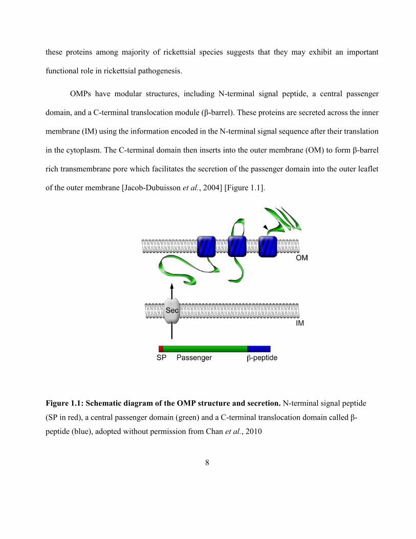
OMPs have modular structures, including N-terminal signal peptide, a central passenger
domain, and a C-terminal translocation module (β-barrel). These proteins are secreted across the inner
membrane (IM) using the information encoded in the N-terminal signal sequence after their translation
in the cytoplasm. The C-terminal domain then inserts into the outer membrane (OM) to form β-barrel
rich transmembrane pore which facilitates the secretion of the passenger domain into the outer leaflet
of the outer membrane [Jacob-Dubuisson et al., 2004] [Figure 1.1].
Figure 1.1: Schematic diagram of the OMP structure and secretion. N-terminal signal peptide
(SP in red), a central passenger domain (green) and a C-terminal translocation domain called β-
peptide (blue), adopted without permission from Chan et al., 2010
9
1.3.2: Outer membrane protein B (OmpB)
OmpB is evolutionarily conserved among all known rickettsial species, including both SFG and
TG rickettsia. Since this study specifically focus on the TG rickettsiea only the research which have
been done on typhus OmpB will be discussed in more detail.
There is a 7-16 nm size crystalline monolayer of protein arranged in tetragonal array attached
externally to the outer membrane of Rickettsia as revealed by electron microscopy studies [Silverman
et al., 1978]. This layer consists of 10-15% of total cellular protein and contains a diverse set of
proteins, most predominantly OmpB [Dasch et al., 1981]. The bacteria invade the host by attaching to
the host cell receptor through this most abundant surface protein [Martinez et al., 2005]. Then they
accumulate inside in substantial quantities causing the host cells to burst and release rickettsiae into the
blood, eventually leading to typhus.
OmpB shows a high level of conservation in terms of amino acid sequence (70 – 95% identity)
amongst different groups of rickettsiae [Blanc et al., 2005]. This conservation is evident not only in β-
barrel domain, which is restricted by its known function, but also in predicted passenger domain. These
areas of conservation suggest that OmpB passenger domain serves a unified function in rickettsial
pathogenesis. Hence a great deal of emphasis has been put forth to determine the contribution of this
domain in rickettsial host cell interactions, which is discussed in detail in the following section.
10
1.3.3: The passenger domain
As explained earlier, OmpB is expressed as a pre-protein and cleaved to release the passenger
domain from the β-barrel translocation domain, leaving the mature passenger domain associated with
the outer leaflet of the outer membrane [Figure 1]. Affinity chromatography methods have shown that
OmpB is the bacterial ligand that interacts with the host receptor Ku70, suggesting that OmpB-Ku70
serves as the adhesion-receptor pair in rickettsia-host cell interactions [Martinez et al., 2005]. The roles
of OmpB in rickettsial pathogenesis with regard to its contribution to bacterial adherence to an invasion
of host cells has also been examined. Heterologous expression of this protein in E.coli has shown that
OmpB plays a vital role in initiating bacterial infection in mammalian cells. OmpB is sufficient to
facilitate bacterial adherence and invasion to non-phagocytic cells when expressed in normally inert
E.coli [Uchiyama et al., 2006, Chan et al., 2009]. Interestingly, Chan et al., (2009) showed that the
purified passenger domain of OmpB itself can interact with Ku70, although a previous study has
proposed it is the β- peptide of OmpB which plays a role in adhesion [Renesto et al., 2006]. These
findings shows that an effector region of this protein is in part contained within the passenger domain.
OmpB has been identified as the immunodominant species-specific surface protein antigen and
has been isolated, purified, and biochemically characterized [Dasch et al., 1981]. Following an
infection with R. prowazekii in mice, guinea pigs, rabbits, and humans it has been observed the
dominant immunological anti-sera responses were directed against OmpB [Carl et al., 1989]. It has
also been shown that purified native typhus OmpB induced strong humoral and cell-mediated immune
responses [Carl et al., 1989]
11
In order to study the immune chemistry of OmpB, purified OmpB from R. prowazekii and R.
typhi was fragmented with CNBr and the origin of major fragments was determined by automated N-
terminal amino acid sequencing. Interestingly, CNBr fragments corresponding to the C-terminal were
not present in purified OmpB’s, suggesting that C-terminal region was removed during the
translocation to the cell surface. Also in an attempt to map the monoclonal antibody binding sites on
these fragments of OmpB antigens, it was found that six out of eight antibody types reacted
predominantly with a single region (amino acids 600-1200) of the OmpB within the passenger domain
suggesting the importance of this domain in generating the immune response [Ching et al., 1992].
1.4: OmpB as vaccine candidates
The immune responses generated from prior rickettsial infections shown to be protective and
detectable for years following an infection. This has led to the hypothesis that the OmpB proteins will
be good candidates for recombinant vaccine production [Sears et al., 2012]. At present there is no
preventative therapy available for protection against rickettsial pathogens. Even the antibiotic treatment
with broad spectrum antibiotics is effective only when administered within the first week of appearance
of symptoms. Thus prompt and proper diagnosis of the disease is required.
The potential use of the whole bacterium immunization procedures through various treatments
of pathogens to prevent the disease have been explored and while maintaining the protective effects of
exposure to the bacteria. For example, formalin-fixed R. rickettsii or R. prowazekii isolated from ticks,
embryonated chicken eggs, chicken embryo fibroblasts, lice or rabbit lungs have been utilized as the
whole cell vaccines in the early to mid-twentieth century [Ellison et al., 2008]. Even though these
12
vaccines failed to protect the individuals completely, they did reduce the severity of the symptoms and
were able to speed up the response to antibiotic treatment [Balayeva et al., 1972].
Humoral responses to live rickettsial immunizations generated antibodies that specifically
recognized high-molecular-weight rickettsial antigens, believed to be OmpB as identified by SDS-
PAGE analysis (115-200 kDa) [Anacker et al., 1987, Feng et al., 2004]. When the monoclonal
antibodies produced from these live rickettsial immunizations were characterized it revealed that only
a subset of antibodies recognizing epitopes belonging to the high molecular protein afforded protection,
leading to the hypothesis that OmpB could produce protective immune response in the host [Anacker
et al., 1985, Xu et al., 1997].
OmpB may be a good candidate as a vaccine and diagnostic tool; however mass production of
the R. prowazekii for the downstream purification of OmpB is not practical due to the intracellular
nature of the organism and the high biosafety levels required. Production of recombinant protein or
fragments should be more feasible. All reactive fragments of partially digested OmpB are in the
passenger domain and appear to be larger than 20 kDa in western blot analysis with patient sera [Ching
et al., 1998]. Among these reactive fragments, one of them was located at the N-terminus (fragment
A.) [Chao et al., 2008]. In addition to fragment A (amino acid residues 33-273) two more fragments
will be considered in this study; fragments AN (amino acid residues 33-744), and K (amino acid
residues 745-1353). These fragments [Figure 1.2] were cloned, expressed and purified to generate
recombinant reagents that mimic rickettsiae derived antigens.
13
Figure 1.2: Schematic diagram showing the recombinant fragments of R.tyhi OmpB. The three
fragments cover the passenger domain (green) and the size of each fragment is indicated. Not drawn
to a scale.
14
Understanding the characteristic biochemical features responsible for the virulence of the native
OmpB is essential to reproduce the correct recombinants and will be discussed in the following section.
1.5: Insight into virulence of Rickettsia
Due to their difficult growth requirements and the lack of genetic tools to manipulate their
genome in the past, little is known about the pathogenesis of rickettsial diseases. One approach to better
understand the factors that contribute to the virulence of Rickettsia is to identify the characteristic
differences between a virulent strain and an avirulent strain. One well studied organism in this regard
is R. prowazekii and the following section will discuss the different phenotypes of R. prowazekii in
detail.
1.5.1: Different R.prowazekii phenotypes
Several strains of R. prowazekii that differ considerably in virulence are recognized. The
prototypic virulent strain is known as Breinl [Ormsbee et al., 1978]. The reference avirulent strain
Madrid E was obtained after serial passages of a virulent strain Madrid I (isolated from an epidemic
typhus patient in Spain) in chicken embryo [Andersson et al., 1998]. Madrid I has lost its virulence
after passages in embryonated eggs and has been used under the name of Madrid E as a vaccine in
human since 1944 [Gudima et al.,1982]. When this avirulent Madrid E strain was subjected to one or
more passages through mice or guinea pigs (instead of chicken embryo) the virulence of the bacterium
was restored. This new strain with regained virulence was named as virulent revertant Evir strain
15
[Balayeva and Nikolskaya 1972]. Recently another strain of R. prowazekii was generated from Madrid
E. The growth of parental Madrid E changed dramatically after 3 months of culture on L929 cells,
making it to grow as fast as virulent strains, including Evir. This new strain was hypothetically termed
Erus considering this change in growth [Bechah et al., 2010]. The most recent virulent strain Rp22 (R.
prowazekii 22) was isolated from a patient in 1999 [Birg et al., 1999] as summarized in Figure 1.3.
Figure 1.3: Representative scheme of R.prowazekii strains showing the origin and evolution of
different phenotypes with varying degrees of virulence (Adopted from Bechah et al.2010 without
permission).
16
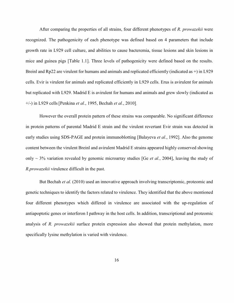
After comparing the properties of all strains, four different phenotypes of R. prowazekii were
recognized. The pathogenicity of each phenotype was defined based on 4 parameters that include
growth rate in L929 cell culture, and abilities to cause bacteremia, tissue lesions and skin lesions in
mice and guinea pigs [Table 1.1]. Three levels of pathogenicity were defined based on the results.
Breinl and Rp22 are virulent for humans and animals and replicated efficiently (indicated as +) in L929
cells. Evir is virulent for animals and replicated efficiently in L929 cells. Erus is avirulent for animals
but replicated with L929. Madrid E is avirulent for humans and animals and grew slowly (indicated as
+/-) in L929 cells [Penkina et al., 1995, Bechah et al., 2010].
However the overall protein pattern of these strains was comparable. No significant difference
in protein patterns of parental Madrid E strain and the virulent revertant Evir strain was detected in
early studies using SDS-PAGE and protein immunoblotting [Balayeva et al., 1992]. Also the genome
content between the virulent Breinl and avirulent Madrid E strains appeared highly conserved showing
only ~ 3% variation revealed by genomic microarray studies [Ge et al., 2004], leaving the study of
R.prowazekii virulence difficult in the past.
But Bechah et al. (2010) used an innovative approach involving transcriptomic, proteomic and
genetic techniques to identify the factors related to virulence. They identified that the above mentioned
four different phenotypes which differed in virulence are associated with the up-regulation of
antiapoptotic genes or interferon I pathway in the host cells. In addition, transcriptional and proteomic
analysis of R. prowazekii surface protein expression also showed that protein methylation, more
specifically lysine methylation is varied with virulence.

17
Table 1.1: Summarized results of the four R. prowazekii phenotypes. (Adopted from Bechah
et al.2010 without permission)
Tested
strain Source
Pathogenicity
Virulence Culture on
L929
Mice Guinea pigs
Bacterial
replication
Tissue
lesions Skin lesions
Rp22,
Breinl human blood + yes yes
yes
Virulent
Evir
passaged on
animals + yes no yes
Less
pathogenic
Erus
grown in
L929 cells
+ no no no Avirulent
Madrid
E
grown in eggs
+/- no no no Avirulent
18
At present the structural feature of rickettsial OmpB methylation at є-amino groups of lysine
residues is not very well understood though it’s very important regarding rickettsial virulence
[Rodionov et al., 1990]. Post-translational modification of proteins by methylation can potentially
alter OmpB structure and function, leading to the hypothesis that methylation of lysine residues of
OmpB correlates with the virulence of R. prowazekii [Turco et al., 1994].
The next few sections will thoroughly focus on the relationship between post-translational
modifications, especially lysine methylation and virulence of Rickettsia that has been previously
suggested [Rodionov et al., 1991, Chao et al., 2004, Ching et al., 1993].
1.6: Relationship between lysine methylation and virulence of Rickettsia
1.6.1: Post translational modifications on proteins
Within the last few decades, scientists have learned that the human proteome is immensely
more complex than the human genome. While it is estimated that the human genome comprises
between 20,000 and 25,000 genes [Collins et al., 2004], the total number of proteins in the human
proteome is estimated at over 1 million [Jensen et al., 2004]. These estimations show that single
genes encode multiple proteins. Genomic recombination, transcription initiation at alternative
promoters, differential transcription termination, and alternative splicing of the transcript are
mechanisms that generate different mRNA transcripts from a single gene [Ayoubi et al., 1996].
The increase in complexity from the level of the genome to the proteome is further facilitated
by protein post-translational modifications (PTMs). It’s defined as the process of covalently altering
one or more amino acids in a protein after the protein has been completely translated and released
19
from the ribosome. PTMs are chemical modifications that play a key role in functional proteomics,
because they regulate activity, localization and interaction with other cellular molecules such as
proteins, nucleic acids, lipids, and cofactor [Walsh C.T, 2005]
PTMs regulate protein structure and function. They play key roles in many cellular processes
such as cellular differentiation [Grotenberg et al., 2007], protein degradation [Geiss-Friedlander et
al., 2007], signaling and regulatory processes [Morrison et al., 2002], regulation of gene expression,
and protein-protein interactions.
Serine and threonine phosphorylation are the most extensively studied PTM in proteins, since
it plays a vital role in intracellular signal transduction and is involved in regulating cell cycle
progression, differentiation, transformation, development, peptide hormone response, and adaptation
[Hubbard et al., 1993, Pawson et al., 1997, Hunter, 2000, Cohen, 2002]. It has been estimated that one
third of mammalian proteins may be phosphorylated and this modification often plays a key role in
modulating protein function.
1.6.2: Post translational modifications on lysines
But over the past decade lysine has become a crucial amino acid due to dynamic nature of its
PTMs and the various effects they have on protein structure and function. Lysine can be modified in
different ways. They can be modified by small chemical changes such as acetylation [Glozak et al.,
2005] or by large “peptide” attachments to lysines such as ubiquitylation [Chau et al., 1989] or
sumoylation [Verger et al., 2003]. These modifications can affect the lysine in two ways by reducing
its positive charge and by changing the structure of the side chain. By doing so, lysine modifications
20
can increase the negative charge, which in turn may alter function directly or could provide a novel
interface for docking of cognate proteins, thereby influencing the protein activity indirectly. In addition
to these, the presence of one PTM inhibits the attachment of the other modifications and the fact that
alternate modifications occur on lysine provides another level of protein regulation [Huang et al.,
2008].
1.6.3: Methylation of lysine
This section will focus on the lysine methylation. Because of the many interesting levels and
degrees of regulation occur for methylation, methylation is considered most exciting in the family
lysine modifications. Methylation requires a methyltransferase enzyme. Methyltransferases in
general can be classified by different substrate specificities (small molecules, lipids, nucleic acids,
proteins etc) and different target atoms for methylation (nitrogen, oxygen, carbon, sulfur, etc).
Lysine methyltransferases are the enzymes which catalyze the transfer of one, two or three methyl
groups to the ε-amino group of lysine residues of proteins [Paik et al., 1990]. The general scheme
of lysine methylation is shown in Figure 1.4. These enzymes (sub-subclass EC 2.1.1) use a reactive
methyl group bound to sulfur in S-adenosyl methionine (AdoMet or SAM) as the methyl donor to
produce S-adenosyl homocysteine (AdoHcy or SAH) as a product along with the methylated
substrate [Chen et al., 1999]. Methylation occurs so often that SAM has been suggested to be the
second most-used co-factor in enzymatic reactions after ATP [Paik et al., 2007]. The transfer of
one-carbon methyl groups to the nitrogen (N-methylation) of amino acid side chains increases the
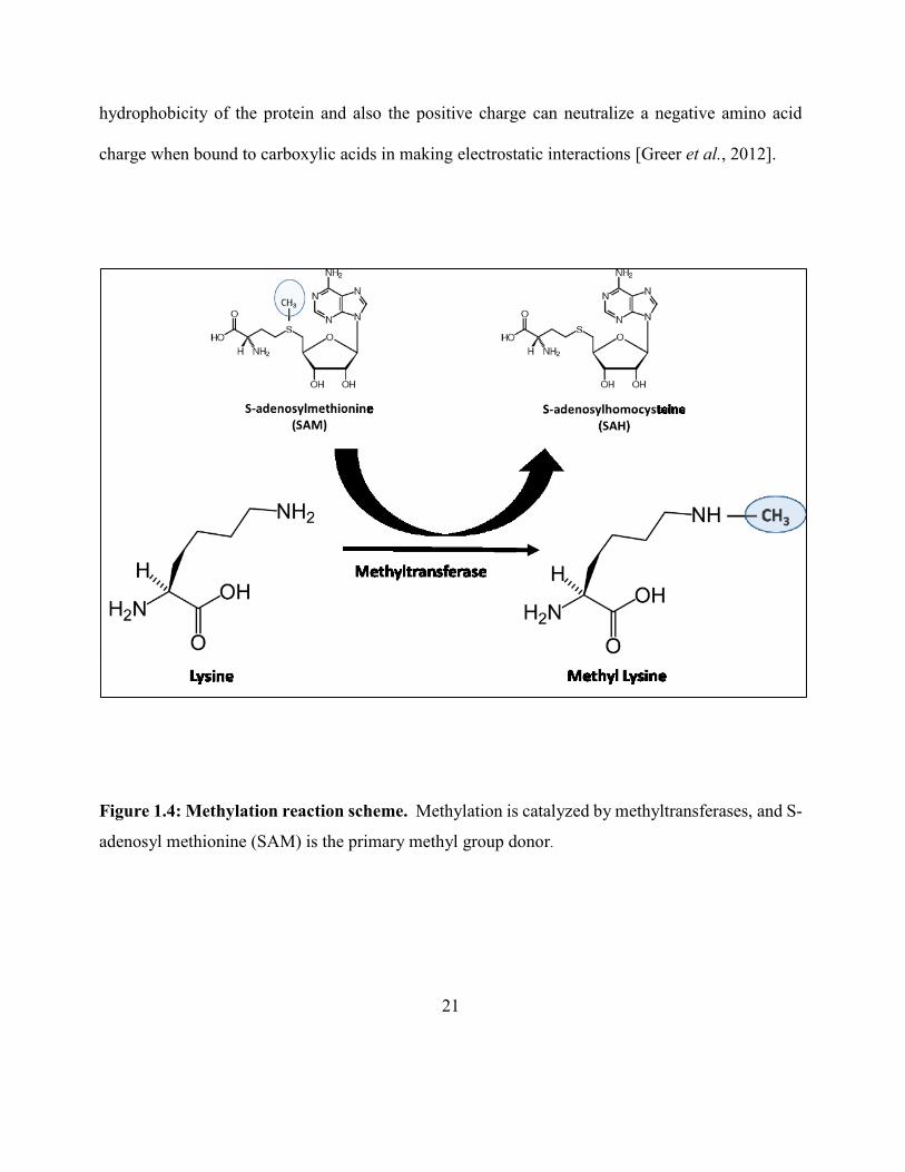
21
hydrophobicity of the protein and also the positive charge can neutralize a negative amino acid
charge when bound to carboxylic acids in making electrostatic interactions [Greer et al., 2012].
Figure 1.4: Methylation reaction scheme. Methylation is catalyzed by methyltransferases, and S-
adenosyl methionine (SAM) is the primary methyl group donor.
22
Lysine methylation can regulate protein function through different mechanisms. For example,
methyllysine can provide docking sites for binding of cognate proteins and also can inhibit other PTMs
occurring on the same lysine residue. The potential of adding one, two or three methyl groups to the
side chain ε-NH2 group can correlate with distinct genomic locations and functions. By far the most
well studied lysine methylation comes from the study of histone substrates [Cheng et al., 2005]. The
methyl groups that are added to the histones act to regulate transcription by blocking or allowing DNA
access to transcription factors. In this way the integrity of the genome and epigenetic regulation of
genes are under the control of the actions of histone methyltransferases. Histone methylation is key in
distinguishing the integrity of the genome and the genes that are expressed by cells, thus giving the
cells their identities [Botuyan et al., 2006, Rea et al., 2000].
Unlike histone proteins, methylation of r i cke t t s i a l membrane proteins has attracted little
attention and requires better characterization at molecular level. OmpB methylation at lysine residues
could conceivably affect bacteria-host cell interactions thereby influencing invasion of host cell, and
inhibit other post- translational modifications such as acetylation, ubiquitination or sumoylation
thereby influencing its survival in the host cell by avoiding destruction and premature death [Polevoda
et al., 2007]. A better understanding of how lysine methylation affect the rickettsial virulence can be
achieved by comparing different methylation profiles of OmpB’s in different R. prowazekii strains.
23
1.7: Different OmpB lysine methylation profiles
1.7.1: Multiple methylation of Rickettsia OmpB
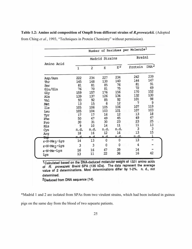
The biochemical properties of OmpB purified from R. prowazekii have been characterized [Carl
et al., 1990]. While more than half of the lysine residues predicted from sequence were not detected
during the conventional amino acid composition analysis, ε-N-mono-methyl, di-methyl and tri-methyl
lysines were detected as natural components of proteins [Paik et al. 1990]. OmpB from the avirulent
live vaccine strain Madrid E contained more ε-N-monomethyllysine and no detectable ε-N-
trimethyllysine compared to OmpB from two virulent strains, revertant Madrid Evir and Breinl
[Balayeva and Nicholsava 1972]. A study by Ching et al., to further investigate the possible relationship
between the level of methylation and virulence revealed that, except for lysine the variation among
other amino acid compositions are comparable between two avirulent Madrid strains (E and Er) and
two virulent Madrid strains (isolated from blood of two patients) These were also comparable to what
it was predicted from the DNA sequence of Breinl. In three virulent strains (Madrid 1, 2 and Breinl)
the amounts of lysine, ε-N-monomethyl, ε-N-dimethyl and ε-N-trimethyl were similar among them.
But in two avirulent strains no dimethyl or trimethyl lysines were detected and the amount of
monomethyl lysine was more than two fold compared to virulent strains. The avirulent strains
contained at least 50% higher lysine and monomethyllysine amounts than all types of lysines combined
in virulent strains, which was similar to number of lysines (42) predicted from Breinl DNA sequence.
The fact that they observed significantly higher numbers for lysine and monomethyllysine in
OmpB for avirulent strain may suggest that, when going from virulent to avirulent strain, a possible
mutation could occur in a methyltransferase or in some other related enzyme, affecting methylation
24
and hence attenuating virulence [Ching et al.,1993]. The possibility of a mutation in OmpB leading to
higher number of lysines in avirulent strain is unlikely because other amino acid contents remain
unchanged and also reversal of avirulent to virulent strain occurred under proper selective pressure
[Kazar et al.,1973].
These findings suggest that OmpBs from virulent Rickettsial strains are more extensively
methylated. Although a specific role of lysine methylation in virulence in not known, it is possible that
hypermethylation affect the transport of proteins to specific organelles and provide protection for
protein from proteolytic degradation. This could explain the previous observation of poor growth of
avirulent Madrid E in macrophages [Turco et al., 1982], That is, without full methylation, OmpB may
be an ineffective barrier and may subject to proteolytic degradation.
25
Table 1.2: Amino acid composition of OmpB from different strains of R.prowazekii. (Adopted
from Ching et al., 1993, “Techniques in Protein Chemistry” without permission).
*Madrid 1 and 2 are isolated from SPAs from two virulent strains, which had been isolated in guinea
pigs on the same day from the blood of two sepearte patients.
26
1.7.2: Genetic differences between virulent and avirulent strains
In section 1.7.1 it was discussed the possibility of a mutation on a methyltransferase or a
related enzyme which could affect the levels of methylation between virulent and avirulent strains.
In an attempt to identify the reverse mutation that may determine the virulence of R.prowazekii,
Zhang et al.2006 took an innovative approach. The genetic differences between the avirulent Madrid
E strain and its virulent revertant Evir were identified as follows. With Evir genome sequence not being
available, they compared the genomes of R. prowazekii E strain with the genomes of R. conorii
(Mediterranean spotted fever) [Yagupsky et al., 1993], R. typhi (Endemic typhus) [Baxter, 1996], R.
rickettsii (Rocky Mountain spotted fever) [Dahlgren et al., 2012], and R. sibirica (North Asian tick
typhus) [Parola et al., 2005], and identified 16 mutant genes with internal stop codon in the E strain
but not in any other rickettsial genome. Out of these 16, DNA sequencing of E strain and Breinl
confirmed that there were only 8 genuine gene mutations and others were false [Zhang et al., 2006].
Seven genes had the same mutation in virulent revertant Evir and five of these genes have the same
mutation in virulent Breinl strain suggesting these genes are irrelevant to R. prowazekii virulence. Most
importantly, they discovered that one gene (Rp028/027) is inactivated by a frame shift mutation in
avirulent E strain, but the mutation does not exist in both virulent revertant Evir and virulent Breinl
strain. A single nucleotide insertion occurred in E strain resulting a premature stop codon and thereby
truncation of the protein product [Figure 1.5]. This mutation is reverted giving rise to Evir from Madrid
E during one or more passages through mice or guinea pigs and becomes identical to the wild type
virulent Breinl strain.
27
Figure 1.5: The gene encoding the methyltransferase RP027-028 was mutated by a frameshift in
avirulent E strain but not in the virulent revertant Evir or wild type Breinl strain. The single
nucleotide (A) insertion is indicated by an arrow. (Adopted from Zhang et al., 2006 without
permission).
The gene Rp028/027 showed homology to a SAM-dependent methyltransferase, and the
mutation may result in the loss of methyltransferase activity in avirulent strain. This suggests that the
deficiency in lysine methylation in avirulent Madrid E, as discussed in section 1.7.1, is due to this
28
mutation in lysine methyltransferase rather than a mutation in OmpB. A comparative proteomic
approach revealed that the protein encoded by Rp028/Rp027 was detectable in virulent strains and this
result is consistent with the fact that these genes are not transcribed in Madrid E strain [Chao et al.,
2005, Chao et al., 2007]
Bechah et al., recently sequenced the genome of the R. prowazekii virulent strain Rp22, and it
showed that one of the putative methyltransferases in Rp22 (NCBI Reference Sequence:
YP_005998435.1) aligned well with the 2 split genes Rp028 and Rp027 of Mardid E. Rp028 is
homologous to the N-terminal part where Rp027 is to the C-terminal part [Figure 1.6].
However there is another methyltransferase homolog (Rp789) which is expressed in both
virulent and avirulent strains as determined by BLAST search. Genomic and proteomic analysis has
shown that Rp789 is one of the putative methyltransferases which is down regulated in avirulent strain
compared to the virulent Rp22 [Bechah et al., 2010]. These data confirm the existence of
methyltransferase in both virulent and avirulent strains. It is suggested that, the loss of a
methyltransferase due to mutation in Rp028/027 and/or differential expression of Rp789 encoded
proteins may change the level of lysine methylation which in turn could cause the loss of virulence.
Taken together, the level of lysine methyltransferases and the virulence of Rickettsia appear to be
correlated.
29
Figure 1.6: Schematic diagram showing the alignment between the conserved domains of
putative methyltransferase in Rp22 and Rp028 and Rp027 from Mardid E (not drawn to scale,
generated using BLAST NCBI). It contains the AdoMet_MTases superfamily which is a S-
adenosylmethionine-binding site and MethyTransf_Reg superfamily which is a predicted
methyltransferase regulatory domain and COG4797 superfamily which is a predicted regulatory
domain of a methyltransferase.
Some studies have already shown that methylation of OmpB appears to enhance its antigenicity.
A study by Gilmore et al., 1993 showed rabbit antiserum against recombinant OmpB (unmethylated)
is less reactive than antiserum against OmpB purified directly from Rickettsiae [Gilmore et al., 1991].
In another study, fragment A (33-272) of the OmpB from R.typhi has been cloned, purified and
30
chemically methylated in an attempt to generate recombinant OmpB fragments that mimic rickettsiae
derived antigens. Fragment A contained mono-, di-, tri-methylated lysines and showed higher reactivity
than the non-methylated fragment A with patient sera in an enzyme linked immunoabsorbant assay
[Chao et al., 2008].
It suggests that recombinant protein fragment may be able to replace native OmpB as a
diagnostic reagent for not only R. typhi infection but also typhus group rickettsiae infection in general.
But the chemical methylation of recombinant proteins may not have the same selectivity and specificity
as the enzymatic methylation derived proteins in vivo.
1.8: Structural Insights of Outer Membrane Protein Methyltransferases from
Rickettsia
First part of this project is focused on first time identification and in-depth characterization of
methyltransferase enzymes responsible for methylating OmpB substrates. The enzymes are
characterized kinetically using radioactive assays and at molecular level using integrated liquid
chromatography – tandem mass spectrometry (LC-MS/MS). The unusual and interesting methylation
patterns observed in lysine residues when using two different methyltransferases from an avirulent
(R.prowazekii Madrid E) and a virulent strain (R.typhi Wilmington) led to the part 2 of the study. The
goal was to obtain a good understanding about the mechanism of methylation given that its’ unusual
nature. There are no crystal structures reported for methyltransferases which methylate the outer
membrane proteins. To get structural insight of the OmpB methylation we used protein X-ray
crystallography to investigate the structure of the lysine methyltransferases. The crystal structures of
31
two methyltransferases and the respective complexes with SAM were determined. These are the first
reported crystal structures of outer membrane protein methyltransferases. Comparison of the two
structures revealed a number of structural insights into the mechanism of novel enzymatic methylation
of OmpB.
1.9: Hypothesis and Objective
We could hypothyze that, there is a functional methyltransferase(s) responsible for the apparent
difference in virulence among different rickettsial strains. The main objective is to identify those
specific methyltransferases which catalyze methylation of OmpB lysine. As the first step, list of
putative methyltransferases will be selected by structural and bioinformatic genomic analysis of
potential methyltransferases from different rickettsial strains. They will be cloned, expressed and
purified and examined for the methyltransferase activity against recombinant OmpB proteins,
fragments and synthetic peptides from rickettsia using radioactive and western blot assays.
Characterization of this enzymatically methylated substrate is essential for elucidating the roles of
hypermethylation in bacterial virulence and pathogenesis. It’s also required to make sure that
methylated recombinant fragments mimic the structure of the native OmpB and it will be achieved by
mass spectrometry (LC-MS/MS). Optimization of the conditions for the enzymatic assays in terms of
substrates and yields and also determination of enzyme kinetic parameters of the methyltransferase are
intended. This understanding will be vital for the mass production of methylated recombinant OmpB,
and may advance our knowledge of the structure and function of protein lysine methylation in general.
Being able to generate enzymatically methylated recombinant OmpB may also provide an improved
32
method over chemical methylation in the attempt to generate advance diagnostic reagents and vaccine
candidates. Also determination of three dimensional structure of the enzyme-ligand complex may
provide information on the methylation mechanism. This study provides the first in-depth
characterization of methylation of an OMP at the molecular level and may lead to uncover the links
between OmpB methylation and rickettsial virulence, improve OmpB-based diagnostic reagents, and
develop inhibitors against methyltransferases.
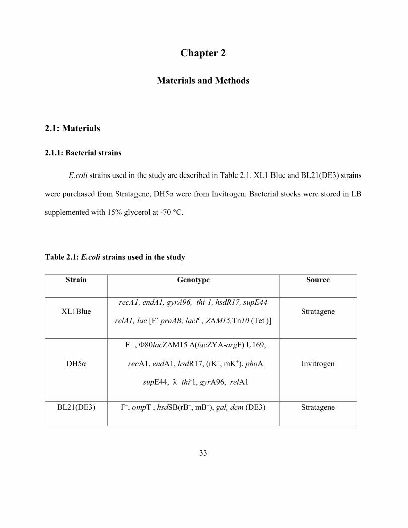
33
Chapter 2
Materials and Methods
2.1: Materials
2.1.1: Bacterial strains
E.coli strains used in the study are described in Table 2.1. XL1 Blue and BL21(DE3) strains
were purchased from Stratagene, DH5α were from Invitrogen. Bacterial stocks were stored in LB
supplemented with 15% glycerol at -70 °C.
Table 2.1: E.coli strains used in the study
Strain Genotype Source
XL1Blue
recA1, endA1, gyrA96, thi-1, hsdR17, supE44
relA1, lac [F´ proAB, lacIq , ZΔM15,Tn10 (Tetr)]
Stratagene
DH5α
F– , Φ80lacZΔM15 Δ(lacZYA-argF) U169,
recA1, endA1, hsdR17, (rK–, mK+), phoA
supE44, λ– thi-1, gyrA96, relA1
Invitrogen
BL21(DE3) F–, ompT , hsdSB(rB–, mB–), gal, dcm (DE3) Stratagene
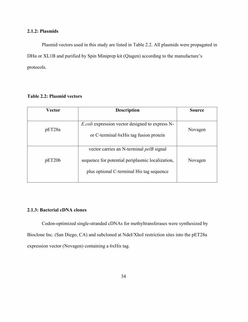
34
2.1.2: Plasmids
Plasmid vectors used in this study are listed in Table 2.2. All plasmids were propagated in
DHα or XL1B and purified by Spin Miniprep kit (Qiagen) according to the manufacture’s
protocols.
Table 2.2: Plasmid vectors
Vector Description Source
pET28a
E.coli expression vector designed to express N-
or C-terminal 6xHis tag fusion protein
Novagen
pET20b
vector carries an N-terminal pelB signal
sequence for potential periplasmic localization,
plus optional C-terminal His tag sequence
Novagen
2.1.3: Bacterial cDNA clones
Codon-optimized single-stranded cDNAs for methyltransferases were synthesized by
Bioclone Inc. (San Diego, CA) and subcloned at NdeI/XhoI restriction sites into the pET28a
expression vector (Novagen) containing a 6xHis tag.

35
2.1.4: Oligonucleotides
All oligonucleotides used in this study were synthesized by Qiagen or Operon. The
oligonucleotides were salt free and synthesized at 10 nM scale. The 100 µM oligonucleotides stocks
were prepared with molecular grade water.
2.1.5: Buffers, media and solutions
10 X PBS was from Biosource. 0.5 M EDTA (pH 8.0), 1.0 M Tris-HCl (pH 7.5) and 10 x
TAE buffer (pH 7.2) were purchased from Quality Biologicals. Lysis buffer for Ni-NTA column
purification contained 50 mM sodium phosphate (pH7.4), 300 mM NaCl, 10 mM Imidazole. Elution
buffer contained 50 mM sodium phosphate (pH7.4), 300 mM NaCl, 250 mM Imidazole. EDTA-
free complete protease inhibitor Tablets were from Roche.
1kb and 100bp DNA ladder with 6x DNA loading buffer were purchased from BioRad. Pre
stained SDS-PAGE standard (BioRad) contains 50% glycerol, 300 mM NaCl, 10 mM Tris, 2 mM
EDTA and 3 mM NaN3. Protein samples were loaded into 12% or 7.5 % Bis-Tis gel with 1x
Laemmli sample buffer (BioRad).
NuPAGE MOPS SDS running buffer (pH7.0) was purchased from Invitrogen for SDS-
PAGE. The gel was stained with coomassie staining solution containing 0.75% (w/v) coomassie
brilliant blue R-250, 7.5% acetic acid and 50% methanol followed by destaining solution containing
35% methanol and 10% acetic acid. Gel drying solution contains 20% methanol and 5% glycerol.
36
2.1.6: Antibodies
250 µg/ml rabbit anti-trimethyl lysine antibodies were fromTrend Pharma & Tech Inc.,
Surrey, Canada. IRDye (800CW) - conjugated anti-rabbit IgG (H+L) (donkey) secondary antibodies
were from Rockland Immunochemicals, Inc., Gilbertsville, PA.
2.1.7: Reagents and other materials
Recombinant OmpB fragments, including recombinant OmpB (A) (33 to 273), recombinant
OmpB(AN) (33 to 744), and OmpB(K) (745 to 1353) based on the genomic sequence of R. typhi
were from Naval Medical Center.
Ampicillin, kanamycin, isopropyl-β-D-1-thiogalactopyranoside (IPTG), ethylene
diaminetetraacetic acid (EDTA), β-mercaptoethanol, dithiothreitol (DTT), ethidium bromide,
agarose, glycerol, acetic acid, trichloroacetic acid (TCA) and ethidium bromide were purchased
from Sigma-Aldrich.
All restriction enzymes and T4 DNA ligase were from New England Biolabs. Proofstart
DNA polymerase was from Qiagen. QuickChange II Site-Directed Mutagenesis kit was from
Stratagene/Agilent. QIAprep Spin mini column for DNA purification, QIAquick DNA gel
extraction kit and Ni-NTA agarose beads were from Qiagen. Trichloroacetic acid (TCA), bovine
serum albumin (BSA), agarose, potassium chloride, sodium chloride, ammonium bicarbonate,
sodium phosphate (monobasic and dibasic), ampicillin, kanamycin, EDTA, iodoacetamide, PMSF,
and urea were purchased from Sigma.
37
Protein concentration assay kit, and blotting grade non-fat dry milk were from BioRad.
Dialysis membranes were from Pierce. Amicon YM-30 columns were from Millipore. Sequencing
grade trypsin was from Roche. S-[3H-Me] adenosylmethionine (10 Ci/mmol) was from Perkin
Elmer. S-adenosylmethionine was from New England Biolabs. SET7 methyltransferase, histones
(Calf thymusssssss) and the histone peptide substrate H3 (1–17) were from New England Biolabs.
N,N-diallyltartardiamide (DATD) was from Bio-Rad.
2.2: Methods
2.2.1: Construction of plasmids
Target DNA sequences were amplified using PCR. The thermostable RedTaq DNA
polymerase (Stratagene) was used in PCR reactions. Reactions were carried out in a total volume of
50 µl. The reaction mixtures contained 1-20 ng DNA template, 1 x PCR buffer, 0.4 mM dNTP mix,
0.4 µM of each primer, 1-2 units of DNA polymerase. The list of the primers used for construction
of the vectors are listed in Appendix 1.2. PCR was carried out for 25 cycles using the profile: 95 °C
(DNA denaturation) for 5 min, 55-65°C (primer annealing) for 30 s, 72°C (extension) for 1 min for
each kb of amplified DNA.
PCR products were purified using Qiagen PCR purification kit. Amplified products and
vectors were digested using restriction enzymes (PCR product was supplemented with 1 x digestion
buffer, 10-20 units of the enzyme, and incubated for 1-2 hours at 37°C). The digested products were
visualized on agarose gel, UV illuminated bands were excised and purified using Qiagen gel
extraction kit.
38
The digested cDNA was ligated into a linearized vector using T4 DNA ligase at 16°C
overnight. The ligated plasmid was transformed into XL1Blue competent cells. Transformed single
colony was used for the propagation of the plasmid at 37°C overnight. All plasmid sequences were
confirmed by DNA sequencing.
2.2.2: Site-directed mutagenesis
Mutations targeting AdoMet binding residues, substrate binding residues and residues
involved in dimer formation were generated using site-directed mutagenesis. All primers and
mutation sites are listed in Appendix 1. Mutagenesis was carried out using Quikchange Lightning
mutagenesis Kits (Stratagene/Agilent) following the manufacture’s protocol. Primers for site-
directed mutagenesis were designed by QuikChange Primer Design online tool (Agilent).
A typical 10 µl mutagenesis reaction contained 100 ng of each primer, about 100 ng of
template, 1µl of dNTP mix, 1µl of 10x reaction buffer, 1µl of PfuTurbo DNA polymerase (2.5U/µl)
and 5 ml of deionized water. After PCR, the reaction mixtures were digested by Dpn1 (20U/µl) at
37°C for 1 hour. Then 5 ml of the reaction mixtures were transformed into DH5α super-competent
cells with standard transformation protocol. The transformed cells were grown overnight on LB
plate with appropriate antibiotics. Plasmids were purified from overnight culture using Qiaprep spin
minprep kit (Qiagen). DNA sequences were confirmed using the sequencing service of Genewiz
Inc.
39
2.2.3: Native OmpB and recombinant OmpB fragment preparation
Native OmpB proteins from R. typhi, R. prowazekii strains Breinl, RP22 and Madrid E were
extracted using 10 mM Tris-HCl (pH 7.6) at 45°C for 30 min and purified as described previously
[Dasch 1981]. Each of the native OmpB proteins showed a single band at Mr 114,000 on SDS
polyacrylamide gel. Recombinant R. typhi OmpB fragments, OmpB(AN) and OmpB(K), that
corresponded to residues M33 to F744 and R745 to G1353, respectively, were expressed and
purified as previously described [Chao et al., 2008]. Briefly, the bacterial proteins that were
expressed in inclusion bodies were dissolved in 8M urea and 1% deoxycholate, purified by ion
exchange chromatography on DEAE-cellulose in 6 M urea. Soluble proteins were obtained by slow
dialysis at stepwise decreasing concentrations of urea. Protein concentrations were determined using
Bio-Rad Protein Assays with bovine serum albumin as the standard.
2.2.4: Bioinformatics analysis
Prior to our structural analysis reported later, no structural information for Rickettsial
methyltransferases was available, therefore bioinformatics approaches were used to form an initial
working model. Annotated genomic sequences of R. prowazekii Madrid E and R. prowazekii RP22
in NCBI and annotated encoded proteins in PIR (Protein Information Resources) were searched and
examined for potential protein methyltransferases. Those methyltransferases targeting DNA, rRNA,
tRNA, mRNA, and small molecule metabolites were eliminated. The remaining genes and encoded
proteins that are annotated as hypothetical proteins with potential S-adenosylmethionine (SAM)
binding domains were selected for further analysis. Structural bioinformatic analysis of the putative
40
methyltransferases was carried out using the software programs HHpred [Soding et al., 2005] and
3DLigandSite (Wass et al., 2010). The structural templates were selected by the software at
respective Web servers. Successful structural modeling was obtained only with the N-terminal half
of the putative methyltransferases. No known three-dimensional structures with significant
homology to the C-terminal half are available. The structural models were displayed using the
software program Chimera (UCSF). The secondary structural prediction of OmpB was carried out
using the Web servers Psipred [Jones 1999], HHpred, and Phyre [Kelly et al., 2009].
2.2.5: Synthesis and cloning of genes encoding putative methyltransferases from R.
prowazekii and R. typhi
Table 2.3 lists the names and accession numbers of genes and encoded proteins of putative
methyltransferases. Codon-optimized cDNAs were synthesized by Bioclone Inc. (San Diego, CA)
and subcloned at NdeI/XhoI restriction sites into the pET28a expression vector (Novagen)
containing a 6xHis tag. Standard cloning procedures were performed, and the inserts were verified
by restriction enzyme digestion and sequencing. The sequences of the cDNAs for the expression in
E. coli are shown in Appendix 1.1.
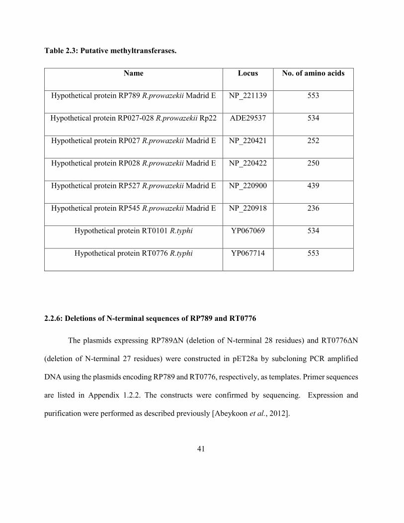
41
Table 2.3: Putative methyltransferases.
Name Locus No. of amino acids
Hypothetical protein RP789 R.prowazekii Madrid E NP_221139 553
Hypothetical protein RP027-028 R.prowazekii Rp22 ADE29537 534
Hypothetical protein RP027 R.prowazekii Madrid E NP_220421 252
Hypothetical protein RP028 R.prowazekii Madrid E NP_220422 250
Hypothetical protein RP527 R.prowazekii Madrid E NP_220900 439
Hypothetical protein RP545 R.prowazekii Madrid E NP_220918 236
Hypothetical protein RT0101 R.typhi YP067069 534
Hypothetical protein RT0776 R.typhi YP067714 553
2.2.6: Deletions of N-terminal sequences of RP789 and RT0776
The plasmids expressing RP789ΔN (deletion of N-terminal 28 residues) and RT0776ΔN
(deletion of N-terminal 27 residues) were constructed in pET28a by subcloning PCR amplified
DNA using the plasmids encoding RP789 and RT0776, respectively, as templates. Primer sequences
are listed in Appendix 1.2.2. The constructs were confirmed by sequencing. Expression and
purification were performed as described previously [Abeykoon et al., 2012].
42
2.2.7: Expression and purification of putative methyltransferases or R.prowazekii and R.typhi
BL21(DE3) competent cells (Agilent Technologies) were transformed with pET28a carrying
one of the genes encoding hypothetical proteins and putative methyltransferases. Protein expression
was carried out by growing the bacteria from single colonies at 37°C in 2 ml of LB broth
supplemented with 100 g/ml kanamycin overnight. The cell culture was transferred to fresh LB
broth with kanamycin and incubated with shaking until the optical density at 600nm (OD600) reached
0.3 to 0.5. The cultures were induced with 0.04 mM isopropyl-D-thiogalactopyranoside (IPTG) and
incubated at 22°C and at 250 rpm overnight. Cells were harvested by centrifugation and lysed, and
the expressed proteins were purified using nickel-nitrilotriacetic acid (Ni-NTA) column
chromatography according to the manufacturer’s instructions (GoldBio, St. Louis, MO). Protein
concentrations were determined using the Bio-Rad Protein Assay and bovine serum albumin as a
standard [Bradford, 1976].
2.2.8: Protein lysine methyltransferase activity assay by incorporation of [3H-Methyl]
Methyltransferase activity was monitored by the transfer of [3H]-methyl from S-[3H-Me]
adenosylmethionine (10 Ci/mmol; Perkin Elmer) to recombinant protein substrates. The purified
recombinant methyltransferases were then examined for protein methyltransferase activity using
recombinant OmpB fragments as substrates. We first developed a methyltransferase assay modified
from the SET7 methyltransferase assay. The amount of methyl groups transferred from S-[3H-Me]-
S- adenosylmethionine to histone 3 peptide was determined by Whatman DE81 immobilized
radioactivity after extensive washing with sodium bicarbonate [Xiao et al., 2003]. Unlike histones,
43
the OmpB fragments are acidic proteins, so Whatman 3MM and 5% trichloroacetic acid (TCA)
replacing DE81 and sodium bicarbonate were used. The rate of methyl transfer from S-[3H-Me]-S-
adenosylmethionine to histone 3 peptide was found to be the same as that using the conventional
SET7 assay.
The 50 µl reaction mixtures contained 8.3 mM sodium phosphate, pH 8.0, 160 µM [3H-Me]
SAM (34 mCi/mmol), and 2 µM recombinant OmpB(AN) or recombinant OmpB(K). The
methylation reaction was initiated by adding limiting amounts of protein methyltransferase and
incubated at 37°C. Aliquots of the reaction mixture were spotted onto Whatman 3MM cellulose
filter paper discs (Fisher Scientific) at various time points. Reactions were stopped by the addition
of 5% TCA. The paper discs were washed with 5% ice-cold TCA three times, followed by ethanol-
ether (1:1 [vol/vol]). The amounts of acid-precipitable radioactivity were determined by liquid
scintillation counting using a Perkin Elmer Wallace 1410 counter. One unit of methyltransferase is
defined as the amount of enzyme required to catalyze the transfer of 1 pmol of methyl group to the
substrate in 10 min.
2.2.9: Quantifying radioactivity in proteins extracted from polyacrylamide gels
Radioactivity associated with methylated OmpB fragments separated by SDS gel
electrophoresis was determined using the method previously described with minor modifications
[Springer 1991]. To quantitate the number of methyl groups transferred to each substrate molecule,
the radioactivity associated with the OmpB(AN) and (K)were measured using the following method.
Protein substrate (3 µM) was incubated with recombinant RP789 or RP027-028 (6 µM) in the
44
presence of radioactive SAM (0.16 mM with specific activity of 68 mCi/mmol) in 8.3 mM
phosphate buffer (pH 8.0) at 37°C overnight. The reaction was stopped by adding SDS sample
buffer. Polyacrylamide gels were polymerized with reagents obtained from Bio-Rad Laboratories
(Richmond, CA) except that N,N’-methylenebisacrylamide was replaced with DATD at a ratio of 1
part of DATD to 10 parts of acrylamide [Springer 1991]. After electrophoresis, the SDS gels were
stained with Coomassie blue, followed by extensive destaining (Bio-Rad). Appropriately sized
slices of the gel were placed in glass scintillation vials, and 0.5 ml of 2% (wt/vol) sodium
metaperiodate was added. The vials were shaken for 30 min to dissolve the gel. Ecoscint A (6 ml;
National Diagnostics, Atlanta, GA) was added to the vial, and the vial was cooled on ice for 20 min
and then counted by liquid scintillation counting.
2.2.10: Western blot analysis of putative lysine methyltransferases
The enzymatic methylation reaction mixtures as described above were incubated at 37°C for
4 h. Protein samples were denatured in standard sample denaturing buffer containing 1% SDS and
10 mM mercaptoethanol. The reaction products were separated by SDS-PAGE and followed by
Western blot analysis with 250 µg/ml rabbit (is it monoclonal from mice?) anti-trimethyllysine
(Trend Pharma & Tech Inc., Surrey, Canada) at a 1:2,000 dilution in Odyssey blocking buffer
(phosphate-buffered saline and 0.1% sodium azide) containing 0.1% Tween 20 at 4°C overnight,
followed by incubation with IRDye (800CW)-conjugated anti-rabbit IgG (H+L) (donkey) secondary
antibody (Rockland Immunochemicals, Inc., Gilbertsville, PA) at a 1:10,000 dilution in blocking
45
buffer for 1 h at room temperature. The signal was detected using an Odyssey infrared imaging
system (Li-Cor) after washing 3 times with 1x PBS containing 0.1% Tween 20.
2.2.11: Steady-state kinetic studies
Initial rates of MT-catalyzed reactions were determined from the linear portions of the time
courses of methylation at varying concentrations of OmpB (AN) up to 2 µM using the radioactivity
assay at 37°C. These initial rates were used to determine the Michaelis–Menten and catalytic
constants. The reactions were initiated by the addition of the specified MT to a final concentration
of 0.26 μM. Michaelis-Menten constants and maximum velocities were obtained by direct fit using
KaleidaGraph. It should be noted that the initial rates thus determined represent the sum of initial
rates of numerous enzymatic methylation reactions at multiple lysine residues in OmpB (AN) to
three different methylation states whose rates may vary with wide ranges. The Michaelis-Menten
and catalytic constants based on the radioactivity assay can only be considered as apparent
Michaelis-Menten and catalytic constants.
2.2.12: Preparation of proteins for LC-MS/MS analysis
OmpB(AN) (10 µg) and OmpB(K) (5 µg) were methylated separately using 10 μg of
specified methyltransferase in 50 μl of reaction mixtures containing 3.2 mM AdoMet (New England
Biolabs) and 8.3 mM sodium phosphate, pH 8.0. After overnight incubation at 37°C, the reaction
mixture was evaporated to 20 μl using Speedvac and mixed with SDS sample buffer. The proteins
were separated by SDS-PAGE and OmpB(AN) and (K) protein bands were excised from the gel
46
and subjected to in-gel digestion. Native OmpB proteins (2 µg each) were separated by SDS-PAGE,
excised from the gel and processed for in-gel digestion. Multiple samples of enzymatically
methylated rOmpB and native OmpB proteins were independently prepared and analyzed using LC-
MS/MS.
2.2.13: In-gel digestion
In-gel digestion of proteins was carried out as described with modifications [Kinter and
Sherman]. Briefly, the excised protein bands from SDS gels were washed using 50% methanol and
5% acetic acid, followed by reduction using 10 mM DTT. The protein samples were alkylated with
100 mM iodoacetamide in the dark. The gel pieces were dehydrated using acetonitrile and
rehydrated with 100 mM (NH4)HCO3 twice. The gel pieces were mixed with one μg of sequencing
grade chymotrypsin (Roche, Madison, WI) in 50 mM (NH4)HCO3 and digested overnight at 25°C.
The digested peptides were extracted with 50% (v/v) acetonitrile and 5% (v/v) formic acid. The
volume was reduced to less than 20 μl by evaporation and the final volume was adjusted to 20 μl
using 1% formic acid. The samples were purified using Zip-Tip with C18 resin (Millipore, Billercia,
MA) according to the manufacturer’s protocol.
2.2.14: LC-MS/MS
LC-MS/MS (liquid chromatography-tandem mass spectrometry) was performed using an
Eksigent nanoLC-Ultra 2D system (Dublin, CA) coupled to an LTQ Orbitrap Elite mass
spectrometer (Thermo Scientific, San Jose, CA). Peptide sample was first loaded onto a Zorbax
47
300SB-C18 trap column (Agilent, Palo Alto, CA) at a flow rate of 6 µl/min for 9 min, and then
separated on a reversed-phase BetaBasic C18 PicoFrit analytical column (New Objective, Woburn,
MA) using a 40-min linear gradient of 5-35% acetonitrile in 0.1% formic acid at a flow rate of 250
nl/min. Eluted peptides were sprayed into the mass spectrometer equipped with a nano-spray
ionization source. Survey MS spectra were acquired in the Orbitrap at a resolution of 60,000. Each
MS scan was followed by six data-dependent MS/MS scans in the linear ion trap with dynamic
exclusion. Other mass spectrometry settings were as follows: spray voltage, 1.5 kV; full MS mass
range, m/z 300 to 2000; normalized collision energy, 35%.
Data files generated from the mass spectrometer were analyzed using Proteome Discoverer
v1.3 software (Thermo Scientific) and the Mascot search engine running on a six processor cluster
at NIH (http://biospec.nih.gov, version 2.3). The search criteria were set to: database, Swiss-Prot
(Swiss Institute of Bioinformatics); taxonomy, Bacteria; enzyme, chymotrypsin; maximal
miscleavages, 3; variable modifications, methylation (K), dimethylation (K), trimethylation (K),
oxidation (M), deamidation (N, Q); fixed modifications, carbamidomethylation (C); peptide
precursor mass tolerance, 25 ppm; MS/MS fragment mass tolerance, 0.8 Da. Peptide-spectrum
matches (PSM) were filtered to achieve an estimated false discovery rate (FDR) of 1%.
In this study, mass errors of 3 ppm or less were routinely achieved with the orbitrap mass
spectrometer used in the present study. This high mass accuracy allowed us to differentiate between
trimethylation and acetylation or between dimethylation and formylation which have a mass
difference of 0.03639 Da, equivalent to 11–45 ppm given that most peptide precursors had an m/z
value between 400 and 1100 with a charge state of either +2 or +3. Even though a peptide mass
48
tolerance of 25 ppm was used in database search, the application of a mass deviation filter (e.g., <5
ppm) established confidence that the tri- or di-methylation sites identified were not due to
acetylation or formylation.
Figure 2.1 shows the overall scheme of this analysis. The spectral matches of a peptide by
MS/MS revealed the amino acid sequence, location and state of methylation. In addition, LC-
MS/MS of the peptides provides the number of peptide spectrum matches of all peptides at
unmethylated, mono-, di- and tri-methylated states throughout the OmpB sequence. For multiple
measurements, independently-prepared samples were used. The MS data are reproducible in types
and locations of methylation in independently prepared samples of OmpB and its fragments. The
number of PSM correlates with the abundance of individual peptide (30). The observed numbers of
PSM vary with the behavior of various peptides in ionization, ion detection and chromatography
and with the amount of protein samples. The variations are progressively lower for greater values
of PSM. For comparison of methylations at different locations in OmpB, normalized fraction of
methylation (from 0 to 1) is used to compare the relative levels of various methylation states at a
specific lysine residue. The peptide coverage in LC-MS/MS analyses is close to the entire sequence
of OmpB or the fragments in all cases. It should be pointed out that in the chymotryptic digests, the
fragments containing K149 and K157, were consistently not detected likely due to retention by the
chromatographic resins.
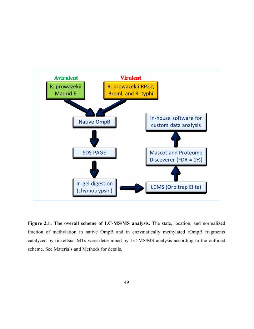
49
Figure 2.1: The overall scheme of LC-MS/MS analysis. The state, location, and normalized
fraction of methylation in native OmpB and in enzymatically methylated rOmpB fragments
catalyzed by rickettsial MTs were determined by LC-MS/MS analysis according to the outlined
scheme. See Materials and Methods for details.
50
2.2.15: Data analysis
Custom software PTMsite was used to compute the numbers of PSM at unmethylated, mono-
, di- and tri-methylated states for all lysine residues throughout OmpB based on the results of LC-
MS/MS of each sample. Normalized fractions of mono-, di- and tri-methylations of a specific lysine
residue were calculated from the observed PSM numbers of respective mono-, di and tri-methylated
peptides divided by the total number of observed PSM numbers including unmethylated, mono-, di-
and tri-methylated peptides of the corresponding lysine residue. For multiple trials of a protein, the
normalized fractions were calculated from the sums of the PSM numbers of each methylated state
at the lysine residue from multiple trials divided by the sum of PSM numbers of all methylation
states of all trials at the same lysine residue.
2.2.16: Molecular weight characterization using SEC-MALS
Wild type RP789 and RT0101, including the mutants targeting dimer formation, were
expressed, purified as described previously (Abeykoon et al., 2014) and tested for their oligomeric
state using size exclusion chromatography with multi-angle light scattering (SEC-MALS) analysis
(Wyatt Technology Corp, Santa Barbara, CA, USA). Proteins samples in 1xPBS supplemented with
TCEP or DTT were separated using HPLC (Agilent 1200 series) using a size exclusion column
(Shodex KW-803) equilibrated in 1XPBS and with in-line UV, MALS, and refractive index (RI)
detectors (DAWN Heleos II and Optilab reX, respectively, Wyatt Technology Corp) for molecular
weight (MW) characterization. For each run, 100 μL of protein sample (2 mg/mL) were injected and
eluted with a flow rate of 0.5 mL/min. UV, MALS, and RI data were collected and analyzed using
51
ASTRA software (Wyatt Technology). Calculated molecular weights of the peaks are presented as
mean with a 95% confidence interval (lower limit, upper limit). Bovine serum albumin (BSA, 66.5
kDa) was run as a control.
2.2.17: Protein Expression, Purification, and Crystallization
The cloning and overexpression of RP789 was performed as previously reported [Abeykoon
et al., 2012] with some modifications where the N-terminal 6xHis tag was removed in hopes of
finding better crystallization conditions. The RP789 coding sequence (1.5 kb) carrying TEV
protease recognition site (gaaaacttatatttccaaggt) immediately upstream to the start codon was
amplified and subcloned at Nde1/Xho1 restriction sites into the pET28a expression vector
(Novagen) containing a 6xHis tag. Standard cloning procedures were performed and the inserts were
verified by restriction enzyme digestion and sequencing. This construct was transformed into
BL21(DE3) competent cells (Agilent Technologies). Protein expression was carried out by growing
the bacteria from single colonies at 37°C in 2 ml of LB broth supplemented with 100µg/ml
kanamycin overnight. The cell culture was transferred to fresh LB broth with kanamycin and
incubated with shaking until the OD600nm reached 0.3-0.5. The cultures were induced with 0.04 mM
IPTG and incubated at 22°C and 250 rpm overnight. Cells were harvested by centrifugation, lysed
and the expressed proteins were purified using Ni-NTA column chromatography according to the
manufacturer’s instructions (GoldBio, St. Louis, MO). The protein was eluted by 250 mM imidazole
and the elutions were pooled and dialysed against 25 mM sodium phosphate (pH 8.0) buffer
containing 1 mM DTPA for 3 times for 1 hour each to remove imidazole and NaCl. The His6X tag
52
was cleaved using TEV protease (protein : protease = 25:1) at 4°C overnight. The cleaved protein
was separated from uncleaved 6xHis tagged protein and TEV protease by passing through the
second Ni-NTA column. Before crystallization, the sample was concentrated to ~5 ml (from 15 ml)
using a Ultracel 30K filter (Millipore) and futher purified by gel filtration across a S100 sephacryl
(GE Healthcare) and crystallization buffer (20mM Tris, pH 7.5, 200mM NaCl). Fractions were
collected and concentrated to bring the final concentration to ~10mg/ml and Tris(2-carboxyethyl)
phosphine hydrochloride (TCEP, 5 mM pH 7.5) was added to prevent the polymerization. Basically
the same procedure was followed to express and purify the Seleno methionine incorporated RP789
with few modifications where the construct was transformed into B839 competent cells (NEB) and
expressed using Seleno-methionine medium base (Athena enzyme systems, 0502). Protein
concentration was determined using NanoDrop UV-Vis spectrophotometer. For the RP789 complex
structure with S-adenosylmethionine (SAM), the protein was incubated with SAM (5 mM, Sigma,
Crystalline) in crystallization buffer for 30 min on ice. For the RP789 complex with S-
adenosylhomocysteine (SAH), the protein was incubated with SAH (3 mM, Sigma, Crystalline) in
crystallization buffer for 30 min on ice.
2.2.18: Crystallization conditions
For crystallization, broad matrix screening was performed using a TTP LabTech Mosquito
crystallization robot and lead conditions optimized accordingly. Final conditions for crystals are
tabulated in Table 2.4 and were harvested directly from the crystallization drop, and then were
plunged into liquid nitrogen for storage until data collection.
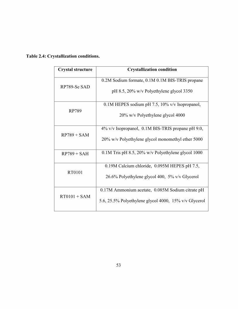
53
Table 2.4: Crystallization conditions.
Crystal structure Crystallization condition
RP789-Se SAD
0.2M Sodium formate, 0.1M 0.1M BIS-TRIS propane
pH 8.5, 20% w/v Polyethylene glycol 3350
RP789
0.1M HEPES sodium pH 7.5, 10% v/v Isopropanol,
20% w/v Polyethylene glycol 4000
RP789 + SAM
4% v/v Isopropanol, 0.1M BIS-TRIS propane pH 9.0,
20% w/v Polyethylene glycol monomethyl ether 5000
RP789 + SAH 0.1M Tris pH 8.5, 20% w/v Polyethylene glycol 1000
RT0101
0.19M Calcium chloride, 0.095M HEPES pH 7.5,
26.6% Polyethylene glycol 400, 5% v/v Glycerol
RT0101 + SAM
0.17M Ammonium acetate, 0.085M Sodium citrate pH
5.6, 25.5% Polyethylene glycol 4000, 15% v/v Glycerol
54
2.2.19: Data collection and Structure Determination
Lead conditions were initially screened using in-house x-ray diffractometer (Rigaku
MicroMax-007 HF microfocus x-ray generator, Raxis IV++ detector). Final datasets were collected
at either the GM/CA-CAT beamline or the SER-CAT beamline at the Advanced Photon Source at
Argonne National Laboratory. All data were processed using HKL2000 and statistics are
summarized in Table 1. The initial RP789 structure was solved by Se-SAD (AutoSol) to bulid a
starting model (AutoBuild) and refined using PHENIX. Molecular replacement was used to solve
the native and complex structures of RP789, the RT0101 native structure, and the RT0101-SAM
structure. A difference map was used to locate the SAM and SAH ligands within those respective
structures. All model building was performed using COOT, and refinement was performed using
PHENIX and CCP4. All structure figures were prepared using PyMOL (Schrödinger), and final
editing was done in Adobe Illustrator. Root mean square deviation (RMSD) analysis was performed
within PyMOL (Schrödinger).
2.2.20: Modeling the putative substrate binding
In the absence of a substrate bound complex crystal structure of either RP789 or RT0101,
we modeled the putative substrate binding site using bioinformatics approaches. First, we did a
sequence alignment of five different Rickettsial methyltransferases (RP789, RT0101, RT0776,
MA5, RP22) that are known to bind OmpB and exhibit methyltransferase activity. The rationale
being that since all methyltransferases are able to bind OmpB, those residues that mediate this
interaction must be shared between all five enzymes. Based on the alignment, we mapped all fully
55
conserved residues to the surface of the RP789 crystal structure which allowed the identification of
several regions of surface area of high conservation within proximity of the SAM (methyl donor)
binding site. Based on this analysis, mutagenesis and kinetics assays were further performed to
determine the role of these conserved regions in substrate binding.
56
Chapter 3
Two protein lysine methyltransferases methylate outer membrane protein B
from Rickettsia
3.1: Introduction
R. prowazekii is the causative agent of epidemic typhus and categorized under Category B
Priority Pathogens by the National Institute of Allergy and Infectious Diseases (NIAID) and select
agents by the Center for Disease Control and Prevention (CDC) for their potential use as tools for
biological terrorism [Walker D, 2003]. Rickettsial OmpB belongs to the autotransporter family
proteins. The reason for OmpB to be categorized under this family is, unlike the α-helical
transmembrane protein in plasma membranes, autotransporter family proteins contain characteristic
β-barrel domain in outer membrane [Barnard et al., 2007, Schulz, 2002]. OmpB is expressed as a
pre-protein and cleaved to release the passenger domain from the β-barrel translocation domain,
leaving the mature passenger domain associated with the outer leaflet of the outer membrane [Jacob-
Dubuisson et al., 2004]. OmpB plays many significant roles in bacterial pathogenesis including,
host cell attachment, invasion, internalization, and intracellular movement [Chan et al., 2009,
Uchiyama et al., 2006, Martinez et al., 2005]. OmpB has been identified as the immunodominant
species-specific surface protein antigen and it has been isolated, purified and [Dasch et al., 1981]
57
shown that purified native OmpB from R. typhus induced strong humoral and cell-mediated immune
responses [Carl et al., 1989, Feng et al., 2004, Bourgeois et al., 1981].
Passenger domain of OmpB (surface protein antigen) undergoes post-translational
modification of methylating the amino group of lysine and it is proposed that methylation play a
major role in bacterial virulence [Rodionov, 1990]. But how methylation affects the virulence is not
well understood. Lysine methylation can regulate protein function through different mechanisms.
For example, methyl lysine can provide docking sites for binding of cognate proteins and also can
inhibit other PTMs occurring on the same lysine residue which may in turn affect bacteria-host cell
interactions influencing invasion of host cell, and inhibit other post-translational modifications
thereby influencing its survival in the host cell by avoiding destruction and premature death
[Polevoda et al., 2007, Huang et al., 2008].
The level of lysine methylation of OmpB and the virulence of Rickettsia appears to be
correlated. The virulent strain Breinl and the virulent revertant strain Evir show a different OmpB
lysine methylation profile compared to avirulent Madrid E strain [Ching et al., 1992]. OmpB has
been shown to be mostly hyper-methylated in the virulent strains in comparison to avirulent strain
which is mostly hypomethylated [Ching et al., 1993]. A comparison of genetic differences between
the avirulent Madrid E strain and virulent revertant Evir strain identified a reverse mutation that
cause the Evir strain to re-gain the virulence when avirulent Madrid E is passaged in animals. The
gene Rp028/Rp027 encoding a methyltransferase was mutated by frameshift in avirulent strain but
not in the virulent revertant Evir or wild type Breinl strains [Zhang et al., 2006]. This may result in
the loss of methyltransferase activity in avirulent strain. A comparative proteomic approach revealed
58
that the protein, Rp028/Rp027, was detectable only in virulent strains [Chao et al., 2005, and 2007].
The observation of the correlation between methylation of lysine residues in rickettsial OmpB and
the bacterial virulence has suggested the importance of an enzymatic system for the methylation of
OmpB. However no rickettsial lysine methyltransferases or their substrates have been characterized.
Studying the effects of hypermethylation on OmpB structure and function will help us to
understand the effects it may have on pathogenesis and host immune response. With the availability
of recombinant OmpB substrate and recombinant lysine methyltransferases, in vitro large scale
production of rickettsial antigens is a real possibility and can be used against patient antibodies for
early detection of typhus. Being able to generate enzymatically methylated recombinant OmpB may
also provide more specificity and selectivity over chemical methylation in the attempt of producing
diagnostic reagents and vaccine candidates. As the first step, a list of putative methyltransferases
was selected by structural and bioinformatic analysis of R. prowazekii genome. They were cloned,
expressed, purified and examined for the methyltransferase activity against recombinant OmpB
proteins. Two rickettsial methyltransferases that can enzymatically methylate recombinant OmpB
were found in this study.
3.2: Results
3.2.1: Genes encode putative protein methyltransferases in R.prowazekii genomes
R. prowazekii Madrid E genome sequence is available at National Center for Biotechnology
Information (NCBI). To identify the putative protein methyltransferases which may methylate the
59
OmpB, gene annotations of R. prowazekii Madrid E was studied [Andersson et al., 1998]. Out of
sixty-two known and potential R. prowazekii methyltransferases that have been annotated, the
enzymes known to methylate small molecules, tRNA, mRNA and rRNA were eliminated. The
remaining unknown and putative methyltransferases were aligned with each other using the local
alignment search tool BLAST in the NCBI (http://blast.ncbi.nlm.nih.gov/Blast.cgi) in order to find
if they can be categorized into independent groups based on their structural homology. Following
this analysis, five different Madrid E methyltransferases were selected, RP789, RP527, RP545,
RP027 and RP028 (Table 3.1). This comparison led to the identification of common motifs
associated with the protein methyltransferases that are important for the activity and also to detect
the differences in sequence that may be important for the differences in the activity. SAM dependent
methyltransferase showed some conserved domains. One is the AdoMet binding site (chemical
binding site) from AdoMet-MTases super family. They may also contain domains similar to
COG4797 (Figure 3.1) super family, a predicted regulatory domain of a methyltransferase and a
MethyTransf-Reg super family, a predicted methyltransferase regulatory domain.
Madrid E is avirulent. A methyltransferase from a virulent strain was also required in the
study to compare with the selected putative methyltransferases of the avirulent strain. The genome
of wild type virulent Breinl strain is not currently available. Instead, R. prowazekii Rp22 genome
was selected which is very closely related to Breinl. A putative methyltransferase from Rp22
(ADE29537) was selected. As stated in the Introduction (section 1.7.2) Madrid E genes RP028 and
RP027 show 100% homology to the N-terminal half (1-243) and C-terminal half (283-535) of the
putative methyltransferase Rp22, respectively. For this reason it is referred to as RP027-028
throughout this study. RP027 and RP028 were mutated in the avirulent Madrid E strain by a
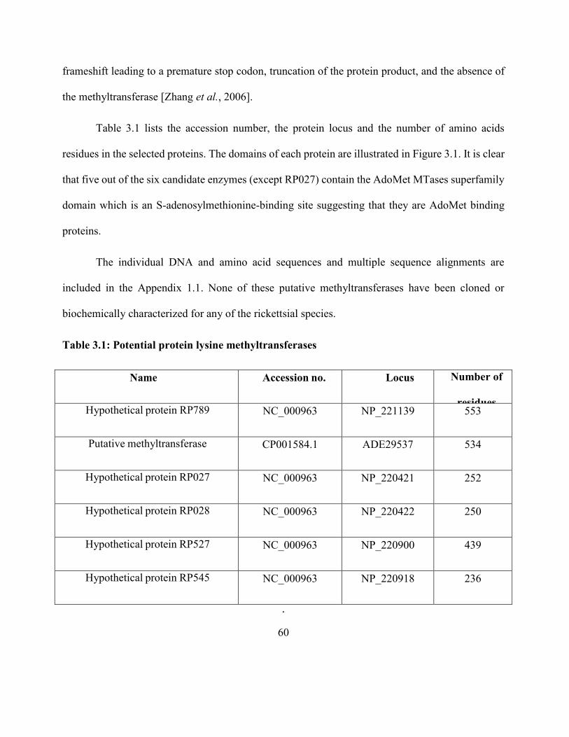
60
frameshift leading to a premature stop codon, truncation of the protein product, and the absence of
the methyltransferase [Zhang et al., 2006].
Table 3.1 lists the accession number, the protein locus and the number of amino acids
residues in the selected proteins. The domains of each protein are illustrated in Figure 3.1. It is clear
that five out of the six candidate enzymes (except RP027) contain the AdoMet MTases superfamily
domain which is an S-adenosylmethionine-binding site suggesting that they are AdoMet binding
proteins.
The individual DNA and amino acid sequences and multiple sequence alignments are
included in the Appendix 1.1. None of these putative methyltransferases have been cloned or
biochemically characterized for any of the rickettsial species.
Table 3.1: Potential protein lysine methyltransferases
Name Accession no. Locus Number of
residues Hypothetical protein RP789
R. prowazekii Madrid E
NC_000963 NP_221139 553
Putative methyltransferase
R. prowazekii Rp22 (RP027-028)
CP001584.1 ADE29537 534
Hypothetical protein RP027
R. prowazekii Madrid E
NC_000963 NP_220421 252
Hypothetical protein RP028
R. prowazekii Madrid E
NC_000963 NP_220422 250
Hypothetical protein RP527
R. prowazekii Madrid E
NC_000963 NP_220900 439
Hypothetical protein RP545
R. prowazekii Madrid E
NC_000963 NP_220918 236
.
61
Figure 3.1: Schematic diagram of the conserved domains of selected putative
meyhyltransferases. In general, they contain the AdoMet-dependent_MTase superfamily which
has a S-adenosylmethionine-binding site and MethyTransf_Reg superfamily which is a predicted
methyltransferase regulatory domain and COG4797 superfamily which is a predicted regulatory
domain of a methyltransferase (Not drawn to a scale).
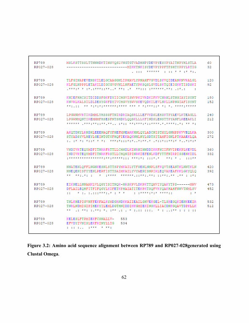
62
Figure 3.2: Amino acid sequence alignment between RP789 and RP027-028generated using
Clustal Omega.
63
Amino acid sequence alignment of RP789 and RP027-028 showed 45% identity and 65%
homology between them in a structural bioinformatics analysis [Figure 3.2]. In order to predict and
compare the 3D structure of AdoMet binding domains of RP789 and RP027-028, the N-terminal
domains of both enzymes (Y48-L213 for RP789 and K3-L185 for RP027-028) were modeled using
3DLigandSite [Wass et al., 2010]. 3DLigandSite is a webserver for predicting ligand binding sites.
Once the amino acid sequence is submitted then another program (Phyre) is run to predict the
structure [Kelley et al, 2009]. This structure is then used to search a structural library to build
homologous models with bound ligands. These ligands are superimposed onto the protein structure
to predict a ligand binding site. Molecular modeling resulted similar 3D models of the AdoMet
binding domains of RP789 and RP027-028 with bound SAM [Figure 3.3], suggesting that both are
AdoMet binding proteins. Due to the absence of structures in the RCSB Protein Database which
showed adequate homology to the C-terminal domains of two proteins, the rest of the protein
couldn’t be modeled.
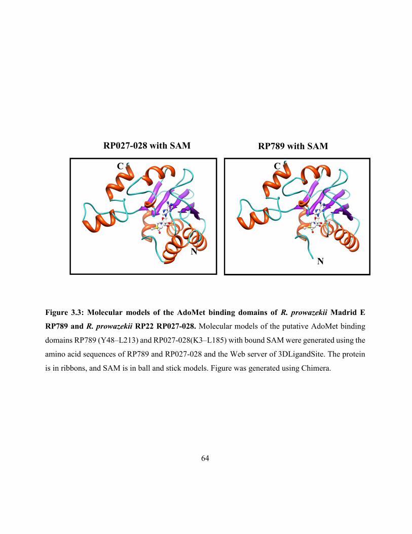
64
Figure 3.3: Molecular models of the AdoMet binding domains of R. prowazekii Madrid E
RP789 and R. prowazekii RP22 RP027-028. Molecular models of the putative AdoMet binding
domains RP789 (Y48–L213) and RP027-028(K3–L185) with bound SAM were generated using the
amino acid sequences of RP789 and RP027-028 and the Web server of 3DLigandSite. The protein
is in ribbons, and SAM is in ball and stick models. Figure was generated using Chimera.
RP027-028 with SAM RP789 with SAM
N
N
C C
65
There are other rickettsial proteins found in NCBI database which show homology to RP789
and RP027-028. More than 40 proteins from various rickettsial species and strains are tabulated
along with their accession numbers and identities to RP789 and RP027-028 in the Appendix 1.4.
3.2.2: Expression and purification of RP789 and RP027-028
The selected putative methyltransferases were over-expressed in E.coli. E.coli codon-optimized
cDNAs that encode potential methyltransfreases were synthesized and subcloned at Nde1/Xho1
restriction sites into pET28a expression vector containing 6xHis tag. Standard cloning procedure was
performed and the insert was verified by restriction enzyme digestion and sequencing. Once the insert
was confirmed to have the correct sequence the plasmid was transformed into BL21(DE3) competent
cells. Proteins were purified using Ni-NTA affinity chromatography. When compared the expression
levels and purity of the six proteins using the SDS-polyacrylamide gel electrophoresis, four out of six
yielded soluble proteins. Out of four soluble proteins, RP789 and RP027-028 showed the highest
expression and purity resulting 0.25 and 0.75 mg protein per 100 ml culture respectively and appeared
on the gel at correct molecular weights as predicted from the amino acid sequence (63.5 kDa for RP789
and 61.5 kDa for RP027-028). RP527 showed the next highest expression level resulting 0.15 mg
protein per 100 ml culture and had the expected molar mass of 50 kDa. The expression level of RP545
was very low and wasn’t significantly improved by increasing the concentration of IPTG to 0.5 or 1
mM instead of 0.04 mM, by changing the incubation time or temperature, or changing the host to
BL21(DE3) Gold pLysS competent cells [Figure 3.4]. The two insoluble proteins RP027 and RP028
expressed in high levels under denaturing conditions in the presence of 8M urea. But the attempt to
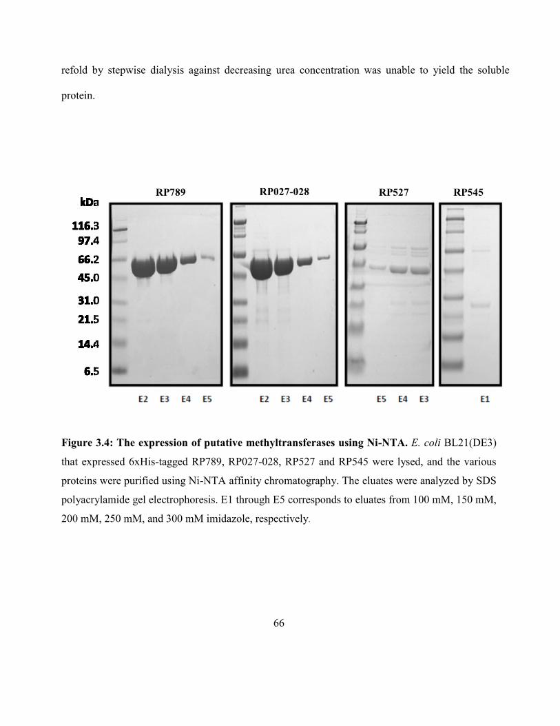
66
refold by stepwise dialysis against decreasing urea concentration was unable to yield the soluble
protein.
Figure 3.4: The expression of putative methyltransferases using Ni-NTA. E. coli BL21(DE3)
that expressed 6xHis-tagged RP789, RP027-028, RP527 and RP545 were lysed, and the various
proteins were purified using Ni-NTA affinity chromatography. The eluates were analyzed by SDS
polyacrylamide gel electrophoresis. E1 through E5 corresponds to eluates from 100 mM, 150 mM,
200 mM, 250 mM, and 300 mM imidazole, respectively.
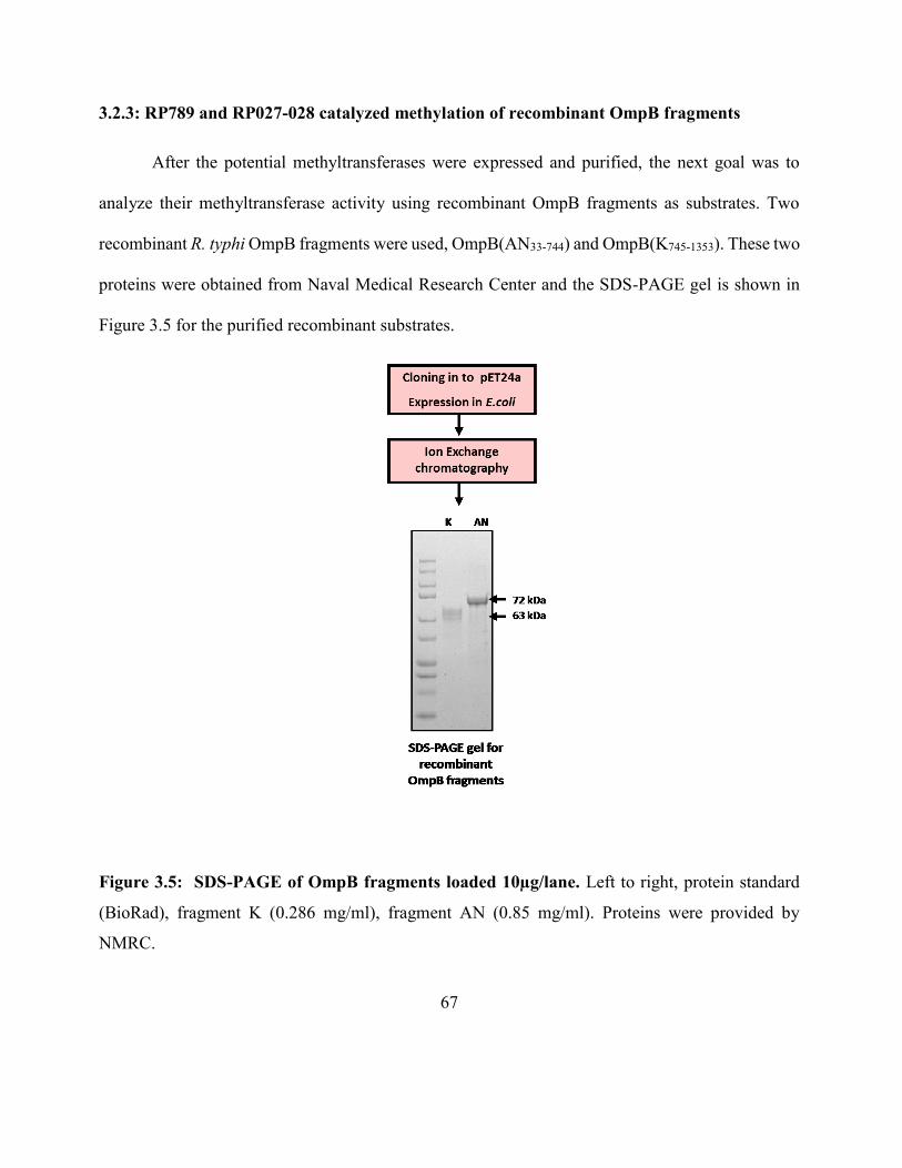
67
3.2.3: RP789 and RP027-028 catalyzed methylation of recombinant OmpB fragments
After the potential methyltransferases were expressed and purified, the next goal was to
analyze their methyltransferase activity using recombinant OmpB fragments as substrates. Two
recombinant R. typhi OmpB fragments were used, OmpB(AN33-744) and OmpB(K745-1353). These two
proteins were obtained from Naval Medical Research Center and the SDS-PAGE gel is shown in
Figure 3.5 for the purified recombinant substrates.
Figure 3.5: SDS-PAGE of OmpB fragments loaded 10µg/lane. Left to right, protein standard
(BioRad), fragment K (0.286 mg/ml), fragment AN (0.85 mg/ml). Proteins were provided by
NMRC.
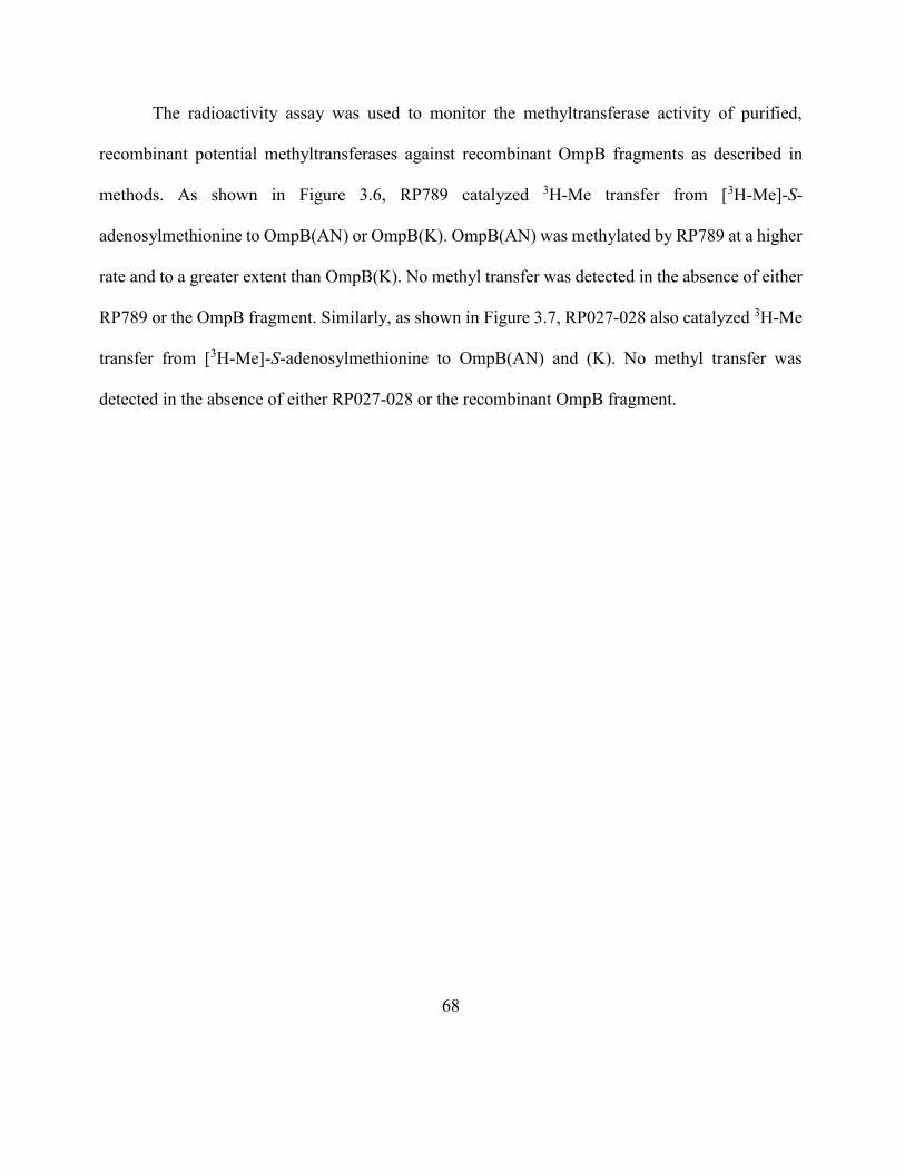
68
The radioactivity assay was used to monitor the methyltransferase activity of purified,
recombinant potential methyltransferases against recombinant OmpB fragments as described in
methods. As shown in Figure 3.6, RP789 catalyzed 3H-Me transfer from [3H-Me]-S-
adenosylmethionine to OmpB(AN) or OmpB(K). OmpB(AN) was methylated by RP789 at a higher
rate and to a greater extent than OmpB(K). No methyl transfer was detected in the absence of either
RP789 or the OmpB fragment. Similarly, as shown in Figure 3.7, RP027-028 also catalyzed 3H-Me
transfer from [3H-Me]-S-adenosylmethionine to OmpB(AN) and (K). No methyl transfer was
detected in the absence of either RP027-028 or the recombinant OmpB fragment.
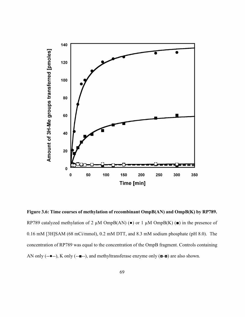
69
Figure 3.6: Time courses of methylation of recombinant OmpB(AN) and OmpB(K) by RP789.
RP789 catalyzed methylation of 2 µM OmpB(AN) (●) or 1 µM OmpB(K) (■) in the presence of
0.16 mM [3H]SAM (68 mCi/mmol), 0.2 mM DTT, and 8.3 mM sodium phosphate (pH 8.0). The
concentration of RP789 was equal to the concentration of the OmpB fragment. Controls containing
AN only (--●--), K only (--■--), and methyltransferase enzyme only (◘-◘) are also shown.
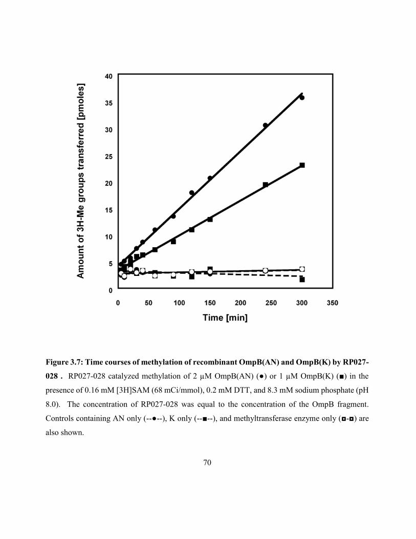
70
Figure 3.7: Time courses of methylation of recombinant OmpB(AN) and OmpB(K) by RP027-
028 . RP027-028 catalyzed methylation of 2 µM OmpB(AN) (●) or 1 µM OmpB(K) (■) in the
presence of 0.16 mM [3H]SAM (68 mCi/mmol), 0.2 mM DTT, and 8.3 mM sodium phosphate (pH
8.0). The concentration of RP027-028 was equal to the concentration of the OmpB fragment.
Controls containing AN only (--●--), K only (--■--), and methyltransferase enzyme only (◘-◘) are
also shown.
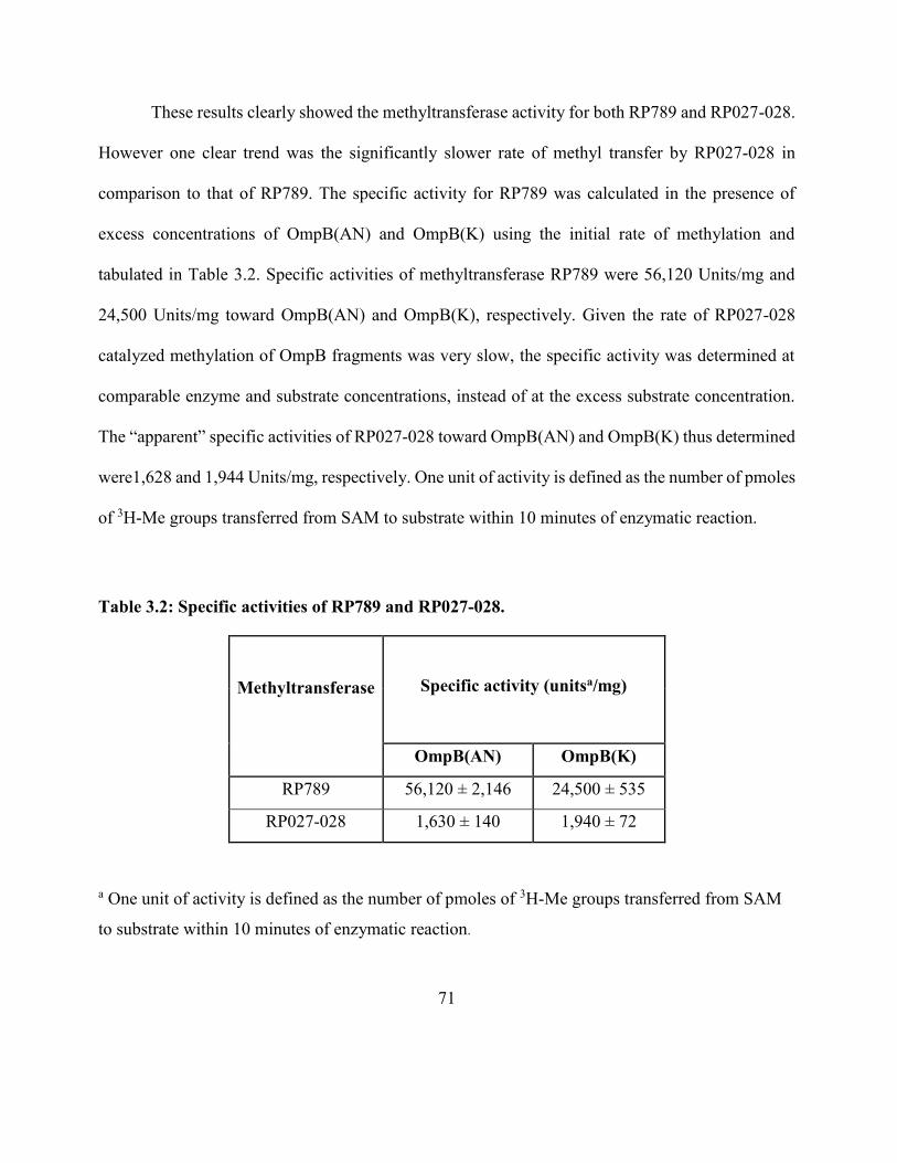
71
These results clearly showed the methyltransferase activity for both RP789 and RP027-028.
However one clear trend was the significantly slower rate of methyl transfer by RP027-028 in
comparison to that of RP789. The specific activity for RP789 was calculated in the presence of
excess concentrations of OmpB(AN) and OmpB(K) using the initial rate of methylation and
tabulated in Table 3.2. Specific activities of methyltransferase RP789 were 56,120 Units/mg and
24,500 Units/mg toward OmpB(AN) and OmpB(K), respectively. Given the rate of RP027-028
catalyzed methylation of OmpB fragments was very slow, the specific activity was determined at
comparable enzyme and substrate concentrations, instead of at the excess substrate concentration.
The “apparent” specific activities of RP027-028 toward OmpB(AN) and OmpB(K) thus determined
were1,628 and 1,944 Units/mg, respectively. One unit of activity is defined as the number of pmoles
of 3H-Me groups transferred from SAM to substrate within 10 minutes of enzymatic reaction.
Table 3.2: Specific activities of RP789 and RP027-028.
Methyltransferase
Specific activity (unitsa/mg)
OmpB(AN) OmpB(K)
RP789 56,120 ± 2,146 24,500 ± 535
RP027-028 1,630 ± 140 1,940 ± 72
a One unit of activity is defined as the number of pmoles of 3H-Me groups transferred from SAM
to substrate within 10 minutes of enzymatic reaction.
72
The two remaining soluble proteins RP527 and RP545 were also tested for their
methyltransferase activity. There was no detectable catalytic activity for purified RP527 or RP545
with either substrates OmpB(AN) or OmpB(K) even at very high enzyme concentration of 13 µM
or under various pH values from 5 through 9 (data not shown). The difficulty of obtaining RP027
and RP028 in pure and soluble form prohibited the meaningful determination of their
methytransferase activity. No further studies for these four proteins were carried out.
3.2.4: RP789 and RP027-028 catalyzed trimethylation of recombinant OmpB
The radioactive assays only showed if the selected proteins had methyltransferase activity
with recombinant OmpB fragment substrates. Out of the six candidates only RP789 and RP027-028
had the ability to catalyze the methylation of OmpB. The next step was to identify if they methylate
lysine residues and if yes, then to what extent these two enzymes can methylate the substrate. As
mentioned in the Introduction rickettsial virulence is correlated to the level of methylation on lysine
and it is important to characterize the lysine methylation in order to develop vaccine candidates or
diagnostic reagents in future.
Protein methyltransferases can catalyze the transfer of a methyl group from SAM, either to
a lysine or arginine. Analysis of OmpB isolated from native R. prowazekii only showed methylated
lysines. Up to three methyl groups can be attached to a single lysine residue. To determine the level
of methylation on OmpB(AN) and OmpB(K) by RP789 and RP027-028, a Western blot analysis
was used. Utilizing antibodies which are specific to trimethylated lysines, we were able to
distinguish between trimethylation and other methylation states. One practical difficulty
73
encountered during western blot was the separation of two protein bands for OmpB(K) and
methyltransferase on the gel, given the closeness of their molecular weights. Hence it was required
to separate the substrate from the enzyme after the overnight reaction before loading the samples on
to the gel. While both methyltransferases, RP789 and RP027-028 contained N-terminal 6xHis tags
the OmpB(K) did not. Hence the enzymes were removed from the overnight reaction mixture using
Ni-NTA affinity chromatography before loading onto the column.
The western bolt was developed as described in Material and Methods. Rabbit anti-trimethyl
lysine antibodies were used as the primary antibody, so it could differentiate between trimethylation
and other levels of methylation. Figure 3.8 shows the Western blot of the methylation products of
OmpB(AN) and OmpB(K) catalyzed by RP789 and RP027-028. Antibodies against trimethyl lysine
reacted with OmpB(AN) only after methylation by either RP789 or RP027-028. There was no clear
trimethylation detected for OmpB(K) on the blot. This could be due to the absence or may be due
to small amounts of detectable trimethyl lysine on OmpB(K). Loss of some OmpB(K) when going
through Ni column after overnight reaction is inevitable and may have led to poor detection.
Recombinant OmpB(AN) or OmpB(K) or RP789 and RP027-028 alone did not react with the
antibodies against trimethyl lysine. Thus, the Western blot analyses confirmed the protein lysine
methyltransferase activity of RP789 and RP027-028 and demonstrated their activity of catalyzing
trimethylation of lysine residues in OmpB fragments.
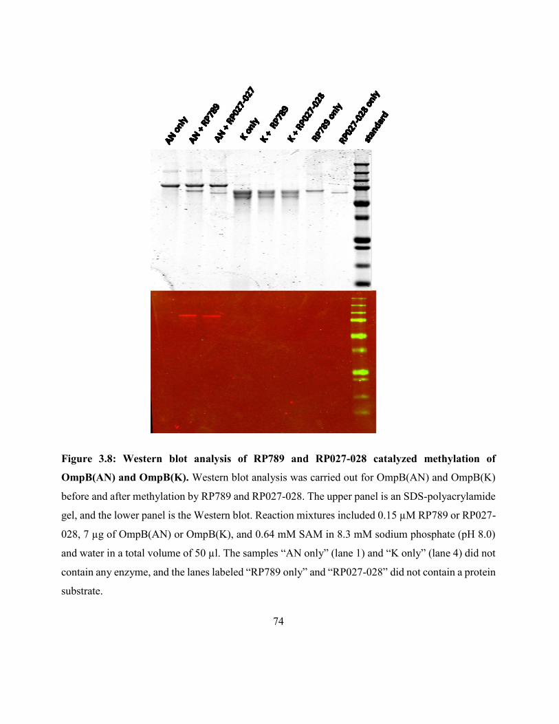
74
Figure 3.8: Western blot analysis of RP789 and RP027-028 catalyzed methylation of
OmpB(AN) and OmpB(K). Western blot analysis was carried out for OmpB(AN) and OmpB(K)
before and after methylation by RP789 and RP027-028. The upper panel is an SDS-polyacrylamide
gel, and the lower panel is the Western blot. Reaction mixtures included 0.15 µM RP789 or RP027-
028, 7 µg of OmpB(AN) or OmpB(K), and 0.64 mM SAM in 8.3 mM sodium phosphate (pH 8.0)
and water in a total volume of 50 µl. The samples “AN only” (lane 1) and “K only” (lane 4) did not
contain any enzyme, and the lanes labeled “RP789 only” and “RP027-028” did not contain a protein
substrate.
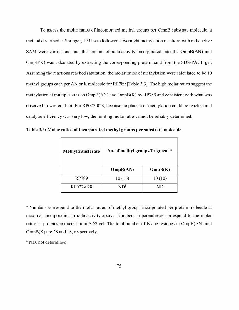
75
To assess the molar ratios of incorporated methyl groups per OmpB substrate molecule, a
method described in Springer, 1991 was followed. Overnight methylation reactions with radioactive
SAM were carried out and the amount of radioactivity incorporated into the OmpB(AN) and
OmpB(K) was calculated by extracting the corresponding protein band from the SDS-PAGE gel.
Assuming the reactions reached saturation, the molar ratios of methylation were calculated to be 10
methyl groups each per AN or K molecule for RP789 [Table 3.3]. The high molar ratios suggest the
methylation at multiple sites on OmpB(AN) and OmpB(K) by RP789 and consistent with what was
observed in western blot. For RP027-028, because no plateau of methylation could be reached and
catalytic efficiency was very low, the limiting molar ratio cannot be reliably determined.
Table 3.3: Molar ratios of incorporated methyl groups per substrate molecule
Methyltransferase
No. of methyl groups/fragment a
OmpB(AN) OmpB(K)
RP789 10 (16) 10 (10)
RP027-028 NDb ND
a Numbers correspond to the molar ratios of methyl groups incorporated per protein molecule at
maximal incorporation in radioactivity assays. Numbers in parentheses correspond to the molar
ratios in proteins extracted from SDS gel. The total number of lysine residues in OmpB(AN) and
OmpB(K) are 28 and 18, respectively.
b ND, not determined
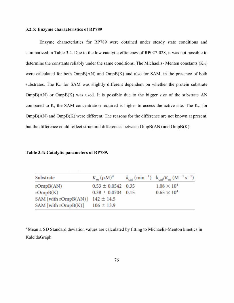
76
3.2.5: Enzyme characteristics of RP789
Enzyme characteristics for RP789 were obtained under steady state conditions and
summarized in Table 3.4. Due to the low catalytic efficiency of RP027-028, it was not possible to
determine the constants reliably under the same conditions. The Michaelis- Menten constants (Km)
were calculated for both OmpB(AN) and OmpB(K) and also for SAM, in the presence of both
substrates. The Km for SAM was slightly different dependent on whether the protein substrate
OmpB(AN) or OmpB(K) was used. It is possible due to the bigger size of the substrate AN
compared to K, the SAM concentration required is higher to access the active site. The Km for
OmpB(AN) and OmpB(K) were different. The reasons for the difference are not known at present,
but the difference could reflect structural differences between OmpB(AN) and OmpB(K).
Table 3.4: Catalytic parameters of RP789.
a Mean ± SD Standard deviation values are calculated by fitting to Michaelis-Menton kinetics in
KaleidaGraph
77
With the objective of further optimizing the catalytic efficiency, the effects of KCl, MgCl2,
and pH on RP789 activity were tested separately using radioactive assays. The reaction rates went
down in the presence of salt. As shown in the Figure 3.9 KCl inhibited the initial rates of enzymatic
methylation of both OmpB(AN) and OmpB(K), when increasing the salt concentrations from 0 –
0.2 M. Similar trend was observed with MgCl2 (data not shown).
The optimum pH value for RP789 enzymatic methylation was also tested and the results are
shown in Figure 3.10. The optimal pH was close to pH 8, when tested using OmpB(AN) as the
substrate, which is lower than that of SET7 methyltransferase, a well-studied methyltransferase for
histone. The amount of 3H-Me groups transferred from SAM to OmpB(AN) within 120 minutes of
reaction was used as the parameter to compare the enzymatic activity at each pH value.
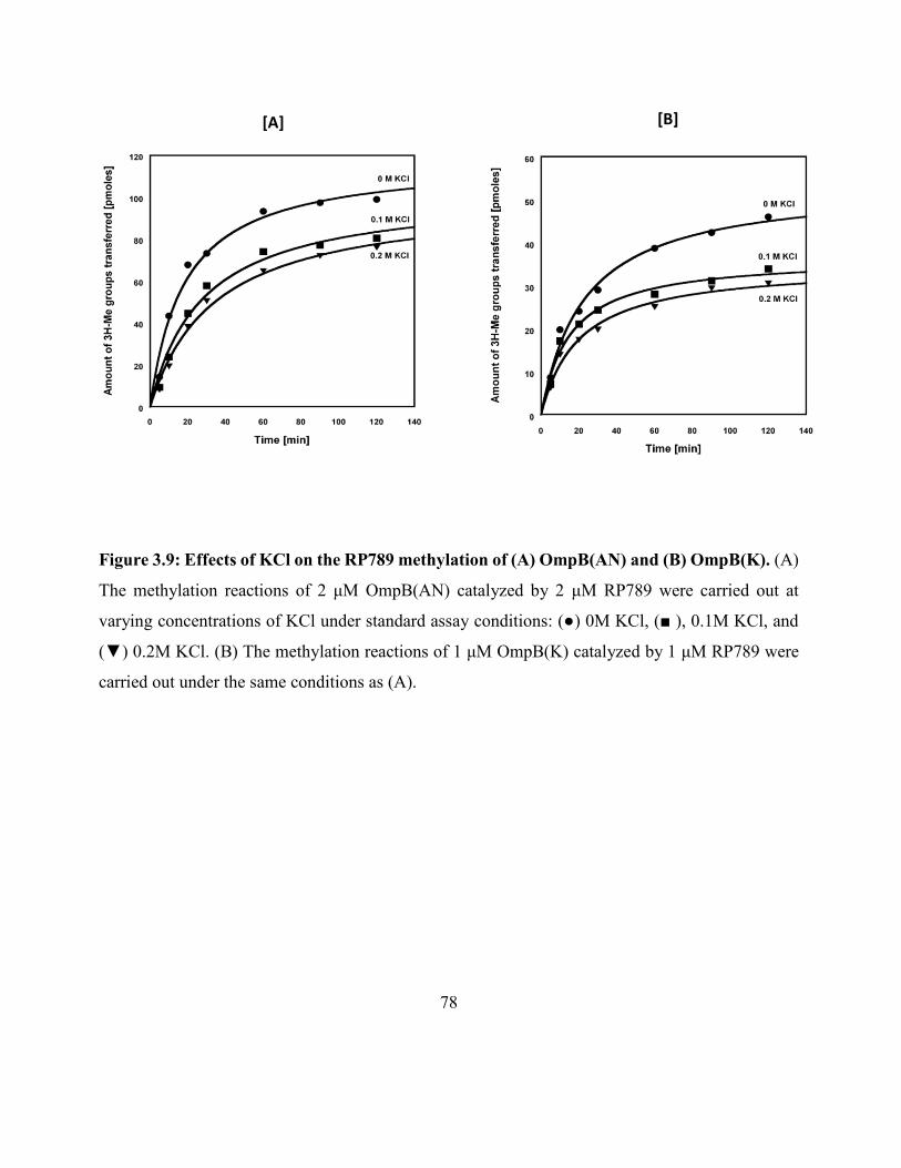
78
Figure 3.9: Effects of KCl on the RP789 methylation of (A) OmpB(AN) and (B) OmpB(K). (A)
The methylation reactions of 2 μM OmpB(AN) catalyzed by 2 μM RP789 were carried out at
varying concentrations of KCl under standard assay conditions: (●) 0M KCl, (■ ), 0.1M KCl, and
(▼) 0.2M KCl. (B) The methylation reactions of 1 μM OmpB(K) catalyzed by 1 μM RP789 were
carried out under the same conditions as (A).
[A] [B]
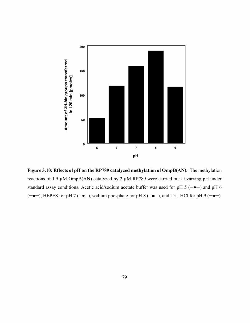
79
Figure 3.10: Effects of pH on the RP789 catalyzed methylation of OmpB(AN). The methylation
reactions of 1.5 µM OmpB(AN) catalyzed by 2 µM RP789 were carried out at varying pH under
standard assay conditions. Acetic acid/sodium acetate buffer was used for pH 5 (─●─) and pH 6
(─■─), HEPES for pH 7 (--●--), sodium phosphate for pH 8 (--■--), and Tris-HCl for pH 9 (─◘─).
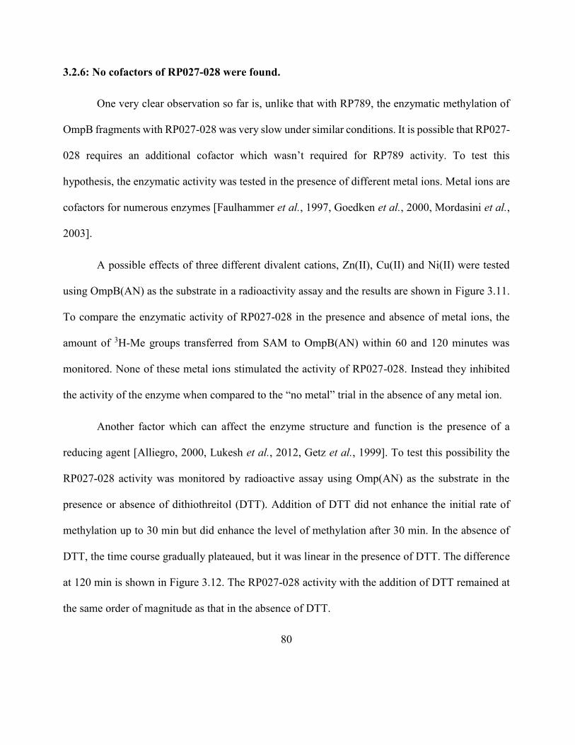
80
3.2.6: No cofactors of RP027-028 were found.
One very clear observation so far is, unlike that with RP789, the enzymatic methylation of
OmpB fragments with RP027-028 was very slow under similar conditions. It is possible that RP027-
028 requires an additional cofactor which wasn’t required for RP789 activity. To test this
hypothesis, the enzymatic activity was tested in the presence of different metal ions. Metal ions are
cofactors for numerous enzymes [Faulhammer et al., 1997, Goedken et al., 2000, Mordasini et al.,
2003].
A possible effects of three different divalent cations, Zn(II), Cu(II) and Ni(II) were tested
using OmpB(AN) as the substrate in a radioactivity assay and the results are shown in Figure 3.11.
To compare the enzymatic activity of RP027-028 in the presence and absence of metal ions, the
amount of 3H-Me groups transferred from SAM to OmpB(AN) within 60 and 120 minutes was
monitored. None of these metal ions stimulated the activity of RP027-028. Instead they inhibited
the activity of the enzyme when compared to the “no metal” trial in the absence of any metal ion.
Another factor which can affect the enzyme structure and function is the presence of a
reducing agent [Alliegro, 2000, Lukesh et al., 2012, Getz et al., 1999]. To test this possibility the
RP027-028 activity was monitored by radioactive assay using Omp(AN) as the substrate in the
presence or absence of dithiothreitol (DTT). Addition of DTT did not enhance the initial rate of
methylation up to 30 min but did enhance the level of methylation after 30 min. In the absence of
DTT, the time course gradually plateaued, but it was linear in the presence of DTT. The difference
at 120 min is shown in Figure 3.12. The RP027-028 activity with the addition of DTT remained at
the same order of magnitude as that in the absence of DTT.
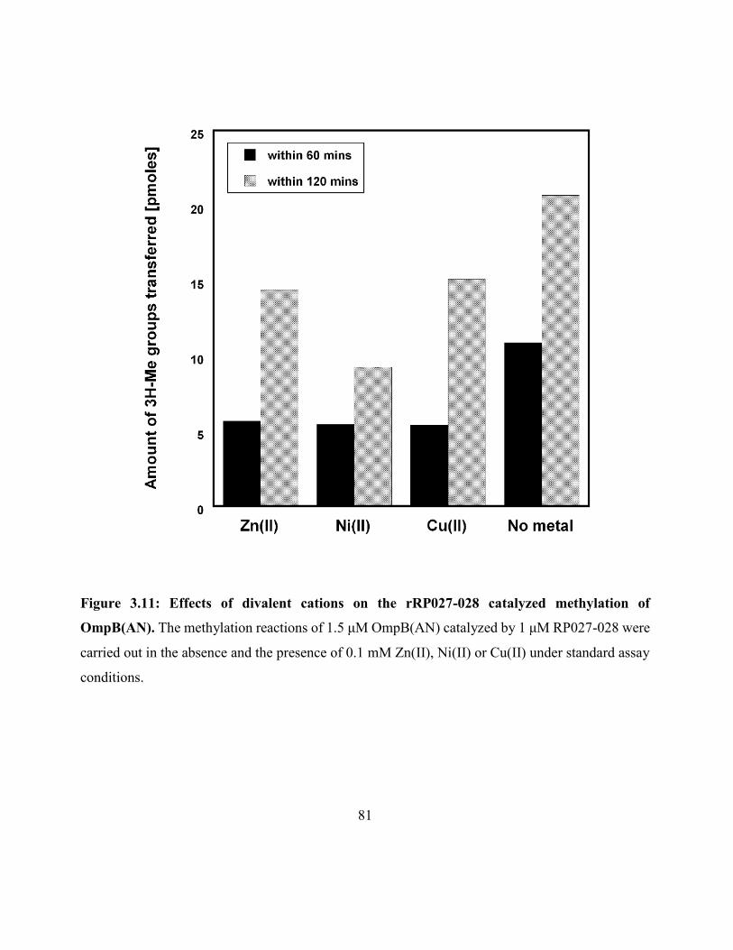
81
Figure 3.11: Effects of divalent cations on the rRP027-028 catalyzed methylation of
OmpB(AN). The methylation reactions of 1.5 μM OmpB(AN) catalyzed by 1 μM RP027-028 were
carried out in the absence and the presence of 0.1 mM Zn(II), Ni(II) or Cu(II) under standard assay
conditions.
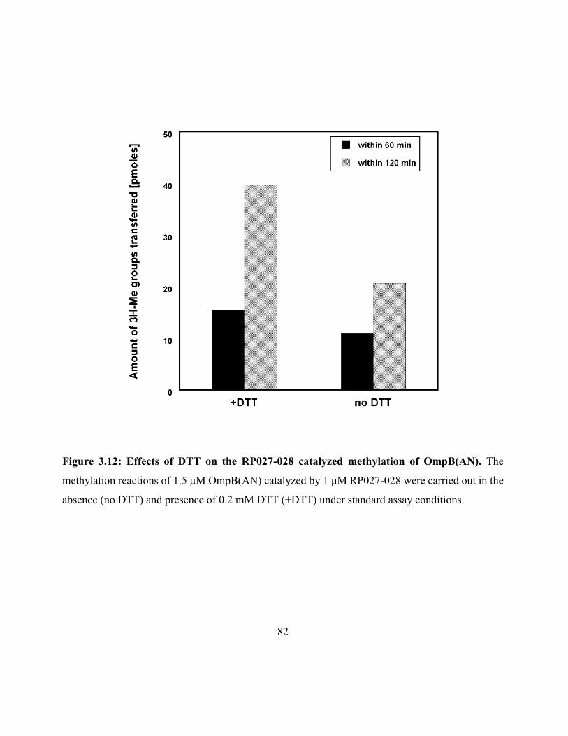
82
Figure 3.12: Effects of DTT on the RP027-028 catalyzed methylation of OmpB(AN). The
methylation reactions of 1.5 μM OmpB(AN) catalyzed by 1 μM RP027-028 were carried out in the
absence (no DTT) and presence of 0.2 mM DTT (+DTT) under standard assay conditions.
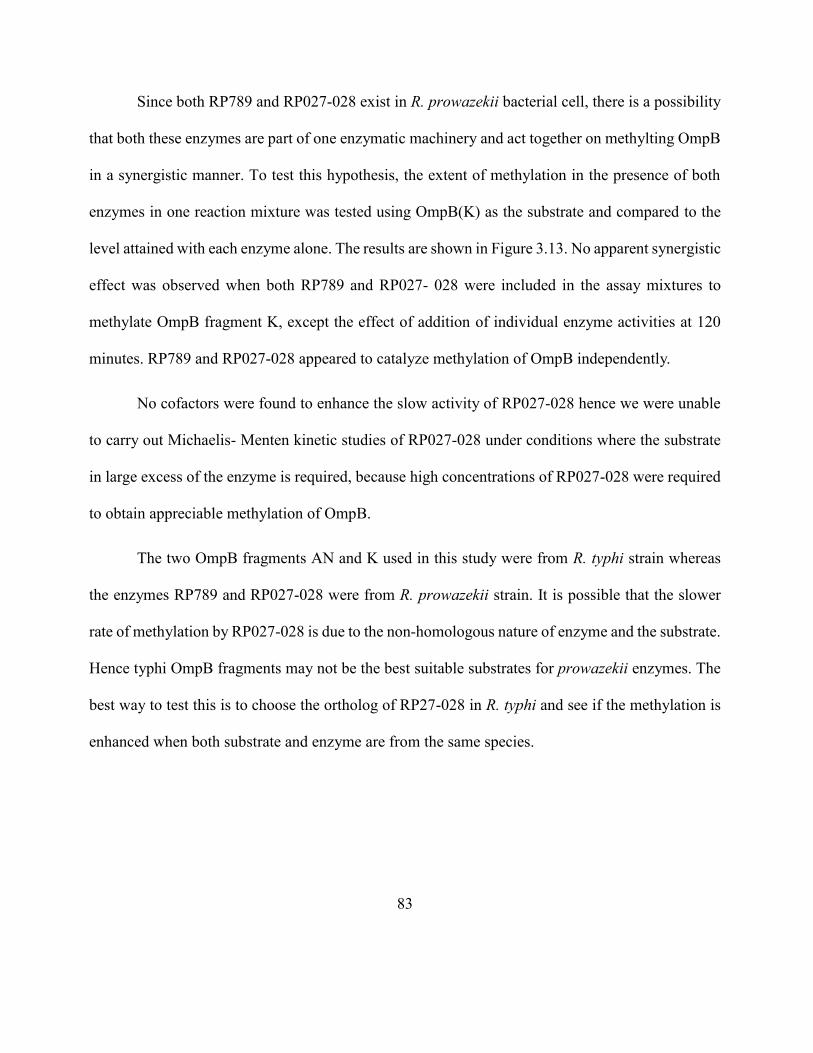
83
Since both RP789 and RP027-028 exist in R. prowazekii bacterial cell, there is a possibility
that both these enzymes are part of one enzymatic machinery and act together on methylting OmpB
in a synergistic manner. To test this hypothesis, the extent of methylation in the presence of both
enzymes in one reaction mixture was tested using OmpB(K) as the substrate and compared to the
level attained with each enzyme alone. The results are shown in Figure 3.13. No apparent synergistic
effect was observed when both RP789 and RP027- 028 were included in the assay mixtures to
methylate OmpB fragment K, except the effect of addition of individual enzyme activities at 120
minutes. RP789 and RP027-028 appeared to catalyze methylation of OmpB independently.
No cofactors were found to enhance the slow activity of RP027-028 hence we were unable
to carry out Michaelis- Menten kinetic studies of RP027-028 under conditions where the substrate
in large excess of the enzyme is required, because high concentrations of RP027-028 were required
to obtain appreciable methylation of OmpB.
The two OmpB fragments AN and K used in this study were from R. typhi strain whereas
the enzymes RP789 and RP027-028 were from R. prowazekii strain. It is possible that the slower
rate of methylation by RP027-028 is due to the non-homologous nature of enzyme and the substrate.
Hence typhi OmpB fragments may not be the best suitable substrates for prowazekii enzymes. The
best way to test this is to choose the ortholog of RP27-028 in R. typhi and see if the methylation is
enhanced when both substrate and enzyme are from the same species.
84
Figure 3.13: Possible synergistic effect between RP789 and RP027-028 using OmpB(K). Equi-
molar mixture of RP789 and RP027-028 catalyzed methylation of 1 µM OmpB(K) (●) compared
with OmpB(K) with RP789 (■) or OmpB(K) with RP027-028 alone(♦) in the presence of 0.16 mM
[3H]SAM (68 mCi/mmol), 0.2 mM DTT, and 8.3 mM sodium phosphate (pH 8.0). The
concentrations of enzymes were equal to the concentration of the OmpB fragment. Control
containing methyltransferase enzymes only (◘) is also shown.
85
The RP027-028 ortholog from R. typhi shows 93% identity to RP027-028 from R.
prowazekii. The recombinant ortholog from R. typhi (hypothetical protein RT0101; accession
number YP_067069) was prepared by the same method as that used for RP027-028. R. typhi RP027-
028 showed specific methyltransferase activity toward OmpB(AN) and OmpB(K) within 2-fold of
that of R. prowazekii RP027-028 (data not shown). Similar to R. prowazekii RP027-028, no
stimulation by Zn(II), Cu(II), or Ni(II) of the initial rates of methylation of rOmpB(AN) by R. typhi
RP027-028 was found.
3.2.7: No proteins other than OmpB were methylated by RP789 and RP027-028.
Thus far in this study the focus was only on two OmpB substrates, OmpB(AN) and
OmpB(K). Is it possible that RP789 and RP027-028 can methylate other proteins in addition to
OmpB? In order to determine how specific RP789 and RP027-028 are towards OmpB, we searched
for any additional substrates in E.coli. E.coli cell lysate and supernatant (SN) were used as a pool
of possible protein substrates. Lysate contained all the soluble and insoluble endogenous proteins
of E.coli, whereas supernatant portion contained only the soluble fraction of endogenous proteins.
The activity was tested using standard radioactivity assay. Neither RP789 nor RP027- 028 showed
significant methyltransferase activity toward E. coli proteins (Figure 3.14). E. coli lysate and
supernatant did not show greater methyl incorporation in the presence of RP789 and RP027-028
than in the absence of RP789 and RP027-028. The lysate showed methyl incorporation above the
baseline, may likely due to the presence of endogenous methyltransferases and substrates in the E.
coli lysate.
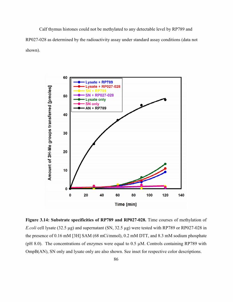
86
Calf thymus histones could not be methylated to any detectable level by RP789 and
RP027-028 as determined by the radioactivity assay under standard assay conditions (data not
shown).
Figure 3.14: Substrate specificities of RP789 and RP027-028. Time courses of methylation of
E.coli cell lysate (32.5 µg) and supernatant (SN, 32.5 µg) were tested with RP789 or RP027-028 in
the presence of 0.16 mM [3H] SAM (68 mCi/mmol), 0.2 mM DTT, and 8.3 mM sodium phosphate
(pH 8.0). The concentrations of enzymes were equal to 0.5 µM. Controls containing RP789 with
OmpB(AN), SN only and lysate only are also shown. See inset for respective color descriptions.
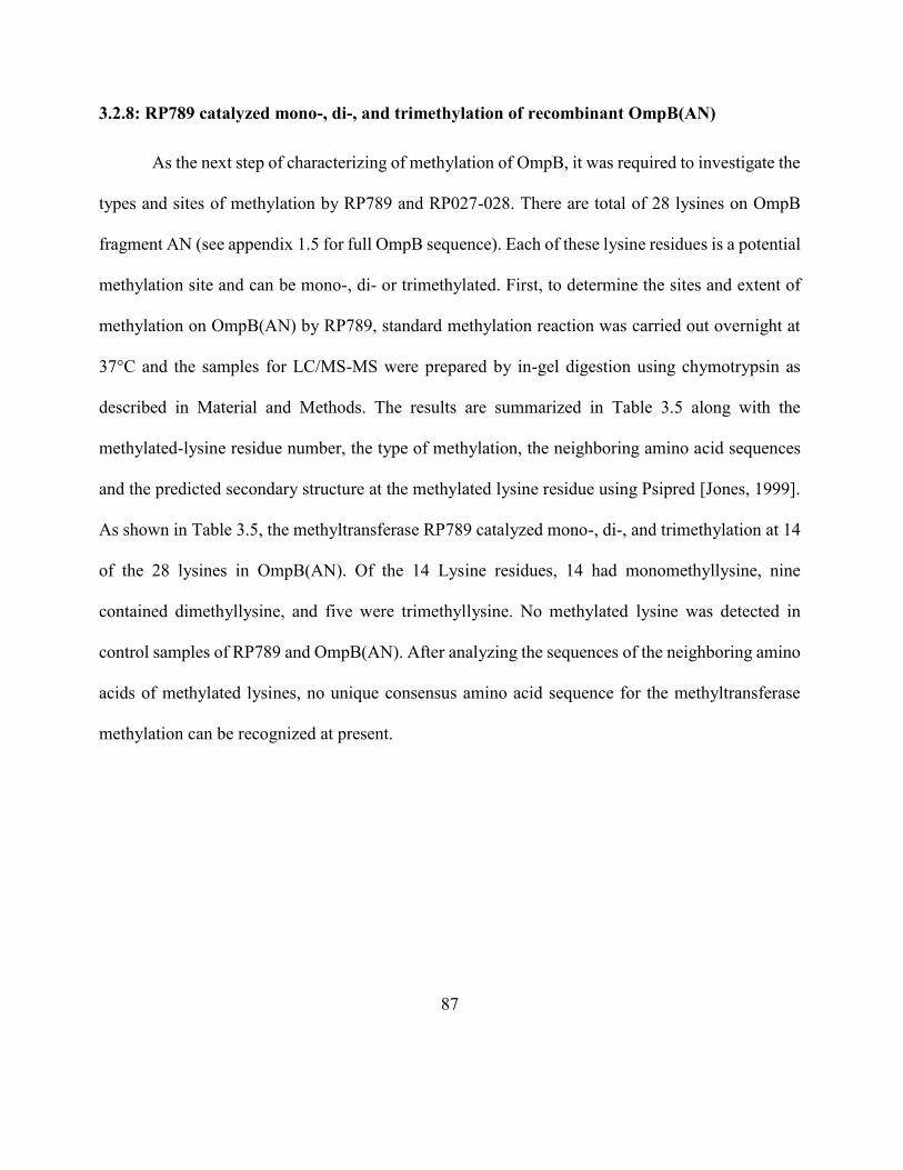
87
3.2.8: RP789 catalyzed mono-, di-, and trimethylation of recombinant OmpB(AN)
As the next step of characterizing of methylation of OmpB, it was required to investigate the
types and sites of methylation by RP789 and RP027-028. There are total of 28 lysines on OmpB
fragment AN (see appendix 1.5 for full OmpB sequence). Each of these lysine residues is a potential
methylation site and can be mono-, di- or trimethylated. First, to determine the sites and extent of
methylation on OmpB(AN) by RP789, standard methylation reaction was carried out overnight at
37°C and the samples for LC/MS-MS were prepared by in-gel digestion using chymotrypsin as
described in Material and Methods. The results are summarized in Table 3.5 along with the
methylated-lysine residue number, the type of methylation, the neighboring amino acid sequences
and the predicted secondary structure at the methylated lysine residue using Psipred [Jones, 1999].
As shown in Table 3.5, the methyltransferase RP789 catalyzed mono-, di-, and trimethylation at 14
of the 28 lysines in OmpB(AN). Of the 14 Lysine residues, 14 had monomethyllysine, nine
contained dimethyllysine, and five were trimethyllysine. No methylated lysine was detected in
control samples of RP789 and OmpB(AN). After analyzing the sequences of the neighboring amino
acids of methylated lysines, no unique consensus amino acid sequence for the methyltransferase
methylation can be recognized at present.
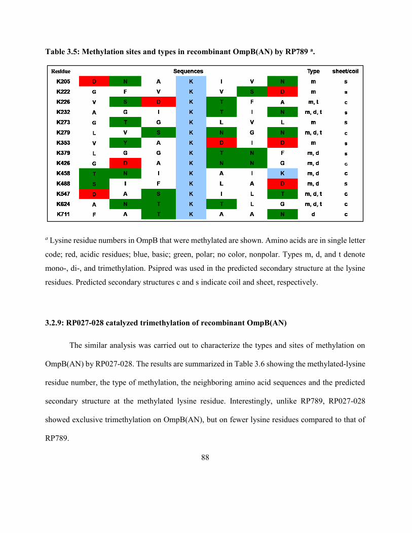
88
Table 3.5: Methylation sites and types in recombinant OmpB(AN) by RP789 a.
a Lysine residue numbers in OmpB that were methylated are shown. Amino acids are in single letter
code; red, acidic residues; blue, basic; green, polar; no color, nonpolar. Types m, d, and t denote
mono-, di-, and trimethylation. Psipred was used in the predicted secondary structure at the lysine
residues. Predicted secondary structures c and s indicate coil and sheet, respectively.
3.2.9: RP027-028 catalyzed trimethylation of recombinant OmpB(AN)
The similar analysis was carried out to characterize the types and sites of methylation on
OmpB(AN) by RP027-028. The results are summarized in Table 3.6 showing the methylated-lysine
residue number, the type of methylation, the neighboring amino acid sequences and the predicted
secondary structure at the methylated lysine residue. Interestingly, unlike RP789, RP027-028
showed exclusive trimethylation on OmpB(AN), but on fewer lysine residues compared to that of
RP789.
89
Table 3.6: Methylation sites and types in recombinant OmpB(AN) by RP027-028 a.
a Lysine residue numbers in OmpB that were methylated are shown. Amino acids are in single letter
code; red, acidic residues; blue, basic; green, polar; no color, nonpolar. Types m, d, and t denote
mono-, di-, and trimethylation. Psipred was used in the predicted secondary structure at the lysine
residues. Predicted secondary structures c and s indicate coil and sheet, respectively.
A total of seven lysine residues were trimethylated by RP027-028, compared to 14 we
observed with RP789. All seven sites showed only trimethylation. Interestingly, no mono- or
dimethyl lysine residues were detected in OmpB(AN) that was methylated by RP027-028. Again,
no unique consensus amino acid sequence can be recognized at present.
3.2.10: Methylated OmpB(K) showed similar patterns.
OmpB(AN) fragment covers only the N-terminal part of full length OmpB (residues 33-
741). Fragment K (residues 745-1353) covers the C-terminal part of the passenger domain. To be
able to characterize the methylation patterns on whole passenger domain of OmpB by each
methyltransferase enzyme, it is required to confirm that similar patterns are observed for both
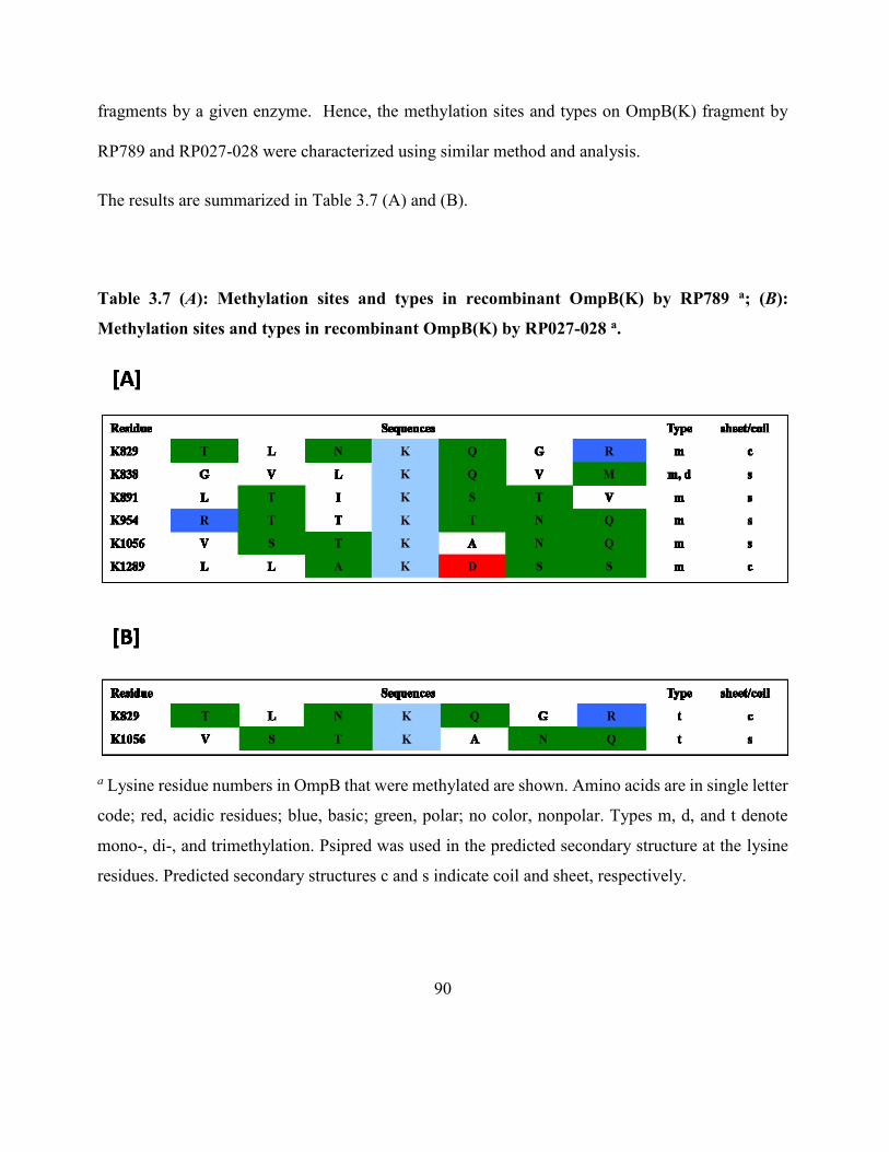
90
fragments by a given enzyme. Hence, the methylation sites and types on OmpB(K) fragment by
RP789 and RP027-028 were characterized using similar method and analysis.
The results are summarized in Table 3.7 (A) and (B).
Table 3.7 (A): Methylation sites and types in recombinant OmpB(K) by RP789 a; (B):
Methylation sites and types in recombinant OmpB(K) by RP027-028 a.
a Lysine residue numbers in OmpB that were methylated are shown. Amino acids are in single letter
code; red, acidic residues; blue, basic; green, polar; no color, nonpolar. Types m, d, and t denote
mono-, di-, and trimethylation. Psipred was used in the predicted secondary structure at the lysine
residues. Predicted secondary structures c and s indicate coil and sheet, respectively.
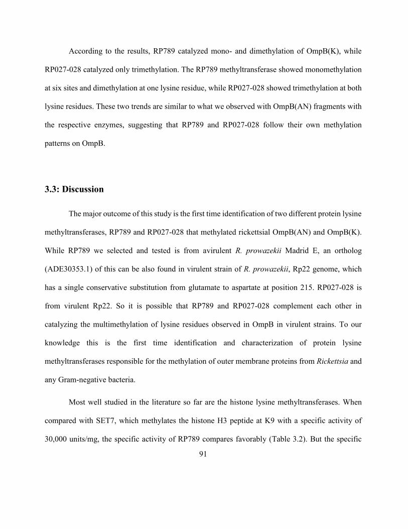
91
According to the results, RP789 catalyzed mono- and dimethylation of OmpB(K), while
RP027-028 catalyzed only trimethylation. The RP789 methyltransferase showed monomethylation
at six sites and dimethylation at one lysine residue, while RP027-028 showed trimethylation at both
lysine residues. These two trends are similar to what we observed with OmpB(AN) fragments with
the respective enzymes, suggesting that RP789 and RP027-028 follow their own methylation
patterns on OmpB.
3.3: Discussion
The major outcome of this study is the first time identification of two different protein lysine
methyltransferases, RP789 and RP027-028 that methylated rickettsial OmpB(AN) and OmpB(K).
While RP789 we selected and tested is from avirulent R. prowazekii Madrid E, an ortholog
(ADE30353.1) of this can be also found in virulent strain of R. prowazekii, Rp22 genome, which
has a single conservative substitution from glutamate to aspartate at position 215. RP027-028 is
from virulent Rp22. So it is possible that RP789 and RP027-028 complement each other in
catalyzing the multimethylation of lysine residues observed in OmpB in virulent strains. To our
knowledge this is the first time identification and characterization of protein lysine
methyltransferases responsible for the methylation of outer membrane proteins from Rickettsia and
any Gram-negative bacteria.
Most well studied in the literature so far are the histone lysine methyltransferases. When
compared with SET7, which methylates the histone H3 peptide at K9 with a specific activity of
30,000 units/mg, the specific activity of RP789 compares favorably (Table 3.2). But the specific
92
activity of RP027-028 was 10 to 30 times lower than that of RP789. Even though the attempts to
find out the existence of any additional modifications or cofactors of RP027-028 were negative, that
possibility cannot be ruled out at present. Hence the reason for low catalytic efficiency of RP027-
028 is not clear. Addition of E. coli proteins did not alter RP027-028 methyltransferase activity,
suggesting that no cofactors were present in E. coli. Addition of divalent cations, such as Zn(II),
Ni(II), and Cu(II), did not appreciably enhance methyltransferase activity, suggesting that none of
these metal ions could be expected to be a cofactor. Even though it was assumed that low catalytic
activity of R.prowazekii RP027-028 may be due to the non-homologous origin of R.typhi OmpB
substrate used, that assumption can be dismissed, since when RP027-028 ortholog from same
rickettsia strain as the OmpB substrate was used there was no significant increase in rate.
What is reported in this chapter is a qualitative analysis of the methylation profiles of OmpB
catalyzed by two methyltransferase RP789 and RP027-028. LC/MS-MS analysis showed that
RP789 from avirulent Madrid E strain catalyzed mono-, di-, and trimethylation and RP027-028 from
virulent Rp22 strain catalyzed exclusively trimethylation. Given what has been reported already on
how the hypermethylation is correlated to the rickettsial virulence, this was a very interesting
observation. It could suggest that these two enzymes follow two different mechanisms giving rise
to qualitatively different methylation profiles. In order to exclusively catalyze trimethylation of
lysine, RP027-028 had to remain bound to the methylation target site of the substrate protein during
the entire course of three rounds of methylation of the lysine residue. On the other hand, the
methyltransferase RP789 could dissociate from the target sites of the substrate protein after the
addition of a methyl group to a lysine or methylated lysine residue. The trimethylation activity of
RP027- 028 is consistent with the idea that RP027-028 accounts at least in part for the
93
hypermethylation of OmpB and consequently the virulence of the virulent strains. Even though the
mass spectrometry data showed that RP027-028 can trimethylate OmpB(K) fragment the western
blot analysis did not. As explained in the results it could be due to reduced reactivity by the primary
antibodies, caused may be due to lower levels of OmpB(K) present on the gel/membrane after going
through the Ni column to separate enzyme from substrate.
The observation of large differences in the catalytic efficiencies between RP789 and RP027-
028 may also be explained based on the assumption, that these two enzymes follow different
reaction mechanisms. Literature reports significant differences in catalytic efficiencies when two
different methylation mechanisms, namely, distributive and processive are followed [Dirk et al.,
2007]. In distributive methylation, methyltransferase dissociates from the protein substrate after the
transfer of each methyl group. This may lead to more random methylation and result a mixture of
mono-, di-, or trimethyl lysines. The kinetics of RP789 likely follows a distributive mechanism. In
processive methylation, methyltransferase transfers methyl groups to the same lysine residue
consecutively in three rounds without dissociation from the protein substrate. For each round of
methyl transfer, processive enzyme must dissociate S-adenosylhomocysteine and rebind S-
adenosylmethionine. The structural similarity between S-adenosylhomocysteine and S-
adenosylmethionine would slow down dissociation of S-adenosylhomocysteine and binding of S-
adenosylmethionine. The kinetics of RP027-028 likely follows a processive mechanism.
In addition to mass spectrometry data, the molar ratios of methyl groups calculated per
OmpB fragment molecule provided evidence that OmpB fragments are methylated at multiple sites.
While the radioactivity assay provided the average number of methyl groups per OmpB(AN) or
94
OmpB(K), LC/MS-MS data provided the locations, states and total number of methylated sites.
There is clearly an apparent difference between the number of sites identified by LC/MS-MS
compared to the average number of methyl groups as determined by the radioactivity assays.
Although there is no definite explanation as of now, one likely scenario would be that methylated
OmpB was microheterogeneous in the number and sites of methylation due to varying degrees and
sites of methylation among different substrate protein molecules.
One unusual feature we observed for these two methyltransferases is the multisite
methylation of their substrate, OmpB. Most reported protein methyltransferases are responsible to
transfer methyl group/s to a designated single lysine or arginine residue. Multisite methylation
indicates the possibility of the processive binding by these methyltransferases to consecutive target
sites without dissociating from the OmpB molecule. This kind of “site hopping” behavior has been
observed in DNA methyltransferases in epigenetic gene regulation but not so far reported in protein
methyltransferases [Casadesus et al., 2006].
When the mass spectrometry data was analyzed for neighboring amino acid sequences of
methylated lysines, no clear unique consensus amino acid sequence was identified, suggesting that
neither RP789 nor RP027-028 target any specific sequence for methylation. Even though OmpB
family of protein are studied in phylogenetic analysis [Roux et al., 2000] and on their role in
rickettsial evolution [Blanc et al., 2005], the importance of three dimensional structure in substrate
recognition by methyltransferases is not well studied. There is no crystal structure reported for the
passenger domain of OmpB so far, hence the 3-D fold is not known. We studied three fragments of
passenger domain (introduction, section 1.4) in this project; fragments A (33-273), AN (33-741)
95
and K (745-1353). The observation that five lysine residues in recombinant OmpB(A) failed to be
enzymatically methylated but were methylated in OmpB(AN) (Table 3.5) could be due to the
absence of properly folded tertiary structure in OmpB(A), unlike that in OmpB(AN). This suggests
that three-dimensional structure of OmpB likely plays roles in substrate recognition and antibody
epitopes. In an attempt to predict the secondary structure using Psipred [Jones, 1999] it was
discovered that the passenger domain consists of alternating short β-strands and loops. The
methylation sites of RP789 and RP027-028 were located either at coil or β-strands as shown in
LC/MS-MS data. When using online databases of available crystal structures to predict the three
dimensional structure, parts of OmpB passenger domain aligned well with β-helical proteins,
characterized by parallel β-strands associating in a helical pattern with three faces. Few examples
were polysialic acid O-acetyltransferase (PDB ID: 2WLG), prions [Govaerts et al., 2004, Krishnan
et al., 2005] and amyloid cross-β spine [Nelson, 2005, Sawaya, 2007]. The structural significance
of this specific β-helical structure of OmpB passenger domain in rickettsial pathogenesis is not yet
known.
OmpB are highly acidic proteins. The transfer of one-carbon methyl groups to nitrogen (N-
methylation) to lysine side chains increases the hydrophobicity of the protein and can neutralize a
negative charge on lysine when bound to carboxylic acids in making non covalent bonds [Greer et al.,
2012]. This way it can affect the electrostatic interactions and hydrogen bonding in protein- protein
interactions and consequently the function of OmpB. OmpB has been shown to mediate rickettsial
attachment to and invasion of endothelial cells [Chan et al., 2009]. Methylation at mul t ip l e lysine
residues could conceivably affect bacteria-host cell interactions thereby influencing invasion of host
96
cell. Methylated lysines may even alter the interactions with host intracellular proteins once inside the
host cells facilitating the bacterial survival and avoiding cell destruction.
It has been demonstrated that chemical methylation of OmpB(A) increased its serological
reactivity. While some patient derived sera could not recognize unmethylated recombinant OmpB(A)
they yielded very high binding signals with chemically methylated recombinant OmpB(A). In vitro
enzymatic methylation of OmpB may provide a new path for producing methylated OmpB as
diagnostic antigens and facilitate the production in large quantities. Antibodies against OmpB are found
in patients without detectable rickettsial bacteria [Anacker et al., 1984]. Enzymatically methylated
OmpB may potentially enhance the potency of OmpB in immunochemical reactions and provide
diagnostic reagents with enhanced sensitivity and specificity and vaccine candidates with greater
effectiveness.
3.4: Conclusion
Rickettsiae are obligatory intracellular infectious Gram-negative bacteria responsible for major
diseases as typhus and spotted fever. The outer membrane protein B has been identified as the
immunodominat agent and plays roles as protective envelope and adhesins. Previous literature reports
the possible correlation between the hypermethylation of lysine residues in OmpB and the bacterial
virulence. This suggests the existence of an enzymatic system for OmpB methylation, but no
methyltransferase enzyme has been identified or characterized. We carried out bioinformatics analysis
of genomic DNA sequences of Rickettsia to identify putative methyltransferases. The
methyltransferase activities of the purified proteins were analyzed by methyl incorporation of
97
radioactively labeled S-adenosylmethionine into recombinant fragments of OmpB. Two putative
recombinant methyltransferases (RP789 and RP027-028) methylated recombinant OmpB fragments.
The specific activity of RP789 is 10- to 30-fold higher than that of RP027-028. Western blot analysis
using specific antibodies against trimethyl lysine showed that both RP789 and RP027-028 catalyzed
trimethylation of recombinant OmpB fragments. Liquid chromatography tandem mass spectrometry
analysis showed that RP789 catalyzed mono-, di-, and trimethylation of lysine, while RP027-028
catalyzed exclusively trimethylation. To our knowledge, RP789 and RP027-028 are the first
biochemically characterized lysine methyltransferases of outer membrane proteins from Gram-
negative bacteria.
The identification and characterization of RP789 and RP027-028 in this study will help us to
better understand the role of methylation of lysines in bacterial virulence. This understanding will also
be vital for the mass production of methylated recombinant OmpB, and may advance our knowledge
of the structure and function of protein lysine methylation in general. Being able to generate
enzymatically methylated recombinant OmpB may also provide an improved method over chemical
methylation in the attempt to generate advance diagnostic reagents and vaccine candidates.
Methylated bacterial surface proteins are not limited to OmpB. Similar methods to the present
studies can be readily applied to many cell surface proteins for the development of improved
diagnostic reagents and vaccine candidates.
In summary, identification, production and characterization of rickettsial lysine
methyltransferases will provide new tools to investigate the mechanism of methylation of OmpB,

98
effects of methylation on the structure and function of OmpB, and development of methylated
OmpB-based diagnostic assays and vaccine candidates.
99
Chapter 4
Multimethylation in Rickettsia OmpB catalyzed by Lysine Methyltransferases
4.1: Introduction
Rickettsiae are the causative agents of major rickettsial diseases including epidemic typhus,
endemic typhus, spotted fever and scrub typhus and are defined as obligatory intracellular, Gram-
negative bacteria [Hackstadt., 1996, Walker et al., 2008]. The bacteria contain a family of outer
membrane proteins (Omp) in the outer leaflet of their outer membrane, a structural feature common
among all Gram-negative bacteria. Omp is responsible for first interactions with the extracellular
environment hence important in molecular transport and bacterial pathogenesis [Koebnik et al.,
2000, Buchanan, 1999]. These proteins are predicted to be involved in mediating interactions with
target host cells such as host cell attachment, invasion, internalization, and intracellular movement
[Uchiyama et al., 2006, Chan et al., 2009, Li et al.,1998]. Purified native typhus Omp have been
shown to induce strong humoral and cellular immune response in mice, guinea pigs and rabbit
models infected with Rickettsiae [Dasch, 1981., Bourgeois et al., 1981, Feng et al., 2004].
Recovery from typhus confers robust immunity and protection from re-infection suggesting
that protective antigen(s) must exist [Carl et al.,1989]. This antigenic protein is known as outer
membrane protein B or OmpB. The fact that this protein is evolutionarily conserved amongst all
rickettsial species suggests OmpB serves a unified function in pathogenesis in rickettsiae. These
100
proteins have modular structures including an N-terminal signal peptide, a central passenger domain
and a C-terminal translocation domain (β-peptide) [Barnard et al., 2007, Schulz, 2002]. The
passenger domain of rickettsial OmpB has been shown to participate in adhesion to mammalian
cells in vitro, suggesting this may be OmpB’s role in the virulence of Rickettsia [Chan et al., 2009,
Uchiyama et al., 2006, Renesto et al., 2006].
OmpB undergoes post-translational modification of methylation at the -amino group of
lysine and it has been proposed that methylation plays a major role in bacterial virulence. OmpB
was shown to be hyper-methylated in the virulent strains compared to avirulent strain [Ching et al.,
1993, Rodionov, 1990]. Methylation may be important in the host immunogenic response to OmpB
itself and to rickettsial virulence [Bechah et al., 2010, Zhang et al., 2006, Chao et al., 2007]. Rabbit
antiserum against recombinant OmpB (unmethylated) is less reactive than antiserum against OmpB
purified directly from Rickettsia suggesting that methylation of OmpB may enhance its antigenicity
[Gilmore et al., 1991]. In addition, the immunoreactivity of chemically methylated recombinant
OmpB is enhanced against sera from infected patients [Chao et al., 2008]. It suggests that
methylated recombinant OmpB may be able to replace native OmpB as a diagnostic reagent to help
advance vaccine development against Rickettsia [Chan et al., 2011].
In our previously published work, we reported the first time identification and
characterization of two rickettsial lysine methyltransferases, RP789 and RP027-028 from
R.prowazekii [Abeykoon et al., 2012]. RP027-028, as well as its homolog in R. typhi, RT0101,
catalyze trimethylation, and RP789 catalyzes monomethylation of recombinant OmpB fragments,
monitored by incorporation of radioactive [3H-Me] from [3H-Me]-S-adenosylmethionine and
101
western blot analysis, while none of these methyltransferases can methylate either calf thymus
histone or E. coli proteins. Also the methylation profiles from LC-MS/MS showed multiple
methylations at multiple lysine residues that differ from what has been reported so far for other
methyltransferases. Hence these enzymes appear to belong to a novel family of lysine
methyltransferases. The fact that these methyltransferases recognize OmpB at many diverse sites
suggests an unusual substrate recognition mechanism. Characterization of methylation in rickettsial
OmpB at the molecular level is essential for explaining the roles of methylation in bacterial virulence
and pathogenesis and will be the focus of this part of the project. Findings from this study will help
to better understand structure, function and mechanism of this new class of protein lysine
methyltransferases. Ability to generate enzymatically methylated recombinant OmpB may also
provide an improved method over chemical methylation in the attempt to generate advance
diagnostic reagents and vaccine candidates.
This study primarily utilized semiquantitative integrated liquid chromatography-tandem
mass spectroscopy (LC-MS/MS) techniques to characterize the location, state and level of
methylation of recombinant OmpB by different methyltransferases from virulent and avirulent
rickettsial strains. The results were compared with the native OmpB isolated from rickettsia to
understand any correlation. This was the first time characterization of methylation of OmpB (or any
Omp) at molecular level and on how methylation of OmpB may contribute to the virulence of
Rickettsia.
102
4.2: Results
4.2.1: Kinetics of methylation by rickettsial methyltransferases
The full length passenger domain of OmpB was not soluble and hence formed inclusion
bodies. But the two recombinant R. typhi OmpB fragments, OmpB(AN33-741) and OmpB(K745-1353)
were successfully purified and refolded to yield soluble protein substrates for analysis as described
in methods [Figure 1.2]. We monitored the steady state kinetics of four methyltransferases using
OmpB(AN) as the substrate, RP789 and RP027-028 from R. prowazekii and RT0776 and RT0101
from R. typhi. The monomethyltransferases, RP789 and RT0776 (94% sequence identity) and
trimethyltransferases, RP027 and RT0101 (93% sequence identity) are orthologous proteins found
in each strain. Table 4.1 summarizes the steady state kinetic parameters. They are calculated based
on the initial rates of the methylation of up to 2 µM OmpB(AN) catalyzed by 0.26 µM each of the
four methyltranferases by monitoring the incorporation of radioactive [3H-Me] from [3H-Me]-S-
adenosylmethionine to OmpB(AN). Overall, the two monomethyltransferase showed higher kcat
values compared to trimethyltransferases. RP789 and RT0101 showed higher enzyme efficiency
(kcat / KM).
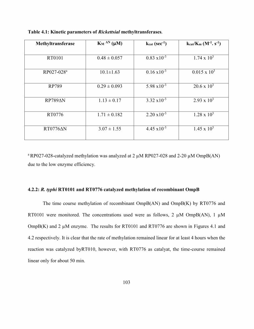
103
Table 4.1: Kinetic parameters of Rickettsial methyltransferases.
Methyltransferase KM AN (µM) kcat (sec-1) kcat/Km (M-1. s-1)
RT0101 0.48 ± 0.057 0.83 x10⁻³ 1.74 x 10³
RP027-028a 10.1±1.63 0.16 x10⁻³ 0.015 x 10³
RP789 0.29 ± 0.093 5.98 x10⁻³ 20.6 x 10³
RP789ΔN 1.13 ± 0.17 3.32 x10⁻³ 2.93 x 10³
RT0776 1.71 ± 0.182 2.20 x10⁻³ 1.28 x 10³
RT0776ΔN 3.07 ± 1.55 4.45 x10⁻³ 1.45 x 10³
a RP027-028-catalyzed methylation was analyzed at 2 µM RP027-028 and 2-20 µM OmpB(AN)
due to the low enzyme efficiency.
4.2.2: R. typhi RT0101 and RT0776 catalyzed methylation of recombinant OmpB
The time course methylation of recombinant OmpB(AN) and OmpB(K) by RT0776 and
RT0101 were monitored. The concentrations used were as follows, 2 µM OmpB(AN), 1 µM
OmpB(K) and 2 µM enzyme. The results for RT0101 and RT0776 are shown in Figures 4.1 and
4.2 respectively. It is clear that the rate of methylation remained linear for at least 4 hours when the
reaction was catalyzed byRT010, however, with RT0776 as catalyat, the time-course remained
linear only for about 50 min.
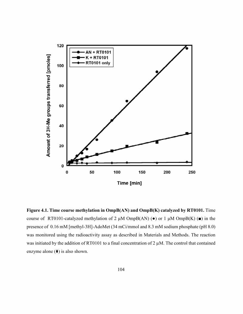
104
Figure 4.1. Time course methylation in OmpB(AN) and OmpB(K) catalyzed by RT0101. Time
course of RT0101-catalyzed methylation of 2 μM OmpB(AN) (●) or 1 μM OmpB(K) (■) in the
presence of 0.16 mM [methyl-3H]-AdoMet (34 mCi/mmol and 8.3 mM sodium phosphate (pH 8.0)
was monitored using the radioactivity assay as described in Materials and Methods. The reaction
was initiated by the addition of RT0101 to a final concentration of 2 µM. The control that contained
enzyme alone (⧫) is also shown.
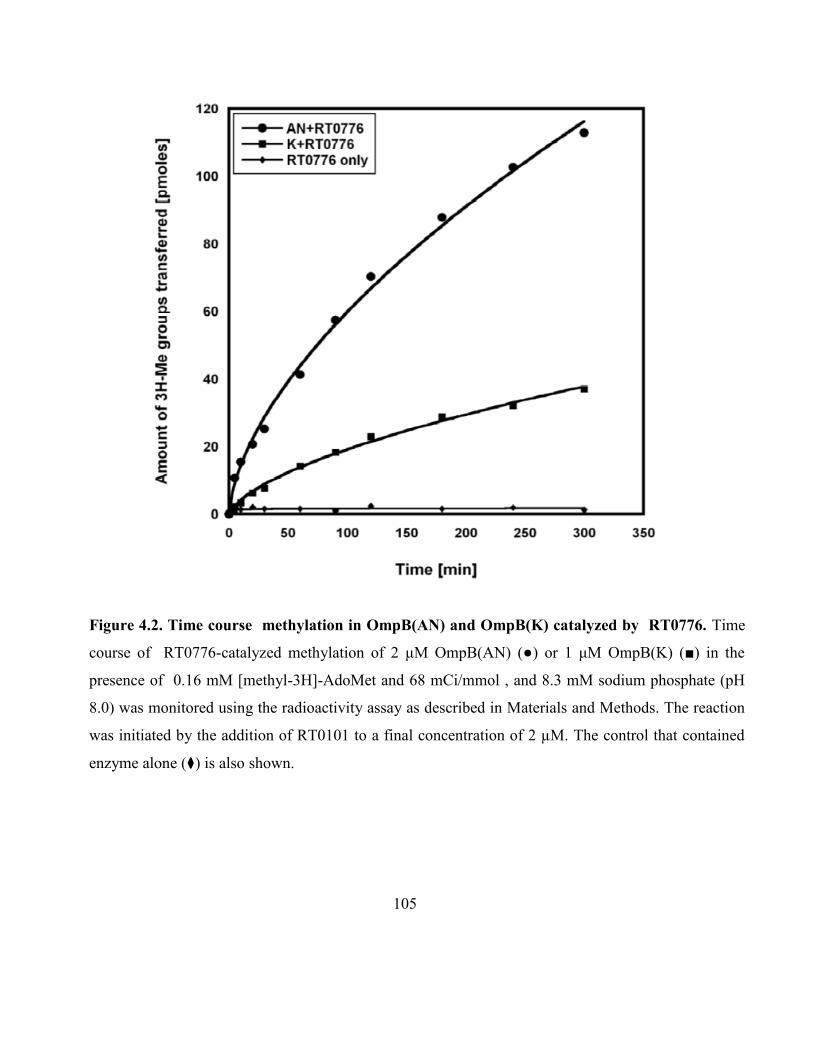
105
Figure 4.2. Time course methylation in OmpB(AN) and OmpB(K) catalyzed by RT0776. Time
course of RT0776-catalyzed methylation of 2 μM OmpB(AN) (●) or 1 μM OmpB(K) (■) in the
presence of 0.16 mM [methyl-3H]-AdoMet and 68 mCi/mmol , and 8.3 mM sodium phosphate (pH
8.0) was monitored using the radioactivity assay as described in Materials and Methods. The reaction
was initiated by the addition of RT0101 to a final concentration of 2 µM. The control that contained
enzyme alone (⧫) is also shown.
106
After confirming the methylation activity, the methylation profiles of recombinant
OmpB(AN) and OmpB(K) were determined. Two fragments were enzymatically methylated using
2 µM RT0101 and 3.2 mM SAM and incubated overnight at 37°C. The methylated fragments were
separated on SDS-PAGE followed by in-gel digestion using chymotrypsin and the resultant peptides
were analyzed by LC-MS/MS. The PSM numbers for all mono-, di-, tri- and non-methylated lysine
residues for OmpB(AN) and OmpB(K) are combined and tabulated in Appendix 2 Table S1.
Sixteen trimethyllysine residues were observed and a total of 133 trimethyllysine-containing PSM
were distributed among the 16 locations, while only nine monomethyllysine-containing and one
dimethyllysine-containing PSM were observed (Table S1). RT0101 can be essentially categorized
as a trimethyltransferase due to the predominance of trimethyllysine-containing PSM observed. The
monomethyllysine and dimethyllysine observed could be the intermediates of trimethylation. Figure
4.3 shows the observed normalized fractions of trimethylations of all lysine residues on OmpB
catalyzed by RT0101.
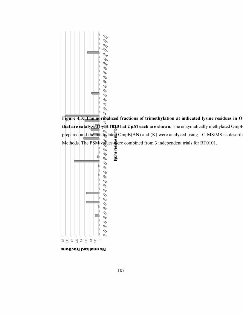
107
Figure 4.3: The normalized fractions of trimethylation at indicated lysine residues in OmpB(AN) and (K)
that are catalyzed by RT0101 at 2 µM each are shown. The enzymatically methylated OmpB(AN) and (K) were
prepared and the methylated OmpB(AN) and (K) were analyzed using LC-MS/MS as described in Materials and
Methods. The PSM values were combined from 3 independent trials for RT0101.
108
The specific lysine residues are trimethylated at varying levels from less than 1% to 36% as
revealed by the normalized fractions. Based on the amino acid sequences adjacent to trimethylated
lysines, no single consensus sequence can be discerned. But two motifs were recognized during the
attempt to uncover amino acid sequences catalyzed by the trimethyltransferas RT0101. This task
was accomplished using Motif-X [Chou et al., 2011]. Motif-X (short for motif extractor) is a web
server designed to extract over-represented patterns from any sequence data set. The remaining
sequences occur either 3 or less than 3 times among the sixteen sequences that are trimethylated by
RT0101.
(1) KX(G/A/V/I)N- present at K205, K232, K279, K667, K711, and K723
(2) KT(I/L/F) - present at K226, K232, K624, K635, and K667
The sequences at K232 and K667 conform to both motifs.
The attempts to trimethylate the peptides which contain one (ANTK624TLG) or two
(IGANTK624TLGRFNVGSSK635TILNA) of these recognition sequences by RT0101 were not
successful (data not shown) suggesting that the conformation of OmpB or other unknown factors
may contribute to substrate recognition.
Methylation catalyzed by RT0101 showed PSM numbers for mono- and di-methylated
lysines at 9 and 1, respectively, which are disproportionally low compared to 133 PSM for
trimethyllysines observed (Appendix 2 Table S1). This observation agrees with the speculation that
trimethylation may occur most likely via a processive mechanism rather than a distributive
mechanism. In the processive mechanism mono- or di-methyllysine is formed but remains bound to
the enzyme until the substrate is fully trimethylated before the product is released from the enzyme.
109
When the time course of methylation of 2 µM OmpB(AN) catalyzed by 2 µM RT0101 was
analyzed for two hours, the PSM of trimethylated peptides increased steadily to 13, 28, and 56 at
30, 60, and 120 min, respectively, while the numbers of PSM of monomethylated peptides were 1,
1 and 6 at 30, 60 and 120 min, respectively. Hence monomethyllysine is likely an intermediate
during trimethylation and RT0101-catalyzed trimethylation of OmpB may proceed via a processive
mechanism.
In addition to RT0101 catalyzed methylation, LC-MS/MS analysis of RT0776-catalyzed
methylation of OmpB(AN) and OmpB(K) was also performed. The PSM numbers for all mono-,
di-, tri- and non-methylated lysine residues for OmpB(AN) and OmpB(K) are combined and
tabulated in Appendix 2 Table S2. Monomethylation was found at 34 lysine residuesand a total of
379 monomethyllysine-containing PSM were distributed among those 34 locations. Compared to
that, only six dimethyllysine-containing and two trimethyllysine-containing PSM were observed
(Table S2). RT0776 can be essentially categorized as a monomethyltransferase due to the
predominance of monomethyllysine-containing PSM observed. The observation of extensive
monomethylation [Figure 4.4] suggests that these enzymes possessan active site that can
accommodate a wide variety of recognition sequences.
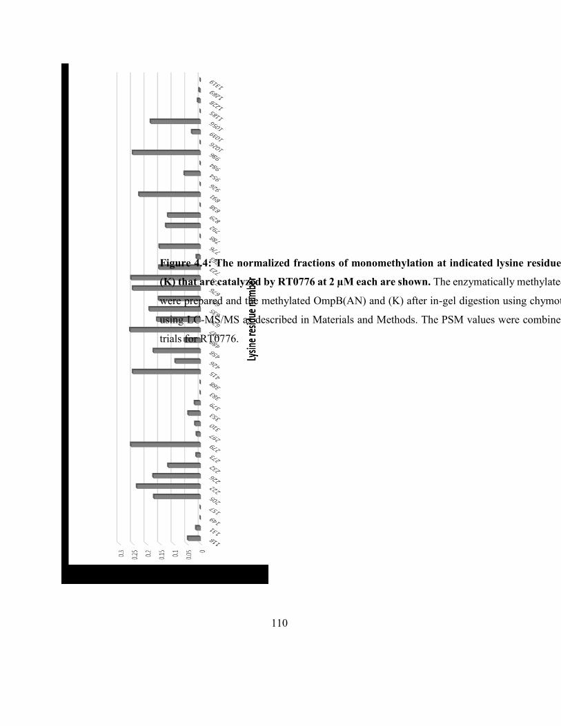
110
Figure 4.4: The normalized fractions of monomethylation at indicated lysine residues in OmpB(AN) and
(K) that are catalyzed by RT0776 at 2 µM each are shown. The enzymatically methylated OmpB(AN) and (K)
were prepared and the methylated OmpB(AN) and (K) after in-gel digestion using chymotrypsin were analyzed
using LC-MS/MS as described in Materials and Methods. The PSM values were combined from 3 independent
trials for RT0776.
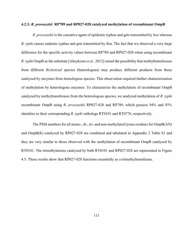
111
4.2.3: R. prowazekii RP789 and RP027-028 catalyzed methylation of recombinant OmpB
R. prowazekii is the causative agent of epidemic typhus and gets transmitted by lice whereas
R. typhi causes endemic typhus and gets transmitted by flea. The fact that we observed a very large
difference for the specific activity values between RP789 and RP027-028 when using recombinant
R. typhi OmpB as the substrate [Abeykoon et al., 2012] raised the possibility that methyltransferases
from different Rickettsial species (heterologous) may produce different products from those
catalyzed by enzymes from homologous species. This observation required further characterization
of methylation by heterologous enzymes. To characterize the methylation of recombinant OmpB
catalyzed by methyltransferases from the heterologous species, we analyzed methylation of R. typhi
recombinant OmpB using R. prowazekii RP027-028 and RP789, which possess 94% and 93%
identities to their corresponding R. typhi orthologs RT0101 and RT0776, respectively.
The PSM numbers for all mono-, di-, tri- and non-methylated lysine residues for OmpB(AN)
and OmpB(K) catalyzed by RP027-028 are combined and tabulated in Appendix 2 Table S1 and
they are very similar to those observed with the methylation of recombinant OmpB catalyzed by
RT0101. The trimethylations catalyzed by both RT0101 and RP027-028 are represented in Figure
4.5. These results show that RP027-028 functions essentially as a trimethyltransferase.
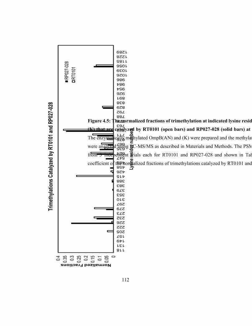
112
Figure 4.5: The normalized fractions of trimethylation at indicated lysine residues in OmpB(AN) and
(K) that are catalyzed by RT0101 (open bars) and RP027-028 (solid bars) at 2 µM each are shown.
The enzymatically methylated OmpB(AN) and (K) were prepared and the methylated OmpB(AN) and (K)
were analyzed using LC-MS/MS as described in Materials and Methods. The PSM values were combined
from 3 independent trials each for RT0101 and RP027-028 and shown in Table S1. The correlation
coefficient of the normalized fractions of trimethylations catalyzed by RT0101 and RP027-028 is 0.65.
113
Similarly, the PSM numbers for all mono-, di-, tri- and non-methylated lysine residues for
OmpB(AN) and OmpB(K) catalyzed by RP789 are combined and tabulated in Appendix 2 Table
S2 along with the values for RT0776. RP789 catalyzed 743, 47 and 46 PSM for mono-, di- and tri-
methyllysine containg peptides respectively and can be categorized as a monomethyltransferase.
The observations for R. prowazekii methyltransferases agree with the results observed for
their R. typhi orthologs. But in the case of RP789, it catalyzed significantly higher normalized
fractions of all mono-, di- and trimethylations compared to RT0776. This could be due to the
heterogeneous nature of the system. For example RP789 and recombinant OmpB are from two
different species whereas RT0776 and OmpB are from R. typhi, a homologous system [Figure 4.6].
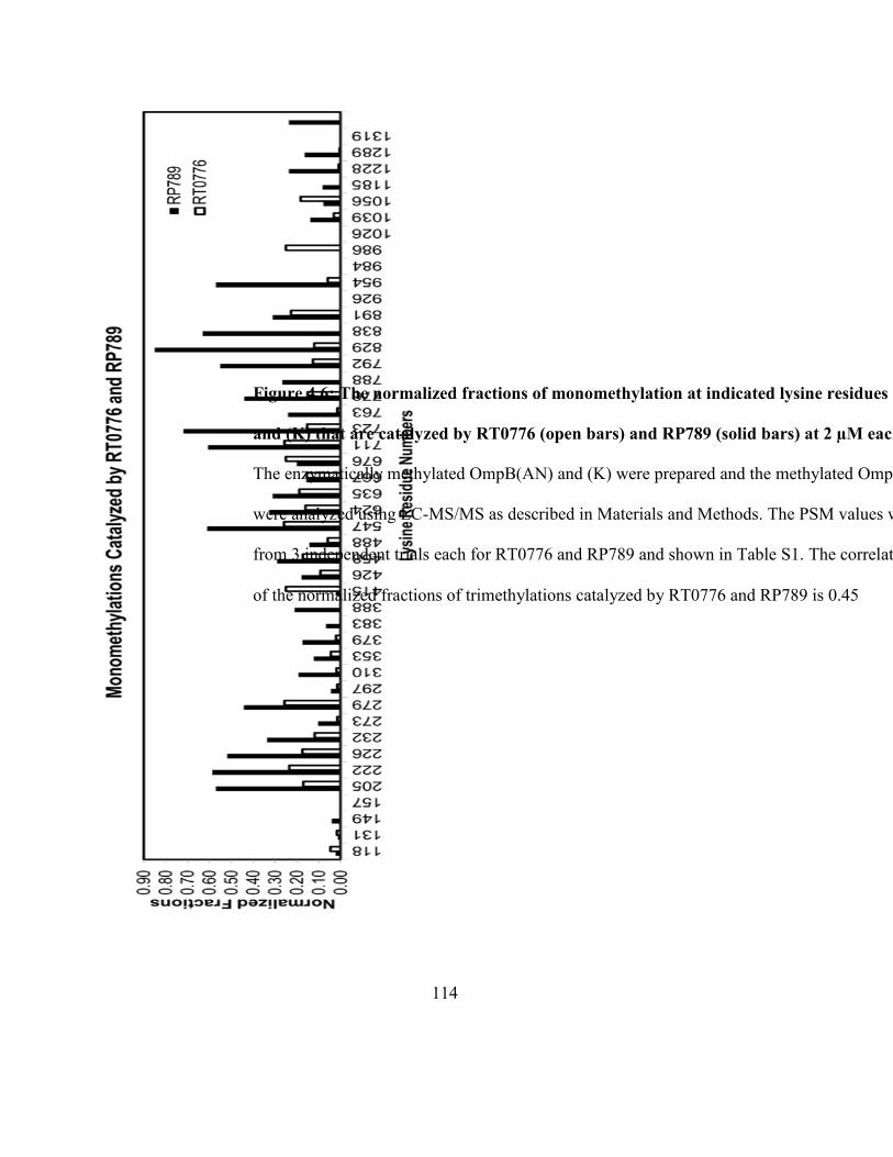
114
Figure 4.6: The normalized fractions of monomethylation at indicated lysine residues in OmpB(AN)
and (K) that are catalyzed by RT0776 (open bars) and RP789 (solid bars) at 2 µM each are shown.
The enzymatically methylated OmpB(AN) and (K) were prepared and the methylated OmpB(AN) and (K)
were analyzed using LC-MS/MS as described in Materials and Methods. The PSM values were combined
from 3 independent trials each for RT0776 and RP789 and shown in Table S1. The correlation coefficient
of the normalized fractions of trimethylations catalyzed by RT0776 and RP789 is 0.45
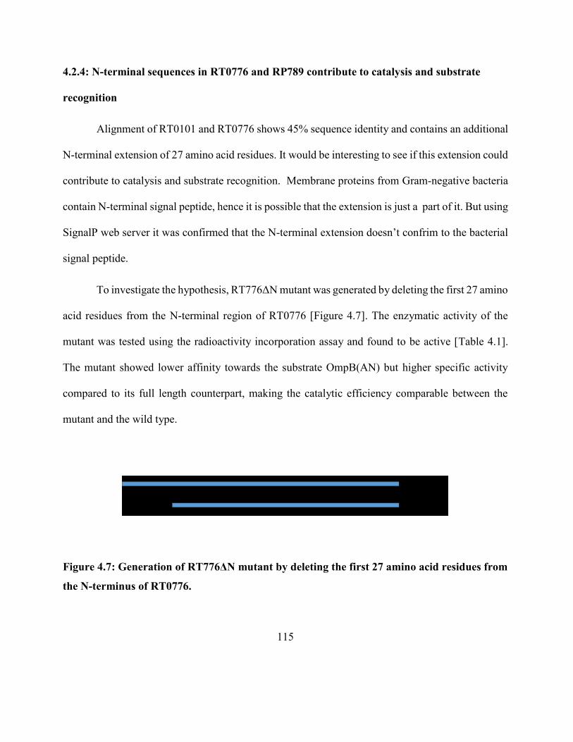
115
4.2.4: N-terminal sequences in RT0776 and RP789 contribute to catalysis and substrate
recognition
Alignment of RT0101 and RT0776 shows 45% sequence identity and contains an additional
N-terminal extension of 27 amino acid residues. It would be interesting to see if this extension could
contribute to catalysis and substrate recognition. Membrane proteins from Gram-negative bacteria
contain N-terminal signal peptide, hence it is possible that the extension is just a part of it. But using
SignalP web server it was confirmed that the N-terminal extension doesn’t confrim to the bacterial
signal peptide.
To investigate the hypothesis, RT776ΔN mutant was generated by deleting the first 27 amino
acid residues from the N-terminal region of RT0776 [Figure 4.7]. The enzymatic activity of the
mutant was tested using the radioactivity incorporation assay and found to be active [Table 4.1].
The mutant showed lower affinity towards the substrate OmpB(AN) but higher specific activity
compared to its full length counterpart, making the catalytic efficiency comparable between the
mutant and the wild type.
Figure 4.7: Generation of RT776ΔN mutant by deleting the first 27 amino acid residues from
the N-terminus of RT0776.
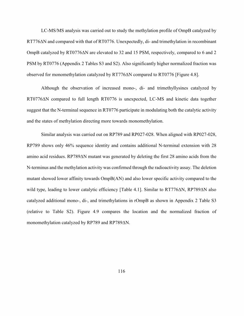
116
LC-MS/MS analysis was carried out to study the methylation profile of OmpB catalyzed by
RT776ΔN and compared with that of RT0776. Unexpectedly, di- and trimethylation in recombinant
OmpB catalyzed by RT0776ΔN are elevated to 32 and 15 PSM, respectively, compared to 6 and 2
PSM by RT0776 (Appendix 2 Tables S3 and S2). Also significantly higher normalized fraction was
observed for monomethylation catalyzed by RT776ΔN compared to RT0776 [Figure 4.8].
Although the observation of increased mono-, di- and trimethyllysines catalyzed by
RT0776ΔN compared to full length RT0776 is unexpected, LC-MS and kinetic data together
suggest that the N-terminal sequence in RT0776 participate in modulating both the catalytic activity
and the states of methylation directing more towards monomethylation.
Similar analysis was carried out on RP789 and RP027-028. When aligned with RP027-028,
RP789 shows only 46% sequence identity and contains additional N-terminal extension with 28
amino acid residues. RP789ΔN mutant was generated by deleting the first 28 amino acids from the
N-terminus and the methylation activity was confirmed through the radioactivity assay. The deletion
mutant showed lower affinity towards OmpB(AN) and also lower specific activity compared to the
wild type, leading to lower catalytic efficiency [Table 4.1]. Similar to RT776ΔN, RP789ΔN also
catalyzed additional mono-, di-, and trimethylations in rOmpB as shown in Appendix 2 Table S3
(relative to Table S2). Figure 4.9 compares the location and the normalized fraction of
monomethylation catalyzed by RP789 and RP789ΔN.
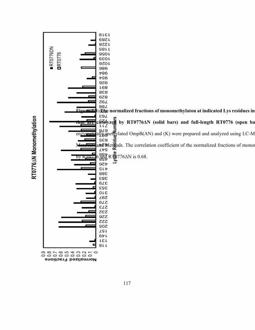
117
Figure 4.8: The normalized fractions of monomethylaton at indicated Lys residues in OmpB(AN) and (K)
that are catalyzed by RT0776ΔN (solid bars) and full-length RT0776 (open bars) are shown. The
enzymatically methylated OmpB(AN) and (K) were prepared and analyzed using LC-MS/MS as described in
Materials and Methods. The correlation coefficient of the normalized fractions of monomethylation catalyzed
by RT0776 and RT0776ΔN is 0.68.
118
Figure 4.9: The normalized fractions of monomethylaton at indicated Lys residues in OmpB(AN) and
(K) that are catalyzed by RP789ΔN (solid bars) and full-length RP789 (open bars) are shown. The
enzymatically methylated OmpB(AN) and (K) were prepared and analyzed using LC-MS/MS as described in
Materials and Methods. The correlation coefficient of the normalized fractions of monomethylation catalyzed
by RT0789 and RT0789ΔN is 0.75.
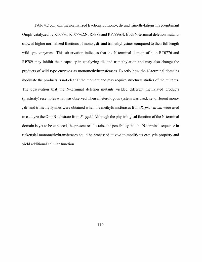
119
Table 4.2 contains the normalized fractions of mono-, di- and trimethylations in recombinant
OmpB catalyzed by RT0776, RT0776ΔN, RP789 and RP789ΔN. Both N-terminal deletion mutants
showed higher normalized fractions of mono-, di- and trimethyllysines compared to their full length
wild type enzymes. This observation indicates that the N-terminal domain of both RT0776 and
RP789 may inhibit their capacity in catalyzing di- and trimethylation and may also change the
products of wild type enzymes as monomethyltransferases. Exactly how the N-terminal domains
modulate the products is not clear at the moment and may require structural studies of the mutants.
The observation that the N-terminal deletion mutants yielded different methylated products
(plasticity) resembles what was observed when a heterologous system was used, i.e. different mono-
, di- and trimethyllysines were obtained when the methyltransferases from R. prowazekii were used
to catalyze the OmpB substrate from R. typhi. Although the physiological function of the N-terminal
domain is yet to be explored, the present results raise the possibility that the N-terminal sequence in
rickettsial monomethyltransferases could be processed in vivo to modify its catalytic property and
yield additional cellular function.
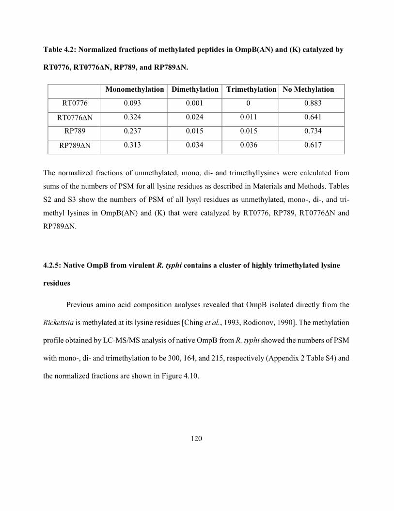
120
Table 4.2: Normalized fractions of methylated peptides in OmpB(AN) and (K) catalyzed by
RT0776, RT0776ΔN, RP789, and RP789ΔN.
Monomethylation Dimethylation Trimethylation No Methylation
RT0776 0.093 0.001 0 0.883
RT0776N 0.324 0.024 0.011 0.641
RP789 0.237 0.015 0.015 0.734
RP789N 0.313 0.034 0.036 0.617
The normalized fractions of unmethylated, mono, di- and trimethyllysines were calculated from
sums of the numbers of PSM for all lysine residues as described in Materials and Methods. Tables
S2 and S3 show the numbers of PSM of all lysyl residues as unmethylated, mono-, di-, and tri-
methyl lysines in OmpB(AN) and (K) that were catalyzed by RT0776, RP789, RT0776ΔN and
RP789ΔN.
4.2.5: Native OmpB from virulent R. typhi contains a cluster of highly trimethylated lysine
residues
Previous amino acid composition analyses revealed that OmpB isolated directly from the
Rickettsia is methylated at its lysine residues [Ching et al., 1993, Rodionov, 1990]. The methylation
profile obtained by LC-MS/MS analysis of native OmpB from R. typhi showed the numbers of PSM
with mono-, di- and trimethylation to be 300, 164, and 215, respectively (Appendix 2 Table S4) and
the normalized fractions are shown in Figure 4.10.
121
Figure 4.10. Normalized fractions of mono- (red), di- (green) and tri- (purple) methylation at indicated
lysine residues in native OmpB from R. typhi are shown. Native OmpB protein (2 µg) from R. typhi
Wilmington was prepared and analyzed by LC-MS/MS as described in Materials and Methods. The PSM
values were combined from 2 independent trials and are shown in Table S4.
122
Interestingly, the locations of trimethylated lysines observed when recombinant R. typhi
OmpB is methylated by RT0101 coincided well with the locations of trimethyllysines observed for
native R. typhi OmpB [Figure 4.11]. Thus RT0101 likely accounts for the trimethylation of OmpB
in vivo.
One important observation is that there is a cluster of four lysine residues (K667, K676,
K711 and K723) in native R.typhi with normalized fraction of trimethylation close to 100%. From
Figure 4.10 it is clear that RT0101 also trimethylated the same cluster of lysines. Similar trend was
observed when recombinant R. typhi OmpB was methylated by RP027-028 [Figure 4.4]. However,
the normalized fractions of trimethylation in native OmpB are appreciably higher than those
observed in vitro with enzymatically methylated recombinant OmpB using RT0101.
4.2.6: Methylation in native OmpBs from virulent and avirulent strains of R. prowazekii
R.prowazekii is the causative agent of epidemic typhus and different strains of this bacteria
exist which differ in virulence. A systematic study of the relationship between the OmpB
methylation levels and the virulence has not been reported. The next attempt was to determine the
methylation of native OmpB isolated directly from three strains, Madrid E, RP22 and Breinl and
see how it is related to the virulence. Out of the three strains, Madrid E is avirulent and RP22 and
Breinl are both highly virulent.
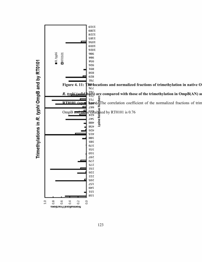
123
Figure 4. 11: The locations and normalized fractions of trimethylation in native OmpB purified from
R. typhi (solid bars) are compared with those of the trimethylation in OmpB(AN) and (K) catalyzed by
RT0101 (open bars). The correlation coefficient of the normalized fractions of trimethylation in native
OmpB and those catalyzed by RT0101 is 0.76
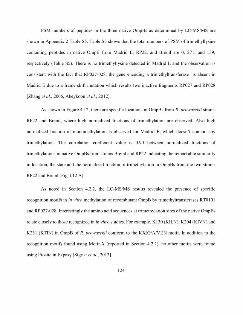
124
PSM numbers of peptides in the three native OmpBs as determined by LC-MS/MS are
shown in Appendix 2 Table S5. Table S5 shows that the total numbers of PSM of trimethyllysine
containing peptides in native OmpB from Madrid E, RP22, and Breinl are 0, 271, and 139,
respectively (Table S5). There is no trimethyllysine detected in Madrid E and the observation is
consistent with the fact that RP027-028, the gene encoding a trimethyltransferase is absent in
Madrid E due to a frame shift mutation which results two inactive fragments RP027 and RP028
[Zhang et al., 2006, Abeykoon et al., 2012].
As shown in Figure 4.12, there are specific locations in OmpBs from R. prowazekii strains
RP22 and Breinl, where high normalized fractions of trimethylation are observed. Also high
normalized fraction of monomethylation is observed for Madrid E, which doesn’t contain any
trimethylation. The correlation coeffcient value is 0.90 between normalized fractions of
trimethylations in native OmpBs from strains Breinl and RP22 indicating the remarkable similarity
in location, the state and the normalized fraction of trimethylation in OmpBs from the two strains
RP22 and Breinl [Fig 4.12 A].
As noted in Section 4.2.2, the LC-MS/MS results revealed the presence of specific
recognition motifs in in vitro methylation of recombinant OmpB by trimethyltransferases RT0101
and RP027-028. Interestingly the amino acid sequences at trimethylation sites of the native OmpBs
relate closely to those recognized in in vitro studies. For example, K130 (KILN), K204 (KIVN) and
K231 (KTIN) in OmpB of R. prowazekii conform to the KX(G/A/VI)N motif. In addition to the
recognition motifs found using Motif-X (reported in Section 4.2.2), no other motifs were found
using Prosite in Expasy [Sigrist et al., 2013].
125
Figure 4. 12. Methylation in native OmpBs purified from R. prowazekii strains Breinl, RP22
and Madrid E. The locations and normalized fractions of (A) tri-, (B) di-, and (C) mono-
methylation in native OmpBs from R. prowazekii strains Breinl (blue), RP22 (red), and Madrid E
(green) are shown. Native OmpBs purified from respective strains (2 µg each) were prepared and
analyzed by LC-MS/MS as described in Materials and Methods. The PSM numbers of
unmethylated, mono-, di- and trimethyllysine containing peptides in OmpBs from R. prowazekii
strains Breinl, RP22, and Madrid E are shown in Table S5. The PSM numbers were obtained from
1, 2 and 3 independent trials of OmpBs from Breinl, RP22 and Madrid E, respectively.
126
As mentioned in Section 4.2.5, R. typhi OmpB contains a cluster of trimethylated lysines
with normalized fractions close to 100%. A similar observation of highly trimethylated lysines
clusters was made for R. prowazekii strains RP22 and Breinl. OmpB lysine residues K623, K634,
K666, K710 and K722 form one such cluster. However, an additional cluster of highly trimethylated
sites occurs at K120 to K231. Also a two doublets of highly trimethylated lysine residues are present
in K309 to K414 with the trimethylation normalized fraction reaching nearly 100% at these sites.
Although the amino acid sequences of OmpBs of R. typhi and R. prowazekii has 87% homology,
the sequence differences at these lysine residues may account for the additional trimethyllysine in
R. prowazekii. The level of virulence of R. prowazekii strains RP22 and Breinl are higher than that
of mildly virulent R. typhi [Botelho-Nevers et al., 2011, Georgiades et al., 2011].
The presence of both mono- and trimethyllysines in native OmpBs of R. prowazekii strains
RP22 and Breinl suggests that both RP89 and RP027-028 are active in those strains. The normalized
values closer to 100% observed for trimethylation support the assumption that in vivo trimethylation
is catalyzed by a trimethyltransferase and may proceed via a processive mechanism and the
monomethylation is catalyzed by a monomethyltransferase. In the trimethyltransferase-catalyzed
reaction, the reaction intermediates, monomethyllysine and dimethyllysine remain enzyme bound.
Due to this reason monomethyltransferase does not take part in the production of trimethyllysine.
Ching et al., reported an analysis comparing the amino acid composition of several R.
powazekii strains that differ in virulence [Ching et al., 1993]. Hence it has been already known that
the virulent strains of R. prowazekii contain more trimethyllysines compared to the avirulent strains.
However, the present LC-MS/MS analyses show the location, state, and normalized fractions of the
127
modified lysine residues in OmpBs of three R. prowazekii strains and one R. typhi strain. Our study
revealed the presence of multiple highly trimethylated lysine clusters for highly virulent R.
prowazekii strains RP22 and Breinl, a single cluster of trimethyllysines in mildly virulent R.typhi
and no trimethyllysine for avirulent R.prowazekii Madrid E. From these results it is clear that the
virulence of the bacteria is correlated to the number of highly trimethylated lysine clusters present
in OmpBs of these four strains of Rickettsia.
From these findings it is clear that the Rickettsial virulence is associated not only with the
total amount of trimethyl lysines, but the number of trimethyl lysine clusters present in OmpB. How
does the presence of trimethyllysine clusters affect the virulence? This phenomenon could be
explained using cation–π electrostatic interactions. These interactions are enhanced as more positive
charges are clustered and interact better with π-electron clouds as a result [Daze et al., 2013].
Another plausible explanation is the presence of charge independent interactions [Lu et al., 2009].
Trimethylation in calmodulin has been shown to modulate NAD kinase through charge independent
interactions [Roberts et al., 1986]. Another example is methylation in histone lysines.
Trimethylation of histone lysine plays a significant role in chromatin remodeling [Granados et al.,
1980] and this is facilitated by the interactions between the positively charged methylated lysines,
with the negatively charged polynucleotides [Hughes et al., 2007].
128
4.2.7: OmpB purified from the avirulent strain Madrid E is minimally methylated by RT0101
and RP027-028
From the LC-MS/MS results obtained for the methylation of native OmpBs and recombinant
OmpB methylated by RT0101 and RT027-028, it was noted the normalized fractions are
significantly different between the two. This observation raised the question of whether the native
OmpBs can be further methylated by methyltransferases in vitro. To test this possibility, the
avirulent R. prowazekii strain Madrid E was chosen as the substrate as it does not contain any
trimethyllysine but mostly the monomethyllysine. Native Madrid E was methylated using RT0101
and RT027-028. We observed that native OmpB from Madrid E is minimally trimethylated by either
RT0101 or RP027-028 [Figure 4.13]
Only a few trimethyllysines were generated in native Madrid E by methyltransferases in
addition to the mono- and dimethyllysine which were already present. Our results show only K352
in native OmpB was significantly trimethylated by RP027-028.
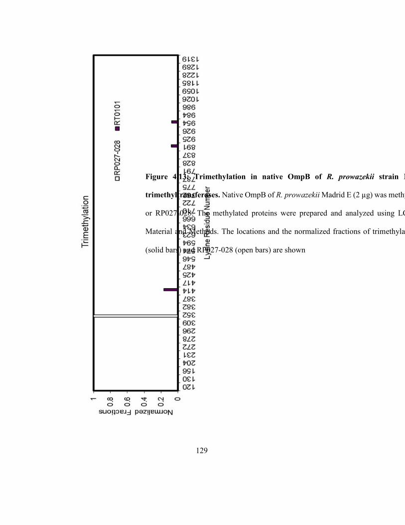
129
Figure 4.13: Trimethylation in native OmpB of R. prowazekii strain Madrid E catalyzed by
trimethyltransferases. Native OmpB of R. prowazekii Madrid E (2 µg) was methylated using 2 µM RT0101
or RP027-028. The methylated proteins were prepared and analyzed using LC-MS/MS as described in
Material and Methods. The locations and the normalized fractions of trimethylation catalyzed by RT0101
(solid bars) and RP027-028 (open bars) are shown
130
4.3: Discussion
The objective of this study was the in-depth analysis and comparison of methylation profiles
between recombinant OmpB catalyzed by four different methyltransferases and in native OmpB
purified directly from virulent and avirulent bacteria. The results showed that their profiles agree
closely in location and state of methylation.
The semiquantitative integrated liquid chromatography-tandem mass spectroscopy was used
to chracterize the methylation of recombinantly expressed fragments of R. typhi OmpB exposed in
vitro to trimethyltransferases of R. prowazekii RP027- 028 and of R. typhi RT0101, and to
monomethyltransferases of R. prowazekii RP789 and of R. typhi RT0776. Also the methylation
profiles of OmpBs purified from R. typhi, R. prowazekii strains Breinl, RP22 and Madrid E were
studied.
When the recombinant OmpB was methylated by RT0101, the highest number of peptide
spectral matches was for trimethyltion. The high proportion of trimethyllysine-containing PSM
clearly shows that RT0101 functions mainly as a trimethyltransferase. The additional mono- and
dimethylysines observed in small PSM numbers could possibly be the intermediates generated
during the trimethylation reaction which remain bound to the enzyme throughout the reaction. This
observation indicates that the trimethylation reaction likely proceeds via a processive instead of a
distributive mechanism.
Even though there was no single consensus amino acid sequence which is recognized by
RT0101, we were able to discover two sequence motifs for RT0101 catalyzed trimethylation,
KX(G/A/V/I)N and KT(I/L/F). This is an unusual but a novel feature for the rickettsial
131
methyltransferases in contrast to the already reported histone methyltransferases, which recognize
a single amino acid sequence for methylation.
In contrast to the methylation by RT0101, RT0776 predominantly catalyzed
monomethylation. In addition the methylation sites scattered across the OmpB sequence in contrast
to a few very specific sites reported with RT0101. This observation clearly indicates that RT0776
mainly functions as a monomethyltransferase which holds an active site that can accommodate a
wide variety of recognition sequences.
When recombinant R. typhi OmpB fragments were methylated using R. prowazekii
methyltransferase RP789, the highest number of PSM obtained was for monomethyllysine, whereas
when RP027-028 was used it was for trimethyllysine. These results show that RP027-028 and
RP789 function essentially as a trimethyltransferase and monomethyltransferase, respectively
similar to their R. typhi orthologs. BRP789 catalyzed significantly higher normalized fractions of
mono-, di- and trimethylations compared to RT0776. This could be due to the heterologous nature
of the system, in which RP789 and recombinant OmpB are from two different species, while
RT0776 and OmpB are from R. typhi, a homogenous system. The significant differences of the
products catalyzed by RT0776 and RP789 for the same substrate are unusual for enzymes sharing
high identity. This indicates that methyltransferases from a different species may not catalyze
exactly the way a methyltransferase from the same species does. But compared to both
trimethyltransferases, the monomethyltransferases’ catalysis is more pervasive throughout OmpB
suggesting that they both possess active sites which can accommodate a variety of recognition
sequences.
132
In addition to the methylation patterns, a significant difference in catalytic efficiencies
between the two was observed. This raised the questions, wheather this differences due to the non-
homologous nature of the enzymatic system (substrate and enzyme are from two different species)
or, is it because the two enzymes follow two different methylation mechanisms (processive vs.
distributive) leading to different reaction rates. But as reported above, when used the homologous
system with R.typhi OmpB substrate and R.typhi methyltransferases RT0101 and RT776 (the
orthologs of RP027-028 and RP789 respectively) similar pattern in reaction rates were observed.
Hence the possibility of non-homologous nature of the enzyme can be disregarded.
Therefore the most probable explanation for the differences in catalytic efficiencies is the different
mechanisms followed by the monomethyltransferases and trimethyltransferases.
The fact that the native OmpBs from virulent strains contain almost quantitative
trimethylation at specific lysine residues compared to the in vitro system suggests the existence of
a highly efficient cellular system in vivo through which those lysine residues in OmpB can be fully
trimethylated. Although there is not much known about the mechanism of in vivo trimethylation,
the in vitro system using the recombinant trimethyltransferase and OmpB can be used as model to
study the in vivo system. The observation that a high concentration of methyltransferase is required
to attain an appreciable level of methylated recombinant OmpB is contrary to the evidence
demonstrating the presence of a highly efficient in vivo trimethylation system. But the inhibition of
complete methylation could be due to the slow dissociation of the trimethylated lysine products
from the enzyme in vitro. There could be other cellular factors which may contribute to the complete
methylation in vivo. The presence of proteins which may interact with the methylated OmpB and
133
facilitate the release of the product from the enzyme and/or reduce the population of enzyme-
substrate complex which would shift the equilibrium toward complete methylation is a possibility.
In addition, the reduction of methylated OmpB-enzyme complex due to possible subcellular
translocation (e.g. cytoplasm, periplasmic space) can drive the methylation to completion. Another
reason for the efficient in vivo methylation may be the better accessibility of OmpB methylation
sites to the methyltransferases, that is, in vivo methylation could occur before or during folding of
the OmpB passenger domain where methylation sites could be more accessible than in the case of
the fully folded OmpB.
Native OmpBs from highly virulent R. prowazekii strains Breinl and RP22 contain multiple
clusters of trimethyllysine in contrast to a single cluster in OmpB from mildly virulent R. typhi.
Furthermore, OmpB from the avirulent strain Madrid E contains mostly monomethyllysine and no
trimethyllysine. These trimethyllysines may affect the protein – protein interactions in several ways;
namely, by increasing the hydrophobicity of the protein, by providing new docking sites and/or by
inhibiting other post translational modofications on the protein. A number of trimethyllysine-
binding domains have been found in recent years [Musselman et al., 2012]. For example interaction
between methylated H3K9 with HP1 facilitates the condensation of nucleosomes to heterochromatin
in gene silencing [Zentner et al., 2013]. If the presence of individual hypermethylated lysine cluster
can increase the valency of a protein and enhance the protein-protein interactions as described, then
the presence of multiple trimethyllysine clusters can even further enhance the affinity of the protein
to interact with other proteins.
134
The molecular basis of the rickettsial virulence is poorly understood. A study of what other
rickettsial and human proteins can interact with the OmpBs containing hypermethylated lysine
clusters will help to better understand the molecular links to virulence. But one drawback is the
unavailability of the three dimensional structure of OmpB, either from a crystallographic study or
from a molecular modeling analysis. Therefore how the methylation affects the overall 3-D structure
of OmpB is not clear and hence difficult to predict how it would interact with other putative
interacting proteins.
The native OmpB from Madrid E was minimally trimethylated by RT0101 or RP027-028.
This observation shows that the presence of already methylated lysine residues on native OmpB can
prevent the further methylation of these lysine residues to form trimethyllysine. This observation is
in agreement with the assumption that trimethylation by RT0101 and RP027-028 follow processive
mechanism where the reaction intermediates, mono- and dimethyllysines remain bound to the
enzyme.
4.4: Conclusion
Lysine methylation is one of the post translational modifications occurs in outer membrane
proteins that has been implicated in bacterial virulence and pathogenesis. This study was carried out
to investigate and to characterize the methylation profiles of recombinant rickettsial OmpB
catalyzed by four different methyltransferases and in native OmpB purified directly from virulent
and avirulent bacteria. The results showed that their profiles agree closely in location and state of
methylation. The recombinant OmpB methylation results lead to the conclusion that RT0776 and
135
RP789 catalyze primarily monomethylation and RT0101 and RP027-028 catalyze almost
exclusively trimethylation and hence characterized as monomethyltransferases and
trimethyltransferases respectively.
One interesting outcome from this study is the near complete trimethylation of specific
lysine residues found in native OmpB of virulent Rickettsia. This may be due to an efficient catalytic
system present in vivo for trimethylation of OmpB. The most studied methyltransferases, for
example histone methyltransferases, recognize and methylate a single lysine residue present in a
specific amino acid sequence. But Rickettsial methyltransferases can recognize and methylate a sets
of various amino acid sequences. The recognition motifs discovered in this study could be used to
predict lysines which can be trimethylated in other outer membrane proteins.
When a methyltransferase and OmpB from different rickettsial strains are used for the
reaction, the methylation patterns are changed. Also, when the N-terminal extensions present in
RP789 and RT0776 are deleted and used for methylation the methylation profiles produced are
significantly altered.
From these results it is clear that the virulence of the bacteria is correlated to the number of
highly trimethylated lysine clusters present in OmpBs of four strains of Rickettsia. The LC-MS/MS
results obtained for the trimethyltransfarase reactions suggest that RP027-028 and RT0101 catalyze
reaction via processive mechanism. The fact that the predominantly monomethylated native OmpB
from Madrid E could not be further methylated by recombinant trimethyltranferases suggests that
monomethylation have an antagonistic effect on trimethylation.
136
Even though this is the first study to report an in-depth characterization of methylation of
OMPs from Gram-negative bacteria, the details on how the methylation would affect the virulence
remain to be investigated. The new findings from this study will definitely help to link the OMP
methylation to bacterial virulence and also open up the possibility of understanding the mechanism
behind methylation and hence targeting methyltransferases [Daze et al., 2012, Wagner et al., 2012]
to find new therapeutic strategies against Rickettsia.
137
Chapter 5
Structural insight into substrate recognition and catalysis in the
methyltransferases RP789 and RT0101 in Rickittsia
5.1: Introduction
Protein methylation is a post translational modification which has been studied since 1970s
[Paik et al., 1971, Paik et al., 2007]. Regulatory potential of protein methylation is enhanced due to
the possibility that it could occur at ε-amino group of Lys to form mono-, di- and trimethyllysine,
or to the guanidinium group of Arg to produce monomethylarginine and symmetric and asymmetric
dimethylarginine. Methylation of Lys and Are in histones is well studied due to their roles in
epigenetics [Barski et al., 2007, Rea et al., 2000]. There are specific methyltransferases which target
Lys and Arg and more than forty such human methyltransferases have been found in the human
geneome . In histones, methylation regulates the transcription of associated genes [Martin et al.,
2005, Cheng et al., 2005]. According to the histone code hypothesis, methylation of histones at
promoter, coding sequence or enhancer of genes may affect the gene expression differently. More
recently, methylation of non-histone proteins has been found to regulate additional biological
processes through methylation of proteins such as p53 [Chuikov et al., 2004] and calmodulin
[Roberts et al., 1986]. Methylation of chemotaxis receptor regulates bacterial chemotaxis
[Djordjevic et al., 1997].
138
Rickettsia are obligate intracellular bacteria and are the causative agents of typhus and
spotted fever [Hackstadt, 1996]. There is a crystalline monolayer of protein arranged externally to
the outer membrane of Rickettsia [Silverman et al., 1978]. This layer consists of 10-15% of total
cellular protein and contains a diverse set of proteins, most predominantly OmpB [Dasch et al.,
1981]. OmpB provides the first line of communication with the extracellular environment and plays
roles in cell adhesion, attachment and invasion [Cardwell et al., 2009]. A previous amino acid
analysis has shown that OmpB contains methylated lysines and the amounts of trimethyllysine of
OmpB from several species correlate with the virulence of the species [Ching et al., 1993, Chao et
al., 2004]. The data from mass spectrometric analysis supports this correlation [Abeykoon et al.,
2014]
An in depth analysis carried out on methylation locations, states and levels of lysine residues
throughout OmpB purified from several Rickettsial species using semi-quantitative integrated LC-
MS/MS revealed that in addition to the trimethylated lysines, OmpB from highly virulent strains
contains multiple clusters of highly trimethylated lysine residues, while mildly virulent strain
contains one cluster of highly trimethylated lysines and OmpB from aviulent strain is devoid of such
clusters and contains instead monomethyllsine residues [Abeykoon et al., 2014].
Every methylation reaction requires a methyltransferase enzyme. First time identification
and characterization of rickettsial AdoMet-dependent methyltransferases that catalyze the
methylation of recombinant OmpB have been reported by our laboratory [Abeykoon et al., 2012].
Two main types of Rickettsial methyltrasferases have been identified. One type of OmpB
methyltransferases, RP789 from R. prowazekii and RT776 from R. typhi, functions primarily as
139
monomethyltransferase in vitro while another, RP027-028 from R. prowazekii and RT0101 from R.
typhi, functions exclusively as trimethyltransferase. Monomethyltransferases catalyze
monomethylation predominantly and di- and trimethylation at much lower levels. They catalyze
methylation of lysine residues throughout the OmpB suggesting the enzyme contain active sites
which can accommodate variety of recognition sequences. Trimethyltransferases catalyze primarily
trimethylation in vitro at relatively specific lysine residues at the same locations as those found in
OmpB purified from Rickettsia [Abeykoon et al., 2014]. Even though these methyltransferases
recognize multiple sequences, they are specific to OmpB but not histones or E.coli proteins. Results
from kinetic analysis and mass spectrometry studies suggest that the catalytic action of
trimethyltransferases is consistent with a processive mechanism. The monomethyltransferases of R.
typhi show 45% identity with the trimethyltransferase and an extension of 27 residues at the N-
terminus. As reported before, the catalysis is not effected by the N-terminal extension but the
product specificity was altered upon deletion of N-terminal domain. Using monomethyltransferase
from a heterologous species yields significantly different product specificity. Methylation by
rickettsial methyltransferases is quite unusual as they catalyzes the methylation of multiple lysine
residues in a protein in a manner which has not been reported before. In order to better understand
the structural bases of the unusual methylation characteristics, the three dimensional structures of
the mono- and trimethyltransferases are determined using protein X-ray crystallography. The
structures of monomethyltransferase, RP789, and trimethyltransferase, RT0101, and their respective
complexes with AdoMet are reported in this chapter.
140
5.2: Results
5.2.1: Overall structures of RP789 and RT0101
Crystallization of RP789 and RP027-028 from R. prowazekii and RT0776 and RT0101 from
R. typhi was performed as outlined in the Sections 2.2.17 and 2.2.18. The crystals were obtained for
RP789 and RT0101, where the initial hits were observed under several different conditions and later
optimized to obtain a single crystal (Figure 5.1 A). The first crystals were of native RP789 that
diffracted to a resolution of 2.6 Å in space group P21 (Figure 5.1 B, C and D). However, without a
close homolog, the structure could not be solved by molecular replacement. To solve the RP789
structure, selenomethionine substituted RP789 was prepared using B834(DE3) cells (methionine
auxotroph) (EMD Millipore) in SelenMet Media (Molecular Dimensions) supplemented with
selenomethionine (40 mg/L). Crystals of selenomethionine substituted RP789 were grown under the
same conditions as the wild type and diffracted to a resolution of 2.9 Å. Selenium single wavelength
anomalous dispersion (Se-SAD) phasing was then performed in PHENIX [Adams et al., 2002]
using AutoSol to calculate an initial experimental electron density map and an initial model built
within AutoBuild (Figure 5.1 D and E). The model was then used for molecular replacement using
the native dataset and the structure further refined within PHENIX and built with COOT [Emsley
et al., 2010]. The crystal structure of RT0101 was then determined by molecular replacement using
a homology model based on the RP789 structure built using CHAINSAW [Stein, 2008] within
CCP4 [CCP4, 1994]. The RP789 and RT0101 crystal structures were refined to a final resolution of
2.6 Å and 3.1 Å, respectively (Table 5.1).
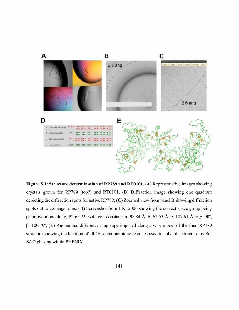
141
Figure 5.1: Structure determination of RP789 and RT0101. (A) Representative images showing
crystals grown for RP789 (top?) and RT0101; (B) Diffraction image showing one quadrant
depicting the diffraction spots for native RP789; (C) Zoomed view from panel B showing diffraction
spots out to 2.6 angstroms; (D) Screenshot from HKL2000 showing the correct space group being
primitive monoclinic, P2 or P21 with cell constants a=98.84 Å, b=62.53 Å, c=107.61 Å, ,=90º,
=100.79º; (E) Anomalous difference map superimposed along a wire model of the final RP789
structure showing the location of all 26 selenomethione residues used to solve the structure by Se-
SAD phasing within PHENIX.
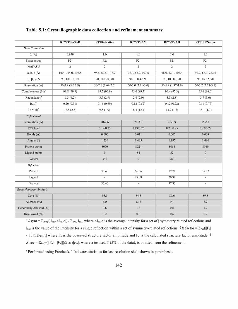
142
Table 5.1: Crystallographic data collection and refinement summary
RP789/Se-SAD RP789/Native RP789/SAM RP789/SAH RT0101/Native
Data Collection
λ (Å) 0.979 1.0 1.0 1.0 1.0
Space group P21 P21 P21 P21 P21
Mol/ASU 2 2 2 2 2
a, b, c (Å) 100.1, 65.0, 108.8 98.5, 62.5, 107.9 98.0, 62.9, 107.6 98.0, 62.1, 107.4 97.2, 66.9, 222.6
º 90, 101.18, 90 90, 100.78, 90 90, 100.42, 90 90, 100.88, 90 90, 89.82, 90
Resolution (Å) 50-2.9 (3.0 2.9) 50-2.6 (2.69-2.6) 50-3.0 (3.11-3.0) 50-1.9 (1.97-1.9) 50-3.2 (3.21-3.1)
Completeness (%)* 99.8 (99.9) 99.5 (96.9) 95.0 (89.7) 99.4 (97.3) 95.6 (96.0)
Redundancy* 6.3 (6.2) 3.7 (2.9) 2.4 (2.0) 3.3 (2.8) 3.7 (3.6)
Rsym†* 0.20 (0.91) 0.16 (0.69) 0.12 (0.52) 0.12 (0.72) 0.11 (0.77)
I / σ (I)* 12.5 (2.3) 9.5 (1.9) 8.4 (1.5) 13.9 (1.5) 15.1 (1.7)
Refinement
Resolution (Å) 20-2.6 20-3.0 20-1.9 15-3.1
R§/Rfree¶ 0.19/0.25 0.19/0.26 0.21/0.25 0.22/0.28
Bonds (Å) 0.006 0.011 0.007 0.008
Angles (º) 1.239 1.495 1.197 1.490
Protein atoms 8070 8024 8068 8160
Ligand atoms 0 54 52 0
Waters 340 0 782 0
B-factors
Protein 33.40 66.36 19.70 39.87
Ligand - 78.38 20.98 -
Waters 36.40 - 37.85 -
Ramachandran Analysis¥
Core (%) 93.1 84.3 89.6 89.8
Allowed (%) 6.0 13.8 9.1 8.2
Generously Allowed (%) 0.6 1.3 0.6 1.7
Disallowed (%) 0.2 0.6 0.6 0.2
† Rsym = hkl,j (|Ihkl-<Ihkl>|) / hkl,j Ihkl, where <Ihkl> is the average intensity for a set of j symmetry related reflections and
Ihkl is the value of the intensity for a single reflection within a set of symmetry-related reflections. § R factor = hkl(||Fo|
- |Fc||)/hkl|Fo| where Fo is the observed structure factor amplitude and Fc is the calculated structure factor amplitude. ¶
Rfree = hkl,T(||Fo| - |Fc||)/hkl,T|Fo|, where a test set, T (5% of the data), is omitted from the refinement.
¥ Performed using Procheck. * Indicates statistics for last resolution shell shown in parenthesis.
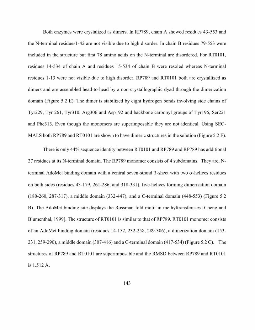
143
Both enzymes were crystalized as dimers. In RP789, chain A showed residues 43-553 and
the N-terminal residues1-42 are not visible due to high disorder. In chain B residues 79-553 were
included in the structure but first 78 amino acids on the N-terminal are disordered. For RT0101,
residues 14-534 of chain A and residues 15-534 of chain B were resoled whereas N-terminal
residues 1-13 were not visible due to high disorder. RP789 and RT0101 both are crystallized as
dimers and are assembled head-to-head by a non-crystallographic dyad through the dimerization
domain (Figure 5.2 E). The dimer is stabilized by eight hydrogen bonds involving side chains of
Tyr229, Tyr 261, Tyr310, Arg306 and Asp192 and backbone carbonyl groups of Tyr196, Ser221
and Phe313. Even though the monomers are superimposable they are not identical. Using SEC-
MALS both RP789 and RT0101 are shown to have dimeric structures in the solution (Figure 5.2 F).
There is only 44% sequence identity between RT0101 and RP789 and RP789 has additional
27 residues at its N-terminal domain. The RP789 monomer consists of 4 subdomains. They are, N-
terminal AdoMet binding domain with a central seven-strand -sheet with two -helices residues
on both sides (residues 43-179, 261-286, and 318-331), five-helices forming dimerization domain
(180-260, 287-317), a middle domain (332-447), and a C-terminal domain (448-553) (Figure 5.2
B). The AdoMet binding site displays the Rossman fold motif in methyltransferases [Cheng and
Blumenthal, 1999]. The structure of RT0101 is similar to that of RP789. RT0101 monomer consists
of an AdoMet binding domain (residues 14-152, 232-258, 289-306), a dimerization domain (153-
231, 259-290), a middle domain (307-416) and a C-terminal domain (417-534) (Figure 5.2 C). The
structures of RP789 and RT0101 are superimposable and the RMSD between RP789 and RT0101
is 1.512 Å.
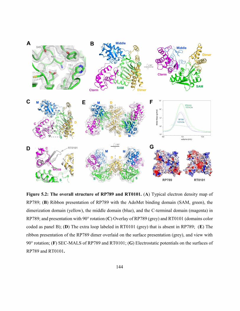
144
Figure 5.2: The overall structure of RP789 and RT0101. (A) Typical electron density map of
RP789; (B) Ribbon presentation of RP789 with the AdoMet binding domain (SAM, green), the
dimerization domain (yellow), the middle domain (blue), and the C-terminal domain (magenta) in
RP789; and presentation with 90° rotation (C) Overlay of RP789 (grey) and RT0101 (domains color
coded as panel B); (D) The extra loop labeled in RT0101 (grey) that is absent in RP789; (E) The
ribbon presentation of the RP789 dimer overlaid on the surface presentation (grey), and view with
90° rotation; (F) SEC-MALS of RP789 and RT0101; (G) Electrostatic potentials on the surfaces of
RP789 and RT0101.
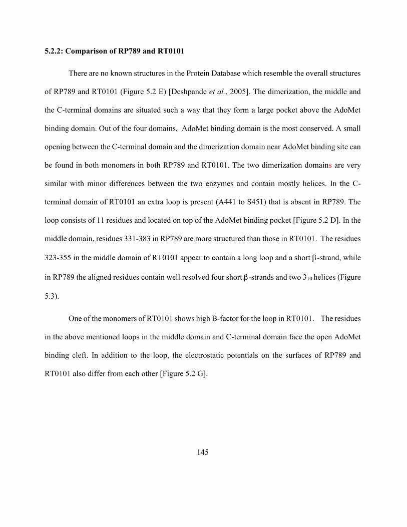
145
5.2.2: Comparison of RP789 and RT0101
There are no known structures in the Protein Database which resemble the overall structures
of RP789 and RT0101 (Figure 5.2 E) [Deshpande et al., 2005]. The dimerization, the middle and
the C-terminal domains are situated such a way that they form a large pocket above the AdoMet
binding domain. Out of the four domains, AdoMet binding domain is the most conserved. A small
opening between the C-terminal domain and the dimerization domain near AdoMet binding site can
be found in both monomers in both RP789 and RT0101. The two dimerization domains are very
similar with minor differences between the two enzymes and contain mostly helices. In the C-
terminal domain of RT0101 an extra loop is present (A441 to S451) that is absent in RP789. The
loop consists of 11 residues and located on top of the AdoMet binding pocket [Figure 5.2 D]. In the
middle domain, residues 331-383 in RP789 are more structured than those in RT0101. The residues
323-355 in the middle domain of RT0101 appear to contain a long loop and a short -strand, while
in RP789 the aligned residues contain well resolved four short -strands and two 310 helices (Figure
5.3).
One of the monomers of RT0101 shows high B-factor for the loop in RT0101. The residues
in the above mentioned loops in the middle domain and C-terminal domain face the open AdoMet
binding cleft. In addition to the loop, the electrostatic potentials on the surfaces of RP789 and
RT0101 also differ from each other [Figure 5.2 G].
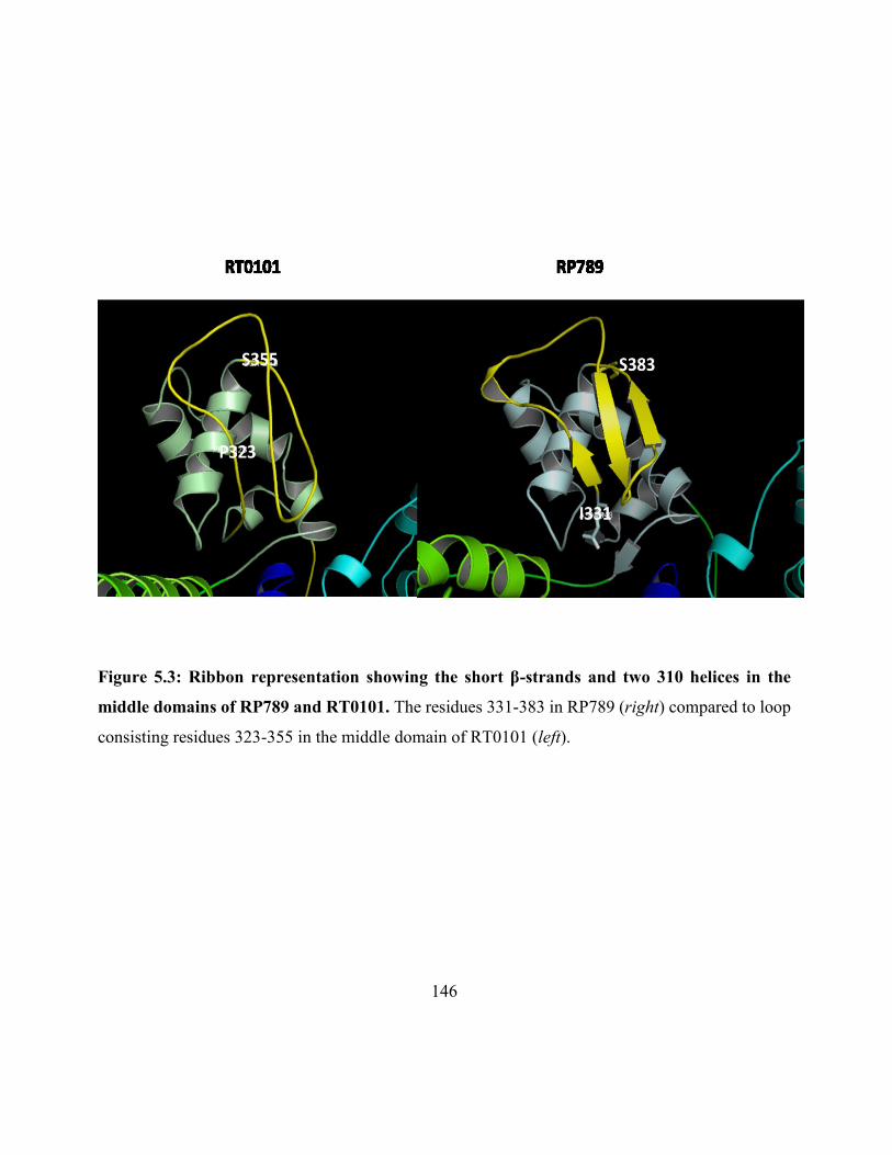
146
Figure 5.3: Ribbon representation showing the short β-strands and two 310 helices in the
middle domains of RP789 and RT0101. The residues 331-383 in RP789 (right) compared to loop
consisting residues 323-355 in the middle domain of RT0101 (left).
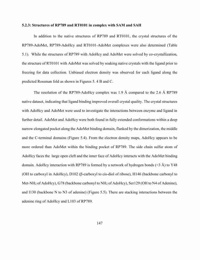
147
5.2.3: Structures of RP789 and RT0101 in complex with SAM and SAH
In addition to the native structures of RP789 and RT0101, the crystal structures of the
RP789-AdoMet, RP789-AdoHcy and RT0101-AdoMet complexes were also determined (Table
5.1). While the structures of RP789 with AdoHcy and AdoMet were solved by co-crystallization,
the structure of RT0101 with AdoMet was solved by soaking native crystals with the ligand prior to
freezing for data collection. Unbiased electron density was observed for each ligand along the
predicted Rossman fold as shown in Figures 5. 4 B and C.
The resolution of the RP789-AdoHcy complex was 1.9 Å compared to the 2.6 Å RP789
native dataset, indicating that ligand binding improved overall crystal quality. The crystal structures
with AdoHcy and AdoMet were used to investigate the interactions between enzyme and ligand in
further detail. AdoMet and AdoHcy were both found in fully extended conformations within a deep
narrow elongated pocket along the AdoMet binding domain, flanked by the dimerization, the middle
and the C-terminal domains (Figure 5.4). From the electron density maps, AdoHcy appears to be
more ordered than AdoMet within the binding pocket of RP789. The side chain sulfur atom of
AdoHcy faces the large open cleft and the inner face of AdoHcy interacts with the AdoMet binding
domain. AdoHcy interaction with RP789 is formed by a network of hydrogen bonds (<3 Å) to Y48
(OH to carboxyl in AdoHcy), D102 (β-carboxyl to cis-diol of ribose), H146 (backbone carbonyl to
Met-NH2 of AdoHcy), G78 (backbone carbonyl to NH2 of AdoHcy), Ser129 (OH to N4 of Adenine),
and I130 (backbone N to N3 of adenine) (Figure 5.5). There are stacking interactions between the
adenine ring of AdoHcy and L103 of RP789.
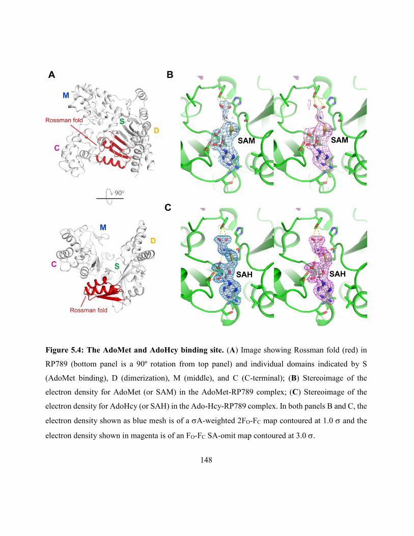
148
Figure 5.4: The AdoMet and AdoHcy binding site. (A) Image showing Rossman fold (red) in
RP789 (bottom panel is a 90º rotation from top panel) and individual domains indicated by S
(AdoMet binding), D (dimerization), M (middle), and C (C-terminal); (B) Stereoimage of the
electron density for AdoMet (or SAM) in the AdoMet-RP789 complex; (C) Stereoimage of the
electron density for AdoHcy (or SAH) in the Ado-Hcy-RP789 complex. In both panels B and C, the
electron density shown as blue mesh is of a A-weighted 2FO-FC map contoured at 1.0 and the
electron density shown in magenta is of an FO-FC SA-omit map contoured at 3.0 .
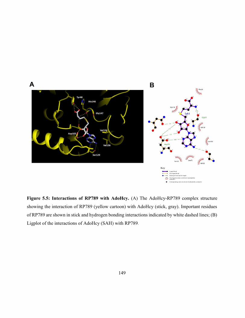
149
Figure 5.5: Interactions of RP789 with AdoHcy. (A) The AdoHcy-RP789 complex structure
showing the interaction of RP789 (yellow cartoon) with AdoHcy (stick, gray). Important residues
of RP789 are shown in stick and hydrogen bonding interactions indicated by white dashed lines; (B)
Ligplot of the interactions of AdoHcy (SAH) with RP789.
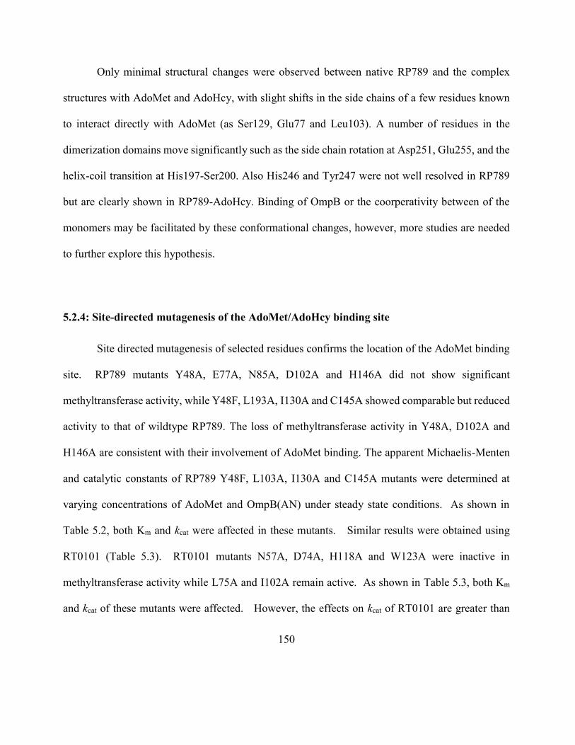
150
Only minimal structural changes were observed between native RP789 and the complex
structures with AdoMet and AdoHcy, with slight shifts in the side chains of a few residues known
to interact directly with AdoMet (as Ser129, Glu77 and Leu103). A number of residues in the
dimerization domains move significantly such as the side chain rotation at Asp251, Glu255, and the
helix-coil transition at His197-Ser200. Also His246 and Tyr247 were not well resolved in RP789
but are clearly shown in RP789-AdoHcy. Binding of OmpB or the coorperativity between of the
monomers may be facilitated by these conformational changes, however, more studies are needed
to further explore this hypothesis.
5.2.4: Site-directed mutagenesis of the AdoMet/AdoHcy binding site
Site directed mutagenesis of selected residues confirms the location of the AdoMet binding
site. RP789 mutants Y48A, E77A, N85A, D102A and H146A did not show significant
methyltransferase activity, while Y48F, L193A, I130A and C145A showed comparable but reduced
activity to that of wildtype RP789. The loss of methyltransferase activity in Y48A, D102A and
H146A are consistent with their involvement of AdoMet binding. The apparent Michaelis-Menten
and catalytic constants of RP789 Y48F, L103A, I130A and C145A mutants were determined at
varying concentrations of AdoMet and OmpB(AN) under steady state conditions. As shown in
Table 5.2, both Km and kcat were affected in these mutants. Similar results were obtained using
RT0101 (Table 5.3). RT0101 mutants N57A, D74A, H118A and W123A were inactive in
methyltransferase activity while L75A and I102A remain active. As shown in Table 5.3, both Km
and kcat of these mutants were affected. However, the effects on kcat of RT0101 are greater than
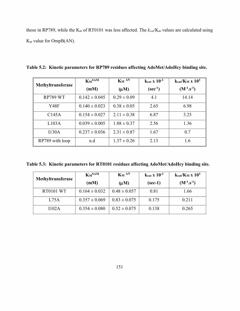
151
those in RP789, while the Km of RT0101 was less affected. The kcat/Km values are calculated using
Km value for OmpB(AN).
Table 5.2: Kinetic parameters for RP789 residues affecting AdoMet/AdoHcy binding site.
Methyltransferase KM
SAM
(mM)
KM AN
(M)
kcat x 10-3
(sec-1)
kcat/KM x 103
(M-1.s-1)
RP789 WT 0.142 ± 0.045 0.29 ± 0.09 4.1 14.14
Y48F 0.140 ± 0.023 0.38 ± 0.05 2.65 6.98
C145A 0.154 ± 0.027 2.11 ± 0.38 6.87 3.25
L103A 0.039 ± 0.005 1.88 ± 0.37 2.56 1.36
I130A 0.237 ± 0.036 2.31 ± 0.87 1.67 0.7
RP789 with loop n.d 1.37 ± 0.26 2.13 1.6
Table 5.3: Kinetic parameters for RT0101 residues affecting AdoMet/AdoHcy binding site.
Methyltransferase KM
SAM
(mM)
KM AN
(M)
kcat x 10-3
(sec-1)
kcat/KM x 103
(M-1.s-1)
RT0101 WT 0.164 ± 0.032 0.48 ± 0.057 0.81 1.66
L75A 0.357 ± 0.069 0.83 ± 0.075 0.175 0.211
I102A 0.354 ± 0.080 0.52 ± 0.075 0.138 0.265
152
5.2.5: Site-directed mutagenesis of the extra loop in RT0101
RP789 functions primarily as a monomethyltransferase but is also capable of producing low
levels of di- and trimethyllysines. RT0101 functions almost exclusively as a trimethyltransferase
via a processive mechanism [Abeykoon et al., 2014]. The catalytic efficiency by RP789 in terms
of kcat/Km is appreciably higher than that of RT0101. One distinct difference between three
dimensional structures of RP789 and RT010 is the presence of an additional small loop in RT0101
that projects toward the large open pocket. A small insertion in RT0101 amino acid sequence
(N444-M449) accounts for the addition of the loop. The function of the loop is explored using site-
directed mutagenesis by deletion of the loop from RT0101 and by insertion of the loop to RP789.
As shown in Figure 5.6, deletion of the loop (N444 to M449) from RT0101 completely inactivated
its methyltransferase activity while insertion of the loop to RP789 has almost no effect on the
methyltransferase activity as analyzed using the radioactivity assay of methyltransferase activity.
The LC-MS/MS analysis carried out on recombinant OmpB methylated by RP789 mutant showed
a methylation profile similar to its wild type with predominant monomethylation (Figure 5.7). Hence
insertion of the loop to RP789 did not cause a significant alteration in its product specificity and did
not convert RP789 to a trimethyltransferase as RT0101.
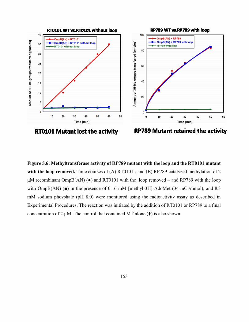
153
Figure 5.6: Methyltransferase activity of RP789 mutant with the loop and the RT0101 mutant
with the loop removed. Time courses of (A) RT0101-, and (B) RP789-catalyzed methylation of 2
μM recombinant OmpB(AN) (●) and RT0101 with the loop removed – and RP789 with the loop
with OmpB(AN) (■) in the presence of 0.16 mM [methyl-3H]-AdoMet (34 mCi/mmol), and 8.3
mM sodium phosphate (pH 8.0) were monitored using the radioactivity assay as described in
Experimental Procedures. The reaction was initiated by the addition of RT0101 or RP789 to a final
concentration of 2 µM. The control that contained MT alone (⧫) is also shown.
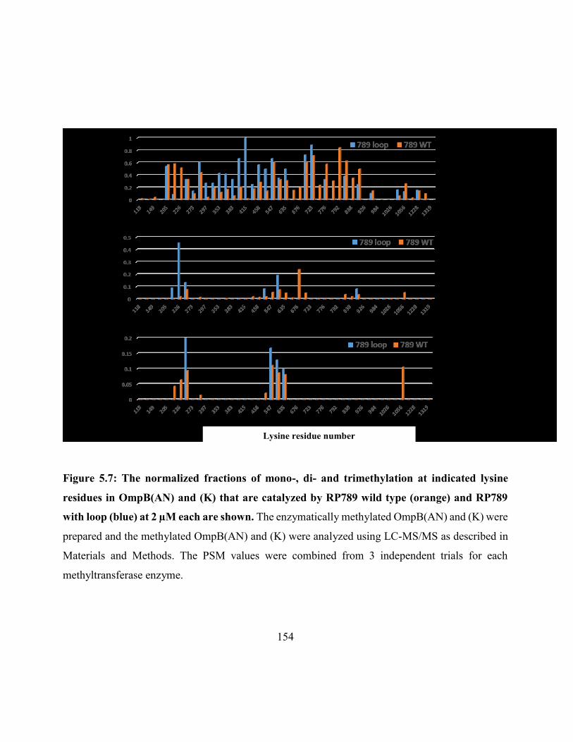
154
Figure 5.7: The normalized fractions of mono-, di- and trimethylation at indicated lysine
residues in OmpB(AN) and (K) that are catalyzed by RP789 wild type (orange) and RP789
with loop (blue) at 2 µM each are shown. The enzymatically methylated OmpB(AN) and (K) were
prepared and the methylated OmpB(AN) and (K) were analyzed using LC-MS/MS as described in
Materials and Methods. The PSM values were combined from 3 independent trials for each
methyltransferase enzyme.
Lysine residue number
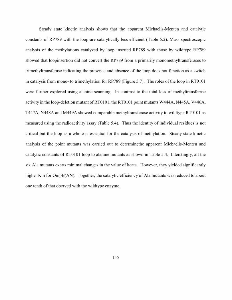
155
Steady state kinetic analysis shows that the apparent Michaelis-Menten and catalytic
constants of RP789 with the loop are catalytically less efficient (Table 5.2). Mass spectroscopic
analysis of the methylations catalyzed by loop inserted RP789 with those by wildtype RP789
showed that loopinsertion did not convert the RP789 from a primarily monomethyltransferases to
trimethyltransferase indicating the presence and absence of the loop does not function as a switch
in catalysis from mono- to trimethylation for RP789 (Figure 5.7). The roles of the loop in RT0101
were further explored using alanine scanning. In contrast to the total loss of methyltransferase
activity in the loop-deletion mutant of RT0101, the RT0101 point mutants W444A, N445A, V446A,
T447A, N448A and M449A showed comparable methyltransferase activity to wildtype RT0101 as
measured using the radioactivity assay (Table 5.4). Thus the identity of individual residues is not
critical but the loop as a whole is essential for the catalysis of methylation. Steady state kinetic
analysis of the point mutants was carried out to determinethe apparent Michaelis-Menten and
catalytic constants of RT0101 loop to alanine mutants as shown in Table 5.4. Interstingly, all the
six Ala mutants exerts minimal changes in the value of kcata. However, they yielded significantly
higher Km for OmpB(AN). Together, the catalytic efficiency of Ala mutants was reduced to about
one tenth of that oberved with the wildtype enzyme.
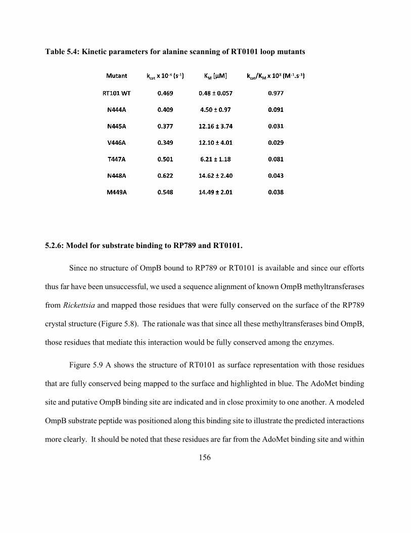
156
Table 5.4: Kinetic parameters for alanine scanning of RT0101 loop mutants
5.2.6: Model for substrate binding to RP789 and RT0101.
Since no structure of OmpB bound to RP789 or RT0101 is available and since our efforts
thus far have been unsuccessful, we used a sequence alignment of known OmpB methyltransferases
from Rickettsia and mapped those residues that were fully conserved on the surface of the RP789
crystal structure (Figure 5.8). The rationale was that since all these methyltransferases bind OmpB,
those residues that mediate this interaction would be fully conserved among the enzymes.
Figure 5.9 A shows the structure of RT0101 as surface representation with those residues
that are fully conserved being mapped to the surface and highlighted in blue. The AdoMet binding
site and putative OmpB binding site are indicated and in close proximity to one another. A modeled
OmpB substrate peptide was positioned along this binding site to illustrate the predicted interactions
more clearly. It should be noted that these residues are far from the AdoMet binding site and within
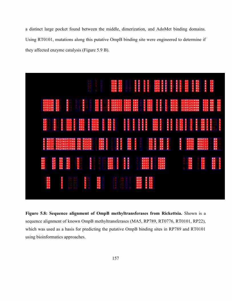
157
a distinct large pocket found between the middle, dimerization, and AdoMet binding domains.
Using RT0101, mutations along this putative OmpB binding site were engineered to determine if
they affected enzyme catalysis (Figure 5.9 B).
Figure 5.8: Sequence alignment of OmpB methyltransferases from Rickettsia. Shown is a
sequence alignment of known OmpB methyltransferases (MA5, RP789, RT0776, RT0101, RP22),
which was used as a basis for predicting the putative OmpB binding sites in RP789 and RT0101
using bioinformatics approaches.
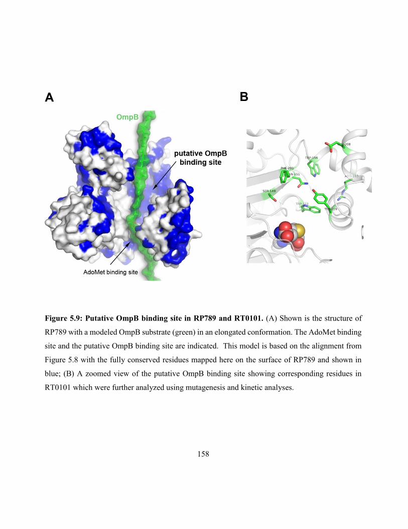
158
Figure 5.9: Putative OmpB binding site in RP789 and RT0101. (A) Shown is the structure of
RP789 with a modeled OmpB substrate (green) in an elongated conformation. The AdoMet binding
site and the putative OmpB binding site are indicated. This model is based on the alignment from
Figure 5.8 with the fully conserved residues mapped here on the surface of RP789 and shown in
blue; (B) A zoomed view of the putative OmpB binding site showing corresponding residues in
RT0101 which were further analyzed using mutagenesis and kinetic analyses.
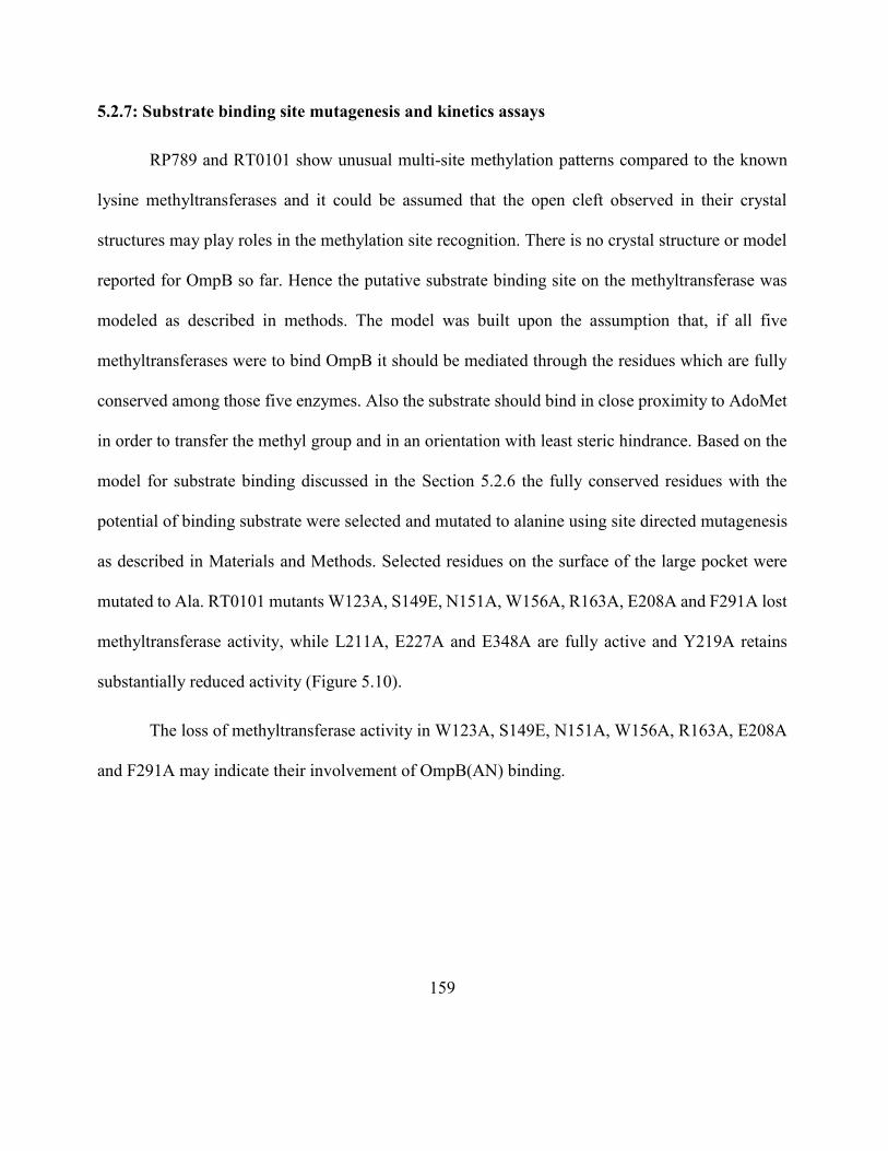
159
5.2.7: Substrate binding site mutagenesis and kinetics assays
RP789 and RT0101 show unusual multi-site methylation patterns compared to the known
lysine methyltransferases and it could be assumed that the open cleft observed in their crystal
structures may play roles in the methylation site recognition. There is no crystal structure or model
reported for OmpB so far. Hence the putative substrate binding site on the methyltransferase was
modeled as described in methods. The model was built upon the assumption that, if all five
methyltransferases were to bind OmpB it should be mediated through the residues which are fully
conserved among those five enzymes. Also the substrate should bind in close proximity to AdoMet
in order to transfer the methyl group and in an orientation with least steric hindrance. Based on the
model for substrate binding discussed in the Section 5.2.6 the fully conserved residues with the
potential of binding substrate were selected and mutated to alanine using site directed mutagenesis
as described in Materials and Methods. Selected residues on the surface of the large pocket were
mutated to Ala. RT0101 mutants W123A, S149E, N151A, W156A, R163A, E208A and F291A lost
methyltransferase activity, while L211A, E227A and E348A are fully active and Y219A retains
substantially reduced activity (Figure 5.10).
The loss of methyltransferase activity in W123A, S149E, N151A, W156A, R163A, E208A
and F291A may indicate their involvement of OmpB(AN) binding.
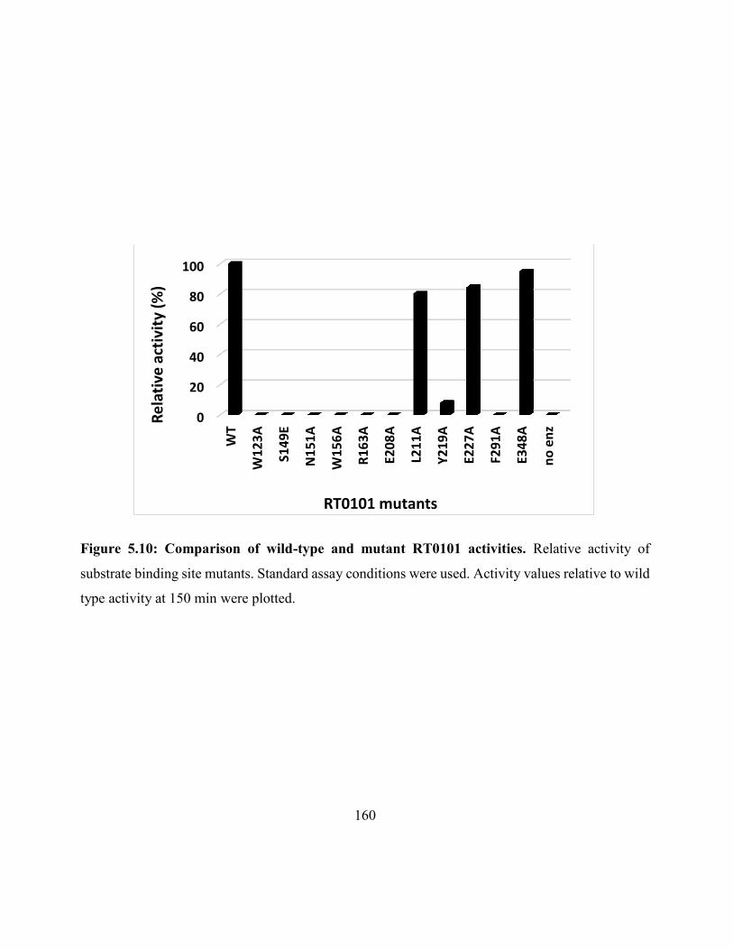
160
Figure 5.10: Comparison of wild-type and mutant RT0101 activities. Relative activity of
substrate binding site mutants. Standard assay conditions were used. Activity values relative to wild
type activity at 150 min were plotted.
0
20
40
60
80
100
WT
W12
3A
S14
9E
N1
51A
W15
6A
R1
63A
E20
8A
L21
1A
Y2
19A
E22
7A
F291
A
E34
8A
no
en
z
Re
lati
ve a
ctiv
ity
(%)
RT0101 mutants
161
5.3: Discussion
Unlike typical methyltransferases, RP789 and RT0101 catalyze methylation at multiple sites
with various sequences, which is an unusual property among known lysine methyltransferases.
There are many similarities and unique features among methyltransferase as reveled by RP789 and
RT0101 structures. The methyltransferases are crystallized as dimers. Each monomer consists of an
AdoMet binding domain, a dimerization domain, and two additional C-terminal domains. The
assemblies of the four structural domains in RP789 and RT0101 have not been found in any other
proteins. This is the first study to provide the crystal structure of protein lysine methyltransferases
that catalyze methylation at multiple sites of a protein and reveals the structural features of the multi-
site mono- and trimethyltransferases.
There are two other biochemically characterized rickettsial methyltransferases which belong
to monomethyltransferase and trimethyltransferase classes. RT776 and RP027-028 have 93% and
93% identity to RP789 and RT0101, respectively. The attempts to produce the crystal structures of
RT0776 and RP027-028 by homology modeling, has generated nearly identical three dimensional
structures to those of RP789 and RT0101, respectively.
Between the dimerization domain, middle domain and the C-terminal domain there is a wide
open pocket and the Rossman fold, the motif which AdoMet binds, is located at the bottom of this
pocket. This is common for both enzymes. This open pocket should be able to house the bulky
OmpB substrate with the least steric hindrance and may accommodate different sequences with -
amino group of lysine residues in sufficient proximity to the methyl group of bound AdoMet. The
ability of both methyltransferases in catalyzing multiple lysine residues at multiple methylation
162
levels may be explained by the presence of the large pocket and the ready accessibility of AdoMet
when bound in the wide open cleft. This hypothesis could also support the results obtained by site
directed mutagenesis. Trp, Arg and Tyr residues are frequently found to be hotspots in protein-
protein interactions [Bogan et al., 1998]. In RT0101, W123, W156, R163 and Y219 are located on
the surface of the large open cleft and their side chains are accessible to solvent. Mutations of either
of W123 and W156 to Ala in RT0101 inactivated the methyltransferase. Similarly, mutations of
R163 and Y219 to Ala eliminated the methyltransferase activity of RT0101. Additional mutations
including S149, N151, E208 and F291 to Ala also inactivated the methyltransferase activity. The
fact that these residues have their side chains exposed to the open cleft and located far from the
AdoMet binding site suggest that they may be involved in OmpB binding. In contrast, mutations of
L211, E227 and E348 did not appreciably affect the methyltransferase activity. There is currently
no crystal structure reported for OmpB and our limited attempts to co-crystallize the
methyltransferase with a 7 amino acid peptide (ANTKTLG – OH) containing one of the methylation
recognition motifs from OmpB has been unsuccessful so far. Our work has shown that the
methyltransferases do not catalyze methylation of these smaller OmpB peptides even at high
concentrations (1 mM), however, the peptides were able to inhibit the methylation of OmpB in our
assays.
One major structural difference between RP789 and RT0101 is the presence of a loop of
N444 to M449 in RT0101 that projects into the open cleft at the C-terminal domain. Whether the
loop plays a role in product specificity in a manner similar to the Phe/Tyr switch [Couture et al.,
2008] or not is yet to be determined. Deletion of the loop inactivates the methyltransferase activity
of RT0101. The observed differences in the apparent Michaelis-Menten constants and catalytic
163
constants between RP789 and its loop insertion mutant shown in Table 5.2 appear to be inconsistent
with the identical time courses shown in Figure 5.6. One plausible interpretation is that the
differences are due to the high concentrations of RP789 and OmpB(AN) at 2 M each for the
experiment in Figure 5.6 but 0.26 M and varying and low concentrations of OmpB(AN) for those
in Table 5.2. The time courses in both cases represent the weighed sum of over thirty methylation
reactions with various Michaelis-Menten and catalytic constants. At high enzyme and substrate
concentrations, the slow methylation reactions will be weighed high but not at low enzyme and
substrate concentrations. Thus the differences are minimized at high concentrations under the
conditions used in Figure 5.6. The data in Table5.4 indicate that Ala scanning of RT1010 loop will
have no effect on kcat but reduce its affinity toward AN, as indicated with increasing Km values. The
reciprocal experiment by inserting the loop to RP789 did not inactivate its methyltransferase
activity. However, RP789 with the inserted loop did not catalyze primarily trimethylation in the
manner as RT0101. These results reveal that the loop alone is insufficient to convert RP789 to a
trimethyltransferase. Since the sequence identity between RP789 and RT0101 is 45% (Figure 5.11),
residues other than the loop likely plays roles in mediating the product specificity of
methyltransferases.
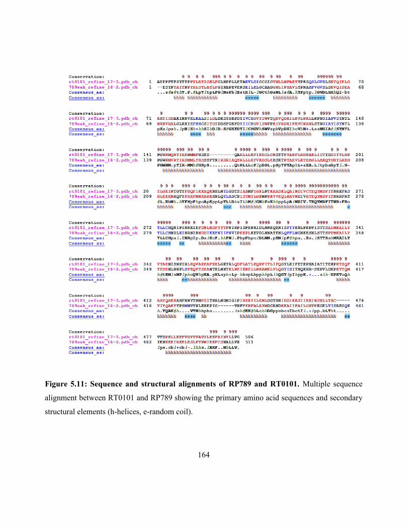
164
Figure 5.11: Sequence and structural alignments of RP789 and RT0101. Multiple sequence
alignment between RT0101 and RP789 showing the primary amino acid sequences and secondary
structural elements (h-helices, e-random coil).
165
In solution, both RP789 and RT0101 exist as dimers. The interface between the two
monomers consists of mainly two helices from each monomer and Tyr196. Tyr229, Tyr261, and
Ser221 in one monomer are within H-bond distances to Arg306, Phe313, Arg192, and Tyr310,
respectively, in the other monomer. Given the size of the OmpB substrate and the observed
methylation pattern at multiple lysine residues, it could be speculated that these methyltransferases
could methylate two Lys residues at the same time in the same OmpB molecule by the two active
sites of the dimeric enzyme. Another possibility is that it could provide the structure for carrying
out methylation at multiple Lys residues processively within the OmpB.
Between the native methyltransferase and the complex with AdoMet, there is a small blue
shift and increase of fluorescence intensity upon binding AdoMet. This suggests that AdoMet
induces a protein conformational change in such a way that the environment of Trp residues become
more shielded from solvent when the ligand is bound [Vivian et al., 2001].
Due to the unavailability of the three dimensional structure of OmpB, molecular modeling
is used to predict the structure using I-Tasser [Roy et al., 2010]. According to the model, OmpB
contains mostly loops and β-helices. The secondary structure prediction of OmpB using PSIPRED
[Buchan et al., 2013] is consistent with the presence of β-helices. Interestingly, we have previously
revaled the presence of highly trimethylated lysine clusters on OmpB [Abeykoon et al., 2014]. Our
model shows that these clusters are located mostly in the loops instead of β-helices. Furthermore,
the openness of the pocket in RT0101 appears to facilitate OmpB docking base on our GRAMM-X
[Tovchigrechko et al., 2006] analysis.
166
In an attempt to find evolutionarily conserved sequences in RP789 and RT0101 using the
online server Consurf [Ashkenazy et al., 2010] it revealed that most highly conserved residues are
located in the AdoMet binding domain at the N-terminal half of RP789 and RT0101. There are
many AdoMet binding proteins reported in literature and AdoMet binding domain of RT0101 and
RP789 showed homology to many known three dimensional structures when analyzed using
VAST+ [Madej et al., 2014].
5.4: Conclusion
Highly virulent strains of Rickettsia contain OmpB with clusters of highly trimethylated
lysine residues, while OmpB from avirulent strain contains mainly monomethyllysine but not
trimethyllysine. Two families of lysine methyltransferases which catalyze the OmpB methylation
have been identified and characterized. One type (RP789, RT0776) catalyzes primarily
monomethylation at a majority of lysine residues throughout OmpB, while another (RP027-028,
RT0101) catalyzes essentially trimethylation at relatively specific lysine residues. The unusual
feature of this methylation is, unlike the typical methytransferases, the ability of methylating at
multiple sites with varying sequences at the methylation sites.
Using protein X-ray crystallography, we have determined the three dimensional structures
of RP789 and RT0101 and their respective complexes with AdoMet and AdoHcy. Both RP789 and
RT0101 exist in solution as dimers as demonstrated by SEC-MALS. Each monomer consists of four
subdomains; an AdoMet binding domain, a dimerization domain, a middle domain and a C-terminal
domain.
167
There are no known structures in the Protein Database which resemble the overall structures
of RP789 and RT0101. The AdoMet binding domain contains seven -strands and conforms to the
Rossman fold in methyltransferases. The dimerization, the middle and the C-terminal domains are
situated such a way that they form a deep open pocket along the AdoMet binding domain, which is
the most conserved of all domains.
AdoMet and AdoHcy were found in an extended conformation within the binding pocket.
The side chain sulfur atom of AdoHcy faces the open cleft and the inner face of AdoHcy interacts
with the AdoMet binding domain. Site directed mutagenesis of selected amino acid residues within
the AdoMet binding pocket followed by steady state kinetic analysis confirms the AdoMet binding
site. Further, mutagenesis and kinetics analysis also support the identification of the putative OmpB
binding pocket located in proximity of the AdoMet binding site.
One distinct difference between three dimensional structures of RP789 and RT010 is the
presence of an additional small loop in RT0101 that projects toward the large open pocket. Deletion
of the loop (N444 to M449) from RT0101 completely inactivated its methyltransferase activity
while insertion of the loop to RP789 has little effect on the methyltransferase activity. Mutation of
individual residues on the RT0101 loop showed reduction of substrate binding.
While the AdoMet binding domain is well conserved, the overall architectures of RP789 and
RT0101 are unique among known structures of methyltransferases with respect to the additional
domains that appear to help mediate substrate interactions and possibly allosteric cooperativity.
Still, given the similarity between the two methyltransferases, it is intriguing that each retains broad
specificity while catalyzing the generation of very specific and distinct products in their respective

168
methylation reactions. The crystal structures and mutation studies presented here have given
valuable molecular insight into this family of enzymes and has provided the necessary foundation
to unravel the mechanism for the methylation of OmpB by the rickettsial methyltransferases.
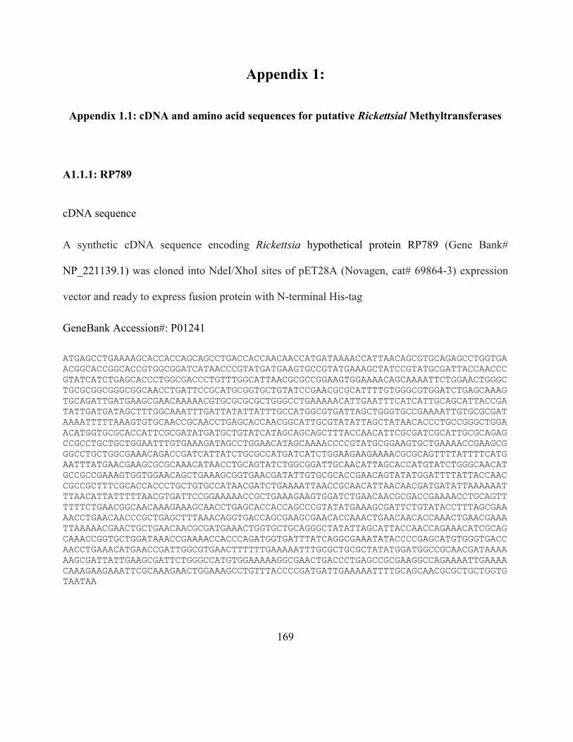
169
Appendix 1:
Appendix 1.1: cDNA and amino acid sequences for putative Rickettsial Methyltransferases
A1.1.1: RP789
cDNA sequence
A synthetic cDNA sequence encoding Rickettsia hypothetical protein RP789 (Gene Bank#
NP_221139.1) was cloned into NdeI/XhoI sites of pET28A (Novagen, cat# 69864-3) expression
vector and ready to express fusion protein with N-terminal His-tag
GeneBank Accession#: P01241
ATGAGCCTGAAAAGCACCACCAGCAGCCTGACCACCAACAACCATGATAAAACCATTAACAGCGTGCAGAGCCTGGTGA
ACGGCACCGGCACCGTGGCGGATCATAACCCGTATGATGAAGTGCCGTATGAAAGCTATCCGTATGCGATTACCAACCC
GTATCATCTGAGCACCCTGGCGACCCTGTTTGGCATTAACGCGCCGGAAGTGGAAAACAGCAAAATTCTGGAACTGGGC
TGCGCGGCGGGCGGCAACCTGATTCCGCATGCGGTGCTGTATCCGAACGCGCATTTTGTGGGCGTGGATCTGAGCAAAG
TGCAGATTGATGAAGCGAACAAAAACGTGCGCGCGCTGGGCCTGAAAAACATTGAATTTCATCATTGCAGCATTACCGA
TATTGATGATAGCTTTGGCAAATTTGATTATATTATTTGCCATGGCGTGATTAGCTGGGTGCCGAAAATTGTGCGCGAT
AAAATTTTTAAAGTGTGCAACCGCAACCTGAGCACCAACGGCATTGCGTATATTAGCTATAACACCCTGCCGGGCTGGA
ACATGGTGCGCACCATTCGCGATATGATGCTGTATCATAGCAGCAGCTTTACCAACATTCGCGATCGCATTGCGCAGAG
CCGCCTGCTGCTGGAATTTGTGAAAGATAGCCTGGAACATAGCAAAACCCCGTATGCGGAAGTGCTGAAAACCGAAGCG
GGCCTGCTGGCGAAACAGACCGATCATTATCTGCGCCATGATCATCTGGAAGAAGAAAACGCGCAGTTTTATTTTCATG
AATTTATGAACGAAGCGCGCAAACATAACCTGCAGTATCTGGCGGATTGCAACATTAGCACCATGTATCTGGGCAACAT
GCCGCCGAAAGTGGTGGAACAGCTGAAAGCGGTGAACGATATTGTGCGCACCGAACAGTATATGGATTTTATTACCAAC
CGCCGCTTTCGCACCACCCTGCTGTGCCATAACGATCTGAAAATTAACCGCAACATTAACAACGATGATATTAAAAAAT
TTAACATTATTTTTAACGTGATTCCGGAAAAACCGCTGAAAGAAGTGGATCTGAACAACGCGACCGAAAACCTGCAGTT
TTTTCTGAACGGCAACAAAGAAAGCAACCTGAGCACCACCAGCCCGTATATGAAAGCGATTCTGTATACCTTTAGCGAA
AACCTGAACAACCCGCTGAGCTTTAAACAGGTGACCAGCGAAGCGAACACCAAACTGAACAACACCAAACTGAACGAAA
TTAAAAACGAACTGCTGAACAACGCGATGAAACTGGTGCTGCAGGGCTATATTAGCATTACCAACCAGAAACATCGCAG
CAAACCGGTGCTGGATAAACCGAAAACCACCCAGATGGTGATTTATCAGGCGAAATATACCCCGAGCATGTGGGTGACC
AACCTGAAACATGAACCGATTGGCGTGAACTTTTTTGAAAAATTTGCGCTGCGCTATATGGATGGCCGCAACGATAAAA
AAGCGATTATTGAAGCGATTCTGGGCCATGTGGAAAAAGGCGAACTGACCCTGAGCCGCGAAGGCCAGAAAATTGAAAA
CAAAGAAGAAATTCGCAAAGAACTGGAAAGCCTGTTTACCCCGATGATTGAAAAATTTTGCAGCAACGCGCTGCTGGTG
TAATAA
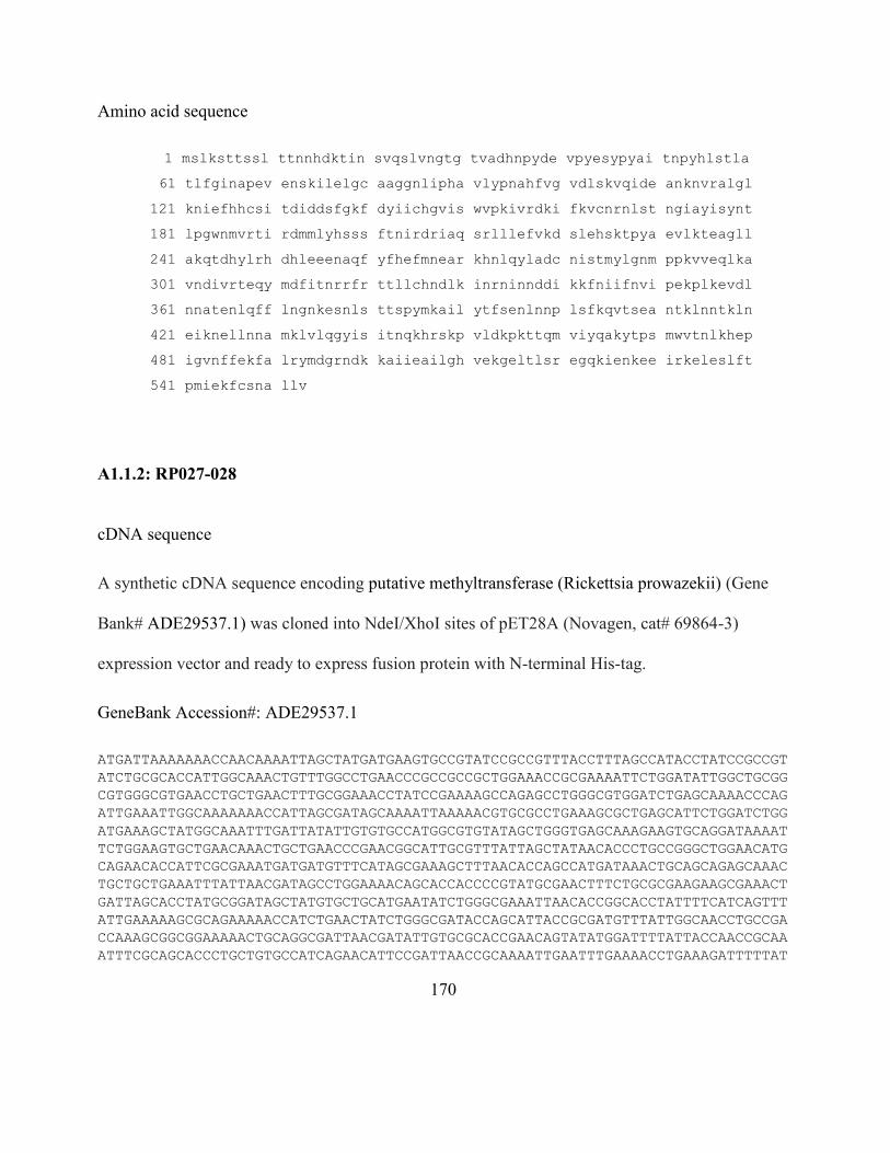
170
Amino acid sequence
1 mslksttssl ttnnhdktin svqslvngtg tvadhnpyde vpyesypyai tnpyhlstla
61 tlfginapev enskilelgc aaggnlipha vlypnahfvg vdlskvqide anknvralgl
121 kniefhhcsi tdiddsfgkf dyiichgvis wvpkivrdki fkvcnrnlst ngiayisynt
181 lpgwnmvrti rdmmlyhsss ftnirdriaq srlllefvkd slehsktpya evlkteagll
241 akqtdhylrh dhleeenaqf yfhefmnear khnlqyladc nistmylgnm ppkvveqlka
301 vndivrteqy mdfitnrrfr ttllchndlk inrninnddi kkfniifnvi pekplkevdl
361 nnatenlqff lngnkesnls ttspymkail ytfsenlnnp lsfkqvtsea ntklnntkln
421 eiknellnna mklvlqgyis itnqkhrskp vldkpkttqm viyqakytps mwvtnlkhep
481 igvnffekfa lrymdgrndk kaiieailgh vekgeltlsr egqkienkee irkeleslft
541 pmiekfcsna llv
A1.1.2: RP027-028
cDNA sequence
A synthetic cDNA sequence encoding putative methyltransferase (Rickettsia prowazekii) (Gene
Bank# ADE29537.1) was cloned into NdeI/XhoI sites of pET28A (Novagen, cat# 69864-3)
expression vector and ready to express fusion protein with N-terminal His-tag.
GeneBank Accession#: ADE29537.1
ATGATTAAAAAAACCAACAAAATTAGCTATGATGAAGTGCCGTATCCGCCGTTTACCTTTAGCCATACCTATCCGCCGT
ATCTGCGCACCATTGGCAAACTGTTTGGCCTGAACCCGCCGCCGCTGGAAACCGCGAAAATTCTGGATATTGGCTGCGG
CGTGGGCGTGAACCTGCTGAACTTTGCGGAAACCTATCCGAAAAGCCAGAGCCTGGGCGTGGATCTGAGCAAAACCCAG
ATTGAAATTGGCAAAAAAACCATTAGCGATAGCAAAATTAAAAACGTGCGCCTGAAAGCGCTGAGCATTCTGGATCTGG
ATGAAAGCTATGGCAAATTTGATTATATTGTGTGCCATGGCGTGTATAGCTGGGTGAGCAAAGAAGTGCAGGATAAAAT
TCTGGAAGTGCTGAACAAACTGCTGAACCCGAACGGCATTGCGTTTATTAGCTATAACACCCTGCCGGGCTGGAACATG
CAGAACACCATTCGCGAAATGATGATGTTTCATAGCGAAAGCTTTAACACCAGCCATGATAAACTGCAGCAGAGCAAAC
TGCTGCTGAAATTTATTAACGATAGCCTGGAAAACAGCACCACCCCGTATGCGAACTTTCTGCGCGAAGAAGCGAAACT
GATTAGCACCTATGCGGATAGCTATGTGCTGCATGAATATCTGGGCGAAATTAACACCGGCACCTATTTTCATCAGTTT
ATTGAAAAAGCGCAGAAAAACCATCTGAACTATCTGGGCGATACCAGCATTACCGCGATGTTTATTGGCAACCTGCCGA
CCAAAGCGGCGGAAAAACTGCAGGCGATTAACGATATTGTGCGCACCGAACAGTATATGGATTTTATTACCAACCGCAA
ATTTCGCAGCACCCTGCTGTGCCATCAGAACATTCCGATTAACCGCAAAATTGAATTTGAAAACCTGAAAGATTTTTAT
171
ACCACCTTTAACATTCGCCCGATTAGCAGCGAAAACAAAATTGATCTGAACAACGAACAGGAAAACATTAGCTTTTATT
ATGAAAACCTGCCGGAACCGTTTATTAGCACCACCAGCGCGATTATGAAAGCGATTCTGTATGTGTATGCGGAAAACAT
TAGCAACCCGATTCGCCTGGAACAGGTGGCGAAAGAAGCGTTTAAAAAACTGGGCAAATATCAGCTGCAGGATTTTCTG
GCGATTCTGGAACAGCATTTTATTACCTTTATTTTTCAGGGCTATCTGAAAATTTTTGAAACCAAACCGCATGCGATTG
CGACCATTACCGAAAAACCGAAAACCAGCCAGTTTGTGCGCTATCAGGCGAAACATGCGCATTTTAACAACGTGACCAA
CATGCTGAGCGTGACCAACCGCCTGAACGATATGATTGGCATTCCGATTCATGAAAAATATATTCTGGAAATGCTGGAT
GGCACCCATAACATTGATGATATTAAAAAAGGCATGATTGAAAAAATTAACAGCAAACTGCTGATTGCGTGCGATAACA
AAGGCCAGGCGGTGACCGATCCGAAACTGCTGAAAGAATTTGTGGATTATATTGTGAACATTAGCCTGGAAAAATTTCG
CATTAACTATCTGCTGATTGGCTAATAA
Amino acid sequence
1 mslksttssl ttnnhdktin svqslvngtg tvadhnpyde vpyesypyai tnpyhlstla
61 tlfginapev enskilelgc aaggnlipha vlypnahfvg vdlskvqide anknvralgl
121 kniefhhcsi tdiddsfgkf dyiichgvis wvpkivrdki fkvcnrnlst ngiayisynt
181 lpgwnmvrti rdmmlyhsss ftnirdriaq srllldfvkd slehsktpya evlkteagll
241 akqtdhylrh dhleeenaqf yfhefmnear khnlqyladc nistmylgnm ppkvveqlka
301 vndivrteqy mdfitnrrfr ttllchndlk inrninnddi kkfniifnvi pekplkevdl
361 nnatenlqff lngnkesnls ttspymkail ytfsenlnnp lsfkqvtsea ntklnntkln
421 eiknellnna mklvlqgyis itnqkhrskp vldkpkttqm viyqakytps mwvtnlkhep
481 igvnffekfa lrymdgrndk kaiieailgh vekgeltlsr egqkienkee irkeleslft
541 pmiekfcsna llv
A1.1.3: RP027
cDNA sequence
A synthetic cDNA sequence encoding Hypothetical protein RP027 (Gene Bank# NP_220421.1) was
cloned into NdeI/XhoI sites of pET28A (Novagen, cat# 69864-3) expression vector and ready to
express fusion protein with N-terminal His-tag
172
GeneBank Accession#: P01241
ATGGATTTTATTACCAACCGCAAATTTCGCAGCACCCTGCTGTGCCATCAGAACATTCCGATTAACCGCAAAATTGAAT
TTGAAAACCTGAAAGATTTTTATACCACCTTTAACATTCGCCCGATTAGCAGCGAAAACAAAATTGATCTGAACAACGA
ACAGGAAAACATTAGCTTTTATTATGAAAACCTGCCGGAACCGTTTATTAGCACCACCAGCGCGATTATGAAAGCGATT
CTGTATGTGTATGCGGAAAACATTAGCAACCCGATTCGCCTGGAACAGGTGGCGAAAGAAGCGTTTAAAAAACTGGGCA
AATATCAGCTGCAGGATTTTCTGGCGATTCTGGAACAGCATTTTATTACCTTTATTTTTCAGGGCTATCTGAAAATTTT
TGAAACCAAACCGCATGCGATTGCGACCATTACCGAAAAACCGAAAACCAGCCAGTTTGTGCGCTATCAGGCGAAACAT
GCGCATTTTAACAACGTGACCAACATGCTGAGCGTGACCAACCGCCTGAACGATATGATTGGCATTCCGATTCATGAAA
AATATATTCTGGAAATGCTGGATGGCACCCATAACATTGATGATATTAAAAAAGGCATGATTGAAAAAATTAACAGCAA
ACTGCTGATTGCGTGCGATAACAAAGGCCAGGCGGTGACCGATCCGAAACTGCTGAAAGAATTTGTGGATTATATTGTG
AACATTAGCCTGGAAAAATTTCGCATTAACTATCTGCTGATTGGCTAATAA
Amino acid sequence
1 mdfitnrkfr stllchqnip inrkiefenl kdfyttfnir pissenkidl nneqenisfy
61 yenlpepfis ttsaimkail yvyaenisnp irleqvakea fkklgkyqlq dflaileqhf
121 itfifqgylk ifetkphaia titekpktsq fvryqakhah fnnvtnmlsv tnrlndmigi
181 pihekyilem ldgthniddi kkgmiekins klliacdnkg qavtdpkllk efvdyivnis
241 lekfrinyll ig
A1.1.4: RP028
cDNA sequence
A synthetic cDNA sequence encoding Rickettsia prowazekii Hypothetical protein RP028 (Gene
Bank# NP_220422.1) was cloned into NdeI/XhoI sites of pET28A (Novagen, cat# 69864-3)
expression vector and ready to express fusion protein with N-terminal His-tag
GeneBank Accession#: P01241
ATGATTAAAAAAACCAACAAAATTAGCTATGATGAAGTGCCGTATCCGCCGTTTACCTTTAGCCATACCTATCCGCCGT
ATCTGCGCACCATTGGCAAACTGTTTGGCCTGAACCCGCCGCCGCTGGAAACCGCGAAAATTCTGGATATTGGCTGCGG
CGTGGGCGTGAACCTGCTGAACTTTGCGGAAACCTATCCGAAAAGCCAGAGCCTGGGCGTGGATCTGAGCAAAACCCAG
ATTGAAATTGGCAAAAAAACCATTAGCGATGCGAAAATTAAAAACGTGCGCCTGAAAGCGCTGAGCATTCTGGATCTGG
ATGAAAGCTATGGCAAATTTGATTATATTGTGTGCCATGGCGTGTATAGCTGGGTGAGCAAAGAAGTGCAGGATAAAAT
173
TCTGGAAGTGCTGAACAAACTGCTGAACCCGAACGGCATTGCGTTTATTAGCTATAACACCCTGCCGGGCTGGAACATG
CAGAACACCATTCGCGAAATGATGATGTTTCATAGCGAAAGCTTTAACACCAGCCATGATAAACTGCAGCAGAGCAAAC
TGCTGCTGAAATTTATTAACGATAGCCTGGAAAACAGCACCACCCCGTATGCGAACTTTCTGCGCGAAGAAGCGAAACT
GATTAGCACCTATGCGGATAGCTATGTGCTGCATGAATATCTGGGCGAAATTAACACCGGCACCTATTTTCATCAGTTT
ATTGAAAAAGCGCAGAAAAAACCGTTTAAACTGCTGCGCTAATAA
Amino acid sequence
1 mikktnkisy devpyppftf shtyppylrt igklfglnpp pletakildi gcgvgvnlln
61 faetypksqs lgvdlsktqi eigkktisda kiknvrlkal sildldesyg kfdyivchgv
121 yswvskevqd kilevlnkll npngiafisy ntlpgwnmqn tiremmmfhs esfntshdkl
181 qqsklllkfi ndslensttp yanflreeak listyadsyv lheylgeint gtyfhqfiek
241 aqkkpfkllr
A1.1.5: RP545
cDNA sequence
A synthetic cDNA sequence encoding Uncharacterized methylase RP545e (Swiss-Prot: Q9ZD05.1)
was cloned into NdeI/XhoI sites of pET28A (Novagen, cat# 69864-3) expression vector and ready
to express fusion protein with N-terminal His-tag
Swiss-Prot: Q9ZD05.1
ATGCTGAAAATTATTAGCGGCAAATATAAAAACCAGATTATTCCGACCGCGCAGAACATTAAATATCGCCCGAGCACCG
GCAAACTGAAAGAAGCGATTTTTAGCATTCTGACCAGCGGCGAATTTATTGGCAACAAACTGTTTAACGAAAACACCCA
GATTCTGGATCTGTTTGCGGGCAGCGGCAGCCTGGCGTTTGAAAGCCTGAGCCGCGGCGCGGGCTTTGCGACCCTGATT
GATATTGATACCTATAGCCTGAAAATTGCGGCGGGCTTTGCGAAAAGCCTGAACATTGAAAACAACGTGCATTTTATTA
ACATTAACGCGCTGAACCTGCAGAAAACCACCCGCTATCTGAGCAAACAGACCGATCGCAACGAATTTATTACCACCGC
GGAAAGCTATATTGGCATTAGCAAACATAAAAGCACCAACATTACCTATAAACTGCCGCTGAAAGAACAGTTTTGCAAC
ATGAGCAACAAAGTGTTTGATCTGGTGTTTATTGATCCGCCGTATAACAAAGATATTGTGCCGAAAGTGATGAAACTGC
TGATTAAAAACAACTGGCTGAAAAACGGCACCATTATTGTGATTGAAATGAGCAAAACCGATGATTATGATCTGGATAA
AAACATTGAAATTATTCGCGCGAAACTGTATGGCCAGAGCAAACTGCTGGTGCTGCGCTATAGCAGCCATCTGCGCTAA
TAA
174
Amino acid sequence
1 mlkiisgkyk nqiiptaqni kyrpstgklk eaifsiltsg efignklfne ntqildlfag
61 sgslafesls rgagfatlid idtyslkiaa gfakslnien nvhfininal nlqkttryls
121 kqtdrnefit taesyigisk hkstnitykl plkeqfcnms nkvfdlvfid ppynkdivpk
181 vmklliknnw lkngtiivie msktddydld knieiirakl ygqskllvlr ysshlr
A1.1.6: RP527
cDNA sequence
A synthetic cDNA sequence encoding Hypothetical protein RP527 (Gene Bank# NP_220900) was
cloned into NdeI/XhoI sites of pET28A (Novagen, cat# 69864-3) expression vector and ready to
express fusion protein with N-terminal His-tag
ATGAGTAATTTCAAGAAAATAAAAAATATTACAATTTCTAAAGATTTACCAGAAGTACAAGAACATTATCAAGATTATC
CTTATCCTTTCAGAGATCCAAATCACGAAAAAGAACGTTTACTTACAATTTGTGGTGAGTTCTTAGGAGAATTAAACCA
TTTTTTATATAAAGGAAAAGAAAATTTTAATAATAATTTTAGATGCTTAATAGCAGGTGGTGGTACCGGTGATTCGGCT
ATTTATCTTGCTGAACAGTTAAAAGATAAAAATGCTGAAATAATATATCTAGATTTTAGTAAGCCTAGCATGGAAATAG
CACAAAAACGTGCAGAAGTACGAGGGCTTAAAAATATAAAATGGATTCATAATTCAATATTAAATATACCTAATCTAAA
ACTTGGAAAATTCGATTATATAAATTGTACTGGCGTGTTACATCATTTAGCAAATCCCGATGAAGGTCTAAAAAATCTC
AAAGCTTCTTTAAAGCCGACAGGTGGTATGGGCTTAATGGTATATGCTAAATATGGACGTACTGGTGTTTATCAAATTC
AAGATTTAATGAAAATGATAAATAAAAATGCTCAAAATCGTATTGAAGAAATAATGAATGGGAAACTAATTCTAGATAA
CTTGCCTGCAACTAATTGGTATAAAAGAGGTTGGGATTTATTTTCAGATTACCAAAATTTTGGTGATATTGGAGTTTAT
GATATGTTCTTGCACAAACAAGATAGAGCATATTCTATTCCTGAGTTATATGAATTCATAGAAAAAGCAGGCTTAAATT
TTGTTGATTATCTTGATCCTTTAGAAAAAATCTTATTAAGAACAGAAAATTATATAAAAGATTTTTCACTCTTGCAAAA
AGTGAAACAGATGGATAAAGTAACTCAAGAAGCAATTTCTGAAATACTTACAGGTAATATTATTAAACATTCATTTTAT
GTATCAAATCAAAAAGATAGTGTAGCATCTCTTGATAATCTTGATAATATACCATACTTCCATAATATCGTAAATTTTG
CTAAACAAATTTATGAATATTTGGAATCTAATTCTTCATCAATAAATGTTATTAATTTTACTATTAATAATGGGATATT
AAATAATATTAATATCTCAATGTCAATATTTAGAAGTACGAAATATATTTTTAAATATATGATAGAAGAGAAAAGTTTA
AGAGAAATATTTGATGCGGTACGCAGTGAATTAAACCAAGAAATAAGTGATGAAGCATTATTAAATGAAGTTAAAAATG
TATTTATGCCATTACTTCATACAAATGTTTTATTGTTAAAATCAAAATACTTGTAA
175
Amino acid sequence
1 msnfkkikni tiskdlpevq ehyqdypypf rdpnhekerl lticgeflge lnhflykgke
61 nfnnnfrcli agggtgdsai ylaeqlkdkn aeiiyldfsk psmeiaqkra evrglknikw
121 ihnsilnipn lklgkfdyin ctgvlhhlan pdeglknlka slkptggmgl mvyakygrtg
181 vyqiqdlmkm inknaqnrie eimngklild nlpatnwykr gwdlfsdyqn fgdigvydmf
241 lhkqdraysi pelyefieka glnfvdyldp lekillrten yikdfsllqk vkqmdkvtqe
301 aiseiltgni ikhsfyvsnq kdsvasldnl dnipyfhniv nfakqiyeyl esnsssinvi
361 nftinngiln ninismsifr stkyifkymi eekslreifd avrselnqei sdeallnevk
421 nvfmpllhtn vlllkskyl
A1.1.7: RT0101
cDNA sequence
A synthetic cDNA sequence encoding hypothetical protein RT0101was cloned into NdeI/XhoI
sites of pET28A (Novagen, cat# 69864-3) expression vector and ready to express fusion protein
with N-terminal His-tag
catATGATCAAGAAGGCAAATAAAATCTCCTACGATGAAGTTCCATACCCTCCTTTCACGTTCAGCTACACGTATCCTC
CGTACCTGCGTACCATTGGTAAGCTGTTTGGCCTGAACCCACCGCTGCTGGAAACGGCGAAAGTGCTGGATATTGGTTG
TGGTATCGGTGTAAACCTGCTGAACTTCGCTGAGACCTACCCGAAATCCCAGTCCCTGGGCGTGGATCTGTCCAAAACT
CAGATCGAACTGGGTAAGAAAACCATCTCTGATGCGAAGATTAATAATGTTGAACTGAAAGCGCTGTCTATCCTGGACC
TGGACGAATCCTATGGTAAGTTTGACTATATTGTTTGTCATGGCGTTTATAGCTGGGTGTCTCAGGAAGTTCAGGATAA
AATTCTGGAGGTGCTGAACAAACTGCTGAACCCGAACGGTATTGCATTTGTTTCTTACAACACCCTGCCGGGTTGGAAC
ATGCAAAATACGATCCGCGAAATGATGATGTTCCATTCCGAGTCTTTCAACACCTCCCACGACAAGCTGCAACAAGCTC
GCCTGCTGCTGAAGTTCATCAACGATTCCCTGGGTAACTCTACCACGCCGTACGCGAACTTCCTGCGCGACGAAGCGAA
ACTGATCTCTACTTACGATGACAGCTACGTGCTGCATGAATACCTGGGCGAAATCAATACCGGCACCTACTTCCACCAG
TTCATCGAAAAGGCACAGAAGAACCACCTGAATTACCTGGGTGACACCTCTATCGCTGCTATGTTCATCGGTAACCTGC
CTACCAAAGCAGCAAGCAAACTGCAAGCTATTAACGATATCGTATGTACCGAGCAATATATGGATTTCATTACGAACCG
TAAATTCCGTTCTACCCTGCTGTGTCACCAGAACATTCCGATTAATCGCAAAATTGAATTTGATAACCTGAAGGACTTC
TATACCACCTTTAATATTCGTCCGATCTCTCCGGAAAACAAAATTGATCTGAACAACGAACAGGAAAATATCTCCTTCT
ACTATGAAAACCTGCCGGAACCGTTTATCAGCACGACCTCCGCAATTATGAAAGCGATCCTGTACGTTTACGCAGAAAA
CATTTCTAACCCGATTCGTCTGGAGCAAGTGGCTAAAGAAGCGTTCAAGAAGCTGGGCAAGTACCGTCTGCAAGACTTT
CTGGCCACTCTGGAACAGCATTTCATCACTCTGATCTTCCAGGGCTACCTGAAAATTTTCGAAACCAAACCACACGCCA
TTGCTACCATTACGGAAAAGCCGAAGACCTCTCAATTTGCTCGTTACCAGGCAAAACATGCGCACTTCAATAACGTGAC
CAATATGTTCTCCATCACGAACCGCCTGAATGACATGATTGGCATTCCGATTCATGAGAAATATATCCTGGAAATGCTG
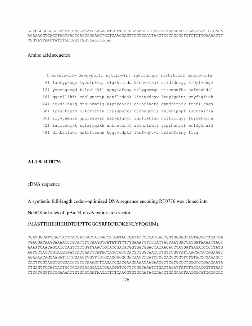
176
GATGGCACGCACAACATTGACGATATCAAGAAATCCATTATCGAAAAGATCAACTCTAAACTGCTGACCGCCTGCGACA
ACAAAGGTCAGGTAGTCACTGACCCGAAACTGCTGAAGGAGTTCGTGGATTACGTTGTAGCGGTGTCTCTGGAAAAGTT
CCGTATTAACTATCTGCTGGTTGGTtagctcgag
Amino acid sequence
1 mikkankisy devpyppftf sytyppylrt igklfglnpp lletakvldi gcgigvnlln
61 faetypksqs lgvdlsktqi elgkktisda kinnvelkal sildldesyg kfdyivchgv
121 yswvsqevqd kilevlnkll npngiafvsy ntlpgwnmqn tiremmmfhs esfntshdkl
181 qqarlllkfi ndslgnsttp yanflrdeak listyddsyv lheylgeint gtyfhqfiek
241 aqknhlnylg dtsiaamfig nlptkaaskl qaindivcte qymdfitnrk frstllchqn
301 ipinrkiefd nlkdfyttfn irpispenki dlnneqenis fyyenlpepf isttsaimka
361 ilyvyaenis npirleqvak eafkklgkyr lqdflatleq hfitlifqgy lkifetkpha
421 iatitekpkt sqfaryqakh ahfnnvtnmf sitnrlndmi gipihekyil emldgthnid
481 dikksiieki nsklltacdn kgqvvtdpkl lkefvdyvva vslekfriny llvg
A1.1.8: RT0776
cDNA sequence
A synthetic full-length codon-optimized DNA sequence encoding RT0776 was cloned into
NdeI/XhoI sites of pBio44 E.coli expression vector
(MASTTHHHHHHDTDIPTTGGGSRPDDDDKENLYFQGHM).
CCATGGCATCCACTACTCACCATCACCATCACCATGATACTGATATCCCGACCACCGGTGGGGGTAGTAGGCCTGATGA
CGACGACAAGGAAAACCTGTACTTCCAAGGCCATATGTCTCTGAAATCTTCTACTACTAATGACCACGATAAAACTACT
AAAATCAACAGCATCCAGTCTCTGGTGAACTGTACCGATACGGTGGCGGACCATAACACCTATGATGAGATCCCTTATG
AGTCCTACCCGTACGCGATTACCAACCCATACCACCTGTCCACTCTGGCAACCCTGTTCGGTATTAACGCCCCGGAAGT
AGAAAACAGCAAGATTCTGGAACTGGGTTGTGCAGCAGGCGGTAACCTGATCCCGCACGCTGTTCTGTACCCGAAAGCT
CACTTCGTAGGTGTGGATCTGTCCAAAGTTCAGATCGACGAAGCAAACAAGAACGTTCGTGCCCTGGGTCTGAAGAATA
TTGAGTTCCACCACTGCTCCATCACGGACATCAACGATTCTTTCGGCAAATTTGACTACATTATCTGCCACGGTGTAAT
TTCCTGGGTCCCGAAAATTGTGCGCGATAAGATTTTCAAGGTGTGCAATAGCAACCTGAGCACTAACGGCATCGCGTAC
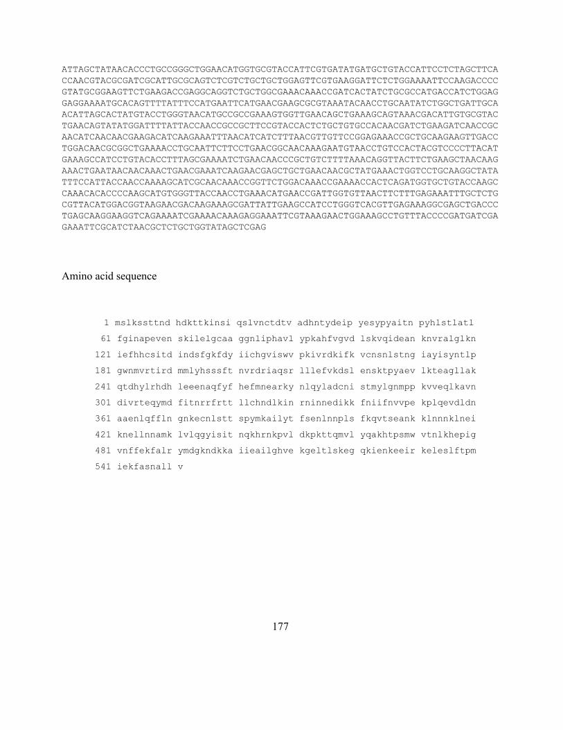
177
ATTAGCTATAACACCCTGCCGGGCTGGAACATGGTGCGTACCATTCGTGATATGATGCTGTACCATTCCTCTAGCTTCA
CCAACGTACGCGATCGCATTGCGCAGTCTCGTCTGCTGCTGGAGTTCGTGAAGGATTCTCTGGAAAATTCCAAGACCCC
GTATGCGGAAGTTCTGAAGACCGAGGCAGGTCTGCTGGCGAAACAAACCGATCACTATCTGCGCCATGACCATCTGGAG
GAGGAAAATGCACAGTTTTATTTCCATGAATTCATGAACGAAGCGCGTAAATACAACCTGCAATATCTGGCTGATTGCA
ACATTAGCACTATGTACCTGGGTAACATGCCGCCGAAAGTGGTTGAACAGCTGAAAGCAGTAAACGACATTGTGCGTAC
TGAACAGTATATGGATTTTATTACCAACCGCCGCTTCCGTACCACTCTGCTGTGCCACAACGATCTGAAGATCAACCGC
AACATCAACAACGAAGACATCAAGAAATTTAACATCATCTTTAACGTTGTTCCGGAGAAACCGCTGCAAGAAGTTGACC
TGGACAACGCGGCTGAAAACCTGCAATTCTTCCTGAACGGCAACAAAGAATGTAACCTGTCCACTACGTCCCCTTACAT
GAAAGCCATCCTGTACACCTTTAGCGAAAATCTGAACAACCCGCTGTCTTTTAAACAGGTTACTTCTGAAGCTAACAAG
AAACTGAATAACAACAAACTGAACGAAATCAAGAACGAGCTGCTGAACAACGCTATGAAACTGGTCCTGCAAGGCTATA
TTTCCATTACCAACCAAAAGCATCGCAACAAACCGGTTCTGGACAAACCGAAAACCACTCAGATGGTGCTGTACCAAGC
CAAACACACCCCAAGCATGTGGGTTACCAACCTGAAACATGAACCGATTGGTGTTAACTTCTTTGAGAAATTTGCTCTG
CGTTACATGGACGGTAAGAACGACAAGAAAGCGATTATTGAAGCCATCCTGGGTCACGTTGAGAAAGGCGAGCTGACCC
TGAGCAAGGAAGGTCAGAAAATCGAAAACAAAGAGGAAATTCGTAAAGAACTGGAAAGCCTGTTTACCCCGATGATCGA
GAAATTCGCATCTAACGCTCTGCTGGTATAGCTCGAG
Amino acid sequence
1 mslkssttnd hdkttkinsi qslvnctdtv adhntydeip yesypyaitn pyhlstlatl
61 fginapeven skilelgcaa ggnliphavl ypkahfvgvd lskvqidean knvralglkn
121 iefhhcsitd indsfgkfdy iichgviswv pkivrdkifk vcnsnlstng iayisyntlp
181 gwnmvrtird mmlyhsssft nvrdriaqsr lllefvkdsl ensktpyaev lkteagllak
241 qtdhylrhdh leeenaqfyf hefmnearky nlqyladcni stmylgnmpp kvveqlkavn
301 divrteqymd fitnrrfrtt llchndlkin rninnedikk fniifnvvpe kplqevdldn
361 aaenlqffln gnkecnlstt spymkailyt fsenlnnpls fkqvtseank klnnnklnei
421 knellnnamk lvlqgyisit nqkhrnkpvl dkpkttqmvl yqakhtpsmw vtnlkhepig
481 vnffekfalr ymdgkndkka iieailghve kgeltlskeg qkienkeeir keleslftpm
541 iekfasnall v
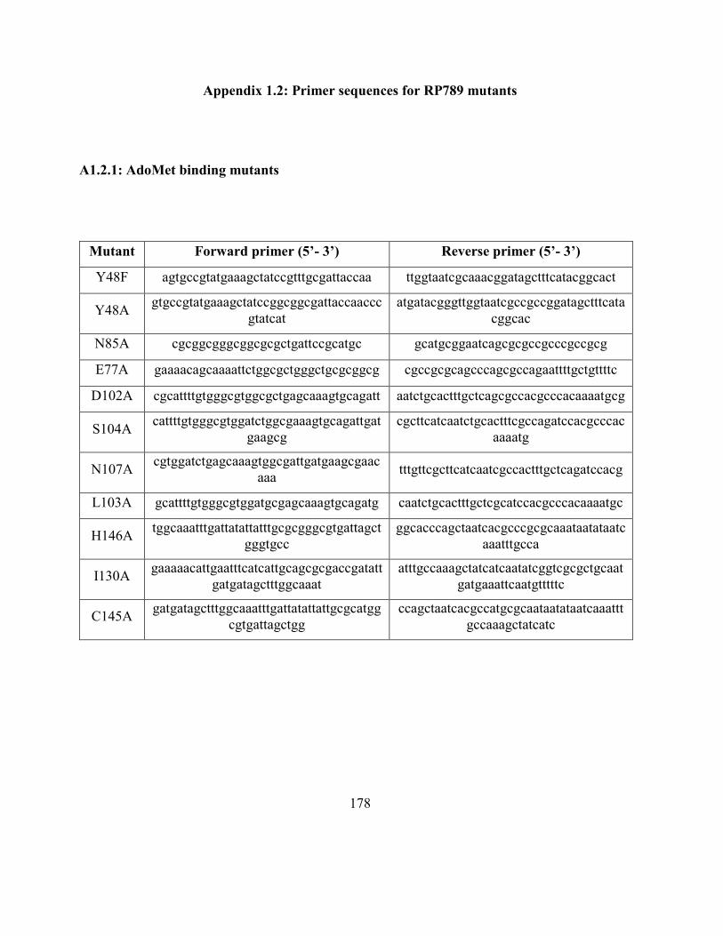
178
Appendix 1.2: Primer sequences for RP789 mutants
A1.2.1: AdoMet binding mutants
Mutant Forward primer (5’- 3’) Reverse primer (5’- 3’)
Y48F agtgccgtatgaaagctatccgtttgcgattaccaa ttggtaatcgcaaacggatagctttcatacggcact
Y48A gtgccgtatgaaagctatccggcggcgattaccaaccc
gtatcat
atgatacgggttggtaatcgccgccggatagctttcata
cggcac
N85A cgcggcgggcggcgcgctgattccgcatgc gcatgcggaatcagcgcgccgcccgccgcg
E77A gaaaacagcaaaattctggcgctgggctgcgcggcg cgccgcgcagcccagcgccagaattttgctgttttc
D102A cgcattttgtgggcgtggcgctgagcaaagtgcagatt aatctgcactttgctcagcgccacgcccacaaaatgcg
S104A cattttgtgggcgtggatctggcgaaagtgcagattgat
gaagcg
cgcttcatcaatctgcactttcgccagatccacgcccac
aaaatg
N107A cgtggatctgagcaaagtggcgattgatgaagcgaac
aaa tttgttcgcttcatcaatcgccactttgctcagatccacg
L103A gcattttgtgggcgtggatgcgagcaaagtgcagatg caatctgcactttgctcgcatccacgcccacaaaatgc
H146A tggcaaatttgattatattatttgcgcgggcgtgattagct
gggtgcc
ggcacccagctaatcacgcccgcgcaaataatataatc
aaatttgcca
I130A gaaaaacattgaatttcatcattgcagcgcgaccgatatt
gatgatagctttggcaaat
atttgccaaagctatcatcaatatcggtcgcgctgcaat
gatgaaattcaatgtttttc
C145A gatgatagctttggcaaatttgattatattattgcgcatgg
cgtgattagctgg
ccagctaatcacgccatgcgcaataatataatcaaattt
gccaaagctatcatc
179
A1.2.2: N-terminal deletion
Mutant Forward primer (5’- 3’) Reverse primer (5’- 3’)
RP789ΔN gtcgaccatatgatgaccggcaccgtggcggatc
ataac caccacctcgagttattacaccagcagcgcgttgctgca
A1.2.3: Loop insertion
Add NNVTNM sequence between 470th Serine (1410 bp) and 471st Methionine (1411 bp)
Base pair sequence is AATAACGTGACCAATATG
Mutant Forward primer (5’- 3’) Reverse primer (5’- 3’)
RP789 with
loop
gaaatataccccgagcaataacgtgaccaata
tgatgtgggtgaccaacc
ggttggtcacccacatcatattggtcacgttattgctcggg
gtatatttc
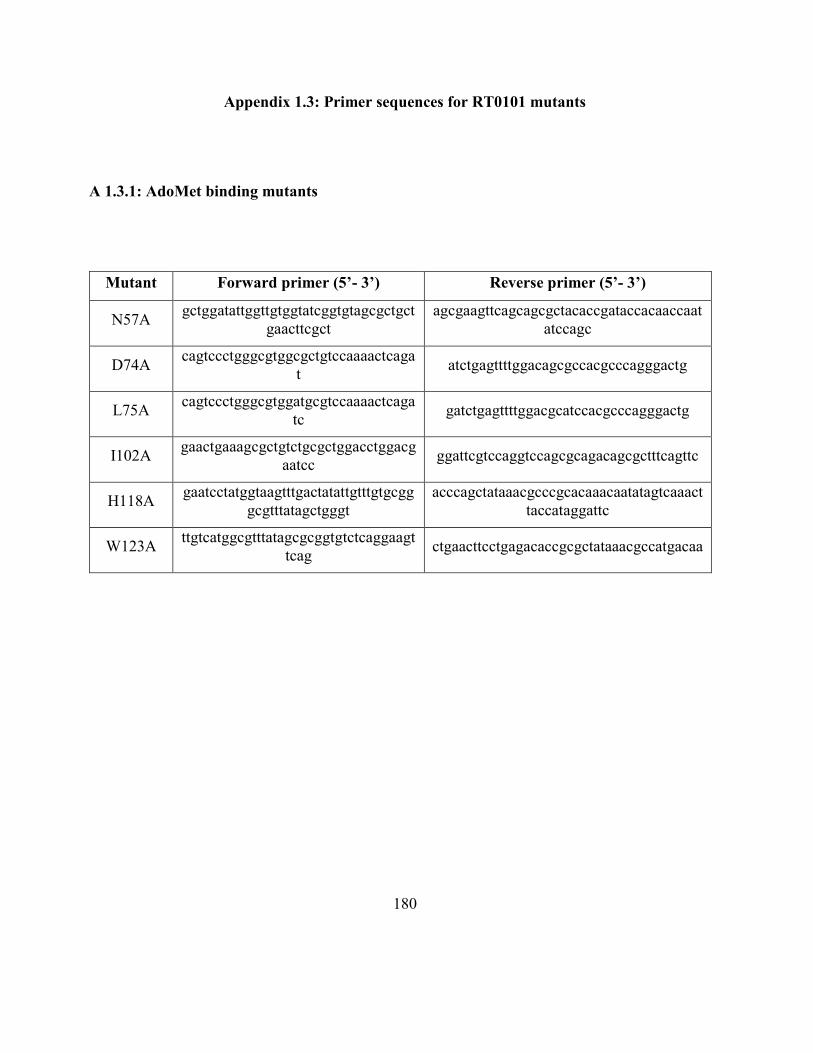
180
Appendix 1.3: Primer sequences for RT0101 mutants
A 1.3.1: AdoMet binding mutants
Mutant Forward primer (5’- 3’) Reverse primer (5’- 3’)
N57A gctggatattggttgtggtatcggtgtagcgctgct
gaacttcgct
agcgaagttcagcagcgctacaccgataccacaaccaat
atccagc
D74A cagtccctgggcgtggcgctgtccaaaactcaga
t atctgagttttggacagcgccacgcccagggactg
L75A cagtccctgggcgtggatgcgtccaaaactcaga
tc gatctgagttttggacgcatccacgcccagggactg
I102A gaactgaaagcgctgtctgcgctggacctggacg
aatcc ggattcgtccaggtccagcgcagacagcgctttcagttc
H118A gaatcctatggtaagtttgactatattgtttgtgcgg
gcgtttatagctgggt
acccagctataaacgcccgcacaaacaatatagtcaaact
taccataggattc
W123A ttgtcatggcgtttatagcgcggtgtctcaggaagt
tcag ctgaacttcctgagacaccgcgctataaacgccatgacaa
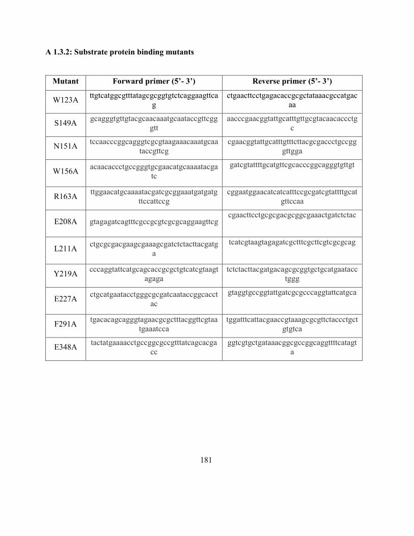
181
A 1.3.2: Substrate protein binding mutants
Mutant Forward primer (5’- 3’) Reverse primer (5’- 3’)
W123A ttgtcatggcgtttatagcgcggtgtctcaggaagttca
g
ctgaacttcctgagacaccgcgctataaacgccatgac
aa
S149A gcagggtgttgtacgcaacaaatgcaataccgttcgg
gtt
aacccgaacggtattgcatttgttgcgtacaacaccctg
c
N151A tccaacccggcagggtcgcgtaagaaacaaatgcaa
taccgttcg
cgaacggtattgcatttgtttcttacgcgaccctgccgg
gttgga
W156A acaacaccctgccgggtgcgaacatgcaaaatacga
tc
gatcgtattttgcatgttcgcacccggcagggtgttgt
R163A ttggaacatgcaaaatacgatcgcggaaatgatgatg
ttccattccg
cggaatggaacatcatcatttccgcgatcgtattttgcat
gttccaa
E208A gtagagatcagtttcgccgcgtcgcgcaggaagttcg cgaacttcctgcgcgacgcggcgaaactgatctctac
L211A ctgcgcgacgaagcgaaagcgatctctacttacgatg
a
tcatcgtaagtagagatcgctttcgcttcgtcgcgcag
Y219A cccaggtattcatgcagcaccgcgctgtcatcgtaagt
agaga
tctctacttacgatgacagcgcggtgctgcatgaatacc
tggg
E227A ctgcatgaatacctgggcgcgatcaataccggcacct
ac
gtaggtgccggtattgatcgcgcccaggtattcatgca
F291A tgacacagcagggtagaacgcgctttacggttcgtaa
tgaaatcca
tggatttcattacgaaccgtaaagcgcgttctaccctgct
gtgtca
E348A tactatgaaaacctgccggcgccgtttatcagcacga
cc
ggtcgtgctgataaacggcgccggcaggttttcatagt
a
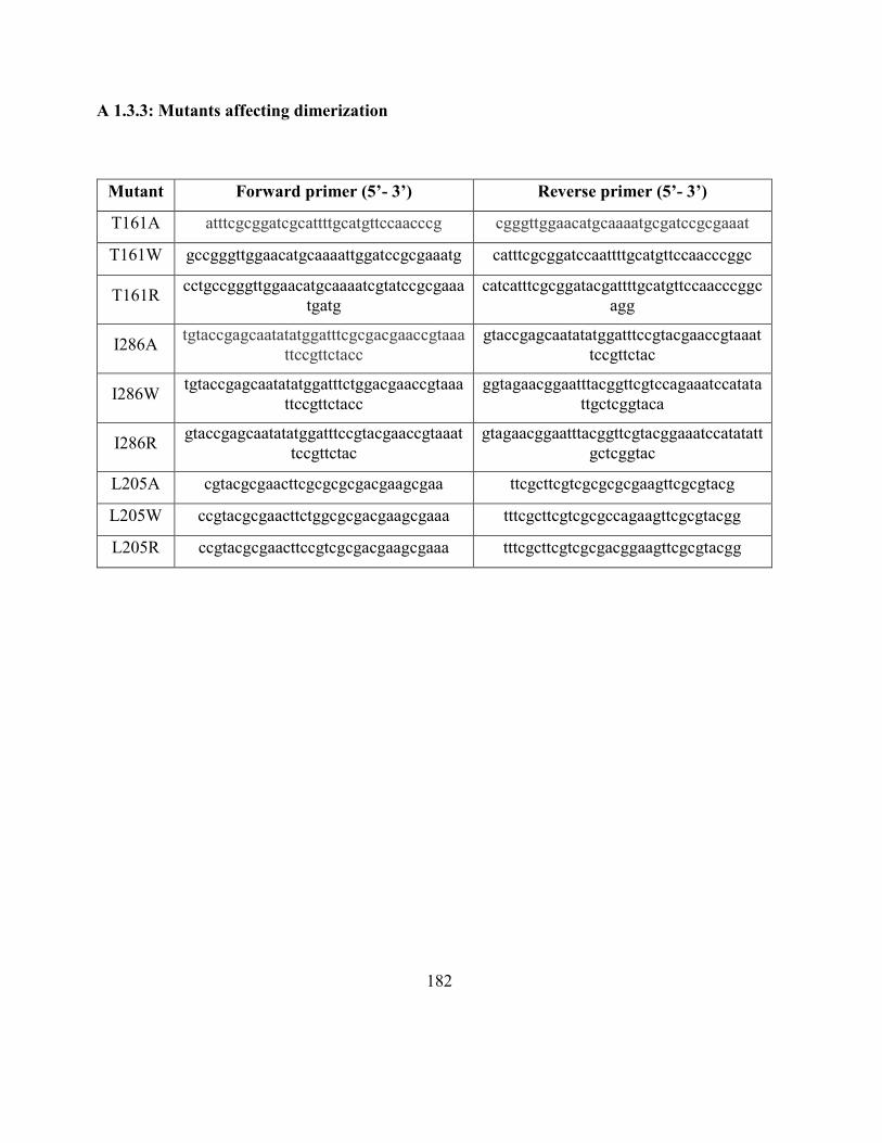
182
A 1.3.3: Mutants affecting dimerization
Mutant Forward primer (5’- 3’) Reverse primer (5’- 3’)
T161A atttcgcggatcgcattttgcatgttccaacccg cgggttggaacatgcaaaatgcgatccgcgaaat
T161W gccgggttggaacatgcaaaattggatccgcgaaatg catttcgcggatccaattttgcatgttccaacccggc
T161R cctgccgggttggaacatgcaaaatcgtatccgcgaaa
tgatg
catcatttcgcggatacgattttgcatgttccaacccggc
agg
I286A tgtaccgagcaatatatggatttcgcgacgaaccgtaaa
ttccgttctacc
gtaccgagcaatatatggatttccgtacgaaccgtaaat
tccgttctac
I286W tgtaccgagcaatatatggatttctggacgaaccgtaaa
ttccgttctacc
ggtagaacggaatttacggttcgtccagaaatccatata
ttgctcggtaca
I286R gtaccgagcaatatatggatttccgtacgaaccgtaaat
tccgttctac
gtagaacggaatttacggttcgtacggaaatccatatatt
gctcggtac
L205A cgtacgcgaacttcgcgcgcgacgaagcgaa ttcgcttcgtcgcgcgcgaagttcgcgtacg
L205W ccgtacgcgaacttctggcgcgacgaagcgaaa tttcgcttcgtcgcgccagaagttcgcgtacgg
L205R ccgtacgcgaacttccgtcgcgacgaagcgaaa tttcgcttcgtcgcgacggaagttcgcgtacgg
183
A 1.3.4: Loop insertion and single amino acid mutants on the loop
Delete 6 amino acids between 443rd Phe and 450th Phe
The deleted amino acid sequence is NNVTNM
Mutant Forward primer (5’- 3’) Reverse primer (5’- 3’)
RT0101
loop
deleted
caaaacatgcgcacttcttctccatcacgaaccg cggttcgtgatggagaagaagtgcgcatgttttg
N444A caggcaaaacatgcgcacttcgcgaacgtgaccaa
tatgttctc
ggagaacatattggtcacgttcgcgaagtgcgcatgtttt
gcctg
N445A ccaggcaaaacatgcgcacttcaatgcggtgacca
atatgttctc
ggagaacatattggtcaccgcattgaagtgcgcatgtttt
gcctgg
V446A catgcgcacttcaataacgcgaccaatatgttctcca
tc
gatggagaacatattggtcgcgttattgaagtgcgcatg
T447A tgcgcacttcaataacgtggcgaatatgttctccatc
acga
tcgtgatggagaacatattcgccacgttattgaagtgcg
ca
N448A gcgcacttcaataacgtgaccgcgatgttctccatca
cgaaccgc
gcggttcgtgatggagaacatcgcggtcacgttattgaa
gtgcgc

184
Appendix 1.4: Identity, accession number and species/strain of RP789 and RP027-028
homologous proteins
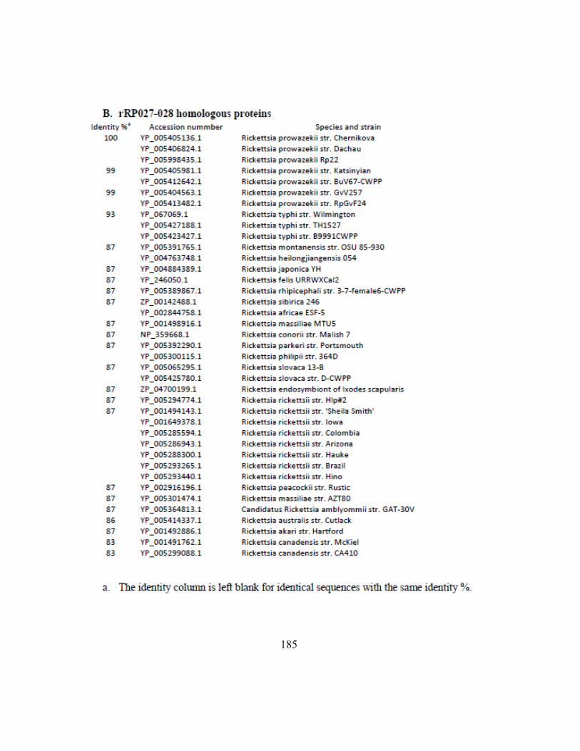
185
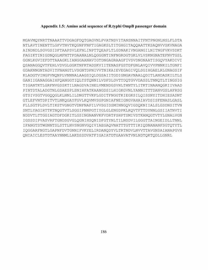
186
Appendix 1.5: Amino acid sequence of R.typhi OmpB passenger domain
MGAVMQYNRTTNAAATTVDGAGFDQTGAGVNLPVATNSVITANSNNAITFNTPNGNLNSLFLDTA
NTLAVTINENTTLGFVTNVTKQGNFFNFTIGAGKSLTITGHGITAQQAATTKSAQNVVSKVNAGA
AINDNDLSGVGSIDFTAAPSVLEFNLINPTTQEAPLTLGDNAKIVNGANGILNITNGFVKVSDKT
FAGIKTINIGDNQGLMFNTTPDAANALNLQGGGNTINFNGRDGTGKLVLVSKNGNATEFNVTGSL
GGNLKGVIEFDTTAAAGKLIANGGAANAVIGTDNGAGRAAGFIVSVDNGNAATISGQVYAKDIVI
QSANAGGQVTFEHLVDVGLGGKTNFKTADSKVIITENASFGSTDFGNLAVQIVVPNNKILTGNFI
GDAKNNGNTAGVITFNANGTLVSGNTDPNIVVTNIKAIEVEGAGIVQLSGIHGAELRLGNAGSIF
KLADGTVINGPVNQNPLVNNNALAAGSIQLDGSAIITGDIGNGAVNAALQDITLANDASKILTLS
GANIIGANAGGAIHFQANGGTIQLTSTQNNILVDFDLDVTTDQTGVVDASSLTNNQTLTINGSIG
TIGANTKTLGRFNVGSSKTILNAGDVAINELVMENDGSVHLTHNTYLITKTINAANQGKIIVAAD
PINTDTALADGTNLGSAESPLSNIHFATKAANGDSILHIGKGVNLYANNITTTDANVGSLHFRSG
GTSIVSGTVGGQQGLKLNNLILDNGTTVKFLGDITFNGGTKIEGKSILQISSNYITDHIESADNT
GTLEFVNTDPITVTLNKQGAYFGVLKQVMVSGPGNIAFNEIGNGVAHAIAVDSISFENASLGASL
FLLSGTPLDVLTIKSTVGNGTVDNFNAPILVVSGIDSMINNGQVIGDQKNIIALSLGSDNSITVN
SNTLYAGIRTTKTNQGTVTLSGGIPNNPGTIYGLGLENGDPKLKQVTFTTDYNNLGSIIATNVTI
NDDVTLTTGGIAGTDFDGKITLGSINGNANVKFVDRTFSHPTSMIVSTKANQGTVTYLGNALVGN
IGSSDIPVASVRFTGNDSGVGLQGNIHSQNIDFGTYNLTILNSDVILGGGTTAINGEIDLLTNNL
IFANGTSTWGNNTSLSTTLNVSNGNVGQIVIAEGAQVNATTTGTTTIKIQDNANANFSGTQTYTL
IQGGARFNGTLGAPNFDVTGNNIFVKYELIRDANQDYVLTRTNDVLNVVTTAVGNSAIANAPGVH
QNIAICLESTDTAAYNNMLLAKDSSDVATFIGAIATDTGAAVATVNLNDTQKTQDLLGNRL
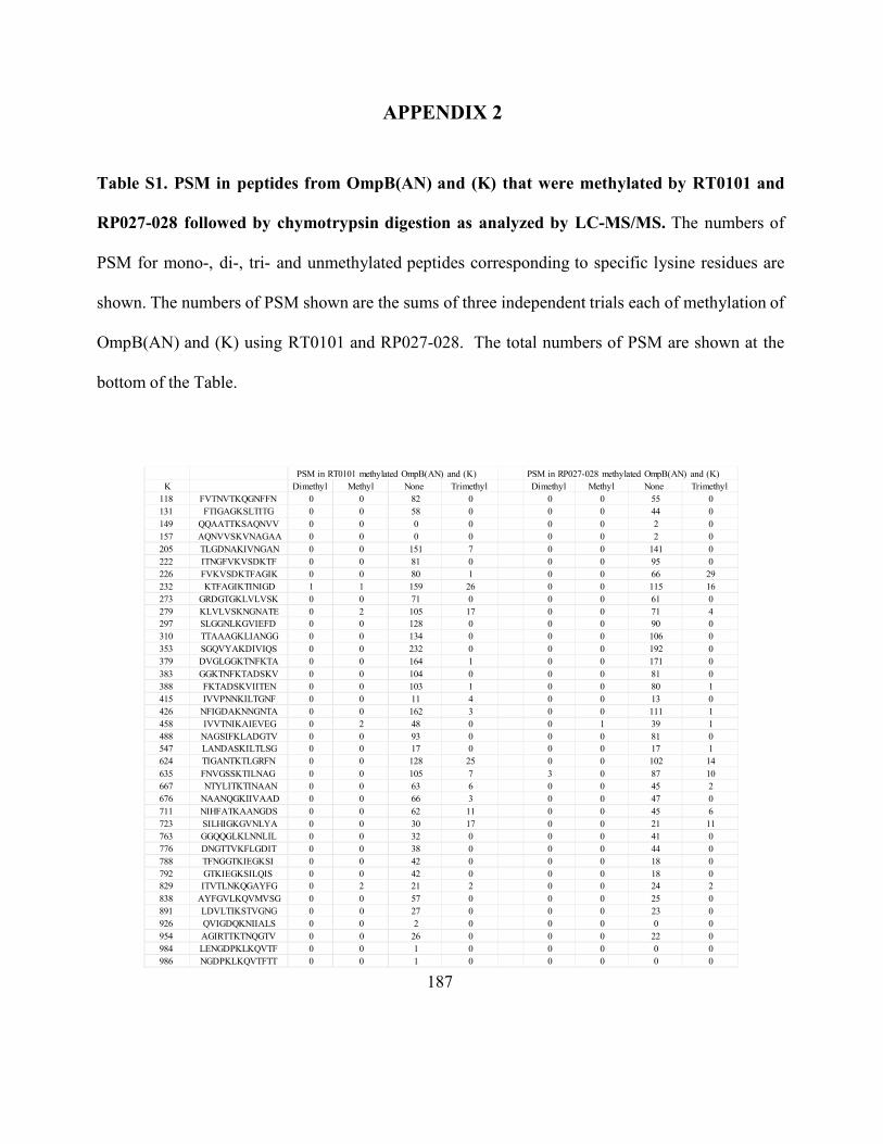
187
APPENDIX 2
Table S1. PSM in peptides from OmpB(AN) and (K) that were methylated by RT0101 and
RP027-028 followed by chymotrypsin digestion as analyzed by LC-MS/MS. The numbers of
PSM for mono-, di-, tri- and unmethylated peptides corresponding to specific lysine residues are
shown. The numbers of PSM shown are the sums of three independent trials each of methylation of
OmpB(AN) and (K) using RT0101 and RP027-028. The total numbers of PSM are shown at the
bottom of the Table.
PSM in RT0101 methylated OmpB(AN) and (K) PSM in RP027-028 methylated OmpB(AN) and (K)
K Dimethyl Methyl None Trimethyl Dimethyl Methyl None Trimethyl
118 FVTNVTKQGNFFN 0 0 82 0 0 0 55 0
131 FTIGAGKSLTITG 0 0 58 0 0 0 44 0
149 QQAATTKSAQNVV 0 0 0 0 0 0 2 0
157 AQNVVSKVNAGAA 0 0 0 0 0 0 2 0
205 TLGDNAKIVNGAN 0 0 151 7 0 0 141 0
222 ITNGFVKVSDKTF 0 0 81 0 0 0 95 0
226 FVKVSDKTFAGIK 0 0 80 1 0 0 66 29
232 KTFAGIKTINIGD 1 1 159 26 0 0 115 16
273 GRDGTGKLVLVSK 0 0 71 0 0 0 61 0
279 KLVLVSKNGNATE 0 2 105 17 0 0 71 4
297 SLGGNLKGVIEFD 0 0 128 0 0 0 90 0
310 TTAAAGKLIANGG 0 0 134 0 0 0 106 0
353 SGQVYAKDIVIQS 0 0 232 0 0 0 192 0
379 DVGLGGKTNFKTA 0 0 164 1 0 0 171 0
383 GGKTNFKTADSKV 0 0 104 0 0 0 81 0
388 FKTADSKVIITEN 0 0 103 1 0 0 80 1
415 IVVPNNKILTGNF 0 0 11 4 0 0 13 0
426 NFIGDAKNNGNTA 0 0 162 3 0 0 111 1
458 IVVTNIKAIEVEG 0 2 48 0 0 1 39 1
488 NAGSIFKLADGTV 0 0 93 0 0 0 81 0
547 LANDASKILTLSG 0 0 17 0 0 0 17 1
624 TIGANTKTLGRFN 0 0 128 25 0 0 102 14
635 FNVGSSKTILNAG 0 0 105 7 3 0 87 10
667 NTYLITKTINAAN 0 0 63 6 0 0 45 2
676 NAANQGKIIVAAD 0 0 66 3 0 0 47 0
711 NIHFATKAANGDS 0 0 62 11 0 0 45 6
723 SILHIGKGVNLYA 0 0 30 17 0 0 21 11
763 GGQQGLKLNNLIL 0 0 32 0 0 0 41 0
776 DNGTTVKFLGDIT 0 0 38 0 0 0 44 0
788 TFNGGTKIEGKSI 0 0 42 0 0 0 18 0
792 GTKIEGKSILQIS 0 0 42 0 0 0 18 0
829 ITVTLNKQGAYFG 0 2 21 2 0 0 24 2
838 AYFGVLKQVMVSG 0 0 57 0 0 0 25 0
891 LDVLTIKSTVGNG 0 0 27 0 0 0 23 0
926 QVIGDQKNIIALS 0 0 2 0 0 0 0 0
954 AGIRTTKTNQGTV 0 0 26 0 0 0 22 0
984 LENGDPKLKQVTF 0 0 1 0 0 0 0 0
986 NGDPKLKQVTFTT 0 0 1 0 0 0 0 0
188
1026 GTDFDGKITLGSI 0 0 14 0 0 0 16 0
1039 NGNANVKFVDRTF 0 0 43 0 0 0 40 0
1056 SMIVSTKANQGTV 0 2 12 2 0 0 12 2
1185 TGTTTIKIQDNAN 0 0 42 0 0 0 20 0
1228 GNNIFVKYELIRD 0 0 66 0 0 0 44 0
1289 NNMLLAKDSSDVA 0 0 113 0 0 0 82 0
1319 NLNDTQKTQDLLG 0 0 73 0 0 0 52 0
Total of PSM 1 9 3089 133 3 1 2461 100
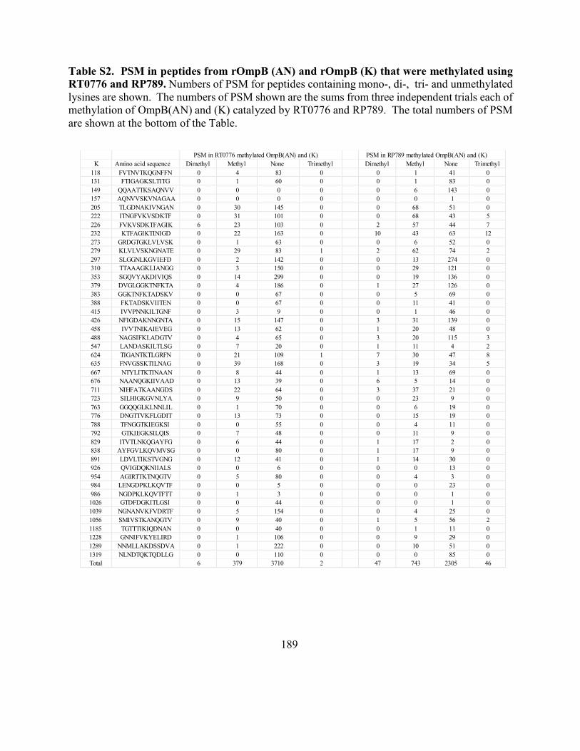
189
Table S2. PSM in peptides from rOmpB (AN) and rOmpB (K) that were methylated using
RT0776 and RP789. Numbers of PSM for peptides containing mono-, di-, tri- and unmethylated
lysines are shown. The numbers of PSM shown are the sums from three independent trials each of
methylation of OmpB(AN) and (K) catalyzed by RT0776 and RP789. The total numbers of PSM
are shown at the bottom of the Table.
PSM in RT0776 methylated OmpB(AN) and (K) PSM in RP789 methylated OmpB(AN) and (K)
K Amino acid sequence Dimethyl Methyl None Trimethyl Dimethyl Methyl None Trimethyl
118 FVTNVTKQGNFFN 0 4 83 0 0 1 41 0
131 FTIGAGKSLTITG 0 1 60 0 0 1 83 0
149 QQAATTKSAQNVV 0 0 0 0 0 6 143 0
157 AQNVVSKVNAGAA 0 0 0 0 0 0 1 0
205 TLGDNAKIVNGAN 0 30 145 0 0 68 51 0
222 ITNGFVKVSDKTF 0 31 101 0 0 68 43 5
226 FVKVSDKTFAGIK 6 23 103 0 2 57 44 7
232 KTFAGIKTINIGD 0 22 163 0 10 43 63 12
273 GRDGTGKLVLVSK 0 1 63 0 0 6 52 0
279 KLVLVSKNGNATE 0 29 83 1 2 62 74 2
297 SLGGNLKGVIEFD 0 2 142 0 0 13 274 0
310 TTAAAGKLIANGG 0 3 150 0 0 29 121 0
353 SGQVYAKDIVIQS 0 14 299 0 0 19 136 0
379 DVGLGGKTNFKTA 0 4 186 0 1 27 126 0
383 GGKTNFKTADSKV 0 0 67 0 0 5 69 0
388 FKTADSKVIITEN 0 0 67 0 0 11 41 0
415 IVVPNNKILTGNF 0 3 9 0 0 1 46 0
426 NFIGDAKNNGNTA 0 15 147 0 3 31 139 0
458 IVVTNIKAIEVEG 0 13 62 0 1 20 48 0
488 NAGSIFKLADGTV 0 4 65 0 3 20 115 3
547 LANDASKILTLSG 0 7 20 0 1 11 4 2
624 TIGANTKTLGRFN 0 21 109 1 7 30 47 8
635 FNVGSSKTILNAG 0 39 168 0 3 19 34 5
667 NTYLITKTINAAN 0 8 44 0 1 13 69 0
676 NAANQGKIIVAAD 0 13 39 0 6 5 14 0
711 NIHFATKAANGDS 0 22 64 0 3 37 21 0
723 SILHIGKGVNLYA 0 9 50 0 0 23 9 0
763 GGQQGLKLNNLIL 0 1 70 0 0 6 19 0
776 DNGTTVKFLGDIT 0 13 73 0 0 15 19 0
788 TFNGGTKIEGKSI 0 0 55 0 0 4 11 0
792 GTKIEGKSILQIS 0 7 48 0 0 11 9 0
829 ITVTLNKQGAYFG 0 6 44 0 1 17 2 0
838 AYFGVLKQVMVSG 0 0 80 0 1 17 9 0
891 LDVLTIKSTVGNG 0 12 41 0 1 14 30 0
926 QVIGDQKNIIALS 0 0 6 0 0 0 13 0
954 AGIRTTKTNQGTV 0 5 80 0 0 4 3 0
984 LENGDPKLKQVTF 0 0 5 0 0 0 23 0
986 NGDPKLKQVTFTT 0 1 3 0 0 0 1 0
1026 GTDFDGKITLGSI 0 0 44 0 0 0 1 0
1039 NGNANVKFVDRTF 0 5 154 0 0 4 25 0
1056 SMIVSTKANQGTV 0 9 40 0 1 5 56 2
1185 TGTTTIKIQDNAN 0 0 40 0 0 1 11 0
1228 GNNIFVKYELIRD 0 1 106 0 0 9 29 0
1289 NNMLLAKDSSDVA 0 1 222 0 0 10 51 0
1319 NLNDTQKTQDLLG 0 0 110 0 0 0 85 0
Total 6 379 3710 2 47 743 2305 46
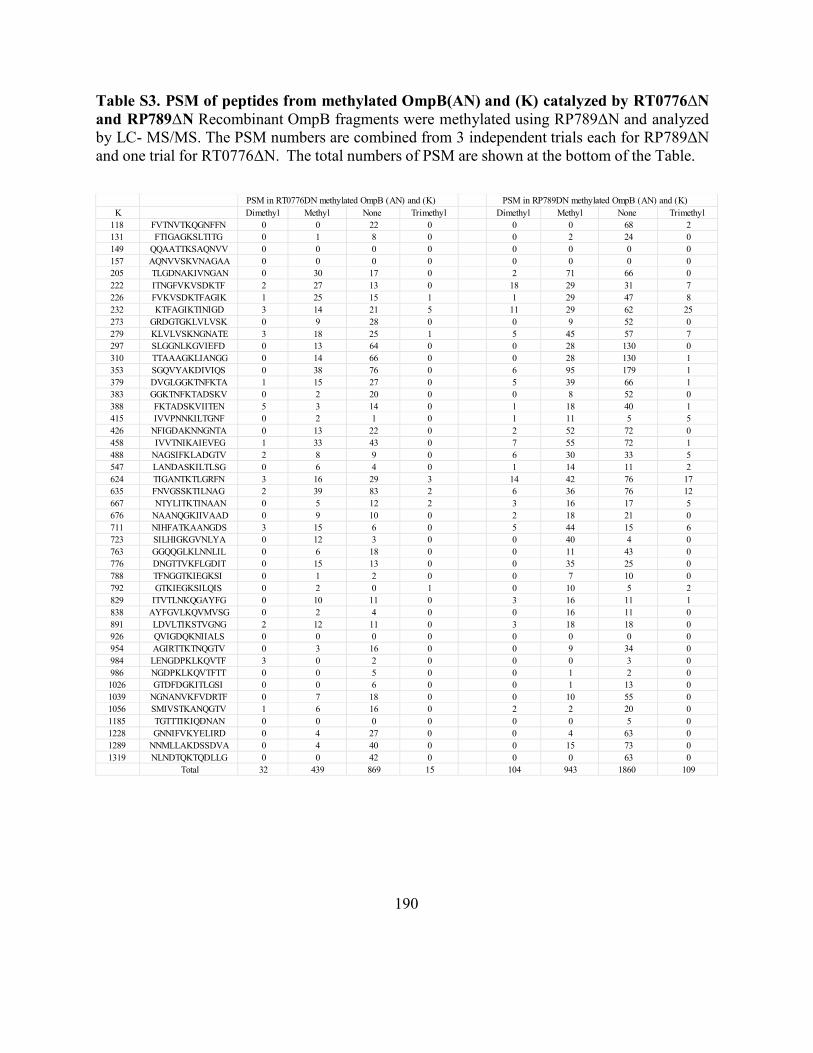
190
Table S3. PSM of peptides from methylated OmpB(AN) and (K) catalyzed by RT0776ΔN
and RP789ΔN Recombinant OmpB fragments were methylated using RP789ΔN and analyzed
by LC- MS/MS. The PSM numbers are combined from 3 independent trials each for RP789ΔN
and one trial for RT0776ΔN. The total numbers of PSM are shown at the bottom of the Table.
PSM in RT0776DN methylated OmpB (AN) and (K) PSM in RP789DN methylated OmpB (AN) and (K)
K Dimethyl Methyl None Trimethyl Dimethyl Methyl None Trimethyl
118 FVTNVTKQGNFFN 0 0 22 0 0 0 68 2
131 FTIGAGKSLTITG 0 1 8 0 0 2 24 0
149 QQAATTKSAQNVV 0 0 0 0 0 0 0 0
157 AQNVVSKVNAGAA 0 0 0 0 0 0 0 0
205 TLGDNAKIVNGAN 0 30 17 0 2 71 66 0
222 ITNGFVKVSDKTF 2 27 13 0 18 29 31 7
226 FVKVSDKTFAGIK 1 25 15 1 1 29 47 8
232 KTFAGIKTINIGD 3 14 21 5 11 29 62 25
273 GRDGTGKLVLVSK 0 9 28 0 0 9 52 0
279 KLVLVSKNGNATE 3 18 25 1 5 45 57 7
297 SLGGNLKGVIEFD 0 13 64 0 0 28 130 0
310 TTAAAGKLIANGG 0 14 66 0 0 28 130 1
353 SGQVYAKDIVIQS 0 38 76 0 6 95 179 1
379 DVGLGGKTNFKTA 1 15 27 0 5 39 66 1
383 GGKTNFKTADSKV 0 2 20 0 0 8 52 0
388 FKTADSKVIITEN 5 3 14 0 1 18 40 1
415 IVVPNNKILTGNF 0 2 1 0 1 11 5 5
426 NFIGDAKNNGNTA 0 13 22 0 2 52 72 0
458 IVVTNIKAIEVEG 1 33 43 0 7 55 72 1
488 NAGSIFKLADGTV 2 8 9 0 6 30 33 5
547 LANDASKILTLSG 0 6 4 0 1 14 11 2
624 TIGANTKTLGRFN 3 16 29 3 14 42 76 17
635 FNVGSSKTILNAG 2 39 83 2 6 36 76 12
667 NTYLITKTINAAN 0 5 12 2 3 16 17 5
676 NAANQGKIIVAAD 0 9 10 0 2 18 21 0
711 NIHFATKAANGDS 3 15 6 0 5 44 15 6
723 SILHIGKGVNLYA 0 12 3 0 0 40 4 0
763 GGQQGLKLNNLIL 0 6 18 0 0 11 43 0
776 DNGTTVKFLGDIT 0 15 13 0 0 35 25 0
788 TFNGGTKIEGKSI 0 1 2 0 0 7 10 0
792 GTKIEGKSILQIS 0 2 0 1 0 10 5 2
829 ITVTLNKQGAYFG 0 10 11 0 3 16 11 1
838 AYFGVLKQVMVSG 0 2 4 0 0 16 11 0
891 LDVLTIKSTVGNG 2 12 11 0 3 18 18 0
926 QVIGDQKNIIALS 0 0 0 0 0 0 0 0
954 AGIRTTKTNQGTV 0 3 16 0 0 9 34 0
984 LENGDPKLKQVTF 3 0 2 0 0 0 3 0
986 NGDPKLKQVTFTT 0 0 5 0 0 1 2 0
1026 GTDFDGKITLGSI 0 0 6 0 0 1 13 0
1039 NGNANVKFVDRTF 0 7 18 0 0 10 55 0
1056 SMIVSTKANQGTV 1 6 16 0 2 2 20 0
1185 TGTTTIKIQDNAN 0 0 0 0 0 0 5 0
1228 GNNIFVKYELIRD 0 4 27 0 0 4 63 0
1289 NNMLLAKDSSDVA 0 4 40 0 0 15 73 0
1319 NLNDTQKTQDLLG 0 0 42 0 0 0 63 0
Total 32 439 869 15 104 943 1860 109
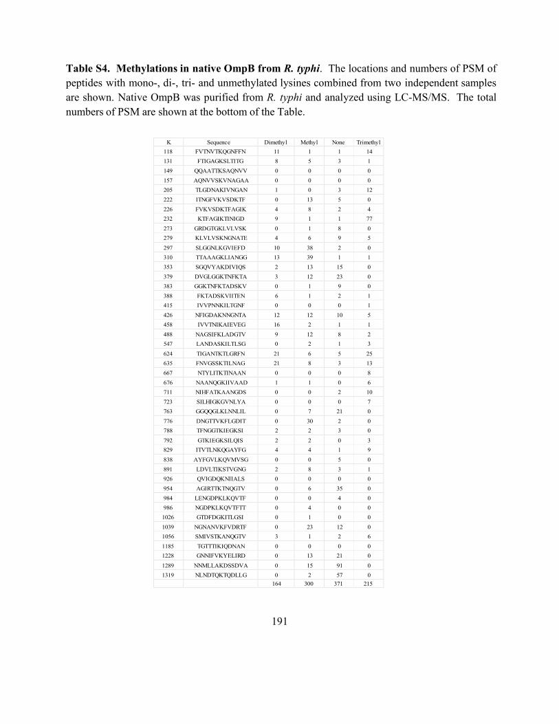
191
Table S4. Methylations in native OmpB from R. typhi. The locations and numbers of PSM of
peptides with mono-, di-, tri- and unmethylated lysines combined from two independent samples
are shown. Native OmpB was purified from R. typhi and analyzed using LC-MS/MS. The total
numbers of PSM are shown at the bottom of the Table.
K Sequence Dimethyl Methyl None Trimethyl
118 FVTNVTKQGNFFN 11 1 1 14
131 FTIGAGKSLTITG 8 5 3 1
149 QQAATTKSAQNVV 0 0 0 0
157 AQNVVSKVNAGAA 0 0 0 0
205 TLGDNAKIVNGAN 1 0 3 12
222 ITNGFVKVSDKTF 0 13 5 0
226 FVKVSDKTFAGIK 4 8 2 4
232 KTFAGIKTINIGD 9 1 1 77
273 GRDGTGKLVLVSK 0 1 8 0
279 KLVLVSKNGNATE 4 6 9 5
297 SLGGNLKGVIEFD 10 38 2 0
310 TTAAAGKLIANGG 13 39 1 1
353 SGQVYAKDIVIQS 2 13 15 0
379 DVGLGGKTNFKTA 3 12 23 0
383 GGKTNFKTADSKV 0 1 9 0
388 FKTADSKVIITEN 6 1 2 1
415 IVVPNNKILTGNF 0 0 0 1
426 NFIGDAKNNGNTA 12 12 10 5
458 IVVTNIKAIEVEG 16 2 1 1
488 NAGSIFKLADGTV 9 12 8 2
547 LANDASKILTLSG 0 2 1 3
624 TIGANTKTLGRFN 21 6 5 25
635 FNVGSSKTILNAG 21 8 3 13
667 NTYLITKTINAAN 0 0 0 8
676 NAANQGKIIVAAD 1 1 0 6
711 NIHFATKAANGDS 0 0 2 10
723 SILHIGKGVNLYA 0 0 0 7
763 GGQQGLKLNNLIL 0 7 21 0
776 DNGTTVKFLGDIT 0 30 2 0
788 TFNGGTKIEGKSI 2 2 3 0
792 GTKIEGKSILQIS 2 2 0 3
829 ITVTLNKQGAYFG 4 4 1 9
838 AYFGVLKQVMVSG 0 0 5 0
891 LDVLTIKSTVGNG 2 8 3 1
926 QVIGDQKNIIALS 0 0 0 0
954 AGIRTTKTNQGTV 0 6 35 0
984 LENGDPKLKQVTF 0 0 4 0
986 NGDPKLKQVTFTT 0 4 0 0
1026 GTDFDGKITLGSI 0 1 0 0
1039 NGNANVKFVDRTF 0 23 12 0
1056 SMIVSTKANQGTV 3 1 2 6
1185 TGTTTIKIQDNAN 0 0 0 0
1228 GNNIFVKYELIRD 0 13 21 0
1289 NNMLLAKDSSDVA 0 15 91 0
1319 NLNDTQKTQDLLG 0 2 57 0
164 300 371 215
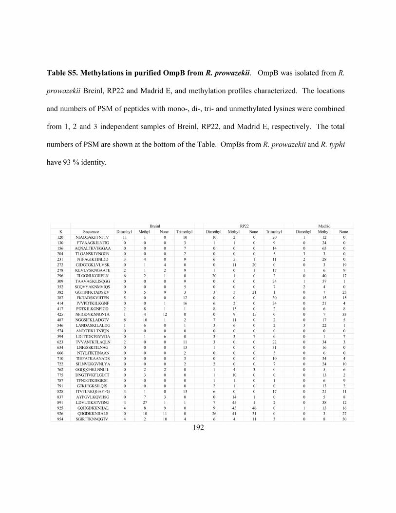
192
Table S5. Methylations in purified OmpB from R. prowazekii. OmpB was isolated from R.
prowazekii Breinl, RP22 and Madrid E, and methylation profiles characterized. The locations
and numbers of PSM of peptides with mono-, di-, tri- and unmethylated lysines were combined
from 1, 2 and 3 independent samples of Breinl, RP22, and Madrid E, respectively. The total
numbers of PSM are shown at the bottom of the Table. OmpBs from R. prowazekii and R. typhi
have 93 % identity.
Breinl RP22 Madrid
K Sequence Dimethyl Methyl None Trimethyl Dimethyl Methyl None Trimethyl Dimethyl Methyl None
120 NIAQQAKFFNFTV 11 1 0 10 10 2 0 20 1 12 0
130 FTVAAGKILNITG 0 0 0 3 1 1 0 9 0 24 0
156 AQNALTKVHGGAA 0 0 0 7 0 0 0 14 0 65 0
204 TLGANSKIVNGGN 0 0 0 2 0 0 0 5 3 3 0
231 NTFAGIKTINIDD 3 4 0 9 6 5 1 11 2 28 0
272 GIDGTGKLVLVSK 0 1 4 0 0 11 20 0 0 3 19
278 KLVLVSKNGAATE 2 1 2 9 1 0 1 17 1 6 9
296 TLGGNLKGIIELN 6 2 1 0 20 1 0 2 0 40 17
309 TAAVAGKLISQGG 0 0 0 9 0 0 0 24 1 57 1
352 SGQVYAKNMVIQS 0 0 0 5 0 0 0 7 2 4 0
382 GGTTNFKTADSKV 0 5 9 3 3 5 21 1 0 7 23
387 FKTADSKVIITEN 5 0 0 12 0 0 0 30 0 15 15
414 IVVPDTKILKGNF 0 0 1 16 6 2 0 24 0 21 4
417 PDTKILKGNFIGD 2 8 1 1 8 15 0 2 0 6 8
425 NFIGDVKNNGNTA 1 4 12 0 0 9 15 0 0 7 33
487 NGGSIFKLADGTV 8 10 1 2 7 11 0 2 0 17 5
546 LANDASKILALDG 1 6 0 1 3 6 0 2 3 22 1
574 ANGGTIKLTNTQN 0 0 0 0 0 0 0 0 0 0 0
594 LDITTDKTGVVDA 0 1 6 0 3 3 7 0 0 1 7
623 TVVANTKTLAQLN 2 0 0 11 3 0 0 22 0 34 3
634 LNIGSSKTILNAG 0 0 0 13 1 0 0 31 0 16 0
666 NTYLITKTINAAN 0 0 0 2 0 0 0 5 0 6 0
710 TIHFATKAANADS 0 0 0 3 0 0 0 10 0 34 4
722 SILNVGKGVNLYA 0 0 0 2 2 0 0 7 0 24 10
762 GGQQGHKLNNLIL 0 2 2 0 1 4 3 0 0 5 6
775 DNGTTVKFLGDTT 0 3 0 0 1 10 0 0 0 13 2
787 TFNGGTKIEGKSI 0 0 0 0 1 1 0 1 0 6 9
791 GTKIEGKSILQIS 0 0 0 0 2 1 0 0 0 13 2
828 ITVTLNKQGAYFG 3 1 0 13 6 0 0 17 0 21 11
837 AYFGVLKQVIISG 0 7 3 0 0 14 1 0 0 5 8
891 LDVLTIKSTVGNG 4 27 1 1 7 45 1 2 0 38 12
925 GQIIGDKKNIIAL 4 8 9 0 9 43 46 0 1 13 16
926 QIIGDKKNIIALS 0 10 11 0 26 41 31 0 0 3 27
954 SGIRTTKNNQGTV 4 2 10 4 6 4 11 3 0 8 30

193
984 LENGSPKLKQVTF 0 0 2 0 0 0 11 0 0 0 9
986 NGSPKLKQVTFTT 0 1 1 0 4 5 0 0 0 5 2
1026 GTDFDAKITLGSV 2 0 0 0 10 1 0 3 0 0 1
1059 VATQANKGTVTYL 0 0 0 1 0 0 0 0 1 1 0
1185 TGTTTIKIQDNAN 0 0 0 0 0 0 0 0 0 0 0
1228 GSNIFVKYELIRD 0 12 12 0 0 19 16 0 0 25 67
1289 NNMLLAKDPSDVA 0 9 66 0 0 11 44 0 0 6 70
1319 NLNDTQKTQDLLS 0 0 5 0 0 0 10 0 0 0 6
58 125 159 139 147 270 239 271 15 614 437
194
BIBLIOGRAPHY
Abeykoon, A.H., Chao, C., Wang, G., Gucek, M., Yang, D.C.H., and Ching, W. (2012) Two
Protein Lysine Methyltransferases Methylate Outer Membrane Protein B from Rickettsia.
J.Bacteriol. 194, 6410-6418
Abeykoon, A., Wang, G., Chao, C., Chock, P.B., Gucek, M., Ching, W., and Yang, D.C.H.
(2014) Multimethylation of Rickettsia OmpB Catalyzed by Lysine Methyltransferases.
J.Biol.Chem. 289, 7691-7701
Adams, P., Grosse-Kunstleve, R., Hung, L., Ioerger, T., McCoy, A., Moriarty, N., Read, R.,
Sacchettini, J., Sauter, N., and Terwilliger, T. (2002) PHENIX: building new software for
automated crystallographic structure determination. Acta Crystallographica Section D-
Biological Crystallography. 58, 1948-1954
Alliegro, M. (2000) Effects of dithiothreitol on protein activity unrelated to thiol-disulfide
exchange: For consideration in the analysis of protein function with Cleland's reagent.
Anal.Biochem. 282, 102-106
Anacker, R., List, R., Mann, R., Hayes, S., Thomas, L. (1985) Characterization of Monoclonal-
Antibodies Protecting Mice Against Rickettsia-Rickettsii. J.Infect.Dis. 151, 1052-1060
195
Anacker, R., Mcdonald, G., List, R., Mann, R. (1987) Neutralizing Activity of Monoclonal-
Antibodies to Heat-Sensitive and Heat-Resistant Epitopes of Rickettsia-Rickettsii Surface-
Proteins. Infect.Immun. 55, 825-827
Andersson, J.O., and Andersson, S.G.E. (2000) A century of typhus, lice and Rickettsia.
Res.Microbiol. 151, 143-150
Andersson, S.G.E., Zomorodipour, A., Andersson, J.O., Sicheritz-Ponten, T., Alsmark,
U.C.M., Podowski, R.M., Naslund, A.K., Eriksson, A.S., Winkler, H.H., and Kurland, C.G.
(1998) The genome sequence of Rickettsia prowazekii and the origin of mitochondria. Nature.
396, 133-140
Ashkenazy, H., Erez, E., Martz, E., Pupko, T., and Ben-Tal, N. (2010) ConSurf 2010:
calculating evolutionary conservation in sequence and structure of proteins and nucleic acids.
Nucleic Acids Res. 38, W529-W533
Ayoubi, T., and VanDeVen, W. (1996) Regulation of gene expression by alternative
promoters. Faseb Journal. 10, 453-460
Azad, A. (1990) Epidemiology of Murine Typhus. Annu.Rev.Entomol. 35, 553-569
196
Balayeva, N.M., Eremeeva, M.E., Ignatovich, V.F., Dmitriev, B.A., Lapina, E.B., and
Belousova, L.S. (1992) Protein Antigens of Genetically Related Rickettsia-Prowazekii Strains
with Different Virulence. Acta Virologica. 36, 52-56
Balayeva, N.M., and Nikolska.Vn (1972) Enhanced Virulence of Vaccine Strain E of
Rickettsia-Prowazeki on Passaging in White Mice and Guinea-Pigs. Acta Virologica. 16, 80-
&
Barnard, T.J., Dautin, N., Lukacik, P., Bernstein, H.D., and Buchanan, S.K. (2007)
Autotransporter structure reveals intra-barrel cleavage followed by conformational changes.
Nature Structural & Molecular Biology. 14, 1214-1220
Barski, A., Cuddapah, S., Cui, K., Roh, T., Schones, D.E., Wang, Z., Wei, G., Chepelev, I.,
and Zhao, K. (2007) High-resolution profiling of histone methylations in the human genome.
Cell. 129, 823-837
Baxter, J.D. (1996) The typhus group. Clin.Dermatol. 14, 271-278
Bechah, Y., El Karkouri, K., Mediannikov, O., Leroy, Q., Pelletier, N., Robert, C., Medigue,
C., Mege, J., and Raoult, D. (2010) Genomic, proteomic, and transcriptomic analysis of
virulent and avirulent Rickettsia prowazekii reveals its adaptive mutation capabilities. Genome
Res. 20, 655-663
197
Birg, M.L., La Scola, B., Roux, V., Brouqui, P., and Raoult, D. (1999) Isolation of Rickettsia
prowazekii from blood by shell vial cell culture. J.Clin.Microbiol. 37, 3722-3724
Bise, G., and Coninx, R. (1997) Epidemic typhus in a prison in Burundi.
Trans.R.Soc.Trop.Med.Hyg. 91, 133-134
Blanc, G., Ngwamidiba, M., Ogata, H., Fournier, P., Claverie, J., and Raoult, D. (2005)
Molecular evolution of Rickettsia surface antigens: Evidence of positive selection.
Mol.Biol.Evol. 22, 2073-2083
Bogan, A., and Thorn, K. (1998) Anatomy of hot spots in protein interfaces. J.Mol.Biol. 280,
1-9
Botelho-Nevers, E., and Raoult, D. (2011) Host, pathogen and treatment-related prognostic
factors in rickettsioses. European Journal of Clinical Microbiology & Infectious Diseases. 30,
1139-1150
Botuyan, M.V., Lee, J., Ward, I.M., Kim, J., Thompson, J.R., Chen, J., and Mer, G. (2006)
Structural basis for the methylation state-specific recognition of histone H4-K20 by 53BP1
and Crb2 in DNA repair. Cell. 127, 1361-1373
198
Bourgeois, A.L., Dasch, G.A., (1981) The species specific surface protein antigens of
Rickettsia typhi: immunogenicity andprotective efficiency in guinea pig. in Rickettsiae and
Rickettsial diseases 71-80
Bradford MM (1976) A rapid and sensitive method for the quantitation of
microgram quantities of protein utilizing the principle of protein-dye
binding. Anal. Biochem. 72, 248-254
Brenner, D.J., Oconnor, S.P., Winkler, H.H., and Steigerwalt, A.G. (1993) Proposals to Unify
the Genera Bartonella and Rochalimaea, with Descriptions of Bartonella-Quintana Comb-Nov,
Bartonella-Vinsonii Comb-Nov, Bartonella-Henselae Comb-Nov, and Bartonella-Elizabethae
Comb-Nov, and to Remove the Family Bartonellaceae from the Order Rickettsiales.
Int.J.Syst.Bacteriol. 43, 777-786
Buchan, D.W.A., Minneci, F., Nugent, T.C.O., Bryson, K., and Jones, D.T. (2013) Scalable
web services for the PSIPRED Protein Analysis Workbench. Nucleic Acids Res. 41, W349-
W357
Buchanan, S.K. (1999) beta-Barrel proteins from bacterial outer membranes: structure,
function and refolding. Curr.Opin.Struct.Biol. 9, 455-461
199
Carl, M., And Dasch, G. (1989) The Importance of the Crystalline Surface-Layer Protein
Antigens of Rickettsiae in T-Cell Immunity. J.Autoimmun. 2, 81-91
Carl, M., Dobson, M., Ching, W., And Dasch, G. (1990) Characterization of the Gene
Encoding the Protective Paracrystalline-Surface-Layer Protein of Rickettsia-Prowazekii -
Presence of a Truncated Identical Homolog in Rickettsia-Typhi. Proc.Natl.Acad.Sci.U.S.A. 87,
8237-8241
Carpenter, C.F., Gandhi, T.K., Kong, L.K., Corey, G.R., Chen, S.M., Walker, D.H., Dumler,
J.S., Breitschwerdt, E., Hegarty, B., and Sexton, D.J. (1999) The incidence of ehrlichial and
rickettsial infection in patients with unexplained fever and recent history of tick bite in central
North Carolina. J.Infect.Dis. 180, 900-903
Casadesus, J., and Low, D. (2006) Epigenetic gene regulation in the bacterial world.
Microbiology and Molecular Biology Reviews. 70, 830-+
Chan,Yvonne G.Y., Riley, S.P., Martinez, J.J (2010) Adherence to and invasion of host cells
by spotted fever group Rickettsia species, Frontiers in Microbiology1, 1-10
Chan, Y.G.Y., Cardwell, M.M., Hermanas, T.M., Uchiyama, T., and Martinez, J.J. (2009)
Rickettsial outer-membrane protein B (rOmpB) mediates bacterial invasion through Ku70 in
an actin, c-Cbl, clathrin and caveolin 2-dependent manner. Cell.Microbiol. 11, 629-644
200
Chan, Y.G., Riley, S.P., Chen, E., and Martinez, J.J. (2011) Molecular Basis of Immunity to
Rickettsial Infection Conferred through Outer Membrane Protein B. Infect.Immun. 79, 2303-
2313
Chao, C.C., Chelius, D., Zhang, T., Daggle, L., and Ching, W.M. (2004) Proteome analysis of
Madrid E strain of Rickettsia prowazekii. Proteomics. 4, 1280-1292
Chao, C.C., Chelius, D., Zhang, T., Mutumanje, E., and Ching, W.M. (2005) Proteomic
analysis of Rickettsia prowazekii. Rickettsioses: from Genome to Proteome, Pathobiology, and
Rickettsiae as an International Threat. 1063, 87-89
201
Chao, C., Wu, S., and Ching, W. (2004) Using LC-MS with de novo software to fully
characterize the multiple methylations of lysine residues in a recombinant fragment of an outer
membrane protein from a virulent strain of Rickettsia prowazekii. Biochimica Et Biophysica
Acta-Proteins and Proteomics. 1702, 145-152
Chao, C., Chelius, D., Zhang, T., Mutumanje, E., and Ching, W. (2007) Insight into the
virulence of Rickettsia prowazekii by proteomic analysis and comparison with an avirulent
strain. Biochimica Et Biophysica Acta-Proteins and Proteomics. 1774, 373-381
Chao, C., Zhang, Z., Wang, H., Alkhalil, A., and Ching, W. (2008) Serological reactivity and
biochemical characterization of methylated and unmethylated forms of a recombinant protein
fragment derived from outer membrane protein B of Rickettsia typhi. Clinical and Vaccine
Immunology. 15, 684-690
Chau, V., Tobias, J., Bachmair, A., Marriott, D., Ecker, D., Gonda, D., And Varshavsky, A.
(1989) A Multiubiquitin Chain is Confined to Specific Lysine in a Targeted Short-Lived
Protein. Science. 243, 1576-1583
Chen, D., Ma, H., Hong, H., Koh, S., Huang, S., Schurter, B., Aswad, D., and Stallcup, M.
(1999) Regulation of transcription by a protein methyltransferase. Science. 284, 2174-2177
202
Cheng, X. D., and Blumenthal, M. J. (1999) S-Adenosylmethionine-Dependent
Methyltransferases: Structures and Functions, , World Scientific Publishing Co, Danvers, MA
Cheng, X., Collins, R., and Zhang, X. (2005) Structural and sequence motifs of protein
(histone) methylation enzymes. Annu.Rev.Biophys.Biomol.Struct. 34, 267-294
Ching,W.M, Wang, H, Davis J, Dasch GA (1993) Amino acid analysis and multiple
methylation of lysine residues in the surface protein antigen of Rickettsia prowazekii In
Techniques in Protein Chemistry
307-314
Ching, W., Carl, M., And Dasch, G. (1992) Mapping of Monoclonal-Antibody Binding-Sites
on Cnbr Fragments of the S-Layer Protein Antigens of Rickettsia-Typhi and Rickettsia-
Prowazekii. Mol.Immunol. 29, 95-105
Ching, W., Wang, H., Eamsila, C., Kelly, D., and Dasch, G. (1998) Expression and refolding
of truncated recombinant major outer membrane protein antigen (r56) of Orientia
tsutsugamushi and its use in enzyme-linked immunosorbent assays. Clin.Diagn.Lab.Immunol.
5, 519-526
203
Chuikov, S., Kurash, J., Wilson, J., Xiao, B., Justin, N., Ivanov, G., McKinney, K., Tempst,
P., Prives, C., Gamblin, S., Barlev, N., and Reinberg, D. (2004) Regulation of p53 activity
through lysine methylation. Nature. 432, 353-360
Cohen, P. (2002) Protein phosphatase 1 - targeted in many directions. J.Cell.Sci. 115, 241-256
Collins, F., Lander, E., Rogers, J., Waterston, R., and Int Human Genome Sequencing Conso
(2004) Finishing the euchromatic sequence of the human genome. Nature. 431, 931-945
Couture, J., Dirk, L.M.A., Brunzelle, J.S., Houtz, R.L., and Trievel, R.C. (2008) Structural
origins for the product specificity of SET domain protein methyltransferases.
Proc.Natl.Acad.Sci.U.S.A. 105, 20659-20664
Dahlgren, F.S., Holman, R.C., Paddock, C.D., Callinan, L.S., and McQuiston, J.H. (2012) Fatal
Rocky Mountain Spotted Fever in the United States, 1999-2007. Am.J.Trop.Med.Hyg. 86, 713-
719
Dasch, G. (1981) Isolation of Species-Specific Protein Antigens of Rickettsia-Typhi and
Rickettsia-Prowazekii for Immunodiagnosis and Immunoprophylaxis. J.Clin.Microbiol. 14,
333-341
204
Dasch, G., Samms, J., And Williams, J. (1981) Partial-Purification and Characterization of the
Major Species-Specific Protein Antigens of Rickettsia-Typhi and Rickettsia-Prowazekii
Identified by Rocket Immunoelectrophoresis. Infect.Immun. 31, 276-288
Daze, K.D., and Hof, F. (2013) The Cation-pi Interaction at Protein-Protein Interaction
Interfaces: Developing and Learning from Synthetic Mimics of Proteins That Bind Methylated
Lysines. Acc.Chem.Res. 46, 937-945
205
Daze, K.D., Pinter, T., Beshara, C.S., Ibraheem, A., Minaker, S.A., Ma, M.C.F.,
Courtemanche, R.J.M., Campbell, R.E., and Hof, F. (2012) Supramolecular hosts that
recognize methyllysines and disrupt the interaction between a modified histone tail and its
epigenetic reader protein. Chemical Science. 3, 2695-2699
Deshpande, N., Addess, K., Bluhm, W., Merino-Ott, J., Townsend-Merino, W., Zhang, Q.,
Knezevich, C., Xie, L., Chen, L., Feng, Z., Green, R., Flippen-Anderson, J., Westbrook, J.,
Berman, H., and Bourne, P. (2005) The RCSB Protein Data Bank: a redesigned query system
and relational database based on the mmCIF schema. Nucleic Acids Res. 33, D233-D237
Dirk, L.M.A., Flynn, E.M., Dietzel, K., Couture, J., Trievel, R.C., and Houtz, R.L. (2007)
Kinetic manifestation of processivity during multiple methylations catalyzed by SET domain
protein methyltransferases. Biochemistry (N.Y.). 46, 3905-3915
Djordjevic, S., and Stock, A. (1997) Crystal structure of the chemotaxis receptor
methyltransferase CheR suggests a conserved structural motif for binding S-
adenosylmethionine. Structure. 5, 545-558
Ellison, D.W., Clark, T.R., Sturdevant, D.E., Virtaneva, M., Porcella, S.F., and Hackstadt, T.
(2008) Genomic comparison of virulent Rickettsia rickettsii Sheila Smith and avirulent
Rickettsia rickettsii Iowa. Infect.Immun. 76, 542-550
206
Emsley, P., Lohkamp, B., Scott, W.G., and Cowtan, K. (2010) Features and development of
Coot. Acta Crystallographica Section D-Biological Crystallography. 66, 486-501
Erce, M.A., Pang, C.N.I., Hart-Smith, G., and Wilkins, M.R. (2012) The methylproteome and
the intracellular methylation network. Proteomics. 12, 564-586
Faulhammer, D., and Famulok, M. (1997) Characterization and divalent metal-ion dependence
of in vitro selected deoxyribozymes which cleave DNA/RNA chimeric oligonucleotides.
J.Mol.Biol. 269, 188-202
Feng, H., Whitworth, T., Olano, J., Popov, V., and Walker, D. (2004) Fc-dependent polyclonal
antibodies and antibodies to outer membrane proteins A and B, but not to lipopolysaccharide,
protect SCID mice against fatal Rickettsia conorii infection. Infect.Immun. 72, 2222-2228
Ge, H., Chuang, Y.Y.E., Zhao, S.P., Tong, M., Tsai, M.H., Temenak, J.J., Richards, A.L., and
Ching, W.M. (2004) Comparative genomics of Rickettsia prowazekii Madrid E and Breinl
strains. J.Bacteriol. 186, 556-565
Geiss-Friedlander, R., and Melchior, F. (2007) Concepts in sumoylation: a decade on. Nature
Reviews Molecular Cell Biology. 8, 947-956
207
Georgiades, K., and Raoult, D. (2011) Genomes of the Most Dangerous Epidemic Bacteria
Have a Virulence Repertoire Characterized by Fewer Genes but More Toxin-Antitoxin
Modules. Plos One. 6, e17962
Getz, E., Xiao, M., Chakrabarty, T., Cooke, R., and Selvin, P. (1999) A comparison between
the sulfhydryl reductants tris(2-carboxyethyl)phosphine and dithiothreitol for use in protein
biochemistry. Anal.Biochem. 273, 73-80
Gilmore, R., Cieplak, W., Policastro, P., And Hackstadt, T. (1991) The 120 Kilodalton Outer-
Membrane Protein (Romp-B) of Rickettsia-Rickettsii is Encoded by an Unusually Long Open
Reading Frame - Evidence for Protein Processing from a Large Precursor. Mol.Microbiol. 5,
2361-2370
Glozak, M., Sengupta, N., Zhang, X., and Seto, E. (2005) Acetylation and deacetylation of
non-histone proteins. Gene. 363, 15-23
Goedken, E., Keck, J., Berger, J., and Marqusee, S. (2000) Divalent metal cofactor binding in
the kinetic folding trajectory of Escherichia coli ribonuclease HI. Protein Science. 9, 1914-
1921
Gonder, J.C., Kenyon, R.H., and Pedersen, C.E. (1980) Epidemic Typhus Infection in
Cynomolgus Monkeys (Macaca-Fascicularis). Infect.Immun. 30, 219-223
208
Govaerts, C., Wille, H., Prusiner, S., and Cohen, F. (2004) Evidence for assembly of prions
with left-handed beta 3-helices into trimers. Proc.Natl.Acad.Sci.U.S.A. 101, 8342-8347
Granados, E., And Bello, J. (1980) Interactions of Poly(n-Epsilon,n-Epsilon,n-Epsilon-
Trimethyllysine) and Poly(n-Delta,n-Delta,n-Delta-Trimethylornithine) with Polynucleotides
- Salt Dissociation and Thermal-Denaturation. Biochemistry (N.Y.). 19, 3227-3233
Greer, E.L., and Shi, Y. (2012) Histone methylation: a dynamic mark in health, disease and
inheritance. Nature Reviews Genetics. 13, 343-357
Grotenbreg, G., and Ploegh, H. (2007) Chemical biology - Dressed-up proteins. Nature. 446,
993-995
Gudima, O. (1982) Reproduction of Vaccine and Virulent Rickectsia-Prow Azeki Strains in
Continuous Cell-Lines at Different Temperatures. Acta Virologica. 26, 390-394
Hackstadt, T. (1996) The biology of rickettsiae. Infectious Agents and Disease-Reviews Issues
and Commentary. 5, 127-143
Hackstadt, T., Messer, R., Cieplak, W., And Peacock, M. (1992) Evidence for Proteolytic
Cleavage of the 120-Kilodalton Outer-Membrane Protein of Rickettsiae - Identification of an
Avirulent Mutant Deficient in Processing. Infect.Immun. 60, 159-165
209
Henderson, I., and Lam, A. (2001) Polymorphic proteins of Chlamydia spp. - autotransporters
beyond the Proteobacteria. Trends Microbiol. 9, 573-578
Huang, J., and Berger, S.L. (2008) The emerging field of dynamic lysine methylation of non-
histone proteins. Curr.Opin.Genet.Dev. 18, 152-158
Hubbard, M., And Cohen, P. (1993) On Target with a New Mechanism for the Regulation of
Protein-Phosphorylation. Trends Biochem.Sci. 18, 172-177
Hughes, R.M., Wiggins, K.R., Khorasanizadeh, S., and Waters, M.L. (2007) Recognition of
trimethyllysine by a chromodomain is not driven by the hydrophobic effect.
Proc.Natl.Acad.Sci.U.S.A. 104, 11184-11188
Hunter, T. (2000) Signaling - 2000 and beyond. Cell. 100, 113-127
Jacob-Dubuisson, F., Fernandez, R., and Coutte, L. (2004) Protein secretion through
autotransporter and two-partner pathways. Biochimica Et Biophysica Acta-Molecular Cell
Research. 1694, 235-257
Jensen, O. (2004) Modification-specific proteomics: characterization of post-translational
modifications by mass spectrometry. Curr.Opin.Chem.Biol. 8, 33-41
210
Jones, D. (1999) Protein secondary structure prediction based on position-specific scoring
matrices. J.Mol.Biol. 292, 195-202
Jones, D. (1999) Protein secondary structure prediction based on position-specific scoring
matrices. J.Mol.Biol. 292, 195-202
Kazar, J., Brezina, R., And Urvolgyi, J. (1973) Studies on E Strain of Rickettsia-Prowazeki.
Bull.World Health Organ. 49, 257-265
Kelley, L.A., and Sternberg, M.J.E. (2009) Protein structure prediction on the Web: a case
study using the Phyre server. Nature Protocols. 4, 363-371
Kelley, L.A., and Sternberg, M.J.E. (2009) Protein structure prediction on the Web: a case
study using the Phyre server. Nature Protocols. 4, 363-371
Kelly, D.J., Richards, A.L., Temenak, J., Strickman, D., and Dasch, G.A. (2002) The past and
present threat of rickettsial diseases to military medicine and international public health.
Clinical Infectious Diseases. 34, S145-S169
Kinter, M., and Sherman, N. E. (2000) Protein Sequencing and Identification using Tandem
Mass Spectroscopy, , Wiley-Interscience
211
Koebnik, R., Locher, K.P., and Van Gelder, P. (2000) Structure and function of bacterial outer
membrane proteins: barrels in a nutshell. Mol.Microbiol. 37, 239-253
Krishnan, R., and Lindquist, S. (2005) Structural insights into a yeast prion illuminate
nucleation and strain diversity. Nature. 435, 765-772
Letaief, A., Kaabia, N., Chakroun, M., Khalifa, M., Bouzouaia, N., and Jemni, L. (2005)
Clinical and laboratory features of murine typhus in central Tunisia: a report of seven cases.
International Journal of Infectious Diseases. 9, 331-334
Li, H., and Walker, D.H. (1998) rOmpA is a critical protein for the adhesion of Rickettsia
rickettsii to host cells. Microb.Pathog. 24, 289-298
LI, H., and WALKER, D. (1992) Characterization of Rickettsial Attachment to Host-Cells by
Flow-Cytometry. Infect.Immun. 60, 2030-2035
Lu, Z., Lai, J., and Zhang, Y. (2009) Importance of Charge Independent Effects in Readout of
the Trimethyllysine Mark by HP1 Chromodomain. J.Am.Chem.Soc. 131, 14928-14931
Lukesh,John C.,,III, Palte, M.J., and Raines, R.T. (2012) A Potent, Versatile Disulfide-
Reducing Agent from Aspartic Acid. J.Am.Chem.Soc. 134, 4057-4059
212
Madej, T., Lanczycki, C.J., Zhang, D., Thiessen, P.A., Geer, R.C., Marchler-Bauer, A., and
Bryant, S.H. (2014) MMDB and VAST+: tracking structural similarities between
macromolecular complexes. Nucleic Acids Res. 42, D297-D303
Martin, C., and Zhang, Y. (2005) The diverse functions of histone lysine methylation. Nature
Reviews Molecular Cell Biology. 6, 838-849
Martinez, J., Seveau, S., Veiga, E., Matsuyama, S., and Cossart, P. (2005) Ku70, a component
of DNA-dependent protein kinase, is a mammalian receptor for Rickettsia conorii. Cell. 123,
1013-1023
McLeod, M., Qin, X., Karpathy, S., Gioia, J., Highlander, S., Fox, G., McNeill, T., Jiang, H.,
Muzny, D., Jacob, L., Hawes, A., Sodergren, E., Gill, R., Hume, J., Morgan, M., Fan, G.,
Amin, A., Gibbs, R., Hong, C., Yu, X., Walker, D., and Weinstock, G. (2004) Complete
genome sequence of Rickettsia typhi and comparison with sequences of other rickettsiae.
J.Bacteriol. 186, 5842-5855
Moe, J.B., and Pedersen, C.E. (1980) The Impact of Rickettsial Diseases on Military
Operations. Mil.Med. 145, 780-786
213
Mordasini, T., Curioni, A., and Andreoni, W. (2003) Why do divalent metal ions either
promote or inhibit enzymatic reactions? - The case of BamHI restriction endonuclease from
combined quantum-classical simulations. J.Biol.Chem. 278, 4381-4384
Morrison, R., Kinoshita, Y., Johnson, M., Uo, T., Ho, J., McBee, J., Conrads, T., and Veenstra,
T. (2002) Proteomic analysis in the neurosciences. Molecular & Cellular Proteomics. 1, 553-
560
Musselman, C.A., Lalonde, M., Cote, J., and Kutateladze, T.G. (2012) Perceiving the
epigenetic landscape through histone readers. Nature Structural & Molecular Biology. 19,
1218-1227
Nelson, R., Sawaya, M., Balbirnie, M., Madsen, A., Riekel, C., Grothe, R., and Eisenberg, D.
(2005) Structure of the cross-beta spine of amyloid-like fibrils. Nature. 435, 773-778
Ogarkova, O.A., Turova, T.P., and Balayeva, N.M. (1985) The use of Molecular Hybridization
to Evaluate the Divergence of some Altered Rickettsia-Prowazekii Strains. Acta Virologica.
29, 329-333
Ormsbee, R., Peacock, M., Gerloff, R., Tallent, G., and Wike, D. (1978) Limits of Rickettsial
Infectivity. Infect.Immun. 19, 239-245
Paik, W., And Kim, S. (1971) Protein Methylation. Science. 174, 114-&
214
Paik,Woon Ki., Kim, Sangduk (1990) Protein Methylation 1-22
Paik, W.K., Paik, D.C., and Kim, S. (2007) Historical review: the field of protein methylation.
Trends Biochem.Sci. 32, 146-152
Palmer, E.L., Martin, M.L., and Mallavia, L. (1974) Ultrastructure of Surface of Rickettsia-
Prowazeki and Rickettsia-Akari. Appl.Microbiol. 28, 713-716
Parola, P., Paddock, C., and Raoult, D. (2005) Tick-borne rickettsioses around the world:
Emerging diseases challenging old concepts. Clin.Microbiol.Rev. 18, 719-+
Pawson, T., and Scott, J. (1997) Signaling through scaffold, anchoring, and adaptor proteins.
Science. 278, 2075-2080
Penkina, G., Ignatovich, V., And Balayeva, N. (1995) Interaction of Rickettsia-Prowazekii
Strains of Different Virulence with White-Rat Macrophages. Acta Virologica. 39, 205-209
Polevoda, B., and Sherman, F. (2007) Methylation of proteins involved in translation.
Mol.Microbiol. 65, 590-606
Rachek, L.I., Hines, A., Tucker, A.M., Winkler, H.H., and Wood, D.O. (2000) Transformation
of Rickettsia prowazekii to erythromycin resistance encoded by the Escherichia coli ereB gene.
J.Bacteriol. 182, 3289-3291
215
Rachek, L.I., Tucker, A.M., Winkler, H.H., and Wood, D.O. (1998) Transformation of
Rickettsia prowazekii to rifampin resistance. J.Bacteriol. 180, 2118-2124
Raoult, D., Woodward, T., and Dumler, J.S. (2004) The history of epidemic typhus.
Infect.Dis.Clin.North Am. 18, 127-+
Rea, S., Eisenhaber, F., O'Carroll, N., Strahl, B., Sun, Z., Schmid, M., Opravil, S., Mechtler,
K., Ponting, C., Allis, C., and Jenuwein, T. (2000) Regulation of chromatin structure by site-
specific histone H3 methyltransferases. Nature. 406, 593-599
Renesto, P., Samson, L., Ogata, H., Azza, S., Fourquet, P., Gorvel, J., Heinzen, R.A., and
Raoult, D. (2006) Identification of two putative rickettsial adhesins by proteomic analysis.
Res.Microbiol. 157, 605-612
Reporter, R., Dassey, D., and Mascola, L. (1996) Murine typhus still exists in the United States.
Clinical Infectious Diseases. 23, 205-205
Roberts, D., Rowe, P., Siegel, F., Lukas, T., And Watterson, D. (1986) Trimethyllysine and
Protein Function - Effect of Methylation and Mutagenesis of Lysine-115 of Calmodulin on
Nad Kinase Activation. J.Biol.Chem. 261, 1491-1494
216
Rodionov, A.V. (1990) The nature of the post-translational modification of the common
species-specific outer membrane protein ofRickettsia prowazekii. Bioorganic Khim 16 1687-
1688
Rodionov, A.V., Eremeeva, M.E., and Balayeva, N.M. (1991) Isolation and Partial
Characterization of the Mr 100 Kd Protein from Rickettsia-Prowazekii Strains of Different
Virulence. Acta Virologica. 35, 557-565
Roux, V., and Raoult, D. (2000) Phylogenetic analysis of members of the genus Rickettsia
using the gene encoding the outer-membrane protein rOmpB (ompB).
Int.J.Syst.Evol.Microbiol. 50, 1449-1455
Roy, A., Kucukural, A., and Zhang, Y. (2010) I-TASSER: a unified platform for automated
protein structure and function prediction. Nature Protocols. 5, 725-738
Sawaya, M.R., Sambashivan, S., Nelson, R., Ivanova, M.I., Sievers, S.A., Apostol, M.I.,
Thompson, M.J., Balbirnie, M., Wiltzius, J.J.W., McFarlane, H.T., Madsen, A.O., Riekel, C.,
and Eisenberg, D. (2007) Atomic structures of amyloid cross-beta spines reveal varied steric
zippers. Nature. 447, 453-457
Schulz, G. (2002) The structure of bacterial outer membrane proteins. Biochimica Et
Biophysica Acta-Biomembranes. 1565, 308-317
217
Schwartz, D., Chou, M.F., and Church, G.M. (2009) Predicting Protein Post-translational
Modifications Using Meta-analysis of Proteome Scale Data Sets. Molecular & Cellular
Proteomics. 8, 365-379
Sears, K.T., Ceraul, S.M., Gillespie, J.J., Allen, E.D., Jr., Popov, V.L., Ammerman, N.C.,
Rahman, M.S., and Azad, A.F. (2012) Surface Proteome Analysis and Characterization of
Surface Cell Antigen (Sca) or Autotransporter Family of Rickettsia typhi. Plos Pathogens. 8,
e1002856
Sigrist, C.J.A., de Castro, E., Cerutti, L., Cuche, B.A., Hulo, N., Bridge, A., Bougueleret, L.,
and Xenarios, I. (2013) New and continuing developments at PROSITE. Nucleic Acids Res.
41, E344-E347
Silverman, D., Wisseman, C., Waddell, A., And Jones, M. (1978) External Layers of
Rickettsia-Prowazekii and Rickettsia-Rickettsii - Occurrence of a Slime Layer. Infect.Immun.
22, 233-246
Soding, J., Biegert, A., and Lupas, A. (2005) The HHpred interactive server for protein
homology detection and structure prediction. Nucleic Acids Res. 33, W244-W248
SPRINGER, W. (1991) A Method for Quantifying Radioactivity Associated with Protein in
Silver-Stained Polyacrylamide Gels. Anal.Biochem. 195, 172-176
218
Stein, N. (2008) CHAINSAW: a program for mutating pdb files used as templates in molecular
replacement. Journal of Applied Crystallography. 41, 641-643
Tovchigrechko, A., and Vakser, I.A. (2006) GRAMM-X public web server for protein-protein
docking. Nucleic Acids Res. 34, W310-W314
Turco, J., and Winkler, H.H. (1994) Cytokine Sensitivity and Methylation of Lysine in
Rickettsia-Prowazekii Evir and Interferon-Resistant R-Prowazekii Strains. Infect.Immun. 62,
3172-3177
Turco, J., And Winkler, H. (1982) Differentiation between Virulent and Avirulent Strains of
Rickettsia-Prowazekii by Macrophage-Like Cell-Lines. Infect.Immun. 35, 783-791
Uchiyama, T., Kawano, H., and Kusuhara, Y. (2006) The major outer membrane protein
rOmpB of spotted fever group rickettsiae functions in the rickettsial adherence to and invasion
of Vero cells. Microb.Infect. 8, 801-809
Verger, A., Perdomo, J., and Crossley, M. (2003) Modification with SUMO - A role in
transcriptional regulation. EMBO Rep. 4, 137-142
Vivian, J., and Callis, P. (2001) Mechanisms of tryptophan fluorescence shifts in proteins.
Biophys.J. 80, 2093-2109
219
Wagner, T., and Jung, M. (2012) New lysine methyltransferase drug targets in cancer.
Nat.Biotechnol. 30, 622-623
Walker, D.H. (1988) Diagnosis of Rickettsial Diseases. Pathol.Annu. 23, 69-96
Walker, D.H. (2009) The realities of biodefense vaccines against Rickettsia. Vaccine. 27, D52-
D55
Walker, D.H. (2007) Rickettsiae and rickettsial infections: The current state of knowledge.
Clinical Infectious Diseases. 45, S39-S44
Walker, D.H., and Ismail, N. (2008) Emerging and re-emerging rickettsioses: endothelial cell
infection and early disease events. Nature Reviews Microbiology. 6, 375-386
Walker, D. (2003) Principles of the malicious use of infectious agents to create terror - Reasons
for concern for organisms of the genus Rickettsia. Rickettsiology: Present and Future
Directions. 990, 739-742 ER
Walker, D. (1989) Rocky-Mountain Spotted-Fever - a Disease in Need of Microbiological
Concern. Clin.Microbiol.Rev. 2, 227-240 ER
WALKER, T. (1984) Rickettsial Interactions with Human-Endothelial Cells-Invitro -
Adherence and Entry. Infect.Immun. 44, 205-210
220
Walsh C, T. (2005) Posttranslational Modification of Proteins: Expanding Nature's Inventory,
Roberts and Company publishers, Englewood, CO
Wass, M.N., Kelley, L.A., and Sternberg, M.J.E. (2010) 3DLigandSite: predicting ligand-
binding sites using similar structures. Nucleic Acids Res. 38, W469-W473
Wass, M.N., Kelley, L.A., and Sternberg, M.J.E. (2010) 3DLigandSite: predicting ligand-
binding sites using similar structures. Nucleic Acids Res. 38, W469-W473
Xiao, B., Jing, C., Wilson, J., Walker, P., Vasisht, N., Kelly, G., Howell, S., Taylor, I.,
Blackburn, G., and Gamblin, S. (2003) Structure and catalytic mechanism of the human histone
methyltransferase SET7/9. Nature. 421, 652-656
Xu, W., Beati, L., and Raoult, D. (1997) Characterization of and application of monoclonal
antibodies against Rickettsia africae, a newly recognized species of spotted fever group
Rickettsia. J.Clin.Microbiol. 35, 64-70
Yagupsky, P., And Wolach, B. (1993) Fatal Israeli Spotted-Fever in Children. Clinical
Infectious Diseases. 17, 850-853
Zentner, G.E., and Henikoff, S. (2013) Regulation of nucleosome dynamics by histone
modifications. Nature Structural & Molecular Biology. 20, 259-266

221
Zhang, J.Z., Hao, J.F., Walker, D.H., and Yu, X.J. (2006) A mutation inactivating the
methyltransferase gene in avirulent Madrid E strain of Rickettsia prowazekii reverted to wild
type in the virulent revertant strain Evir. Vaccine. 24, 2317-2323