Embed Size (px)
Citation preview
1
CHARACTERIZATION OF CANINE BONE MARROW-DERIVED STROMAL CELLS: A POTENTIAL CELL SOURCE FOR TREATMENT OF NEUROLOGICAL DISORDERS
By
HIROAKI KAMISHINA
A DISSERTATION PRESENTED TO THE GRADUATE SCHOOL OF THE UNIVERSITY OF FLORIDA IN PARTIAL FULFILLMENT
OF THE REQUIREMENTS FOR THE DEGREE OF DOCTOR OF PHILOSOPHY
UNIVERSITY OF FLORIDA
2007

2
© 2007 Hiroaki Kamishina

3
To my wife Harumi for her constant encouragement and support.

4
ACKNOWLEDGMENTS
There are a number of people whose assistance proved invaluable. I first express my
gratitude to my supervisory committee chair, Dr. Roger Clemmons, for giving me the
opportunity to learn and develop my skills under his guidance. I would also like to acknowledge
my remaining committee members, Dr. James Farese, Dr. Rowan Milner, Dr. Floyd Thompson,
and Dr. Paul Reier for their expertise and guidance in a number of areas.
I thank the members of Dr. Reep’s research group. It was a great honor to be able to
work with them. I wish to thank Ms. Margaret Stoll who helped me in preparing histological
specimens. Thanks also go to Ms. Linda Lee-Ambrose for her technical help and my colleagues
Jennifer Cheeseman and Takashi Uemura for their encouragement and valuable discussion.
I thank my family Harumi and Yuto for their patience and endless support. Credit is also
due to my parents for their encouragement and support.
Financial support through the College of Veterinary Medicine, the American German
Shepherd Dog Charitable Foundation, and the Amerman Family Foundation are also
acknowledged.
5
TABLE OF CONTENTS page
ACKNOWLEDGMENTS ...............................................................................................................4
LIST OF TABLES...........................................................................................................................8
LIST OF FIGURES .........................................................................................................................9
LIST OF ABBREVIATIONS........................................................................................................11
ABSTRACT...................................................................................................................................14
CHAPTER
1 INTRODUCTION AND BACKGROUND INFORMATION ..............................................16
Definition and Terminology of Stem Cells ............................................................................16 Classification of Stem Cells....................................................................................................17 Adult Stem Cells.....................................................................................................................17 Bone Marrow-Derived Stromal Cells (BMSCs).....................................................................19
Historical Background of BMSC Research.....................................................................19 Isolation, Expansion, and Cell Surface Markers of BMSCs ...........................................21 Multipotentiality of BMSCs: Mesengenesis ..................................................................23
Neural Transdifferentiation of BMSCs ..................................................................................25 Evidence of in vitro Neural Transdifferentiation of BMSCs ..........................................25 Evidence of in vivo Neural Transdifferentiation of BMSCs ...........................................27 Controversies in Neural Transdifferentiation of BMSCs................................................28
Potential Mechanisms of Action.............................................................................................30 Neuronal Replacement by BMSCs..................................................................................30 Production of Soluble Factors by BMSCs.......................................................................31 Axonal Regrowth Stimulatory Effects of BMSCs ..........................................................32 Remyelination by Transplanted BMSCs .........................................................................33
BMSC Transplantation for CNS Injury..................................................................................34 Experimental Studies in Animal Models of CNS Disorders ...........................................34 Spinal Cord Injury ...........................................................................................................35 Brain Injury .....................................................................................................................36 Neurodegenerative Disorders ..........................................................................................38 Human Clinical Trials .....................................................................................................39
2 CHARACTERIZATION OF CANINE BONE MARROW STROMAL CELLS.................41
Background and Introduction .................................................................................................41 Materials and Methods ...........................................................................................................43
Bone Marrow Collection .................................................................................................43 BMSC Culture .................................................................................................................44 Colony-Forming Unit Assay ...........................................................................................44
6
Growth Kinetics...............................................................................................................45 Flow Cytometric Characterization ..................................................................................45 Differentiation Assays .....................................................................................................46
Results.....................................................................................................................................47 Morphological Observation of Canine BMSCs ..............................................................47 The Frequency of Canine BMSCs...................................................................................49 Growth Kinetics of Canine BMSCs ................................................................................49 Flow Cytometric Profile of Canine BMSCs....................................................................50 Osteogenic and Adipogenic Differentiation of Canine BMSCs......................................52
Conclusion and Discussion.....................................................................................................56
3 IN VITRO NEURAL DIFFERENTIATION OF CANINE BONE MARROW STROMAL CELLS................................................................................................................61
Background and Introduction .................................................................................................61 Materials and Methods ...........................................................................................................62
Preparation of Canine BMSCs ........................................................................................62 Neural Differentiation of Canine BMSCs .......................................................................63 Immunocytochemistry for Neural Specific Markers .......................................................63 Western Blotting and Densitometric Analyses................................................................64 Electrophysiological Recording ......................................................................................65
Results.....................................................................................................................................66 Morphological Observation during Neural Induction .....................................................66 Immunocytochemical Characterization of Neurally Induced Canine BMSCs................66 Western Blot Analysis of Neural Specific Proteins ........................................................69 Electrophysiological Recording ......................................................................................69
Conclusion and Discussion.....................................................................................................70
4 IN VIVO NEURAL DIFFERENTIATION OF CANINE BONE MARROW STROMAL CELLS ....................................................................................................................................76
Background and Introduction .................................................................................................76 Materials and Methods ...........................................................................................................78
Preparation of Canine BMSCs and Fibroblasts...............................................................78 Transplantation of Canine BMSCs and Fibroblasts into Neonatal Mouse Brain............79 Immunohistochemistry to Evaluate Transdifferentiation of Canine BMSCs..................79 Chromosome Painting to Evaluate Cell Fusion...............................................................80
Results.....................................................................................................................................81 Distribution and Phenotypic Fates of Adult Canine BMSCs and Fibroblasts.................81 Distribution and Phenotypic Fates of Young Canine BMSCs ........................................82 Fluorescence In Situ Hybridization for Chromosome painting.......................................82
Conclusion and Discussion.....................................................................................................83
7
5 EFFECTS OF CANINE BONE MARROW STROMAL CELLS ON NEURITE EXTENSION FROM DORSAL ROOT GANGLION NEURONS IN VITRO .....................90
Background and Introduction .................................................................................................90 Materials and Methods ...........................................................................................................93
Preparation of Canine BMSCs and Fibroblasts...............................................................93 Immunocytochemical Analysis for Expression of Extracellular and Adhesion
Molecules.....................................................................................................................93 Direct Co-culture of BMSCs and Dorsal Root Ganglion Neurons .................................94 Culture of DRG Neurons in Conditioned Medium .........................................................94 Measurements of Neurite Outgrowth ..............................................................................95
Results.....................................................................................................................................95 Expression of Extracellular Matrix Molecules................................................................95 Direct co-culture of DRG on BMSC Monolayer ............................................................96 DRG Cultured in Conditioned Medium ..........................................................................98
Conclusion and Discussion.....................................................................................................99
6 SUMMARY AND CONCLUSION .....................................................................................104
LIST OF REFERENCES.............................................................................................................107
BIOGRAPHICAL SKETCH .......................................................................................................127

8
LIST OF TABLES
Table page 2-1 Summary of bone marrow samples....................................................................................47
2-2 Number and frequency of CFU-F from five bone marrow samples. .................................49
9
LIST OF FIGURES
Figure page 2-1 Morphological observation of canine primary BMSCs .....................................................48
2-2 The CFU-F assays from five adult canine bone marrow samples .....................................49
2-3 Cell growth kinetics as a function of initial cell densities .................................................51
2-4 Representative results of flow cytometric analysis of canine BMSCs ..............................51
2-5 Morphological changes of canine P1 BMSCs during osteogenic induction......................53
2-6 Osteogenic differentiation of canine P0 BMSCs.. .............................................................54
2-7 Morphological changes during osteogenic differentiation of rat P1 BMSCs.. ..................55
2-8 Osteogenic differentiation of rat P1 BMSCs (day12)........................................................56
2-9 Adipogenic differentiation of canine P1 BMSCs. .............................................................57
3-1 Phase-contrast photomicrographs of canine BMSCs and fibroblasts ................................67
3-2 Immunofluorescent micrographs of canine BMSCs..........................................................68
3-3 Western blotting of neuronal (ß III-tubulin) and glial proteins (GFAP) ...........................69
3-4 Result of whole-cell voltage clamp recording of neurally induced BMSCs .....................70
4-1 Montage immunofluorescence photomicrograph of mouse brain with engrafted adult canine BMSCs ...................................................................................................................81
4-2 DiI-positive BMSCs isolated from young donors present in the olfactory bulb ...............83
4-3 Various morphologies of BMSCs in the subventricular zone ...........................................84
4-4 BMSC from a young donor located in the subventricular zone.........................................84
4-5 Immunostaining of BMSCs in the subventricular zone for GFAP and NeuN expression.. ........................................................................................................................85
4-6 Fluorescence in situ hybridization for chromosome painting............................................85
5-1 Immunofluorescent photomicrographs of canine BMSCs.................................................96
5-2 Representative photomicrographs of DRG neurons cultured on three different substrates for 48 hours .......................................................................................................97

10
5-3 Quantitative neurite measurements of neurite outgrowth from DRG neurons cultured on laminin, fibroblasts, or BMSCs ....................................................................................98
5-4 Quantitative neurite measurements of neurite outgrowth from DRG neurons cultured in control, fibroblast-conditioned, or BMSC-conditioned media. .....................................99
11
LIST OF ABBREVIATIONS
ALCAM activated leukocyte cell adhesion molecule
BBB Basso-Beattie-Bresnahan
BDNF brain-derived neurotrophic factor
bFGF basic fibroblast growth factor
BHA butylated hydroxyanisole
BME β-mercaptoethanol
BMP-2 bone morphogenic protein-2
BMSC bone marrow-derived stromal cell
BNP brain natriuretic peptide
BSA bovine serum albumin
CFU-F colony-forming unit fibroblast
CNS central nervous system
DAPI 4’,6-diamino-2-phenylindole
dbcAMP dibutyryl cyclic AMP
DMEM Dulbecco’s modified Eagle’s medium
DMSO dimethylsulfoxide
DRG dorsal root ganglion
DTT dithiothreitol
ECM extracellular matrix
EDTA ethylenediaminetetraacetic acid
EG cell embryonic germ cell
EGF epidermal growth factor
ES cell embryonic stem cell
FACS fluorescence activated cell sorting
12
FBS fetal bovine serum
FITC fluorescein isothiocyanate
GDNF glial cell line–derived neurotrophic factor
GFAP glial fibrillary acidic protein
HGF hepatocyte growth factor
HSC hematopoietic stem cell
IBMX isobutylmethylxanthine
ICAM intercellular adhesion molecule
LIF leukemia inhibitory factor
MAPC multipotent adult progenitor cell
MBP myelin basic protein
MHC major histocompatibility complex
MNC mononucleated cell
MPC mesodermal progenitor cell
NCAM neural cell adhesion molecule
NeuN neuronal nuclear antigen
NGF nerve growth factor
NICD notch intracellular domain
NSE neuron-specific enolase
NT-3 neurotrophin-3
OEC olfactory ensheathing cell
PBS phosphate buffered saline
PDGF platelet-derived growth factor
PNS peripheral nervous system
PPAR-γ peroxisome proliferator-activated receptor-γ
13
RA retinoic acid
RMS rostral migratory stream
RT room temperature
RUNX-2 runt-related transcription factor 2
SCI spinal cord injury
SVZ subventricular zone
TBS tris buffered saline
TGF-β transforming growth factor-beta
VCAM vascular cell adhesion molecule
VEGF vascular endothelial growth facto
14
Abstract of Dissertation Presented to the Graduate School of the University of Florida in Partial Fulfillment of the Requirements for the Degree of Doctor of Philosophy
CHARACTERIZATION OF CANINE BONE MARROW-DERIVED STROMAL CELLS – A
POTENTIAL CELL SOURCE FOR TREATMENT OF NEUROLOGICAL DISORDERS
By
Hiroaki Kamishina
December 2007 Chair: Roger M. Clemmons Major: Veterinary Medical Sciences
Bone marrow-derived stromal cells (BMSCs) represent a promising cell source for
treatment of traumatic and ischemic injury of the central nervous system (CNS). Increasing
evidence suggests that BMSCs hold multiple modes of action in promoting repair process of
various CNS injuries. Based on these findings, initial clinical studies of autologous BMSC
transplantation in human spinal cord injury patients are being conducted. The potential
therapeutic value of BMSCs is certainly not limited to human applications. For example, dogs
can sustain traumatic spinal cord injuries at relatively high incidence. A need thus exists for
developing novel treatments and cell-based therapies for veterinary practice. These canine
patients also afford an important animal-to-human translational opportunity.
Our study first systematically characterized adult canine BMSCs, in an attempt to
understand the frequency of BMSCs in canine bone marrow, growth kinetics in culture,
phenotypic profile, and differentiation potentials. Next, neural differentiation properties of
canine BMSCs were studied in vitro and in vivo. Finally, the effects of canine BMSCs on
neurite extension were studied in vitro. Our data suggest that adult canine bone marrow contains
approximately 1 BMSC/2.38 × 104 bone marrow mononucleated cells. Under standard culture
15
techniques, canine BMSCs grow rigorously to generate morphologically heterogeneous
populations of plastic-adherent cells. The flow cytometric profile of canine BMSCs was similar
to those of rodent and human counterparts. Standard protocols for osteogenesis and adipogenesis
induced differentiation of primary canine BMSCs into respective lineages. Canine BMSCs
intrinsically express neuronal and glial markers in vitro, and upon transplantation into a neonatal
mouse brain, a small portion of canine BMSCs isolated from young donors, but not from adult
donors, migrated into the subventricular zone as well as the olfactory bulb where they exhibited
neuronal phenotypes. When co-cultured with dorsal root ganglion neurons, canine BMSCs
promoted neurite extension via production of extracellular matrix molecules. We conclude that
BMSCs can be isolated from adult canine bone marrow and expanded ex vivo. Canine BMSCs
have the potential to differentiate into osteoblasts and adipocytes in vitro although the standard
culture method does not support expansion of osteogenic cell populations in passaged cultures.
Bone marrow of young dogs contains neurogenic cells; however, it is not known whether cells
with similar properties exist in adult canine bone marrow. Nonetheless, adult canine BMSCs
have the potential for promoting neuritic outgrowth in tissue culture.
16
CHAPTER 1 INTRODUCTION AND BACKGROUND INFORMATION
There have been a number of breakthroughs in the field of cell biology that have led to our
current knowledge of cell plasticity, including the demonstration of the incredible plasticity of
the adult somatic nucleus giving birth to the cloned sheep Dolly (Wilmut et al., 1997).
Development of the novel technique by Thomson (1998) for isolation and culture of human
embryonic stem (ES) cells has been a huge stride as well. Even more intriguing idea, derived
from these two findings, is “therapeutic cloning” where a nucleus from a patient’s somatic cell
may be used to create patient-specific ES cells, thus avoiding immune rejection of transplants.
More recently, adult stem cells have gained considerable attention on the grounds that the use of
adult stem cells may circumvent logistical and ethical issues posed by ES and fetal-derived stem
cells. Bone marrow-derived stromal cells (BMSCs) are a particularly promising cell source, and
recent evidence suggests that these cells may be effective in treating diseases of the central
nervous system (CNS). In this chapter, I present a brief overview of stem cell research, in
general and BMSCs in particular. I also present scientific rationales for the use of BMSCs in
treatment of CNS injury, as well as some of the controversies surrounding the authenticity of the
plasticity of BMSCs. In the following chapters, I present our findings on characterization of
canine BMSCs and their neural transdifferentiation properties in vitro and in vivo, as well as their
stimulatory effects on neuritic extension in vitro.
Definition and Terminology of Stem Cells
A stem cell is defined as an undifferentiated cell that is capable of both replicating itself (a
process termed self-renewal) and producing multipotent daughter cells (a process termed
differentiation). Daughter cells produced from stem cells are precursor cells that usually
proliferate before giving rise to fully differentiated cells. Therefore, these precursor cells are
17
also called transit amplifying cells. The terms precursor cell and progenitor cell are used
interchangeably; however, some progenitor cells are considered to have more developmental
potential than other precursor cells. There is inconsistency as to how to define the self-renewal
ability of stem cells; some definitions require stem cells be able to self-renew indefinitely,
whereas others do not. Multipotentiality (or multipotency) refers to the ability to give rise to
multiple cell types and has been used as another definition of stem cells; however, this is not a
required definition of a stem cell. For example, spermatogonial stem cells in the testis produce
only spermatozoa (Meachem et al., 2001).
Classification of Stem Cells
Stem cells can be operationally classified based on their developmental potential.
Pluripotent stem cells refer to those that can generate all the cells in the body including germ
cells. Embryonic stem cells (ES cells) and embryonic germ cells (EG cells) are produced in
culture from the epiblast cells of a blastocyst and primordial germ cells of an early embryo,
respectively. Because ES cells and EG cells cannot produce extra-embryonic tissues that are
necessary for embryonic development, these cells are not totipotent. Most of the stem cells in
animal organs are multipotent, but there are also unipotent stem cells as in the case of
spermatogonial stem cells.
Stem cells can also be categorized by their embryonic, fetal, or adult origin. Fetal and
adult stem cells can be further divided according to their tissue of origin and referred to as organ-
specific or tissue-specific stem cells; those that are found in adult organs are specifically called
adult stem cells.
Adult Stem Cells
It was originally thought that adult stem cells were only present in certain organs that have
high cell turnover rates such as blood, gut, skin, testis, and the respiratory tract. These adult stem
18
cells are responsible for life-long replenishment of damaged and lost cells in the organs in which
they reside. The hematopoietic stem cell (HSC) in the bone marrow is a classical example of
adult stem cell and the most extensively studied adult stem cell. A single HSC can reconstitute
the entire hematopoietic system in an irradiated mouse by producing all the blood cells and the
immune system. HSCs were the first adult stem cells to be used clinically and their therapeutic
potentials have been fully recognized for numerous hematologic and immune diseases (Kroger et
al., 2002).
In recent years, it has become increasingly clear that most, if not all, adult organs contain
stem cells. The discovery of stem cells in the adult mammalian central nervous system (neural
stem cells) was particularly unexpected because the adult central nervous system has a limited
regenerative capacity (Gritti et al., 1996; Lois and Alvarez-Buylla, 1993; Morshead et al., 1994).
It is now known that neural stem cells reside in specific areas in the adult brain including the
hippocampus and olfactory bulb where adult neurogenesis takes place (Altman and Das, 1966).
However, the identity of neural stem cells in vivo is still not fully understood (Laywell et al.,
2000).
Much of the interest in adult stem cell research relates to its great therapeutic potentials.
There are three major rationales for advancing adult stem cell research in the context of cell
therapy. First, the use of adult stem cells for therapy avoids ethical problems related to the use of
embryos and fetuses for isolation of ES cells and fetal stem cells. Second, it is possible to isolate
adult stem cells from the patient requiring the treatment, allowing autologous transplantation to
be performed. This will avoid immunological rejection of transplants and the use of
immunosuppressive therapy. Third, because adult stem cells are thought to be more restricted in
their lineage specification than ES cells and fetal stem cells, the risk of forming tumors is
19
believed to be reduced. This idea may conflict in one way to the ability of adult stem cells to
transdifferentiate into multiple cell types. Among various types of adult stem cells, BMSCs have
generated tremendous attention because of their accessibility and multipotency. In particular, the
perspectives of the use of BMSCs for neurological diseases have expanded considerably during
the last decade (Dezawa et al., 2004). Although promising, many fundamental questions about
basic biology of BMSCs remain, such as their true identity and differentiation properties that are
clinically meaningful.
Bone Marrow-Derived Stromal Cells (BMSCs)
Historical Background of BMSC Research
Discovery of the presence of bone-forming cells in bone marrow came from early
transplantation studies performed in 1950s and 1960s. In these studies, whole bone marrow was
transplanted in ectopic sites (e.g. anterior chamber of the eye or under the renal capsule) and
shown to form an osseous tissue (Petrakova et al., 1963; Tavassoli and Crosby, 1968; Urist and
Mc, 1952) and marrow stromal microenvironment for hematopoiesis (Tavassoli and Crosby,
1968). Initial attempts to identify and isolate these osteogenic cells in bone marrow were made
by Friedenstein and colleagues in 1960s and 1970s. They found characteristic fibroblastic
colony-forming cells (CFU-F) from the bone marrow of guinea pig, which could be isolated by
using their plastic adherent property, cultured in vitro, and shown to have osteogenic potential in
vivo (Friedenstein et al., 1970; Friedenstein et al., 1968; Friedenstein et al., 1966). Thus,
adherent, fibroblastic, colony-forming cells in the bone marrow were recognized to form bone.
In the early 1980s, Ashton et al. (1980) showed that rabbit bone marrow stromal cells
consistently generated bone and cartilage in diffusion chambers implanted into the peritoneal
cavity of the host animals. It was not clear, however, whether generation of bone and cartilage
represented different stages of a skeletal developmental process or separate activities by distinct
20
cell populations. Friedenstein et al. (1987) later reported that a portion of CFU-F colonies have
an extensive proliferative capacity after passaging and proposed that precursor cells with stem
cell characteristics reside within the CFU-F compartment. At around this time, the stromal stem
cell hypothesis was proposed in which there exists a hierarchy of cellular organization with
different developmental stages supported by a small number of self-renewing stem cells,
analogous to the organization of the hematopoietic system.
It was only in the late 1990s when the first detailed description of multipotential
mesenchymal stem cells was published by Pittenger et al. (1999). This group performed
extensive characterization of human BMSCs by means of flow cytometric analysis, gene
expression analysis, and multi-lineage differentiation assays. It was clearly shown that clonal
BMSCs derived from single cells demonstrated multipotentiality by differentiating into
osteoblasts, chondrocytes, and adipocytes, indicating unequivocally the presence of multipotent
mesenchymal stem cells in the adult human bone marrow. They also noted that some of the
isolated colonies had limited differentiation potential; thus, adherent colonies obtained from
adult human bone marrow are composed of mixed cell populations of multipotential BMSCs and
more lineage restricted progenitor cells.
Recent studies suggested the presence of a rare cell population with more primitive stem
cell characteristics within the BMSC compartment in adult bone marrow. Jiang et al. (2002a)
reported that in murine bone marrow, there are populations of cells with extensive proliferative
and differentiation capacity. These cells were termed multipotent adult progenitor cell (MAPC).
Murine MAPCs express transcription factors important in maintaining undifferentiated ES cells
such as Oct-4, Rex-1, and SSEA-1, and interestingly, require leukemia inhibitory factor (LIF) for
expansion, a feature found in murine ES cells but not in human ES cells (Odorico et al., 2001;
21
Williams et al., 1988). Murine MAPCs differentiated in vitro not only into mesenchymal cells
but also visceral mesodermal, neuroectodermal, and endodermal cells, and upon transplantation
into an early blastocyst, contributed to most, if not all, somatic cell types. Reyes et al. (2001)
suggested that human bone marrow also contains a primitive cell population, termed mesodermal
progenitor cells (MPCs), that have extensive proliferative and differentiation potential. These
studies provided evidence that BMSCs are mixed populations of stem and progenitor cells,
which also include pluripotent stem cells.
Isolation, Expansion, and Cell Surface Markers of BMSCs
Currently used technique for isolation of BMSCs relies on the adhesive property of
BMSCs to tissue culture plastic, a technique similar to the original one described by Friedenstein
(1970) and later optimized by Caplan et al. (1991a). In rodents, bone marrow is typically
harvested by flushing excised long bones (e.g. femurs, tibiae, and humeri). In humans and large
animals including dogs, bone marrow can be harvested from long bones or the iliac crest of the
pelvis by aspiration. After bone marrow collection, the marrow is often subjected to
fractionation via density gradient centrifugation using Percoll or Ficoll to isolate mononucleated
cells. These cells can be cultured in a standard medium such as Dulbecco’s modified Eagle’s
medium (DMEM), containing 10-20% fetal bovine serum (FBS). Primary cells form symmetric
colonies after low-density plating or single-cell sorting (Brockbank et al., 1985; Colter et al.,
2000; Javazon et al., 2001; Kuznetsov et al., 1997), an important feature of the BMSC. As
demonstrated by Owen and Friedenstein (1988) and DiGirolamo et al. (1999), these colonies are,
however, heterogeneous in both appearance (morphology and size) and differentiation potential.
Initially, culture is consisted of heterogeneous cell populations, but becomes morphologically
homogeneous over time by depletion of non-adhesive hematopoietic cells (Bruder et al., 1997).
Primary cultures are usually maintained for 10-14 days, and are then detached by trypsinization
22
followed by sub-culturing. Passaged cells are spindle-shaped and fibroblast-like in their
undifferentiated state, which grow in uniform monolayer.
Although commonly used isolation and expansion techniques are easy and do not require
special equipments, hematopoietic contaminations remain problematic in some species,
particularly in the mouse (Phinney et al., 1999). Therefore, many attempts have been made to
develop techniques for isolation and expansion of a pure population of BMSCs. Most of these
techniques are dependent on utilizing cell surface antigen specific antibodies combined with
either cell sorting technique or immunological selection methods. However, due to a lack of
unique cell markers on BMSCs (Majumdar et al., 2003), most of these selection techniques are
designed to eliminate unwanted cell types, mainly hematopoietic cells, from the starting
materials (Tropel et al., 2004), although attempts have been made to purify BMSCs using an
antibody Stro-1 (Gronthos et al., 1994; Simmons and Torok-Storb, 1991).
Investigation of cell surface marker profiles of BMSCs has been carried out for the
purpose of characterizing BMSCs and developing better purification methods. This task has
been difficult since BMSCs do not express unique cell surface markers (Digirolamo et al., 1999;
Haynesworth et al., 1992a), in a similar manner to CD34 positive hematopoietic stem cells.
Therefore, BMSCs are identified by a combination of immunophenotypic profiles. These
include negative profiles against hematopoietic lipopolysaccharide receptor CD34, CD14, and
the leukocyte common antigen CD45, as well as endothelial markers such as CD31, von
Willebrand factor, and P-selectin (Pittenger et al., 1999). Human and murine BMSCs express a
number of receptors for cytokines (IL-1, IL-3, IL-4, IL-6, IL-7), adhesion molecules (ICAM,
VCAM, ALCAM, integrins), and growth factors (bFGF, PDGF) (Majumdar et al., 2003). The
expression profiles of some of these cell surface molecules are not static and influenced by their
23
developmental stage and other cytokines (Barry, 2003; Majumdar et al., 2003). This may
explain significant variations of the cell surface marker profiles of BMSCs reported from
different laboratories (Majumdar et al., 1998; Peister et al., 2004).
Haynesworth et al. (1992a) developed the monoclonal antibody SH-2 raised against
human BMSCs, which reacts with an epitope present on the transforming growth factor-beta
(TGF-β or CD105) and was later used in immunomagnetic selection methods (Barry et al.,
1999). The antibodies SH-3 and SH-4 were also raised against human BMSCs, which recognize
distinct epitopes on CD73 (membrane-bound ecto-5’-nucleotidase) (Barry et al., 2001a).
Multipotentiality of BMSCs: Mesengenesis
As first shown by Pittenger et al. (1999), in vitro differentiation into three major
mesenchymal cell types (osteoblasts, chondrocytes, and adipocytes) has been the widely
accepted and perhaps the most reliable single requirement to identify BMSC populations. To
induce differentiation into each cell type, different combinations of reagents are used, which
slightly vary among different species. Differentiation along osteoblastic cells typically requires
β-glycerolphosphate, ascorbic acid-2-phosphate, and glucocorticoids such as dexamethasone
(Jaiswal et al., 1997). BMP-2 has been reported to further stimulate osteogenic differentiation of
BMSCs isolated from rodents and dogs (Volk et al., 2005), but not to the equivalent degree in
humans (Einhorn, 2003; Govender et al., 2002). When cultured in monolayer in the presence of
these stimulators, BMSCs acquire osteoblastic (cuboidal) morphology with concomitant up-
regulation of related genes (alkaline phosphatase, osteocalcin, osteopontin, RUNX-2, etc).
Chondrogenic differentiation can be induced in a three dimensional culture in the absence of
FBS and in the presence of the TGF-β superfamily (Mackay et al., 1998). Under these
conditions, BMSCs lose their fibroblastic morphology and initiate expressing cartilage-specific
extracellular matrix molecules (Barry et al., 2001b). Differentiation into adipocytes can be
24
induced in monolayer in the presence of isobutylmethylxanthine (IBMX) via activation of the
ligand-induced transcription factor peroxisome proliferator-activated receptor-γ (PPAR-γ)
(Suzawa et al., 2003). Differentiated cells are easily identified by their morphology and the
presence of large lipid-filled vacuoles.
In vivo mesengenic differentiation of BMSCs has been demonstrated in a number of
studies by their contribution to a tissue repair process in osseous and cartilaginous tissues upon
transplantation. Transplantation of BMSCs loaded on porous ceramics in a bone defect led to
formation of copious amounts of bone and facilitated bone healing in the canine model (Arinzeh
et al., 2003; Bruder et al., 1998; Kadiyala et al., 1997). In initial clinical trials of allogenic
BMSC transplantation in children with osteogenesis imperfecta, a genetic disorder in which
osteoblasts produce defective type I collagen, leading to osteopenia and multiple fractures, new
dense bone formation and increases in the total body bone mineral content were reported
(Horwitz et al., 2002; Horwitz et al., 1999). Ponticiello et al. (2000) transplanted scaffolds
loaded with human BMSCs in an osteochondral defect in the rabbit femoral condyle and showed
filling of defective lesion with cartilaginous tissues.
In vivo differentiation of BMSCs into cardiomyocytes has also been reported and attracted
many investigators as a potential source for cellular cardiomyoplasty. Toma et al. (2002)
reported that human BMSCs, when delivered by infusion to an immunocompromised mouse,
could engraft to the normal myocardium and differentiate into cardiomyocytes. Engrafted cells
could persist in the heart and displayed normal cellular organization of cardiomyocytes.
Preclinical studies of autologous BMSC (Vulliet et al., 2004) or bone marrow cell (Li et al.,
2003; Memon et al., 2005) transplantation for cardiomyoplasty have been performed in the
25
canine mode of ischemic myocardium and successfully translated into clinical studies on patients
with myocardial infarction (Li et al., 2003; Stangel and Hartung, 2002).
Neural Transdifferentiation of BMSCs
Evidence of in vitro Neural Transdifferentiation of BMSCs
Early in vitro studies (Sanchez-Ramos et al., 2000; Woodbury et al., 2000) reporting
differentiation of BMSCs along neuronal phenotypes have aroused considerable interest. This
phenomenon was considered to represent “trans” differentiation based on the fact that BMSCs
(mesoderm origin) differentiated into neurons (ectoderm origin), crossing the developmental
germ layer boundaries. In these studies, human or mouse BMSCs were treated with different
combinations of reagents (e.g. β-mercaptoethanol [BME], dimethylsulfoxide [DMSO], and
butylated hydroxyanisole [BHA]) and growth factors (e.g. epidermal growth factor [EGF],
retinoic acid [RA], and BDNF). Neuronal differentiation was confirmed by their morphological
changes and expression of a panel of neuronal markers such as nestin, neuron-specific enolase,
TrkA, and βIII-tubulin. Following these studies, Deng et al. (2001) reported that human BMSCs
differentiated into neurons in vitro when treated with isobutylmethylxanthine (IBMX) and
dibutyryl cyclic AMP (dbcAMP), two reagents known to increase the intracellular concentration
of cAMP. They reported based on the expression pattern of neuronal markers that this treatment
induced human BMSCs to commit to early neuronal cells but not mature neurons.
Several independent groups re ported that native neuronal cells can instruct BMSCs to
differentiate into neurons via cell-to-cell contact in addition to their trophic effects. BMSCs
were co-cultured with various types of neurons such as fetal mesencephalic cells (Sanchez-
Ramos et al., 2000), neonatal hippocampal neurons (Abouelfetouh et al., 2004), and neonatal
cerebellar granule neurons (Wislet-Gendebien et al., 2005). Wislet-Gendebien et al. (2005) was
the first to report that when co-cultured with cerebellar granule neurons, rat BMSCs
26
differentiated into neuron-like cells that sequentially expressed voltage-gated potassium and
sodium channels and became excitable in response to depolarizing voltage steps via whole-cell
patch clamp.
Modification of the gene expression pattern has been used to generate
electrophysiologically excitable neurons from BMSCs. Kohyama et al. (2001) transfected
mouse BMSCs with Noggin and subsequently cultured them in a medium supplemented with
NGF, NT-3, and BDNF. This treatment gradually induced BMSCs to differentiate into mature
neurons which had developed voltage-gated ion channels and the ability to uptake calcium in
response to potassium and neurotransmitters (e.g. acetylcholine and glutamate). Because Noggin
is known to play a role in neural development during early embryogenesis (Smith and Harland,
1992) and adult neurogenesis (Lim et al., 2000) by antagonizing bone morphogenic protein
signaling (Zimmerman et al., 1996), it was suggested that Noggin may inhibit osteogenic
activities of BMSCs and induce neural differentiation.
Dezawa et al. (2004) transfected human and rat BMSCs with Notch intracellular domain
(NICD) and subsequently treated them with bFGF, forskolin, and ciliary neurotrophic factor.
Upon activation by neighboring cells through Delta/Serrate/Lag-1 ligands, NICD is cleaved and
enters the nucleus where it influences the expression of transcription factors related to progenitor
pool maintenance, cell fate decision, and, in case of nervous system, terminal specification of
cells as neurons and glial cells (Gaiano et al., 2000; Lundkvist and Lendahl, 2001; Morrison et
al., 2000). They reported that transfection of NICD was highly efficient and specific in
generating neuronal cells with electrophysiological properties from both human and rat BMSCs,
without generation of glial cells. Further treatment with glial cell line–derived neurotrophic
factor (GDNF) increased the proportion of tyrosine hydroxylase-positive and dopamine-
27
producing cells, which upon intrastriatal implantation, improved behavior outcomes in a rat
model of Parkinson disease.
Evidence of in vivo Neural Transdifferentiation of BMSCs
Initial evidence that BMSCs produce neural cells in vivo was provided by Pereira et al.
who implanted systemically wild-type mouse BMSCs into osetogenesis imperfect transgenic
mice (Pereira et al., 1998). In the study, donor-derived DNA was detected in numerous non-
hematopoietic tissues including the brain, although the engraftment rate was low and phenotypes
of engrafted cells were not described. Azizi et al. (1998) injected human BMSCs directly into
the adult rat brain striatum and reported that BMSCs migrated through the host brain tissue in a
manner similar to that of implanted neural stem cells. They reported that transplanted BMSCs
lost immunoreactivity against fibronectin and collagen-I, putative BMSC markers in culture, but
expression of neuronal markers were not examined. Kopen et al. (1999) transplanted murine
BMSCs in the lateral ventricle of neonatal mouse brains and reported that BMSCs migrated
throughout the forebrain, cerebellum, and brain stem where engrafted cells expressed GFAP or
neurofilament. More recently, Deng et al. (2006) transplanted murine BMSCs into the lateral
ventricle of neonatal mouse brains and found that these cells migrated and differentiated into
olfactory bulb granule cells and periventricular astrocytes. These events were independent on
cell fusion, evaluated by sex chromosome-specific in situ hybridization. These previous studies
indicate that BMSCs may be capable of responding to environmental cues provided by the
developing and adult CNS and adopting neuronal phenotypes in vivo. It remains, however, to be
confirmed whether engrafted BMSC-derived neuronal cells possess electrophysiological
properties and become integrated into an existing or newly established neural circuitry.
28
Controversies in Neural Transdifferentiation of BMSCs
Findings of previous in vitro and in vivo studies, as described above, appeal that under
certain conditions adult stem cells may be reprogrammed and generate cells of other germ layer
origins including neuroectodermal derivatives. Consequently, neuronal transdifferentiation of
BMSCs has generated great excitement since cellular therapy using autologous BMSCs may
become possible for diseases of the CNS. However, this concept has been repeatedly challenged
during the last few years and suggested for a need of more careful interpretations. Some of the
findings that led to this controversy are described below.
Two independent groups reported that adult stem cells can fuse with ES cells and adopt
the phenotypes of other cell types. Terada et al. (2002) showed that murine BMSCs
spontaneously fused with ES cells in culture under the presence of interleukin-3. Ying et al.
(2002), in the same year, reported that co-cultured neural stem cells from transgenic mice fused
with ES cells and produced non-neural derivatives. Generated tetraploid hybrid cells had full
pluripotent character and contributed to multilineage chimera formation. Following these
studies, several groups have described in vivo cell fusion after transplantation of BMSCs.
Vassilopoulos et al. (2003) implanted transgenic murine BMSCs into livers of mice that lacked
fumarylacetoacetate hydrolase. Transplanted BMSCs fused with recipient hepatocytes, which
resulted in restoration of normal liver function. Therefore, this study pointed to the therapeutic
potential of cell fusion. Fusion of bone marrow-derived cells with cerebellar Purkinje cells and
cardiomyocytes were also reported (Alvarez-Dolado et al., 2003; Weimann et al., 2003a;
Weimann et al., 2003b). These studies suggest that cell fusion may explain some of the early
observations that were interpreted as transdifferentiation or cell plasticity and prompt to include
genotyping of potentially transdifferentiated cells for careful evaluation of such observations.
29
Contamination of other cell types is another concern in investigation of
transdifferentiation. This concern may be particularly important for BMSCs because, as
mentioned earlier, adult bone marrow is suggested to contain pluripotent stem cells (Jiang et al.,
2002a; Reyes et al., 2001) (e.g. MAPC and MPCs) which may be within the BMSC fraction.
These pluripotent cells are thought to be quiescent, but under certain conditions, may be
triggered to differentiate into a wide range of cell types, some of which could be cells of other
germ layer origins. It was further suggested that, in addition to bone marrow, adult tissues such
as brain and muscle also contain quiescent pluripotent stem cells (Jiang et al., 2002b). If the
fraction of bone marrow contain MAPCs and was transplanted into the brain, neuronal
transdifferentiation could result from differentiation of MAPCs. If the whole bone marrow is to
be used for transplantation, hematopoietic stem cells may also contribute to neural
transdifferentiation of donor cells as suggested previously (Cogle et al., 2004; Mezey et al.,
2003). Therefore, these studies highlight the importance of proper identification of transplanted
cells and the origin of differentiated cells.
Early in vitro findings (Sanchez-Ramos et al., 2000; Woodbury et al., 2002; Woodbury et
al., 2000) claiming neural transdifferentiation of BMSCs have been questioned as to whether the
observed morphological and phenotypic changes represented true neural differentiation or
perhaps cytotoxic changes caused by induction medium. In these studies, when BMSCs were
cultured in neural induction medium, conversion of fibroblastic morphology into neuron-like
morphology occurred rapidly (within a few hours). Lu et al. (2004) reported that using various
chemicals (e.g. BME, MHA, DMSO) similar morphological changes can be induced on
fibroblasts, HEK293 cells (human embryonic kidney cell line), and PC-12 cells (cell line derived
from a pheochromocytoma). They noted that the morphological changes primarily resulted from
30
shrinkage of cytoskeleton rather than genuine neurofilament extension. Further,
immunoreactivity against neuronal markers (NSE and NeuN) was detected on these induced
cells, which was, however, not detectable as changes in corresponding mRNA levels with RT-
PCR. Similar results were also reported by Neuhuber et al. (2004). Rismanchi et al. (2003)
found that the initial neural differentiation protocol reported by Woodbury (2000) resulted in
significantly increased cell death of differentiated cells and that maintenance of neuronal
morphology required continued exposure to the induction medium. It is thus unlikely that
morphological changes into neuron-like cells resulted from tightly regulated process of changes
in the gene expression pattern and cellular organization in early studies showing rapid neural
transdifferentiation of BMSCs. As such, interpretation of neural differentiation of BMSCs in
vitro needs great caution and systematic approaches including assays at the molecular level.
Potential Mechanisms of Action
There has been convincing evidence that transplantation of BMSCs in experimental
models of CNS injury leads to significant improvement in functional as well as
electrophysiological measures. Although these studies suggest therapeutic potentials of BMSC
transplantation, mechanisms underlying their effects are still largely unknown. A few studies
addressing this question propose potential modes of action of transplanted BMSCs, which could
be divided into four categories as follows; 1) Neuronal replacement, 2) Production of soluble
factors, 3) Axonal regrowth stimulatory effects, and 4) Generation of myelinating cells. Many
think that these effects play roles in combination, potentially synergistically to one another.
Neuronal Replacement by BMSCs
The idea of neuronal replacement by BMSCs is based on early studies showing neuronal
transdifferentiation of rat and human BMSCs in vitro and in vivo as described above. However,
this topic is still under intensive controversy. Even if BMSCs can generate functional neurons in
31
vivo, a practical problem still exists on the grounds that BMSC-derived neurons may need to be
integrated in the host neural circuitry in order to produce functional improvement. This mode of
action itself is therefore unlikely to be sufficient to explain significant functional improvements
observed in previous studies with experimental CNS injury. On the other hand, it can be said
that neuronal transdifferentiation and replacement by BMSCs may not be the essential
requirement for functional recovery to occur after CNS injury.
Production of Soluble Factors by BMSCs
Recent studies have revealed that BMSCs naturally produce various cytokines,
neurotrophic factors, and angiogenic growth factors, including nerve growth factor (NGF) (Chen
et al., 2005; Chen et al., 2002a; Chen et al., 2002b; Crigler et al., 2006; Garcia et al., 2004; Li et
al., 2002), glial cell-derived neurotrophic factor (GDNF) (Chen et al., 2005), brain-derived
growth factor (BDNF) (Chen et al., 2002a; Chen et al., 2002b; Crigler et al., 2006; Li et al.,
2002), vascular endothelial growth factor (VEGF) (Chen et al., 2002a; Chen et al., 2002b),
hepatocyte growth factor (HGF) (Chen et al., 2002a; Chen et al., 2002b), and brain natriuretic
peptide (BNP) (Song et al., 2004). Neurotrophic factors such as NGF and BDNF support
survival of damaged neurons (DeKosky et al., 1994; Goss et al., 1998; Hammond et al., 1999; Lu
et al., 2001) and stimulate neurite regrowth (Mocchetti and Wrathall, 1995), whereas vasoactive
peptides such as VEGF (Chen et al., 2003b; Chen et al., 2002a; Chen et al., 2002b) and BNP
(Song et al., 2004) induce angiogenesis and reduce secondary edema thereby participate in the
reparative processes after CNS injury. In addition, soluble factors secreted by BMSCs may
stimulate differentiation of neural progenitor cells (Wu et al., 2003).
A previous observation that transplanted BMSCs lack voltage-gated ion channels
necessary for the generation of action potentials while promoting functional recovery of
paralyzed rats supports the hypothesis based on the growth factor-mediated effects (Hofstetter et
32
al., 2002). Fibroblasts genetically engineered to express neurotrophin-3 (NT-3) (Grill et al.,
1997), or to secrete NGF (Nakahara et al., 1996) or BDGF (Schwartz et al., 2003) have been
shown to be of therapeutic benefit in rat SCI models. Therefore, these observations further
corroborate the effects of neural growth factors secreted by transplanted cells on the CNS repair.
Another possibility is that BMSCs may have the ability to degrade nerve-inhibitory
molecules present in the site of CNS injury thereby allow regenerating axons to grow. Human
BMSCs have been shown to produce membrane type I matrix metalloproteinase and matrix
metalloproteinase-2 (Son et al., 2006) which degrade nerve-inhibitory molecules (d'Ortho et al.,
1997; Fosang et al., 1992; Passi et al., 1999).
Axonal Regrowth Stimulatory Effects of BMSCs
Perhaps the most plausible mechanism of BMSCs in promoting recovery from CNS injury
is mediated by the ability of BMSCs to stimulate axonal regrowth. This effect is thought to be
mediated by several distinct functions of BMSCs, which are likely to work in combination.
First, as descried above, BMSCs produce an array of neurotrophic factors, some of which are
known to enhance axonal regrowth in vitro and in vivo (Auffray et al., 1996; Chen et al., 2002a;
Chen et al., 2002b; Crigler et al., 2006; Li et al., 2002). Second, BMSCs produce extracellular
matrix and adhesion molecules that are known to have strong influence on the attachment and
extension of neuronal bodies and their axons. Among these are fibronectin and laminin, which
human BMSCs are shown to produce (Grayson et al., 2004; Hofstetter et al., 2002), are potent
neurite extension-promoting substrates. Adhesion molecules such as E-cadherin (Oblander et al.,
2007), N-cadherin (Puch et al., 2001; Schense et al., 2000), and neural cell adhesion molecule
(NCAM) (Crigler et al., 2006; Kimura et al., 2004) have all been implicated to play a role in
axonal regeneration. Through production of these molecules BMSCs are thought to provide
permissive microenvironment for regenerating axons (Hofstetter et al., 2002). Third, it was
33
proposed that BMSCs may guide regenerating axons by acting as a “cellular bridge” and
“towing” axons (Wright et al., 2007). In a rat contused spinal cord, BMSCs were shown to form
bundles to which regenerating axons were intimately associated (Hofstetter et al., 2002). It has
been further suggested that BMSCs may provide a nerve-permissive environment by degrading
nerve-inhibitory molecules (such as neural proteoglycans, myelin associated glycoprotein, and
Nogo-A) with metalloproteinase and matrix metalloproteinase-2 (d'Ortho et al., 1997; Fosang et
al., 1992; Passi et al., 1999; Son et al., 2006). Thus, BMSCs may modify nerve-inhibitory
molecules and provide physical, nerve-permissive cellular bridges for axons to regenerate and
guide axons to cross the lesion site that is otherwise severely hostile to regenerating axons.
Remyelination by Transplanted BMSCs
It has been known that peripheral myelinating cells, Schwann cells, can myelinate
demyelinated axons and enhance axonal regeneration in the spinal cord when transplanted into a
lesion site (Blakemore, 1977; Honmou et al., 1996). Olfactory ensheathing cells (OECs) are
another type of peripheral myelinating cells which are found in the olfactory nerve and the outer
layer of the olfactory bulb, which normally do not myelinate axons but can remyelinate
demyelinated axons when transplanted into the CNS lesion (Franklin et al., 1996; Imaizumi et
al., 1998). Although attracting as transplants, harvest of a sufficient number of autologous
Schwann cells or OECs poses practical difficulty. There are, however, interesting studies
demonstrating that bone marrow cells may act like peripheral myelinating cells when
transplanted in the CNS and restore conduction properties of the damaged spinal cord. Sasaki et
al. (2001) transplanted an acutely isolated mononuclear cell fraction of rat bone marrow into rat
spinal cords that were demyelinated by x-irradiation followed by microinjection of ethidium
bromide. They found that donor cells remyelinated demyelinated host axons with predominately
a peripheral myelination pattern. When CD34+ cells were transplanted no remyelination was
34
observed, indicating that BMSCs remyelinated axons. Remyelination of the rat demyelinated
spinal cord was also reported following systemic delivery of the mononuclear cell fraction of rat
bone marrow, which showed both CNS and PNS patterns of myelination and resulted in
restoration of conduction velocity (Akiyama et al., 2002a). The same group later demonstrated
that rat BMSCs have the ability to form both central and peripheral myelin around demyelinated
CNS axons and restore conduction properties of the spinal cord (Akiyama et al., 2002b).
An alternative approach may be to coax BMSCs in myelinating cells in vitro prior to
transplantation in order to maximize the myelination efficacy. Several studies suggest that this
may be possible by inducing BMSCs to differentiate into Schwann cells. Dezawa et al. (2001)
reported that when rat BMSCs were treated with beta-mercaptoethanol followed by retinoic acid
and cultured in the presence of forskolin, bFGF, PDGF and heregulin, these cells differentiated
into cells resembling morphologically and immunophenotypically Schwann cells. Upon
transplantation into the cut ends of the sciatic nerve, differentiated cells remyelinated and
enhanced axon regeneration. Using the same protocol, Chen et al. (2006) reported that rat
BMSC-derived Schwann cells contributed to reconstruction of the sciatic nerve and
improvement of functional analyses. Kamada et el. (2005) demonstrated that transplantation of
rat BMSC-derived Schwann cells resulted in enhanced axonal regeneration and functional
recovery in rats with completely transected spinal cord.
BMSC Transplantation for CNS Injury
Experimental Studies in Animal Models of CNS Disorders
Following early in vitro studies of BMSC plasticity, the clinical efficacy and safety of
BMSC transplantation for various CNS disorders have been investigated. Most of these studies
are performed in preclinical models of CNS injury, primarily in rodent models, but some have
been translated in human clinical trials. Intensive research has been undertaken in the fields of
35
spinal cord injury (SCI) and brain stroke models, but neurodegenerative disorders have also been
investigated.
Spinal Cord Injury
Functional recovery after SCI is very limited because of the limited ability of damaged
axons and neurons to regenerate in the site of injury. This is largely due to the hostile
microenvironment at the injury site containing inflammatory mediators, nerve inhibitory
molecules, and lack of trophic factors. As discussed, it has been suggested that neuronal
replacement by BMSCs is unlikely the prime mechanism underlying functional recovery after
SCI. Alternatively, evidence suggests that BMSCs help overcome the inhibitory barriers by
providing a permissive environment for regenerating axons.
The efficacy of BMSC transplantation on functional recovery has been reported in rat
contusion SCI models. Chopp et al. (2000) transplanted BMSCs intramedullary in rats with SCI
1 week after injury and observed functional recovery, as measured by the Basso-Beattie-
Bresnahan (BBB) scores, over a 5-week period. Wu et al. (2003) showed that rat BMSCs
promoted differentiation of neurosphere cells in vitro, and upon transplantation in rats with
contusion SCI immediately after injury, reduced the size of cavity and promoted functional
recovery. In both studies, authors speculated that functional recovery was primarily mediated by
trophic support by BMSCs because only a small portion of BMSCs showed neuronal
phenotypes. In contrast, Hofstetter et al. (2002) reported that significant larger numbers of
surviving cells and functional improvement were achieved in rats with contusion SCI when
BMSCs were given 1 week after injury compared to immediate treatment. They found 5 weeks
after injury that BMSCs formed bundles associated with longitudinally arranged immature
astrocytes, bridging the epicenter of the injury. Robust bundles of neurofilament-positive fibers
and some 5-hydroxytryptaminepositive fibers were found mainly at the interface between the
36
graft and scar tissues. Functional recovery from SCI has also been reported after BMSC
administration in a mouse model with (Lee et al., 2003) and without (Koda et al., 2005) neuronal
differentiation of BMSCs.
The effects of BMSCs on chronic SCI are less known; so far there are 2 studies addressing
this question. Zurita et al. (2004) transplanted BMSCs into rats with contusion SCI 3 month
after the injury and evaluated functional improvement over a 4 weeks period. They reported that
progressive functional recovery was clearly observed in animals treated with BMSCs.
Histological sections of the spinal cord revealed that grafted BMSCs formed cellular bridges
within the centromedullary cavity and expressed neuronal as well as astrocytic phenotypes.
They also noted marked ependymal cell proliferation expressing nestin, a marker of neural stem
cells, which might have become activated by initial injury and/or subsequent grafting of BMSCs
as endogenous restorative and regenerative processes. The same group (Zurita and Vaquero,
2006) reported a one year follow-up of the same procedures in rats with contusion SCI receiving
intralesional BMSC transplantation. They found that functional recovery, evaluated by BBB
scores, started a few weeks after transplantation, increased over the following months, and
stabilized 1 year post transplantation at which point the functional recovery was almost
complete. Histochemistry revealed that donor cells formed tissue bundles bridging the central
cord cavity.
Brain Injury
Two types of experimental brain injury model are commonly studied in the context of
BMSC transplantation; a contusion model has been used to mimic human traumatic brain injury
whereas an ischemic model has been used to mimic brain stroke.
In rats with contusion brain injury, intra-arterial and intravenous administration of rat
BMSCs resulted in functional recovery (Chopp and Li, 2002). BMSCs administered
37
intravenously targeted damaged tissues in the brain and led to functional recovery; these effects
were not further promoted by intra-arterial injection. As for the case of SCI, the effects of
BMSCs on functional recovery were attributed to enhancement of endogenous reparative
processes as only a small number of grafted cells adopted neural phenotypes (Chopp and Li,
2002; Mahmood et al., 2002). In a subsequent study by the group emphasized the role of NGF
and BDNF in neurogenesis, synaptogenesis, and reduction in apoptotic cells in the boundary
zone of the injury site (Mahmood et al., 2004a). Intracerebral administration of BMSCs was also
shown to be beneficial in rats with traumatic injury, leading to long lasting functional recovery
over a 3 months period (Mahmood et al., 2004b; Mahmood et al., 2006). Therapeutic effects
were dose dependent and thought be mediated by proliferation of endogenous progenitor cells
(Mahmood et al., 2004b; Mahmood et al., 2006). Additional studies showed that transplantation
of human BMSCs also provided a similar degree of functional recovery in the rat model of
traumatic brain injury (Mahmood et al., 2003; Mahmood et al., 2005).
BMSC transplantation has also provided promising results in the rat model of ischemic
brain injury. It was shown that intracerebral transplantation of rat bone marrow with (Chen et
al., 2000) and without BDNF (Li et al., 2000) resulted in neural differentiation of grafted cells
and improved motor function. In a following study, Chen et al. (2003a) found that rat BMSCs
administered intravenously preferentially migrated in the ipsilateral ischemic hemisphere where
they reduced apoptosis and promoted proliferation of endogenous progenitor cells, leading to
improved function recovery. Long-term effects of rat BMSC transplantation in stroke were
reported by Li et al. (2005). In their study, significant improvement of neurological deficits was
observed 4 months post-transplantation. BMSC treatment reduced the thickness of the scar wall
and reduced the numbers of microglia/macrophages within the scar wall. Further, they found
38
increased expression of growth-associated protein-43 (GAP43) (a marker of axonal growth
cones), among reactive astrocytes in the scar boundary zone and in the subventricular zone in the
treated rats. The beneficial effects of BMSC transplantation (1 month post-injury) on a chronic
model of stroke has also been reported (Shen et al., 2007). Functional recovery following
intravenous transplantation of human BMSCs in the rat ischemic model has been reported by
several groups. Most of these studies emphasize the importance of soluble factors released by
BMSCs, including neurotrophic factors (Chen et al., 2002b), vasoactive factors (Chen et al.,
2003b; Chen et al., 2002b; Song et al., 2004), and various cytokines (Kinnaird et al., 2004).
Neurodegenerative Disorders
Although the potential use of BMSCs for the treatment of neurodegenerative diseases has
been implicated, there is only limited information about the prospect of BMSCs for treatment of
neurodegenerative diseases. Unlike other types of CNS injury (e.g. traumatic and ischemic),
neurodegenerative diseases often affect specific populations of neurons. Previous studies
demonstrated that BMSCs can be induced to differentiate into neurons expressing glutamic acid
decarboxylase (Li et al., 2004), choline acetyltransferase (ChAT) (Li et al., 2004), or tyrosine
hydroxylase (Dezawa et al., 2004). These findings encourage researchers to pursue development
of a new therapeutic strategy for neurodegenerative diseases using BMSCs.
As discussed earlier Dezawa et al. (2004) reported generation of tyrosine hydroxylase
positive neurons from rat and human BMSCs by NICD transfection and GDNF treatment. These
neurons produced dopamine and, upon transplantation in a 6-hydroxy dopamine rat model of
Parkinson’s disease, promoted functional recovery. Alternatively, BMSCs may be used as a
vector to deliver therapeutic molecules to a target tissue. Schwarz et al. (1999) genetically
engineered rat and human BMSCs to synthesize L-DOPA using retroviruses encoding tyrosine
hydroxylase and GTP cyclohydrolase I. Although transduced BMSCs synthesized L-DOPA and,

39
upon transplantation into the striatum of 6-hydroxy dopamine-lesioned rats, led to functional
recovery, expression of transgenes ceased at about 9 days and therapeutic effects were short-
lived.
An attempt to treat Alzheimer’s disease with BMSCs has also been reported recently (Wu
et al., 2007). Transplantation of BMSCs in a rat model of Alzheimer’s disease (induced by
injection of Aβ amyloid protein in the hippocampus) increased the number of ChAT neurons in
the CA1 zone of the hippocampus and improved learning and memory of the lesioned rats.
Some of the transplanted GFP-positive BMSCs seemed to have differentiated into ChAT neurons
and might have contributed to the functional recovery.
Human Clinical Trials
The first report on human trial of BMSCs transplantation was reported in 2005 by Park et
al. (2005). In their study, five human patients with complete acute SCI received intra-lesional
transplantation of autologous whole bone marrow and subcutaneous injection of granulocyte
macrophage-colony stimulating factor (GM-CSF). One patient received GM-CSF only. They
reported that side effects following the procedure were limited to those caused by GM-CSF
injection, including transient a fever, myalgic pain, and leukocytosis, and no serious
complications increasing mortality and morbidity were found. Sensory improvements were
noticed immediately after the operations followed by significant motor improvements noticed 3
to 7 months postoperatively. Neurological improvement, judged by the American Spiral Injury
Association Impairment Scale (AIS), was achieved in four of five patients receiving bone
marrow transplantation and GM-CSF, and no worsening of neurological symptoms was found.
They concluded that bone marrow transplantation and GM-CSF administration represent a safe
protocol to efficiently manage SCI patients, especially those with acute complete injury. It

40
should be noticed, however, that since they used whole bone marrow, it is not clear which cell
populations in the bone marrow primarily contributed to the observed functional recovery.
The same group reported the results of a phase I/II open-label and nonrandomized study of
bone marrow transplantation and GM-CSF injection in 35 complete spinal cord injury patients
(Yoon et al., 2007). They reported that the procedures were not associated with any serious
adverse events increasing morbidities. The AIS grade increased in 30.4% of the acute (within 14
days postinjury) and subacute (between 14 days and 8 weeks postinjury) treated patients (AIS A
to B or C), whereas no significant improvement was observed in the chronic treatment group
(more than 8 weeks postinjury).
41
CHAPTER 2 CHARACTERIZATION OF CANINE BONE MARROW STROMAL CELLS
Background and Introduction
Recently, bone marrow stromal cells (BMSCs), also referred to as bone marrow-derived
mesenchymal stem cells, have gained considerable attention as a potential source for cellular
transplantation therapies for a variety of diseases due to their proliferative capacity, accessibility,
and multipotentiality. Clinical application of BMSCs depends on development of preclinical
animal models. Canine models have been used to evaluate various aspects of human diseases,
some of which were developed to evaluate the safety and clinical efficacy of BMSC
transplantation. The most widely accepted canine models that are being used in the context of
BMSC research are myocardiac infarction models (Li et al., 2003; Memon et al., 2005; Vulliet et
al., 2004) and bone defect models (Arinzeh et al., 2003; Bruder et al., 1998). These canine
models have provided important insights into stem cell biology under a setting that is perhaps
closer to human patients than many laboratory animal models are.
Previous studies on canine BMSCs have focused on osteogenic (Arinzeh et al., 2003;
Bruder et al., 1998; Kadiyala et al., 1997; Volk et al., 2005) and ,to a less extent, chondrogenic
(Kadiyala et al., 1997) differentiation properties because of the high prevalence of disorders in
the skeletal system in dogs and the potential of such disorders to be an animal model in bone and
cartilage research. These studies demonstrated proof of concept for the clinical use of BMSCs
for bone and cartilage regeneration. Despite the potentials of canine models in BMSC research
and their clinical relevance in veterinary medicine, information regarding the basic biology of
canine BMSCs has been rather scarce compared to that of BMSCs from other species. Thus far,
little is known about phenotypic properties, purification and culture expansion techniques, and
differentiation abilities of canine BMSCs, all of which will become imperative to develop
42
preclinical studies using canine BMSCs. In human and rodent BMSC research, emphases have
been put on the development of prospective identification and optimal expansion techniques in
order to selectively expand authentic BMSCs. This relates to the fact that BMSCs have been
classically isolated by the use of their physical property to adhere to plastic substrates
(Friedenstein, 1995; Goshima et al., 1991b) which results in a high degree of hematopoietic
contamination. Although there has been much progress in identifying cell surface proteins
expressed on BMSCs (Haynesworth et al., 1992a), there is no specific BMSC marker that can be
used to prospectively identify, isolate, and expand BMSCs in a similar way to hematopoietic
stem cells. Therefore, identification of unique BMSC cell surface markers and development of
culture expansion techniques have been a focus of may investigators.
While phenotypic characterization is needed to develop techniques for selective expansion
of BMSCs, functional assays are used to demonstrate the multipotency of expanded cell
populations. Classically, multipotency of BMSCs has been demonstrated by their ability to
differentiate into three major mesodermal cell types in vitro and/or in vivo, namely osteoblasts,
chondrocytes, and adipocytes (Haynesworth et al., 1992b; Jaiswal et al., 1997; Pittenger et al.,
1999). Furthermore, it has been shown that in addition to these cell types, BMSCs differentiate
into myoblasts (Wakitani et al., 1995), cardiomyocytes (Toma et al., 2002) and tendocytes (Jiang
et al., 2002a) in vitro. Although demonstration of multipotency has been the gold standard to
characterize BMSCs there is evidence that responses to given differentiation stimuli are
noticeably diverse among different species. Interspecies variation of osteogenic differentiation
properties of BMSCs has been most intensively studied to date (Diefenderfer et al., 2003a;
Diefenderfer et al., 2003b; Osyczka et al., 2004; Volk et al., 2005). The differentiation capacity
of BMSCs into cell types other than those of mesodermal origin has been demonstrated in recent
43
studies and explained in part by their tremendous plasticity. The possibility of generating
various cell types from BMSCs has expanded potential applications of BMSC-based therapies,
and we addressed in vitro and in vivo neural transdifferentiation properties of canine BMSCs in
Chapter 3 and Chapter 4, respectively.
In the present study, we characterized phenotypic and functional properties of canine
BMSCs. First, we estimated the frequency of BMSCs in canine bone marrow by use of CFU-F
assay and investigated the growth kinetics in vitro. Next, flow cytometry was used to
characterize the phenotypic properties and homogeneity of expanded canine BMSCs. Last, we
evaluated the differentiation properties of canine BMSCs by observing osteogenic and
adipogenic differentiation in vitro and comparing the results to those of rat BMSCs.
Materials and Methods
Bone Marrow Collection
A total of 5 adult mongrel canine cadavers were collected from a local animal shelter after
euthanasia. Exact ages of these dogs were unknown; however, we collected only those which
had a complete set of adult dentures with minimum dental calculus deposition, allowing
inclusion of “young adults” in the study. The use of these animals was approved by the
Institutional Animal Care and Use Committee of the University of Florida. Bone marrow was
aseptically aspirated from iliac crests into sterilized 10 mL syringes containing 2,000 units of
Heparin, using 16-G Jamshidi needles. All bone marrow collections were performed within 30
minutes after euthanasia.
Rat bone marrow was obtained from a total of 3 young adult male Sprague-Dawley rats
(90–150 g) that were euthanized for an unrelated project. Specifically, both femurs and tibiae
were removed, both ends cut, and bone marrow obtained by flushing it with complete medium
through a 21-G needle inserted into the proximal end. The complete medium consisted of
44
Dulbecco's Modified Eagle's Medium (1g/L glucose) supplemented with 20% fetal bovine serum
(FBS), 100 U/mL penicillin G, 100 µg/mL streptomycin sulfate, and 0.25 µg/mL amphotericin
B.
BMSC Culture
Canine BMSCs were isolated and cultured using techniques described by Kadiyala et al.
(1997) with some modifications. Briefly, the marrow was washed with Hank’s balanced salt
solution (HBSS) and mixed with two volumes of complete culture medium. Aliquots of the bone
marrow suspension were layered over Ficoll and centrifuged at 400 × g for 30 minutes to enrich
mononucleated cells. Enriched mononuclear cells were plated in T-75 culture flasks at a cell
density of 1.5 × l05 cells/cm2 in 15mL of complete medium for each animal and incubated at
37°C in a humidified 5% CO2 environment. After 48 hours, flasks were washed with phosphate
buffered saline (PBS) to remove nonadherent cells. The cultures were fed biweekly and
passaged upon reaching approximately 80% confluence by releasing the cells with 0.05%
trypsin/0.53 mM EDTA. Released cells were collected by centrifugation at 800 × g for 5
minutes, washed twice with HBSS, counted by use of a hematocytometer, and replated at 8 × l03
cells/cm2 for subsequent passages. Portions of Ficoll-separated mononucleated cells from each
dog were suspended in 10% dimethylsulfoxide (DMSO) and 90% FBS and frozen in aliquots in
liquid nitrogen for later use.
Rat bone marrow was plated in 2 T-75 flasks and cultured in complete medium. Upon
reaching semiconfluency, primary cells were detached by 0.05% trypsin/0.53 mM EDTA and
frozen in liquid nitrogen for later use.
Colony-Forming Unit Assay
In order to estimate the frequency of BMSCs in the canine bone marrow, CFU-F assay was
performed according to the method described by Meirelles et al. (2003) with modifications.

45
After Ficoll separation, mononucleated cells were plated in 60mm culture dishes with increasing
cell numbers (4.2 × 10⁵, 8.4 × 10⁵, and 1.68 × 10⁶). Cells were cultured for 12 days in complete
medium after which colonies were fixed, stained with Diff-Quick (Dade Diagnostics, Inc.), and
observed under an inverted microscope. Colonies containing more than 50 cells were scored.
All assays were performed in duplicate.
Growth Kinetics
The rate of cell growth was measured in samples obtained from three bone marrow
samples. Frozen Ficoll-separated mononucleated cells were thawed, counted with a
hematocytometer, and seeded at 1.5 × 10⁶/cm2 in T-75 culture flasks. Adherent cells were
expanded and first passaged (P1) BMSCs were used to measure cell proliferation. On day 0, P1
BMSCs were plated in 6-well culture plates at low (10 cell/cm2), medium (100 cells/cm2), and
high (1,000 cells/cm2) cell densities and cultured for 12 days in complete medium. Medium
changes were performed every three days. Each day, 2 wells from each cell density group were
trypsinized and the cell numbers were determined by use of a hematocytometer.
Flow Cytometric Characterization
To evaluate the cell surface markers and the homogeneity of canine BMSCs, P3 BMSCs
were analyzed by flow cytometry. Culture medium was removed and attached cells were
washed three times with PBS. The cells were detached from the flasks by incubation with 0.05%
Trypsin/0.53mM EDTA. Detached cells were collected by centrifugation, washed with FACS
buffer (PBS containing 0.5% BSA and 0.1% sodium azide), and counted. Aliquots containing 1
× 106 cells were incubated with primary antibodies for 30 minutes on ice. The primary
antibodies used were R-phycoerythrin (R-PE) conjugated mouse anti-canine CD34 (BD
Biosciences; 1:1), FITC-conjugated rat anti-canine CD45 (Serotec; 1:15), mouse anti-canine
CD90 (Thy-1) (VMRD Inc; 1:80), mouse anti-canine MHC-I (VMRD Inc; 1:133), and mouse
46
anti-canine MHC-II (VMRD Inc; 1:133). APC-conjugated goat anti-mouse IgM (Caltag
Laboratories; 1:5) was used to label anti-CD90 antibodies and FITC-conjugated rat anti-mouse
IgG2a (BD Biosciences; 1:25) was used to label anti-MHC I and MHC II antibodies for 30
minutes on ice. Aliquots containing the equal number of cells were incubated with respective
isotype controls under the same conditions. Cells without antibodies were used as a negative
control. Labeled cells were then washed with FACS buffer, pelleted, and fixed in 0.5%
paraformaldehyde and 0.1% sodium azide in PBS. Data were analyzed by collecting 10,000
events on FACS Calibur cytometer (Becton Dickinson Immunocytometry Systems) using Cell
Quest software (BD Biosciences).
Differentiation Assays
Osteogenic and adipogenic differentiation properties of canine BMSCs were evaluated
using the protocols described by Pittenger et al. (1999) with slight modifications. To maximize
the possibility and rate of cell differentiation, P1 canine BMSCs were used for the assays.
Frozen Ficoll-separated canine BMSCs were plated in T-75 culture flasks and expanded in
complete medium until semiconfluency. Expanded primary cells were released by 0.05%
trypsin/0.53 mM EDTA and subcultured at 8 × l03 cells/cm2 as first passage BMSCs in 6-well
culture plates. P0 BMSCs from 2 dogs were also subjected to osteogenic differentiation. Rat
primary (Po) BMSCs were plated in 6-well culture plates and used for comparative observations.
Osteogenic induction was initiated by replacing the complete medium with medium
containing 50µM ascorbate 2-phosphate (Sigma), 10mM β-glycerophosphate (MP Biomedical),
100nM dexamethasone (MP Biomedical), 10% FBS, in low-glucose (1g/L) DMEM. In selected
cultures, bone morphogenic protein-2 (recombinant human BMP-2, Pepro Tech) was added at
50ng/mL as per Volk et al. (2005). Osteogenic induction media were replaced every three days
and continued for 12 days at 37°C in a humidified 5% CO2 environment. On day 12,
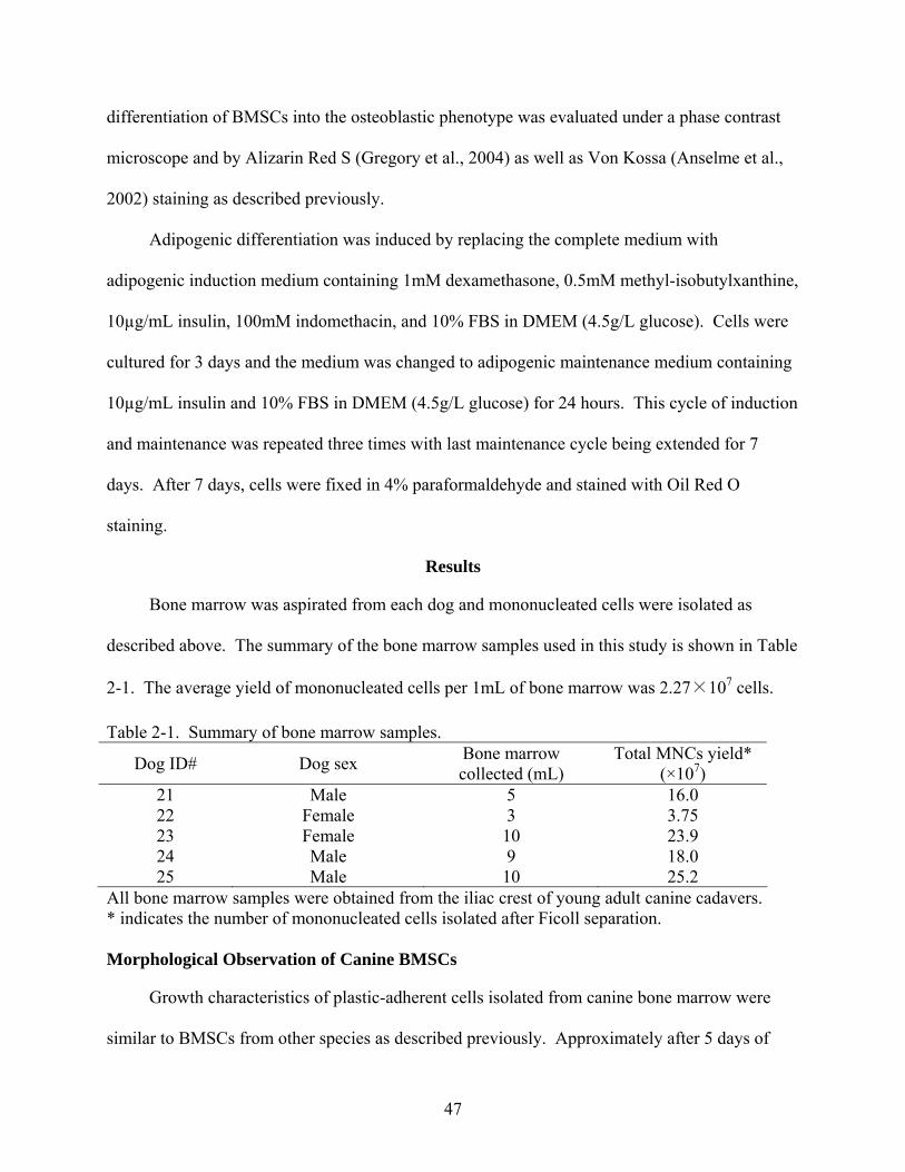
47
differentiation of BMSCs into the osteoblastic phenotype was evaluated under a phase contrast
microscope and by Alizarin Red S (Gregory et al., 2004) as well as Von Kossa (Anselme et al.,
2002) staining as described previously.
Adipogenic differentiation was induced by replacing the complete medium with
adipogenic induction medium containing 1mM dexamethasone, 0.5mM methyl-isobutylxanthine,
10µg/mL insulin, 100mM indomethacin, and 10% FBS in DMEM (4.5g/L glucose). Cells were
cultured for 3 days and the medium was changed to adipogenic maintenance medium containing
10µg/mL insulin and 10% FBS in DMEM (4.5g/L glucose) for 24 hours. This cycle of induction
and maintenance was repeated three times with last maintenance cycle being extended for 7
days. After 7 days, cells were fixed in 4% paraformaldehyde and stained with Oil Red O
staining.
Results
Bone marrow was aspirated from each dog and mononucleated cells were isolated as
described above. The summary of the bone marrow samples used in this study is shown in Table
2-1. The average yield of mononucleated cells per 1mL of bone marrow was 2.27×10⁷ cells.
Table 2-1. Summary of bone marrow samples.
Dog ID# Dog sex Bone marrow collected (mL)
Total MNCs yield* (×107)
21 Male 5 16.0 22 Female 3 3.75 23 Female 10 23.9 24 Male 9 18.0 25 Male 10 25.2
All bone marrow samples were obtained from the iliac crest of young adult canine cadavers. * indicates the number of mononucleated cells isolated after Ficoll separation. Morphological Observation of Canine BMSCs
Growth characteristics of plastic-adherent cells isolated from canine bone marrow were
similar to BMSCs from other species as described previously. Approximately after 5 days of
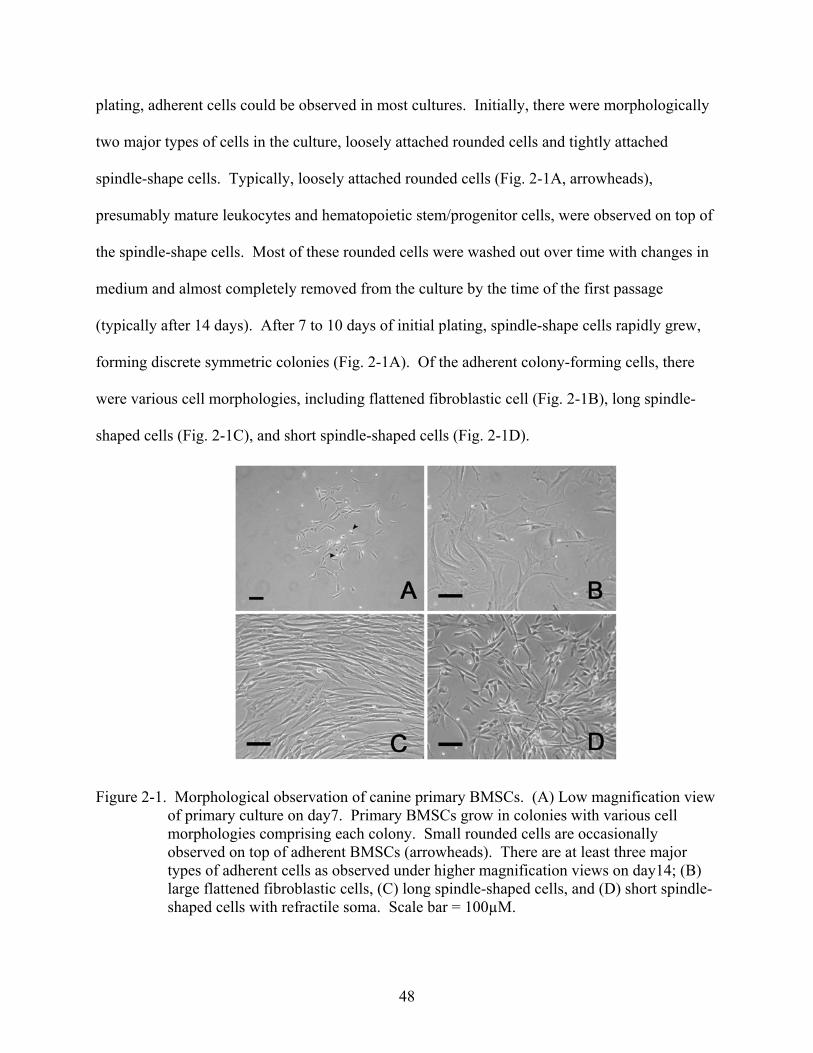
48
plating, adherent cells could be observed in most cultures. Initially, there were morphologically
two major types of cells in the culture, loosely attached rounded cells and tightly attached
spindle-shape cells. Typically, loosely attached rounded cells (Fig. 2-1A, arrowheads),
presumably mature leukocytes and hematopoietic stem/progenitor cells, were observed on top of
the spindle-shape cells. Most of these rounded cells were washed out over time with changes in
medium and almost completely removed from the culture by the time of the first passage
(typically after 14 days). After 7 to 10 days of initial plating, spindle-shape cells rapidly grew,
forming discrete symmetric colonies (Fig. 2-1A). Of the adherent colony-forming cells, there
were various cell morphologies, including flattened fibroblastic cell (Fig. 2-1B), long spindle-
shaped cells (Fig. 2-1C), and short spindle-shaped cells (Fig. 2-1D).
Figure 2-1. Morphological observation of canine primary BMSCs. (A) Low magnification view of primary culture on day7. Primary BMSCs grow in colonies with various cell morphologies comprising each colony. Small rounded cells are occasionally observed on top of adherent BMSCs (arrowheads). There are at least three major types of adherent cells as observed under higher magnification views on day14; (B) large flattened fibroblastic cells, (C) long spindle-shaped cells, and (D) short spindle-shaped cells with refractile soma. Scale bar = 100µM.
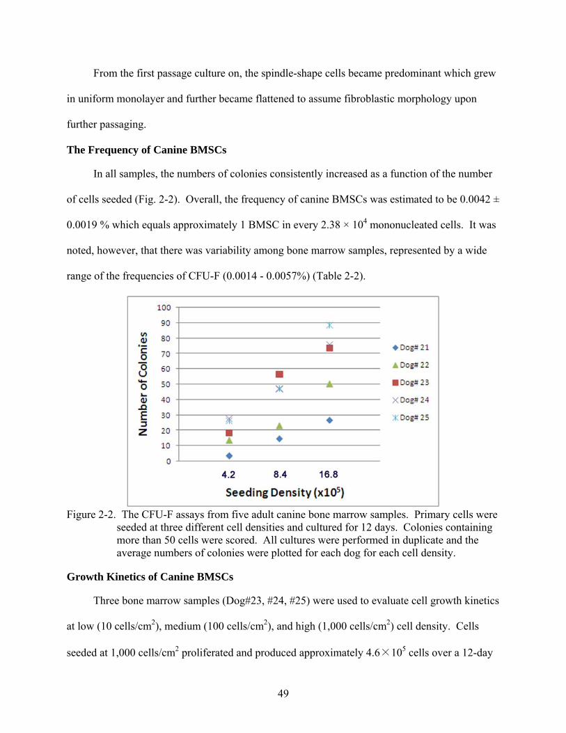
49
From the first passage culture on, the spindle-shape cells became predominant which grew
in uniform monolayer and further became flattened to assume fibroblastic morphology upon
further passaging.
The Frequency of Canine BMSCs
In all samples, the numbers of colonies consistently increased as a function of the number
of cells seeded (Fig. 2-2). Overall, the frequency of canine BMSCs was estimated to be 0.0042 ±
0.0019 % which equals approximately 1 BMSC in every 2.38 × 10⁴ mononucleated cells. It was
noted, however, that there was variability among bone marrow samples, represented by a wide
range of the frequencies of CFU-F (0.0014 - 0.0057%) (Table 2-2).
Figure 2-2. The CFU-F assays from five adult canine bone marrow samples. Primary cells were
seeded at three different cell densities and cultured for 12 days. Colonies containing more than 50 cells were scored. All cultures were performed in duplicate and the average numbers of colonies were plotted for each dog for each cell density.
Growth Kinetics of Canine BMSCs
Three bone marrow samples (Dog#23, #24, #25) were used to evaluate cell growth kinetics
at low (10 cells/cm2), medium (100 cells/cm2), and high (1,000 cells/cm2) cell density. Cells
seeded at 1,000 cells/cm2 proliferated and produced approximately 4.6×10⁵ cells over a 12-day
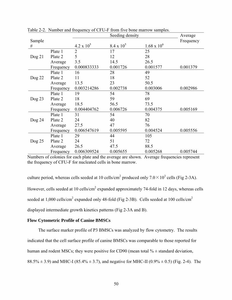
50
Table 2-2. Number and frequency of CFU-F from five bone marrow samples. Seeding density Average
Sample # 4.2 x 10⁵ 8.4 x 10⁵ 1.68 x 10⁶
Frequency
Plate 1 2 17 25 Dog 21 Plate 2 5 12 28 Average 3.5 14.5 26.5 Frequency 0.000833333 0.001726 0.001577 0.001379 Plate 1 16 28 49 Dog 22 Plate 2 11 18 52 Average 13.5 23 50.5 Frequency 0.003214286 0.002738 0.003006 0.002986 Plate 1 19 54 78 Dog 23 Plate 2 18 59 69 Average 18.5 56.5 73.5 Frequency 0.004404762 0.006726 0.004375 0.005169 Plate 1 31 54 70 Dog 24 Plate 2 24 40 82 Average 27.5 47 76 Frequency 0.006547619 0.005595 0.004524 0.005556 Plate 1 29 44 105 Dog 25 Plate 2 24 51 72 Average 26.5 47.5 88.5
Frequency 0.006309524 0.005655 0.005268 0.005744 Numbers of colonies for each plate and the average are shown. Average frequencies represent the frequency of CFU-F for nucleated cells in bone marrow.
culture period, whereas cells seeded at 10 cells/cm2 produced only 7.0×10³ cells (Fig 2-3A).
However, cells seeded at 10 cells/cm2 expanded approximately 74-fold in 12 days, whereas cells
seeded at 1,000 cells/cm2 expanded only 48-fold (Fig 2-3B). Cells seeded at 100 cells/cm2
displayed intermediate growth kinetics patterns (Fig 2-3A and B).
Flow Cytometric Profile of Canine BMSCs
The surface marker profile of P3 BMSCs was analyzed by flow cytometry. The results
indicated that the cell surface profile of canine BMSCs was comparable to those reported for
human and rodent MSCs; they were positive for CD90 (mean total % ± standard deviation,
88.5% ± 3.9) and MHC-I (85.4% ± 3.7), and negative for MHC-II (0.9% ± 0.5) (Fig. 2-4). The
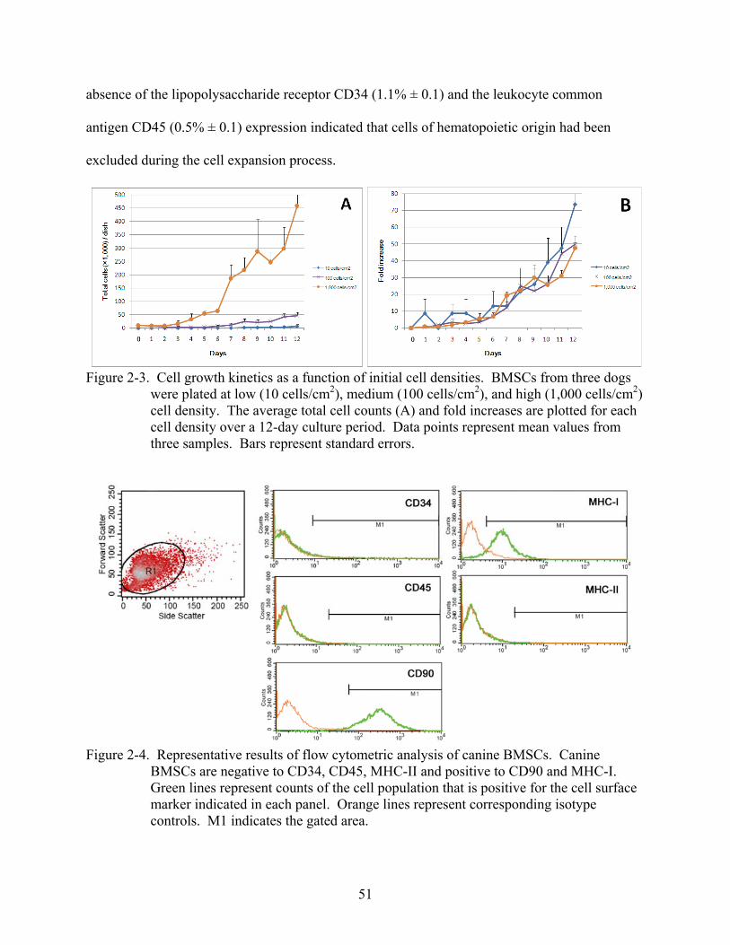
51
absence of the lipopolysaccharide receptor CD34 (1.1% ± 0.1) and the leukocyte common
antigen CD45 (0.5% ± 0.1) expression indicated that cells of hematopoietic origin had been
excluded during the cell expansion process.
Figure 2-3. Cell growth kinetics as a function of initial cell densities. BMSCs from three dogs
were plated at low (10 cells/cm2), medium (100 cells/cm2), and high (1,000 cells/cm2) cell density. The average total cell counts (A) and fold increases are plotted for each cell density over a 12-day culture period. Data points represent mean values from three samples. Bars represent standard errors.
Figure 2-4. Representative results of flow cytometric analysis of canine BMSCs. Canine
BMSCs are negative to CD34, CD45, MHC-II and positive to CD90 and MHC-I. Green lines represent counts of the cell population that is positive for the cell surface marker indicated in each panel. Orange lines represent corresponding isotype controls. M1 indicates the gated area.
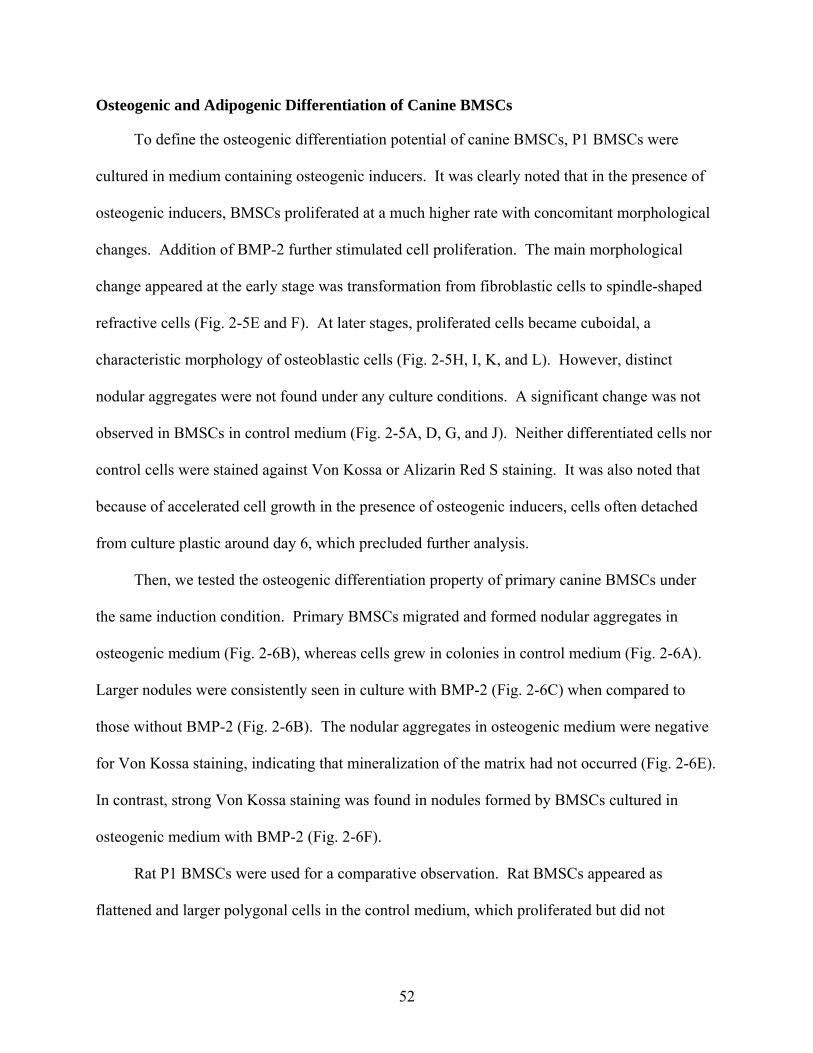
52
Osteogenic and Adipogenic Differentiation of Canine BMSCs
To define the osteogenic differentiation potential of canine BMSCs, P1 BMSCs were
cultured in medium containing osteogenic inducers. It was clearly noted that in the presence of
osteogenic inducers, BMSCs proliferated at a much higher rate with concomitant morphological
changes. Addition of BMP-2 further stimulated cell proliferation. The main morphological
change appeared at the early stage was transformation from fibroblastic cells to spindle-shaped
refractive cells (Fig. 2-5E and F). At later stages, proliferated cells became cuboidal, a
characteristic morphology of osteoblastic cells (Fig. 2-5H, I, K, and L). However, distinct
nodular aggregates were not found under any culture conditions. A significant change was not
observed in BMSCs in control medium (Fig. 2-5A, D, G, and J). Neither differentiated cells nor
control cells were stained against Von Kossa or Alizarin Red S staining. It was also noted that
because of accelerated cell growth in the presence of osteogenic inducers, cells often detached
from culture plastic around day 6, which precluded further analysis.
Then, we tested the osteogenic differentiation property of primary canine BMSCs under
the same induction condition. Primary BMSCs migrated and formed nodular aggregates in
osteogenic medium (Fig. 2-6B), whereas cells grew in colonies in control medium (Fig. 2-6A).
Larger nodules were consistently seen in culture with BMP-2 (Fig. 2-6C) when compared to
those without BMP-2 (Fig. 2-6B). The nodular aggregates in osteogenic medium were negative
for Von Kossa staining, indicating that mineralization of the matrix had not occurred (Fig. 2-6E).
In contrast, strong Von Kossa staining was found in nodules formed by BMSCs cultured in
osteogenic medium with BMP-2 (Fig. 2-6F).
Rat P1 BMSCs were used for a comparative observation. Rat BMSCs appeared as
flattened and larger polygonal cells in the control medium, which proliferated but did not
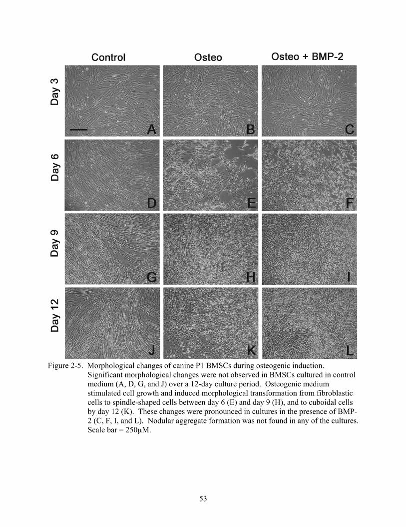
53
Figure 2-5. Morphological changes of canine P1 BMSCs during osteogenic induction.
Significant morphological changes were not observed in BMSCs cultured in control medium (A, D, G, and J) over a 12-day culture period. Osteogenic medium stimulated cell growth and induced morphological transformation from fibroblastic cells to spindle-shaped cells between day 6 (E) and day 9 (H), and to cuboidal cells by day 12 (K). These changes were pronounced in cultures in the presence of BMP-2 (C, F, I, and L). Nodular aggregate formation was not found in any of the cultures. Scale bar = 250µM.
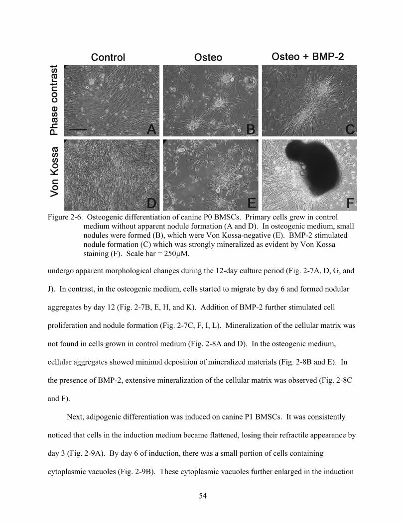
54
Figure 2-6. Osteogenic differentiation of canine P0 BMSCs. Primary cells grew in control
medium without apparent nodule formation (A and D). In osteogenic medium, small nodules were formed (B), which were Von Kossa-negative (E). BMP-2 stimulated nodule formation (C) which was strongly mineralized as evident by Von Kossa staining (F). Scale bar = 250µM.
undergo apparent morphological changes during the 12-day culture period (Fig. 2-7A, D, G, and
J). In contrast, in the osteogenic medium, cells started to migrate by day 6 and formed nodular
aggregates by day 12 (Fig. 2-7B, E, H, and K). Addition of BMP-2 further stimulated cell
proliferation and nodule formation (Fig. 2-7C, F, I, L). Mineralization of the cellular matrix was
not found in cells grown in control medium (Fig. 2-8A and D). In the osteogenic medium,
cellular aggregates showed minimal deposition of mineralized materials (Fig. 2-8B and E). In
the presence of BMP-2, extensive mineralization of the cellular matrix was observed (Fig. 2-8C
and F).
Next, adipogenic differentiation was induced on canine P1 BMSCs. It was consistently
noticed that cells in the induction medium became flattened, losing their refractile appearance by
day 3 (Fig. 2-9A). By day 6 of induction, there was a small portion of cells containing
cytoplasmic vacuoles (Fig. 2-9B). These cytoplasmic vacuoles further enlarged in the induction
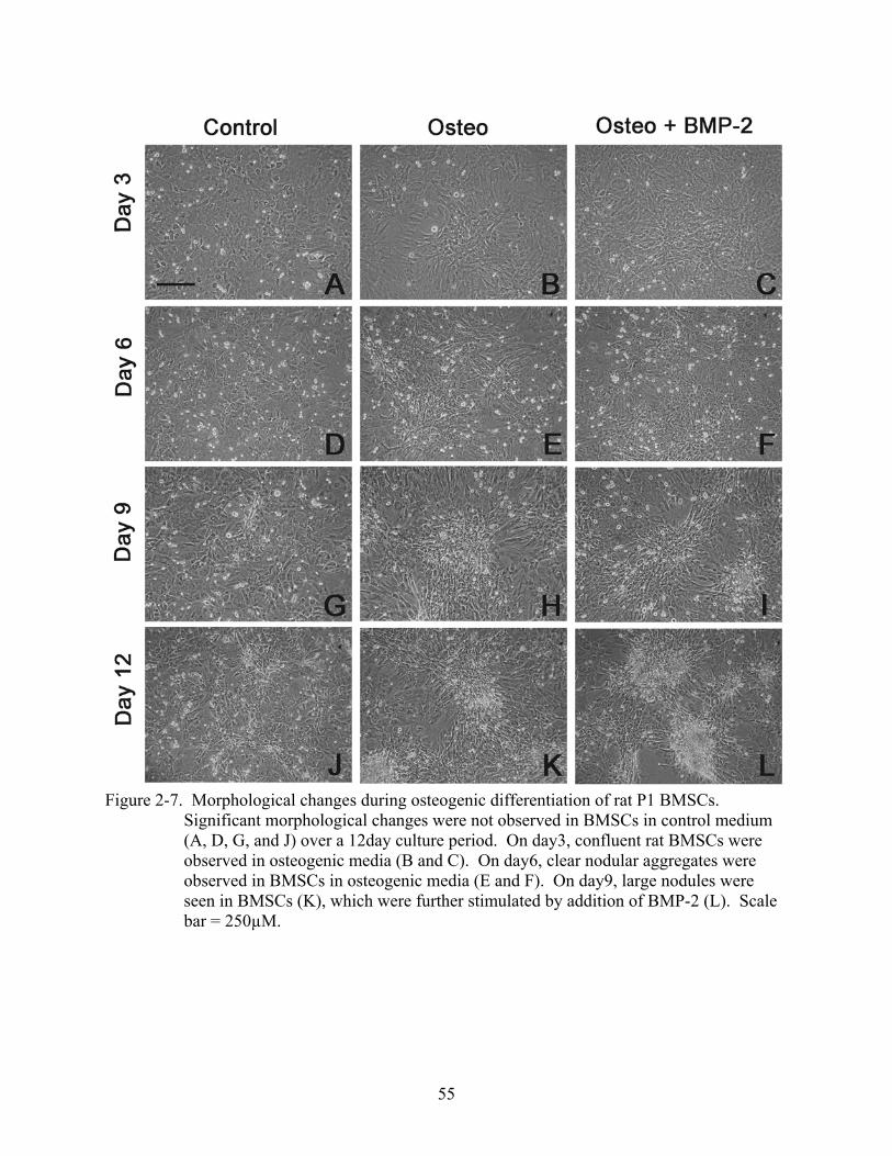
55
Figure 2-7. Morphological changes during osteogenic differentiation of rat P1 BMSCs.
Significant morphological changes were not observed in BMSCs in control medium (A, D, G, and J) over a 12day culture period. On day3, confluent rat BMSCs were observed in osteogenic media (B and C). On day6, clear nodular aggregates were observed in BMSCs in osteogenic media (E and F). On day9, large nodules were seen in BMSCs (K), which were further stimulated by addition of BMP-2 (L). Scale bar = 250µM.
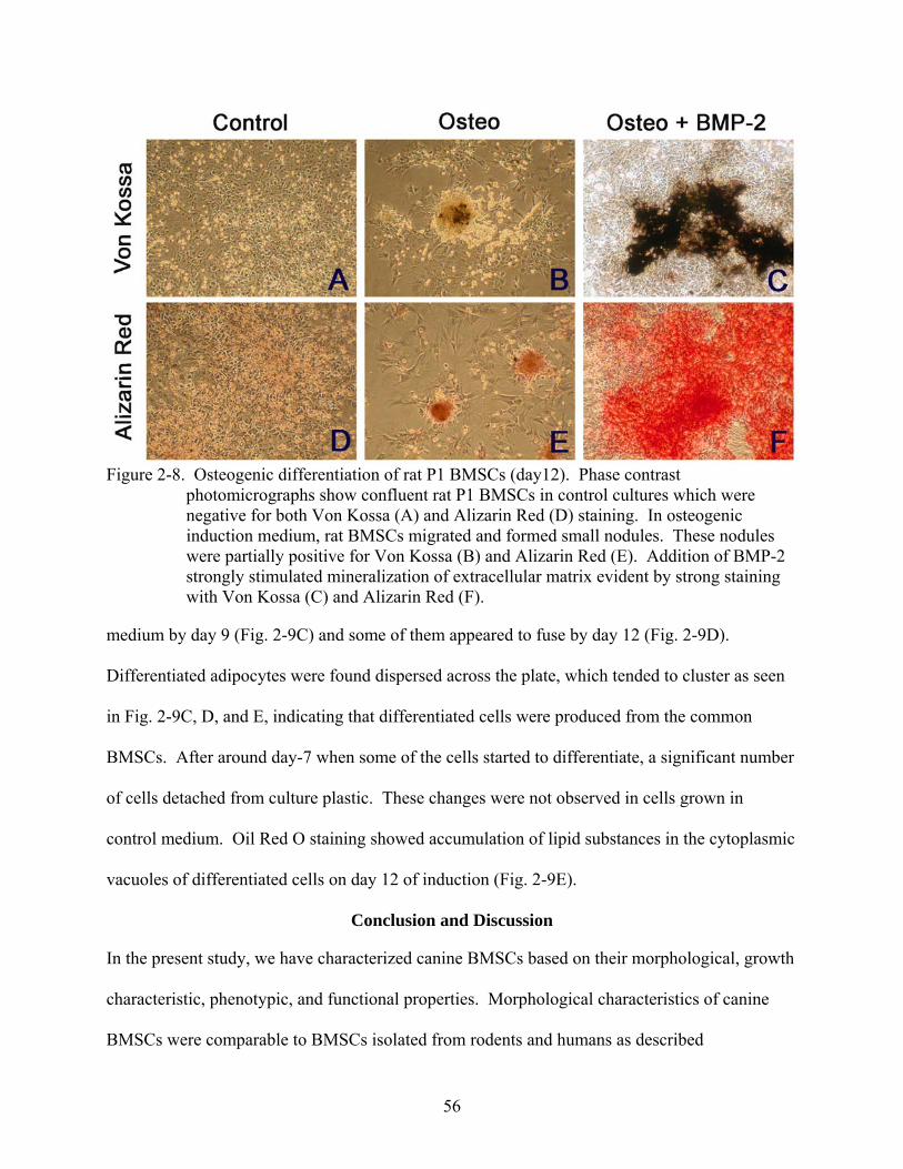
56
Figure 2-8. Osteogenic differentiation of rat P1 BMSCs (day12). Phase contrast
photomicrographs show confluent rat P1 BMSCs in control cultures which were negative for both Von Kossa (A) and Alizarin Red (D) staining. In osteogenic induction medium, rat BMSCs migrated and formed small nodules. These nodules were partially positive for Von Kossa (B) and Alizarin Red (E). Addition of BMP-2 strongly stimulated mineralization of extracellular matrix evident by strong staining with Von Kossa (C) and Alizarin Red (F).
medium by day 9 (Fig. 2-9C) and some of them appeared to fuse by day 12 (Fig. 2-9D).
Differentiated adipocytes were found dispersed across the plate, which tended to cluster as seen
in Fig. 2-9C, D, and E, indicating that differentiated cells were produced from the common
BMSCs. After around day-7 when some of the cells started to differentiate, a significant number
of cells detached from culture plastic. These changes were not observed in cells grown in
control medium. Oil Red O staining showed accumulation of lipid substances in the cytoplasmic
vacuoles of differentiated cells on day 12 of induction (Fig. 2-9E).
Conclusion and Discussion
In the present study, we have characterized canine BMSCs based on their morphological, growth
characteristic, phenotypic, and functional properties. Morphological characteristics of canine
BMSCs were comparable to BMSCs isolated from rodents and humans as described
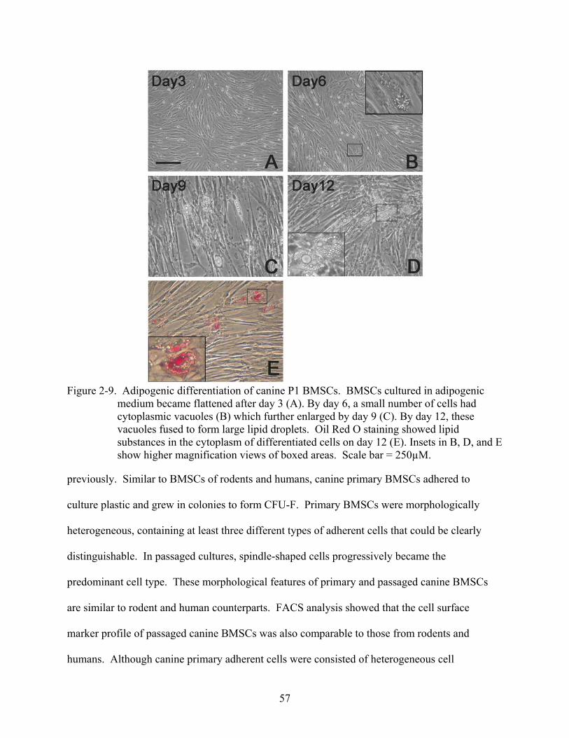
57
Figure 2-9. Adipogenic differentiation of canine P1 BMSCs. BMSCs cultured in adipogenic
medium became flattened after day 3 (A). By day 6, a small number of cells had cytoplasmic vacuoles (B) which further enlarged by day 9 (C). By day 12, these vacuoles fused to form large lipid droplets. Oil Red O staining showed lipid substances in the cytoplasm of differentiated cells on day 12 (E). Insets in B, D, and E show higher magnification views of boxed areas. Scale bar = 250µM.
previously. Similar to BMSCs of rodents and humans, canine primary BMSCs adhered to
culture plastic and grew in colonies to form CFU-F. Primary BMSCs were morphologically
heterogeneous, containing at least three different types of adherent cells that could be clearly
distinguishable. In passaged cultures, spindle-shaped cells progressively became the
predominant cell type. These morphological features of primary and passaged canine BMSCs
are similar to rodent and human counterparts. FACS analysis showed that the cell surface
marker profile of passaged canine BMSCs was also comparable to those from rodents and
humans. Although canine primary adherent cells were consisted of heterogeneous cell
58
populations, significant hematopoietic contamination was not evident; therefore, the plastic
adhesion method seemed sufficient for isolation of canine BMSCs. This is in contrast to murine
BMSCs in which the plastic adhesion method results in significant contamination of granulo-
monocytic cells in both primary and passaged cultures (Phinney et al., 1999; Xu et al., 1983),
thus requiring preceding immunodepletion using CD11b antibody (Kopen et al., 1999). It is
unknown whether morphologically heterogeneous cell populations in the canine primary
adherent cultures represent distinct classes of stem cells, perhaps containing rare pluripotent
stem cell populations present in human and murine bone marrow (Jiang et al., 2002a; Jiang et al.,
2002b; Reyes et al., 2001), or cells of the same lineage with different developmental stages. At
present, the latter is likely the case as human BMSCs are also morphologically heterogeneous
even when cloned from single-cell-derived colonies (Colter et al., 2000; Zohar et al., 1997).
The frequency of BMSCs in canine bone marrow has been reported by Kadiyala (1997)
to be approximately 0.004% (1 per 2.5 × 10⁴ mononucleated cells), although detailed data was
not presented in their study. In the present study, CFU-F assays demonstrated that the frequency
of canine BMSCs was approximately 0.0042% (1 per 2.38 × 10⁴ mononucleated cells). This
frequency of canine BMSCs is twofold and fourfold higher than the previously reported
frequencies of murine (Friedenstein et al., 1976) and human BMSCs (Bruder et al., 1997),
respectively. However, the number of CFU-F can be influenced by culture conditions (e.g. FBS
concentration, type of basal medium, pH of medium, etc) (Xu et al., 1983); therefore, further
studies are needed to optimize the culture condition for canine BMSCs. We found that the
numbers of CFU-F were variable among bone marrow samples and several factors could account
for this finding. Age is a known factor that has impact on the frequency of BMSCs. In the
present study, we did not have information regarding the exact age of the dogs. Although the
59
dogs in the present study were considered to represent “young adult” based on their denture,
differences in their maturity might have influenced the frequency of BMSCs. The degree of
peripheral blood contamination is another factor since CFU-F is calculated based on the number
of mononucleated cells in the sample. This could be significantly influenced by postmortem
changes such as coagulation of peripheral blood. This problem may arise when bone marrow is
collected by aspiration but not by flushing long bones which is a common technique for
collection of rodent BMSCs.
Growth kinetics of canine BMSCs were evaluated under three different cell seeding
densities (10 cells/cm2, 100 cells/cm2, and 1,000 cells/cm2). It was clearly shown that initial cell
densities have a profound influence on the proliferation rate of canine BMSCs, as the case for
human BMSCs (Sekiya et al., 2002). Among the three cell densities tested, there was an inverse
relationship between the total yield of BMSCs and expansion rate (fold increase). This suggests
that when BMSCs are seeded at a lower density, they undergo more rapid proliferation. It also
implies that, because increasing cell density is known to trigger cell differentiation (Caplan et al.,
1983), there must be a compromise between the total cell yield and the content of
undifferentiated cells. Further studies are necessary to define the optimal culture condition,
which generates the maximal cell yield with the greatest content of multipotent canine BMSCs.
Our functional assays demonstrated that passaged canine BMSCs showed limited
osteogenic differentiation ability. Although some morphological changes, which resembled
osteoblastic morphology, were observed in passaged BMSCs cultured in osteogenic medium,
formation of nodular aggregates was never found in passaged BMSCs, indicating that full
differentiation toward osteoblasts did not occur. Osteogenic inducers stimulated cell growth,
which often resulted in detachment of cell monolayer around after day 6 of induction and
60
precluded further culture of BMSCs. Despite previous studies (Kadiyala et al., 1997; Volk et al.,
2005) describing differentiation of passaged canine BMSCs into osteoblasts, these results were
not reproducible in our hands. In contrast, primary canine BMSCs differentiated into osteoblasts
and formed nodules which were intensively mineralized. Primary BMSCs cultured in medium
without osteogenic inducers did not show similar changes; thus, contamination of osteoblastic
cells in the samples seemed unreasonable. When passaged BMSCs were grown in adipogenic
inducers, a small number of cells differentiated into mature adipocytes. Upon exposure to a high
concentration of dexamethasone, a significant number of undifferentiated cells detached from the
culture plate. These results demonstrated that a small number of BMSCs possess adipogenic
capacity in the passaged BMCS culture. All together, the results of our functional assays suggest
that canine bone marrow contains inducible mesenchymal progenitors with the ability to giving
rise to osteoblasts and adipocytes. Upon passaging, a significant number of these stem cells
either lose their multipotency or did not survive in the standard culture condition. Further studies
are needed to develop culture techniques which enable expansion of canine BMSCs in their
undifferentiated state.
61
CHAPTER 3 IN VITRO NEURAL DIFFERENTIATION OF CANINE BONE MARROW STROMAL
CELLS
Background and Introduction
A variety of cell types such as embryonic stem cells (McDonald et al., 1999; Wernig et al.,
2004) and neural progenitor/stem cells (Ogawa et al., 2002; Pluchino et al., 2003), and
developing tissues harboring stem cells such as fetal spinal cord tissues (Anderson et al., 1995;
Houle, 1992) have been shown to promote CNS regeneration in experimental studies. However,
clinical application of these sources is severely limited due to issues associated with the
availability of the transplants, immunological reactions, and most importantly with ethical
considerations. Cells derived from bone marrow, particularly bone marrow-derived stromal cells
(BMSCs, also referred to as mesenchymal stem cells), possess potentials to circumvent many of
these obstacles; therefore, may represent the most realistic cell source.
Recent in vitro studies demonstrated that BMSCs can generate not only mesodermal cells
but also endodermal and ectodermal cell types, suggesting that these cells are less restricted than
were previously thought. Neural differentiation of cells derived from rat and human bone
marrow has been reported by several independent groups (Deng et al., 2001; Sanchez-Ramos et
al., 2000; Woodbury et al., 2000). These studies have aroused considerable interest in the
potential use of BMSCs for neurological disorders. Unlike ES cells and neural stem cells where
embryos or neonatal neural tissues are necessary to obtain these cells, BMSCs are easily
accessible by bone marrow aspiration. Furthermore, BMSCs are easily expandable in culture
without losing their multipotentiality (Pittenger et al., 1999). Because of these advantages,
BMSCs have received great attention as a promising source of cells for a variety of therapies
including neuroregeneration.
62
Early studies on neural differentiation of bone marrow-derived stem cells were based on
morphological changes and immunoreactivity against some of the neural markers (Sanchez-
Ramos et al., 2000; Woodbury et al., 2000). The significance of the morphological changes
observed in these studies, however, has been questioned by others and attributed to actin
cytoskeleton shrinkage rather than genuine neurofilament extension (Bertani et al., 2005; Lu et
al., 2004; Neuhuber et al., 2004). As such, interpretation of neural differentiation of BMSCs in
vitro needs great caution and systematic approaches including assays at the molecular level are
desirable. While it is still the subject of debate whether neurogenesis of BMSCs represents
genuine “trans-differentiation” or an epiphenomenon caused by induced chemical stress,
functionalities of MSC-derived neurons, such as their abilities to secrete neurotransmitters and/or
fire action potentials, have been reported by several groups (Hermann et al., 2004; Kohyama et
al., 2001; Tao et al., 2005; Wislet-Gendebien et al., 2005). These in vitro studies have raised
tremendous interest that BMSCs have the potential to differentiate into functional neural cells.
Based on the previous findings in human and rodent BMSC studies, we hypothesized that
canine BMSCs can be culture-expanded while maintaining their multipotentiality and that these
cells bear neural properties. We induced a rapid neural differentiation on canine BMSCs as
previously described by Deng et al. (2001) and evaluated expression of neuronal and glial-
specific proteins, using immunocytochemical and western blotting analyses. We further
evaluated the electrophysiological properties of neurally induced BMSCs using the whole-cell
voltage clamp technique.
Materials and Methods
Preparation of Canine BMSCs
Bone marrow aspirates from 5 canine cadavers were used to isolate and culture canine
BMSCs. Detailed descriptions of bone marrow collection, cell isolation, and culture expansion
63
can be found in Chapter 2. Third passage (P3) BMSCs were used for the assays described in this
chapter.
Neural Differentiation of Canine BMSCs
Neural differentiation of P3 BMSCs was induced by replacing the complete medium by
neural induction medium. The neural induction medium consisted of 0.5mM methyl-
isobutylxanthine (IBMX) and 1mM dibutyryl cAMP (dbcAMP) in DMEM/F12 as used in neural
differentiation of human bone marrow stromal cells (Deng et al., 2001). To support growth of
neural cells the medium was supplemented with 1% N2 supplements. For
immunocytochemistry, a total of 1 × 10⁴ P3 BMSCs were suspended in 100 µl of complete
medium and plated on coverslips placed in each well of 12-well culture plates. After 12 hours,
900 µl of complete medium was added and cells were cultured until reaching 70% confluency.
Consequently, the medium was changed to 1 ml of neural induction medium and cultured at
37°C in a humidified 5% CO2 environment for 5 hours. For Western blot analysis, P3 BMSCs
were plated at 8,000 cells/cm2 on 60 mm culture dishes in complete medium. Upon reaching
70% confluency, cells were washed with PBS and cultured in neural induction medium for 5
hours. Cells were also cultured in complete media and served as control. We also used canine
fibroblast cultures (also from passage 3) (Coriell Institute for Medical Research) as a control for
the assays described below.
Immunocytochemistry for Neural Specific Markers
We performed immunocytochemistry to evaluate whether canine express neuronal (βШ-
tubulin), astrocyte-specific (GFAP), and oligodendrocyte-specific (MBP) proteins. Cells on
coverslips cultured in complete medium or neural differentiation medium were gently washed
with PBS and fixed in ice-cold 4% paraformaldehyde for 30 minutes at room temperature (RT).
Fixed cells were permeabilized in 1.0% Triton X-100 in PBS for 10 minutes at RT. Non-specific
64
binding was blocked with a blocking solution (1.0% BSA in PBS) for 30 minutes at RT. Cells
were then incubated with primary antibodies, including anti-ß Ш-tubulin (Promega Corp,
1:1,000), anti-GFAP (BD Biosciences, 1:1,000), anti-MBP (HyTest Ltd, 1:50,000), for 1 hour at
RT. The primary antibodies were removed, cells washed with PBS, and cells incubated with one
of secondary antibodies for 1 hour in dark at RT. The secondary antibodies (all from Jackson
ImmunoResearch Laboratories Inc.) used were Cy2 conjugated goat anti-mouse IgG1 (1:400),
Rhodamine conjugated goat anti-mouse IgG2b (1:100), FITC conjugated goat anti-mouse IgG2a
(1:400). Cells were also incubated without primary antibodies to control for non-specific
staining by secondary antibodies. Cells were washed with PBS and mounted with a mounting
medium containing DAPI (Vector Laboratories). Stained cells were observed under a
fluorescent microscope (Zeiss Axioplan II, Carl Zeiss MicroImaging) with appropriate filters.
Western Blotting and Densitometric Analyses
To confirm expressions of neuronal (ß Ш-tubulin) and glial (GFAP and MBP) markers on
canine BMSCs, cultures were detached with 0.05% Trypsin/0.53mM EDTA for western blot
analyses. Cells were lysed with a lysis solution (0.18M Tris-HCl, 40% Glycerol, 4% SDS,
0.04% BPB, 0.05M DTT). Canine spinal cord lysate was obtained using the same lysis solution
and used as positive control. Total protein concentrations of each sample were determined by a
protein assay kit (Micro BCA protein assay, Pierce). Samples containing 20 µg of protein were
loaded in each lane of 12% polyacrylamide gels and electrophoretically separated. Separated
proteins were transferred to nitrocellulose membranes, blocked with TBS containing 5% non-fat
dry milk and 0.1% Tween20 for 1 hour at RT, and incubated with primary antibodies (βШ-
tubulin; 1:500, GFAP; 1:4,000, MBP; 1:50,000) overnight at 4°C. The membranes were also
probed with β-actin (Abcam Inc, 1:1,000) as loading controls. After washing the membranes
with TTBS (0.05% Tween20 in TBS solution) for three times, the membranes were incubated
65
with alkaline phosphate conjugated secondary antibodies (Jackson ImmunoResearch
Laboratories Inc., 1:5,000) for 1 hour at RT. The membranes were washed three times in TTBS
and developed in a solution containing NBT and BCIP. Photographs of the membranes were
taken using a molecular imager (Flour-S, Multi-imager, Bio-Rad Laboratories) and densitometric
analyses performed using a software program (Quantity One, Bio-Rad Laboratories). Western
blotting and densitometric analyses were duplicated in all samples.
Electrophysiological Recording
Two representative BMSC cultures were used for electrophysiological recordings. Neural
differentiation was performed as described above. Spontaneous and depolarizing pulse-elicited
action potentials were recorded with the whole-cell voltage clamp configuration in current clamp
mode (Hamill et al., 1981). Experiments were performed at room temperature (23–24°C) with
an Axopatch 200B amplifier and a Digidatal 1200B interface (Axon Instruments, Burlingame,
CA). Data acquisition and analyses were performed with the use of Axoscope 7.0 and pClamp
6.2. Differentiated BMSCs were bathed in Tyrode’s solution containing 140 mM NaCl, 5.4 mM
KCl, 2.0 mM CaCl2, 2.0 mM MgCl2, 0.3 mM NaH2PO4, 10 mM HEPES, and 10 mM dextrose,
pH adjusted to 7.4 with NaOH. BMSCs in the culture dish (volume 1.5 ml) were superfused at a
rate of 2–4 ml/min. The patch electrodes (Kimax-5.1, Kimble Glass, Toledo, OH) had
resistances of 3–4 MOhms when filled with an internal pipette solution containing 140 mM KCl,
4 mM MgCl2, 4 mM ATP, 0.1 mM guanosine 59-triphosphate, 10 mM dextrose, and 10 mM
HEPES, pH adjusted to 7.2 with KOH. The whole-cell configuration was formed by applying
negative pressure to the patch electrode.
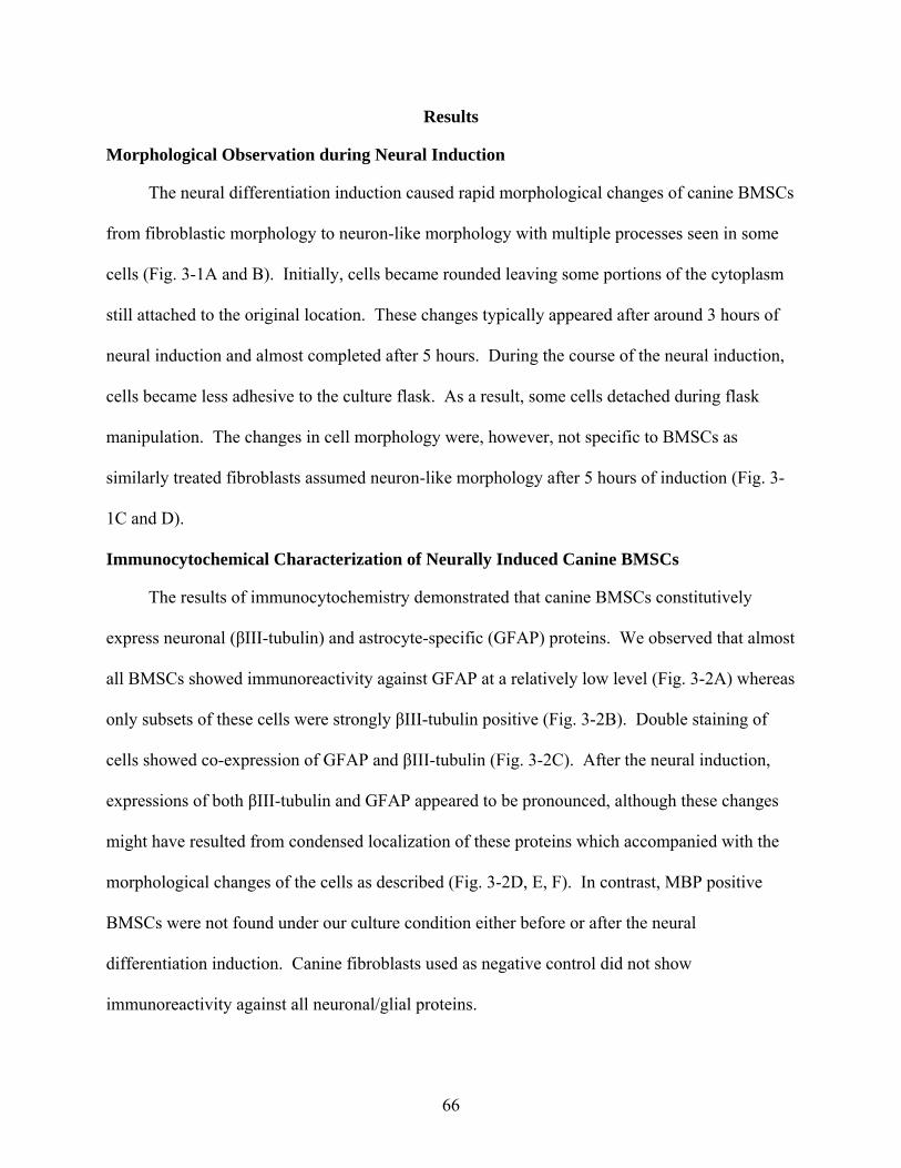
66
Results
Morphological Observation during Neural Induction
The neural differentiation induction caused rapid morphological changes of canine BMSCs
from fibroblastic morphology to neuron-like morphology with multiple processes seen in some
cells (Fig. 3-1A and B). Initially, cells became rounded leaving some portions of the cytoplasm
still attached to the original location. These changes typically appeared after around 3 hours of
neural induction and almost completed after 5 hours. During the course of the neural induction,
cells became less adhesive to the culture flask. As a result, some cells detached during flask
manipulation. The changes in cell morphology were, however, not specific to BMSCs as
similarly treated fibroblasts assumed neuron-like morphology after 5 hours of induction (Fig. 3-
1C and D).
Immunocytochemical Characterization of Neurally Induced Canine BMSCs
The results of immunocytochemistry demonstrated that canine BMSCs constitutively
express neuronal (βIII-tubulin) and astrocyte-specific (GFAP) proteins. We observed that almost
all BMSCs showed immunoreactivity against GFAP at a relatively low level (Fig. 3-2A) whereas
only subsets of these cells were strongly βIII-tubulin positive (Fig. 3-2B). Double staining of
cells showed co-expression of GFAP and βIII-tubulin (Fig. 3-2C). After the neural induction,
expressions of both βIII-tubulin and GFAP appeared to be pronounced, although these changes
might have resulted from condensed localization of these proteins which accompanied with the
morphological changes of the cells as described (Fig. 3-2D, E, F). In contrast, MBP positive
BMSCs were not found under our culture condition either before or after the neural
differentiation induction. Canine fibroblasts used as negative control did not show
immunoreactivity against all neuronal/glial proteins.
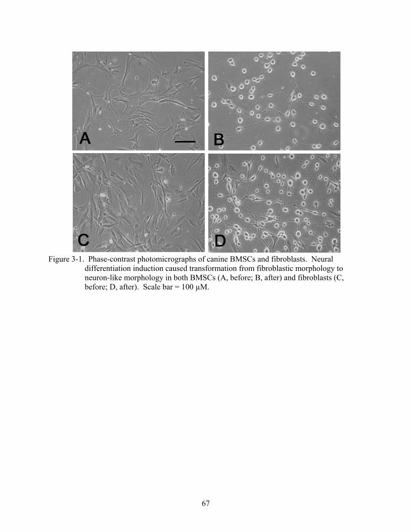
67
Figure 3-1. Phase-contrast photomicrographs of canine BMSCs and fibroblasts. Neural
differentiation induction caused transformation from fibroblastic morphology to neuron-like morphology in both BMSCs (A, before; B, after) and fibroblasts (C, before; D, after). Scale bar = 100 µM.
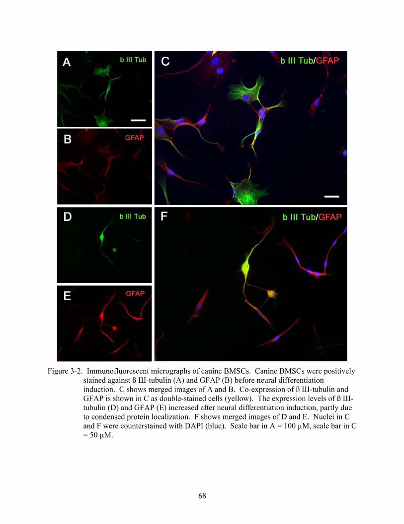
68
Figure 3-2. Immunofluorescent micrographs of canine BMSCs. Canine BMSCs were positively
stained against ß Ш-tubulin (A) and GFAP (B) before neural differentiation induction. C shows merged images of A and B. Co-expression of ß Ш-tubulin and GFAP is shown in C as double-stained cells (yellow). The expression levels of ß Ш-tubulin (D) and GFAP (E) increased after neural differentiation induction, partly due to condensed protein localization. F shows merged images of D and E. Nuclei in C and F were counterstained with DAPI (blue). Scale bar in A = 100 µM, scale bar in C = 50 µM.
69
Western Blot Analysis of Neural Specific Proteins
We performed western blot analyses to confirm the presence of neuronal and glial protein
expressions on canine BMSCs and to evaluate their expression levels before and after the neural
differentiation induction. The presences of distinct bands corresponding to ß III-tubulin and
GFAP were observed in untreated canine BMSCs from all samples (Fig. 3-3). Densitometric
analyses confirmed that the expression level of GFAP in BMSCs consistently increased after the
neural differentiation induction (39.0 ± 20.6 % increase relative to ß-actin). On the other hand,
the expression level of ß III-tubulin in BMSCs did not significantly differ before and after the
neural differentiation induction (9.6 ± 18.5 % increase relative to ß-actin). Bands corresponding
to MBP were not detected in BMSCs before and after the neural differentiation induction.
Expressions of these proteins were not detectable in fibroblasts.
Figure 3-3. Western blotting of neuronal (ß III-tubulin) and glial proteins (GFAP). Loaded
samples were obtained from (A) spinal cord lysate (positive control); (B) fibroblasts before neural induction; (C) fibroblasts after neural induction; (D) BMSCs before neural induction; (E) BMSCs after neural induction.
Electrophysiological Recording
In order to assess the electrophysiological properties of neurally induced canine BMSCs,
we applied the whole-cell voltage clamp technique. We observed neither spontaneous action
potentials nor depolarizing pulse-elicited action potentials from induced canine BMSCs. This
suggested that although canine BMSCs constitutively expressed a neuronal marker and neurally
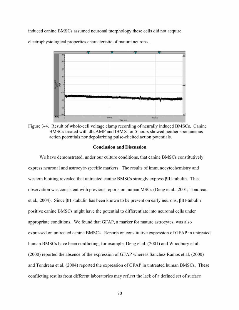
70
induced canine BMSCs assumed neuronal morphology these cells did not acquire
electrophysiological properties characteristic of mature neurons.
Figure 3-4. Result of whole-cell voltage clamp recording of neurally induced BMSCs. Canine
BMSCs treated with dbcAMP and IBMX for 5 hours showed neither spontaneous action potentials nor depolarizing pulse-elicited action potentials.
Conclusion and Discussion
We have demonstrated, under our culture conditions, that canine BMSCs constitutively
express neuronal and astrocyte-specific markers. The results of immunocytochemistry and
western blotting revealed that untreated canine BMSCs strongly express βIII-tubulin. This
observation was consistent with previous reports on human MSCs (Deng et al., 2001; Tondreau
et al., 2004). Since βIII-tubulin has been known to be present on early neurons, βIII-tubulin
positive canine BMSCs might have the potential to differentiate into neuronal cells under
appropriate conditions. We found that GFAP, a marker for mature astrocytes, was also
expressed on untreated canine BMSCs. Reports on constitutive expression of GFAP in untreated
human BMSCs have been conflicting; for example, Deng et al. (2001) and Woodbury et al.
(2000) reported the absence of the expression of GFAP whereas Sanchez-Ramos et al. (2000)
and Tondreau et al. (2004) reported the expression of GFAP in untreated human BMSCs. These
conflicting results from different laboratories may reflect the lack of a defined set of surface
71
markers for BMSCs, resulting in inclusion of unidentified subsets of BMSCs with slightly
different phenotypic profiles. In our study, although expanded canine BMSCs appeared to be
morphologically and phenotypically homogeneous, it was possible that several subsets of
BMSCs existed in our culture. This was illustrated by discrete properties of expanded cells in
their immunoreactivities against ß III-tubulin and GFAP; GFAP was expressed in nearly all
BMSCs whereas ß III-tubulin expression was restricted in approximately 75% of all BMSCs. It
was interesting to note that canine BMSCs expressing neuron-specific proteins also expressed
astrocyte-specific proteins (approximately 75% of all BMSCs). Therefore, similar to human
BMSCs (Reyes et al., 2001; Tondreau et al., 2004), canine MSCs are considered undifferentiated
but may also be characterized as “multi-differentiated”, which may explain their high plasticity.
The absence of MBP expression on canine BMSCs was somewhat predictable as this protein is
only expressed in mature oligodendrocytes and discrete molecular signals may be required to
induce BMSCs to differentiate towards this lineage. Markers for earlier stages of
oligodendrocytes need to be investigated to further characterize the plasticity of canine BMSCs
in their neural differentiation capacity. The effects of the neural differentiation induction on cell
morphology and neuronal/glial marker expressions were evaluated on canine BMSCs. The
induction protocol using dbcAMP and IBMX has been previously described and known to
induce a rapid neuron-like morphological change in human BMSCs as a result of elevated
intracellular cyclic AMP levels (Deng et al., 2001). In our study, after the neural induction,
canine BMSCs exhibited a neuron-like morphology as early as 3 hours after the induction.
However, a similar morphological change was also observed in canine fibroblasts at about the
same rapidity. These observations suggested that the transformation of canine BMSCs from the
fibroblastic morphology to the neuron-like morphology was not a specific event associated with
72
the multipotentiality of canine BMSCs. Therefore, these dramatic morphological changes should
not be overvalued and special care must be taken to interpret results of in vitro neural
differentiation of BMSCs. As pointed previously by several investigators, a rapid morphological
change of BMSCs after neural induction may represent cytotoxic effects of the reagents in the
induction medium, leading to cell shrinkage and subsequent adoption of the neuron-like
morphology (Bertani et al., 2005; Lu et al., 2004; Neuhuber et al., 2004). Commonly used
reagents, solely or in combination, that have effects on BMSCs to induce neuron-like
morphology by cytoskeletal shrinkage include butylated hydroxyanisole (BHA),
dimethylsulphoxide (DMSO), and ß-mercaptoethanol (BME).
Based on the results of immunocytochemistry and western blot analysis, we found that
neural induction caused up-regulation of the expression level of GFAP on canine BMSCs.
Therefore, these results may indicate that canine BMSCs can be stimulated to differentiate along
mature astrocytes. Similar results have been reported in human BMSCs (Tondreau et al., 2004)
and murine BMSCs treated with dbcAMP/IBMX (Deng et al., 2006). Although strong
expression of ß III-tubulin was observed on canine BMSCs before and after neural induction, the
expression levels were not affected by this treatment. As mentioned, canine BMSCs became less
adhesive to the culture flask and there was a significant reduction of the cell number during the
neural differentiation induction. As a result, all analyses were performed 5 hours after the
induction in the present study. However, it would be interesting to further investigate whether a
longer induction period would cause emergence of more mature neuron markers and stable
phenotypes.
Electrophysiological recordings of neurally induced canine BMSCs revealed that these
cells did not acquire the ability to fire spontaneous or depolarizing pulse-elicited action
73
potentials. It may be the case that induced BMSCs committed to the neuronal lineage, as shown
by expression of the early neuronal marker, but have not fully differentiated into a mature
phenotype. Again, a longer induction protocol may be required for full maturation in neural
differentiation of canine BMSCs. Alternatively, other techniques may be more useful to evaluate
the process of differentiation and maturation of neurally induced BMSCs. For example,
expression of voltage-gated ion channels and recordings of inward sodium currents are more
sensitive methods to monitor the process of neural maturation. At current, acquisition of
electrophysiological properties by BMSCs was only achieved by gene transfection (Dezawa et
al., 2004; Kohyama et al., 2001) and treatment with DMSO and BHA on rat BMSCs was
reported to be insufficient in inducing differentiation into mature neuron phenotypes with
electrophysiological properties (Hofstetter et al., 2002).
With the advancement of our understanding of regulatory mechanisms underlying neural
differentiation, it may become possible to generate a specific neural cell type from canine
BMSCs. This approach has been most intensively studied in embryonic stem (ES) cells in an
attempt to generate functional neural precursor cells (Zhang et al., 2001). This approach holds
an advantage in that pre-induction of ES cells into neural progenitor cells may reduce the
possibility of developing tumor (i.e. teratomas) in the transplantation site. Further, induction of
ES cells into more specific neural cell types has been proposed; for example, generation of
dopaminergic neurons from human (Brederlau et al., 2006; Perrier et al., 2004), monkey
(Kawasaki et al., 2002; Takagi et al., 2005), and murine (Thinyane et al., 2005) ES cells has been
reported for potential treatments for Parkinson’s disease. It has also been shown that purified
oligodendrocytes can be generated from human and murine ES cells (Glaser et al., 2005; Nistor
et al., 2005). These ES cells-derived oligodendrocytes have extensive myelinating capacity in
74
vivo, therefore, hold promise for treatment of demyelinating diseases in humans. Dezawa et al.
(2004) showed that rat and human BMSCs can be specifically induced to generate functional
neurons, using gene transfection with Notch intracellular domain. In vitro differentiation of rat
BMSCs into myelinating cells with phenotypic and functional characteristics of Schwann cells
has been previously described (Dezawa et al., 2001; Keilhoff et al., 2006). Kamada et el. (2005)
demonstrated that transplantation of rat BMSC-derived Schwann cells resulted in enhanced
axonal regeneration and functional recovery in rats with completely transected spinal cord.
These studies on BMSC differentiation into specific neural cells are particularly promising in
that if stable functional phenotypes can be achieved BMSCs may become the strongest candidate
for cellular therapies since autologous transplantation is clearly advantageous in a clinical
setting. Induction of canine BMSCs into specific functional neural cell types may be possible by
understanding the transcription factors involved in neurogenesis. Global gene expression
profiling, which has become available through DNA microarray for the canine genome, would
aid developing such techniques.
In conclusion, a large-scale expansion of transplants is one of the critical prerequisites for
clinical applications of cellular transplantation therapies. Canine BMSCs can be readily isolated
from bone marrow of the patient, thus allowing autologous transplantation. These cells can be
further expanded in culture while retaining multi-differentiation capacity. Canine BMSCs hold
neural differentiation properties; therefore, may have the potential for usages in treating various
neurodegenerative diseases and spinal cord injuries in dogs. Further investigations on
mechanisms of neurogenic abilities of canine BMSCs are warranted, which may lead to the
developments of novel therapeutic strategies to target specific CNS diseases. Applying these
novel therapies in canine patients will provide important insights into the safety and clinical

75
efficacy of the treatment, which are the essential elements in translational research of
neurological disorders in human patients.
76
CHAPTER 4 IN VIVO NEURAL DIFFERENTIATION OF CANINE BONE MARROW STROMAL CELLS
Background and Introduction
Neurotransplantation has generated tremendous attention as a potential therapeutic
approach for disorders of the central nervous system (CNS). Cells and tissues derived from
fetuses have been used to treat CNS disorders such as Parkinson’s disease (Kordower et al.,
1995; Spencer et al., 1992) and spinal cord injury (SCI) (Thompson et al., 2001) and led to
significant improvement of clinical signs in some patients. Neural stem cells (Ogawa et al.,
2002; Pluchino et al., 2003; Teng et al., 2002) and embryonic stem cells (Keirstead et al., 2005;
McDonald et al., 1999; Nistor et al., 2005) have also been extensively studied and shown to
ameliorate CNS diseases in experimental models. However, these cell/tissue types pose serious
obstacles with respect to the harvesting of donor cells/tissues and the possibility of
tumorigenesis. Transplantation of bone marrow-derived cells, which has been effectively used to
treat diseases of the hematopoietic system, may provide a viable alternative strategy. Bone
marrow contains bone marrow stromal cells (BMSCs, also referred to as mesenchymal stem
cells), nonhematopoietic cells that provide a bone marrow microenvironment and regulate
hematopoiesis (Dormady et al., 2001) and also serve as a stem cell reservoir for mesenchymal
cells (Pittenger et al., 1999). Due to their multipotency, BMSCs have been shown to regenerate
mesenchymal tissues by differentiating into osteoblasts (Bruder et al., 1997; Kadiyala et al.,
1997), chondrocytes (Kadiyala et al., 1997; Noth et al., 2007; Williams et al., 2003), or skeletal
muscle cells (Dezawa et al., 2005). BMSCs are easily accessible via bone marrow aspiration,
extensively expandable ex vivo, and amenable for gene transfection (Lu et al., 2006). BMSCs,
therefore, represent a promising candidate cell source for cellular therapy for various diseases
including those of the CNS.
77
Perspectives on BMSCs for neurotransplantation have rapidly grown during the last
decade, based on several reports on BMSC transdifferentiation into neural phenotypes both in
vitro (Sanchez-Ramos et al., 2000; Woodbury et al., 2000) and in vivo (Arnhold et al., 2006;
Azizi et al., 1998). These studies demonstrated that adult stem cells may be capable of adopting
appropriate phenotypic fates in response to environmental cues. Therapeutic benefits of BMSC
transplantation has been repeatedly shown in various experimental models of CNS injuries,
which include ischemic (Lee et al., 2003; Zhao et al., 2002), traumatic (Wu et al., 2003), and
degenerative (Wu et al., 2007) lesions in the brain or the spinal cord. Although functional
recovery from CNS injury may not result solely from neural transdifferentiation and cellular
replacement by transplanted BMSCs, investigations of neural transdifferentiation properties of
BMSCs provide insights into their potentials in the treatment of CNS diseases and aid in refining
the design of new clinical trials.
Initial clinical trials in humans with SCI using autologous bone marrow cells have already
been reported (Park et al., 2005; Yoon et al., 2007), and the potential therapeutic value of bone
marrow cells is certainly not limited to human applications. For example, dogs can sustain
traumatic SCI at relatively high incidence. A need thus exists for developing novel treatments
and cell-based therapies for veterinary practice. Based on existing evidence, BMSCs are a
reasonable candidate cell platform. Veterinary clinical applications also afford an important
animal-to-human translation opportunity, especially the pathology of clinical SCI in dogs
appears very similar to that documented in humans, as well as in cats and numerous examples of
experimental SCI (Jeffery et al., 2006; Smith and Jeffery, 2006).
The objective of the present study was to determine by immunohistochemistry whether
canine BMSCs can migrate and adopt neural phenotypes in the developing mouse brain. We
78
also evaluated by species-specific chromosome painting technique whether cell fusion events
contributed to transdifferentiation properties of canine BMSCs.
Materials and Methods
Preparation of Canine BMSCs and Fibroblasts
Bone marrow from four adult canine cadavers, obtained from a local animal shelter, was
used to isolate and culture canine BMSCs. Exact ages of these dogs were unknown; however,
we collected from only those which had a complete set of adult dentures with minimum dental
calculus deposition, thus meeting inclusion criteria for “young adults”. Bone marrow was also
collected from two young dogs (estimated age between 3 to 5 months old). The use of these
animals was approved by the Institutional Animal Care and Use Committee of University of
Florida. In all animals, bone marrow was collected from a femur by flushing the medullary canal
immediately after euthanasia. Mononucleated cells were isolated on a Ficoll density gradient,
washed in PBS, and suspended in culture medium consisting of DMEM (1g/L glucose)
supplemented with 10% fetal bovine serum (FBS) and antibiotics (100 U/ml penicillin G, 100
µg/ml streptomycin sulfate, and 0.25 µg/ml amphotericin B). Primary adherent cells were grown
until semiconfluency after which cells were trypsinized and labeled with the fluorescent
carbocyanine dye, DiI (Invitrogen). Third passage canine fibroblasts (Coriell Institute for
Medical Research) were used for comparative purposes. Fluorescent labeling was performed
according to a protocol of Laywell et al. (1996) with modifications. Briefly, trypsinized BMSCs
or fibroblasts were washed three times in PBS and resuspended in PBS containing DiI (final
concentration, 40µg/mL). The cells were incubated in the DiI-containing PBS for 5 minutes at
37°C followed by 15 minutes at 4°C before being washed three times in PBS. DiI labeled
BMSCs and fibroblasts were frozen at -80°C until transplantation was performed.
79
Transplantation of Canine BMSCs and Fibroblasts into Neonatal Mouse Brain
Postnatal day-2 immunocompromised mice (NOD.Cg-Prkdcscid Il2rgtm1Wjl/SzJ, The
Jackson Laboratory) were used as recipients (n=29). On the day of transplantation, DiI-labeled
BMSCs or fibroblasts were thawed, washed three times in PBS, and the cell number was
determined by use of a hematocytometer. Cell suspensions were made in PBS (2.0 × 10⁵/µL)
and transferred on ice to the mouse facility. DiI-labeled BMSCs or fibroblasts were transplanted
into the left lateral ventricle of postnatal day-2 immunocompromised mice as described
previously (Deng et al., 2006). Under hypothermic anesthesia, 2.0 × 10⁵ cells in 1µL of PBS
were slowly injected into the left lateral ventricle, through a 30G needle attached to a 5µL
Hamilton syringe. Ten day post-transplantation, mice were euthanized with CO2 gas and
transcardially perfused with 4% paraformaldehyde in PBS. The brains were excised and
postfixed in 0.4% paraformaldehyde containing 30% sucrose for 2-3 days. Brains were
sectioned with a freezing microtome into 40µM sagittal or coronal slices for
immunohistochemistry or 20 µM sagittal slices for fluorescence in situ hybridization.
Immunohistochemistry to Evaluate Transdifferentiation of Canine BMSCs
Immunohistochemical staining of free-floating brain slices were performed, using neuron-
specific βIII-tubulin (1:500; Promega, Madison, WI) and AlexaFluor 488-conjugated NeuN
(1:50; Chemicon), and astrocyte-specific GFAP (1:100; BD Biosciences, Franklin Lakes, NJ)
antibodies. Brain slices were washed three times in PBS, and permeabilized in 0.4% Triton X-
100 in PBS for 30 minutes at 25ºC. Non-specific binding was blocked with a blocking solution
(3.0% normal goat serum in PBS) for 60 minutes at 25ºC. Brain slices were then incubated
overnight at 4ºC with the primary antibodies. The primary antibodies were removed, slices were
washed three times in PBS, and incubated for 60 minutes in dark at 25ºC with secondary
antibodies. The secondary antibodies were Cy2 conjugated goat anti-mouse IgG1 (1:400;
80
Jackson ImmunoResearch Laboratories, Inc., West Grove, PA) or IgG2b (1:200; Jackson
ImmunoResearch Laboratories, Inc., West Grove, PA). Stained slices were observed under a
fluorescent microscope (Zeiss Axioplan II, Carl Zeiss Microimaging Inc.) to evaluate
engraftment and phenotypes of transplanted canine BMSCs.
Chromosome Painting to Evaluate Cell Fusion
Twenty micron sagittal sections were used for assaying possible fusion events associated
with DiI-labeled donor BMSCs in the neonatal mouse brain. Brain sections were first viewed
under a fluorescence microscope to identify sections with DiI-positive donor cells. Identified
sections were then treated with 0.2N HCl for 30 minutes, and retrieved in 1M sodium
thiocyanate (NaSCN) for 30 minutes at 85°C. The sections were digested with 4mg/mL pepsin
(Sigma; diluted in 0.9% NaCl pH2.0) for 60 minutes at 37°C. After equilibrating in 2x SSC for
1 minute, the sections were dehydrated through graded alcohols. The tissue was then denatured
with FITC-conjugated canine X-chromosome probes (Cambio, UK) and biotin-conjugated
mouse X- chromosome probes (Cambio, UK) for 10 minutes at 60°C and hybridized at 37°C
overnight. After hybridization, slides were washed first in 1:1 formamide:2x SSC, then in
2xSSC. Dual color detection was performed using a commercially available kit (Cambio, UK) to
visualize mouse X-chromosomes with Cy-5 and enhance FITC signals of canine X-
chromosomes. Some slides were reacted only with mouse X- chromosome probes and
visualization was performed with streptavidin-conjugated Alexa Fluor® 555 (Invitrogen). Slides
were then coverslipped in mounting medium containing DAPI (Vector, Burlingame, CA) and
evaluated under a fluorescence microscope.
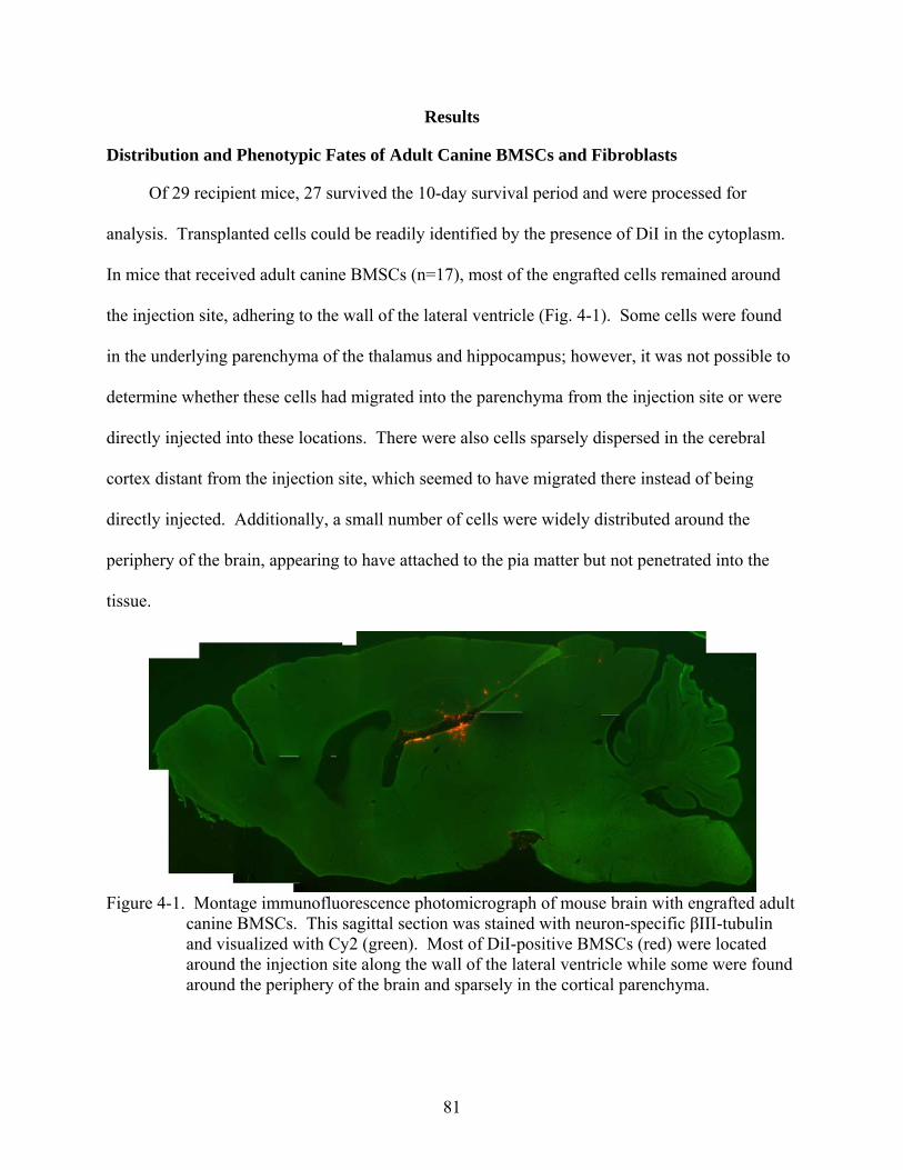
81
Results
Distribution and Phenotypic Fates of Adult Canine BMSCs and Fibroblasts
Of 29 recipient mice, 27 survived the 10-day survival period and were processed for
analysis. Transplanted cells could be readily identified by the presence of DiI in the cytoplasm.
In mice that received adult canine BMSCs (n=17), most of the engrafted cells remained around
the injection site, adhering to the wall of the lateral ventricle (Fig. 4-1). Some cells were found
in the underlying parenchyma of the thalamus and hippocampus; however, it was not possible to
determine whether these cells had migrated into the parenchyma from the injection site or were
directly injected into these locations. There were also cells sparsely dispersed in the cerebral
cortex distant from the injection site, which seemed to have migrated there instead of being
directly injected. Additionally, a small number of cells were widely distributed around the
periphery of the brain, appearing to have attached to the pia matter but not penetrated into the
tissue.
Figure 4-1. Montage immunofluorescence photomicrograph of mouse brain with engrafted adult
canine BMSCs. This sagittal section was stained with neuron-specific βIII-tubulin and visualized with Cy2 (green). Most of DiI-positive BMSCs (red) were located around the injection site along the wall of the lateral ventricle while some were found around the periphery of the brain and sparsely in the cortical parenchyma.
82
The majority of the engrafted adult BMSCs exhibited a spindle-shaped appearance similar
to their in vitro characteristic morphology. A small population of cells that migrated into the
parenchyma underlying the ventricle showed process-bearing morphology. However,
immunohistochemistry showed that engrafted cells expressed neither neuron-specific (βIII-
tubulin and NeuN) nor astrocyte-specific (GFAP) markers. Engrafted canine fibroblasts (n=3)
showed a similar tendency of distribution and phenotypic patterns to adult BMSCs with the
exception that fibroblasts remained spindle-shaped morphology.
Distribution and Phenotypic Fates of Young Canine BMSCs
In contrast to adult BMSCs and fibroblasts, BMSCs isolated from young donors (n=7)
demonstrated a different behavior. Although most BMSCs of young donors remained around the
injection site in the lateral ventricle similar to adult BMSCs, a small number of cells were
located in the olfactory bulb (Fig 4-2). These cells in the olfactory bulb were βIII-tubulin
positive and thought to have migrated from the lateral ventricle through the rostral migratory
stream (RMS), a known pathway of neuronal precursors migrating from the subventricular zone
(SVZ) to the olfactory bulb. There were also DiI-positive donor cells in the RMS. In the SVZ,
most of engrafted BMSCs exhibited typical fibroblastic morphology (Fig 4-3A), but some cells
assumed astrocyte-like (Fig 4-3B) or bipolar neuron-like (Fig 4-3C) morphology.
Immunostaining revealed that a significant number of BMSCs in the SVZ were βIII-tubulin
positive (Fig 4-4). Expression of neither NueN nor GFAP was found in any region containing
BMSCs (Fig 4-5).
Fluorescence In Situ Hybridization for Chromosome painting
We first attempted to visualize canine X-chromosomes and mouse X- chromosomes using
different fluorescence-conjugated antibodies to evaluate possible fusion events between
transplanted canine BMSCs and host neural cells. However, due to different required
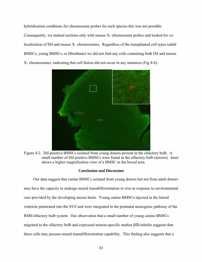
83
hybridization conditions for chromosome probes for each species this was not possible.
Consequently, we stained sections only with mouse X- chromosome probes and looked for co-
localization of DiI and mouse X- chromosomes. Regardless of the transplanted cell types (adult
BMSCs, young BMSCs, or fibroblasts) we did not find any cells containing both DiI and mouse
X- chromosomes, indicating that cell fusion did not occur in any instances (Fig 4-6).
Figure 4-2. DiI-positive BMSCs isolated from young donors present in the olfactory bulb. A
small number of DiI positive BMSCs were found in the olfactory bulb (arrows). Inset shows a higher magnification view of a BMSC in the boxed area.
Conclusion and Discussion
Our data suggest that canine BMSCs isolated from young donors but not from adult donors
may have the capacity to undergo neural transdifferentiation in vivo in response to environmental
cues provided by the developing mouse brain. Young canine BMSCs injected in the lateral
ventricle penetrated into the SVZ and were integrated in the postnatal neurogenic pathway of the
RMS/olfactory bulb system. Our observation that a small number of young canine BMSCs
migrated to the olfactory bulb and expressed neuron-specific marker βIII-tubulin suggests that
these cells may possess neural transdifferentiation capability. This finding also suggests that a
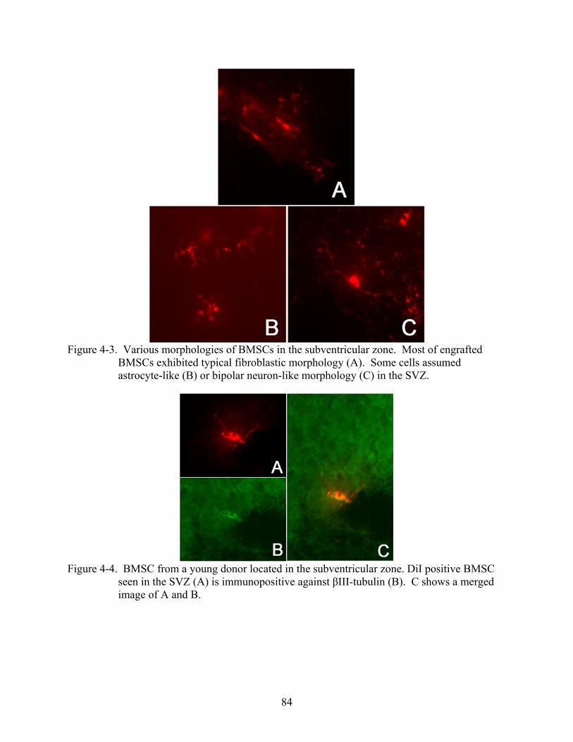
84
Figure 4-3. Various morphologies of BMSCs in the subventricular zone. Most of engrafted
BMSCs exhibited typical fibroblastic morphology (A). Some cells assumed astrocyte-like (B) or bipolar neuron-like morphology (C) in the SVZ.
Figure 4-4. BMSC from a young donor located in the subventricular zone. DiI positive BMSC
seen in the SVZ (A) is immunopositive against βIII-tubulin (B). C shows a merged image of A and B.
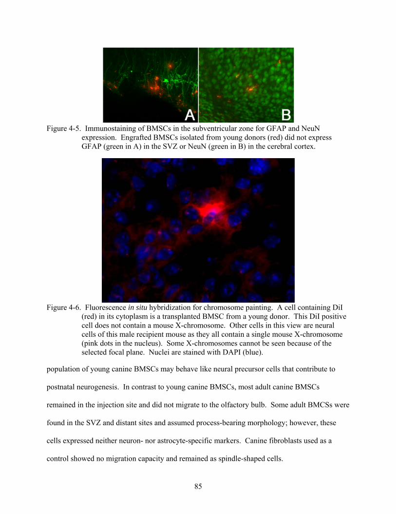
85
Figure 4-5. Immunostaining of BMSCs in the subventricular zone for GFAP and NeuN
expression. Engrafted BMSCs isolated from young donors (red) did not express GFAP (green in A) in the SVZ or NeuN (green in B) in the cerebral cortex.
Figure 4-6. Fluorescence in situ hybridization for chromosome painting. A cell containing DiI
(red) in its cytoplasm is a transplanted BMSC from a young donor. This DiI positive cell does not contain a mouse X-chromosome. Other cells in this view are neural cells of this male recipient mouse as they all contain a single mouse X-chromosome (pink dots in the nucleus). Some X-chromosomes cannot be seen because of the selected focal plane. Nuclei are stained with DAPI (blue).
population of young canine BMSCs may behave like neural precursor cells that contribute to
postnatal neurogenesis. In contrast to young canine BMSCs, most adult canine BMSCs
remained in the injection site and did not migrate to the olfactory bulb. Some adult BMCSs were
found in the SVZ and distant sites and assumed process-bearing morphology; however, these
cells expressed neither neuron- nor astrocyte-specific markers. Canine fibroblasts used as a
control showed no migration capacity and remained as spindle-shaped cells.
86
Our observation of the migration and differentiation properties of young canine BMSCs is
similar to those of previous studies in which murine BMSCs were engrafted into the lateral
ventricle of the neonatal mouse brain (Deng et al., 2006; Kopen et al., 1999). However, the
extent of migration and adopted phenotype was much more restricted in our study compared to
these previous studies. For example, Kopen et al. (1999) reported that murine BMSCs injected
into the lateral ventricle of neonatal mouse brains migrated extensively throughout the forebrain
and cerebellum and differentiated into both neurons and astrocytes. They found that engrafted
BMSCs preferentially populated neuron rich regions including the Islands of Calleja, the
olfactory bulb, and the internal granular layer of the cerebellum, events similar to those occur in
the ongoing developmental processes in early postnatal life. Based on these observations, they
suggested that BMSCs mimic the behavior of neural progenitor cells. Deng et al. (2006) more
recently showed that, upon intraventricular injection, murine BMSCs integrated into the
postnatal neurogenic pathway of the RMS/olfactory bulb system by migrating appropriately and
differentiating into olfactory granule cells, supporting the contention that the bone marrow
derived adult stem cell indeed possesses neural transdifferentiation capability under the influence
of environment cues from the brain.
We found that although most of BMSCs remained in the injection site, a small number of
young canine BMSCs penetrated into the SVZ and assumed various morphologies resembling
neurons or astrocytes. Immunostaining suggested that some of these cells in the SVZ express
βIII-tubulin. We believe that migration of young canine BMSCs into the SVZ and
differentiation toward the neuronal phenotype occurred in a specific way in response to
microenvironmental cues provided by the SVZ. The SVZ forms adjacent to the lateral ventricle
during embryogenesis and is known to contain multipotent neural stem cells (Doetsch et al.,
87
1997; Lois and Alvarez-Buylla, 1993; Weiss et al., 1996). The SVZ is also the area with active
neurogenesis in the postnatal and adult brain (Altman, 1969; Altman and Das, 1966; Luskin,
1993). Our observation that migrating BMSCs were found in the RMS and also localized in the
olfactory bulb further suggested that young canine BMSCs were integrated into the
RMS/olfactory bulb neurogenic system. The olfactory bulb is one destination of neural
progenitors generated in the SVZ (Altman, 1969; Lois and Alvarez-Buylla, 1994).
Little is currently known about what drives implanted BMSCs to differentiate into neural
cells in vivo. Postnatal brain development occurs and this process is governed in part by various
growth factors of which the expression levels are regulated precisely in a controlled
spatiotemporal manner. For example, levels of fibroblast growth factor 2 (FGF-2 or bFGF)
expression increase dramatically during late embryonic and early postnatal stages of
development in rodent brain (Caday et al., 1990; Ford-Perriss et al., 2001). FGF-2 is known to
promote proliferation and self-renewal of neural stem cells derived from the neuroepithelium of
the developing cortex (Gritti et al., 1996) and the adult SVZ (Bartlett et al., 1995). FGF-2 is also
a potent mitogenic factor for BMSCs and it is known that FGF-2 promotes self-renewal of
BMSCs in vitro (Locklin et al., 1995; Oliver et al., 1990; Tsutsumi et al., 2001). Therefore, high
levels of FGF-2 present in the developing brain might have stimulated proliferation and more
importantly the self-renewal capacity of canine BMSCs. It is also likely that neurotrophic
factors played a role in instructing BMSCs to differentiate into the neuronal phenotype.
Neurotrophic factors for which BMSCs express receptors, such as NGF (Caneva et al., 1995) and
BDNF (Li et al., 2007), might have induced neural differentiation of BMSCs when coupled with
other cues present in the SVZ.
88
What contributed to the observed difference between adult donor BMSCs and young donor
BMSCs in their behavior in the mouse brain? It may simply be that bone marrow from young
dogs contained a larger number of stem cells, which survived transplantation, than adult bone
marrow. Alternatively, it could be that transdifferentiated BMSCs were derived from a more
primitive stem cell population present in the BMSC fraction of the young donors. The presence
of a rare pluripotent stem cell population has been reported in adult mouse (Jiang et al., 2002a)
and human (Reyes et al., 2001) bone marrow and shown to have transdifferentiation capacity.
These cells can only be expanded at extremely low cell density on fibronectin and in the
presence of LIF and PDGF. Existence of a similar cell population in adult bone marrow has not
been reported for other species including canines. As we utilized the standard culture condition
(without growth factors at standard cell density) to prepare all BMSCs for transplantation, cells
with multipotent differentiation properties may not have been expanded from the adult bone
marrow sample. Identification of cell populations with multipotential differentiation properties
resident in canine bone marrow needs to be investigated.
In conclusion, BMSCs isolated from young donors exhibited neuronal phenotype upon
transplantation into the developing mouse brain. Transplanted young canine BMSCs responded
to instructive cues provided by the SVZ and migrated to the olfactory bulb via the RMS. These
migratory and differentiation properties of young canine BMSCs resemble behavior of
indigenous neural progenitors. Adult canine BMSCs failed to demonstrate a similar degree of
migration and differentiation neither did canine fibroblasts used as a control. The results suggest
that a primitive stem cell population with neural transdifferentiation capacity may exist in the
BMSC compartment isolated from young dogs. Identification of the responsible cell population

89
in the young canine BMSCs and the development of selective culture techniques may aid further
understanding of the identity of neurogenic stem cell populations in the canine bone marrow.
90
CHAPTER 5 EFFECTS OF CANINE BONE MARROW STROMAL CELLS ON NEURITE EXTENSION
FROM DORSAL ROOT GANGLION NEURONS IN VITRO
Background and Introduction
Bone marrow stromal cells (BMSCs), also referred to as mesenchymal stem cells, have
been known to play a key role in regulating hematopoiesis through interactions with
hematopoietic stem cells within the bone marrow microenvironment (Dormady et al., 2001).
More recent investigations have provided evidence indicating that BMSCs may also serve as a
stem cell reservoir for mesodermal cells and thus participate in regeneration of mesodermal
tissues (Pittenger et al., 1999). In addition, several lines of evidence suggest that bone marrow
derived cells from rodents and humans may be capable of transdifferentiation and thereby have
the capacity to generate cells in vitro (Deng et al., 2001; Sanchez-Ramos et al., 2000; Woodbury
et al., 2000) as well as in vivo (Azizi et al., 1998; Cogle et al., 2004; Deng et al., 2006; Lee et al.,
2003; Mezey et al., 2003) expressing various neuronal markers.
After transplantation of BMSCs, functional recovery has been reported in animal models
involving ischemic (Chen et al., 2001; Lee et al., 2003; Li et al., 2002; Zhao et al., 2002) and
traumatic lesions (Chopp et al., 2000; Hofstetter et al., 2002; Lee et al., 2003; Ohta et al., 2004)
of the CNS. Although studies have shown the presence of transplanted BMSCs in host CNS
tissues (Arnhold et al., 2006; Azizi et al., 1998; Kopen et al., 1999), which in some cases appear
to have distinct neuronal features (Cogle et al., 2004), there is currently no definitive evidence
that BMSCs significantly contribute directly to neuronal replacement after CNS injuries (Ohta et
al., 2004). Alternatively, BMSCs have been shown to have other tissue repair properties such as
promoting remyelination (Akiyama et al., 2002b) and production of various neurotrophic and
angiogenic growth factors, including nerve growth factor (NGF) (Auffray et al., 1996; Chen et
al., 2005; Chen et al., 2002a; Chen et al., 2002b; Crigler et al., 2006; Garcia et al., 2004; Li et al.,
91
2002), glial cell-derived neurotrophic factor (GDNF) (Chen et al., 2005), brain-derived growth
factor (BDNF) (Chen et al., 2002a; Chen et al., 2002b; Crigler et al., 2006; Li et al., 2002),
vascular endothelial growth factor (VEGF) (Chen et al., 2002a; Chen et al., 2002b), hepatocyte
growth factor (HGF) (Chen et al., 2002a; Chen et al., 2002b), and brain natriuretic peptide (BNP)
(Song et al., 2004). Neurotrophic factors such as NGF and BDNF support survival of damaged
neurons (DeKosky et al., 1994; Goss et al., 1998; Hammond et al., 1999; Lu et al., 2001), and it
has been suggested that production of these factors is increased in ischemic brain lesion models
after BMSC transplantation which in turn can lead to reduced apoptosis in the penumbra,
proliferation of endogenous cells in the subventricular zone, and functional recovery (Li et al.,
2002). Vasoactive peptides such as VEGF (Chen et al., 2002a; Chen et al., 2002b) and BNP
(Song et al., 2004) may also participate in the reparative processes after CNS injury by inducing
angiogenesis and reducing secondary edema after injury.
BMSCs also appear to stimulate neuritic outgrowth which may likewise be attributed to
their ability to secrete neurotrophic factors (Mocchetti and Wrathall, 1995), as well as
extracellular matrix (ECM) and adhesion molecules such as fibronectin and laminin (Grayson et
al., 2004; Hofstetter et al., 2002), which are known to promote attachment and extension of
axons. Through production of these molecules BMSCs are thought to provide permissive
microenvironment for regenerating axons (Hofstetter et al., 2002). The importance of this ECM
milieu in which axonal regrowth takes place is well recognized after early studies of peripheral
nerve grafts into CNS lesions (David and Aguayo, 1981; Richardson et al., 1980).
Initial clinical trials in humans using autologous BMSCs have already been reported
(Park et al., 2005; Yoon et al., 2007), and the potential therapeutic value of BMSCs is certainly
not limited to human applications. For example, dogs can sustain traumatic spinal cord injuries
92
at relatively high incidence. A need thus exists for developing novel treatments and cell-based
therapies for veterinary practice. Based on existing evidence, BMSCs are a reasonable candidate
cell platform. Veterinary clinical applications also afford an important animal-to-human
translation opportunity, especially the pathology of clinical spinal cord injury (SCI) in dogs
appears very similar to that documented in humans, as well as in cats and numerous examples of
experimental SCI (Smith and Jeffery, 2006).
Previous studies (Arinzeh et al., 2003; Bruder et al., 1998; Kadiyala et al., 1997; Volk et
al., 2005) of canine BMSCs have focused on their osteogenic properties because of the wide
clinical applicability. The osteogenic property of canine BMSCs has also been exploited in the
field of tissue engineering, particularly in an attempt to promote periodontal tissue regeneration
(Hasegawa et al., 2006). Cardiomyogenic property of canine BMSCs has been studied and
shown to result in improvement of cardiac function in a chronic myocardial infarction model
after transplantation of autologous BMSCs (Bartunek et al., 2007). These studies have
demonstrated “proof of concept” for using BMSCs with clinical benefits. However, our
understanding of the basic biology of canine BMSCs is rather scarce and their potential use for
treatment of neurological diseases has not been studied.
In the present study, we investigated the effects of canine BMSCs on neuritogenesis from
dorsal root ganglion neurons (DRG) in vitro. Our results suggest that canine BMSCs promote
neurite outgrowth from DRG neurons with development of complex and highly branched
neuritic arborizations. These effects were pronounced in co-cultures of BMSCs and DRG
neurons, indicating that production of non-diffusible molecules might be the prime attribute of
BMSCs in promoting neuritogenesis.
93
Materials and Methods
Preparation of Canine BMSCs and Fibroblasts
Bone marrow aspirates from three canine cadavers, obtained from a local animal shelter,
were used to isolate and culture canine BMSCs. Exact ages of these dogs were unknown;
however, we collected from only those which had a complete set of adult dentures with
minimum dental calculus deposition, thus meeting inclusion criteria for “young adults”. The use
of these animals was approved by the Institutional Animal Care and Use Committee of
University of Florida. Detailed protocols for bone marrow collection, cell isolation, and culture
expansion have been described previously (Kamishina et al., 2006). Briefly, bone marrow
aspirates were obtained from the iliac crest immediately after euthanasia. Mononucleated cells
were isolated on a Ficoll density gradient, washed in PBS, and suspended in culture medium
consisting of DMEM (1g/L glucose) supplemented with 10% fetal bovine serum (FBS) and
antibiotics (100 U/ml penicillin G, 100 µg/ml streptomycin sulfate, and 0.25 µg/ml amphotericin
B). Homogeneous BMSC cultures were established in the third passage cultures. Third passage
canine fibroblasts were used for comparative purposes. BMSCs or fibroblasts were plated at a
cell density of 8 × 10³/cm2 on 18mm glass coverslips placed in 12-well culture plates for direct
co-culture experiments or in 6-well plates to yield conditioned media.
Immunocytochemical Analysis for Expression of Extracellular and Adhesion Molecules
We evaluated whether canine BMSCs express some of the putative ECM and adhesion
molecules that are known to favor neurite outgrowth. Third passage canine BMSCs and
fibroblasts were grown on glass coverslips until confluency and immunostained as follows.
Cells were fixed in 4% paraformaldehyde for 30 minutes at 25 ºC, washed three times in PBS,
and permeabilized in 1.0% Triton X-100 in PBS for 10 minutes at 25 ºC. Non-specific binding
was blocked with a blocking solution (1.0% BSA in PBS) for 30 minutes at 25 ºC. Cells were
94
then incubated overnight at 4 ºC with primary antibodies directed against following proteins;
laminin (C4 and D18, both from the Developmental Studies Hybridoma Bank at the University
of Iowa [DSHB]), E-cadherin (DSHB), N-cadherin (DSHB), neural cell adhesion molecule
(NCAM; DSHB), and fibronectin (Sigma). The primary antibodies were removed, cells washed
with PBS, and cells incubated for 1 hour in dark at 25 ºC with secondary antibodies. The
secondary antibodies were Cy2 conjugated goat anti-mouse IgG1 (1:400) or Rhodamine-Red X
conjugated goat anti-mouse IgG2a (1:400). Cells were also incubated without primary
antibodies to control for non-specific staining by secondary antibodies. Cells were washed with
PBS and mounted with a mounting medium. Stained cells were observed under a fluorescent
microscope (Olympus BX50, Olympus Corporation, Tokyo Japan) with appropriate filters.
Direct Co-culture of BMSCs and Dorsal Root Ganglion Neurons
Rat fetal DRG neurons (Cambrex Corporation) were plated on 1) monolayer of BMSCs, 2)
monolayer of fibroblasts, or 3) laminin-coated (2 µg/ml, Sigma) coverslips at a cell density of
3.8 × 10³/cm2 in the same medium as above. After 48 hours, cells were fixed and processed for
immunocytochemistry as described above. The primary and secondary antibodies used were
anti-neurofilament 200 (1:50, Sigma) and Alexa Fluor® 350 (1:200, Invitrogen), respectively.
Cells were also incubated without primary antibodies to control for non-specific staining by
secondary antibodies. Stained cells were observed under a fluorescent microscope equipped
with a digital camera (Retiga 1300, QImaging, Surrey, BC Canada) and pictures of DRG neurons
were taken from non-overlapping fields using a ×10 objective lens. These experiments were
performed in triplicate.
Culture of DRG Neurons in Conditioned Medium
Third passage BMSCs or fibroblasts were cultured in 6-well culture plates for 72 hours
after which conditioned media were collected. In this experiment, 6-well plates were coated with
95
laminin (2 µg/mL) and poly-D-lysine (30 µg/mL) and DRG neurons were plated in each well at a
cell density of 1.0 × 10³/cm2. DRG neurons were also plated in culture media that were
incubated for 72 hours at 37 ºC without conditioning cells. DRG neurons were cultured in
conditioned media for 48 hours after which cells were washed with PBS and fixed as described.
DRG neurons were stained with SimplyBlue SafeStain (Invitrogen) for 8 hours at 25 ºC, washed
three times in PBS, and examined under a phase contrast microscope as previously described
(Price et al., 2006). Digital pictures of DRG neurons were acquired from non-overlapping fields
using a ×10 objective lens. These experiments were performed in duplicate.
Measurements of Neurite Outgrowth
Digital images were transferred into image analysis software (NIH ImageJ ver 1.37v) for
neurite morphometric analyses. All neuronal processes were considered neurites as axons and
dendrites were not distinguishable from one another. Primary neurites were defined as processes
directly emerging from the cell body which usually have a thicker diameter than branching
neurites. All primary and branching neurites were manually traced on the digital images. The
following parameters were measured as previously described (Chakrabortty et al., 2000); 1) total
neurite length/neuron, 2) total primary neurite length/neuron, 3) mean length of primary
neurite/neuron, 4) mean number of primary neurites/neuron, and 5) mean number of branching
neurites/neuron. Data are presented as mean ± standard deviation. Statistical differences among
groups were tested by One-way ANOVA followed by Tukey’s HSD with p-value set at .05
(SPSS).
Results
Expression of Extracellular Matrix Molecules
After 72 hours of plating, third passage canine BMSCs formed a uniform monolayer
consisting of homogenous polygonal cells. Immunocytochemistry demonstrated that canine
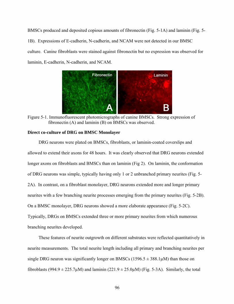
96
BMSCs produced and deposited copious amounts of fibronectin (Fig. 5-1A) and laminin (Fig. 5-
1B). Expressions of E-cadherin, N-cadherin, and NCAM were not detected in our BMSC
culture. Canine fibroblasts were stained against fibronectin but no expression was observed for
laminin, E-cadherin, N-cadherin, and NCAM.
Figure 5-1. Immunofluorescent photomicrographs of canine BMSCs. Strong expression of
fibronectin (A) and laminin (B) on BMSCs was observed.
Direct co-culture of DRG on BMSC Monolayer
DRG neurons were plated on BMSCs, fibroblasts, or laminin-coated coverslips and
allowed to extend their axons for 48 hours. It was clearly observed that DRG neurons extended
longer axons on fibroblasts and BMSCs than on laminin (Fig 2). On laminin, the conformation
of DRG neurons was simple, typically having only 1 or 2 unbranched primary neurites (Fig. 5-
2A). In contrast, on a fibroblast monolayer, DRG neurons extended more and longer primary
neurites with a few branching neurite processes emerging from the primary neurites (Fig. 5-2B).
On a BMSC monolayer, DRG neurons showed a more elaborate appearance (Fig. 5-2C).
Typically, DRGs on BMSCs extended three or more primary neurites from which numerous
branching neurites developed.
These features of neurite outgrowth on different substrates were reflected quantitatively in
neurite measurements. The total neurite length including all primary and branching neurites per
single DRG neuron was significantly longer on BMSCs (1596.5 ± 388.1µM) than those on
fibroblasts (994.9 ± 225.7µM) and laminin (221.9 ± 25.0µM) (Fig. 5-3A). Similarly, the total
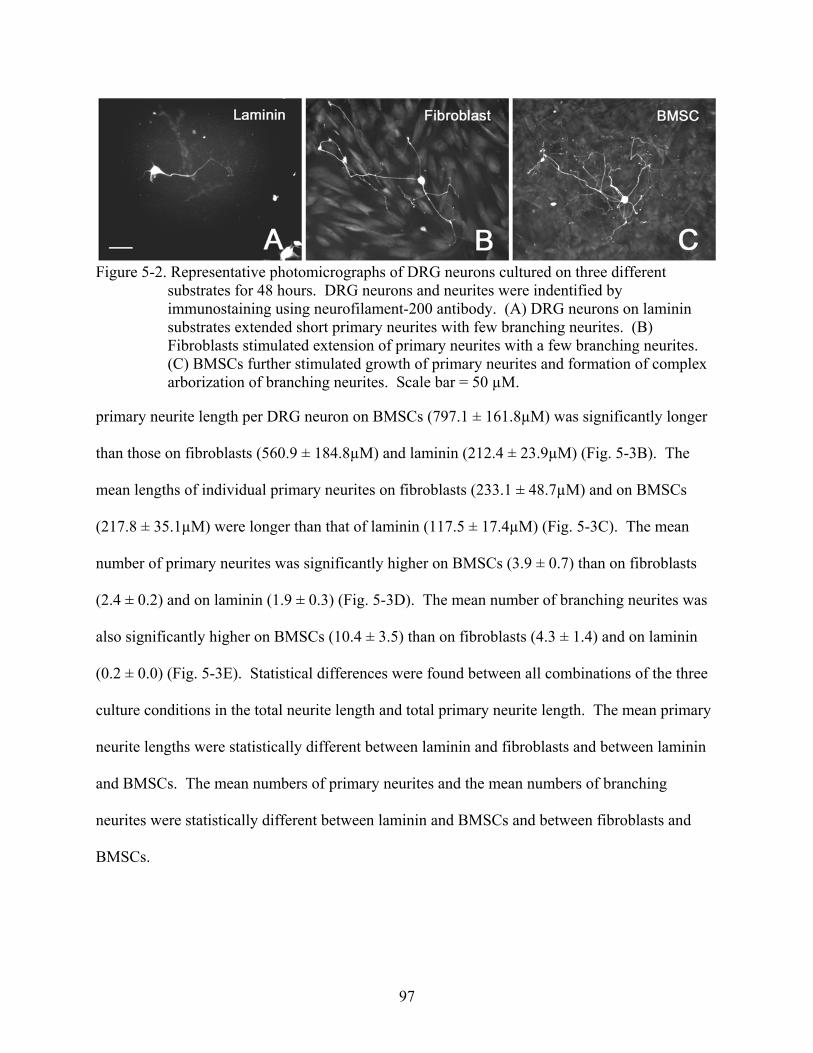
97
Figure 5-2. Representative photomicrographs of DRG neurons cultured on three different
substrates for 48 hours. DRG neurons and neurites were indentified by immunostaining using neurofilament-200 antibody. (A) DRG neurons on laminin substrates extended short primary neurites with few branching neurites. (B) Fibroblasts stimulated extension of primary neurites with a few branching neurites. (C) BMSCs further stimulated growth of primary neurites and formation of complex arborization of branching neurites. Scale bar = 50 µM.
primary neurite length per DRG neuron on BMSCs (797.1 ± 161.8µM) was significantly longer
than those on fibroblasts (560.9 ± 184.8µM) and laminin (212.4 ± 23.9µM) (Fig. 5-3B). The
mean lengths of individual primary neurites on fibroblasts (233.1 ± 48.7µM) and on BMSCs
(217.8 ± 35.1µM) were longer than that of laminin (117.5 ± 17.4µM) (Fig. 5-3C). The mean
number of primary neurites was significantly higher on BMSCs (3.9 ± 0.7) than on fibroblasts
(2.4 ± 0.2) and on laminin (1.9 ± 0.3) (Fig. 5-3D). The mean number of branching neurites was
also significantly higher on BMSCs (10.4 ± 3.5) than on fibroblasts (4.3 ± 1.4) and on laminin
(0.2 ± 0.0) (Fig. 5-3E). Statistical differences were found between all combinations of the three
culture conditions in the total neurite length and total primary neurite length. The mean primary
neurite lengths were statistically different between laminin and fibroblasts and between laminin
and BMSCs. The mean numbers of primary neurites and the mean numbers of branching
neurites were statistically different between laminin and BMSCs and between fibroblasts and
BMSCs.
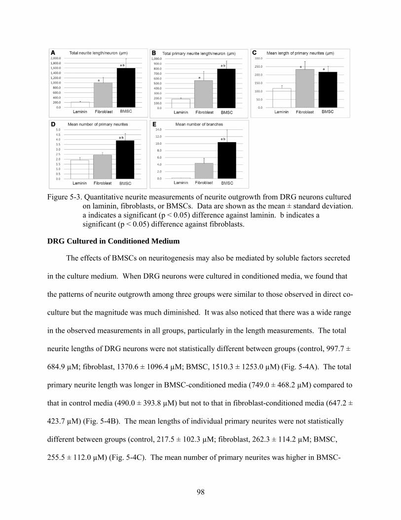
98
Figure 5-3. Quantitative neurite measurements of neurite outgrowth from DRG neurons cultured
on laminin, fibroblasts, or BMSCs. Data are shown as the mean ± standard deviation. a indicates a significant (p < 0.05) difference against laminin. b indicates a significant (p < 0.05) difference against fibroblasts.
DRG Cultured in Conditioned Medium
The effects of BMSCs on neuritogenesis may also be mediated by soluble factors secreted
in the culture medium. When DRG neurons were cultured in conditioned media, we found that
the patterns of neurite outgrowth among three groups were similar to those observed in direct co-
culture but the magnitude was much diminished. It was also noticed that there was a wide range
in the observed measurements in all groups, particularly in the length measurements. The total
neurite lengths of DRG neurons were not statistically different between groups (control, 997.7 ±
684.9 µM; fibroblast, 1370.6 ± 1096.4 µM; BMSC, 1510.3 ± 1253.0 µM) (Fig. 5-4A). The total
primary neurite length was longer in BMSC-conditioned media (749.0 ± 468.2 µM) compared to
that in control media (490.0 ± 393.8 µM) but not to that in fibroblast-conditioned media (647.2 ±
423.7 µM) (Fig. 5-4B). The mean lengths of individual primary neurites were not statistically
different between groups (control, 217.5 ± 102.3 µM; fibroblast, 262.3 ± 114.2 µM; BMSC,
255.5 ± 112.0 µM) (Fig. 5-4C). The mean number of primary neurites was higher in BMSC-
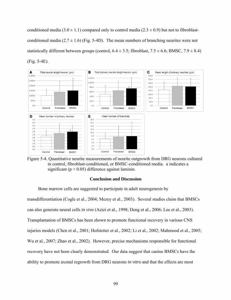
99
conditioned media (3.0 ± 1.1) compared only to control media (2.3 ± 0.9) but not to fibroblast-
conditioned media (2.7 ± 1.6) (Fig. 5-4D). The mean numbers of branching neurites were not
statistically different between groups (control, 6.4 ± 3.5; fibroblast, 7.5 ± 6.6; BMSC, 7.9 ± 8.4)
(Fig. 5-4E).
Figure 5-4. Quantitative neurite measurements of neurite outgrowth from DRG neurons cultured
in control, fibroblast-conditioned, or BMSC-conditioned media. a indicates a significant (p < 0.05) difference against laminin.
Conclusion and Discussion
Bone marrow cells are suggested to participate in adult neurogenesis by
transdifferentiation (Cogle et al., 2004; Mezey et al., 2003). Several studies claim that BMSCs
can also generate neural cells in vivo (Azizi et al., 1998; Deng et al., 2006; Lee et al., 2003).
Transplantation of BMSCs has been shown to promote functional recovery in various CNS
injuries models (Chen et al., 2001; Hofstetter et al., 2002; Li et al., 2002; Mahmood et al., 2005;
Wu et al., 2007; Zhao et al., 2002). However, precise mechanisms responsible for functional
recovery have not been clearly demonstrated. Our data suggest that canine BMSCs have the
ability to promote axonal regrowth from DRG neurons in vitro and that the effects are most
100
prominent via direct contact with neurons. The ability of BMSCs to produce ECM molecules
such as fibronectin and laminin appeared to be the prime attribute for neurite outgrowth-
promoting effects of canine BMSCs.
In the present study, direct co-cultures showed that DRG neurons extended substantially
longer and more complex neurites on BMSCs than on laminin or fibroblasts. The total neurite
length of DRG neurons on BMSCs was about 1.5 times and 7 times longer than those on
fibroblasts and laminin, respectively. The total primary neurite length was also significantly
longer on BMSCs than on fibroblasts or laminin; the difference between BMSCs and fibroblasts
was due to the increased number of primary neurites on BMSCs and not to the increased length
of individual primary neurites. The mean number of branching neurites was also significantly
higher on BMSCs compared to other substrates. These findings suggest that BMSCs promote
neurite outgrowth from DRG neurons by stimulating emergence of both primary and branching
neurites.
We speculate that neurite outgrowth-promoting effects of BMSCs are primarily mediated
by the production of ECM and adhesion molecules. Our results of immunocytochemistry
demonstrated that canine BMSCs produce fibronectin and laminin similar to human BMSCs
(Grayson et al., 2004; Hofstetter et al., 2002). Fibronectin and laminin are expressed by a variety
of cell types and known to enhance neurite extension (Kimura et al., 2004; Orr and Smith, 1988;
Smith and Orr, 1987). A previous in vitro study demonstrated that these ECM molecules
promote adhesion of neurons and neurite extension and the effects are most potent when used in
combination (Orr and Smith, 1988). Our observation that fibroblasts also produce fibronectin
but not laminin points to a significant involvement of laminin in promoting neurite outgrowth.
This view is in line with earlier studies indicating that cell surface factors associated with
101
astrocytes and Schwann cells but not with fibroblasts are primarily responsible for inducing
active neurite outgrowth (Fallon, 1985a; Fallon, 1985b). Nevertheless, in vivo studies showed
that fibronectin may provide a scaffold for invading cellular elements including macrophages
and Schwann cells which in tern stimulate axonal regeneration at the site of spinal cord injury
(King et al., 2003; King et al., 2006). In addition, we examined whether canine BMSCs express
other adhesion molecules that are known to stimulate neurite extension, including E-cadherin
(Oblander et al., 2007), N-cadherin (Puch et al., 2001; Schense et al., 2000), and neural cell
adhesion molecule NCAM (Crigler et al., 2006; Kimura et al., 2004). However, these molecules
were not expressed on canine BMSCs under our culture condition.
A recent in vitro study showed that human BMSCs promote neurite extension from DRG
explants over nerve-inhibitory molecules such as neural proteoglycans, myelin associated
glycoprotein, and Nogo-A (Wright et al., 2007). In the study, BMSCs reduced the inhibitory
effects of these molecules and thereby permitted extension of DRG neurites. The authors
described that DRG explants extended neurites when co-cultured with BMSCs that acted as
“cellular bridges” and also “towed” neurites over the nerve-inhibitory molecules. In contrast,
BMSC-conditioned medium stimulated neurite extension over nerve-permissive substrate (i.e.,
type I collagen) but not over nerve-inhibitory substrates. Therefore, axon-BMSC interactions
appear to be important for BMSCs to promote neurite extension, particularly in injured nervous
tissues where nerve-inhibitory molecules are present. Another possibility is that BMSCs may
degrade nerve-inhibitory molecules thereby allowing regenerating axons to grow in and across
the CNS injury site. Human BMSCs have been shown to produce membrane type I matrix
metalloproteinase and matrix metalloproteinase 2 (Son et al., 2006) which degrade nerve-
102
inhibitory molecules (d'Ortho et al., 1997; Fosang et al., 1992; Passi et al., 1999). Whether
canine BMSCs hold similar enzymatic properties remained to be investigated.
Although our results suggest that ECM molecules are the prime contributors in promoting
neurite outgrowth, involvement of soluble factors cannot be entirely excluded. In fact, some
parameters (i.e., the total primary neurite length/neuron and the number of primary
neurites/neuron) were significantly greater in BMSC-conditioned media compared to control
media. The candidate factors responsible for these changes might be neurotrophic factors (e.g.,
NGF, BDNF, and NT-3) since production of an array of neurotrophic factors has been shown in
human BMSCs (Auffray et al., 1996; Chen et al., 2002a; Chen et al., 2002b; Crigler et al., 2006;
Li et al., 2002). However, there were some uncertainties present in our study, including the
cross-species reactivity of canine neurotrophic factors to rat derived neurons and local
concentrations of soluble factors secreted in the conditioned media. In addition, a soluble form
of laminin has been shown to have effects on neurite outgrowth (Kohno et al., 2005), indicating
the possibility of laminin to play dual roles as an ECM molecule and a soluble factor.
Investigations into identification and quantitation of candidate neurite outgrowth-promoting
soluble factors produced by canine BMSCs are currently underway in our laboratory, using a
range of neutralizing antibodies.
In conclusion, we demonstrated that canine BMSCs have the ability to promote neurite
extension and branching from DRG neurons in vitro, primarily through production of ECM
molecules. Because of their accessibility BMSCs may represent the most promising source for
cellular treatment of CNS injuries. In order to translate our findings to clinical application of
canine BMSCs, further investigations are needed to address several questions. In particular,
although DRG neurons have both CNS and PNS components in vivo, effects of canine BMSCs
103
on pure CNS neurites need to be elucidated. Effects of BMSCs on neuritogenesis are also donor-
dependent (Neuhuber et al., 2005) and there is even a considerable variation in the ability to
produce neurotrophic factors among different populations of BMSCs from the same donor
(Crigler et al., 2006). These previous reports highlight the need for thorough characterization of
canine BMSCs to facilitate the development and refinement of prospective cellular based therapy
for dogs with CNS injuries. Finally, xenotransplantation studies in rodent models of CNS injury
will allow assessment of procedural as well as biological safety associated with transplantation
of canine BMSCs and behavioral benefits in vivo.
104
CHAPTER 6 SUMMARY AND CONCLUSION
This dissertation presents a series of studies conducted to understand the biology of canine
BMSCs. Chapter 2 represents an initial effort to systematically define the growth kinetics,
phenotypic profile, and in vitro differentiation properties of canine BMSCs. It was also our goal
to explore the potential of canine BMSCs for cell therapy for CNS disorders. Consequently, in
vitro and in vivo neural differentiation properties of canine BMSCs were presented in chapter 3
and 4, respectively. Finally, in chapter 5, the effects of BMSCs on neurite extension were
described.
We showed that canine BMSCs share many characteristics with BMSCs of humans and
rodents. First, canine primary BMSCs can be readily isolated by use of their adhesive properties
to culture plastic and expanded as CFU-F under the standard culture condition. From classical
CFU-F assay, we estimated the frequency of canine BMSCs to be approximately 0.0042% (1
BMSC in every 2.38 × 10⁴ mononucleated cells). Passaged canine BMSCs grow as monolayer
and the growth kinetics are dependent on the initial cell density with the low cell density
producing the maximal fold increase of cell expansion. Second, the cell surface marker profile
of canine BMSCs evaluated by flow cytometry is similar to human and rodent counterparts.
Flow cytometry also revealed that expanded cell populations did not contain a significant degree
of contamination of other cell lineages. Third, similar to human and rodent BMSCs, canine
BMSCs consist of heterogeneous cell populations with varying degrees of differentiation
capacities. We found that canine BMSCs do contain a population of cells with the ability to
differentiate into osteoblasts and adipocytes in vitro. However, we speculate that the standard
culture method does not support expansion of multipotent stem cells present in the canine BMSC
105
fraction as passaged canine BMSCs demonstrated limited differentiation along osteoblastic and
adipogenic pathways.
Interestingly, canine BMSCs spontaneously express neuron- and astrocyte-specific
proteins in vitro. The significance of this finding is unknown but it may suggest that canine
BMSCs are not only undifferentiated but also multi-differentiated as has been suggested for
human BMSCs. Elevation of intracellular cyclic AMP levels caused rapid transformation of
canine BMSCs into neuron-like morphology, but the morphological change per se was
interpreted as a result of cytoskeletal shrinkage rather than genuine neurite extension.
Our xenotransplantation study suggested that BMSCs isolated from young canine donors
may have the capacity of neural transdifferentiation in the neonatal mouse brain. Young donor
BMSCs injected into the lateral ventricle of neonatal mouse brains migrated into the SVZ and
assumed neuronal or astrocytic morphology. A small number of BMSCs further migrated in the
RMS and reached the olfactory bulb where they expressed neuron-specific marker. Cell fusion
events did not contribute to these observations of young donor BMSCs. Adult canine BMSCs,
however, did not exhibit a similar degree of migration and differentiation. The results imply that
neurogenic stem cells are present in the BMSC compartment of young dogs, whereas, in the
adult canine BMSC compartment, cells of similar properties either do not exist or are lost during
ex vivo expansion.
Finally, we evaluated the ability of canine BMSCs in promoting neurite outgrowth from
DRG neurons in vitro. When DRGs neurons were co-cultured with canine BMSCs, neurite
outgrowth was strongly promoted. This observation was attributed to the stimulatory effects of
canine BMSCs on development of primary and branching neurites from DRG neurons. When
DRG neurons were cultured in BMSC-conditioned medium, the magnitude of neurite outgrowth
106
was suppressed compared to direct co-culture. We showed that canine BMSCs express copious
amounts of laminin and fibronectin, two potent neurite extension-promoting proteins.
Consequently, canine BMSCs were thought to stimulate neurite outgrowth primarily via
production of ECM molecules.
In summary, canine BMSCs are easily accessible and expandable in vitro; thus, represent a
promising source for various cell therapies. However, there are still a lot of works to be done in
order to better understand the biology of canine BMSCs. Future investigations should emphasize
the development of culture techniques that allow maximum expansion of multipotent canine
BMSCs. In spite of controversies surrounding neural transdifferentiation of bone marrow
derived cells, canine bone marrow (at least from young donors) is likely to contain neurogenic
cells. Upon revealing the identity of these cells and refining culture techniques, it may become
possible to isolate and expand similar cell populations from adult canine bone marrow. We
showed that, with the current standard culture technique, canine BMSCs have the ability to
promote neurite outgrowth from DRG neurons. It is thus warranted to investigate in vivo effects
of canine BMSCs in rodent models of CNS injury. Ultimately, BMSC transplantation in
spontaneous CNS injury in dogs will provide valuable insights into the safety as well as clinical
efficacy associated with this procedure.
107
LIST OF REFERENCES
Abouelfetouh, A., Kondoh, T., Ehara, K., Kohmura, E., 2004. Morphological differentiation of bone marrow stromal cells into neuron-like cells after co-culture with hippocampal slice. Brain Res. 1029, 114-9.
Akiyama, Y., Radtke, C., Honmou, O., Kocsis, J. D., 2002a. Remyelination of the spinal cord following intravenous delivery of bone marrow cells. Glia. 39, 229-36.
Akiyama, Y., Radtke, C., Kocsis, J. D., 2002b. Remyelination of the rat spinal cord by transplantation of identified bone marrow stromal cells. J Neurosci. 22, 6623-30.
Altman, J., 1969. Autoradiographic and histological studies of postnatal neurogenesis. IV. Cell proliferation and migration in the anterior forebrain, with special reference to persisting neurogenesis in the olfactory bulb. J Comp Neurol. 137, 433-57.
Altman, J., Das, G. D., 1966. Autoradiographic and histological studies of postnatal neurogenesis. I. A longitudinal investigation of the kinetics, migration and transformation of cells incorporating tritiated thymidine in neonate rats, with special reference to postnatal neurogenesis in some brain regions. J Comp Neurol. 126, 337-89.
Alvarez-Dolado, M., Pardal, R., Garcia-Verdugo, J. M., Fike, J. R., Lee, H. O., Pfeffer, K., Lois, C., Morrison, S. J., Alvarez-Buylla, A., 2003. Fusion of bone-marrow-derived cells with Purkinje neurons, cardiomyocytes and hepatocytes. Nature. 425, 968-73.
Anderson, D. K., Howland, D. R., Reier, P. J., 1995. Fetal neural grafts and repair of the injured spinal cord. Brain Pathol. 5, 451-57.
Anselme, K., Broux, O., Noel, B., Bouxin, B., Bascoulergue, G., Dudermel, A. F., Bianchi, F., Jeanfils, J., Hardouin, P., 2002. In vitro control of human bone marrow stromal cells for bone tissue engineering. Tissue Eng. 8, 941-53.
Arinzeh, T. L., Peter, S. J., Archambault, M. P., van den Bos, C., Gordon, S., Kraus, K., Smith, A., Kadiyala, S., 2003. Allogeneic mesenchymal stem cells regenerate bone in a critical-sized canine segmental defect. J Bone Joint Surg Am. 85-A, 1927-35.
Arnhold, S., Klein, H., Klinz, F. J., Absenger, Y., Schmidt, A., Schinkothe, T., Brixius, K., Kozlowski, J., Desai, B., Bloch, W., Addicks, K., 2006. Human bone marrow stroma cells display certain neural characteristics and integrate in the subventricular compartment after injection into the liquor system. Eur J Cell Biol. 85, 551-65.
Ashton, B. A., Allen, T. D., Howlett, C. R., Eaglesom, C. C., Hattori, A., Owen, M., 1980. Formation of bone and cartilage by marrow stromal cells in diffusion chambers in vivo. Clin Orthop Relat Res. 294-307.
Auffray, I., Chevalier, S., Froger, J., Izac, B., Vainchenker, W., Gascan, H., Coulombel, L., 1996. Nerve growth factor is involved in the supportive effect by bone marrow--derived stromal cells of the factor-dependent human cell line UT-7. Blood. 88, 1608-18.
108
Azizi, S. A., Stokes, D., Augelli, B. J., DiGirolamo, C., Prockop, D. J., 1998. Engraftment and migration of human bone marrow stromal cells implanted in the brains of albino rats--similarities to astrocyte grafts. Proc Natl Acad Sci U S A. 95, 3908-13.
Barry, F., Boynton, R., Murphy, M., Haynesworth, S., Zaia, J., 2001a. The SH-3 and SH-4 antibodies recognize distinct epitopes on CD73 from human mesenchymal stem cells. Biochem Biophys Res Commun. 289, 519-24.
Barry, F., Boynton, R. E., Liu, B., Murphy, J. M., 2001b. Chondrogenic differentiation of mesenchymal stem cells from bone marrow: differentiation-dependent gene expression of matrix components. Exp Cell Res. 268, 189-200.
Barry, F. P., 2003. Biology and clinical applications of mesenchymal stem cells. Birth Defects Res Part C Embryo Today. 69, 250-6.
Barry, F. P., Boynton, R. E., Haynesworth, S., Murphy, J. M., Zaia, J., 1999. The monoclonal antibody SH-2, raised against human mesenchymal stem cells, recognizes an epitope on endoglin (CD105). Biochem Biophys Res Commun. 265, 134-9.
Bartlett, P. F., Richards, L. R., Kilpatrick, T. J., Talman, P. T., Bailey, K. A., Brooker, G. J., Dutton, R., Koblar, S. A., Nurcombe, V., Ford, M. O., et al., 1995. Factors regulating the differentiation of neural precursors in the forebrain. Ciba Found Symp. 193, 85-99; discussion 117-26.
Bartunek, J., Croissant, J. D., Wijns, W., Gofflot, S., de Lavareille, A., Vanderheyden, M., Kaluzhny, Y., Mazouz, N., Willemsen, P., Penicka, M., Mathieu, M., Homsy, C., De Bruyne, B., McEntee, K., Lee, I. W., Heyndrickx, G. R., 2007. Pretreatment of adult bone marrow mesenchymal stem cells with cardiomyogenic growth factors and repair of the chronically infarcted myocardium. Am J Physiol Heart Circ Physiol. 292, H1095-104.
Bertani, N., Malatesta, P., Volpi, G., Sonego, P., Perris, R., 2005. Neurogenic potential of human mesenchymal stem cells revisited: analysis by immunostaining, time-lapse video and microarray. J Cell Sci. 118, 3925-36.
Blakemore, W. F., 1977. Remyelination of CNS axons by Schwann cells transplanted from the sciatic nerve. Nature. 266, 68-9.
Brederlau, A., Correia, A. S., Anisimov, S. V., Elmi, M., Roybon, L., Paul, G., Morizane, A., Bergquist, F., Riebe, I., Nannmark, U., Carta, M., Hanse, E., Takahashi, J., Sasai, Y., Funa, K., Brundin, P., Eriksson, P. S., Li, J. Y., 2006. Transplantation of human embryonic stem cell-derived cells to a rat model of Parkinson's disease: effect of in vitro differentiation on graft survival and teratoma formation. Stem Cells.
Brockbank, K. G., Piersma, A. H., Ploemacher, R. E., Voerman, J. S., 1985. Stromal cells (CFU-F) in normal and genetically anemic mouse strains. Acta Haematol. 74, 75-80.
109
Bruder, S. P., Jaiswal, N., Haynesworth, S. E., 1997. Growth kinetics, self-renewal, and the osteogenic potential of purified human mesenchymal stem cells during extensive subcultivation and following cryopreservation. J Cell Biochem. 64, 278-94.
Bruder, S. P., Kraus, K. H., Goldberg, V. M., Kadiyala, S., 1998. The effect of implants loaded with autologous mesenchymal stem cells on the healing of canine segmental bone defects. J Bone Joint Surg Am. 80, 985-96.
Caday, C. G., Klagsbrun, M., Fanning, P. J., Mirzabegian, A., Finklestein, S. P., 1990. Fibroblast growth factor (FGF) levels in the developing rat brain. Brain Res Dev Brain Res. 52, 241-6.
Caneva, L., Soligo, D., Cattoretti, G., De Harven, E., Deliliers, G. L., 1995. Immuno-electron microscopy characterization of human bone marrow stromal cells with anti-NGFR antibodies. Blood Cells Mol Dis. 21, 73-85.
Caplan, A. I., Syftestad, G., Osdoby, P., 1983. The development of embryonic bone and cartilage in tissue culture. Clin Orthop Relat Res. 243-63.
Chakrabortty, S., Kitada, M., Matsumoto, N., Taketomi, M., Kimura, K., Ide, C., 2000. Choroid plexus ependymal cells enhance neurite outgrowth from dorsal root ganglion neurons in vitro. J Neurocytol. 29, 707-17.
Chen, J., Li, Y., Chopp, M., 2000. Intracerebral transplantation of bone marrow with BDNF after MCAo in rat. Neuropharmacology. 39, 711-6.
Chen, J., Li, Y., Katakowski, M., Chen, X., Wang, L., Lu, D., Lu, M., Gautam, S. C., Chopp, M., 2003a. Intravenous bone marrow stromal cell therapy reduces apoptosis and promotes endogenous cell proliferation after stroke in female rat. J Neurosci Res. 73, 778-86.
Chen, J., Li, Y., Wang, L., Zhang, Z., Lu, D., Lu, M., Chopp, M., 2001. Therapeutic benefit of intravenous administration of bone marrow stromal cells after cerebral ischemia in rats. Stroke. 32, 1005-11.
Chen, J., Zhang, Z. G., Li, Y., Wang, L., Xu, Y. X., Gautam, S. C., Lu, M., Zhu, Z., Chopp, M., 2003b. Intravenous administration of human bone marrow stromal cells induces angiogenesis in the ischemic boundary zone after stroke in rats. Circ Res. 92, 692-9.
Chen, Q., Long, Y., Yuan, X., Zou, L., Sun, J., Chen, S., Perez-Polo, J. R., Yang, K., 2005. Protective effects of bone marrow stromal cell transplantation in injured rodent brain: synthesis of neurotrophic factors. J Neurosci Res. 80, 611-9.
Chen, X., Katakowski, M., Li, Y., Lu, D., Wang, L., Zhang, L., Chen, J., Xu, Y., Gautam, S., Mahmood, A., Chopp, M., 2002a. Human bone marrow stromal cell cultures conditioned by traumatic brain tissue extracts: growth factor production. J Neurosci Res. 69, 687-91.
110
Chen, X., Li, Y., Wang, L., Katakowski, M., Zhang, L., Chen, J., Xu, Y., Gautam, S. C., Chopp, M., 2002b. Ischemic rat brain extracts induce human marrow stromal cell growth factor production. Neuropathology. 22, 275-9.
Chen, X., Wang, X. D., Chen, G., Lin, W. W., Yao, J., Gu, X. S., 2006. Study of in vivo differentiation of rat bone marrow stromal cells into schwann cell-like cells. Microsurgery. 26, 111-5.
Chopp, M., Li, Y., 2002. Treatment of neural injury with marrow stromal cells. Lancet Neurol. 1, 92-100.
Chopp, M., Zhang, X. H., Li, Y., Wang, L., Chen, J., Lu, D., Lu, M., Rosenblum, M., 2000. Spinal cord injury in rat: treatment with bone marrow stromal cell transplantation. Neuroreport. 11, 3001-5.
Cogle, C. R., Yachnis, A. T., Laywell, E. D., Zander, D. S., Wingard, J. R., Steindler, D. A., Scott, E. W., 2004. Bone marrow transdifferentiation in brain after transplantation: a retrospective study. Lancet. 363, 1432-7.
Colter, D. C., Class, R., DiGirolamo, C. M., Prockop, D. J., 2000. Rapid expansion of recycling stem cells in cultures of plastic-adherent cells from human bone marrow. Proc Natl Acad Sci U S A. 97, 3213-8.
Crigler, L., Robey, R. C., Asawachaicharn, A., Gaupp, D., Phinney, D. G., 2006. Human mesenchymal stem cell subpopulations express a variety of neuro-regulatory molecules and promote neuronal cell survival and neuritogenesis. Exp Neurol. 198, 54-64.
David, S., Aguayo, A. J., 1981. Axonal elongation into peripheral nervous system "bridges" after central nervous system injury in adult rats. Science. 214, 931-3.
DeKosky, S. T., Goss, J. R., Miller, P. D., Styren, S. D., Kochanek, P. M., Marion, D., 1994. Upregulation of nerve growth factor following cortical trauma. Exp Neurol. 130, 173-7.
Deng, J., Petersen, B. E., Steindler, D. A., Jorgensen, M. L., Laywell, E. D., 2006. Mesenchymal stem cells spontaneously express neural proteins in culture and are neurogenic after transplantation. Stem Cells. 24, 1054-64.
Deng, W., Obrocka, M., Fischer, I., Prockop, D. J., 2001. In vitro differentiation of human marrow stromal cells into early progenitors of neural cells by conditions that increase intracellular cyclic AMP. Biochem Biophys Res Commun. 282, 148-52.
Dezawa, M., Ishikawa, H., Itokazu, Y., Yoshihara, T., Hoshino, M., Takeda, S., Ide, C., Nabeshima, Y., 2005. Bone marrow stromal cells generate muscle cells and repair muscle degeneration. Science. 309, 314-7.
111
Dezawa, M., Kanno, H., Hoshino, M., Cho, H., Matsumoto, N., Itokazu, Y., Tajima, N., Yamada, H., Sawada, H., Ishikawa, H., Mimura, T., Kitada, M., Suzuki, Y., Ide, C., 2004. Specific induction of neuronal cells from bone marrow stromal cells and application for autologous transplantation. J Clin Invest. 113, 1701-10.
Dezawa, M., Takahashi, I., Esaki, M., Takano, M., Sawada, H., 2001. Sciatic nerve regeneration in rats induced by transplantation of in vitro differentiated bone-marrow stromal cells. Eur J Neurosci. 14, 1771-6.
Diefenderfer, D. L., Osyczka, A. M., Garino, J. P., Leboy, P. S., 2003a. Regulation of BMP-induced transcription in cultured human bone marrow stromal cells. J Bone Joint Surg Am. 85-A Suppl 3, 19-28.
Diefenderfer, D. L., Osyczka, A. M., Reilly, G. C., Leboy, P. S., 2003b. BMP responsiveness in human mesenchymal stem cells. Connect Tissue Res. 44 Suppl 1, 305-11.
Digirolamo, C. M., Stokes, D., Colter, D., Phinney, D. G., Class, R., Prockop, D. J., 1999. Propagation and senescence of human marrow stromal cells in culture: a simple colony-forming assay identifies samples with the greatest potential to propagate and differentiate. Br J Haematol. 107, 275-81.
Doetsch, F., Garcia-Verdugo, J. M., Alvarez-Buylla, A., 1997. Cellular composition and three-dimensional organization of the subventricular germinal zone in the adult mammalian brain. J Neurosci. 17, 5046-61.
Dormady, S. P., Bashayan, O., Dougherty, R., Zhang, X. M., Basch, R. S., 2001. Immortalized multipotential mesenchymal cells and the hematopoietic microenvironment. J Hematother Stem Cell Res. 10, 125-40.
d'Ortho, M. P., Will, H., Atkinson, S., Butler, G., Messent, A., Gavrilovic, J., Smith, B., Timpl, R., Zardi, L., Murphy, G., 1997. Membrane-type matrix metalloproteinases 1 and 2 exhibit broad-spectrum proteolytic capacities comparable to many matrix metalloproteinases. Eur J Biochem. 250, 751-7.
Einhorn, T. A., 2003. Clinical applications of recombinant human BMPs: early experience and future development. J Bone Joint Surg Am. 85-A Suppl 3, 82-8.
Fallon, J. R., 1985a. Neurite guidance by non-neuronal cells in culture: preferential outgrowth of peripheral neurites on glial as compared to nonglial cell surfaces. J Neurosci. 5, 3169-77.
Fallon, J. R., 1985b. Preferential outgrowth of central nervous system neurites on astrocytes and Schwann cells as compared with nonglial cells in vitro. J Cell Biol. 100, 198-207.
Ford-Perriss, M., Abud, H., Murphy, M., 2001. Fibroblast growth factors in the developing central nervous system. Clin Exp Pharmacol Physiol. 28, 493-503.
112
Fosang, A. J., Neame, P. J., Last, K., Hardingham, T. E., Murphy, G., Hamilton, J. A., 1992. The interglobular domain of cartilage aggrecan is cleaved by PUMP, gelatinases, and cathepsin B. J Biol Chem. 267, 19470-4.
Franklin, R. J., Gilson, J. M., Franceschini, I. A., Barnett, S. C., 1996. Schwann cell-like myelination following transplantation of an olfactory bulb-ensheathing cell line into areas of demyelination in the adult CNS. Glia. 17, 217-24.
Friedenstein, A. J., 1995. Marrow stromal fibroblasts. Calcif Tissue Int. 56 Suppl 1, S17.
Friedenstein, A. J., Chailakhjan, R. K., Lalykina, K. S., 1970. The development of fibroblast colonies in monolayer cultures of guinea-pig bone marrow and spleen cells. Cell Tissue Kinet. 3, 393-403.
Friedenstein, A. J., Chailakhyan, R. K., Gerasimov, U. V., 1987. Bone marrow osteogenic stem cells: in vitro cultivation and transplantation in diffusion chambers. Cell Tissue Kinet. 20, 263-72.
Friedenstein, A. J., Gorskaja, J. F., Kulagina, N. N., 1976. Fibroblast precursors in normal and irradiated mouse hematopoietic organs. Exp Hematol. 4, 267-74.
Friedenstein, A. J., Petrakova, K. V., Kurolesova, A. I., Frolova, G. P., 1968. Heterotopic of bone marrow.Analysis of precursor cells for osteogenic and hematopoietic tissues. Transplantation. 6, 230-47.
Friedenstein, A. J., Piatetzky, S., II, Petrakova, K. V., 1966. Osteogenesis in transplants of bone marrow cells. J Embryol Exp Morphol. 16, 381-90.
Gaiano, N., Nye, J. S., Fishell, G., 2000. Radial glial identity is promoted by Notch1 signaling in the murine forebrain. Neuron. 26, 395-404.
Garcia, R., Aguiar, J., Alberti, E., de la Cuetara, K., Pavon, N., 2004. Bone marrow stromal cells produce nerve growth factor and glial cell line-derived neurotrophic factors. Biochem Biophys Res Commun. 316, 753-4.
Glaser, T., Perez-Bouza, A., Klein, K., Brustle, O., 2005. Generation of purified oligodendrocyte progenitors from embryonic stem cells. Faseb J. 19, 112-4.
Goshima, J., Goldberg, V. M., Caplan, A. I., 1991a. The origin of bone formed in composite grafts of porous calcium phosphate ceramic loaded with marrow cells. Clin Orthop Relat Res. 274-83.
Goshima, J., Goldberg, V. M., Caplan, A. I., 1991b. Osteogenic potential of culture-expanded rat marrow cells as assayed in vivo with porous calcium phosphate ceramic. Biomaterials. 12, 253-8.
113
Goss, J. R., O'Malley, M. E., Zou, L., Styren, S. D., Kochanek, P. M., DeKosky, S. T., 1998. Astrocytes are the major source of nerve growth factor upregulation following traumatic brain injury in the rat. Exp Neurol. 149, 301-9.
Govender, S., Csimma, C., Genant, H. K., Valentin-Opran, A., Amit, Y., Arbel, R., Aro, H., Atar, D., Bishay, M., Borner, M. G., Chiron, P., Choong, P., Cinats, J., Courtenay, B., Feibel, R., Geulette, B., Gravel, C., Haas, N., Raschke, M., Hammacher, E., van der Velde, D., Hardy, P., Holt, M., Josten, C., Ketterl, R. L., Lindeque, B., Lob, G., Mathevon, H., McCoy, G., Marsh, D., Miller, R., Munting, E., Oevre, S., Nordsletten, L., Patel, A., Pohl, A., Rennie, W., Reynders, P., Rommens, P. M., Rondia, J., Rossouw, W. C., Daneel, P. J., Ruff, S., Ruter, A., Santavirta, S., Schildhauer, T. A., Gekle, C., Schnettler, R., Segal, D., Seiler, H., Snowdowne, R. B., Stapert, J., Taglang, G., Verdonk, R., Vogels, L., Weckbach, A., Wentzensen, A., Wisniewski, T., 2002. Recombinant human bone morphogenetic protein-2 for treatment of open tibial fractures: a prospective, controlled, randomized study of four hundred and fifty patients. J Bone Joint Surg Am. 84-A, 2123-34.
Grayson, W. L., Ma, T., Bunnell, B., 2004. Human mesenchymal stem cells tissue development in 3D PET matrices. Biotechnol Prog. 20, 905-12.
Gregory, C. A., Gunn, W. G., Peister, A., Prockop, D. J., 2004. An Alizarin red-based assay of mineralization by adherent cells in culture: comparison with cetylpyridinium chloride extraction. Anal Biochem. 329, 77-84.
Grill, R. J., Blesch, A., Tuszynski, M. H., 1997. Robust growth of chronically injured spinal cord axons induced by grafts of genetically modified NGF-secreting cells. Exp Neurol. 148, 444-52.
Gritti, A., Parati, E. A., Cova, L., Frolichsthal, P., Galli, R., Wanke, E., Faravelli, L., Morassutti, D. J., Roisen, F., Nickel, D. D., Vescovi, A. L., 1996. Multipotential stem cells from the adult mouse brain proliferate and self-renew in response to basic fibroblast growth factor. J Neurosci. 16, 1091-100.
Gronthos, S., Graves, S. E., Ohta, S., Simmons, P. J., 1994. The STRO-1+ fraction of adult human bone marrow contains the osteogenic precursors. Blood. 84, 4164-73.
Hamill, O. P., Marty, A., Neher, E., Sakmann, B., Sigworth, F. J., 1981. Improved patch-clamp techniques for high-resolution current recording from cells and cell-free membrane patches. Pflugers Arch. 391, 85-100.
Hammond, E. N., Tetzlaff, W., Mestres, P., Giehl, K. M., 1999. BDNF, but not NT-3, promotes long-term survival of axotomized adult rat corticospinal neurons in vivo. Neuroreport. 10, 2671-5.
Hasegawa, N., Kawaguchi, H., Hirachi, A., Takeda, K., Mizuno, N., Nishimura, M., Koike, C., Tsuji, K., Iba, H., Kato, Y., Kurihara, H., 2006. Behavior of Transplanted Bone Marrow-Derived Mesenchymal Stem Cells in Periodontal Defects. J Periodontol. 77, 1003-1007.
114
Haynesworth, S. E., Baber, M. A., Caplan, A. I., 1992a. Cell surface antigens on human marrow-derived mesenchymal cells are detected by monoclonal antibodies. Bone. 13, 69-80.
Haynesworth, S. E., Goshima, J., Goldberg, V. M., Caplan, A. I., 1992b. Characterization of cells with osteogenic potential from human marrow. Bone. 13, 81-8.
Hermann, A., Gastl, R., Liebau, S., Popa, M. O., Fiedler, J., Boehm, B. O., Maisel, M., Lerche, H., Schwarz, J., Brenner, R., Storch, A., 2004. Efficient generation of neural stem cell-like cells from adult human bone marrow stromal cells. J Cell Sci. 117, 4411-22.
Hofstetter, C. P., Schwarz, E. J., Hess, D., Widenfalk, J., El Manira, A., Prockop, D. J., Olson, L., 2002. Marrow stromal cells form guiding strands in the injured spinal cord and promote recovery. Proc Natl Acad Sci U S A. 99, 2199-204.
Honmou, O., Felts, P. A., Waxman, S. G., Kocsis, J. D., 1996. Restoration of normal conduction properties in demyelinated spinal cord axons in the adult rat by transplantation of exogenous Schwann cells. J Neurosci. 16, 3199-208.
Horwitz, E. M., Gordon, P. L., Koo, W. K., Marx, J. C., Neel, M. D., McNall, R. Y., Muul, L., Hofmann, T., 2002. Isolated allogeneic bone marrow-derived mesenchymal cells engraft and stimulate growth in children with osteogenesis imperfecta: Implications for cell therapy of bone. Proc Natl Acad Sci U S A. 99, 8932-7.
Horwitz, E. M., Prockop, D. J., Fitzpatrick, L. A., Koo, W. W., Gordon, P. L., Neel, M., Sussman, M., Orchard, P., Marx, J. C., Pyeritz, R. E., Brenner, M. K., 1999. Transplantability and therapeutic effects of bone marrow-derived mesenchymal cells in children with osteogenesis imperfecta. Nat Med. 5, 309-13.
Houle, J., 1992. The structural integrity of glial scar tissue associated with a chronic spinal cord lesion can be altered by transplanted fetal spinal cord tissue. J Neurosci Res. 31, 120-30.
Imaizumi, T., Lankford, K. L., Waxman, S. G., Greer, C. A., Kocsis, J. D., 1998. Transplanted olfactory ensheathing cells remyelinate and enhance axonal conduction in the demyelinated dorsal columns of the rat spinal cord. J Neurosci. 18, 6176-85.
Jaiswal, N., Haynesworth, S. E., Caplan, A. I., Bruder, S. P., 1997. Osteogenic differentiation of purified, culture-expanded human mesenchymal stem cells in vitro. J Cell Biochem. 64, 295-312.
Javazon, E. H., Colter, D. C., Schwarz, E. J., Prockop, D. J., 2001. Rat marrow stromal cells are more sensitive to plating density and expand more rapidly from single-cell-derived colonies than human marrow stromal cells. Stem Cells. 19, 219-25.
Jeffery, N. D., Smith, P. M., Lakatos, A., Ibanez, C., Ito, D., Franklin, R. J., 2006. Clinical canine spinal cord injury provides an opportunity to examine the issues in translating laboratory techniques into practical therapy. Spinal Cord.
115
Jiang, Y., Jahagirdar, B. N., Reinhardt, R. L., Schwartz, R. E., Keene, C. D., Ortiz-Gonzalez, X. R., Reyes, M., Lenvik, T., Lund, T., Blackstad, M., Du, J., Aldrich, S., Lisberg, A., Low, W. C., Largaespada, D. A., Verfaillie, C. M., 2002a. Pluripotency of mesenchymal stem cells derived from adult marrow. Nature. 418, 41-9.
Jiang, Y., Vaessen, B., Lenvik, T., Blackstad, M., Reyes, M., Verfaillie, C. M., 2002b. Multipotent progenitor cells can be isolated from postnatal murine bone marrow, muscle, and brain. Exp Hematol. 30, 896-904.
Kadiyala, S., Young, R. G., Thiede, M. A., Bruder, S. P., 1997. Culture expanded canine mesenchymal stem cells possess osteochondrogenic potential in vivo and in vitro. Cell Transplant. 6, 125-34.
Kamada, T., Koda, M., Dezawa, M., Yoshinaga, K., Hashimoto, M., Koshizuka, S., Nishio, Y., Moriya, H., Yamazaki, M., 2005. Transplantation of bone marrow stromal cell-derived Schwann cells promotes axonal regeneration and functional recovery after complete transection of adult rat spinal cord. J Neuropathol Exp Neurol. 64, 37-45.
Kamishina, H., Deng, J., Oji, T., Cheeseman, J. A., Clemmons, R. M., 2006. Expression of neural markers on bone marrow-derived canine mesenchymal stem cells. Am J Vet Res. 67, 1921-8.
Kawasaki, H., Suemori, H., Mizuseki, K., Watanabe, K., Urano, F., Ichinose, H., Haruta, M., Takahashi, M., Yoshikawa, K., Nishikawa, S., Nakatsuji, N., Sasai, Y., 2002. Generation of dopaminergic neurons and pigmented epithelia from primate ES cells by stromal cell-derived inducing activity. Proc Natl Acad Sci U S A. 99, 1580-5.
Keilhoff, G., Goihl, A., Langnase, K., Fansa, H., Wolf, G., 2006. Transdifferentiation of mesenchymal stem cells into Schwann cell-like myelinating cells. Eur J Cell Biol. 85, 11-24.
Keirstead, H. S., Nistor, G., Bernal, G., Totoiu, M., Cloutier, F., Sharp, K., Steward, O., 2005. Human embryonic stem cell-derived oligodendrocyte progenitor cell transplants remyelinate and restore locomotion after spinal cord injury. J Neurosci. 25, 4694-705.
Kimura, K., Matsumoto, N., Kitada, M., Mizoguchi, A., Ide, C., 2004. Neurite outgrowth from hippocampal neurons is promoted by choroid plexus ependymal cells in vitro. J Neurocytol. 33, 465-76.
King, V. R., Henseler, M., Brown, R. A., Priestley, J. V., 2003. Mats made from fibronectin support oriented growth of axons in the damaged spinal cord of the adult rat. Exp Neurol. 182, 383-98.
King, V. R., Phillips, J. B., Hunt-Grubbe, H., Brown, R., Priestley, J. V., 2006. Characterization of non-neuronal elements within fibronectin mats implanted into the damaged adult rat spinal cord. Biomaterials. 27, 485-96.

116
Kinnaird, T., Stabile, E., Burnett, M. S., Lee, C. W., Barr, S., Fuchs, S., Epstein, S. E., 2004. Marrow-derived stromal cells express genes encoding a broad spectrum of arteriogenic cytokines and promote in vitro and in vivo arteriogenesis through paracrine mechanisms. Circ Res. 94, 678-85.
Koda, M., Okada, S., Nakayama, T., Koshizuka, S., Kamada, T., Nishio, Y., Someya, Y., Yoshinaga, K., Okawa, A., Moriya, H., Yamazaki, M., 2005. Hematopoietic stem cell and marrow stromal cell for spinal cord injury in mice. Neuroreport. 16, 1763-7.
Kohno, K., Kawakami, T., Hiruma, H., 2005. Effects of soluble laminin on organelle transport and neurite growth in cultured mouse dorsal root ganglion neurons: difference between primary neurites and branches. J Cell Physiol. 205, 253-61.
Kohyama, J., Abe, H., Shimazaki, T., Koizumi, A., Nakashima, K., Gojo, S., Taga, T., Okano, H., Hata, J., Umezawa, A., 2001. Brain from bone: efficient "meta-differentiation" of marrow stroma-derived mature osteoblasts to neurons with Noggin or a demethylating agent. Differentiation. 68, 235-44.
Kopen, G. C., Prockop, D. J., Phinney, D. G., 1999. Marrow stromal cells migrate throughout forebrain and cerebellum, and they differentiate into astrocytes after injection into neonatal mouse brains. Proc Natl Acad Sci U S A. 96, 10711-6.
Kordower, J. H., Freeman, T. B., Snow, B. J., Vingerhoets, F. J., Mufson, E. J., Sanberg, P. R., Hauser, R. A., Smith, D. A., Nauert, G. M., Perl, D. P., et al., 1995. Neuropathological evidence of graft survival and striatal reinnervation after the transplantation of fetal mesencephalic tissue in a patient with Parkinson's disease. N Engl J Med. 332, 1118-24.
Kroger, N., Zabelina, T., Renges, H., Kruger, W., Kordes, U., Rischewski, J., Schrum, J., Horstmann, M., Ayuk, F., Erttmann, R., Kabisch, H., Zander, A. R., 2002. Long-term follow-up of allogeneic stem cell transplantation in patients with severe aplastic anemia after conditioning with cyclophosphamide plus antithymocyte globulin. Ann Hematol. 81, 627-31.
Kuznetsov, S. A., Friedenstein, A. J., Robey, P. G., 1997. Factors required for bone marrow stromal fibroblast colony formation in vitro. Br J Haematol. 97, 561-70.
Laywell, E. D., Friedman, P., Harrington, K., Robertson, J. T., Steindler, D. A., 1996. Cell attachment to frozen sections of injured adult mouse brain: effects of tenascin antibody and lectin perturbation of wound-related extracellular matrix molecules. J Neurosci Methods. 66, 99-108.
Laywell, E. D., Rakic, P., Kukekov, V. G., Holland, E. C., Steindler, D. A., 2000. Identification of a multipotent astrocytic stem cell in the immature and adult mouse brain. Proc Natl Acad Sci U S A. 97, 13883-8.
117
Lee, J., Kuroda, S., Shichinohe, H., Ikeda, J., Seki, T., Hida, K., Tada, M., Sawada, K., Iwasaki, Y., 2003. Migration and differentiation of nuclear fluorescence-labeled bone marrow stromal cells after transplantation into cerebral infarct and spinal cord injury in mice. Neuropathology. 23, 169-80.
Li, C. Q., Liu, D., Wu, X. Q., 2004. [Differentiation of rat bone marrow stromal cells into neuron like cells]. Zhong Nan Da Xue Xue Bao Yi Xue Ban. 29, 18-20.
Li, N., Yang, H., Lu, L., Duan, C., Zhao, C., Zhao, H., 2007. Spontaneous expression of neural phenotype and NGF, TrkA, TrkB genes in marrow stromal cells. Biochem Biophys Res Commun. 356, 561-8.
Li, T. S., Hamano, K., Hirata, K., Kobayashi, T., Nishida, M., 2003. The safety and feasibility of the local implantation of autologous bone marrow cells for ischemic heart disease. J Card Surg. 18 Suppl 2, S69-75.
Li, Y., Chen, J., Chen, X. G., Wang, L., Gautam, S. C., Xu, Y. X., Katakowski, M., Zhang, L. J., Lu, M., Janakiraman, N., Chopp, M., 2002. Human marrow stromal cell therapy for stroke in rat: neurotrophins and functional recovery. Neurology. 59, 514-23.
Li, Y., Chen, J., Zhang, C. L., Wang, L., Lu, D., Katakowski, M., Gao, Q., Shen, L. H., Zhang, J., Lu, M., Chopp, M., 2005. Gliosis and brain remodeling after treatment of stroke in rats with marrow stromal cells. Glia. 49, 407-17.
Li, Y., Chopp, M., Chen, J., Wang, L., Gautam, S. C., Xu, Y. X., Zhang, Z., 2000. Intrastriatal transplantation of bone marrow nonhematopoietic cells improves functional recovery after stroke in adult mice. J Cereb Blood Flow Metab. 20, 1311-9.
Lim, D. A., Tramontin, A. D., Trevejo, J. M., Herrera, D. G., Garcia-Verdugo, J. M., Alvarez-Buylla, A., 2000. Noggin antagonizes BMP signaling to create a niche for adult neurogenesis. Neuron. 28, 713-26.
Locklin, R. M., Williamson, M. C., Beresford, J. N., Triffitt, J. T., Owen, M. E., 1995. In vitro effects of growth factors and dexamethasone on rat marrow stromal cells. Clin Orthop Relat Res. 27-35.
Lois, C., Alvarez-Buylla, A., 1993. Proliferating subventricular zone cells in the adult mammalian forebrain can differentiate into neurons and glia. Proc Natl Acad Sci U S A. 90, 2074-7.
Lois, C., Alvarez-Buylla, A., 1994. Long-distance neuronal migration in the adult mammalian brain. Science. 264, 1145-8.
Lu, P., Blesch, A., Tuszynski, M. H., 2001. Neurotrophism without neurotropism: BDNF promotes survival but not growth of lesioned corticospinal neurons. J Comp Neurol. 436, 456-70.
118
Lu, P., Blesch, A., Tuszynski, M. H., 2004. Induction of bone marrow stromal cells to neurons: differentiation, transdifferentiation, or artifact? J Neurosci Res. 77, 174-91.
Lu, Y., Wang, Z., Zhu, M., 2006. Human bone marrow mesenchymal stem cells transfected with human insulin genes can secrete insulin stably. Ann Clin Lab Sci. 36, 127-36.
Lundkvist, J., Lendahl, U., 2001. Notch and the birth of glial cells. Trends Neurosci. 24, 492-4.
Luskin, M. B., 1993. Restricted proliferation and migration of postnatally generated neurons derived from the forebrain subventricular zone. Neuron. 11, 173-89.
Mackay, A. M., Beck, S. C., Murphy, J. M., Barry, F. P., Chichester, C. O., Pittenger, M. F., 1998. Chondrogenic differentiation of cultured human mesenchymal stem cells from marrow. Tissue Eng. 4, 415-28.
Mahmood, A., Lu, D., Chopp, M., 2004a. Intravenous administration of marrow stromal cells (MSCs) increases the expression of growth factors in rat brain after traumatic brain injury. J Neurotrauma. 21, 33-9.
Mahmood, A., Lu, D., Chopp, M., 2004b. Marrow stromal cell transplantation after traumatic brain injury promotes cellular proliferation within the brain. Neurosurgery. 55, 1185-93.
Mahmood, A., Lu, D., Lu, M., Chopp, M., 2003. Treatment of traumatic brain injury in adult rats with intravenous administration of human bone marrow stromal cells. Neurosurgery. 53, 697-702; discussion 702-3.
Mahmood, A., Lu, D., Qu, C., Goussev, A., Chopp, M., 2005. Human marrow stromal cell treatment provides long-lasting benefit after traumatic brain injury in rats. Neurosurgery. 57, 1026-31; discussion 1026-31.
Mahmood, A., Lu, D., Qu, C., Goussev, A., Chopp, M., 2006. Long-term recovery after bone marrow stromal cell treatment of traumatic brain injury in rats. J Neurosurg. 104, 272-7.
Mahmood, A., Lu, D., Wang, L., Chopp, M., 2002. Intracerebral transplantation of marrow stromal cells cultured with neurotrophic factors promotes functional recovery in adult rats subjected to traumatic brain injury. J Neurotrauma. 19, 1609-17.
Majumdar, M. K., Keane-Moore, M., Buyaner, D., Hardy, W. B., Moorman, M. A., McIntosh, K. R., Mosca, J. D., 2003. Characterization and functionality of cell surface molecules on human mesenchymal stem cells. J Biomed Sci. 10, 228-41.
Majumdar, M. K., Thiede, M. A., Mosca, J. D., Moorman, M., Gerson, S. L., 1998. Phenotypic and functional comparison of cultures of marrow-derived mesenchymal stem cells (MSCs) and stromal cells. J Cell Physiol. 176, 57-66.
McDonald, J. W., Liu, X. Z., Qu, Y., Liu, S., Mickey, S. K., Turetsky, D., Gottlieb, D. I., Choi, D. W., 1999. Transplanted embryonic stem cells survive, differentiate and promote recovery in injured rat spinal cord. Nat Med. 5, 1410-2.
119
Meachem, S., von Schonfeldt, V., Schlatt, S., 2001. Spermatogonia: stem cells with a great perspective. Reproduction. 121, 825-34.
Meirelles Lda, S., Nardi, N. B., 2003. Murine marrow-derived mesenchymal stem cell: isolation, in vitro expansion, and characterization. Br J Haematol. 123, 702-11.
Memon, I. A., Sawa, Y., Miyagawa, S., Taketani, S., Matsuda, H., 2005. Combined autologous cellular cardiomyoplasty with skeletal myoblasts and bone marrow cells in canine hearts for ischemic cardiomyopathy. J Thorac Cardiovasc Surg. 130, 646-53.
Mezey, E., Key, S., Vogelsang, G., Szalayova, I., Lange, G. D., Crain, B., 2003. Transplanted bone marrow generates new neurons in human brains. Proc Natl Acad Sci U S A. 100, 1364-9.
Mocchetti, I., Wrathall, J. R., 1995. Neurotrophic factors in central nervous system trauma. J Neurotrauma. 12, 853-70.
Morrison, S. J., Perez, S. E., Qiao, Z., Verdi, J. M., Hicks, C., Weinmaster, G., Anderson, D. J., 2000. Transient Notch activation initiates an irreversible switch from neurogenesis to gliogenesis by neural crest stem cells. Cell. 101, 499-510.
Morshead, C. M., Reynolds, B. A., Craig, C. G., McBurney, M. W., Staines, W. A., Morassutti, D., Weiss, S., van der Kooy, D., 1994. Neural stem cells in the adult mammalian forebrain: a relatively quiescent subpopulation of subependymal cells. Neuron. 13, 1071-82.
Nakahara, Y., Gage, F. H., Tuszynski, M. H., 1996. Grafts of fibroblasts genetically modified to secrete NGF, BDNF, NT-3, or basic FGF elicit differential responses in the adult spinal cord. Cell Transplant. 5, 191-204.
Neuhuber, B., Gallo, G., Howard, L., Kostura, L., Mackay, A., Fischer, I., 2004. Reevaluation of in vitro differentiation protocols for bone marrow stromal cells: disruption of actin cytoskeleton induces rapid morphological changes and mimics neuronal phenotype. J Neurosci Res. 77, 192-204.
Neuhuber, B., Timothy Himes, B., Shumsky, J. S., Gallo, G., Fischer, I., 2005. Axon growth and recovery of function supported by human bone marrow stromal cells in the injured spinal cord exhibit donor variations. Brain Res. 1035, 73-85.
Nistor, G. I., Totoiu, M. O., Haque, N., Carpenter, M. K., Keirstead, H. S., 2005. Human embryonic stem cells differentiate into oligodendrocytes in high purity and myelinate after spinal cord transplantation. Glia. 49, 385-96.
Noth, U., Rackwitz, L., Heymer, A., Weber, M., Baumann, B., Steinert, A., Schutze, N., Jakob, F., Eulert, J., 2007. Chondrogenic differentiation of human mesenchymal stem cells in collagen type I hydrogels. J Biomed Mater Res A.
120
Oblander, S. A., Ensslen-Craig, S. E., Longo, F. M., Brady-Kalnay, S. M., 2007. E-cadherin promotes retinal ganglion cell neurite outgrowth in a protein tyrosine phosphatase-mu-dependent manner. Mol Cell Neurosci. 34, 481-92.
Odorico, J. S., Kaufman, D. S., Thomson, J. A., 2001. Multilineage differentiation from human embryonic stem cell lines. Stem Cells. 19, 193-204.
Ogawa, Y., Sawamoto, K., Miyata, T., Miyao, S., Watanabe, M., Nakamura, M., Bregman, B. S., Koike, M., Uchiyama, Y., Toyama, Y., Okano, H., 2002. Transplantation of in vitro-expanded fetal neural progenitor cells results in neurogenesis and functional recovery after spinal cord contusion injury in adult rats. J Neurosci Res. 69, 925-33.
Ohta, M., Suzuki, Y., Noda, T., Ejiri, Y., Dezawa, M., Kataoka, K., Chou, H., Ishikawa, N., Matsumoto, N., Iwashita, Y., Mizuta, E., Kuno, S., Ide, C., 2004. Bone marrow stromal cells infused into the cerebrospinal fluid promote functional recovery of the injured rat spinal cord with reduced cavity formation. Exp Neurol. 187, 266-78.
Oliver, L. J., Rifkin, D. B., Gabrilove, J., Hannocks, M. J., Wilson, E. L., 1990. Long-term culture of human bone marrow stromal cells in the presence of basic fibroblast growth factor. Growth Factors. 3, 231-6.
Orr, D. J., Smith, R. A., 1988. Neuronal maintenance and neurite extension of adult mouse neurones in non-neuronal cell-reduced cultures is dependent on substratum coating. J Cell Sci. 91 ( Pt 4), 555-61.
Osyczka, A. M., Diefenderfer, D. L., Bhargave, G., Leboy, P. S., 2004. Different effects of BMP-2 on marrow stromal cells from human and rat bone. Cells Tissues Organs. 176, 109-19.
Owen, M., Friedenstein, A. J., 1988. Stromal stem cells: marrow-derived osteogenic precursors. Ciba Found Symp. 136, 42-60.
Park, H. C., Shim, Y. S., Ha, Y., Yoon, S. H., Park, S. R., Choi, B. H., Park, H. S., 2005. Treatment of complete spinal cord injury patients by autologous bone marrow cell transplantation and administration of granulocyte-macrophage colony stimulating factor. Tissue Eng. 11, 913-22.
Passi, A., Negrini, D., Albertini, R., Miserocchi, G., De Luca, G., 1999. The sensitivity of versican from rabbit lung to gelatinase A (MMP-2) and B (MMP-9) and its involvement in the development of hydraulic lung edema. FEBS Lett. 456, 93-6.
Peister, A., Mellad, J. A., Larson, B. L., Hall, B. M., Gibson, L. F., Prockop, D. J., 2004. Adult stem cells from bone marrow (MSCs) isolated from different strains of inbred mice vary in surface epitopes, rates of proliferation, and differentiation potential. Blood. 103, 1662-8.
121
Pereira, R. F., O'Hara, M. D., Laptev, A. V., Halford, K. W., Pollard, M. D., Class, R., Simon, D., Livezey, K., Prockop, D. J., 1998. Marrow stromal cells as a source of progenitor cells for nonhematopoietic tissues in transgenic mice with a phenotype of osteogenesis imperfecta. Proc Natl Acad Sci U S A. 95, 1142-7.
Perrier, A. L., Tabar, V., Barberi, T., Rubio, M. E., Bruses, J., Topf, N., Harrison, N. L., Studer, L., 2004. Derivation of midbrain dopamine neurons from human embryonic stem cells. Proc Natl Acad Sci U S A. 101, 12543-8.
Petrakova, K. V., Tolmacheva, A. A., AIa, F., 1963. [Bone Formation Occurring in Bone Marrow Transplantation in Diffusion Chambers.]. Biull Eksp Biol Med. 56, 87-91.
Phinney, D. G., Kopen, G., Isaacson, R. L., Prockop, D. J., 1999. Plastic adherent stromal cells from the bone marrow of commonly used strains of inbred mice: variations in yield, growth, and differentiation. J Cell Biochem. 72, 570-85.
Pittenger, M. F., Mackay, A. M., Beck, S. C., Jaiswal, R. K., Douglas, R., Mosca, J. D., Moorman, M. A., Simonetti, D. W., Craig, S., Marshak, D. R., 1999. Multilineage potential of adult human mesenchymal stem cells. Science. 284, 143-7.
Pluchino, S., Quattrini, A., Brambilla, E., Gritti, A., Salani, G., Dina, G., Galli, R., Del Carro, U., Amadio, S., Bergami, A., Furlan, R., Comi, G., Vescovi, A. L., Martino, G., 2003. Injection of adult neurospheres induces recovery in a chronic model of multiple sclerosis. Nature. 422, 688-94.
Ponticiello, M. S., Schinagl, R. M., Kadiyala, S., Barry, F. P., 2000. Gelatin-based resorbable sponge as a carrier matrix for human mesenchymal stem cells in cartilage regeneration therapy. J Biomed Mater Res. 52, 246-55.
Price, R. D., Oe, T., Yamaji, T., Matsuoka, N., 2006. A simple, flexible, nonfluorescent system for the automated screening of neurite outgrowth. J Biomol Screen. 11, 155-64.
Puch, S., Armeanu, S., Kibler, C., Johnson, K. R., Muller, C. A., Wheelock, M. J., Klein, G., 2001. N-cadherin is developmentally regulated and functionally involved in early hematopoietic cell differentiation. J Cell Sci. 114, 1567-77.
Reyes, M., Lund, T., Lenvik, T., Aguiar, D., Koodie, L., Verfaillie, C. M., 2001. Purification and ex vivo expansion of postnatal human marrow mesodermal progenitor cells. Blood. 98, 2615-25.
Richardson, P. M., McGuinness, U. M., Aguayo, A. J., 1980. Axons from CNS neurons regenerate into PNS grafts. Nature. 284, 264-5.
Rismanchi, N., Floyd, C. L., Berman, R. F., Lyeth, B. G., 2003. Cell death and long-term maintenance of neuron-like state after differentiation of rat bone marrow stromal cells: a comparison of protocols. Brain Res. 991, 46-55.
122
Sanchez-Ramos, J., Song, S., Cardozo-Pelaez, F., Hazzi, C., Stedeford, T., Willing, A., Freeman, T. B., Saporta, S., Janssen, W., Patel, N., Cooper, D. R., Sanberg, P. R., 2000. Adult bone marrow stromal cells differentiate into neural cells in vitro. Exp Neurol. 164, 247-56.
Sasaki, M., Honmou, O., Akiyama, Y., Uede, T., Hashi, K., Kocsis, J. D., 2001. Transplantation of an acutely isolated bone marrow fraction repairs demyelinated adult rat spinal cord axons. Glia. 35, 26-34.
Schense, J. C., Bloch, J., Aebischer, P., Hubbell, J. A., 2000. Enzymatic incorporation of bioactive peptides into fibrin matrices enhances neurite extension. Nat Biotechnol. 18, 415-9.
Schwartz, E. D., Shumsky, J. S., Wehrli, S., Tessler, A., Murray, M., Hackney, D. B., 2003. Ex vivo MR determined apparent diffusion coefficients correlate with motor recovery mediated by intraspinal transplants of fibroblasts genetically modified to express BDNF. Exp Neurol. 182, 49-63.
Schwarz, E. J., Alexander, G. M., Prockop, D. J., Azizi, S. A., 1999. Multipotential marrow stromal cells transduced to produce L-DOPA: engraftment in a rat model of Parkinson disease. Hum Gene Ther. 10, 2539-49.
Sekiya, I., Larson, B. L., Smith, J. R., Pochampally, R., Cui, J. G., Prockop, D. J., 2002. Expansion of human adult stem cells from bone marrow stroma: conditions that maximize the yields of early progenitors and evaluate their quality. Stem Cells. 20, 530-41.
Shen, L. H., Li, Y., Chen, J., Zacharek, A., Gao, Q., Kapke, A., Lu, M., Raginski, K., Vanguri, P., Smith, A., Chopp, M., 2007. Therapeutic benefit of bone marrow stromal cells administered 1 month after stroke. J Cereb Blood Flow Metab. 27, 6-13.
Simmons, P. J., Torok-Storb, B., 1991. Identification of stromal cell precursors in human bone marrow by a novel monoclonal antibody, STRO-1. Blood. 78, 55-62.
Smith, P. M., Jeffery, N. D., 2006. Histological and ultrastructural analysis of white matter damage after naturally-occurring spinal cord injury. Brain Pathol. 16, 99-109.
Smith, R. A., Orr, D. J., 1987. The survival of adult mouse sensory neurons in vitro is enhanced by natural and synthetic substrata, particularly fibronectin. J Neurosci Res. 17, 265-70.
Smith, W. C., Harland, R. M., 1992. Expression cloning of noggin, a new dorsalizing factor localized to the Spemann organizer in Xenopus embryos. Cell. 70, 829-40.
Son, B. R., Marquez-Curtis, L. A., Kucia, M., Wysoczynski, M., Turner, A. R., Ratajczak, J., Ratajczak, M. Z., Janowska-Wieczorek, A., 2006. Migration of bone marrow and cord blood mesenchymal stem cells in vitro is regulated by stromal-derived factor-1-CXCR4 and hepatocyte growth factor-c-met axes and involves matrix metalloproteinases. Stem Cells. 24, 1254-64.
123
Song, S., Kamath, S., Mosquera, D., Zigova, T., Sanberg, P., Vesely, D. L., Sanchez-Ramos, J., 2004. Expression of brain natriuretic peptide by human bone marrow stromal cells. Exp Neurol. 185, 191-7.
Spencer, D. D., Robbins, R. J., Naftolin, F., Marek, K. L., Vollmer, T., Leranth, C., Roth, R. H., Price, L. H., Gjedde, A., Bunney, B. S., et al., 1992. Unilateral transplantation of human fetal mesencephalic tissue into the caudate nucleus of patients with Parkinson's disease. N Engl J Med. 327, 1541-8.
Stangel, M., Hartung, H. P., 2002. Remyelinating strategies for the treatment of multiple sclerosis. Prog Neurobiol. 68, 361-76.
Suzawa, M., Takada, I., Yanagisawa, J., Ohtake, F., Ogawa, S., Yamauchi, T., Kadowaki, T., Takeuchi, Y., Shibuya, H., Gotoh, Y., Matsumoto, K., Kato, S., 2003. Cytokines suppress adipogenesis and PPAR-gamma function through the TAK1/TAB1/NIK cascade. Nat Cell Biol. 5, 224-30.
Takagi, Y., Takahashi, J., Saiki, H., Morizane, A., Hayashi, T., Kishi, Y., Fukuda, H., Okamoto, Y., Koyanagi, M., Ideguchi, M., Hayashi, H., Imazato, T., Kawasaki, H., Suemori, H., Omachi, S., Iida, H., Itoh, N., Nakatsuji, N., Sasai, Y., Hashimoto, N., 2005. Dopaminergic neurons generated from monkey embryonic stem cells function in a Parkinson primate model. J Clin Invest. 115, 102-9.
Tao, H., Rao, R., Ma, D. D., 2005. Cytokine-induced stable neuronal differentiation of human bone marrow mesenchymal stem cells in a serum/feeder cell-free condition. Dev Growth Differ. 47, 423-33.
Tavassoli, M., Crosby, W. H., 1968. Transplantation of marrow to extramedullary sites. Science. 161, 54-6.
Teng, Y. D., Lavik, E. B., Qu, X., Park, K. I., Ourednik, J., Zurakowski, D., Langer, R., Snyder, E. Y., 2002. Functional recovery following traumatic spinal cord injury mediated by a unique polymer scaffold seeded with neural stem cells. Proc Natl Acad Sci U S A. 99, 3024-9.
Terada, N., Hamazaki, T., Oka, M., Hoki, M., Mastalerz, D. M., Nakano, Y., Meyer, E. M., Morel, L., Petersen, B. E., Scott, E. W., 2002. Bone marrow cells adopt the phenotype of other cells by spontaneous cell fusion. Nature. 416, 542-5.
Thinyane, K., Baier, P. C., Schindehutte, J., Mansouri, A., Paulus, W., Trenkwalder, C., Flugge, G., Fuchs, E., 2005. Fate of pre-differentiated mouse embryonic stem cells transplanted in unilaterally 6-hydroxydopamine lesioned rats: histological characterization of the grafted cells. Brain Res. 1045, 80-7.
Thompson, F. J., Reier, P. J., Uthman, B., Mott, S., Fessler, R. G., Behrman, A., Trimble, M., Anderson, D. K., Wirth, E. D., 3rd, 2001. Neurophysiological assessment of the feasibility and safety of neural tissue transplantation in patients with syringomyelia. J Neurotrauma. 18, 931-45.
124
Thomson, J. A., Itskovitz-Eldor, J., Shapiro, S. S., Waknitz, M. A., Swiergiel, J. J., Marshall, V. S., Jones, J. M., 1998. Embryonic stem cell lines derived from human blastocysts. Science. 282, 1145-7.
Toma, C., Pittenger, M. F., Cahill, K. S., Byrne, B. J., Kessler, P. D., 2002. Human mesenchymal stem cells differentiate to a cardiomyocyte phenotype in the adult murine heart. Circulation. 105, 93-8.
Tondreau, T., Lagneaux, L., Dejeneffe, M., Massy, M., Mortier, C., Delforge, A., Bron, D., 2004. Bone marrow-derived mesenchymal stem cells already express specific neural proteins before any differentiation. Differentiation. 72, 319-26.
Tropel, P., Noel, D., Platet, N., Legrand, P., Benabid, A. L., Berger, F., 2004. Isolation and characterisation of mesenchymal stem cells from adult mouse bone marrow. Exp Cell Res. 295, 395-406.
Tsutsumi, S., Shimazu, A., Miyazaki, K., Pan, H., Koike, C., Yoshida, E., Takagishi, K., Kato, Y., 2001. Retention of multilineage differentiation potential of mesenchymal cells during proliferation in response to FGF. Biochem Biophys Res Commun. 288, 413-9.
Urist, M. R., Mc, L. F., 1952. Osteogenetic potency and new-bone formation by induction in transplants to the anterior chamber of the eye. J Bone Joint Surg Am. 34-A, 443-76.
Vassilopoulos, G., Russell, D. W., 2003. Cell fusion: an alternative to stem cell plasticity and its therapeutic implications. Curr Opin Genet Dev. 13, 480-5.
Volk, S. W., Diefenderfer, D. L., Christopher, S. A., Haskins, M. E., Leboy, P. S., 2005. Effects of osteogenic inducers on cultures of canine mesenchymal stem cells. Am J Vet Res. 66, 1729-37.
Vulliet, P. R., Greeley, M., Halloran, S. M., MacDonald, K. A., Kittleson, M. D., 2004. Intra-coronary arterial injection of mesenchymal stromal cells and microinfarction in dogs. Lancet. 363, 783-4.
Wakitani, S., Saito, T., Caplan, A. I., 1995. Myogenic cells derived from rat bone marrow mesenchymal stem cells exposed to 5-azacytidine. Muscle Nerve. 18, 1417-26.
Weimann, J. M., Charlton, C. A., Brazelton, T. R., Hackman, R. C., Blau, H. M., 2003a. Contribution of transplanted bone marrow cells to Purkinje neurons in human adult brains. Proc Natl Acad Sci U S A. 100, 2088-93.
Weimann, J. M., Johansson, C. B., Trejo, A., Blau, H. M., 2003b. Stable reprogrammed heterokaryons form spontaneously in Purkinje neurons after bone marrow transplant. Nat Cell Biol. 5, 959-66.
Weiss, S., Reynolds, B. A., Vescovi, A. L., Morshead, C., Craig, C. G., van der Kooy, D., 1996. Is there a neural stem cell in the mammalian forebrain? Trends Neurosci. 19, 387-93.
125
Wernig, M., Benninger, F., Schmandt, T., Rade, M., Tucker, K. L., Bussow, H., Beck, H., Brustle, O., 2004. Functional integration of embryonic stem cell-derived neurons in vivo. J Neurosci. 24, 5258-68.
Williams, C. G., Kim, T. K., Taboas, A., Malik, A., Manson, P., Elisseeff, J., 2003. In vitro chondrogenesis of bone marrow-derived mesenchymal stem cells in a photopolymerizing hydrogel. Tissue Eng. 9, 679-88.
Williams, R. L., Hilton, D. J., Pease, S., Willson, T. A., Stewart, C. L., Gearing, D. P., Wagner, E. F., Metcalf, D., Nicola, N. A., Gough, N. M., 1988. Myeloid leukaemia inhibitory factor maintains the developmental potential of embryonic stem cells. Nature. 336, 684-7.
Wilmut, I., Schnieke, A. E., McWhir, J., Kind, A. J., Campbell, K. H., 1997. Viable offspring derived from fetal and adult mammalian cells. Nature. 385, 810-3.
Wislet-Gendebien, S., Hans, G., Leprince, P., Rigo, J. M., Moonen, G., Rogister, B., 2005. Plasticity of cultured mesenchymal stem cells: switch from nestin-positive to excitable neuron-like phenotype. Stem Cells. 23, 392-402.
Woodbury, D., Reynolds, K., Black, I. B., 2002. Adult bone marrow stromal stem cells express germline, ectodermal, endodermal, and mesodermal genes prior to neurogenesis. J Neurosci Res. 69, 908-17.
Woodbury, D., Schwarz, E. J., Prockop, D. J., Black, I. B., 2000. Adult rat and human bone marrow stromal cells differentiate into neurons. J Neurosci Res. 61, 364-70.
Wright, K. T., El Masri, W., Osman, A., Roberts, S., Chamberlain, G., Ashton, B. A., Johnson, W. E., 2007. Bone marrow stromal cells stimulate neurite outgrowth over neural proteoglycans (CSPG), myelin associated glycoprotein and Nogo-A. Biochem Biophys Res Commun. 354, 559-66.
Wu, Q. Y., Li, J., Feng, Z. T., Wang, T. H., 2007. Bone marrow stromal cells of transgenic mice can improve the cognitive ability of an Alzheimer's disease rat model. Neurosci Lett. 417, 281-5.
Wu, S., Suzuki, Y., Ejiri, Y., Noda, T., Bai, H., Kitada, M., Kataoka, K., Ohta, M., Chou, H., Ide, C., 2003. Bone marrow stromal cells enhance differentiation of cocultured neurosphere cells and promote regeneration of injured spinal cord. J Neurosci Res. 72, 343-51.
Xu, C. X., Hendry, J. H., Testa, N. G., Allen, T. D., 1983. Stromal colonies from mouse marrow: characterization of cell types, optimization of plating efficiency and its effect on radiosensitivity. J Cell Sci. 61, 453-66.
Ying, Q. L., Nichols, J., Evans, E. P., Smith, A. G., 2002. Changing potency by spontaneous fusion. Nature. 416, 545-8.
126
Yoon, S. H., Shim, Y. S., Park, Y. H., Chung, J. K., Nam, J. H., Kim, M. O., Park, H. C., Park, S. R., Min, B. H., Kim, E. Y., Choi, B. H., Park, H., Ha, Y., 2007. Complete spinal cord injury treatment using autologous bone marrow cell transplantation and bone marrow stimulation with granulocyte macrophage-colony stimulating factor: Phase I/II clinical trial. Stem Cells. 25, 2066-73.
Zhang, S. C., Wernig, M., Duncan, I. D., Brustle, O., Thomson, J. A., 2001. In vitro differentiation of transplantable neural precursors from human embryonic stem cells. Nat Biotechnol. 19, 1129-33.
Zhao, L. R., Duan, W. M., Reyes, M., Keene, C. D., Verfaillie, C. M., Low, W. C., 2002. Human bone marrow stem cells exhibit neural phenotypes and ameliorate neurological deficits after grafting into the ischemic brain of rats. Exp Neurol. 174, 11-20.
Zimmerman, L. B., De Jesus-Escobar, J. M., Harland, R. M., 1996. The Spemann organizer signal noggin binds and inactivates bone morphogenetic protein 4. Cell. 86, 599-606.
Zohar, R., Sodek, J., McCulloch, C. A., 1997. Characterization of stromal progenitor cells enriched by flow cytometry. Blood. 90, 3471-81.
Zurita, M., Vaquero, J., 2004. Functional recovery in chronic paraplegia after bone marrow stromal cells transplantation. Neuroreport. 15, 1105-8.
Zurita, M., Vaquero, J., 2006. Bone marrow stromal cells can achieve cure of chronic paraplegic rats: functional and morphological outcome one year after transplantation. Neurosci Lett. 402, 51-6.

127
BIOGRAPHICAL SKETCH
Hiroaki Kamishina was born on December 1, 1971, in Oita, Japan. He received his
Bachelor of Veterinary Medical Sciences degree from Rakuno Gakuen University, Japan, in
March 1996. He then worked in a small animal practice for four years. After that, he came to
the University of Florida where he received a Master of Science in veterinary medical sciences in
August 2003 and a Ph.D. in veterinary medical sciences in December 2007.