Embed Size (px)
Citation preview
DISEASES OF AQUATIC ORGANISMSDis Aquat Org
Vol. 64: 175–180, 2005 Published April 18
INTRODUCTION
Turbot Scophthalmus maximus is a commerciallyimportant fish species that occurs mainly in the easternNorth Atlantic and the Mediterranean Sea. Catcheshave declined markedly during the last decade, and asa result aquaculture has become an important sourceof turbot in several countries, including Spain, France,Norway and China.
In Korea, more than 245000 tons of marine fishesand shellfish were cultured in 2001 (based on the offi-cial statistics issued by the National Fisheries Research& Development Institute in 2003), but the aquacultureindustry is now facing difficulties due to over-produc-tion and the importation of live fishes. To overcomethese problems, fish farmers have switched to moreeconomical species, and turbot was selected because itis fast-growing and in high demand due to the popu-larity of white meat fish in Korea.
In 2003, an outbreak of disease at aquaculture farmsin Go-Chang on the west coast of Korea caused a high
rate of mortality (50 to 70%) in cultured juvenile tur-bot. During histopathological examination of the dis-eased fish, enlarged cells were observed in variousinternal tissues, which is suggestive of red sea breamiridovirus (RSIV) disease; this also occurs in red seabream Pagrus major, grouper Epinephelus ocellata(L.), striped beakperch Oplegnathus fasciatus and reddrum Sciaenos ocellata (L.) (Jung et al. 1997, Jung &Oh 2000, Sano et al. 2002, Weng et al. 2002). In addi-tion, a partial iridovirus genome has been detected inthe spleens and kidneys of diseased turbot using PCRdescribed by Kurita et al. (1998). In this paper, wereport the histopathological characteristics, electronmicroscopic features and molecular characters of thevirus detected in the diseased turbot.
MATERIALS AND METHODS
Epizootics. Turbot eggs were imported from Franceand hatched at an aquaculture farm in Go-Chang,
© Inter-Research 2005 · www.int-res.com*Corresponding author. Email: [email protected]
NOTE
Characterization of an iridovirus detected fromcultured turbot Scophthalmus maximus in Korea
Wi-Sik Kim, Myung-Joo Oh, Sung-Ju Jung, Young-Jin Kim, Shin-Ichi Kitamura*
Department of Aqualife Medicine, Yosu National University, San 96-1 Dunduk-dong, Yosu 550-749, South Korea
ABSTRACT: Juvenile turbot Scophthalmus maximus that became sick during an outbreak of diseaseat mariculture facilities at Go-Chang, Korea, in 2003, were examined to identify the cause of the dis-ease. The fish had pale body color, an enlarged abdomen, protruding eyes, an enlarged spleen andkidney, and pale gills and/or liver. Histopathogical examination revealed basophilic enlarged cells inthe kidney, spleen, gills, heart, stomach, intestine, liver, pancreas and skin. Hexagonal viral particleswith a diameter of 136 to 159 nm were observed in the enlarged cells. A specific 1299 bp fragment ofthe major capsid protein (MCP) gene of the turbot iridovirus (TBIV) was amplified by PCR. Sequencehomology was greater than 93.76% between the MCP gene in TBIV and the same gene in 5 virusesin the tentatively proposed genus Tropivirus (family Iridoviridae): red sea bream iridovirus, sea bassiridovirus, grouper sleepy disease iridovirus, African lampeye iridovirus and dwarf gourami irido-virus. These results suggest that the virus detected from turbot is similar to the proposed genusTropivirus.
KEY WORDS: Turbot · Scophthalmus maximus · Iridovirus · RSIV · MCP · Tropivirus
Resale or republication not permitted without written consent of the publisher
Dis Aquat Org 64: 175–180, 2005
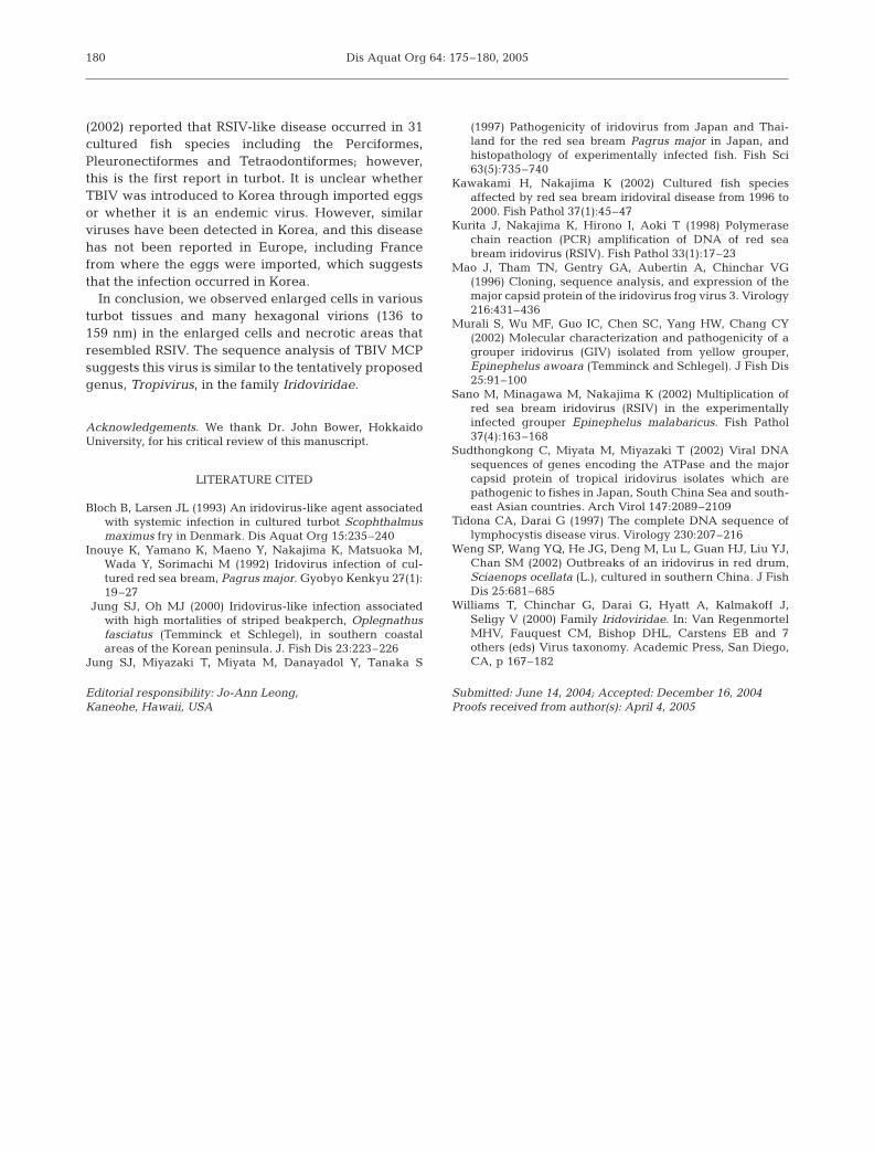
Korea. Juvenile turbot (mean weight 9.0 ± 1.9 g, meanlength 7.9 ± 0.5 cm) began to show signs of disease inJune 2003. Affected fish were lethargic and had areduced appetite, pale body color, enlarged abdomenand protruding eyes. The key internal characteristicsof the diseased fish were an extremely enlarged spleenand kidney, and pale gills and/or liver (Fig. 1).
The fish farmers misdiagnosed the disease as hiramerhabdovirus (HRV) disease or viral hemorrhagic sep-ticemia (VHS) disease, and increased the water tem-perature from 17–18°C to 20–23°C in an attempt tocontrol the disease. The mortality increased rapidlywhen the water temperature was increased. Total mor-tality was about 50 to 70%. After sampling some of thediseased turbot, the remaining fish were killed, and allfacilities were disinfected with sodium hypochlorite.
Histology. The kidney, spleen, gills, heart, stom-ach, intestine, liver, pancreas, skin, eye and brainwere removed from diseased fish and immediatelyfixed in 10% neutral buffered formalin (NBF). Afterfixation, standard histological procedures were usedfor tissue dehydration and paraffin embedding. Tis-sue sections were stained with haematoxylin andeosin (H&E).
Electron microscopy. The spleens were fixed in2.5% glutaraldehyde in 0.1 M phosphate buffer(pH 7.2) at 4°C. After several rinses with 0.1 M phos-phate buffer, each sample was post-fixed with 1%OsO4 for 1 h. Subsequently, the tissue was dehydratedin an ethanol series and embedded in Epon 812. Ultrathin sections were prepared using an RMC-MTX ultra-microtome (SIMS). Sections were stained with lead cit-rate and uranyl acetate. Stained grids were observedunder a Hitachi-7000 electron microscope.
DNA extraction. In preliminary investigations, thethick PCR product appeared from spleen and gillssamples, although a thin band was amplified fromsome organs including stomach, intestine, liver andkidney. Therefore, the spleen and gills of diseasedturbot were chosen for DNA extraction in this study.These tissues were homogenized in a 4-fold volume ofTE buffer (10 mM Tris-HCl, 1 mM EDTA, pH 8.0) andcentrifuged at 2500 × g for 10 min at 4°C. A 20 µlaliquot of Proteinase K (1 mg/ml; TaKaRa) was addedto 200 µl of the supernatant. The mixture was incu-bated at 55°C for 2 h. DNA was isolated using phenoland chloroform. Nucleic acids were precipitated withisopropanol, resuspended with distilled water andstored at –20°C until use.
PCR amplification. The PCR primer set of the majorcapsid protein (MCP) gene was designed from thesequence of red sea bream iridovirus MCP genein the GenBank DNA database (accession numberAY310918). The primer position in the deposited MCPgene is as follows: the forward primer (TBIVMCP-1F)is at bases 12 through 30 (5’-CTCAGGTGCGAACG-TAACC -3’) and the reverse primer (TBIVMCP-1R) isat bases 1288 through 1310 (5’-TTGACTGCAATAAC-GACCAGTTC-3’). This primer set covers 1299/1376bp (94.4%) of the MCP gene. The PCR reactions wereperformed in a final volume of 20 µl of reaction mixturecontaining 50 mM KCl, 10 mM Tris-HCl (pH 9.0),1.5 mM MgCl2, 0.1% Triton X-100, 100 pM of eachprimer, 0.2 mM of each dNTP, 1U Taq DNA poly-merase and template DNA. PCR amplification was per-formed using a GeneAmp 2400 thermal cycler (PerkinElmer) with 30 cycles (95°C for 1 min, 59°C for 1 min,and 72°C for 1 min). The PCR products were analyzedin 1.5% agarose gels containing ethidium bromide andvisualized under UV light.
Cloning of the PCR product and sequencing. The1299 bp specific PCR product was purified using theQIAquick Gel Extraction Kit (Qiagen). The purifiedPCR product was cloned into the pCR 2.1 vector system(Invitrogen), and then transformed to Escherichia colistrain TOP10 (Invitrogen) using standard protocols.The vector was extracted from grown E. coli TOP10and used for sequencing.
Sequencing was performed based on the dideoxynu-cleotide termination method in an automated ABIPRISM 310 DNA sequencer (PE Biosystems) using theABI PRISM Big Dye Terminator Cycle Sequencing FSReady Reaction Kit (PE Biosystems) and the M-13 for-ward and reverse universal primer set. To obtain com-plete sequencing data, another primer set was de-signed in the inserted gene and used for sequencing.The sequence of this primer set was as follows:TBIVMCP-2F (5’-CTGGTCTCCGGTATCACCA-3’)and TBIVMCP-2R (5’-TGACGGGATGACTGAACCT-
176
Fig. 1. Scophthalmus maximus. Diseased turbot with aseverely enlarged spleen (arrow) and pale gills (arrowhead).
Scale bar = 5 mm

Kim et al.: Characterization of turbot iridovirus
3’). Analysis of the results was performed using Gene-tyx Win Ver. 5.1 software. The nucleotide sequence ofthe partial MCP gene of turbot iridovirus (TBIV) wassubmitted to GenBank (accession number AB166788).
The TBIV MCP gene was compared with the MCPgenes of 2 genera of iridoviruses — (1) genus Lympho-cyctivirus (lymphocystis disease virus 1 [LCDV-1;L63545, Tidona & Darai 1997]), (2) genus Ranavirus(grouper iridovirus [GIV; AF364593, Murali et al. 2002]and frog virus 3 [FV3; U36913, Mao et al. 1996]), andthe genus Tropivirus, tentatively proposed bySudthongkong et al. (2002) (red sea bream iridovirus[RSIV; AY310918, Sudthongkong et al. 2002], sea bassiridovirus [SBIV; AB109372, Sudthongkong et al.2002], dwarf gourami iridovirus [DGIV; AY285744,Sudthongkong et al. 2002], grouper sleepy disease iri-dovirus [GSDIV; AY285746, Sudthongkong et al. 2002]and African lampeye iridovirus [ALIV; AY285745,Sudthongkong et al. 2002]).
RESULTS
Histopatholgy and electron microscopy
All moribund fish had enlarged cells in the kidney,spleen, gills, heart, stomach, intestine, liver, pancreasand skin. The enlarged cells had an extremelybasophilic and granular cytoplasm, and their nucleiwere often hypertrophic, fragmented or not visible.
In the spleen, large numbers of enlarged cellsappeared mainly in the splenic pulp and sheathed tis-sue accompanied by hemorrhages (Fig. 2A). The kid-ney had enlarged cells in the glomerulus (Fig. 2B) andhaematopoietic tissue together with necrosis and infil-tration of inflammatory cells. The tubular epithelium ofthe kidney occasionally showed degeneration. In thegills, many enlarged cells were found in the branchialarteries, venous sinus and inter-lamellar epithelia ofthe filaments. The filaments showed necrosis in theinter-lamellar epithelia and hyperemia in the lamellarcapillaries. In the skin, many enlarged cells werelocated adjacent to adipose tissue, and infiltration ofinflammatory cells occurred around the adipose tissueand muscle fibers. In the heart, enlarged cells occurredin the bulbus arteriosus, atrium, ventricles and bulbusendocardium (Fig. 2C). The epicardium was infiltratedby inflammatory cells. In some fish, necrosis wasobserved in the myocardium. In the stomach, the lam-ina propria, submucosa, muscle layer and serous mem-brane contained enlarged cells and inflammatory cells(Fig. 2D). Some fish also had enlarged cells in theepithelial layer. In the intestine, numerous enlargedcells occurred in the lamina propria and submucosabut not in the muscle layer. Inflammatory cells and
necrosis were observed in the lamina propria and sub-mucosa. In the liver, enlarged cells and inflammatorycells were observed in the sinusoids and adjacentparenchymal cells, where multiple discrete necroticfoci were observed. In the pancreas, enlarged cellsoccurred adjacent to the acinar cells, and necrosis wasvisible in the acinar cells. In the eyes, inflammatorycells and necrosis appeared in the choroid, butenlarged cells were not observed. The brain showedno histopathological changes. No histopathologicalchanges including enlarged cells were observed in theany control tissues (Fig. 2E,F).
Electron microscopy revealed many hexagonal-shaped virions in the enlarged cells and necrotic area.Each virion consisted of a central, electron-dense coresurrounded by an electron-translucent zone, and mea-sured 136 to 159 nm from vertex to vertex (the size wascalculated by measuring 50 virions) (Fig. 3).
PCR amplification
The results of PCR using the TBIVMCP-1F andTBIVMCP-1R primer set are shown in Fig. 4. The spe-cific PCR products corresponding to the 1299-bp frag-ment of the MCP gene were detected in the spleen andgills, and the negative control using distilled watershowed no amplification.
Analysis of the MCP nucleotide sequence
The homology of the MCP gene between TBIV andother reported iridoviruses is shown in Table 1. Thenucleotide sequence of TBIV MCP was highly homolo-gous with viruses in the tentatively proposed genusTropivirus (SBIV, 94.38%; RSIV, 94.30%; GSDIV,94.23%; ALIV, 93.92%; and DGIV, 93.76%). When thededuced amino acid sequences were compared, thehomology between TBIV and the genus Tropivirus wasmore than 97.92% (data not shown). On the otherhand, the homology between TBIV and the other 2genera was less than 56.35% for nucleotide sequencesand less than 49.2% for deduced amino acidsequences.
DISCUSSION
We observed enlarged cells in various tissues duringhistopathological observations and many hexagonalvirions (136 to 159 nm) in the enlarged cells andnecrotic areas. The histopathological signs and virionsize are similar to those of red sea bream iridovirus(RSIV) disease in red sea bream (Inouye et al. 1992)
177

Dis Aquat Org 64: 175–180, 2005178
Fig. 2. Scophthalmus maximus. Histopathology of turbot infected with the turbot iridovirus (TBIV). (A) Many enlarged cells withan extremely basophilic (a) and granular cytoplasm (b) in the spleen. (B) Enlarged cell in the glomerulus of the kidney (arrow).(C) Necrosis and many enlarged cells in the bulbus endocardium of the heart. (D) Many enlarged cells in the submucosal layer
of the stomach. (E) Spleen and (F) stomach of control tissue. Scale bars = 50 µm

Kim et al.: Characterization of turbot iridovirus
and striped beakperch (Jung & Oh 2000). Also, turbotiridovirus (TBIV) induced 60% of turbot mortality at23°C in experimental infection, although no mortalitywas observed in the non-injection group (W. S. Kim,M. J. Oh, S. T. Jung, S. I. Kitamura unpubl. data). Thisresult suggests that the mass mortality of turbot wascaused by this virus.
The outbreak of TBIV disease in Go-Chang in 2003occurred at 17 to 18°C, and the mortality increasedrapidly when the water temperature was increased to20–23°C. The optimum temperature for maintainingturbot is around 17°C, and a temperature of 20 to 23°Cmay cause stress in these fish. We suspect that theincreased temperature triggered an increase in mor-tality. Examination of how temperature affects thephysiological condition of turbot and TBIV replicationis needed.
Bloch & Larsen (1993) reported the occurrence of aniridovirus-like agent in cultured turbot fry in Denmark.This agent measured 160 to 185 nm in diameter andwas detected in the fin, gills, liver, kidney, spleen,heart, pancreas, and intestinal collagen. Clinical signsincluded abnormal movements, spasm swimming anddarkened pigmentation in the tail and fins. However,enlarged cells were not observed in any diseased tur-bot tissues reported by Bloch & Larsen (1993). TBIV-infected fish showed no specific signs in their fins, andthe main signs were pale body color, an enlargedabdomen and protruding eyes. These findings suggestthat TBIV is different from the iridovirus-like agentreported by Bloch & Larsen (1993).
The family Iridoviridae comprises 4 genera: Irido-virus, Chloriridovirus, Ranavirus and Lymphocys-tivirus (Williams et al. 2000), and RSIV belongs to thegenus Ranavirus (The Universal Virus Database of theInternational Committee on Taxonomy of Viruses).Recently, Sudthongkong et al. (2002) proposed a newgenus, Tropivirus, in this family for tropical iri-doviruses including RSIV, SBIV, GSDIV, ALIV andDGIV based on nucleotide sequencing of the MCP andATPase genes. We found that sequence homology in
the MCP gene of TBIV was high(>93.75%) with the MCP genes ofthese viruses and low with theMCP genes of the genera Ranavirus(FV3; 56.35%, GIV; 52.06%)and Lymphocystivirus (LCDV-1;53.06%), suggesting that TBIV issimilar to the tentatively proposedgenus Tropivirus.
RSIV-like viruses have beendetected from many fish species inseveral Asian countries, includingKorea, Japan, China, Thailand andIndonesia. Kawakami & Nakajima
179
Fig. 3. Iridovirus particles of infected turbot ranging from136 to 159 nm in the cytoplasm of enlarged cells. Scale bar =
500 nm
Fig. 4. Detection of TBIV MCP gene from the gills and spleenof diseased turbot by PCR. Lanes are M: molecular marker;1: negative control without template; 2: gills of diseasedturbot; 3: spleen of diseased turbot; 4: positive control (red
sea bream iridovirus, RSIV)
Table 1. Comparative analysis (%) of the MCP nucleotide sequence between TBIVand other iridoviruses
TBIV SBIV RSIV GSDIV ALIV DGIV FV3 GIV LCDV-1
TBIV 94.38 94.3 94.23 93.92 93.76 56.35 52.06 53.06SBIV 99.3 99.85 94.84 94.84 56.44 51.35 54.08RSIV 99.31 95.07 95.07 56.62 51.65 53.7GSDIV 94.84 94.84 56.36 51.28 54ALIV 99.62 57.1 51.95 53.02DGIV 56.88 51.8 52.95FV3 69.83 52.18GIV 54.67LCDV-1
Dis Aquat Org 64: 175–180, 2005
(2002) reported that RSIV-like disease occurred in 31cultured fish species including the Perciformes,Pleuronectiformes and Tetraodontiformes; however,this is the first report in turbot. It is unclear whetherTBIV was introduced to Korea through imported eggsor whether it is an endemic virus. However, similarviruses have been detected in Korea, and this diseasehas not been reported in Europe, including Francefrom where the eggs were imported, which suggeststhat the infection occurred in Korea.
In conclusion, we observed enlarged cells in variousturbot tissues and many hexagonal virions (136 to159 nm) in the enlarged cells and necrotic areas thatresembled RSIV. The sequence analysis of TBIV MCPsuggests this virus is similar to the tentatively proposedgenus, Tropivirus, in the family Iridoviridae.
Acknowledgements. We thank Dr. John Bower, HokkaidoUniversity, for his critical review of this manuscript.
LITERATURE CITED
Bloch B, Larsen JL (1993) An iridovirus-like agent associatedwith systemic infection in cultured turbot Scophthalmusmaximus fry in Denmark. Dis Aquat Org 15:235–240
Inouye K, Yamano K, Maeno Y, Nakajima K, Matsuoka M,Wada Y, Sorimachi M (1992) Iridovirus infection of cul-tured red sea bream, Pagrus major. Gyobyo Kenkyu 27(1):19–27
Jung SJ, Oh MJ (2000) Iridovirus-like infection associatedwith high mortalities of striped beakperch, Oplegnathusfasciatus (Temminck et Schlegel), in southern coastalareas of the Korean peninsula. J. Fish Dis 23:223–226
Jung SJ, Miyazaki T, Miyata M, Danayadol Y, Tanaka S
(1997) Pathogenicity of iridovirus from Japan and Thai-land for the red sea bream Pagrus major in Japan, andhistopathology of experimentally infected fish. Fish Sci63(5):735–740
Kawakami H, Nakajima K (2002) Cultured fish speciesaffected by red sea bream iridoviral disease from 1996 to2000. Fish Pathol 37(1):45–47
Kurita J, Nakajima K, Hirono I, Aoki T (1998) Polymerasechain reaction (PCR) amplification of DNA of red seabream iridovirus (RSIV). Fish Pathol 33(1):17–23
Mao J, Tham TN, Gentry GA, Aubertin A, Chinchar VG(1996) Cloning, sequence analysis, and expression of themajor capsid protein of the iridovirus frog virus 3. Virology216:431–436
Murali S, Wu MF, Guo IC, Chen SC, Yang HW, Chang CY(2002) Molecular characterization and pathogenicity of agrouper iridovirus (GIV) isolated from yellow grouper,Epinephelus awoara (Temminck and Schlegel). J Fish Dis25:91–100
Sano M, Minagawa M, Nakajima K (2002) Multiplication ofred sea bream iridovirus (RSIV) in the experimentallyinfected grouper Epinephelus malabaricus. Fish Pathol37(4):163–168
Sudthongkong C, Miyata M, Miyazaki T (2002) Viral DNAsequences of genes encoding the ATPase and the majorcapsid protein of tropical iridovirus isolates which arepathogenic to fishes in Japan, South China Sea and south-east Asian countries. Arch Virol 147:2089–2109
Tidona CA, Darai G (1997) The complete DNA sequence oflymphocystis disease virus. Virology 230:207–216
Weng SP, Wang YQ, He JG, Deng M, Lu L, Guan HJ, Liu YJ,Chan SM (2002) Outbreaks of an iridovirus in red drum,Sciaenops ocellata (L.), cultured in southern China. J FishDis 25:681–685
Williams T, Chinchar G, Darai G, Hyatt A, Kalmakoff J,Seligy V (2000) Family Iridoviridae. In: Van RegenmortelMHV, Fauquest CM, Bishop DHL, Carstens EB and 7others (eds) Virus taxonomy. Academic Press, San Diego,CA, p 167–182
180
Editorial responsibility: Jo-Ann Leong, Kaneohe, Hawaii, USA
Submitted: June 14, 2004; Accepted: December 16, 2004Proofs received from author(s): April 4, 2005