Embed Size (px)
Citation preview
ARTICLE
Characterization of an engineered live bacterialtherapeutic for the treatment of phenylketonuriain a human gut-on-a-chipM. Tyler Nelson 1,6✉, Mark R. Charbonneau 2,6, Heidi G. Coia1,3, Mary J. Castillo2, Corey Holt1,
Eric S. Greenwood 1,4, Peter J. Robinson1,5, Elaine A. Merrill1, David Lubkowicz2 & Camilla A. Mauzy1
Engineered bacteria (synthetic biotics) represent a new class of therapeutics that leverage
the tools of synthetic biology. Translational testing strategies are required to predict synthetic
biotic function in the human body. Gut-on-a-chip microfluidics technology presents an
opportunity to characterize strain function within a simulated human gastrointestinal tract.
Here, we apply a human gut-chip model and a synthetic biotic designed for the treatment of
phenylketonuria to demonstrate dose-dependent production of a strain-specific biomarker, to
describe human tissue responses to the engineered strain, and to show reduced blood
phenylalanine accumulation after administration of the engineered strain. Lastly, we show
how in vitro gut-chip models can be used to construct mechanistic models of strain activity
and recapitulate the behavior of the engineered strain in a non-human primate model. These
data demonstrate that gut-chip models, together with mechanistic models, provide a fra-
mework to predict the function of candidate strains in vivo.
https://doi.org/10.1038/s41467-021-23072-5 OPEN
1 United States Air Force Research Laboratory, 711th Human Performance Wing, Airman Systems Directorate, Bioengineering Division, Wright-Patterson AFB,OH Columbus, USA. 2 Synlogic Inc, Cambridge, MA, USA. 3 National Research Council, The National Academies of Sciences, Engineering, and Medicine,Washington DC, USA. 4Oak Ridge Institute for Science and Education, Oak Ridge, TN, USA. 5 The Henry M. Jackson Foundation, Bethesda, MD, USA. 6Theseauthors contributed equally: M. Tyler Nelson, Mark R. Charbonneau. ✉email: [email protected]
NATURE COMMUNICATIONS | (2021) 12:2805 | https://doi.org/10.1038/s41467-021-23072-5 |www.nature.com/naturecommunications 1
1234
5678
90():,;

Phenylketonuria (PKU) is a rare genetic disease that resultsin reduced activity of the enzyme, phenylalanine hydro-xylase, which converts the essential amino acid, phenyla-
lanine (Phe) to tyrosine. For patients with PKU, dietary proteinconsumption causes prolonged elevation of plasma Phe con-centrations and can lead to severe cognitive impairment, amongother sequelae1,2. There is increasing evidence that live biother-apeutics comprised of engineered microbes (synthetic biotics) canbe used to sense and respond to environmental signals within thehuman body in order to metabolize potentially toxic compounds,including Phe3. We recently described the development of asynthetic biotic strain of E. coli Nissle 1917, called SYNB1618,designed to consume Phe in the human upper gastrointestinaltract4. SYNB1618 degrades Phe by the expression of two distinctmechanisms: (1) the conversion of Phe to trans-cinnamic acid(TCA) by phenylalanine ammonia lyase (PAL), and (2) theconversion of Phe to phenylpyruvic acid by L-amino acid dea-minase (LAAD). TCA is further converted to hippuric acid (HA)in vivo and excreted in urine. We have previously shown that oraladministration of SYNB1618 significantly lowered blood Pheconcentrations in a mouse model of PKU and resulted in dose-dependent production of the PAL-specific urinary biomarker,HA, in healthy non-human primates (NHP). A Phase 1/2a doseescalation study in healthy volunteers and PKU patientsdemonstrated that SYNB1618 was generally well tolerated(Clinicaltrials.gov Identifier: NCT03516487). This study alsorevealed a dose-dependent production of urinary HA uponadministration of SYNB1618, confirming Phe consumption bythe synthetic biotic in human subjects.
The tools of synthetic biology enable rapid and cost-effectivedevelopment of engineered strain prototypes5. However, clinicaldevelopment of therapeutics requires compliance with strictregulatory guidelines and does not scale similarly3. Therefore, it isessential to develop testing strategies that can be applied early inthe development process to characterize the function of syntheticbiotics, to optimize potency, and to establish confidence in theirtranslational potential. For engineered bacterial therapeutics,environmental conditions are important determinants of strainviability and metabolism. Methods that simulate the conditions ofthe human upper gastrointestinal tract, such as the simulatedhuman intestinal microbial ecosystem (SHIME), are useful forcharacterizing the viability and function of engineered strains6.However, these simplified in vitro simulations lack host cells andtissue architecture. By contrast, animal models may be used tostudy synthetic biotic function in the context of the complete hostorganism, though the translational value of animal models variesby species and genotype, due in part to differences in gastro-intestinal (GI) physiology with respect to humans7.
In recent years, significant advances have been made towardorgan-on-a-chip (OOC) microfluidic systems that can be used tostudy the effects of engineered microbes and their products onhuman tissues, including effects on tissue viability8. Thesemicroscale synthetic tissue surrogates enable robust cellular andmolecular analysis, fine control over transport and fluid flowdynamics, and incorporation of complex mechanical stimuli thatcapture aspects of human gut physiology. Gut-on-a-chip in vitromodels have shown promise in overcoming many limitations ofconventional cell culture techniques (e.g., Transwells®) that fail torecapitulate the complexity and function of human gut tissue9,10.For example, dynamic culturing conditions in microfluidic sys-tems enhance gut-endothelial barrier health, aid in maintainingmicrobiome homeostasis, and facilitate nutrient and metabolitetransport under steady-state conditions. Microfluidic culturesthat incorporate both passive and applied mechanical forces canalso drive the development of human gut-like villus projections aswell as the formation of a functional mucus layer11–13. The
benefits of dynamic culture are not limited to host responses butalso address significant experimental obstacles. In particular,dynamic systems can suppress bacterial overgrowth of the hostcell culture and limit inflammatory responses11,13. This technol-ogy represents an opportunity to simulate synthetic biotic activityin the human body, as well as bacterial clearance and the flux ofbacterial and host metabolites between body compartments in adynamic environment (e.g., gut lumen and blood). Moreover,mechanistic data from OOC systems can be used to calibratepredictive mechanistic models of in vivo strain activity.
In this article, we aim to determine whether a human gut-chipmodel, representing the upper gastrointestinal tract, could reca-pitulate the in vivo activity of a synthetic biotic designed for thetreatment of PKU. Single bolus application of a synthetic livebiotherapeutic, SYN5183, is applied to the gut-compartmentresulting in dose dependent increases in the biomarker, trans-cinnamic acid (TCA), and a corresponding 26.9% decrease insystemic Phe. Simulations performed using a mathematicalmodel, calibrated to in vitro gut-chip results, showed a highdegree of correlation with previously published non-human pri-mate results4, highlighting the predictive potential of gut-chiptechnology to accelerate synbiotic development.
ResultsGut-chip model characterization. A human gut-chip model wasdeveloped to simulate the metabolic activity of a synthetic bioticin the human gastrointestinal tract. Fig. 1a displays the two-compartment microfluidic device11 and a schematic describingthe orientation of gut and endothelial cell components. Humanenterocyte-like (Caco2-BBE) and goblet-like cells (HT-29 MTX)were cultured in the upper channel and interfaced with micro-vascular endothelial cells (immortalized cell line) through a thin,porous, flexible membrane, creating a gut-blood barrier. Fig. 1bdisplays a confocal z-stack image of the gut-chip, highlighting thefully vascularized channel and the direct interface with the gutepithelial surface, where cellular projections extend through thepores, resulting in physical association. Void channels runningparallel to and offset from the fluidic channels provide vacuum-induced suction, creating cyclical stretch forces on the membranesimulating peristaltic strain. Continuous dynamic culture (stretchand physiological flow = 60 µL/h; 0.0003 dynes/cm2) of theCaco2 intestinal cells leads to spontaneous 3D morphogenesis,producing villus-like projections (120–150 µm) within the gutcompartment (Fig. 1c). Immunofluorescent staining of f-actinand ZO-1 (tight junctions) in the gut epithelial cells show that thefinger-like projections extend well beyond the membrane surfaceand maintain consistent morphologies and staining throughoutthe length of the channel. Targeted imaging of endothelial cells atthe membrane (Fig. 1d) or on the bottom surface of the lowerchannel (Fig. 1e) reveals a continuous, honeycomb patterned ZO-1 tight-junctional protein staining. Fig. 1f shows a high magni-fication image of gut epithelial cell tight-junction formation (ZO-1, Red). These observations suggest that the human gut-chipexhibits several physiological features and molecular signatures ofa functional gut-blood barrier and that this model system issuitable for evaluating synthetic biotic function and biomarkertransport.
Activity of SYN5183 in a human gut-chip. Activity of the LAADenzyme, expressed by SYNB1618, is dependent on the presence ofoxygen14. Since oxygen concentrations in the human GI tract arelow15,16, and our gut-chip studies are performed in the presenceof oxygen, LAAD activity in the gut-chip would not be repre-sentative of in vivo function. As such, this study utilized theclosely related synthetic biotic strain, SYN5183, which is designed
ARTICLE NATURE COMMUNICATIONS | https://doi.org/10.1038/s41467-021-23072-5
2 NATURE COMMUNICATIONS | (2021) 12:2805 | https://doi.org/10.1038/s41467-021-23072-5 | www.nature.com/naturecommunications

to consume Phe only by expressing the enzyme, PAL. The pro-duct of PAL activity, TCA, serves as a strain-specific biomarker ofPhe consumption that is detectible in plasma (Fig. 2a;4) As abiocontainment feature, SYN5183 lacks the dapA gene, renderingthe strain unable to replicate in the absence of exogenouslysupplemented diaminopimelic acid (DAP;4).
To determine the kinetics of TCA production in a human gut-chip, as well as the relationship between strain activity and dose,SYN5183 was administered in Phe- and DAP-free medium to the
gut compartment as a single dose at a low (L; 1.25 × 10⁸ CFU/mL), medium (M; 6.25 × 10⁸ CFU/mL), or high (H; 1.25 × 10⁹
CFU/mL) level (n= 6 chips/treatment group). Non-treated chips(NT) served as negative controls. Starting 1 h after SYN5183inoculation, simulated intestinal fluid (SIF;17) containing 5 mMPhe was administered continuously to the gut compartment for12 h to represent dietary Phe intake (Fig. 2b). Phe dosing levels inthe gut-chip were selected based on the design of a previouslypublished non-human primate study, considering differences in
a
b
c
d e f
Gut Compartment
Blood Compartment
MicrofluidicDevice
1.0 mm
0.2 mm
17.5 mm
Gut Compartment
100 μm
Gut Compartment
Blood Compartment
Endothelium
PDMSMembrane
PDMS Membrane100 μm
100 μm 100 μm 100 μm
Fig. 1 Characterization of a microfluidic human gut-chip model. a Schematic representation of two-compartment gut-chip model design. Humanenterocyte-like (Caco2-BBE) and goblet-like cells (HT-29 MTX) were co-cultured in the upper channel (gut compartment) and interfaced withmicrovascular endothelial cells in the lower channel (blood compartment), through a thin, porous, flexible membrane. (b, n = 3 gut-chips) Confocal z-stackmicrograph of the human gut-chip model, displaying the fully vascularized endothelial channel (bottom, green—ZO-1) and its interface with the gutepithelial barrier (upper channel, red—ZO-1) post-10 days of maturation. (c, n = 3 gut chips) Continuous dynamic culture (stretch 10% strain, 0.25 Hz andphysiological flow = 60 µL/h; 0.0003 dynes/cm2) of the Caco2 intestinal cells leads to spontaneous 3D morphogenesis, producing villus-like projections(120–150 µm, red—phalloidin Alexa-fluor 555, green—ZO-1) within the gut compartment. (d–f, n = 2 gut chips) Micrographs of the crucial gut-vascularinterface displaying endothelial tight junctions (d, e; green—ZO-1, blue—DAPI) and gut epithelial tight junctions (f; red—ZO-1, blue—DAPI).
NATURE COMMUNICATIONS | https://doi.org/10.1038/s41467-021-23072-5 ARTICLE
NATURE COMMUNICATIONS | (2021) 12:2805 | https://doi.org/10.1038/s41467-021-23072-5 |www.nature.com/naturecommunications 3

available small intestinal absorption surface area4. SYN5183 doseconcentrations in the chips were selected to represent a range ofSYNB1618 doses used in healthy volunteers, consideringdifferences in scale (Clinicaltrials.gov Identifier: NCT03516487).50–200 µL Phe-free medium was applied to cover the outlet portsof the endothelial (blood) compartment and prevent bubbleformation. This resulted in a modest dilution of measured Phe orTCA levels at the 3-h time point (Fig. 2b). Gut and bloodcompartment effluents were sampled every 3 h to enumerateSYN5183, as well as to determine the concentrations of Phe andTCA by liquid chromatography with tandem mass spectrometry
(LC-MS/MS). SYN5183 rapidly cleared the chips and wasundetectable in gut compartment effluents 6 h after strainadministration for both the Low and Medium dose levels, andafter 9 h for the High dose level (Fig. 2c). Administration ofSYN5183 resulted in a dose-dependent depletion of Phe in gutcompartment effluents (Fig. 2d). High dose SYN5183 significantlyreduced gut Phe levels at 3, 6, and 9 h compared to NT controls(p < 0.05; 2-way ANOVA, with Tukey’s post-hoc analysis). Lowand Medium SYN5183 doses displayed a trend of decreased gutPhe concentrations that did not achieve statistical significance (p= 0.067, in comparison to NT). SYN5183 treatment also elevated
a
d
b
f
e
g h
c
NTLMH
NTLM
H
Phe
Phe TCA
TCA
dapAPAL
FNR
FNR
PAL PtacPAL
pheP
PhePGut Compartment
Blood Compartment
PhePhe
Phe
TCA
TCA
Single DoseSYN5183
Phe
TCA
Phe
TCA
**
*
**
**
*
** * *
*
* **
* **
* ** * *
0 3 6 9 12
100101102103104105106107108109
1010
0
Exposure Time (hr)
CFU
/ m
L
3 6 9 120
2
4
6
Exposure Time (hr)
Gut
Phe
(mM
)
3 6 9 120.0
0.2
0.4
0.6
Exposure Time (hr)
Gut
TC
A (m
M)
3 6 9 120
2
4
6
8
Exposure Time (hr)
Bloo
d TC
A (µ
M)
3 6 9 120.15
0.20
0.25
0.30
0.35
Exposure Time (hr)
Bloo
d Ph
e (m
M)
NT L M H0
1 10-7
2 10-7
3 10-7
4 10-7
SYN5183 Dose
P app
(cm
/s)
Fig. 2 Administration of single dose SYN5183 in a human gut-chip model. a Schematic representation of SYN5183 design. SYN5183 containschromosomally inserted genes encoding PheP, a high-affinity Phe transporter that can bring Phe into the cytoplasm, and PAL, which converts Phe to TCA.Induction of these components is carried out partially by the anaerobic-responsive transcriptional activator FNR (fumarate and nitrate reductase regulator),for activation of PAL and PheP. An additional copy of PAL is placed under the control of an Isopropyl β-D-1-thiogalactopyranoside (IPTG) induciblepromoter, Ptac, for strain activation during production of drug product. ΔdapA indicates deletion of the dapA gene, leading to diaminopimelic acidauxotrophy. b Schematic of study design. SYN5183 was administered as a single bolus dose at the start of the experiment in the gut compartment, and SIFcontaining 5.0 mM Phe was continuously administered to the gut compartment. SYN5183 CFU were enumerated from gut compartment effluents, and Pheand TCA were quantified using effluents collected from the gut and blood compartments. c–e Concentrations of SYN5183 (c; CFU/mL, n = 3 chips), Phe (d;mM, n = 3 chips), and TCA (e; mM, n = 3 chips) recovered from gut compartment effluents over time post-dose. f, g Concentrations of TCA (f; mM, n = 3chips) and Phe (g; mM, n = 3 chips) recovered from blood compartment effluents over time post-dose. h Apparent permeability (Papp; cm/s) across thegut barrier 12 h post-dose. For (c–h), H, M, and L correspond to SYN5183 doses of 1.25 × 10⁹ CFU/mL, 6.25 × 10⁸ CFU/mL, and 1.25 × 10⁸ CFU/mL,respectively. NT corresponds to non-treated chips. *p < 0.05, 2-way ANOVA, with a post-hoc Tukey analysis using a 95% confidence interval compared tothe NT group, n = 6 independent gut chips, error bars represent SEM with the middle point representing the mean value. Source data are provided as aSource Data file.
ARTICLE NATURE COMMUNICATIONS | https://doi.org/10.1038/s41467-021-23072-5
4 NATURE COMMUNICATIONS | (2021) 12:2805 | https://doi.org/10.1038/s41467-021-23072-5 | www.nature.com/naturecommunications

gut compartment TCA concentrations in a dose-dependentmanner for all time points, as compared to negative controls(Fig. 2e). As expected, Phe and TCA concentrations in the gutcompartment were time-dependent and associated with theabundance of SYN5183 remaining in the chip (Fig. 2c).
Treatment of PKU patients focuses on the management ofblood Phe concentrations2. Therefore, a meaningful endpoint fornovel therapeutics is the reduction of blood Phe. One uniqueaspect of the human gut-chip model system is an ability toexamine the effects of strain activity within the gut compartmenton Phe and TCA concentrations in the endothelial compartment(blood). Co-administration of SYN5183 and Phe to the gutcompartment resulted in a dose-dependent increase in TCAconcentrations in the blood compartment, indicating transport ofthis biomarker across the gut barrier (Fig. 2f). TCA concentra-tions in the blood compartment decreased over time, consistentwith the clearance of SYN5183. Untreated control chips exhibited0.275 ± 0.05 mM Phe (mean ± SEM) in the blood compartment, avalue that constitutes mild plasma Phe elevation2. Bloodcompartment Phe concentrations 6 h post-administration wereon average 26.9% lower in chips receiving high-dose SYN5183compared to untreated controls (Fig. 2g), suggesting that strainconsumption of dietary Phe can lower systemic Phe concentra-tions. A significant lowering of blood compartment Phe wasobserved up to 12 h after SYN5183 administration, despiteclearance of the strain by 6–9 h (Fig. 2c). However, a dose-dependent effect of Phe lowering in the blood compartment wasnot observed. This result is consistent with Phe lowering bySYNB1618 in non-human primates4. Importantly, administrationof SYN5183 had no significant impact on gut barrier permeability(Papp; Fig. 2h), as measured by the quantification of a fluorescentcascade blue molecule in both compartments over time,indicating that the chips retained a functional gut epithelialbarrier.
SYN5183 was constructed using the probiotic chassis strain, E.coli Nissle 1917 (EcN), an organism with a long history of safe usein humans. EcN is used in Europe for the treatment ofgastrointestinal disorders, including ulcerative colitis, owing inpart to its anti-inflammatory characteristics18. However, bacterialantigens can also mediate the production of inflammatorycytokines13. Human OOC models present an opportunity tostudy the response of human tissues to synthetic biotics undersimulated physiological conditions. To address this, we assayedlactate dehydrogenase (LDH) activity, as an indicator of humancell necrosis and death, in effluents from the gut and bloodcompartments of chips treated with increasing doses of SYN5183.No significant differences were detected with respect to untreatedchips, in either gut or blood compartments (Supplemental Fig. 1a,b). An orthogonal dye exclusion assay conducted using flowcytometry confirmed that SYN5183 had no significant effect ongut (Supplemental Fig. 1c) or endothelial (Supplemental Fig. 1d)cell viability (83 ± 4% and 89 ± 3%, respectively). Gut epithelialstress is often associated with blunted villus structures13.Measurements of gut villus height (Supplemental Fig. 1e) revealedno significant difference in villus height associated with SYN5183administration. Lastly, we assessed the response of human tissueswithin the chips by performing a Luminex cytokine bead-basedenzyme-linked immunosorbent assay (ELISA) on gut compart-ment effluents. Although SYN5183 administration did not impairgut-blood barrier function, IL-2, IL-7, TNF-α, and IL-21concentrations were elevated 12 hr post-dose at the high doselevel, as compared to NT as indicated (p < 0.05, Student’s t testwith False Discovery Rate correction for multiple hypothesistesting; Supplemental Fig. 1f, Supplemental Table 1). Takentogether, these findings demonstrate that a single dose ofSYN5183 consumes Phe and produces the biomarker TCA in
the human gut-chip model system in a dose-dependent manner,and this activity is associated with a significant lowering of Pheconcentrations in the blood compartment but not in a dose-dependent manner. Importantly, no significant effects on humantissue viability, villus architecture, or gut barrier function wereobserved.
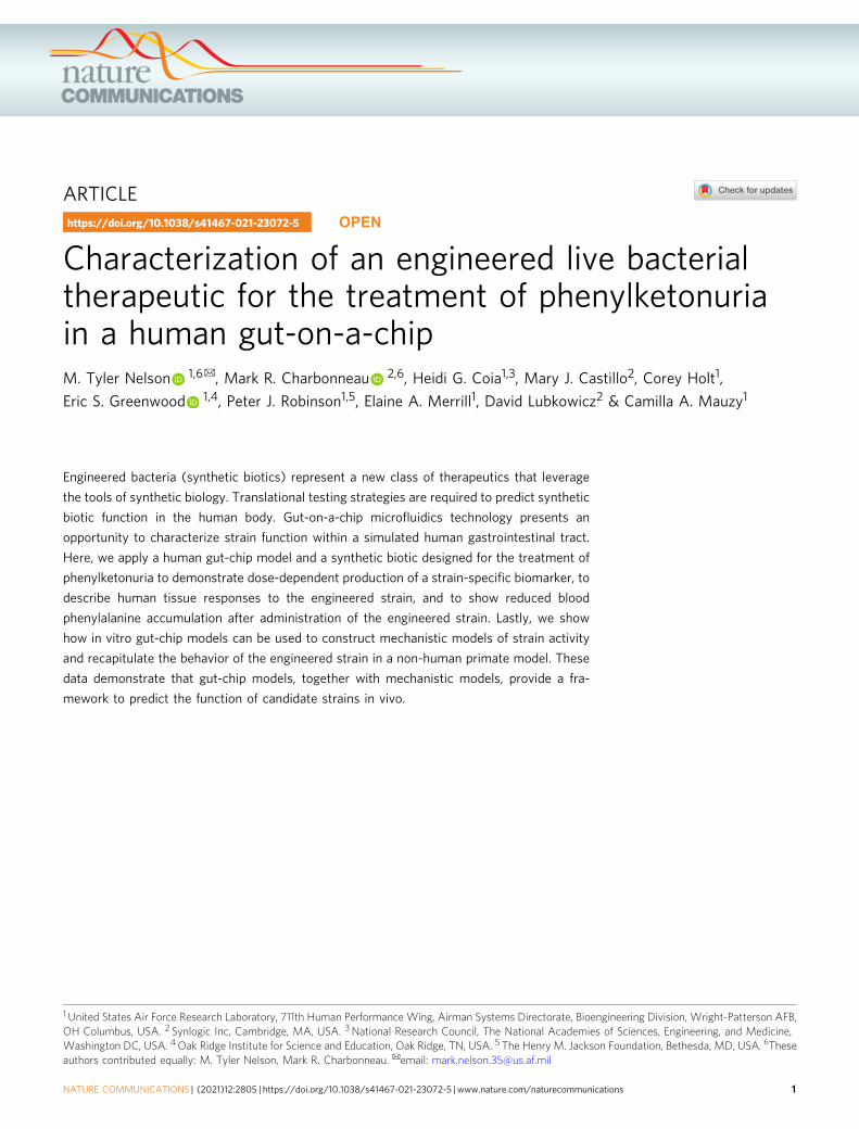
Continuous exposure to high-dose SYN5183 impairs gut bar-rier function. The lack of responses in tissue viability or gutbarrier function with administration of a single dose of SYN5183is consistent with the safety profile of engineered EcN observed inpreclinical studies19. However, synthetic biotics rapidly transitthrough the GI tract in preclinical mouse and NHP models4,19.To investigate tissue responses during extended exposure to thisengineered strain, SIF containing SYN5183 and 5 mM Phe wasadministered to the gut-chips continuously for 12 h at a low (L;1.25 × 10⁸ CFU/mL), medium (M; 6.25 × 10⁸ CFU/mL), or high(H; 1.25 × 10⁹ CFU/mL) dose level (n= 6 chips/treatment group)(Fig. 3a). Fresh SIF containing SYN5183 was introduced after 6 h,and non-treated chips (NT) served as negative controls. CFUplating of gut compartment effluents confirmed that liveSYN5183 was present in the chips throughout this study (Fig. 3b).Continuous SYN5183 administration led to a dose-dependentreduction of gut compartment Phe with concomitant productionof TCA (Fig. 3c, d). A dose-dependent increase of TCA was alsoobserved in the blood compartment, though continuous admin-istration of SYN5183 had no significant impact on blood Pheconcentrations (Fig. 3e, f).
In contrast to a single dose, continuous administration ofSYN5183 resulted in dose-dependent decreases of macro-villusheight and ZO-1 staining in the gut-chips (Fig. 3g, h), suggestingreduced function of the gut barrier. Consistent with thisobservation, continuously administered high-dose SYN5183 alsoresulted in a statistically significant increase in barrier perme-ability (Fig. 3i). This effect on barrier permeability may contributeto the lack of blood Phe lowering in the context of continuousstrain administration, given the low molecular weight of Phe andthe high concentration of Phe administered to the gut compart-ment. Continuous dosing also had a substantial impact on hostcell viability. LDH activity in gut effluents was significantlyincreased at 6 and 12 h for the high-dose group (p < 0.05, 2-wayANOVA, Tukey post-hoc analysis; Supplemental Fig. 2a, b). Flowcytometry conducted at the completion of the study revealed nosignificant effect on epithelial viability in the gut compartment (p= 0.089; ANOVA, Tukey post-hoc analysis Supplemental Fig. 2c).However, a statistically significant decrease in endothelial cellviability was evident in the blood compartment (p= 0.031, 2-wayANOVA, Tukey post-hoc analysis; Supplemental Fig. 2d).
In the gut compartment, a dose-dependent increase in IL-2,TNF-α, and IL-15 was evident at 6 h (p < 0.05, 2-way ANOVA,Tukey post-hoc analysis; Supplemental Fig. 2e, SupplementalTable 2a). However, IL-15 secretion was markedly decreased forthe medium and high dose at 12 h, suggesting that the viability ofthe host gut cells and breakdown of the gut-blood barrier alteredcytokine production and compartment specific concentrations(Supplemental Fig. 2f, Supplemental Table 2b). Conversely, lowdoses of SYN5183 resulted in significant increases of IL-15, IL-2,and TNF-α. Dose-dependent reduction of TNF-β was also evidentin the gut compartment. Interestingly, minimal secreted cytokineactivity was evident in the blood compartment, with the exceptionof the high-dose group at 12 h, where all measured cytokines weresignificantly elevated by up to 600-fold over NT controls(Supplemental Fig. 2g, h, Supplemental Table 2c, d). To determinewhether this markedly increased cytokine secretion in the bloodcompartment was attributable to bacterial translocation, an
NATURE COMMUNICATIONS | https://doi.org/10.1038/s41467-021-23072-5 ARTICLE
NATURE COMMUNICATIONS | (2021) 12:2805 | https://doi.org/10.1038/s41467-021-23072-5 |www.nature.com/naturecommunications 5
additional set of gut-chips (n= 5 chips) were continuously dosedwith a high dose of SYN5183 (1 × 10⁹ CFU/mL) for 24 h, andSYN5183 CFU was enumerated in gut and blood compartmenteffluents. While gut effluents harbored the expected concentrationof SYN5183, no CFU was detected in the blood compartment(Supplemental Fig. 2i). This result indicates that increasedcytokine secretion in the blood compartment is not driven by
bacterial translocation. However, it is possible that the transport ofbacterial compounds, including lipopolysaccharide (LPS),impacted cytokine expression in the vascular compartment.
SYN5183 degrades blood Phe in a human gut-chip. In additionto the consumption of Phe from dietary intake, it is important to
a b
ec
f g
d
NTLMH
h i
Dose 1 Dose 2
LMH
Gut Compartment
Blood Compartment
PhePhe
Phe
TCA
TCA
Continuously DosedSYN5183
Phe
TCA
Phe
TCA
NT
DIC ZO1 Merge 114 μm
106 μm
77 μm
64 μm
L
M
H
**
*
**
*
*
*
* ** *
*
*
*
**
*
*
*
**
*
**
0 3 6 9 12
100101102103104105106107108109
10101011
0
Time (hr)
CFU
/mL
3 6 9 120
2
4
6
8
10
Time (hr)
Gut
Phe
(mM
)
3 6 9 120
1
2
3
4
Time (hr)
Gut
TC
A (m
M)
3 6 9 120.0
0.2
0.4
0.6
Time (hr)
Bloo
d Ph
e (m
M)
3 6 9 120.0
0.1
0.2
0.3
0.4
0.5
Time (hr)
Bloo
d TC
A (m
M)
NTLMH
NT L M H40
60
80
100
120
140
SYN5183 Dose
Villu
s H
eigh
t (µm
)
**
NT L M H0.0
5.0 10-7
1.0 10-6
1.5 10-6
SYN5183 Dose
P app
(cm
/s)
***
ARTICLE NATURE COMMUNICATIONS | https://doi.org/10.1038/s41467-021-23072-5
6 NATURE COMMUNICATIONS | (2021) 12:2805 | https://doi.org/10.1038/s41467-021-23072-5 | www.nature.com/naturecommunications

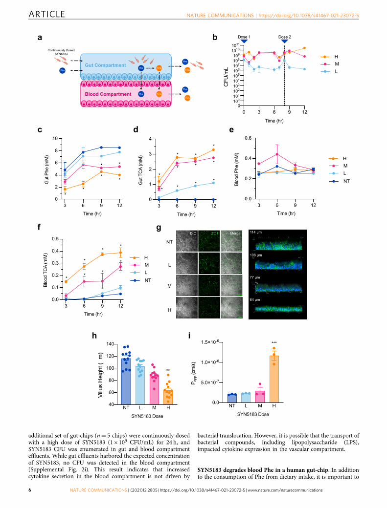
determine whether a single dose of synthetic biotic can accesssystemic pools of Phe. There is evidence that plasma Phe canenter the gastrointestinal lumen, through a process termedenterorecirculation4,20. Though the specific mechanisms of Pherecirculation are not well defined, this notion is supported bystudies in ileostomy patients that demonstrate loss of Phe in guteffluents after being placed on a Phe-free diet21. To address thisusing the human gut- chip model, SYN5183 was administered inPhe-free medium to the gut compartment at a low (L; 1.25 × 10⁸
CFU/mL), medium (M; 6.25 × 10⁸ CFU/mL), or high (H; 1.25 ×10⁹ CFU/mL) dose level (n= 3 chips/treatment group). Non-treated chips (NT) served as negative controls. 1 mM Phe wasthen continuously administered to the blood compartment for 12h, consistent with levels observed in adult PKU patients withoutdietary Phe restriction (Fig. 4a;2). This procedure resulted in0.321 ± 0.003 mM Phe (mean ± SEM) available in the gut com-partment of untreated chips.
SYN5183 cells cleared the chips within 9 h for all dose levels(Fig. 4b) and demonstrated a dose-dependent lowering of gutcompartment Phe with concomitant production of the biomarkerTCA (Fig. 4c, d). All concentrations of SYN5183 resulted insignificant TCA production at 3 h after strain administration ascompared to negative controls, and medium and high doses alsoachieved statistical significance at 6 and 9 h (Fig. 4d). However,only the high dose of SYN5183 resulted in a statisticallysignificant decrease of gut compartment Phe at 3 h (p= 0.021;two-way ANOVA, post-hoc Tukey analysis), 6 h (p= 0.002), and9 h (p= 0.025), as compared to NT chips.
In the blood compartment, TCA concentrations were sig-nificantly elevated compared to untreated controls at 3, 6, and 9 hfor both the medium and high doses of SYN5183 (p < 0.05, two-way ANOVA, post-hoc Tukey analysis; Fig. 4e). In addition, thehigh dose of SYN5183 resulted in a 9.55% decreased area underthe curve (AUC) for blood Phe over the course of the study (p=0.032, two-way ANOVA, post-hoc Tukey analysis; Fig. 4f).Importantly, no significant changes in macro-villus height orbarrier permeability were observed (Fig. 4g, h), indicatingmaintenance of a functional gut-endothelial barrier.
Cytokine profiles 12 h post SYN5183 administration (Supple-mental Fig. 3, Supplemental Table 3) showed no statisticallysignificant differences in cytokine secretion (p < 0.05, Student’s ttest with False Discovery Rate correction for multiple hypothesistesting). These data suggest that SYN5183 can access circulatingPhe and lower systemic concentrations without compromisinggut barrier function.
Computational models to describe Phe kinetics in NHP dosedwith SYNB1618. Although the gut-chip model emulates thegastrointestinal tract, in vitro systems do not account for allaspects of human physiology. Understanding the translationalvalue of synthetic biotics necessitates the development of math-ematical frameworks for integrating data from in vitro and in vivo
model systems to predict the behavior of clinical candidates underdynamic conditions of the human gut. We formulated compu-tational models to describe the activity of SYN5183 in gut-chips(Fig. 5a) and in vivo (Fig. 5b, c; see Supplementary Informationfor a complete description of model development andcalibration).
Briefly, the in vitro computational model represents SYN5183dosing in the gut-chip system (Fig. 5a). Phe consumption by thePAL enzyme, expressed by SYN5183, was assumed to followMichaelis–Menten kinetics, and kinetic parameters were esti-mated from in vitro data. Specifically, fitting the in vitrocomputational model to our gut-chip bolus studies at allSYN5183 dose levels (1.25, 6.25, and 12.5 × 10⁸ CFUs), providedan estimate of Vmax for SYN5183 PAL activity of 0.16 μmol/(108
CFU-h). Km for the PAL pathway was estimated as 0.2 mM fromin vitro Phe consumption assays. This model was also used toestimate a key gut permeability parameter, P, by calibration to thein vitro chip studies (P= 0.12 cm/h for Phe; P= 6 × 10⁻⁴ cm/hfor TCA). Gut wall permeability is an indicator of gut health anda mediator of other physiologically relevant processes, includingwater flow, motility, and pH.
The in vivo computational model is an extension of the gut-chip computational model that enables time course simulation ofblood Phe concentrations after SYN5183 dosing (Fig. 5b). Duringsimulated gastrointestinal transit, Phe is assumed to diffuse intothe blood across an effective gut wall surface area, A(see Supplementary Information for a discussion on thesignificance of A). Specifically, the model represents the uptakeof dietary Phe in the gut according to a permeability-surface areaproduct PA (cm3/h) and the concentration gradient of Phebetween gut and blood compartments. In addition, host tissueprotein breakdown, D (36 μmol/h in humans;22), contributes toblood Phe concentrations, and Phe is assumed to deplete from theblood according to a composite, first-order elimination rateconstant, k (cm3/h). Fasting, steady-state blood Phe levels wereused to express k in terms of D, assuming negligible input of Phefrom the gut in the fasting state. Blood Phe levels, cb, are thusrepresented by Eq. 1:
Vbdcbdt
¼ PA cg � cb� �
þ Dð1� cbcb;ss
Þ ð1Þ
where cg is the concentration of Phe in the gut, cb,ss is the steady-state fasting blood Phe concentration, and Vb is the blood volume.
To assess the translational value of the in vivo computationalmodel, time courses of blood Phe concentrations were simulatedusing this model for comparison to data collected from non-human primates (NHP), orally dosed with the related clinicalcandidate strain, SYNB1618, and a bolus of peptone (containingapproximately 0.25 g Phe) in an earlier study4. Simulated dosingcomprised a bolus of 0.25 g Phe and engineered cells at four doselevels (0, 1.8 × 10¹¹, 3.6 × 10¹¹, or 7.2 × 10¹¹ CFU). The oral boluswas assumed to pass through the relevant portion of the gut with
Fig. 3 Continuous administration of SYN5183 in a human gut-chip model. a Schematic of study design. SYN5183 was administered continuously over 12 hin the gut compartment with 5.0 mM Phe in SIF, SYN5183 CFU were enumerated from gut compartment effluents, and Phe and TCA were quantified usingeffluents collected from the gut and blood compartments. Fresh, SYN5183-containing inoculum was introduced after 8 h. b–d Concentrations of SYN5183(b; CFU/mL), Phe (c; mM), and TCA (d; mM) recovered from gut compartment effluents over time. e, f Concentrations of Phe (e; mM) and TCA (f; mM)recovered from blood compartment effluents over time. g DIC and ZO-1 (green) staining showed consistent tight-junction formation surrounding macro-villus structures at NT and L—dose and loss of ZO-1 signal and disruption of morphology at M and H—doses, and to the right f-actin (green) stainingshows macro-villi structures decreasing in height with increasing dose (n = 3 independent gut chips, 4 ROIs per image quantified for villus height). h, iVillus height (μm) and apparent permeability (Papp; cm/s) across the gut barrier 12 h post-dose. For (b–i), H (orange), M (pink), and L (light blue)correspond to SYN5183 doses of 1.25 × 10⁹ CFU/mL, 6.25 × 10⁸ CFU/mL, and 1.25 × 10⁸ CFU/mL, respectively. NT (dark blue) corresponds to non-treated(NT) chips. *p < 0.05, **p < 0.01, ***p < 0.01; 2-way ANOVA, with a post-hoc Tukey analysis using a 95% confidence interval compared to the NT group, n= 6 gut chips, error bars represent SEM with the middle point representing the mean value. Source data are provided as a Source Data file.
NATURE COMMUNICATIONS | https://doi.org/10.1038/s41467-021-23072-5 ARTICLE
NATURE COMMUNICATIONS | (2021) 12:2805 | https://doi.org/10.1038/s41467-021-23072-5 |www.nature.com/naturecommunications 7
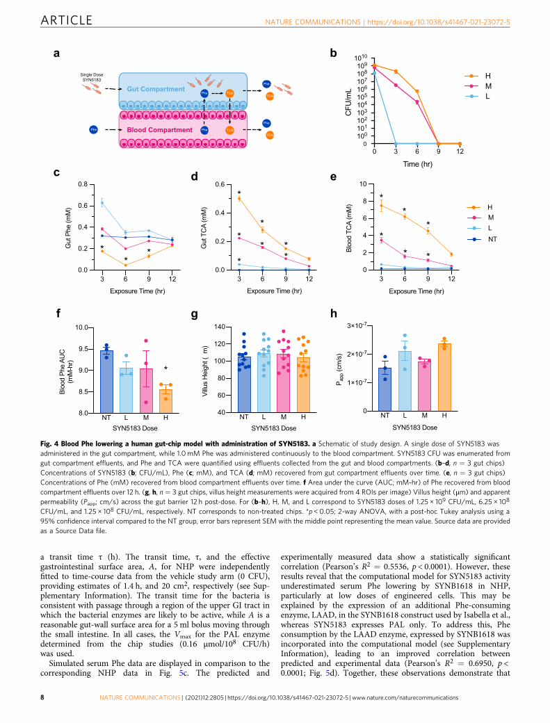
a transit time τ (h). The transit time, τ, and the effectivegastrointestinal surface area, A, for NHP were independentlyfitted to time-course data from the vehicle study arm (0 CFU),providing estimates of 1.4 h, and 20 cm2, respectively (see Sup-plementary Information). The transit time for the bacteria isconsistent with passage through a region of the upper GI tract inwhich the bacterial enzymes are likely to be active, while A is areasonable gut-wall surface area for a 5 ml bolus moving throughthe small intestine. In all cases, the Vmax for the PAL enzymedetermined from the chip studies (0.16 μmol/108 CFU/h)was used.
Simulated serum Phe data are displayed in comparison to thecorresponding NHP data in Fig. 5c. The predicted and
experimentally measured data show a statistically significantcorrelation (Pearson’s R2 = 0.5536, p < 0.0001). However, theseresults reveal that the computational model for SYN5183 activityunderestimated serum Phe lowering by SYNB1618 in NHP,particularly at low doses of engineered cells. This may beexplained by the expression of an additional Phe-consumingenzyme, LAAD, in the SYNB1618 construct used by Isabella et al.,whereas SYN5183 expresses PAL only. To address this, Pheconsumption by the LAAD enzyme, expressed by SYNB1618 wasincorporated into the computational model (see SupplementaryInformation), leading to an improved correlation betweenpredicted and experimental data (Pearson’s R2 = 0.6950, p <0.0001; Fig. 5d). Together, these observations demonstrate that
a b
ec
f g h
d
NTLMH
Gut Compartment
Blood Compartment
Phe
Phe Phe
TCA
TCA
Single DoseSYN5183
Phe
TCA
Phe
TCA
**
*
*
*
**
**
*
**
*
***
0 3 6 9 12
100101102103104105106107108109
1010
0
Time (hr)
CFU
/mL
HML
3 6 9 120.0
0.2
0.4
0.6
0.8
Exposure Time (hr)
Gut
Phe
(mM
)
3 6 9 120.0
0.2
0.4
0.6
Exposure Time (hr)
Gut
TC
A (m
M)
3 6 9 120
2
4
6
8
10
Exposure Time (hr)
Bloo
d TC
A (m
M)
NT L M H8.0
8.5
9.0
9.5
10.0
Bloo
d Ph
e AU
C(m
M-h
r)
*
NT L M H0
1 10-7
2 10-7
3 10-7
SYN5183 Dose
P app
(cm
/s)
NT L M H40
60
80
100
120
140
SYN5183 Dose
Villu
s H
eigh
t (µm
)
SYN5183 Dose
Fig. 4 Blood Phe lowering a human gut-chip model with administration of SYN5183. a Schematic of study design. A single dose of SYN5183 wasadministered in the gut compartment, while 1.0 mM Phe was administered continuously to the blood compartment. SYN5183 CFU was enumerated fromgut compartment effluents, and Phe and TCA were quantified using effluents collected from the gut and blood compartments. (b–d, n = 3 gut chips)Concentrations of SYN5183 (b; CFU/mL), Phe (c; mM), and TCA (d; mM) recovered from gut compartment effluents over time. (e, n = 3 gut chips)Concentrations of Phe (mM) recovered from blood compartment effluents over time. f Area under the curve (AUC; mM-hr) of Phe recovered from bloodcompartment effluents over 12 h. (g, h, n = 3 gut chips, villus height measurements were acquired from 4 ROIs per image) Villus height (μm) and apparentpermeability (Papp; cm/s) across the gut barrier 12 h post-dose. For (b–h), H, M, and L correspond to SYN5183 doses of 1.25 × 10⁹ CFU/mL, 6.25 × 10⁸
CFU/mL, and 1.25 × 10⁸ CFU/mL, respectively. NT corresponds to non-treated chips. *p < 0.05; 2-way ANOVA, with a post-hoc Tukey analysis using a95% confidence interval compared to the NT group, error bars represent SEM with the middle point representing the mean value. Source data are providedas a Source Data file.
ARTICLE NATURE COMMUNICATIONS | https://doi.org/10.1038/s41467-021-23072-5
8 NATURE COMMUNICATIONS | (2021) 12:2805 | https://doi.org/10.1038/s41467-021-23072-5 | www.nature.com/naturecommunications

gut-chip model systems, together with physiologically basedcomputational frameworks, can be used to describe the functionof synthetic biotics in vivo.
DiscussionSynthetic biotics represent a new class of living therapeutics withthe potential to sense and respond to signals within the humanbody, to combine therapeutic effectors, and to address the sig-nificant unmet medical needs of patients3. However, the devel-opment of novel therapeutics is both time and resource
intensive3. Preclinical in vitro and animal models are commonlyemployed to anticipate the safety and efficacy of drug candidatesin humans, but the translational value of these models varies byindication and drug mechanism of action3,4,12,23. Ultimately,most drug candidates that do enter clinical efficacy studies fail tomeet their primary endpoints and to achieve regulatoryapproval4,8,23. Innovative preclinical models are needed to ade-quately predict the performance of putative drug candidates inhuman subjects.
Human OOC technology integrates engineered microfluidicdevices and dynamic tissue cultures that emulate organ-level
a b
d
c1.8 x 1011 CFU
PAL + LAAD PAL + LAAD PAL + LAAD PAL + LAAD
Gut Compartment
in vitro System
Blood Compartment
ApicalInflow
Outflow
I IVII
BasolateralInflow
III
Outflow
Gut Phe, Cg
Blood Phe, Cb
TCA
Gut Lumen
in vivo Equivalent
Blood
GutDosing
VenousOutflow
I IVIIa
ArterialInflow
III
ProteinBreakdown
IIbGut Phe, Cg
Blood Phe, Cb
TCA
0 2 4 6
0.05
0.10
0.15
Time (hr)
Seru
m P
he (m
M)
Vehicle
0 2 4 6
0.05
0.10
0.15
Time (hr)0 2 4 6
0.05
0.10
0.15
Time (hr)
0 2 4 6
0.05
0.10
0.15
Time (hr)
3.6 x 10¹¹ CFU 7.2 x 10¹¹ CFU
1.8 x 10¹¹ CFUVehicle 3.6 x 10¹¹ CFU 7.2 x 10¹¹ CFU
PAL Only PAL Only PAL Only PAL Only
0 2 4 6
0.05
0.10
0.15
Time (hr)
Seru
m P
he (m
M)
0 2 4 6
0.05
0.10
0.15
Time (hr)
0 2 4 6
0.05
0.10
0.15
Time (hr)
0 2 4 6
0.05
0.10
0.15
Time (hr)
Fig. 5 Computational models of Phe kinetics in NHP dosed with SYNB1618. a, b Schematic representations of Phe kinetics in gut-chips (a) and in vivo (b).Arrows labeled I-IV represent equivalent processes in the two systems as follows: (I) Uptake of Phe from gut to blood via permeability-surface areaproduct, PA, (II) Excretion of Phe from blood to the gut, (III) Metabolism of Phe to TCA by SYN5183 or SYNB1618, normalized to CFU number, and (IV)absorption of TCA from gut to blood via PA. For (b), the complex in vivo process of hepatic enterorecirculation (IIb) is not implemented in thecomputational model. Instead, this process is described by direct diffusion from the blood to the gut compartment (IIa), as in the gut chip. c Serum Pheconcentrations for NHP orally dosed with SYNB1618 and the corresponding in vivo model simulations, assuming PAL activity only. d Serum Pheconcentrations for NHP orally dosed with SYNB1618 and the corresponding in vivo model simulations, assuming both PAL and LAAD enzyme activity. For(c, d, in vitro results were acquired from three independent gut chips and extrapolated to published NHP data4), points with errors bars indicateexperimental data from Isabella et al. 4, and solid lines represent simulate data. NHP were dosed at 0 h with a 5.0 g peptone bolus and 0–7.2 × 10¹¹ CFUSYNB16184, error bars represent standard deviation with the middle point representing the mean value. Source data are provided as a Source Data file.
NATURE COMMUNICATIONS | https://doi.org/10.1038/s41467-021-23072-5 ARTICLE
NATURE COMMUNICATIONS | (2021) 12:2805 | https://doi.org/10.1038/s41467-021-23072-5 |www.nature.com/naturecommunications 9

functionality using human cells to bridge the gap between tra-ditional in vitro techniques and animal research, providingenhanced mechanistic insight and relevance to human biology.These platforms enable robust cellular and molecular analysis,fine control over transport and fluid flow dynamics, and incor-poration of complex mechanical stimuli that augment thetranslational value of these laboratory organ models12,13,24–27.Gut-chip models, in particular, have shown significant promise inovercoming many limitations of conventional cell culture tech-niques that fail to capture the complexity and function of humangut tissue. For example, common in vitro model systems of thegut epithelium (e.g., transwell) lack shear stress and fluid flow,which limits the relevance of host-microbe interactions andassessments of barrier function. By contrast, the dynamic cul-turing conditions in microfluidic systems enhance gut-endothelialbarrier health, aid in maintaining microbiome homeostasis, andfacilitate nutrient and metabolite transport13,24,26. Importantly,gut-chip cultures incorporating both passive and appliedmechanical forces develop human gut-like villus projections andan epithelial mucus layer24,26. The benefits of dynamic culturealso address significant experimental obstacles. For example,these models can prevent bacterial overgrowth of the host cellculture and limit inflammatory responses13. This feature of gut-chip models enables a shift of timescale from hours to days andexperimental designs that incorporate clinical dosing regimens.
Preclinical models that capture aspects of human gut phy-siology are critical for interrogating the function of orally admi-nistered synthetic biotics, including the diffusion of bacterialproducts across the gut barrier. In this article, a human gut-chipmodel system was employed to characterize engineered bacterialstrain activity and host tissue responses, as well as to defineparameters for computational models that describe strain func-tion in vivo. One limitation of the approach described here is theuse of immortalized cell lines. More advanced primary crypt orinduced pluripotent stem cell (iPSC) derived human intestinalmodels have indeed been described that demonstrate significantimprovements in capturing host gene expression profiles8,28,29.Additional considerations for primary tissue culture modelsinclude internal review board governance, donor to donorvariability, and increased cost. By contrast, the Caco2 and HT-29cell lines used in this work have been extensively characterizedand are widely available8,24,25,28,29. Importantly, Caco2 gut-chipmodels have proven to be useful for replicating human relevantgut-blood apparent permeability and functional responses. Thesecell line-based models are suitable for the rapid evaluation ofsynthetic biotic prototypes in a human gut microenvironment.
SYNB1618 has been previously shown to reduce circulatingPhe in an NHP model after an oral Phe bolus and to produce theurinary biomarker, HA, in a dose-dependent manner4. In thisstudy, the related strain, SYN5183, demonstrated a dose-dependent production of the strain-specific biomarker, TCA,and reduced blood Phe levels in a human gut-chip by up to 27%.In contrast to static in vitro systems, the dynamic gut-chip modelalso enabled simulation of bacterial residence time within thegastrointestinal tract, and a single dose of SYN5183 was found toclear the gut-chips within 12 h, leading to concomitant clearanceof its product, TCA. Importantly, simultaneous administration ofPhe and SYN5183 to the blood and gut compartments, respec-tively, resulted in a 9.55% cumulative reduction in circulatingPhe. These data support the hypothesis that orally administeredSYN5183 can consume systemic Phe, in addition to dietary Phe,through a process termed enterorecirculation4,20.
In preclinical animal models, engineered EcN strains are welltolerated and rapidly clear the GI tract following oraladministration4,19. Here, we used the human gut-chip model toexamine the effects of synthetic biotic administration at durations
of exposure above those that are feasible in vivo by continuousinfusion of SYN5183 to the gut compartment. This continuousdosing paradigm (Fig. 3) differs from in vivo exposure to thestrain in that this design prevents the rapid bacterial clearanceobserved in preclinical animal models and humans4. In contrastto our studies that simulated in vivo dosing paradigms, weobserved that continuous exposure to high-dose SYN5183 in gut-chips decreased macro-villus height and increased barrier per-meability. The decreased epithelial surface area associated withvillus blunting may alter Phe transport across the gut barrier,consistent with our observation that continuous exposure toSYN5183 abrogated the blood Phe lowering effect observed withclinical dosing designs despite significant differences in gut Pheconcentrations. Indeed, villus blunting due to inflammation hasbeen previously shown to impair nutrient transport, barriermaintenance, and mucosal integrity13,23–27. These findings sug-gest that current clinical dosing strategies are preferred, asincreased exposure to SYN5183 was not beneficial for loweringcirculating Phe.
A notable limitation of our gut-chip model implementation isthe presence of ambient oxygen in the system, which contrastswith the low-oxygen concentrations present in the humanproximal GI tract22,30. Due to this discrepancy, we were unable todirectly assess activity of the highly oxygen sensitive LAADenzyme expressed by the clinical candidate strain SYNB16184.Relatedly, expression of the PheP transporter and PAL enzyme bySYN5183 and SYNB1618 is driven by the low-oxygen responsiveFNR promoter and is expected to be maintained in the low-oxygen conditions of the gut. These components are inducedduring the preparation of biomass (i.e., SYN5183 is “pre-loaded”with PheP and PAL protein). However, continued induction ofthe FNR promoter in the oxygenated gut-chip model is notexpected. This could result in an underestimate of in vivo activity.A complete translational understanding of the potential efficacyof SYNB1618 for PKU patients would require a more thoroughexamination of LAAD function in a model system that simulatesmicroaerobic conditions. Some groups have indeed describedlow-oxygen microfluidics implementations for the study of host-bacterial interactions25, but such systems are not yet widelyavailable. The development of anaerobic or microaerobic gut-chipmodels will also be essential for understanding the function ofsynthetic biotics in the context of a complex endogenousmicrobial community. The studies described in this work exam-ined SYN5183 in isolation, and a complete understanding of thestrain’s function in vivo will require tools that incorporate arepresentative gut microbiota.
Computational models are powerful tools for embeddingexperimentally derived parameters in a broader physiologicalframework. The gut-chip model described here displayed phy-siologically relevant Phe flux (0.03 nmol/cm2*min), indicatingthat the gut-blood barrier biology was representative of humansmall intestinal tissue31,32. The flux, or the rate of transportthrough a porous medium, is a measurement of gut-blood barrier.While increases in apparent permeability directly impacts theflux, it is surface area that dictates the magnitude of solute (Phe)transport behavior. The gut-chip represents a microscale anato-mical unit of human gut tissue, and thus it is important toconsider that the geometry and scale of the model impactsmeasured values, making computational extrapolation necessaryfor interpreting in vitro model outcomes. The computationalmodel outlined in this article combines key parameters extractedfrom gut-chip data with parameters estimating gastrointestinalphysiology (e.g., flow rates, tissue volumes, and surface areas), tomake predictions of circulating Phe levels in vivo. Using datafrom the gut-chip system, this model accurately predicted Phereduction associated with administration of SYNB1618 in NHP.
ARTICLE NATURE COMMUNICATIONS | https://doi.org/10.1038/s41467-021-23072-5
10 NATURE COMMUNICATIONS | (2021) 12:2805 | https://doi.org/10.1038/s41467-021-23072-5 | www.nature.com/naturecommunications

However, a limitation is that this model implementation doesnot capture the variability observed in human Phe metabolism(such as residual levels of PAH activity in PKU patients). Tocapture this, a comprehensive, physiologically based pharmaco-kinetic (PBPK) model of Phe metabolism is required. Such amodel could also include a detailed treatment of enteror-ecirculation, as well as analysis of engineered strain functionwithin the dynamic and heterogeneous conditions of the humangastrointestinal tract. Advances in OOC technology, such as theincorporation of human induced Pluripotent Stem Cells(iPSCs)28, patient-derived primary cell cultures8,29, and anaerobicconditions capable of sustaining a human distal gut microbiota25,may facilitate these efforts. For example, Novak et al.33 recentlydescribed a PBPK representation of multiple connected OOCsystems, highlighting complex interactions between multipleorgan systems33.
OOC technology has been successfully applied to the devel-opment of other therapeutic modalities. For example, gut-chipanalysis of dimethyloxaloylglycine, a small molecule drug candi-date for the prevention of radiation-induced gut permeability,predicted clinical outcomes in humans, while these results werenot reproduced by rodent models23. Furthermore, evaluation ofmilk oligosaccharides and fermentate from SHIME microbialcultures displayed protein- and metabolite-dependent activationof claudin tight-junctional proteins, limiting small moleculetransport as seen in longitudinal human studies involving B.infantis treatment of IBD34,35. To the best of our knowledge, gut-chip models have not previously been used to validate the in vivofunction of synthetic biotics, making this study a foundationalcase study for evaluating dynamic processes such as strainactivity, biomarker production, and host tissue responses, as wellas for extrapolation of in vitro data to provide in vivo predictions.
Together, these data indicate that OOC methods, combinedwith PBPK modeling approaches, provide a powerful set of toolsfor determining the translational value of engineered bacterialtherapeutics. Leveraging this technology has the potential tosignificantly accelerate the development of synthetic biotics, toenhance probability of success in the clinic, and to address theunmet needs of patients.
MethodsGut-on-a-chip development. The microfluidic device and pressure-driven auto-mated pumping system, collectively termed the Human Emulation System, wasprocured from Emulate Bio (Boston, MA). The microfluidic device has twocompartments separated by a thin, flexible, porous membrane (50 µm thick, 7 µmdiameter pores, spaced every 40 µm). The dimensions of the channels are as fol-lows: Upper channel—width = 1 mm, height = 1 mm, culture area 28.0 mm2;Lower channel—width = 1 mm, height = 0.2 mm, and culture area 24.5 mm2. Themicrofluidic device and pumping unit are housed and maintained inside a cellculture incubator at 37 °C, 95% relative humidity (RH). Prior to cell seeding, theupper and lower channels were activated with ER-1 and ER-2 surface preparationsreagents (Emulate Bio, Boston, MA), and subsequently exposed to UV light for 20min as directed by the manufacturer. Both channels were flushed twice with 200 µLof phosphate buffer solution (PBS, Gibco) followed by two flushes with cell culturemedium to remove any residual ER-1. 40 µL of Geltrex reduced-growth factorbasement membrane matrix (Geltrex hESC-Qualified, LDEV, Ready-to-Use,Reduced Growth Factor Basement Membrane Matrix from EHS tumors, Gibco,ThermoFisher Scientific, USA) with an added 100 µg/mL collagen type I(Advanced Biomatrix Inc., USA) was added to the lower and upper compartmentsand incubated overnight at 4 °C. The following day, channels were flushed withseeding media and prepared for cell seeding. HMVEC, human microvascularendothelial cells (TIME hTERT immortalized, cat #CRL-4045, ATCC, Manassas,VA) were cultured in the lower compartment at a density of 1 × 10⁷ cells/mL (28µL) in maintenance medium (Vascular Cell Basal Medium ATCC® PCS-100-030supplemented with a Microvascular Endothelial Growth Kit ATCC® PCS-110-041without VEGF addition). The microfluidic devices were flipped upside down for 2h to encourage endothelial adherence to the lower side of the porous membrane.After which the devices were flipped back upright and a 4:1 ratio of Caco2 (Caco2-BBE, ATCC HTB-37, human colorectal carcinoma) and HT29 (HT29-MTX-E12,Millipore-Sigma, 12040401, human adenocarcinoma) gut epithelial cells was addedto the upper surface of the membrane at a cell seeding density of 3.75 × 10⁶ cells/
mL (40 µL) in experimental medium (DMEM supplemented with 5% fetal bovineserum, ATCC). After 4 h, the microfluidic devices were flushed with fresh degassedmedium, docked with medium reservoirs, and maintained within the HumanEmulation System. A flow rate of 60 µL/hr was applied to both the upper and lowerchannels for the entirety of the experiment. The gut-on-a-chip undergoes spon-taneous differentiation over a 10-day maturation period, of which cyclicalmechanical stretch (10% strain, 0.15 Hz) is applied for the last 7 days to encouragethe development of “macro-villus” structures. Nine devices per condition exhi-biting macro-villus structures were utilized for experimental testing and evaluation.
SYN5183 strain construction. SYN5183 was constructed by removing the L-amino acid deaminase (LAAD) gene from SYNB1618 utilizing standard lambdared strain construction methodology4. In SYNB1618, LAAD was integrated intothe araC locus of E. coli Nissle 1917, knocking out its natural capability to utilizearabinose for growth. To remove LAAD, we transformed pkd46 into SYNB1618 viaelectroporation and amplified the original araC locus from wild-type Nissle viaPCR. This fragment was subsequently used as knock in fragment for lambda redrecombination. After electroporation, clones were plated on M9 minimal mediaplates containing 0.5% arabinose as carbon source. Only cells, with an intact araClocus, and therefore LAAD negative, were able to grow under this condition. Theremoval of LAAD was further verified by PCR and Sanger sequencing. Primers andsequences used for the construction of SYN5183 are provided in Table S4.
SYN5183 dosing. Frozen glycerol stocks of SYN5183 at 1.65 × 10¹¹ CFU/mL werequick thawed diluted in warmed experimental gut medium containing 25%simulated intestinal fluid (full materials and methods located in Supplementarysection). Dosages of 1.25 × 10⁸ (Low, L), 6.25 × 10⁸ (Medium, M), or 1.25 × 10⁹
CFU/mL (High, H) of SYN5183 were selected based on in vivo dosing regimensevaluated previously4. After the maturation of the gut-on-a-chip, 1 h prior to thestart of the experiment, 35 uL of SYN5183 was added to the gut compartment, thechips were then docked with the Pod (cell culture microfluidic reservoir), and heldunder static, no flow conditions. The 1-h static condition allows the SYN5183 tosettle in the chip prior to continuous flow.
Direct bolus. 5 mM phenylalanine (Phe, L-Phenylalanine, Sigma-Aldrich, USA)was added into the experimental medium and continuously flowed through the gutcompartment of the microfluidic device for 12 h. All of the effluent was collectedevery 3 h from both the gut and ‘blood’ compartment outlet waste reservoirs. 200µL of gut and ‘blood’ compartment inlet medium was collected at 3 h as a referencefor permeability and LC-MS/MS analysis.
Continuous dosing. In addition to the 5 mM phenylalanine the respective dose ofSYN5183 was added into the experimental medium and continuously flowedthrough the gut compartment of the microfluidic device for 12 h. After 6 h a seconddose of SYN5183 was added to the inlet reservoir. All of the effluent was collectedevery 3 h from both the gut and “blood” compartment outlet waste reservoirs. 200µL of gut and “blood” compartment inlet medium was collected at 3 h as areference for permeability and LC-MS/MS analysis.
Phe reduction. Simulation of human circulating levels of blood Phe at 1 mM wasadded into the inlet of the ‘blood’ compartment of the microfluidic chip. Every 3 hthe pass-through effluent from both compartments, and 200 µL from the inlets wascollected and stored for future analysis.
SYN5183 CFU Enumeration. Colonies of SYN5183 from gut-on-a-chip effluent(gut compartment) were plated on LB Agar plates (Animal Free Soytone & Dia-minopimelic Acid, Teknova Cat# L1193); 20 µl of effluent was added to 180 µl 1XPBS and serially diluted down to 10−8. Five microliters from each dilution were“spot”-plated at each time point in triplicate, and plates were incubated overnightat 37 °C. Colony counts were taken after 18 h (high dose SYN5183 continuousdosing ‘blood’ compartment effluents were analyzed for bacterial presence after 24h in the gut-chip and showed no countable colonies, further experiments werelimited to the gut compartment).
Cellular morphological and tight-junction analysis. Images of the human gut-on-a-chip villus structures were acquired using differential interface contrast (DIC)and fluorescence (f-actin, ActinGreen 488 (Invitrogen R3110) or ActinRed 555(Invitrogen R3112) microscopy (Nikon, Ti2 scanning confocal microscope). Guttissue tight junctions were evaluated using ZO-1 (primary antibody, Invitrogen, 33-9100) immunofluorescence imaging. Gut-chip tissues were washed with phosphatebuffered saline (PBS) 2x, followed by fixation in a 4% paraformaldehyde (PFA,Electron Microscopy Sciences, Inc.) phosphate buffer solution at room temperaturefor 10 min. Following fixation, 0.1% triton-X 100 phosphate buffer solution con-taining 2.5% bovine serum albumin was added to each channel for 45 min toensure complete membrane permeabilization and blocking to prevent non-specificimmunolabeling. After permeabilization the gut-chip tissue channels were washedwith phosphate buffer, and subsequently primary antibody at a concentration of 5µg/mL was added in permeabilization buffer overnight at 4 °C. After primary
NATURE COMMUNICATIONS | https://doi.org/10.1038/s41467-021-23072-5 ARTICLE
NATURE COMMUNICATIONS | (2021) 12:2805 | https://doi.org/10.1038/s41467-021-23072-5 |www.nature.com/naturecommunications 11

antibody labeling, each tissue channel was washed with permeabilization buffer. 5µg/mL secondary antibody (Goat anti-mouse H &L Alexafluor 488, Invitrogen orGoat anti-mouse H&L AlexaFluor 555, Invitrogen) containing ActinGreen orActinRed per manufacturers guidelines was added to the respective channels atroom temperature for 2 h. Following secondary antibody labeling and actinstaining, the tissue channels were flushed with permeabilization buffer and sub-sequently filled with PBS for confocal imaging. Pipette tips containing excess PBSwere placed into the outlet and inlet ports of the gut-chip to prevent channeldehydration. Immunostained gut-chips were stored at 4°C until use. Immuno-fluorescent cross sections and DIC images were prepared in NIS-Elements Nikonacquisition software. Measurements of villus height and ZO-1 intensity werequantified directly from the images using ImageJ image analysis software.
Quantification of phenylalanine and trans-cinnamic acid. Quantification ofanalytes of interest was performed using a triple quadrupole LC-MS/MS ThermoTSQ Quantum Max system. Standards were prepared in water with the followingconcentrations: 0.032, 0.16, 0.8, 4, 20, 100, and 250 µg/mL. Samples were dilutedappropriately with water prior to processing. In a 96-well plate, 10 µL of thestandards and samples were transferred, followed by the addition of 90 µL deri-vatization solution (50 mM of 2-hydrazinoquinoline (2-HQ), dipyridyl disulfide,and triphenylphosphine in acetonitrile with 1 µg/mL of internal standard Phe-13C9-15N). The plate was heat-sealed with a ThermASeal foil (Sigma), mixed, andincubated at 60 °C for 1 h. The samples were then centrifuged at 1000xg for 5 min.To another plate, 20 µL of the derivatized samples were transferred and furtherdiluted with 180 μL of 0.1% formic acid in water/acetonitrile (140:40).
The injection volume used was 10 µL and the run time was 4.25 min at a flowrate of 0.5 mL/min. Mobile phase A was 0.1% formic acid in water and mobilephase B was 0.1% formic acid in acetonitrile/isopropanol (90:10, v/v).Chromatographic separation was carried out using a Phenomenex Luna 5 µm C18column (3 µm, 100 x 2 mm) with the following gradient: 10% B from 0 to 0.5 min,10→97% B from 0.5 to 2 min, 97% B from 2 to 4 min, 10% B from 4 to 4.25 min.Multiple reaction monitoring (MRM) in positive mode was used for tandem MSanalysis. The following mass transitions were monitored for quantitation: Phe(307.2/186.0), Phe-d5 (312.2/186.0), TCA (290.2/131.0), TCA-d5 (295.1/138.1), andinternal standard Phe-13C9-15N (317.0/186.1).
Paracellular cascade blue flux assay. Fifty microgram per milliliter of cascade blue(0.59 kDa; ThermoFisher, C687) were introduced to the gut channel (60mL h−1)and fluorescence intensity (390 nm/420 nm) of top and bottom channel effluentswere measured using a multi-mode plate reader (BioTek Cytation 5). Apical-to-basolateral flux of the paracellular marker was calculated based on the followingequation: Papp=(dQ/dt)/A.dC. Papp (cm s−1) denotes the apparent permeabilitycoefficient, dQ/dt (g s−1) is molecular flux, A (cm2) is the total area of diffusion anddC (mgmL−1) is the average gradient. Samples were collected at time 0 from theinlets only, and 12 h from both inlets and outlets of each gut-chip compartment. Pappvalues will be calculated by converting relative fluorescence units (RFU) using astandard curve, the mean and standard error will be reported.
Lactate dehydrogenase (LDH). Effluent isolated from the outlet reservoirs of sixindependent chips was added in triplicate to a black-walled, 96-well plate andmixed 1:1 with Cell Membrane Integrity Assay reagent buffer (Promega Inc., USA)and incubated at 37 °C for 2 h as suggested by the manufacturer. The plates weresubsequently evaluated using a multi-well spectrophotometer (Cytation 5, Biotek,USA) at an excitation/emission wavelength of 560/590 nm. Lactate dehydrogenase(LDH) is a cytosolic enzyme, upon cell death the cellular membrane becomescompromised and leaks LDH into the cell culture medium. LDH reactivity with theCell Membrane Integrity Assays provides a direct analysis of LDH concentration.
Inflammatory cytokine analysis. Secreted inflammatory cytokines were evaluatedusing a multiplexed bead-based immunoassay (Cytokine 25-Plex Human Pro-cartaPlex™ Panel 1B, EPX250-12166-901, ThermoFisher Scientific, USA). Thefollowing cytokines were evaluated from 3 independent chips in duplicate followingthe manufacturer’s suggested protocols: GM-CSF; IFN alpha; IFN gamma; IL-1alpha; IL-1 beta; IL-1RA; IL-2; IL-4; IL-5; IL-6; IL-7; IL-9; IL-10; IL-12 p70; IL-13;IL-15; IL-17A; IL-18; IL-21; IL-22; IL-23; IL-27; IL-31; TNF alpha; TNF beta/LT.Briefly, 50 µL of gut and blood outlet effluent was added to the supplied 96-wellplate and mixed with the bead assay. Utilizing the common sandwich enzyme-linked immunosorbent assay (ELISA) approach, pre-mixed beads with captureantibodies for each of the different cytokines were incubated with sample andsubsequently detected with a fluorescent conjugated detection antibody. Thedetected samples isolated on the beads were then fed through an automated, flow-based analyzer (FlexMap 3D, Luminex Inc., USA) and converted to pg/mL using astandard reference curve. ROUT outlier analysis (GraphPad Prism 8) was con-ducted using a 0.5% false discovery rate as defined by GraphPad Inc identifyingoutliers with high confidence and removing them for statistical analysis36. Aver-aged and normalized (normalized as fold-change difference with respect to the NT)results are displayed in a heat map to show condition specific differences insecretion. Statistical results are added to the heat maps to show differences withrespect to the no treatment control.
Host cell viability. Gut-chip host cells (Caco2 and HT-29 in the gut compartment,HMVEC in the blood compartment) at the terminal time point (12 h) were takenoff of the pumping system and manually flushed with PBS. Trypsin-EDTA wasadded into each channel to encourage cellular detachment from the ECM coatedgut-chip PDMS channels. After a 10-min incubation, using pipetting technique thetrypsin solution in each independent channel was mechanically flushed back andforth through the channel at high flow rate to further dislodge the cells. Thecollected gut or blood compartment cells were washed with DMEM cell culturemedium containing 10% FBS, then centrifuged at 300 RCF for 5 min, the super-natant was decanted, and the cells were resuspended in PBS at a concentration of2 × 105 cells/mL for flow cytometry analysis. Viacount (Millipore Guava ViacountReagent, Millipore-Sigma, Cat. # 4000-0040) dye exclusion viability assessmentreagent was added to the cell-PBS suspension at a 1:1 ratio and incubated at roomtemperature for 5 min as indicated by the manufacturer’s suggested protocol. Cellviability was determined using flow cytometry (Guava 12-HT Flow Cytometer,Millipore-Sigma), where cells having a compromised cell membrane were labeledwith fluorescent Viacount reagent indicating cell death. Using manufacturer set-tings, optimized gating parameters, and analysis algorithm nucleated viable cellswere separated and easily identified from dead cells. 1000 events per sample (n = 3)were acquired in duplicate technical measurement indicating a high degree ofprecision and low error. Duplicates were averaged and the biological mean andstandard error of the mean were reported.
In vitro to in vivo extrapolation of blood Phe levels. Please see SupplementaryInformation for a full description of computational model development and analysis.Briefly, Berkley Madonna v10, was utilized to solve using iterative methods differentialequations representing the in vitro to in vivo extrapolation of gut-chip results.
Statistics and reproducibility. All results and error bars in this article arerepresented as mean ± SD. For statistical analyses, a two-way analysis of variance(ANOVA) with Tukey–Kramer multiple comparisons test was performed. Differ-ences between groups were considered statistically significant when P < 0.05.GraphPad PRISM 8 (GraphPad, USA) software was utilized to conduct the sta-tistical analysis and produce graphs. Each experimental condition was repeated onindependent gut-chips. Microsoft Word, Powerpoint, and Excel softwares(Microsoft, USA) were utilized to organize raw data, format figures, and producethe manuscript. All figures were formatted as vectors in Illustrator (Adobe, USA)for final publication.
Reporting summary. Further information on research design is available in the NatureResearch Reporting Summary linked to this article.
Data availabilityThe datasets and associated source data files generated during and/or analyzed during thecurrent study are available from the corresponding author ([email protected]) onreasonable request. Source data is available in the Source Data file. Source data areprovided with this paper.
Code availabilityAll custom code will be made available by request through the corresponding author([email protected]).
Received: 28 October 2020; Accepted: 12 April 2021;
References1. de Groot, M. J., Hoeksma, M., Blau, N., Reijngoud, D. J. & van Spronsen, F. J.
Pathogenesis of cognitive dysfunction in phenylketonuria: review ofhypotheses. Mol. Genet. Metab. 99, S86–S89 (2010).
2. Hanley, W. B. Adult phenylketonuria. Am. J. Med. 117, 590–595 (2004).3. Charbonneau, M. R., Isabella, V. M., Li, N. & Kurtz, C. B. Developing a new
class of engineered live bacterial therapeutics to treat human diseases. Nat.Commun. 11, 1738 (2020).
4. Isabella, V. M. et al. Development of a synthetic live bacterial therapeutic forthe human metabolic disease phenylketonuria. Nat. Biotechnol. 36, 857–864(2018).
5. Hughes, R. A. & Ellington, A. D. Synthetic DNA synthesis and assembly:putting the synthetic in synthetic biology. Cold Spring Harb. Perspect. Biol. 9,a023812 (2017).
6. Molly, K., Vande Woestyne, M. & Verstraete, W. Development of a 5-stepmulti-chamber reactor as a simulation of the human intestinal microbialecosystem. Appl. Microbiol. Biotechnol. 39, 254–258 (1993).
7. Hatton, G. B., Yadav, V., Basit, A. W. & Merchant, H. A. Animal farm:considerations in animal gastrointestinal physiology and relevance to drugdelivery in humans. J. Pharm. Sci. 104, 2747–2776 (2015).
ARTICLE NATURE COMMUNICATIONS | https://doi.org/10.1038/s41467-021-23072-5
12 NATURE COMMUNICATIONS | (2021) 12:2805 | https://doi.org/10.1038/s41467-021-23072-5 | www.nature.com/naturecommunications

8. Kasendra, M. et al. Duodenum Intestine-Chip for preclinical drug assessmentin a human relevant model. Elife 9, https://doi.org/10.7554/eLife.50135 (2020).
9. Henry, O. Y. F. et al. Organs-on-chips with integrated electrodes for trans-epithelial electrical resistance (TEER) measurements of human epithelialbarrier function. Lab Chip 17, 2264–2271 (2017).
10. Huh, D., Hamilton, G. A. & Ingber, D. E. From 3D cell culture to organs-on-chips. Trends Cell Biol. 21, 745–754 (2011).
11. Kim, H. J., Huh, D., Hamilton, G. & Ingber, D. E. Human gut-on-a-chipinhabited by microbial flora that experiences intestinal peristalsis-like motionsand flow. Lab Chip 12, 2165–2174 (2012).
12. Kim, H. J. & Ingber, D. E. Gut-on-a-Chip microenvironment induces humanintestinal cells to undergo villus differentiation. Integr. Biol. 5, 1130 (2013).
13. Kim, H. J., Li, H., Collins, J. J. & Ingber, D. E. Contributions of microbiomeand mechanical deformation to intestinal bacterial overgrowth andinflammation in a human gut-on-a-chip. Proc. Natl Acad. Sci. 113, 201522193(2015).
14. Motta, P., Molla, G., Pollegioni, L. & Nardini, M. Structure-functionrelationships in l-amino acid deaminase, a flavoprotein belonging to a novelclass of biotechnologically relevant enzymes. J. Biol. Chem. 291, 10457–10475(2016).
15. Zheng, L., Kelly, C. J. & Colgan, S. P. Physiologic hypoxia and oxygenhomeostasis in the healthy intestine. A review in the theme: cellular responsesto hypoxia. Am. J. Physiol. Cell Physiol. 309, C350–C360 (2015).
16. Albenberg, L. et al. Correlation between intraluminal oxygen gradient andradial partitioning of intestinal microbiota. Gastroenterology 147, 1055–1063.e1058 (2014).
17. Minekus, M. et al. A standardised static in vitro digestion method suitable forfood—an international consensus. Food Funct. 5, 1113–1124 (2014).
18. Scaldaferri, F. et al. Role and mechanisms of action of Escherichia coli Nissle1917 in the maintenance of remission in ulcerative colitis patients: an update.World J. Gastroenterol. 22, 5505–5511 (2016).
19. Kurtz, C. B. et al. An engineered E. coli Nissle improves hyperammonemiaand survival in mice and shows dose-dependent exposure in healthy humans.Sci. Transl. Med. 11, eaau7975 (2019).
20. Chang, T. M., Bourget, L. & Lister, C. A new theory of enterorecirculation ofamino acids and its use for depleting unwanted amino acids using oralenzyme-artificial cells, as in removing phenylalanine in phenylketonuria. Artif.Cells Blood Substit. Immobil. Biotechnol. 23, 1–21 (1995).
21. Fuller, M. F., Milne, A., Harris, C. I., Reid, T. M. & Keenan, R. Amino acidlosses in ileostomy fluid on a protein-free diet. Am. J. Clin. Nutr. 59, 70–73(1994).
22. Kaufman, S. A model of human phenylalanine metabolism in normal subjectsand in phenylketonuric patients. Proc. Natl Acad. Sci. USA 96, 3160–3164(1999).
23. Jalili-Firoozinezhad, S. et al. Modeling radiation injury-induced cell death andcountermeasure drug responses in a human Gut-on-a-Chip. Cell Death Dis. 9,223 (2018).
24. Sontheimer-Phelps, A. et al. Human colon-on-a-chip enables continuousin vitro analysis of colon mucus layer accumulation and physiology. Cell Mol.Gastroenterol. Hepatol. 9, 507–526 (2019).
25. Jalili-Firoozinezhad, S. et al. A complex human gut microbiome cultured in ananaerobic intestine-on-a-chip. Nat. Biomed. Eng. 3, 520–531 (2019).
26. Shin, W., Hinojosa, C. D., Ingber, D. E. & Kim, H. J. Human intestinalmorphogenesis controlled by transepithelial morphogen gradient and flow-dependent physical cues in a microengineered gut-on-a-chip. iScience 15,391–406 (2019).
27. Villenave, R. et al. Human gut-on-a-chip supports polarized infection ofCoxsackie B1 virus in vitro. PLoS ONE 12, e0169412 (2017).
28. Workman, M. J. et al. Enhanced utilization of induced pluripotent stemcell–derived human intestinal organoids using microengineered chips. Cell.Mol. Gastroenterol. Hepatol. 5, 669–677.e662 (2018).
29. Kasendra, M. et al. Development of a primary human small intestine-on-a-chip using biopsy-derived organoids. Sci. Rep. 8, 2871 (2018).
30. Vockley, J. et al. Phenylalanine hydroxylase deficiency: diagnosis andmanagement guideline. Genet. Med. 16, 188–200 (2014).
31. Adibi, S. A. & Gray, S. J. Intestinal absorption of essential amino acids in man.Gastroenterology 52, 837–845 (1967).
32. Waisbren, S. E. et al. Phenylalanine blood levels and clinical outcomes inphenylketonuria: a systematic literature review and meta-analysis. Mol. Genet.Metab. 92, 63–70 (2007).
33. Novak, R. et al. Robotic fluidic coupling and interrogation of multiplevascularized organ chips. Nat. Biomed. Eng. 4, 407–420 (2020).
34. Kong, C. et al. Human milk oligosaccharides mediate the crosstalk betweenintestinal epithelial Caco-2 cells and lactobacillus PlantarumWCFS1in anin vitro model with intestinal peristaltic shear force. J. Nutr. 150, 2077–2088(2020).
35. Šuligoj, T. et al. Effects of human milk oligosaccharides on the adult gutmicrobiota and barrier function. Nutrients 12, https://doi.org/10.3390/nu12092808 (2020).
36. Motulsky, H. J. & Brown, R. E. Detecting outliers when fitting data withnonlinear regression—a new method based on robust nonlinear regressionand the false discovery rate. BMC Bioinform. 7, 123 (2006).
AcknowledgementsThe authors would like to thank Pasquale Cantarella, Damion Whitfield, and TiffanyWong for expert technical support, and Nancy Kelley-Loughnane for connecting AFRLwith Synlogic Inc. This article was cleared for public release by the 88th Air Base Wing,CASE Number: 88ABW-2020-3112 CLEARED on 8 Oct 2020. All organ-on-a-chip andmodeling efforts were funded by the Department of Defense (DOD), Office of theSecretary of Defense (OSD) through the tri-service Applied Research for the Advance-ment of Science and Technology Priorities Program (ARAP) on Synthetic Biology forMilitary Environments (SBME).
Author contributionsM.T.N. and M.R.C. designed the organ-on-a-chip studies. E.G., C.H., H.C., and M.T.N.built and executed all organ-on-a-chip experiments, including the execution of cellularassays and the corresponding analysis and statistics. P.R. and E.M. developed the com-putational code and executed the in vitro to in vivo extrapolation. M.J.C. conducted LC-MS/MS sample preparation and analysis. C.M. provided project management. M.T.N.,M.R.C., P.R., and E.M. contributed to the writing of the manuscript, while all authorsprovided editing for the completed document.
Competing interestsThe authors declare the following competing interests: M.R.C., M.J.C., and D.L. areemployees of Synlogic, Inc. M.T.N., H.G.C., E.S.G., Lt C.H., P.J.R., E.A.M., and C.A.M.have no competing interests to disclose.
Additional informationSupplementary information The online version contains supplementary materialavailable at https://doi.org/10.1038/s41467-021-23072-5.
Correspondence and requests for materials should be addressed to M.T.N.
Peer review information Nature Communications thanks the anonymous reviewer(s) fortheir contribution to the peer review of this work.
Reprints and permission information is available at http://www.nature.com/reprints
Publisher’s note Springer Nature remains neutral with regard to jurisdictional claims inpublished maps and institutional affiliations.
Open Access This article is licensed under a Creative CommonsAttribution 4.0 International License, which permits use, sharing,
adaptation, distribution and reproduction in any medium or format, as long as you giveappropriate credit to the original author(s) and the source, provide a link to the CreativeCommons license, and indicate if changes were made. The images or other third partymaterial in this article are included in the article’s Creative Commons license, unlessindicated otherwise in a credit line to the material. If material is not included in thearticle’s Creative Commons license and your intended use is not permitted by statutoryregulation or exceeds the permitted use, you will need to obtain permission directly fromthe copyright holder. To view a copy of this license, visit http://creativecommons.org/licenses/by/4.0/.
This is a U.S. Government work and not under copyright protection in the US; foreigncopyright protection may apply 2021
NATURE COMMUNICATIONS | https://doi.org/10.1038/s41467-021-23072-5 ARTICLE
NATURE COMMUNICATIONS | (2021) 12:2805 | https://doi.org/10.1038/s41467-021-23072-5 |www.nature.com/naturecommunications 13