Embed Size (px)
Citation preview
0
Characteristic Conformational Behaviorsof Representative Mycolic Acids in
the Interfacial Monolayer
Masumi VilleneuveGraduate School of Science and Engineering, Saitama University
Japan
1. Introduction
Mycobacterial mycolic acid (MA) are long chain 2-alkyl branched, 3-hydroxy fatty acid withtwo intra-chain groups in the so-called meromycolate chain. On the basis of the nature ofthe functional groups in the meromycolate chains, MAs are categorized into three majorgroups: α-MA with no oxygen-containing intra-chain groups, methoxy-MA (MeO-MA) inwhich the distal group has a methoxy gorup and Keto-MA in which the distal group hasa carbonyl group (Fig. 1) (Watanabe et al., 2001; 2002). MAs are characteristic componentsof mycobacterial cell envelopes, where a major proportion are covalently bonded to theunderlying cell wall arabinogalactan (Goren & Brennan, 1979; McNeil et al., 1991; Minnikin,1982).
In the structural models of the mycobacterial cell envelope proposed previously (Minnikin,1982; Rastogi, 1991), MAs covalently linked to penta-arabinosyl residues of cell wallarabinogalactan are arranged perpendicular to the cell wall, forming a highly structuredmonolayer. Recent computer simulation work supported such arrangement of MAs asproposed in the model (Hong & Hopfinger, 2004). This outer leaflet of mycobacterial cellenvelope is considered to provide the cells with a special permeability barrier responsible forvarious physiological and pathogenic features of mycobacterial cells (Daffé et al., 1999). Thereare various other lipids in the mycobacterial cell envelope and they may also take part in thepermeability function of the cell envelope as suggested (Minnikin, 1982; Puech et al., 2001;Rastogi, 1991). Recently, a Mycobacterium tuberculosis (M. tb) mutant whose MA comprisesonly α-MA (Dubnau et al., 2000), a recombinant mutant having over-produced MeO-MAwith no Keto-MA (Yuan et al., 1998) and a mutant having 40 % less cell wall mycolate (Dafféet al., 1999) have been described. These results show that M. tb can be viable with highlymodified mycolic acid composition and that its pathogenicity may be related to the types ofMAs. Those papers also suggest that MAs on the cell envelope have determining effect onthe permeability barrier function of the cell wall outer hydrophobic layer barrier and differentMAs may contribute to the cell wall permeability barrier functions in different ways.
In very early studies (Staellberg-Stenhagen & Stenhagen, 1945), the multi-component natureof mycolic acids was not yet known, but it was shown that the total MA formed a stablemonolayer on the water surface. It was concluded that MA had extended linear structures, afeature later confirmed by structural analysis (Minnikin, 1982; Minnikin et al., 2002; Rastogi,
17
www.intechopen.com
2 Will-be-set-by-IN-TECH
1991). Both in the monolayer on the water surface and in the proposed cell envelopelipid structure models, MA is considered to take the same structural arrangement, with thehydrophilic 3-hydroxy and 2-carboxyl groups touching the hydrophilic surface and with thealiphatic chains stretching out in parallel, and normal to the hydrophilic surface. Therefore,detailed studies on the artificial MA layers on water surface should help elucidation of theroles and the nature of actual mycolate layers in the mycobacterial cell envelope.
Recently, limited Langmuir monolayer studies have been performed on a selection ofMA (Hasegawa et al., 2000; 2002; Hasegawa & Leblanc, 2003; Hasegawa et al., 2003). Thosestudies reported that, in a compressed monolayer, α-mycolic acid from Mycobacterium avium,apparently took a conformation with three parallel chains, and on further compression, anextended structure, but that the corresponding M. tb mycolate appeared to take an extendedconformation. As regards the MeO and Keto MAs from M. tb, they were reported totake triple chain folded conformations (Hasegawa & Leblanc, 2003; Hasegawa et al., 2003).Regrettably, their monolayer experiments were limited at a single temperature of 25 ◦Cwhereas temperature is one of the important factors that influence biological activities of theliving cells.
In this chapter, the temperature effect on the Langmuir monolayer packing of all threeα-, Keto-, and MeO-MAs from representative slow growing mycobacteria are analyzed toelucidate the conformational behavior of MAs in the monolayer and to understand their rolesin the mycobacterial cell envelope.
2. Air/water interface as a biological model membrane
All the biological cells are covered with membranes, through which selective materials areallowed to diffuse in and/or out. It is needless to say that almost all biological activities areintermediated by membranes.
In a real biological membrane system, there always exists specific intermolecular interactionamong the membrane-forming components, which is essential for biological activity howeversometimes hinders simple understanding of the mechanism. Air/water interface across whichdielectric constant changes drastically is a similar self-assembly field to biological interfacewithout particular intermolecular interaction and therefore an ideal model for a biologicalmembrane to study how a certain membrane component behaves there.
Interfacial tension or surface tension, when the interface is between air and water, γ is anessential thermodynamic property for studying interfacial phenomena. A monolayer formedat the air/water interface by a water-insoluble film-forming material is called "Langmuirmonolayer." They have been widely studied by using a so-called trough, a shallow containerof wide base area equipped with surface tensiometer and a movable barrier for changingthe surface area. The performance of amphiphilic molecules such as MAs, lipids, or otherbiologically significant substance, at interfaces depends not only on the nature of the interfacebut also strongly on the environmental conditions such as temperature, pressure, pH and soon. Therefore, we studied the temperature effect on the surface tension and the molecular areain the interfacial monolayer of the representative MAs.
318 Understanding Tuberculosis – Deciphering the Secret Life of the Bacilli
www.intechopen.com
Characteristic Conformational Behaviors of Representative Mycolic Acids in the Interfacial Monolayer 3
3. Structural features of representative mycolic acid samples
The structures of MAs have been characterized (Watanabe et al., 2001; 2002) and MAs aregrouped into three major groups, i.e., α-MAs in which X and Y in Fig. 1 are two cyclopropylsor one cyclopropyl and one double bond, MeO-MAs in which X is a methoxy group with amethyl group at the adjacent distal carbon, and Keto-MAs in which X is a keto group witha methyl group at the distal adjacent carbon. In Mycobacterium avium-intracellulare complex(MAC), further oxidized wax-ester MA is also known. Further, natural mixtures have bothcis- and trans-cyclopropane rings, the latter having an adjacent methyl group (Fig. 1).
The stereochemistry of the proximal carbon and that of the distal carbon of the cis-cyclopropylgroup have been determined to be R and S, respectively, according to the knowedgethat the cis-cyclopropyl group is derived from the same biosynthetic intermediate of theknown stereochemistry (Al Dulayymi et al., 2005). The absolute configurations of thehydroxy-bearing carbon and the carboxyl-bearing carbon in -CH2-CH(COOH)-CH(OH)-CH2-are both R as reported (Asselineau & Asselineau, 1966; Tocanne & Asselineau, 1968) and asdemonstrated by us by easy preparation of its stable chair form acetonide by reduction of MAmethyl ester and subsequent acetonide formation (yield 74%).
The non-oxygenated MA samples assayed were so-called type-1 α-MAs (α1-MAs, X and Yboth cis-cyclopropyls) from M. tb (strain Aoyama B), M. kansasii (strain 304) and MAC (strainKK41-24) and so-called type-3 α-MAs (α3-MAs), from BCG (strain Tokyo 172) (X cis-doublebond, Y cis-cyclopropyl in Fig. 1) and from MAC (X cis-cyclopropyl, Y cis-double bond).Their intrachain groups are either cis-cyclopropyl or cis-double bond but the methylene chainsegment lengths vary greatly.
The oxygenated MA samples were type-1 MeO-MA and Keto-MAs from M. tb (strain AoyamaB) and Mycobacterium bovis BCG (strain Tokyo 172). The structural characteristics andcompositions of the MAs studied are summarized in Fig. 1 and Table 1.
4. Experimental details
4.0.0.1 Preparation of the mycolic acid samples
MA samples used in our study were prepared by hydrolysis of purified relevant α-MA,MeO-MA and Keto-MA methyl esters. The procedures for separation and purification ofthe methyl esters including argentation thin-layer chromatography (TLC) to remove minorcomponents with double bonds and the analytical details are described elsewhere (Watanabeet al., 2001; 2002). Hydrolysis was performed by heating a sealed tube containing MA methylester (70 mg), powdered KOH (200 mg) and 2-propanol (2 ml) in an oil bath kept at 80-85 ◦Cfor 2 hours with stirring. The hydrolysate was acidified with 2 N H2SO4 and treated withhexane, and the mycolic acid obtained was purified by TLC with hexane/AcOEt (4:1, v/v) toremove the byproduct epimer.
4.0.0.2 Other reagents
Distilled reagent grade chloroform (Wako chemicals) was used as the spreading medium.Water was distilled once and deionized by Milli-Q Plus (resistance 18.2 MΩ cm).
319Characteristic Conformational Behaviors of Representative Mycolic Acids in the Interfacial Monolayer
www.intechopen.com

4 Will-be-set-by-IN-TECH
Fig. 1. General structure of MA.
Sample Origin Distal group [X] p-n-m-la cis / trans
ratio of [X]b
Type 1 α
M. tb cis-cyclopropyl 23-11-14-19 1/023-13-14-19
M. kansasii cis-cyclopropyl
21-15-14-19
1/0.03[21-15-16-17or
21-17-14-17]MAC cis-cyclopropyl 21-17-14-17 1/0.14
Type 3 αBCG cis-double bond 23-13-14-19 1/0MAC cis-cyclopropyl 21-17-14-17 1/0.14
Type 1 MeOM. tb
cis-cyclopropyl 23-17-16-17 1/0.4trans-cyclopropyl 23-18-16-17
BCG cis-cyclopropy 23-17-16-17 1/0.03trans-cyclopropyl 23-18-16-17
Type 1 Keto
M. tbcis-cyclopropyl 23-15-18-17 1/0.4
trans-cyclopropyl 23-16-18-17
BCG cis-cyclopropyl 23-15-18-171/0.3323-17-18-17
trans-cyclopropyl 23-16-18-17a. Values of major components.b. Ratios implied by 1H-NMR spectra.
Table 1. Structural features of the MA samples.
320 Understanding Tuberculosis – Deciphering the Secret Life of the Bacilli
www.intechopen.com
Characteristic Conformational Behaviors of Representative Mycolic Acids in the Interfacial Monolayer 5
4.0.0.3 Surface pressure vs. mean molecular surface area (π vs. A) isotherms measurement
The Langmuir monolayers were prepared by spreading a chloroform solution of MA (1 ml, ca.6× 10−5 M) on the water surface. Surface pressure (π) vs. mean molecular area (A) isothermsof the Langmuir monolayer of MA spread on water were measured by a Lauda film balance(FW1). Here, π is defined as π = γ0 − γ, where γ0 is the surface tension of water with nomonolayer. The area of the water surface was about 562 cm2 in this trough. The compressionrate of the monolayer was 14 Å2 molecule−1 min−1. π vs. A isotherms were measured atvarious temperatures in the range of 10 ∼ 46 ◦C. The subphase temperature was controlledwithin the accuracy of ±0.2 ◦C. The room temperature was thermostatted at 23 ± 1 ◦C. Eachmeasurement was repeated 3∼5 times.
4.0.0.4 Ellipsometry:
Ellipsometry (Tompkins & McGahan, 1999) was performed on a Nanofilm EP3 (NFT Co.,Göttingen, Germany) with a home-built trough installed on the stage. The trough wasthermostatted at the temperatures as specified in Table 2 (error within ± 0.2 ◦C). Themonolayer was prepared and compressed with a Teflon-coated barrier to the target π values.The refractive index was taken to be 1.48 in evaluation of monolayer thickness.
5. Conformational behavior of MAs in the monolayer
The behavior of MAs in the interfacial monolayer is most effectively shown by phase diagramof the monolayer. The phase diagrams are constructed by plotting the surface pressuresat the bends on the π vs. A isotherms which correspond to the phase transitions πtr andto film collapsing πcp against the temperature (Villeneuve et al., 2005; 2007; 2010). Asthey will be shown later, at low temperature (T) and low surface pressure (π: definedas the decrement of surface tension due to monolayer formation), all these MAs form acondensed four-chain (so called W-shape) structure in which the 2-alkyl side chain and thethree methylene-chain segments are parallel to each other. The two functional groups appearto allow the meromycolate chain to fold up into a compact parallel arrangement. As T andπ are increased, α- and MeO-MAs tend to take stretchedout structures in which the distalfunctional group in the meromycolate chain leaves the near-hydroxy group location. Theconformation of Keto-MA is little affected by the changes in T and π and the four-chain formis retained.
5.1 α-MA monolayers
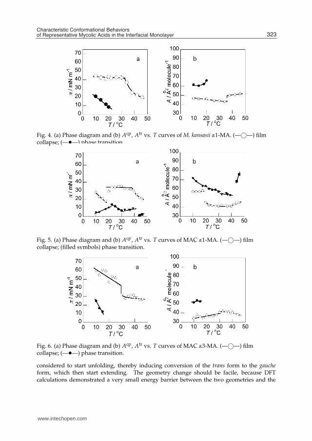
The phase diagrams (π vs. T diamgram), where surface pressure of phase transition πtr andthat of film collapse πcp are plotted against T and the diagram where mean molecular areasat πtr and πcp are plotted against T are shown in Figs. 2-6. Those diagrams, some of whichare quite simple and others more complex, demonstrate that πtr, πcp, Atr and Acp all changeddepending on the temperature. As shown, all those diagrams for the present α-MAs fromdifferent origins gave analogous features: In Figs 2a, 3a, 4a, 5a and 6a, each of the πcp vs. Tdiagram gave a cusp in the range of 32 ∼ 39 ◦C. The T at the cusp were different in differentsamples, and at that point, Acp vs. T curve was discontinuous, as shown in Figs. 2b, 3band 4b, though it is not quite obvious in Figs. 5b and 6b. One characteristic feature noted inthose diagrams is that the πcp values of M. tb complex α-MAs, such as α1-MA from M. tb andα3-MA from BCG were much higher, and accordingly the Acp values much smaller than the
321Characteristic Conformational Behaviors of Representative Mycolic Acids in the Interfacial Monolayer
www.intechopen.com
6 Will-be-set-by-IN-TECH
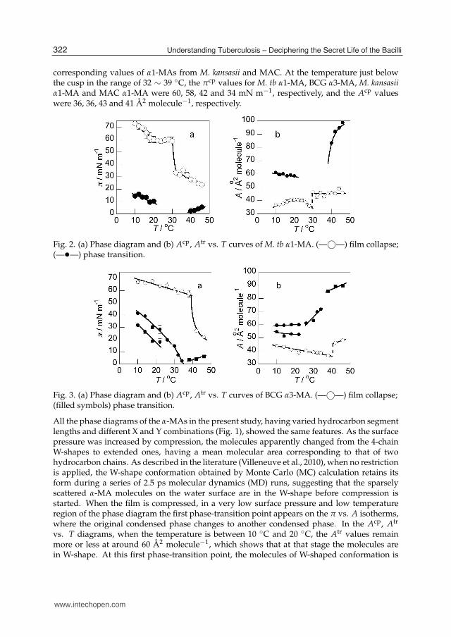
corresponding values of α1-MAs from M. kansasii and MAC. At the temperature just belowthe cusp in the range of 32 ∼ 39 ◦C, the πcp values for M. tb α1-MA, BCG α3-MA, M. kansasiiα1-MA and MAC α1-MA were 60, 58, 42 and 34 mN m−1, respectively, and the Acp valueswere 36, 36, 43 and 41 Å2 molecule−1, respectively.
Fig. 2. (a) Phase diagram and (b) Acp, Atr vs. T curves of M. tb α1-MA. (—©—) film collapse;(—•—) phase transition.
Fig. 3. (a) Phase diagram and (b) Acp, Atr vs. T curves of BCG α3-MA. (—©—) film collapse;(filled symbols) phase transition.
All the phase diagrams of the α-MAs in the present study, having varied hydrocarbon segmentlengths and different X and Y combinations (Fig. 1), showed the same features. As the surfacepressure was increased by compression, the molecules apparently changed from the 4-chainW-shapes to extended ones, having a mean molecular area corresponding to that of twohydrocarbon chains. As described in the literature (Villeneuve et al., 2010), when no restrictionis applied, the W-shape conformation obtained by Monte Carlo (MC) calculation retains itsform during a series of 2.5 ps molecular dynamics (MD) runs, suggesting that the sparselyscattered α-MA molecules on the water surface are in the W-shape before compression isstarted. When the film is compressed, in a very low surface pressure and low temperatureregion of the phase diagram the first phase-transition point appears on the π vs. A isotherms,where the original condensed phase changes to another condensed phase. In the Acp, Atr
vs. T diagrams, when the temperature is between 10 ◦C and 20 ◦C, the Atr values remainmore or less at around 60 Å2 molecule−1, which shows that at that stage the molecules arein W-shape. At this first phase-transition point, the molecules of W-shaped conformation is
322 Understanding Tuberculosis – Deciphering the Secret Life of the Bacilli
www.intechopen.com
Characteristic Conformational Behaviors of Representative Mycolic Acids in the Interfacial Monolayer 7
Fig. 4. (a) Phase diagram and (b) Acp, Atr vs. T curves of M. kansasii α1-MA. (—©—) filmcollapse; (—•—) phase transition.
Fig. 5. (a) Phase diagram and (b) Acp, Atr vs. T curves of MAC α1-MA. (—©—) filmcollapse; (filled symbols) phase transition.
Fig. 6. (a) Phase diagram and (b) Acp, Atr vs. T curves of MAC α3-MA. (—©—) filmcollapse; (—•—) phase transition.
considered to start unfolding, thereby inducing conversion of the trans form to the gaucheform, which then start extending. The geometry change should be facile, because DFTcalculations demonstrated a very small energy barrier between the two geometries and the
323Characteristic Conformational Behaviors of Representative Mycolic Acids in the Interfacial Monolayer
www.intechopen.com
8 Will-be-set-by-IN-TECH
sharp NMR signals of the relevant atoms implied that the conversion between the two shouldtake place easily. Though an exothermic transition is noted in the phase diagrams, the πtr
values tend to decrease generally in the π vs. T diagrams as the temperature increases, whichmeans that the transition taking place at the surface pressure is endothermic. This also impliesthat the conversion from trans to gauche geometry takes place during this phase transition. Thedecrease in the πtr values, however, is not so marked as in the case of MeO-MA (Villeneuve etal., 2007) as will be shown below, probably because the change in the conformation of α-MAsdoes not involve breaking of hydrogen bonding.
Elasticity and fluidity of the biological membranes are important factors in relation to theirfunctions. The elasticity modulus
E = −A(∂π/∂A)T,p (1)
or E values of α-MAs are in the range of 80 to 200 mN m−1, which roughly correspond to thevalues of liquid condensed film of common fatty acids, such as stearic acid (Villeneuve et al.,2005). When π is 10 mN m−1 and T below 20 ◦C, where the molecules are considered to betaking the W-shape according to the phase diagrams, the E values of those MAs are equivalentto those at higher temperatures where the molecules are in extended conformations. Thus,α-MA forms a monolayer which is fluid in whatever conformation the α-MAs might be takingand in which the α-MAs is ready to change the molecular area in response to outside pressureand temperatures.
5.2 Keto-MA monolayers
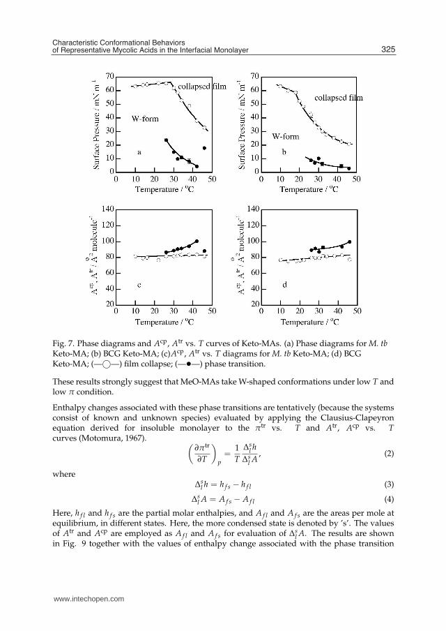
The phase diagrams of Langmuir monolayer and the Acp, Atr vs. T diagrams for Keto-MAsare quite different from those for α-MAs as shown in Fig. 7. The phase diagrams forKeto-MAs are much simpler than those for the α-MAs and the values of Acp are muchlarger for the Keto-MAs than for the α-MAs. Keto-MA forms a condensed monolayer morerigid than a liquid condensed film but less stiff than a solid condensed film over a widerange of temperature and surface pressure. Acp of Keto-MA is shown to be about 80 Å2
molecule−1. Moreover, the monolayer is in a condensed state as indicated by the elasticmodulus, e.g., about E = 1000 mN m−1 for M. tb and about E = 300 mN m−1 for BCG.Accordingly, it seems reasonable to assume that in the Keto-MA molecules the meromycolatechain bends at the cyclopropane and at the carbonyl group to form a 4-chain structure whosefour hydrocarbons are packed tightly in parallel, with the carbonyl group touching the watersurface and hydrated.
5.3 MeO-MA monolayers
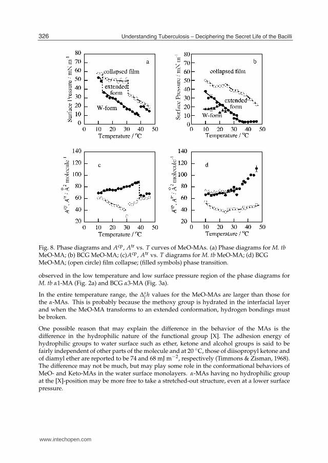
The phase diagrams of monolayer and the mean molecular area vs. T diagrams for MeO-MAsfrom M. tb and BCG are shown in Fig. 8. In each of the phase diagram, a characteristicphase transition is shown, of which surface pressure greatly decreased with an increasein temperature. This phase transition is reversible as has been confirmed by repeatedcompression-expansion measurement of the π vs. A isotherm. Atr takes values from 70 to90 Å2 molecule−1 for M. tb and from 70 to 110 Å2 molecule−1 for BCG∗. Even with such largemean molecular areas, the monolayers are in condensed states. For example the elastic modulibelow πtr of the two MA samples are E = 100 ∼ 350 mN m−1. On the other hand, Acp takesvalues less than 60 Å2 molecule−1 down to 38 Å2 molecule−1 for both MeO-MA samples∗.
324 Understanding Tuberculosis – Deciphering the Secret Life of the Bacilli
www.intechopen.com
Characteristic Conformational Behaviors of Representative Mycolic Acids in the Interfacial Monolayer 9
Fig. 7. Phase diagrams and Acp, Atr vs. T curves of Keto-MAs. (a) Phase diagrams for M. tbKeto-MA; (b) BCG Keto-MA; (c)Acp, Atr vs. T diagrams for M. tb Keto-MA; (d) BCGKeto-MA; (—©—) film collapse; (—•—) phase transition.
These results strongly suggest that MeO-MAs take W-shaped conformations under low T andlow π condition.
Enthalpy changes associated with these phase transitions are tentatively (because the systemsconsist of known and unknown species) evaluated by applying the Clausius-Clapeyronequation derived for insoluble monolayer to the πtr vs. T and Atr, Acp vs. Tcurves (Motomura, 1967).
(
∂πtr
∂T
)
p
=1T
∆sl h
∆sl A
, (2)
where∆
sl h = h f s − h f l (3)
∆sl A = A f s − A f l (4)
Here, h f l and h f s are the partial molar enthalpies, and A f l and A f s are the areas per mole atequilibrium, in different states. Here, the more condensed state is denoted by ’s’. The valuesof Atr and Acp are employed as A f l and A f s for evaluation of ∆
sl A. The results are shown
in Fig. 9 together with the values of enthalpy change associated with the phase transition
325Characteristic Conformational Behaviors of Representative Mycolic Acids in the Interfacial Monolayer
www.intechopen.com
10 Will-be-set-by-IN-TECH
Fig. 8. Phase diagrams and Acp, Atr vs. T curves of MeO-MAs. (a) Phase diagrams for M. tbMeO-MA; (b) BCG MeO-MA; (c)Acp, Atr vs. T diagrams for M. tb MeO-MA; (d) BCGMeO-MA; (open circle) film collapse; (filled symbols) phase transition.
observed in the low temperature and low surface pressure region of the phase diagrams forM. tb α1-MA (Fig. 2a) and BCG α3-MA (Fig. 3a).
In the entire temperature range, the ∆sl h values for the MeO-MAs are larger than those for
the α-MAs. This is probably because the methoxy group is hydrated in the interfacial layerand when the MeO-MA transforms to an extended conformation, hydrogen bondings mustbe broken.
One possible reason that may explain the difference in the behavior of the MAs is thedifference in the hydrophilic nature of the functional group [X]. The adhesion energy ofhydrophilic groups to water surface such as ether, ketone and alcohol groups is said to befairly independent of other parts of the molecule and at 20 ◦C, those of diisopropyl ketone andof diamyl ether are reported to be 74 and 68 mJ m−2, respectively (Timmons & Zisman, 1968).The difference may not be much, but may play some role in the conformational behaviors ofMeO- and Keto-MAs in the water surface monolayers. α-MAs having no hydrophilic groupat the [X]-position may be more free to take a stretched-out structure, even at a lower surfacepressure.
326 Understanding Tuberculosis – Deciphering the Secret Life of the Bacilli
www.intechopen.com

Characteristic Conformational Behaviors of Representative Mycolic Acids in the Interfacial Monolayer 11
Fig. 9. Enthalpy change associated with phase transition. M. tb α 1-MA (—©—); M. tbMeO-MA (—•—); BCG α 3-MA (—�—); BCG MeO-MA (—�—).
* The Atr and Acp must be corrected to the values mentioned here from the ones reported inthe literature (Villeneuve et al., 2007).
5.4 Other data supporting conformational behavior of Keto- and MeO-MAs deduced from
the monolayer study
5.4.0.5 Ellipsometry
Conformational transition from a 4-chain structure to an extended one suggested from the πvs. A study is supported by the in-situ ellipsometry. Results of the ellipsometric measurementare summarized in Table 2. Thus the thickness of the monolayer of MeO-MA at T = 32 ◦Cand π = 30 mN m−1 is larger by a factor of 1.7 ∼ 2 than that of MeO-MA at T = 18 ◦C andπ = 18 mN m−1 and those of Keto-MA at T = 18 ◦C and π = 30 mN m−1 and T = 32 ◦Cand π = 20 mN m−1. The thickness of MeO-MA monolayer changes from 2.91 nm for M. tband 2.78 nm for BCG at T = 18 ◦C and π = 18 mN m−1, which is almost the same value withthe thickness of Keto-MA, drastically to 4.96 nm (M. tb) and 5.62 nm (BCG) at T = 32 ◦C andπ = 30 mN m−1. As for Keto-MA whose Acp values imply that its carbonyl group is hydratedat the water surface to give a four-chain molecular conformation, the monolayer thickness isunchanged irrespective of the surface pressure or temperature (Table 2).
Origin Keto-MA MeO-MAT / ◦C π / mN m−1 thickness / nm T / ◦C π /mN m−1 thickness / nm
M. tb18 30 2.90 ± 0.07 18 18 2.91 ± 0.05
32 30 4.96 ± 0.88
BCG 18 30 2.90 ± 0.05 18 18 2.78 ± 0.0432 20 2.80 ± 0.08 32 30 5.62 ± 0.18
Table 2. Thickness of Langmuir monolayer estimated by ellipsometry.
327Characteristic Conformational Behaviors of Representative Mycolic Acids in the Interfacial Monolayer
www.intechopen.com

12 Will-be-set-by-IN-TECH
5.4.0.6 Computer simulation
The MAs subjected to MC calculations and MD simulations were cis-cyclopropyl MeO-MAwith n-m-l of 17-16-17, trans-cyclopropyl MeO-MA with n-m-l of 18-16-17, cis-cyclopropylKeto-MA with n-m-l of 15-18-17 and 17-18-17 and trans-cyclopropyl Keto-MA with n-m-l of16-18-17 (Table 1). The structural models of MeO- and Keto-MA produced by MC calculationswere all of 4-chain structure as illustrated in Figs. 10a and b. The fact suggests that this type ofarrangement of carbon chain segments is appropriate for energetically stable conformations.After the molecular dynamics (MD) simulation, Keto-MA having 4-chain structure normallyretained the original 4-chain form (Fig. 10e) and seldom gave extended structures. Onthe other hand, MeO-MA, whose starting structure is as in Figs. 10a and b, often gaveextended structures (Fig. 10c), though some models retained 4-chain structures as seen inFig. d (Villeneuve et al., 2007).
Intra-molecular hydrogen bonding involving either the oxo or methoxy group and the3-hydroxy carboxylic acid group may contribute to some extent to the retaining of the 4-chainstructure. However, one of the major causes for the difference observed in the results ofMD simulation of the two types of MAs seems to be in the difference in the lengths of themethylene chain segments or in the n-m-l values. As described previously (Watanabe etal., 2002), the major component of the MeO-MA is cis-cyclopropyl-containing MeO-MA acidwith the n-m-l value of 17-16-17, and the minor component with a trans-cyclopropane withthe n-m-l value of 18-16-17. In those MeO-MAs, n is larger than m. In the starting modelsfor MD of MeO-MA, having the energetically stabilized 4-chain structure produced by MC(Figs. 10a and b), a bulky group consisting of a methoxy group and the adjacent methylgroup is at a position to obstruct the compact arrangement of the 4 chains, as demonstratedby the molecular minimized energy levels: for the models in the literature (Villeneuve etal., 2007), the minimized energy levels of cis- and trans-MeO-MAs are −13 ∼ −19 kcalmol−1 and −7 ∼ −12 kcal mol−1, respectively, whereas those of cis- and trans-Keto-MAsare −24 ∼ −32 kcal mol−1 and −28 ∼ −38 kcal mol−1. The vibrations of the bulky grouplocating at or above the location of the 3-hydroxy carboxylate group during MD simulationmay disturb the neat arrangement of the neighbouring chains to induce faster and moreefficient deviation from the original 4-chain structure. In Keto-MA, the n-m-l value for themajor cis-cyclopropane containing component is 15-18-17 or 17-18-17 and that for the majortrans-cyclopropane containing component is 16-18-17, n being smaller than m. Thus, theoxo and the adjacent methyl groups are normally at the end or stretching out of the squarepillar-like 4-chain structure. It allows a more compact solid arrangement of the methylenechains in the molecule and the more quiet vibration of the alpha-methyl oxo group may tendto be less disturbing for the 4-chain arrangement during the MD.
The fact that the oxo group is normally situating at the end or out of the 4-chain pillar structuremay contribute to the more stable 4-chain structure of Keto-MA in Langmuir monolayers;it assures that the oxo group touches and bonds to the water surface firmly. On the otherhand, in MeO-MA, the methoxy group may often be above the level of the location of thecarboxyl group, which touches the water surface. Therefore, although the hydrophilicity ofthe methoxy group approaches that of an oxo group, the methoxy group may not be able tointeract decisively with the water surface.
328 Understanding Tuberculosis – Deciphering the Secret Life of the Bacilli
www.intechopen.com

Characteristic Conformational Behaviors of Representative Mycolic Acids in the Interfacial Monolayer 13
Fig. 10. Structures of MAs in MD study. (a) and (b) Top and side views of MeO-MA moleculeobtained by MC followed by minimization; (c) MeO-MA taking a stretched-out structureafter 20 ps in MD; (d) MeO-MA retaining a 4-cain structure after 20 ps in MD; (e) structure ofKeto-MA obtained after 20 ps in MD.
6. A tentative interpretation of the model membrane in terms of biological activity
6.1 Generality
In relating the present results to the biological role of mycolic acids, the most valuable findingis the special behavior of the Keto-MA. It has exceptional rigidity in monolayers, over a
329Characteristic Conformational Behaviors of Representative Mycolic Acids in the Interfacial Monolayer
www.intechopen.com
14 Will-be-set-by-IN-TECH
wide temperature range (Fig. 7), apparently assuming a W-shaped conformation with fourhydrocarbon chains packing together in parallel. To our knowledge, this is the first time thatfatty acid packing of this type has been detected in nature. The initial studies (Minnikin &Polgar, 1967, 1; 2) on the location of functional groups in cyclopropyl MA showed that thesefatty acids are assembled according to a generalized template, with groups spaced at regularintervals separated by relatively uniform lengths of hydrocarbon chains (Table 1); these resultshave been thoroughly substantiated in recent studies (Watanabe et al., 2001; 2002). The currentresults offer the first justification for this exquisite regular architecture of mycobacterial MAs.The true reason for the presence of oxygenated functions in MAs is also revealed as beingnecessary for conformational stabilization probably through hydrophilic interactions. In thismodel monolayer study, the hydrophilic interaction is most likely to be with the aqueoussub-phase. In the cell envelope of mycobacteria, however, the interaction of the keto groupcould either be with the covalently attached arabinogalactan or, more intriguingly, withthe mycolic acid 3-hydroxy group. This latter interaction could take place in an inter- orintramolecular fashion.
Biological activities of living cells rely upon, not only the relevant chemical reactions but alsoon relating physicochemical or physical processes. In this study, we have shown that MAs ofdifferent chemical structures form Langmuir monolayers having distinctive physicochemicalfeatures and each MA exhibits multiple phase transitions depending upon the temperatureand the surface pressure. Dubnau (Dubnau et al., 2000) et al. reported that in a M. tbstrain whose cell wall-linked mycolate consists solely of α-MA, the permeation rate was verylow, and Yuan et al. (Yuan et al., 1998) reported that in a recombinant mycobacterial strainwhose Keto-MA is completely replaced by MeO-MA showed poor growth in macrophagesand a decreased rate of permeation for hydrophilic substances. Those studies imply thatdifferent mycolates in the cell envelope contribute differently to the permeability functionof the mycolate layer of the cell envelope. It seems possible that each component mycolatetakes different conformation in the cell envelope mycolate layer as suggested by the presentstudy, and that the different forms of different mycolates have different effects on the cellfunction, though we should be careful in applying the monolayer results to the natural cellwall mycolate layer functions.
6.2 Roles of α-MAs
Recent papers revealed better defined features of the outer membrane lipid bilayer ofmycobacterial cells by the cyro-electron tomography (Hoffmann et. al., 2008; Niederweis etal., 2010; Zuber et al., 2008). On the basis of the thickness of the layer, in those papers, thecell-bound MAs constituting the basis of the inner leaflet of the outer membrane lipid bilayerare suggested to be in the W-shape. However, if MAs in the lipid bilayer are to take basicallythe W-shape, it does not necessarily mean that all the MAs are to stay in the W-shape. Inthe biological lipid bilayers, at biological temperatures, it is well known that the componentmolecules are able to move quite easily to shift their locations in the layer or to change theconformation. Our present monolayer studies and computer simulation results showed thatthe α-MAs are ready to change the conformation from the W-shape to any of the variousextended shapes as required or favored by its environment. In the outer membrane lipidlayer, α-MAs, a large molecular weight component, may change the conformation to variousextended shapes and probably by dynamically waving and bending the variously extended
330 Understanding Tuberculosis – Deciphering the Secret Life of the Bacilli
www.intechopen.com
Characteristic Conformational Behaviors of Representative Mycolic Acids in the Interfacial Monolayer 15
long hydrocarbon chains, may take an initiative active part in making the lipid layer moremobile and biologically compatible.
The observations of α-MAs, especially of the flexible conformational behavior of the moleculesand of various characteristics closely and directly related to the physical nature of the lipidlayer may imply importance of the presence of α-MAs always in about 50 % of the wholecell-bound MAs in mycobacterial cells.
6.3 Roles of oxygenated-MAs
The possible special influence of MAs with a trans-cyclopropane ring on membrane functionor pathogenicity of the cells has been highlighted (Glickman et al., 2000; 2001). When thefeatures of the Langmuir monolayers of MeO- and Keto-MAs from BCG in the present studywere compared with those of the corresponding MAs from M. tb in our study (Villeneuveet al., 2005), some differences were noted. The collapse pressures of Keto-MA and thesurface pressures of MeO-MA at the phase transition from the 4-chain conformation to theextended one were lower in those of Keto- and MeO-MAs from BCG, respectively, than inthose from M. tb, at all the temperatures assayed. The structures of the molecular componentsof the MAs from the two mycobacteria are essentially identical and the difference is onlyin the ratios. The ratios between the cis-cyclopropane and trans-cyclopropane contents are1/0.03 and 1/0.22, respectively, in MeO-MAs from BCG and M. tb and is 1/0.33 and 1/3.5,respectively, in Keto-MAs from BCG and M. tb. The differences noted in the respectivephase diagrams or isotherms may be attributed to subtle differences in the properties ofthe cis-cyclopropane rings and the trans-cyclopropane rings with an adjacent methyl branch.Apparently an increased ratio of trans-isomers seems to stabilize the four-chain conformationof the oxygenated MAs. The MD studies did not demonstrate any clear difference between theconformational behaviors of cis-cyclopropane-containing and trans-cyclopropane-containingMAs. This confirms the previous conclusion (Villeneuve et al., 2005) that trans-cyclopropaneunits, with an adjacent methyl branch, are able to allow folding of MAs in a similar mannerto that allowed by cis-cyclopropane rings. Further studies on individual molecular species ofoxygenated mycolates should be of great value to clear this problem.
Many factors are involved in the process of the onset of infectious diseases. In the case oftuberculosis, the primary and characteristic factor relating to the onset of the disease shouldbe the intrinsic capacity of the M. tb cells to resist and reject the attacks by the defensemechanisms of human host cells. If the mycolate layer of the cell envelope is to play adetermining role in the permeability barrier function, as suggested (Minnikin, 1982; Puechet al., 2001; Rastogi, 1991), then the layer is to take an active part in regulating the in and outpassages of essential factors vital for the living bacteria and thus to control the viability ofthe bacterial cells. The component MAs, therefore, may be considered to be responsible forthe viability of the M. tb cells in human cells and detailed analysis of the physicochemicalfeatures of the component MAs may help to clear part of the problems relating to the humantuberculosis.
MAs from pathogenic M. tb and from non-pathogenic BCG are the same in thechemical structures of each component and slightly different in the ratios between thetrans-cyclopropane-containing and cis-cyclopropane-containing components. One markeddifference between the MAs from the two is in the ratios between the non-oxygenated MA(alpha-MA) and the oxygenated MAs. The ratio in the former is roughly 1:1, whereas that
331Characteristic Conformational Behaviors of Representative Mycolic Acids in the Interfacial Monolayer
www.intechopen.com
16 Will-be-set-by-IN-TECH
in BCG reaches 1:3.5, in which the oxygenated MA is often mostly Keto-MA (Watanabeet al., 2001). The actual surface pressure in the cell envelope mycolate layer is unknown,but whatever the environmental conditions may be, as demonstrated in the present study,Keto-MAs form compact, relatively solid domains with a minimum thickness in the mycolatemonolayer. Such Keto-MA units may provide a relatively impermeable stable foundation inthe outer leaflet of the cell envelope. A larger number of, or a larger proportion of this type ofless permeable compact domains in BCG cell envelope may provide the cells with the featuresthat distinguish BCG cells from M. tb cells. The presence of such solid domains may provideBCG cells with slower and lower multiplication rate and fairly good or moderate resistance tothe killing system of the host cells so that the cells can survive quietly for a long time, whichis an essential and necessary requirement for a good live vaccine.
The extended structures of MeO-MA, produced by MD simulation, are not of two long straightmethylene chains. As exemplified in Fig. 1c, the long chains curve and bend, implying thatthey are ready to change their conformation in response to the environmental conditions.Probably MeO-MA and also alpha-MA are to be considered to provide less condensedorganelles suitable for facilitating selective permeability and interaction with complex cellsurface free lipids.
7. References
Al Dulayymi, J. R., Baird, M. S., Roberts, E. (2005), The synthesis of a single enantiomer of amajor α-mycolic acid of M. tuberculosis, Tetrahedron Vol. 6, pp. 11939-11951.
Asselineau, C. & Asselineau, J. (1966), Stéréochimique de l’acide corynomycolique, Bull. Soc.Chim. France pp. 1992-1999.
Jackson, M., Raynaud, C., Lanèelle, M.-A., Guilhot, C., Laurent-Winter, C., Ensergueix, D.,Gicquel, B., Daffé, M. (1999), Inactivation of the antigen 85C gene profoundly affectsthe mycolate content and alters the permeability of the mycobacterium tuberculosis cellenvelope, Mol. Microbiol. Vol. 31, pp. 1573-1587.
Dubnau, E., Chan, J., Raynaud, C., Mohan, V. P., Lanèelle, M.-A., Yu, K., Quémard, A.,Smith, I., Daffée, M., (2000), Oxygenated mycolic acids are necessary for virulenceof Mycobacterium tuberculosis in mice, Mol. Microbiol. Vol. 36, pp. 630-637.
Glickman, M. S., Cox, J. S., Jacobs Jr., W. R., (2000), A novel mycolic acid cyclopropanesynthetase is required for cording, persistence and virulence of Mycobacteriumtuberculosis, Mol. Cell Vol. 5, pp. 717-727.
Glickman, M. S., Cahill, S. M., Jacobs Jr., W. R. (2001), The Mycobacterium tuberculosis cmaA2gene encodes a mycolic acid trans-cyclopropane synthetase, J. Biol. Chem. Vol. 276,pp. 2228-2233.
Goren, M. B. & Brennan, P. J. (1979), Mycobacterial lipids: Chemistry and biologic activities,In: Tuberculosis, Youmans, G.P. (Ed.), page numbers (first-last), Saunders, ISBN,Philadelphia.
Hasegawa, T., Nishijo, J., Watanabe, M., Funayama, K., Imae, T. (2000), Conformationalcharacterization of a-mycolic acid in a monolayer film by the Langmuir-Blodgetttechnique and atomic force microscopy, Langmuir Vol. 16, pp. 7325-7330.
Hasegawa, T., Nishijo, J., Watanabe, M., Umemura, J., Ma, Y., Sui, G., Huo, Q., Leblanc, R.M. (2002), Characteristics of long-chain fatty acid monolayers studied by infraredexternal-reflection spectroscopy, Langmuir Vol. 18, pp. 4758-4764.
332 Understanding Tuberculosis – Deciphering the Secret Life of the Bacilli
www.intechopen.com
Characteristic Conformational Behaviors of Representative Mycolic Acids in the Interfacial Monolayer 17
Hasegawa, T. & Leblanc, R. M. (2003), Aggregation properties of mycolic acid molecules inmonolayer films: a comparative study of compounds from various acid-fast bacterialspecies Biochim. Biophys. Acta Vol. 1617, pp. 89-95.
Hasegawa, T., Amino, S. Kitamura, S., Matsumoto, R., Katada, S., Nishijo, J. (2003), Studyof the molecular conformation of α- and keto-mycolic acid monolayers by theLangmuir-Blodgett technique and Fourier transform infrared reflection-adsorptionspectroscopy, Langmuir Vol. 19, pp. 105-109.
Hoffmann, C., Leis, A., Niederweis, M., Plitzko, J. M., Engelhardt, H. (2008), Disclosure ofthe mycobacterial outer membrane: Cryo-electron tomography and vitreous sectionsreveal the lipid bilayer structure, PNAS Vol. 105, pp. 3963-3967.
Hong, X. & Hopfinger, A. J. (2004), Construction, molecular modeling, and simulation ofmycobacterium tuberculosis cell walls, Biomacromolecules Vol. 5, pp. 1052-1065.
Motomura, K. (1967), Thermodynamics and phase transitions in monolayers, J.C.I.S. Vol. 23,pp. 313-318.
M. McNeil, M. Daffé, P. J. Brennan (1991), Location of the mycolyl ester substituents in the cellwalls of mycobacteria, J. Biol. Chem. Vol. 266, pp. 13217-13223.
Minnikin, D. E. & Polgar, N. (1967), The mycolic acids from human and avian tubercle bacilli,Chem. Comm. pp. 916-918.
Minnikin, D. E. & Polgar, N. (1967), The mycolic acids from human and avian tubercle bacilli,Chem. Comm. pp. 1172-1174.
D. E. Minnikin (1982). Lipids: Complex Lipids, Their Chemistry, Biosynthesis and Roles, In:The Biology of the Mycobacteria Vol. 1, Ratledge, C. & Stanford, J. L. (Ed.), pp. 95-184,Academic Press, New York.
Minnikin, D. E., Kremer, L., Dover, L. G., Besra, G. S. (2002), The methyl-branchedfortifications of Mycobacterium tuberculosis, Chem. Biol. Vol. 9, pp. 545-553.
Niederweis, M., Danielchanka, O., Huff, J., Hoffmann, C., Engelhardt, H. (2010),Mycobacterial outer membranes: in search of proteins, Trends in Microbiology Vol.18, pp. 109-116.
Puech, V., Chami, M., Lemassu, A., Lanèelle, M.-A., Schiffler, B., Gounon, P., Bayan, N., Benz,R., Daffé, M. (2001), Structure of the cell envelope of corynebacteria: importance ofthe non-covalently bound lipids in the formation of the cell wall permeability barrierand fracture plane, Microbiology Vol. 147, pp. 1356-1382.
Rastogi, N. (1991), Recent observations concerning structure and function relationships inthe mycobacterial cell envelope: elaboration of a model in terms of mycobacterialpathogenicity, virulence and drug-resistance, Res. Microbiol. Vol. 142, pp. 464-476.
Staellberg-Stenhagen, S. & Stenhagen, E. (1945) A monolayer and X-ray studies of mycolicacid from the human tubercle bacillus, J. B. C. Vol. 150, pp. 255-262.
Timmons, C. O. & Zisman, W. A. (1968) The relation of initial spreading pressure of polarcompounds on water to interfacial tension, work of adhesion, and solubility, J.C.I.S.Vol. 28, pp. 106-117.
Tocanne, J. F. & Asselineau, C. (1968), Étude stéréochimique des acides aliphatiques α-ramifiésβ-hydroxylés. Configuration de l’acide corynomycolique, Bull. Soc. Chim. Fr. pp.4519-4525.
Tompkins, H. G. & McGahan, W. A. (1999). Spectroscopic Ellipsometry and Reflectometry: A User’sGuide, Wiley-Interscience, New York.
333Characteristic Conformational Behaviors of Representative Mycolic Acids in the Interfacial Monolayer
www.intechopen.com

18 Will-be-set-by-IN-TECH
Villeneuve, M. Kawai, M., Kanashima, H., Watanabe, M., Minnikin, D. E., Nakahara, H. (2005),Temperature dependence of the Langmuir monolayer packing of mycolic acids fromMycobacterium tuberculosis, Biochim. Biophys. Acta Vol. 1715, pp. 71-80.
Villeneuve, M. Kawai, M., Watanabe, M., Aoyagi, Y., Hitotsuyanagi, Y., Takeya, K., Gouda,H., Hirono, S., Minnikin, D. E., Nakahara, H. (2007), Conformational behavior ofoxygenated mycobacterial mycolic acids from Mycobacterium bovis BCG, Biochim.Biophys. Acta Vol. 1768, pp. 1717-1726.
Villeneuve, M. Kawai, M., Watanabe, M., Aoyagi, Y., Hitotsuyanagi, Y., Takeya, K., Gouda,H., Hirono, S., Minnikin, D. E., Nakahara, H. (2010), Differential conformationalbehavior of α-mycolic acids in Langmuir monolayers and computer simulations,Chemistry and Physics of Lipids Vol. 163, pp. 569-579.
Watanabe, M., Aoyagi, Y. Ridell, M. Minnikin, D. E. (2001), Separation and characterizationof individual mycolic acids in representative mycobacteria, Microbiology Vol. 147, pp.1825-1837.
Watanabe, M., Aoyagi, Y. , Mitome, H. ,Fujita, T., Naoki, H., Ridell, M., Minnikin, D. E. (2002),Location of functional groups in mycobacterial meromycolate chains; the recognitionof new structural principles in mycolic acids, Microbiology Vol. 148, pp. 1881-1902.
Yuan, Y., Zhu, Y. Q., Crane, D. D., Barry III, C. E. (1998), The effect of oxygenated mycolicacid composition on cell wall function and macrophage growth in mycobacteriumtuberculosis, Mol. Microbiol. Vol. 29, pp. 1449-1458.
Zuber, B., Chami, M., Houssin, C., Dubochet, J., Griffiths, G., Daffé, M. (2008), Directvisualization of the outer membrane of mycobacteria and corynebacteria in theirnative state, Journal of Bacteriology Vol. 190, 5672-5680.
334 Understanding Tuberculosis – Deciphering the Secret Life of the Bacilli
www.intechopen.com
Understanding Tuberculosis - Deciphering the Secret Life of theBacilliEdited by Dr. Pere-Joan Cardona
ISBN 978-953-307-946-2Hard cover, 334 pagesPublisher InTechPublished online 17, February, 2012Published in print edition February, 2012
InTech EuropeUniversity Campus STeP Ri Slavka Krautzeka 83/A 51000 Rijeka, Croatia Phone: +385 (51) 770 447 Fax: +385 (51) 686 166www.intechopen.com
InTech ChinaUnit 405, Office Block, Hotel Equatorial Shanghai No.65, Yan An Road (West), Shanghai, 200040, China
Phone: +86-21-62489820 Fax: +86-21-62489821
Mycobacterium tuberculosis, as recent investigations demonstrate, has a complex signaling expression, whichallows its close interaction with the environment and one of its most renowned properties: the ability to persistfor long periods of time under a non-replicative status. Although this skill is well characterized in other bacteria,the intrinsically very slow growth rate of Mycobium tuberculosis, together with a very thick and complex cellwall, makes this pathogen specially adapted to the stress that could be generated by the host against them. Inthis book, different aspects of these properties are displayed by specialists in the field.
How to referenceIn order to correctly reference this scholarly work, feel free to copy and paste the following:
Masumi Villeneuve (2012). Characteristic Conformational Behaviors of Representative Mycolic Acids in theInterfacial Monolayer, Understanding Tuberculosis - Deciphering the Secret Life of the Bacilli, Dr. Pere-JoanCardona (Ed.), ISBN: 978-953-307-946-2, InTech, Available from:http://www.intechopen.com/books/understanding-tuberculosis-deciphering-the-secret-life-of-the-bacilli/characteristic-conformational-behaviors-of-representative-mycolic-acids-in-the-interfacial-monolayer

© 2012 The Author(s). Licensee IntechOpen. This is an open access articledistributed under the terms of the Creative Commons Attribution 3.0License, which permits unrestricted use, distribution, and reproduction inany medium, provided the original work is properly cited.


![Personality: An individual’s characteristic patterns of thoughts, feelings, and behaviors [persisting over time and across situations] Sensitive, Reactive](https://img.pdfslide.us/doc/110x75/56649e5c5503460f94b53f4a/personality-an-individuals-characteristic-patterns-of-thoughts-feelings.jpg)