Embed Size (px)
Citation preview

Characterisation of Leukocytes in a Human Skin BlisterModel of Acute Inflammation and ResolutionWilliam Jenner1., Madhur Motwani1., Kristin Veighey1, Justine Newson1, Tatsiana Audzevich1,
Anna Nicolaou2, Sharon Murphy2, Raymond MacAllister1, Derek W. Gilroy1*
1 Centre for Clinical Pharmacology and Therapeutics, Division of Medicine, University College London, London, United Kingdom, 2 Manchester Pharmacy School, Faculty
of Medical and Human Sciences, University of Manchester, Manchester, United Kingdom
Abstract
There is an increasing need to understand the leukocytes and soluble mediators that drive acute inflammation and bringabout its resolution in humans. We therefore carried out an extensive characterisation of the cantharidin skin blister modelin healthy male volunteers. A novel fluorescence staining protocol was designed and implemented, which facilitated theidentification of cell populations by flow cytometry. We observed that at the onset phase, 24 h after blister formation, thepredominant cells were CD16hi/CD66b+ PMNs followed by HLA-DR+/CD14+ monocytes/macrophages, CD11c+ and CD141+
dendritic cells as well as Siglec-8+ eosinophils. CD3+ T cells, CD19+ B cells and CD56+ NK cells were also present, but incomparatively fewer numbers. During resolution, 72 h following blister induction, numbers of PMNs declined whilst thenumbers of monocyte/macrophages remain unchanged, though they upregulated expression of CD16 and CD163. Incontrast, the overall numbers of dendritic cells and Siglec-8+ eosinophils increased. Post hoc analysis of these data revealedthat of the inflammatory cytokines measured, TNF-a but not IL-1b or IL-8 correlated with increased PMN numbers at theonset. Volunteers with the greatest PMN infiltration at onset displayed the fastest clearance rates for these cells atresolution. Collectively, these data provide insight into the cells that occupy acute resolving blister in humans, the solublemediators that may control their influx as well as the phenotype of mononuclear phagocytes that predominate theresolution phase. Further use of this model will improve our understanding of the evolution and resolution of inflammationin humans, how defects in these over-lapping pathways may contribute to the variability in disease longevity/chronicity,and lends itself to the screen of putative anti-inflammatory or pro-resolution therapies.
Citation: Jenner W, Motwani M, Veighey K, Newson J, Audzevich T, et al. (2014) Characterisation of Leukocytes in a Human Skin Blister Model of AcuteInflammation and Resolution. PLoS ONE 9(3): e89375. doi:10.1371/journal.pone.0089375
Editor: John Wallace, McMaster University, Canada
Received December 20, 2013; Accepted January 18, 2014; Published March 6, 2014
Copyright: � 2014 Jenner et al. This is an open-access article distributed under the terms of the Creative Commons Attribution License, which permitsunrestricted use, distribution, and reproduction in any medium, provided the original author and source are credited.
Funding: This work was funded by the British Heart Foundation (WJ,RM) and The Wellcome Trust (DWG). The funders had no role in study design, data collectionand analysis, decision to publish, or preparation of the manuscript.
Competing Interests: The authors have declared that no competing interests exist.
* E-mail: [email protected]
. These authors contributed equally to this work.
Introduction
Inflammation is characterised by the sequential release of
mediators (including histamine, bradykinin and 5HT), resulting in
the immediate influx of polymorphonuclear leukocytes (PMNs)
followed by phagocytosing monocyte/macrophages, leading to
leukocyte clearance and resolution [1]. Indeed, for the past 40
years research focused on identifying factors which initiate/
perpetuate inflammation with the objective of developing drugs to
alleviate diseases driven by on-going or dysregulated inflammation
[2]. More recently, emphasis has shifted to the other end of the
inflammatory spectrum, resolution, in order to understand how
immune-mediated responses switch off. Advances in this area will
help shed light on the aetiology of chronic inflammation and
provide drug development opportunities based upon endogenous
pro-resolution mediators/pathways [3]. However, elucidating the
factors that drive inflammation, control its severity and longevity
were, for the most part, characterised using rodent models of
pleuritis, peritonitis or paw swelling [4,5,6,7]. This included the
response to innate (carrageenan) or specific (methylated bovine
serum albumin) antigens.
In contrast, comparatively few human in vivo models of acute
and resolving inflammation are available. Such models would
allow us to better understand how the immune system is altered in
people with chronic inflammatory diseases, and to determine the
efficacy of novel immune-modifying agents. Performing such
investigations requires existence of models that are representative
of individual’s innate inflammatory response, have low within-
subject variability, and are non-invasive, such that they can be
used appropriately in patients with exisiting inflammatory
conditions. Of the human models currently in use for characteris-
ing and quantifying the inflammatory response, skin window
techniques [8], and skin blisters induced by traumatic suction [9]
or cantharidin [10] have proven useful in developing our
understanding of the inflammatory phenotype. However, detailed
analysis of trafficking cell populations that account for the onset
and resolution of inflammation alongside traditional soluble
mediators (cytokines and lipids) is lacking. These data would also
confirm whether inherent mechanisms underlying the innate
inflammatory response in humans are similar to those identified by
rodent studies.
PLOS ONE | www.plosone.org 1 March 2014 | Volume 9 | Issue 3 | e89375

In the current study we therefore carried out detailed
characterisation of leukocytes and soluble mediators occupying
human cantharidin skin blisters at the onset of the inflammatory
response and during its resolution.
Materials and Methods
Ethics StatementThis study was approved by the UCL ethics committee for
human research (Ref: 2907/002). Written informed consent was
obtained from all volunteers.
Cantharidin blistersThe technique for inducing, aspirating, and processing the
cantharidin skin blister and oedema has been previously described
[11]. In short, blisters were elicited by applying 12.5 ml of 0.1%
cantharidin (Cantharone, Dormer Laboratories) to the ventral
aspect of the forearms of 20 non-smoking, healthy male volunteers
aged 18–45 years. On day 1, two skin blisters were induced on one
forearm with one blister aspirated on day 2 (24 hours) and the
other on day 4 (72 hours). Peripheral blood samples were obtained
following venepuncture at the antecubital fossa and leukocytes
isolated following flash lysis to remove erythrocytes.
Flow cytometryBlister and circulating leukocytes were enumerated and then
analysed for surface marker expression on a flow cytometer (LSR
Fortessa, BD Biosciences). Due to a lack of published data on
blister leukocyte differentiation using flow cytometry, a novel
staining and subsequent gating strategy was designed to identify
individual cell populations. Leukocytes were incubated with
combinations of antibodies to CD3 (APC, Clone: UCHT1, BD),
CD19 (PE-Cy 7, Clone: SJ25C1, BD), CD56 (PerCP-Cy5.5, Clone:
B159, BD), HLA-DR (V450, Clone: L243, BD), CD14 (Alexa Fluor
700, Clone: M5E2, BD), CD16 (FITC, Clone: 3G8, BD), CD141
(PE, Clone: M80, Biolegend), CD163 (PE, Clone: M130, BD),
CD11c (PE-Cy7, Clone: B-ly6, BD), Siglec-8 (PE, Clone:7C9,
Biolegend) and Annexin-V/7AAD Apoptosis Detection Kit (BD)
using respective isotype antibodies and fluorescence-minus-one
(FMO) controls, and compensated for dual labelling. Separation of
cell subtypes was performed using a cell sorter (FACS Aria, BD
Biosciences) with subtypes undergoing histological staining using a
modified Wright’s method (Shandon Kwif-Diff Stain Kit, Thermo
Scientific). Flow cytometry analysis was completed using FlowJo
software (Tree Star Inc).
Cytokine analysisCytokine expression profiles were measured using the MSD
Bio-Plex human cytokine assay (Merck, Sharp & Dohme LTD).
Our assay was customized to quantify the concentration of IL-1,
IL-6, IL-8, IL-10, IFNc, IL-12p70 and TNF-a within the blisters.
All samples were run in duplicate.
Extraction and analysis of lipid mediatorsLipid mediators in human plasma were analysed by liquid
chromatography coupled to electrospray ionization tandem mass
spectrometry (LC/ESI-MS/MS) based on protocols published
previously [12,13]. Briefly, samples were collected and stored
immediately at 280uC. Plasma samples (500 mL) were defrosted
on ice and adjusted to 15% (v/v) methanol: water (final volume
4 mL). Internal standards, PGB2-d4 (40 ng) and 12-HETE-d8
(40 ng) (Cayman Chemical Company, Ann Arbor, USA) were
added and the pH of resulting solutions adjusted to 3.0 (1 M
HCL). Acidified samples were immediately applied to precondi-
tioned solid-phase cartridges (C18-E, Phenomenex, Macclesfield,
UK) and lipid mediators eluted with methyl formate. LC/ESI-
MS/MS analysis was performed on a HPLC pump (Waters
Alliance 2695) coupled to an electrospray ionisation triple
quadrupole mass spectrometer (Quattro Ultima, Waters, UK).
Chromatographic separation was performed on a C18 Luna
column (5 mm, 15062.0 mm, 21 Phenomenex) for eicosanoids
and a C18 Kinetex column (2.6 mm, 10062.1 mm, Phenomenex)
for hydroxy-fatty acids. Analytes were monitored on multiple
reaction monitoring mode as reported [12,13] with the following
additions: 15-hydroxyeicosatrienoic acid (HETrE) m/z 321.221,
10-hydroxydocosahexaenoicacid (HDHA) m/z 343.153, 14-
HDHA m/z 343.161, 13-HDHA m/z 343.193 and 17- HDHA
m/z 343.201.
Calculations and statistical analysisCell populations are expressed as the absolute number of cells
(logarithmic scale, median 6 interquartile range) and as the
percentage of total cells (linear scale, mean 6 standard deviation).
Statistical analysis was performed using GraphPad Prism 4
(GraphPad Software). Between time-point differences in cells,
cytokines, and lipids were assessed using paired t test for normally
distributed data, or Wilcoxon matched pairs test for skewed data
sets. Correlations between variables were calculated using Spear-
man’s rank correlation. p,0.05 was considered statistically
significant.
Results
Characterisation of peripheral blood leukocytesTo develop an effective gating strategy for identification of
inflammatory leukocyte sub-populations in skin blisters, we first
validated the surface phenotype of known peripheral blood
leukocyte populations by flow cytometry. After exclusion of cell
debris and doublets (Figure 1 [i]), the remaining mixed cell
population was firstly gated for CD3+ T cells and CD19+ B cells
(Figure 1 [iii]). Resulting CD32/CD192 cells were then gated on
CD56 (Figure 1 [iv]) and CD16 to identify CD16+ and CD162
subpopulations of CD56+ NK cells (Figure 1 [v]). The remaining
leukocytes were then differentiated on HLA-DR expression
(Figure 1 [vi]). This allowed for separate classification of
mononuclear and granulocytic populations, as has been previously
described [14]. Analysis of HLA-DR+ cells (Figure 2 [i]) revealed
the expected distribution of classical CD14hi/CD162, intermedi-
ate CD14hi/CD16+, and non-classical CD14lo/CD16+ monocytes
(Figure 2 [ii]), possessing varying degrees of the scavenger receptor
marker, CD163 (Figure 1B [iii]–[v]). Within this HLA-DR+
population we also identified CD142 CD162 dendritic cells,
which upon extended characterisation were identified as having
mixed CD141 and CD11c expression (Figure 2 [vi]), as previously
described [15].
The HLA-DR2 population (Figure 2 [i]) comprised of two sub-
populations with varying degree of expression for CD16, labelled
here as CD16hi and CD16lo (Figure 2 [vii]). CD16hi population
was identified as PMNs, confirmed by expression of CD66b
(Figure 2 [viii]). On extended characterisation, the CD16lo cells
were identified as Siglec8+ eosinophils (Figure 2 [ix–x]).
Characterisation of inflammatory cell infiltrates into skinblisters at 24 h - onset
Skin blisters aspirated at 24 h following cantharidin application,
showed a robust inflammatory cell infiltrate, as detected by
histology (Figure 3 [ii]). By utilising the gating strategy developed
for peripheral blood leukocytes, 24 h blister exudate revealed
Leukocyte Characterisation in Skin Blister
PLOS ONE | www.plosone.org 2 March 2014 | Volume 9 | Issue 3 | e89375

CD3+ T and CD19+ B lymphocytes (Figure 3 [iii]) and CD56+/
CD16+/2 NK cells (Figure 3 [iv–v]). The remaining population
was then probed for HLA-DR expression as for circulating
leukocytes, and separated into the HLA-DR+ and HLA-DR2 cells
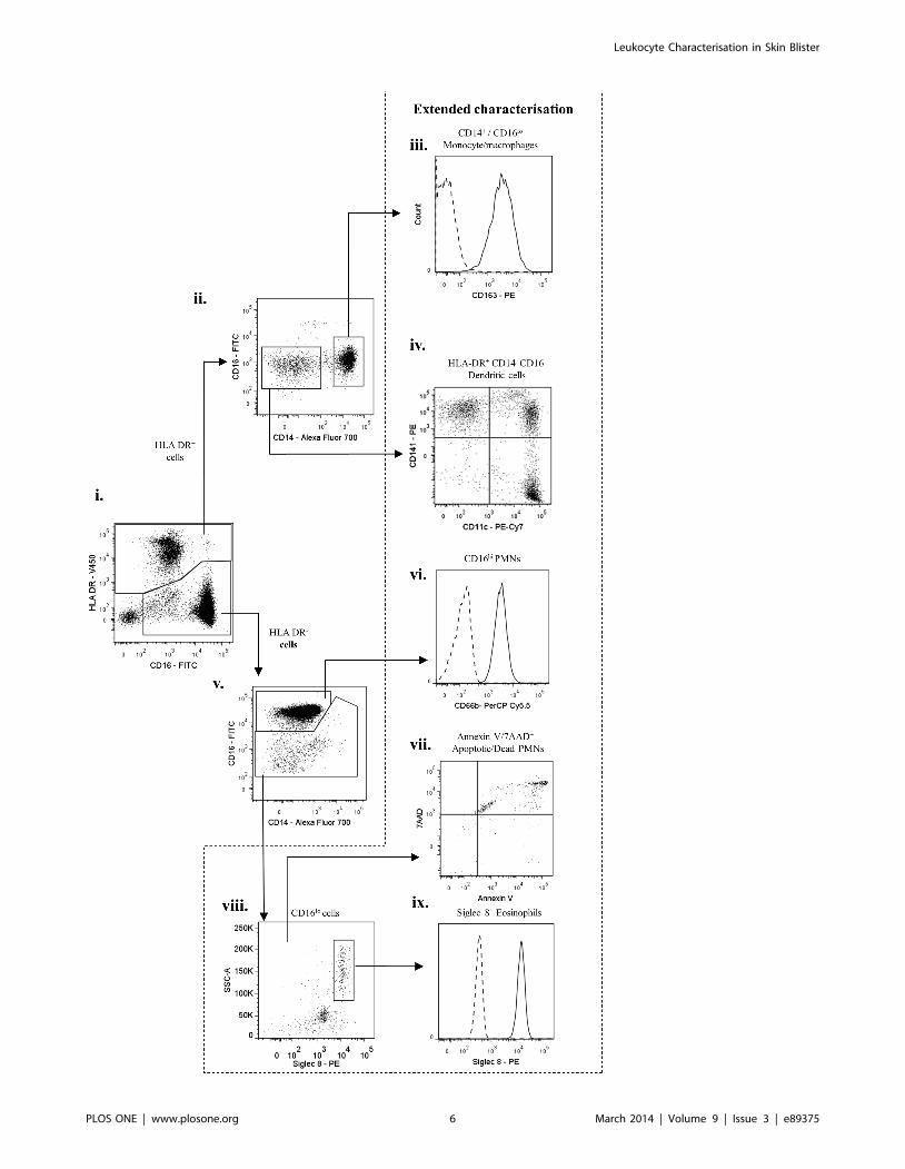
(Figure 3 [vi]). The HLA-DR+ population (Figure 4 [ii]) comprised
of CD14+/CD16lo monocyte/macrophages expressing CD163
(Figure 4 [iii]), and a CD142 CD162 dendritic cell population,
which upon extended characterisation, comprised of three
dendritic cell subpopulations: CD11c+, CD141+, and CD11c+/
CD141+ (Figure 4 [iv]).
The HLA-DR2 population comprised of CD16hi and CD16lo
populations (Figure 4 [v]). CD16hi population labelled positive for
CD66b and was identified as PMNs (Figure 4 [vi]). The CD16lo
cells comprised two different populations with varying autofluo-
rescence and side scatter. Flow-assisted cell sorting followed by
modified Wright’s staining on cytospin revealed these cells to be a
mixture of eosinophils and apoptotic PMNs. The autofluorescent
CD16loSSChi population stained positive for Siglec-8 (Figure 4
[viii,ix]), whereas CD16loSSClo population stained positive for
apoptotic cell marker, Annexin-V/7AAD (Figure 4 [vii,viii]) and
were characterised as apoptotic PMNs as previously reported [16].
Therefore, in a 24 h cantharidin-induced skin blister, PMNs are
the predominant cell type along with monocytes/macrophages,
dendritic cells and comparatively fewer lymphocytes and eosino-
phils.
Characterisation of inflammatory cell infiltrates into skinblisters at 72 h –resolution
Flow cytometric characterisation of 72 h blister is outlined in
Figure 5 and Figure 6. The gating strategy employed to 24 h
blister was similarly applied at 72 h. In three individuals, one of
Figure 1. Characterisation of peripheral blood leukocytes from healthy volunteers- I. Representative dot plots for flow cytometric gatingare shown for healthy male volunteers (n = 17). Blood was drawn from the forearm not bearing skin blisters. Erythrocytes were lysed and theremaining leukocytes incubated with antibodies and processed by flow cytometry. Gating strategies firstly identified CD3+ T cells, CD19+ B cells andCD56+ CD16+/2 NK cells. The remaining lymphocyte-deplete population was gated into HLA-DR+ and HLA-DR2 cells. Arrows indicate gating strategy.doi:10.1371/journal.pone.0089375.g001
Leukocyte Characterisation in Skin Blister
PLOS ONE | www.plosone.org 3 March 2014 | Volume 9 | Issue 3 | e89375

Leukocyte Characterisation in Skin Blister
PLOS ONE | www.plosone.org 4 March 2014 | Volume 9 | Issue 3 | e89375

the 24 h or 72 h blisters could not be aspirated, and as such the
temporal changes in total leukocytes and oedema from 24 h to
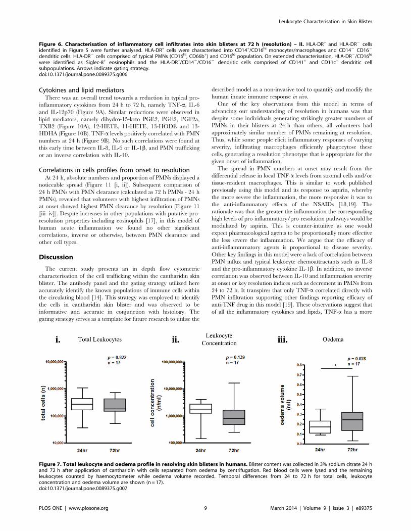
72 h were quantified for 17 individuals (Figure 7). There was no
overall difference in total leukocytes from 24–72 h (Figure 7 [i–ii]),
but oedema volume increased significantly (Figure 7 [iii]).
Individual leukocyte profiles show a decrease in CD19+ B cells
(Figure 8 [ii]) and a modest increase in CD56+ NK cells (Figure 8
[iii–iv]). The absolute numbers of PMNs decreased significantly
from 24 h to 72 h (Figure 8 [vi]). The number of monocytes/
macrophages did not change significantly (Figure 8 [v]), but
tended towards a reduction from 24–72 h. There was an increase
in the number of apoptotic PMNs and eosinophils from 24 to
72 hours (Figure 8 [vii–viii]).
Figure 2. Characterisation of peripheral blood leukocytes from healthy volunteers- II. HLA-DR+ and HLA-DR2 cells identified in Figure 1were further analysed. HLA-DR+ cells were characterised into CD14hi/CD162 , CD14hi/CD16+, CD14lo/CD16+ monocytes and CD142/CD162 dendriticcells. HLA-DR2 cells comprised of typical PMNs (CD16hi, CD66+) and a CD16lo population. On extended characterisation, HLA-DR2/CD16lo cells wereidentified as Siglec-8+ eosinophils and the HLA-DR+/CD142/CD162 dendritic cells comprised of CD141+ and CD11c+ dendritic cell subpopulations.Arrows indicate gating strategy.doi:10.1371/journal.pone.0089375.g002
Figure 3. Characterisation of inflammatory cell infiltrates into skin blisters at 24 h (onset) – I. Representative dot plots for flowcytometric gating are shown for healthy male volunteers (n = 17). Blister contents were collected from one blister 24 h after application of cantharidinin 3% sodium citrate, with cells separated from oedema by centrifugation. Leukocytes were enumerated by haemocytometer and oedema volumerecorded. Leukocytes were incubated with antibodies and processed by flow cytometry. Gating strategies firstly identified CD3+ T cells, CD19+ B cellsand CD56+CD16+/2 NK cells. The remaining lymphocyte-deplete population was gated into HLA-DR+ and HLA-DR2 cells. Arrows indicate gatingstrategy.doi:10.1371/journal.pone.0089375.g003
Leukocyte Characterisation in Skin Blister
PLOS ONE | www.plosone.org 5 March 2014 | Volume 9 | Issue 3 | e89375
Leukocyte Characterisation in Skin Blister
PLOS ONE | www.plosone.org 6 March 2014 | Volume 9 | Issue 3 | e89375

Figure 4. Characterisation of inflammatory cell infiltrates into skin blisters at 24 h (onset) – II. HLA-DR+ and HLA-DR2 cells identified inFigure 3 were further analysed. HLA-DR+ cells were characterised into CD14+/CD16lo monocytes/macrophages and HLA-DR+/CD142/CD162 dendriticcells. HLA-DR2 cells comprised of typical PMNs (CD16hi, CD66+) and a CD16lo population. On extended characterisation, HLA-DR2/CD16lo wereidentified as a mixture of Siglec-8+ eosinophils and Annexin V/7AAD+ apoptotic/dead PMNs. The HLA-DR+/CD142/CD162 dendritic cells comprised ofCD141+ and CD11c+ dendritic cell subpopulations. Arrows indicate gating strategy.doi:10.1371/journal.pone.0089375.g004
Figure 5. Characterisation of inflammatory cell infiltrates into skin blisters at 72 h (resolution) – I. Representative dot plots for flowcytometric gating are shown for healthy male volunteers (n = 17). Blister contents were collected from the remaining blister 72 h after application ofcantharidin in 3% sodium citrate, with cells separated from oedema by centrifugation. Leukocytes were enumerated by haemocytometer andoedema volume recorded. Leukocytes were incubated with antibodies and processed by flow cytometry. Gating strategies firstly identified CD3+ Tcells, CD19+ B cells and CD56+CD16+/2 NK cells. The remaining lymphocyte-deplete population was gated into HLA-DR+ and HLA-DR2 cells. Arrowsindicate gating strategy.doi:10.1371/journal.pone.0089375.g005
Leukocyte Characterisation in Skin Blister
PLOS ONE | www.plosone.org 7 March 2014 | Volume 9 | Issue 3 | e89375

Leukocyte Characterisation in Skin Blister
PLOS ONE | www.plosone.org 8 March 2014 | Volume 9 | Issue 3 | e89375
Cytokines and lipid mediatorsThere was an overall trend towards a reduction in typical pro-
inflammatory cytokines from 24 h to 72 h, namely TNF-a, IL-6
and IL-12p70 (Figure 9A). Similar reductions were observed in
lipid mediators, namely dihydro-15-keto PGE2, PGE2, PGF2a,
TXB2 (Figure 10A), 12-HETE, 11-HETE, 13-HODE and 13-
HDHA (Figure 10B). TNF-a levels positively correlated with PMN
numbers at 24 h (Figure 9B). No such correlations were found at
this early time between IL-8, IL-6 or IL-1b, and PMN trafficking
or an inverse correlation with IL-10.
Correlations in cells profiles from onset to resolutionAt 24 h, absolute numbers and proportion of PMNs displayed a
noticeable spread (Figure 11 [i, ii]). Subsequent comparison of
24 h PMNs with PMN clearance (calculated as 72 h PMNs - 24 h
PMNs), revealed that volunteers with highest infiltration of PMNs
at onset showed highest PMN clearance by resolution (Figure 11
[iii–iv]). Despite increases in other populations with putative pro-
resolution properties including eosinophils [17], in this model of
human acute inflammation we found no other significant
correlations, inverse or otherwise, between PMN clearance and
other cell types.
Discussion
The current study presents an in depth flow cytometric
characterisation of the cell trafficking within the cantharidin skin
blister. The antibody panel and the gating strategy utilized here
accurately identify the known populations of immune cells within
the circulating blood [14]. This strategy was employed to identify
the cells in cantharidin skin blister and was observed to be
informative and accurate in conjunction with histology. The
gating strategy serves as a template for future research to utilise the
described model as a non-invasive tool to quantify and modify the
human innate immune response in vivo.
One of the key observations from this model in terms of
advancing our understanding of resolution in humans was that
despite some individuals generating strikingly greater numbers of
PMNs in their blisters at 24 h than others, all volunteers had
approximately similar number of PMNs remaining at resolution.
Thus, while some people elicit inflammatory responses of varying
severity, infiltrating macrophages efficiently phagocytose these
cells, generating a resolution phenotype that is appropriate for the
given onset of inflammation.
The spread in PMN numbers at onset may result from the
differential release in local TNF-a levels from stromal cells and/or
tissue-resident macrophages. This is similar to work published
previously using this model and its response to aspirin, whereby
the more severe the inflammation, the more responsive it was to
the anti-inflammatory effects of the NSAIDs [18,19]. The
rationale was that the greater the inflammation the corresponding
high levels of pro-inflammatory/pro-resolution pathways would be
modulated by aspirin. This is counter-intuitive as one would
expect pharmacological agents to be proportionally more effective
the less severe the inflammation. We argue that the efficacy of
anti-inflammmatory agents is proportional to disease severity.
Other key findings in this model were a lack of correlation between
PMN influx and typical leukocyte chemoattractants such as IL-8
and the pro-inflammatory cytokine IL-1b. In addition, no inverse
correlation was observed between IL-10 and inflammation severity
at onset or key resolution indices such as decrement in PMNs from
24 to 72 h. It transpires that only TNF-a correlated directly with
PMN infiltration supporting other findings reporting efficacy of
anti-TNF drug in this model [19]. These observations suggest that
of all the inflammatory cytokines and lipids, TNF-a has a more
Figure 6. Characterisation of inflammatory cell infiltrates into skin blisters at 72 h (resolution) – II. HLA-DR+ and HLA-DR2 cellsidentified in Figure 5 were further analysed. HLA-DR+ cells were characterised into CD14+/CD16hi monocytes/macrophages and CD142 CD162
dendritic cells. HLA-DR2 cells comprised of typical PMNs (CD16hi, CD66b+) and CD16lo population. On extended characterisation, HLA-DR2/CD16lo
were identified as Siglec-8+ eosinophils and the HLA-DR+/CD142/CD162 dendritic cells comprised of CD141+ and CD11c+ dendritic cellsubpopulations. Arrows indicate gating strategy.doi:10.1371/journal.pone.0089375.g006
Figure 7. Total leukocyte and oedema profile in resolving skin blisters in humans. Blister content was collected in 3% sodium citrate 24 hand 72 h after application of cantharidin with cells separated from oedema by centrifugation. Red blood cells were lysed and the remainingleukocytes counted by haemocytometer while oedema volume recorded. Temporal differences from 24 to 72 h for total cells, leukocyteconcentration and oedema volume are shown (n = 17).doi:10.1371/journal.pone.0089375.g007
Leukocyte Characterisation in Skin Blister
PLOS ONE | www.plosone.org 9 March 2014 | Volume 9 | Issue 3 | e89375
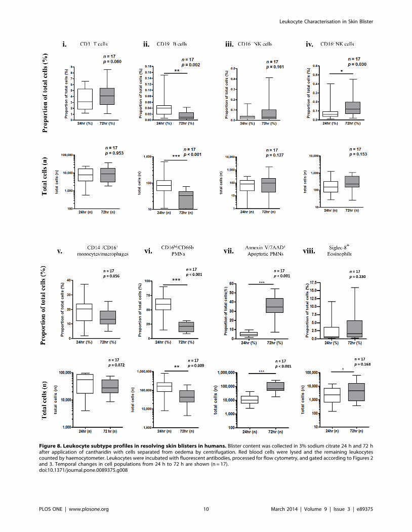
Figure 8. Leukocyte subtype profiles in resolving skin blisters in humans. Blister content was collected in 3% sodium citrate 24 h and 72 hafter application of cantharidin with cells separated from oedema by centrifugation. Red blood cells were lysed and the remaining leukocytescounted by haemocytometer. Leukocytes were incubated with fluorescent antibodies, processed for flow cytometry, and gated according to Figures 2and 3. Temporal changes in cell populations from 24 h to 72 h are shown (n = 17).doi:10.1371/journal.pone.0089375.g008
Leukocyte Characterisation in Skin Blister
PLOS ONE | www.plosone.org 10 March 2014 | Volume 9 | Issue 3 | e89375

Figure 9. Cytokine profiles in resolving skin blisters in humans. Blister content was collected in 3% sodium citrate 24 h and 72 h afterapplication of cantharidin with cells separated from oedema by centrifugation. Red blood cells were lysed and the remaining leukocytes counted byhaemocytometer while oedema volume recorded and processed by assay for cytokine concentration (A). Correlations were made between cytokineconcentration and PMN infiltration at 24 h and 72 h (B) (n = 17).doi:10.1371/journal.pone.0089375.g009
Leukocyte Characterisation in Skin Blister
PLOS ONE | www.plosone.org 11 March 2014 | Volume 9 | Issue 3 | e89375

Leukocyte Characterisation in Skin Blister
PLOS ONE | www.plosone.org 12 March 2014 | Volume 9 | Issue 3 | e89375

prominent role in controlling cell trafficking in acute inflammatory
reaction in this skin injury model.
Furthermore study results identified an increase in overall
numbers of dendritic cells, eosinophils and apoptotic PMNs during
resolution. This offers insight into the potential importance of
eosinophils in resolution of some inflammatory responses in
humans. This theory supports recent murine studies showing that
during resolution of acute zymosan-induced peritonitis, eosino-
phils congregate in the abdominal cavity and generate 15-
lipoxygenase lipid mediators that are known to enhance the
phagocytic ability of macrophages [20]. This must be borne in
Figure 10. Lipid mediators in resolving skin blisters in humans. Blister content was collected in 3% sodium citrate 24 h and 72 h afterapplication of cantharidin with cells separated from oedema by centrifugation. Red blood cells were lysed and the remaining leukocytes counted byhaemocytometer while oedema volume recorded and analysed by liquid chromatography coupled to electrospray ionization tandem massspectrometry for eicosanoid levels (n = 17).doi:10.1371/journal.pone.0089375.g010
Figure 11. Correlations in cell profiles from onset to resolution. A wide range of PMN numbers infiltrate skin blisters early in the response(onset), and an immune response induces appropriate resolution. Correlation between change in PMN numbers (72 h minus 24 h) and total PMNnumbers at onset are expressed as either total cells or as percentages.doi:10.1371/journal.pone.0089375.g011
Leukocyte Characterisation in Skin Blister
PLOS ONE | www.plosone.org 13 March 2014 | Volume 9 | Issue 3 | e89375
mind against the vast literature describing eosinophils as being
pathogenic in allergic diseases [21].
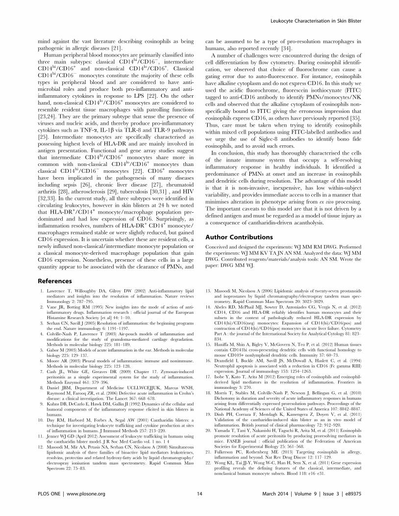
Human peripheral blood monocytes are primarily classified into
three main subtypes: classical CD14hi/CD162, intermediate
CD14hi/CD16+ and non-classical CD14lo/CD16+. Classical
CD14hi/CD162 monocytes constitute the majority of these cells
types in peripheral blood and are considered to have anti-
microbial roles and produce both pro-inflammatory and anti-
inflammatory cytokines in response to LPS [22]. On the other
hand, non-classical CD14lo/CD16+ monocytes are considered to
resemble resident tissue macrophages with patrolling functions
[23,24]. They are the primary subtype that sense the presence of
viruses and nucleic acids, and thereby produce pro-inflammatory
cytokines such as TNF-a, IL-1b via TLR-8 and TLR-9 pathways
[25]. Intermediate monocytes are specifically characterised as
possessing highest levels of HLA-DR and are mainly involved in
antigen presentation. Functional and gene array studies suggest
that intermediate CD14hi/CD16+ monocytes share more in
common with non-classical CD14lo/CD16+ monocytes than
classical CD14hi/CD162 monocytes [22]. CD16+ monocytes
have been implicated in the pathogenesis of many diseases
including sepsis [26], chronic liver disease [27], rheumatoid
arthritis [28], atherosclerosis [29], tuberculosis [30,31] , and HIV
[32,33]. In the current study, all three subtypes were identified in
circulating leukocytes, however in skin blisters at 24 h we noted
that HLA-DR+/CD14+ monocyte/macrophage population pre-
dominated and had low expression of CD16. Surprisingly, as
inflammation resolves, numbers of HLA-DR+ CD14+ monocyte/
macrophages remained stable or were slightly reduced, but gained
CD16 expression. It is uncertain whether these are resident cells, a
newly influxed non-classical/intermediate monocyte population or
a classical monocyte-derived macrophage population that gain
CD16 expression. Nonetheless, presence of these cells in a large
quantity appear to be associated with the clearance of PMNs, and
can be assumed to be a type of pro-resolution macrophages in
humans, also reported recently [34].
A number of challenges were encountered during the design of
cell differentiation by flow cytometry. During eosinophil identifi-
cation, we observed that choice of fluorochrome can cause a
gating error due to auto-fluorescence. For instance, eosinophils
have alkaline cytoplasm and do not express CD16. In this study we
used the acidic fluorochrome, fluorescein isothiocynate (FITC)
tagged to anti-CD16 antibody to identify PMNs/monocytes/NK
cells and observed that the alkaline cytoplasm of eosinophils non-
specifically bound to FITC giving the erroneous impression that
eosinophils express CD16, as others have previously reported [35].
Thus, care must be taken when trying to identify eosinophils
within mixed cell populations using FITC-labelled antibodies and
we urge the use of Siglec-8 antibodies to identify bono fide
eosinophils, and to avoid such errors.
In conclusion, this study has thoroughly characterised the cells
of the innate immune system that occupy a self-resolving
inflammatory response in healthy individuals. It identified a
predominance of PMNs at onset and an increase in eosinophils
and dendritic cells during resolution. The advantage of this model
is that it is non-invasive, inexpensive, has low within-subject
variability, and provides immediate access to cells in a manner that
minimises alteration in phenotype arising from ex vivo processing.
The important caveats to this model are that it is not driven by a
defined antigen and must be regarded as a model of tissue injury as
a consequence of cantharidin-driven acantholysis.
Author Contributions
Conceived and designed the experiments: WJ MM RM DWG. Performed
the experiments: WJ MM KV TA JN AN SM. Analyzed the data: WJ MM
DWG. Contributed reagents/materials/analysis tools: AN SM. Wrote the
paper: DWG MM WJ.
References
1. Lawrence T, Willoughby DA, Gilroy DW (2002) Anti-inflammatory lipid
mediators and insights into the resolution of inflammation. Nature reviews
Immunology 2: 787–795.
2. Vane JR, Botting RM (1995) New insights into the mode of action of anti-
inflammatory drugs. Inflammation research : official journal of the European
Histamine Research Society [et al] 44: 1–10.
3. Serhan CN, Savill J (2005) Resolution of inflammation: the beginning programs
the end. Nature immunology 6: 1191–1197.
4. Colville-Nash P, Lawrence T (2003) Air-pouch models of inflammation and
modifications for the study of granuloma-mediated cartilage degradation.
Methods in molecular biology 225: 181–189.
5. Gabor M (2003) Models of acute inflammation in the ear. Methods in molecular
biology 225: 129–137.
6. Moore AR (2003) Pleural models of inflammation: immune and nonimmune.
Methods in molecular biology 225: 123–128.
7. Cash JL, White GE, Greaves DR (2009) Chapter 17. Zymosan-induced
peritonitis as a simple experimental system for the study of inflammation.
Methods Enzymol 461: 379–396.
8. Daniel JBM, Department of Medicine UCLLWCEJJUK, Marcus WNH,
Raymond M, Farooq ZR, et al. (2006) Defective acute inflammation in Crohn’s
disease: a clinical investigation. The Lancet 367: 668–678.
9. Kuhns DB, DeCarlo E, Hawk DM, Gallin JI (1992) Dynamics of the cellular and
humoral components of the inflammatory response elicited in skin blisters in
humans.
10. Day RM, Harbord M, Forbes A, Segal AW (2001) Cantharidin blisters: a
technique for investigating leukocyte trafficking and cytokine production at sites
of inflammation in humans. J Immunol Methods 257: 213–220.
11. Jenner WJ GD (April 2012) Assessment of leukocyte trafficking in humans using
the cantharidin blister model. J R Soc Med Cardio vol. 1 no. 1 4
12. Masoodi M, Mir AA, Petasis NA, Serhan CN, Nicolaou A (2008) Simultaneous
lipidomic analysis of three families of bioactive lipid mediators leukotrienes,
resolvins, protectins and related hydroxy-fatty acids by liquid chromatography/
electrospray ionisation tandem mass spectrometry. Rapid Commun Mass
Spectrom 22: 75–83.
13. Masoodi M, Nicolaou A (2006) Lipidomic analysis of twenty-seven prostanoids
and isoprostanes by liquid chromatography/electrospray tandem mass spec-
trometry. Rapid Commun Mass Spectrom 20: 3023–3029.
14. Abeles RD, McPhail MJ, Sowter D, Antoniades CG, Vergis N, et al. (2012)
CD14, CD16 and HLA-DR reliably identifies human monocytes and their
subsets in the context of pathologically reduced HLA-DR expression by
CD14(hi)/CD16(neg) monocytes: Expansion of CD14(hi)/CD16(pos) and
contraction of CD14(lo)/CD16(pos) monocytes in acute liver failure. Cytometry
Part A : the journal of the International Society for Analytical Cytology 81: 823–
834.
15. Haniffa M, Shin A, Bigley V, McGovern N, Teo P, et al. (2012) Human tissues
contain CD141hi cross-presenting dendritic cells with functional homology to
mouse CD103+ nonlymphoid dendritic cells. Immunity 37: 60–73.
16. Dransfield I, Buckle AM, Savill JS, McDowall A, Haslett C, et al. (1994)
Neutrophil apoptosis is associated with a reduction in CD16 (Fc gamma RIII)
expression. Journal of immunology 153: 1254–1263.
17. Isobe Y, Kato T, Arita M (2012) Emerging roles of eosinophils and eosinophil-
derived lipid mediators in the resolution of inflammation. Frontiers in
immunology 3: 270.
18. Morris T, Stables M, Colville-Nash P, Newson J, Bellingan G, et al. (2010)
Dichotomy in duration and severity of acute inflammatory responses in humans
arising from differentially expressed proresolution pathways. Proceedings of the
National Academy of Sciences of the United States of America 107: 8842–8847.
19. Dinh PH, Corraza F, Mestdagh K, Kassengera Z, Doyen V, et al. (2011)
Validation of the cantharidin-induced skin blister as an in vivo model of
inflammation. British journal of clinical pharmacology 72: 912–920.
20. Yamada T, Tani Y, Nakanishi H, Taguchi R, Arita M, et al. (2011) Eosinophils
promote resolution of acute peritonitis by producing proresolving mediators in
mice. FASEB journal : official publication of the Federation of American
Societies for Experimental Biology 25: 561–568.
21. Fulkerson PC, Rothenberg ME (2013) Targeting eosinophils in allergy,
inflammation and beyond. Nat Rev Drug Discov 12: 117–129.
22. Wong KL, Tai JJ-Y, Wong W-C, Han H, Sem X, et al. (2011) Gene expression
profiling reveals the defining features of the classical, intermediate, and
nonclassical human monocyte subsets. Blood 118: e16–e31.
Leukocyte Characterisation in Skin Blister
PLOS ONE | www.plosone.org 14 March 2014 | Volume 9 | Issue 3 | e89375

23. Ziegler-Heitbrock HW, Fingerle G, Strobel M, Schraut W, Stelter F, et al.
(1993) The novel subset of CD14+/CD16+ blood monocytes exhibits features oftissue macrophages. Eur J Immunol 23: 2053–2058.
24. Auffray C, Fogg D, Garfa M, Elain G, Join-Lambert O, et al. (2007) Monitoring
of blood vessels and tissues by a population of monocytes with patrollingbehavior. Science 317: 666–670.
25. Cros J, Cagnard N, Woollard K, Patey N, Zhang SY, et al. (2010) HumanCD14dim monocytes patrol and sense nucleic acids and viruses via TLR7 and
TLR8 receptors. Immunity 33: 375–386.
26. Fingerle G, Pforte A, Passlick B, Blumenstein M, Strobel M, et al. (1993) Thenovel subset of CD14+/CD16+ blood monocytes is expanded in sepsis patients.
Blood 82: 3170–3176.27. Zimmermann HW, Seidler S, Nattermann J, Gassler N, Hellerbrand C, et al.
(2010) Functional contribution of elevated circulating and hepatic non-classicalCD14CD16 monocytes to inflammation and human liver fibrosis. PloS one 5:
e11049.
28. Hepburn AL, Mason JC, Davies KA (2004) Expression of Fcgamma andcomplement receptors on peripheral blood monocytes in systemic lupus
erythematosus and rheumatoid arthritis. Rheumatology 43: 547–554.29. Schlitt A, Heine GH, Blankenberg S, Espinola-Klein C, Dopheide JF, et al.
(2004) CD14+CD16+ monocytes in coronary artery disease and their
relationship to serum TNF-alpha levels. Thrombosis and haemostasis 92: 419–424.
30. Lugo-Villarino G, Neyrolles O (2013) Dressed not to kill: CD16+ monocytes
impair immune defence against tuberculosis. European journal of immunology
43: 327–330.
31. Castano D, Garcia LF, Rojas M (2011) Increased frequency and cell death of
CD16+ monocytes with Mycobacterium tuberculosis infection. Tuberculosis 91:
348–360.
32. Vanham G, Edmonds K, Qing L, Hom D, Toossi Z, et al. (1996) Generalized
immune activation in pulmonary tuberculosis: co-activation with HIV infection.
Clinical and experimental immunology 103: 30–34.
33. Thieblemont N, Weiss L, Sadeghi HM, Estcourt C, Haeffner-Cavaillon N (1995)
CD14lowCD16high: a cytokine-producing monocyte subset which expands
during human immunodeficiency virus infection. European journal of
immunology 25: 3418–3424.
34. Evans BJ, Haskard DO, Sempowksi G, Landis RC (2013) Evolution of the
Macrophage CD163 Phenotype and Cytokine Profiles in a Human Model of
Resolving Inflammation. International Journal of Inflammation 2013.
35. Bedner E, Halicka HD, Cheng W, Salomon T, Deptala A, et al. (1999) High
affinity binding of fluorescein isothiocyanate to eosinophils detected by laser
scanning cytometry: a potential source of error in analysis of blood samples
utilizing fluorescein-conjugated reagents in flow cytometry. Cytometry 36: 77–
82.
Leukocyte Characterisation in Skin Blister
PLOS ONE | www.plosone.org 15 March 2014 | Volume 9 | Issue 3 | e89375