Embed Size (px)
Citation preview
INFECTION AND IMMUNITY, Apr. 1988, p. 815-8220019-9567/88/040815-08$02.00/0Copyright C 1988, American Society for Microbiology
Type 1 Fimbriate Escherichia coli Stimulates a Unique Pattern ofDegranulation by Human Polymorphonuclear LeukocytesROBERT STEADMAN,* NICHOLAS TOPLEY, DAVID E. JENNER,t MALCOLM DAVIES,
AND JOHN D. WILLIAMS
Department of Renal Medicine, Kidney Research Unit for Wales Foundation Institute Royal Infirmary, Newport Road,Cardiff CF2 JFZ, United Kingdom
Received 10 August 1987/Accepted 22 December 1987
Uropathogenic strains of Escherichia coli bearing mannose-sensitive (type 1) fimbriae promote a uniquepattern of degranulation from human polymorphonuclear leukocytes (PMN). Significant quantities of theprimary (10) and tertiary (30) granule markers, neutral protease-myeloperoxidase and N-acetyl-o-D-glucosa-minidase, respectively, were released by PMN in a dose- and time-dependent manner when stimulated by thesedefined bacterial strains. Organisms bearing mannose-resistant (P) fimbriae promoted release of only thesecondary (2°) granule marker, vitamin B12-binding protein. When this pattern of degranulation was comparedto that produced by PMN in response to a variety of soluble and particulate stimuli, only the calcium ionophoreA23187 similarly triggered 1° and 30 granule marker release. All the other stimuli tested-zymosan,serum-treated and unopsonized; n-formylmethionyl-leucyl-phenylalanine; and phorbol myristate acetate-promoted release of only the 2° granule marker. These results demonstrate selectivity of PMN degranulationin response to a number of transmembrane signals. In addition, the capacity of E. coli to promote PMNdegranulation is dependent on its phenotypic fimbrial expression, a surface characteristic which correlatessignificantly with its relative surface hydrophobicity as measured by binding to octyl Sepharose. Those bacteriademonstrating the greatest hydrophobicity were capable of triggering discharge of all three granule markerproteins. Thus, the mannose-sensitive fimbriae of uropathogenic E. coli may contribute significantly to theirpotential pathophysiologic role in renal scarring.
Chronic pyelonephritis is characterized by progressiverenal scarring. Recent evidence obtained by using animalmodels has demonstrated that this scarring is directly pro-
portional to the magnitude of the initial inflammatory re-
sponse, which in turn is dependent on the interaction ofinvading bacteria with host inflammatory cells (15, 34, 36).Escherichia coli strains isolated from patients with urinarytract infections possess a higher frequency of defined viru-lence markers than do strains which compose the normalfecal flora. These urinary tract pathogens express a re-stricted range of 0 serotypes (30), resist the killing effects ofserum (20), and may elaborate alpha-hemolysin (6). Afterisolation and subculture, these organisms usually expresssurface fimbriae. Type 1, mannose-sensitive (MS) fimbriaepromote the mannose-dependent hemagglutination of guineapig erythrocytes (27, 37), whereas mannose-resistant (MR)fimbriae, of which P fimbriae are a subgroup (41), promotethe mannose-independent hemagglutination of human eryth-rocytes. Although both fimbrial types increase adherence touroepithelial cells (7, 22), type 1 (MS) fimbriae also mediateadherence to and phagocytosis by human neutrophils (polyr-morphonuclear leukocytes [PMN]) (4), and there is a directand highly significant correlation between the possession oftype 1 (MS) fimbriae, relative bacterial surface hydrophobic-ity, and the capacity of the organism to activate the PMNrespiratory burst (19).The activation of phagocytic cells by particulate stimuli
occurs as a result of either specific receptor-ligand binding
* Corresponding author.t Present address: I.C.I. Diagnostics Group, Gadbrook Park,
Rudheath, Northwick, Cheshire, United Kingdom.
(26) or a receptor-independent physicochemical interactionof a particle with the cell surface (42). This stimulus-cellinteraction has been characterized in terms of respiratoryburst activation (18), the release of intracellular proteins (3),and the generation of pro-inflammatory mediators (44).There is considerable evidence that the reactive oxygenmetabolites and proteases released by phagocytic cells dur-ing this inflammatory response can contribute directly totissue damage (13, 14, 23). The human PMN has a well-defined population of intracellular enzymes which are re-
leased from at least two distinct classes of cytoplasmicgranule in response to particulate or soluble stimuli (3, 35).The primary (10) (azurophil) granule contains a number ofacid hydrolases and neutral proteases (such as elastase), as
well as myeloperoxidase (MPO). These enzymes are dis-charged into the phagocytic vacuole and are believed to beresponsible for microbial killing through the action of thevarious proteases and by the generation of hydrogen perox-ide and toxic oxygen radicals (14). The secondary (2°)(specific) granule contains a number of proteins, whichinclude lysozyme, lactoferrin, and vitamin B12-binding pro-teins, as well as neutral proteases such as collagenase. Fewof these molecules have any defined antimicrobial activity,but they may be secreted and function extracellularly to limitchemotaxis and increase PMN adhesiveness (16, 31). Iso-lated 20 granule protein release has been demonstrated afteractivation of cells by a variety of ligands (35), emphasizingthe nonspecific, secretory nature of 20 granule release. Aperoxidase-negative tertiary (30) granule containing N-acetyl-,3-D-glucosaminidase, P-galactosidase, and P-glucuro-nidase has also been described (35), but its function has notbeen defined in detail. Few studies have analyzed the degree
815
Vol. 56, No. 4
816 STEADMAN ET AL.
of participation of each granule during degranulation inresponse to either phagocytic or chemical stimulation (3, 32,46).The present study investigates the capacity of unopso-
nized uropathogenic strains of E. coli to trigger PMN degra-nulation and correlates this capacity with the phenotypicexpression of fimbrial type. In addition, the unique pattern ofdegranulation induced by certain of these strains of E. coli iscompared with the pattern of degranulation produced byPMN stimulated with a variety of reference compounds.
MATERIALS AND METHODS
Preparation of human neutrophils. Normal human leuko-cytes were isolated from citrated peripheral blood by dex-tran sedimentation and were rendered plasma-free and plate-let-poor by being washed with phosphate-buffered salinewithout calcium or magnesium, pH 7.3. PMN were purifiedby density gradient centrifugation at 400 x g for 35 min at23°C on Ficoll-Paque (Pharmacia Ltd., Milton Keynes,United Kingdom). The PMN were counted in a modifiedNeubauer counting chamber after hypotonic lysis of theerythrocytes. The cell preparations were judged to be 98%PMN on the basis of their morphology after examination ofWright-stained centrifuged preparations (Cytospin II; Shan-don Southern Products, Runcorn, Cheshire, United King-dom).
Preparation of reference stimuli. Unless stated otherwise,all chemicals were purchased from Sigma Chemical Co.Ltd., Poole, Dorset, United Kingdom. The calcium iono-phore A23187 (Cambridge Bioscience, Cambridge, UnitedKingdom), n-formylmethionyl-leucyl-phenylalanine, andphorbol myristate acetate (PMA) were stored at 10 mMconcentrations in dimethyl sulfoxide at -70°C and diluted toan appropriate concentration in Krebs Ringer phosphatebuffer (pH 7.4) containing 0.54 mM Ca2", 1.2 mM Mg2", and11.0 mM D-glucose (KRPG) immediately before use. Zymo-san A (500 mg) was boiled for 20 min in 50 ml of 0.9% saline,washed, and resuspended to a final concentration of 10mg/ml (5 x 10⁸ particles per ml). Serum-treated zymosanwas prepared by suspending 20 mg of boiled zymosan in 1 mlof KRPG, adding 3 ml of fresh pooled human serum, andincubating this mixture with continuous mixing for 30 min at37°C. The serum-treated zymosan was then washed threetimes, suspended to a volume of 4 ml in KRPG, and storedat -200C.
Bacterial strains. Eleven uropathogenic isolates of E. coliwere studied. Each was serially subcultured for 18 h either innutrient broth (Oxoid No. 2) or on nutrient agar (Oxoid Ltd.,Basingstoke, United Kingdom) at least three times to facil-itate the maximal expression of a required fimbrial type.Bacteria were harvested by centrifugation, washed twice,and suspended to an optical density of 2.0 at 560 nm in aspectrophotometer (Unicam SP500 series 2; Pye Unicam,Cambridge, United Kingdom) phosphate-buffered saline, pH7.3 (equivalent to 109 CFU/ml).The hemagglutination of guinea pig or human erythrocytes
by each bacterial strain was tested immediately prior to theiruse in order to assess their fimbrial expression (Table 1).Fimbriation was confirmed by transmission electron micros-copy (17).
Bacterial phagocytosis. Purified human PMN were washedand resuspended in RPMI 1640 (GIBCO Ltd., Paisley,Scotland) containing 0.2% (wt/vol) bovine serum albumin ata concentration of 3 x 10⁶ cells per ml. One-milliliterportions of the suspension were layered onto 35-mm tissue
culture plates, incubated at 37°C in 5% CO2 for 30 min, andthen washed three times with 2 ml of RPMI 1640 (containing5 mM MgCl2). More than 97% of the adherent cells wereidentified as PMN on the basis of their morphology.The PMN monolayers were then incubated for 20 min with
different strains of E. coli suspended to a concentration of 4x 10⁸ bacteria per ml in RPMI 1640 containing MgCl2. Themonolayers were then washed three times with phosphate-buffered saline (pH 7.3), air dried, fixed, and stained withWright's stain. The percentage of cells ingesting five or morebacteria was estimated by light microscopy. At least 200PMN per plate were counted.Measurement of bacterial hydrophobicity. Bacterial sur-
face hydrophobicity was assessed by modified hydrophobicinteraction chromatography (N. Topley, R. Steadman, R. K.Mackenzie, J. D. Williams, M. Davies, and A. W. Asscher,Rev. Infect. Dis., in press). Briefly, Octyl Sepharose CL4Bcolumns (15 by 5 mm) (Pharmacia) were prepared, equili-brated with 15 volumes of 0.25 M ammonium sulfate in 10mM sodium phosphate buffer, pH 6.8 (ASP buffer), andstored at 4°C until required. Samples of bacteria (opticaldensity at 560 nm = 1.0) (100 ,ul) were diluted 1:10 in ASPbuffer, and 500 ,ul of this dilution was loaded onto the columnand eluted with 2 ml of ASP buffer. Bacterial ATP wasextracted from eluted and from control bacterial suspensionsand measured by using the firefly bioluminescence assaysystem (Topley et al., in press) in a Lumac M2010 bio-counter (Lumac/3MbV, Schaesberg, The Netherlands). Thedegree of binding of bacteria to the Octyl Sepharose wasthen calculated and expressed as percent hydrophobicity.
Protein release experiments. One hundred-microliter vol-umes of KRPG containing 5 x 10⁵ PMN were diluted with300 pl of KRPG and preincubated for 5 min at 37°C beforethe addition of stimulus in KRPG (100 ,ul). Triplicate sampleswere incubated with 100 ,ul of each stimulus at the appropri-ate concentration for periods of up to 60 min and separatedby centrifugation for 1 min at 11,000 x g in a BeckmanMicrofuge B (Beckman RIIC Ltd., High Wycombe, Buck-
TABLE 1. Surface characteristics of individual E. coli strains
Phenotypic BidnE. coli 0 hemagglutinationn P-fimbrial toBindingstrain serotype" expression' to dotl
MS MR Sepharose
504 06 +++ - - 76.8 ± 4.512 04 +++ - - 71.1 ± 3.5ER2 04 - ++ + 33.8 ± 8.6KV NT ++ - - 70.5 ± 3.4AB NT - - - 28.8 ± 6.749 08 +++ - - 84.7 ±6.2NK1 04 - ++ + 21.2 ± 8.2SC 01 - ++ + 13.9 ± 6.1168 04 +++ - - 84.6 ± 6.4103 AA +++ - - N/A63 NT - + + 6.8 ± 2.3
" Abbreviations: NT, not typable (smooth but not one of the following 0serotypes: 1, 2, 4, 5, 6, 7, 8, 9, 11, 17, 18, 25, or 75); AA, autoagglutinable.
b Hemagglutination of individual E. coli strains on the day of the experi-ment. MS represents the capacity of D-mannose (2.5%, wt/vol) to inhibit theagglutination of guinea pig erythrocytes by MS fimbriate organisms; MRrepresents the mannose-resistant agglutination of human erythrocytes by MRfimbriate organisms.
' P-fimbriation was assessed by the receptor-specific particle agglutinationtest for E. coli (BACH-test, KabiVitrum, Stockholm, Sweden) of strainsgrown on nutrient agar (39).
d Results are expressed as the means + standard deviations for at leastthree separate determinations.
INFECT. IMMUN.
PMN DEGRANULATION: TYPE I FIMBRIAE 817
inghamshire, United Kingdom), and the supernatant wasremoved for the enzyme assays. Controls, which wereperformed for each experiment, consisted of supernatantfrom (i) unstimulated cells without incubation, (ii) cellsincubated in KRPG for periods of up to 60 min withoutstimulation, (iii) stimuli incubated in KRPG without PMN,and (iv) a KRPG blank incubated without PMN or stimulus.Each stimulus was used in three separate experiments withcells from three different donors.Enzyme assays. The percent release of enzymes from the
PMN was calculated, after subtraction of the appropriateblank values, as a percentage of that released from cellsdisrupted by sonication, for two 1-min periods at an 8-p.mpeak-to-peak distance at 4°C in a 150-W ultrasonic disinte-grator (Measuring and Scientific Equipment, Ltd., Crawley,United Kingdom). This form of disruption gave reproducibleresults for the maximal levels of total intracellular enzymeactivities measured when compared with freeze-thawing,hypotonic lysis, and Triton X-100 extraction (data notshown).
Control incubations. Granule marker release by PMN inresponse to E. coli was compared in all cases with theappropriate control cells incubated in buffer alone. Therewas no detectable enzyme activity when E. coli strains wereincubated without PMN in KRPG for 60 min at 37°C.
In all experiments, <5% of the cytoplasmic marker lacticdehydrogenase was released, indicating that most PMNremained intact throughout the 60 min of stimulation.MPO. One hundred microliters of supernatant from each
stimulation was incubated at 30°C with 1 ml of substrateconsisting of 0.3 mM o-dianisidine, 0.03% (vol/vol) H202,and 0.05% (vol/vol) Triton X-100 (BDH Ltd., Poole, Dorset,United Kingdom) in 0.1 M citric acid (pH 5.5). The reactionwas stopped after 5 min with 1 ml of 3.25 M perchloric acid.The A560 was measured in a spectrophotometer (Cecil CE292; Cecil Instruments Ltd., Cambridge, United Kingdom).
Neutral protease. Fifty microliters of supernatant wasadded to 50 p.l (0.1 mg) of [3H]casein (0.41 ,uCi/mg) (21) inphosphate buffer (pH 7.0) containing 1 mM CaC12 andincubated for 16 h at 37°C. Ice-cold 11% (wt/vol) trichloro-acetic acid (BDH Ltd.) was added. After incubation for 30min at 4°C, the nonhydrolyzed casein was precipitated bycentrifugation at 11,000 x g for 5 min.One hundred and seventy-five microliters of supernatant
containing the hydrolyzed product was then assayed forreleased radioactivity by addition to 4 ml of scintillant(Optiphase MP; LKB Instruments Ltd., Croydon, UnitedKingdom) and counted in an LKB Rackbeta scintillationcounter for 1 min.N-Acetyl p-D-glucosaminidase. One hundred microliters of
supernatant and 100 ,lI of substrate (0.2 mM 4-methylumbel-liferyl N-acetyl f-D-glucosamine [Koch Light Ltd., Have-hill, United Kingdom] in 0.1 M sodium citrate-phosphatebuffer, pH 4.3) were incubated for 16 h at 37°C. The reactionwas stopped by the addition of 2 ml of 5 mM EDTA (BDHLtd.) in 50 mM glycine buffer, pH 10.4. Fluorescenceemission at 448 nm was measured with excitation at 360 nmin an Aminco Bowman spectrophotofluorimeter (AmericanInstrument Co. Inc., Silver Spring, Md.).Vitamin B12-binding protein. One hundred microliters of
supernatant and 250 ,u1 of [57Co]cyanocobalamin (4.44 ng/mlin water, 0.067 p.Ci/ml) (Amersham International plc, Car-diff, United Kingdom) were incubated at room temperaturefor 30 min. One milliliter of activated charcoal (Norit GSX;BDH Ltd.) (5%, wt/vol) coated with 1% (wt/vol) bovineserum albumin was added and left at room temperature for
10 min before centrifugation for 10 min at 2,500 x g toprecipitate charcoal-adsorbed, non-protein-bound vitaminB12. One milliliter of supernatant was counted for 1 min in agamma counter (Kontron Instruments, St. Albans, UnitedKingdom).
Lactate dehydrogenase. Three hundred microliters of su-pernatant was added to 200 ,u1 of 10 mM ,-NADH in 2.4 mlof 0.1 M phosphate buffer, pH 7.4. The reaction was startedwith 100 ,ul of potassium pyruvate (1 mg/ml), and the changein extinctions at 340 nm (E340) was measured over 4 min in aCecil CE292 spectrophotometer by using the reaction ratecalculator.
Gelatin-degrading activity. One hundred microliters ofsupernatant was incubated at 37°C with 50 ,ul of 0.4 M Trishydrochloride, pH 7.5 (containing 10 mM CaCl2), and with100 p.1 of [14C]gelatin (1 mg/ml, 0.045 ,uCi/mg) from dena-tured interstitial rat skin collagen (95% type I, 5% type III)(5). Fifty microliters of ice-cold 100% (wt/vol) trichloroaceticacid was then added and incubated at 4°C for a further 30min. The insoluble protein was pelleted in a BeckmanMicrofuge at 11,000 x g for 10 min. One hundred and fiftymicroliters of the supernatant was then dissolved in 4 ml ofOptiphase MP (LKB Ltd.) and counted in an LKB Rackbetascintillation counter for 1 min.
Elastase activity. One hundred microlite-rs of supernatantwas added to 100 ,ul of insoluble [3H]elastin (25 ,ug/ml; 0.05,uCi/mg) (40) in 0.1 M Tris hydrochloride, pH 8.2, containing0.1% (vol/vol) Triton X-100 and incubated at 40°C for 16 h.Nondegraded elastin was precipitated by centrifugation in aBeckman Microfuge at 11,000 x g for 10 min, and 100 p.l ofthe supernatant was dissolved in 4 ml of Optiphase MPscintillant and counted in an LKB Rackbeta scintillationcounter for 1 min.
Trasylol binding. Five hundred microliters of supernatantfrom sonicated cells or from cells stimulated by E. coli 504was mixed with 0.5 ml of 0.05 M Tris hydrochloride buffer,pH 8.3, containing the serine protease inhibitor, Trasylol(Bayer Pharmaceuticals Ltd., Haywards Heath, UnitedKingdom), covalently bound to Sepharose beads (2). Thesuspension was incubated with occasional mixing for 30 minand then was centrifuged for 1 min at 11,000 x g. Thesupernatant was assayed for activity against [3H]elastin.After being washed in 0.05 M Tris hydrochloride buffer, pH8.3, the pellet was suspended in 1 ml of 0.05 M sodiumacetate buffer, pH 5.0, and incubated at 4°C for 30 min. Themixture was then centrifuged for 1 min at 11,000 x g, and theactivity against [3H]elastin released into the supernatant wasassayed.
Neutral protease inhibition. The serine protease inhibitorphenylmethylsulfonyl fluoride (PMSF) was stored at 40C inpropan-2-ol at a concentration of 500 mM and was used inthe enzyme assays at a concentration of 5 mM. EDTA (100mM) was used in the enzyme assays to inhibit metallopro-teases at a concentration of 5 mM. p-Aminophenylmercuricacetate (PAMA) was prepared to a concentration of 10 mMin 0.05 M NaOH and used at 1 mM in the enzyme assays.
Statistics. All data in this paper was analyzed by usingStudent's t test with Bessell's correction unless otherwisestated in the text.
RESULTS
PMN granule marker release in response to bacterial stim-ulation. Stimulation of 5 x 10⁵ human PMN by 10 differentstrains of E. coli of defined fimbrial type (Table 1) resulted inthe release of granule marker enzymes in a dose- (Fig. 1) and
VOL. 56, 1988
818 STEADMAN ET AL.
50-
40-
30120-
10-
T60 A B C
0. - - --
50
40130]20-
50°]d*- -1i - il i
300
50140] l301 T20{-j~ p id ~ j
1 10 100200500 1 10 100200500
Bacteria/PMN ratio
1 10 100 200 500
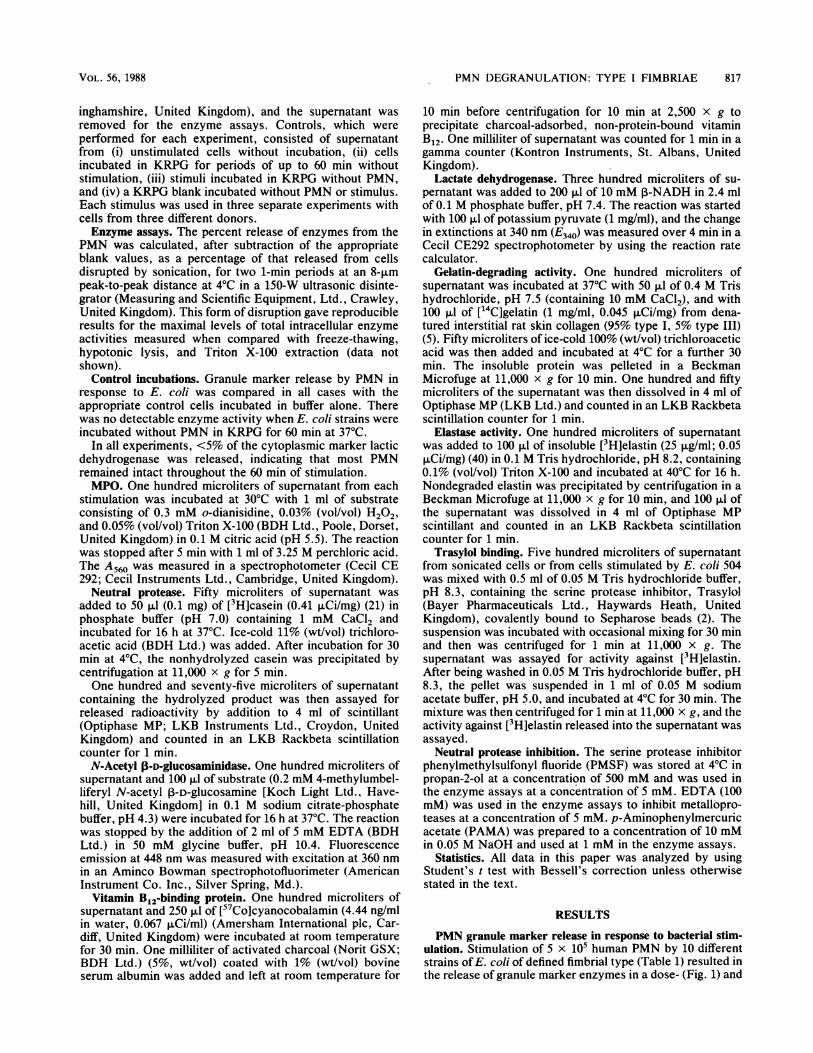
FIG. 1. Dose-dependent effect of E. coli on the capacity of 5 x10⁵ human PMN to release granule marker activity over a 60-minperiod. Three representative strains of E. coli are shown: MSfimbriate strain 504 (A), MR(P) fimbriate strain SC (B), and nonfim-briate strain AB (C). Data are expressed as the means ± standarddeviations for three separate experiments with each strain usingPMN from different donors. Release of granule markers fromunstimulated cells was measured over a 60-min period (T60j.
marker proteins (10, t = 1.90, P < 0.05; 20, t = 5.68, P <0.005; 30, t = 3.55, P < 0.05). Other compounds stimulatedsignificant release of only the 20 granule marker (Fig. 3).
Neutral protease release from human PMN. Type 1 (MS)fimbriate E. coli 504 and the calcium ionophore A23187caused significant release of proteolytic activity against[3H]casein (P < 0.05), [3H]elastin (P < 0.05), and [14C]gelatin (P < 0.02) (Fig. 4). All the other stimulants examined,except the MR(P) fimbriate E. coli SC, caused release ofsignificant quantities of only [14C]gelatin-degrading activity.This MR fimbriate organism caused release of significantquantities of vitamin B12-binding protein alone (Fig. 1) in theabsence of release of gelatinase activity (Fig. 4).The activity released against [3H]casein and [3H]elastin
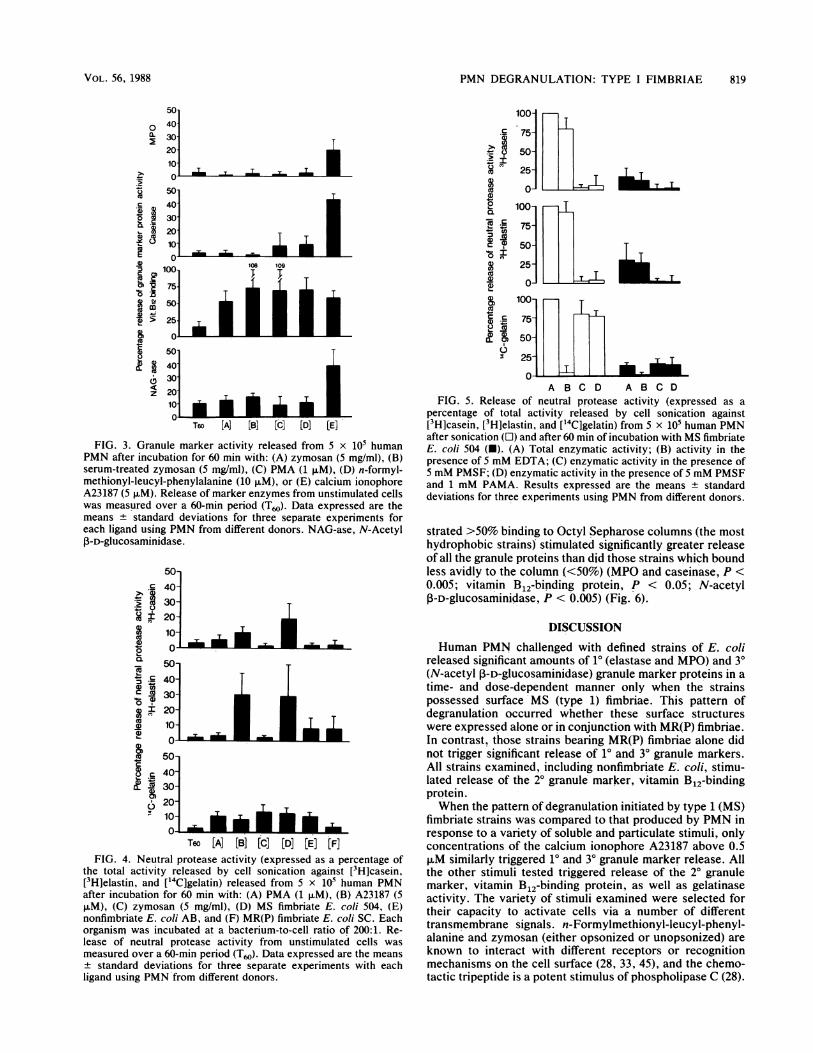
from PMN stimulated with E. coli 504 or from sonicatedPMN was totally inhibited by 5 mM PMSF, a serine proteaseinhibitor, but not by 5 mM EDTA, an inhibitor of metallo-proteases (Fig. 5). Conversely, [14C]gelatin-degrading activ-ity released by PMN was totally inhibited by 5 mM EDTAbut not by 5 mM PMSF. The inclusion of 1 mM PAMAtogether with 5 mM PMSF did not increase the amount ofcasein-, elastin-, or gelatin-degrading activity, indicating thata metalloprotease with activity against these substrates wasnot present in a latent form.The release of a serine protease active against [3H]elastin
by PMN stimulated with E. coli 504 was confirmed by usingTrasylol covalently linked to Sepharose beads. In threeexperiments, 95.5 ± 3% (mean ± standard deviation) of theactivity released from PMN stimulated by E. coli 504 wasbound to the Trasylol, of which 77 ± 12% was recovered byelution with 0.05 M sodium acetate, pH 5.0. After sonicationof control cells, 93 ± 3% of the elastase activity was boundby Trasylol, of which 97 ± 4% was recovered after elution atpH 5.0.
Bacterial hydrophobicity. E. coli strains which demon-
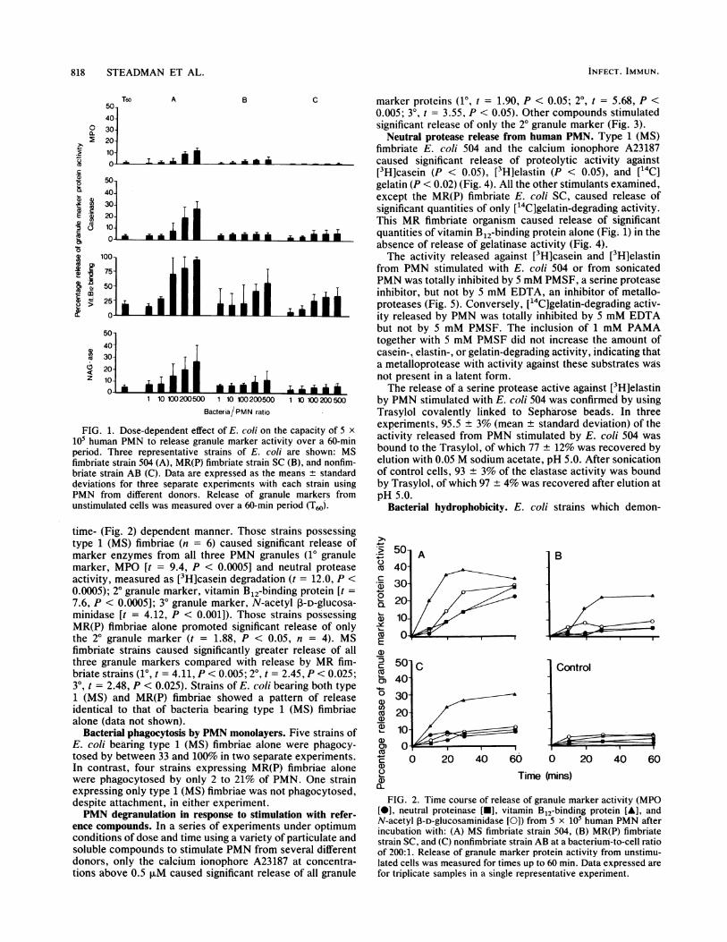
time- (Fig. 2) dependent manner. Those strains possessingtype 1 (MS) fimbriae (n = 6) caused significant release ofmarker enzymes from all three PMN granules (10 granulemarker, MPO [t = 9.4, P < 0.0005] and neutral proteaseactivity, measured as [3H]casein degradation (t = 12.0, P <0.0005); 20 granule marker, vitamin B12-binding protein [t =7.6, P < 0.0005]; 30 granule marker, N-acetyl P-D-glucosa-minidase [t = 4.12, P < 0.001]). Those strains possessingMR(P) fimbriae alone promoted significant release of onlythe 20 granule marker (t = 1.88, P < 0.05, n = 4). MSfimbriate strains caused significantly greater release of allthree granule markers compared with release by MR fim-briate strains (10, t = 4.11, P < 0.005; 20, t = 2.45, P < 0.025;309 t = 2.48, P < 0.025). Strains of E. coli bearing both type1 (MS) and MR(P) fimbriae showed a pattern of releaseidentical to that of bacteria bearing type 1 (MS) fimbriaealone (data not shown).
Bacterial phagocytosis by PMN monolayers. Five strains ofE. coli bearing type 1 (MS) fimbriae alone were phagocy-tosed by between 33 and 100% in two separate experiments.In contrast, four strains expressing MR(P) fimbriae alonewere phagocytosed by only 2 to 21% of PMN. One strainexpressing only type 1 (MS) fimbriae was not phagocytosed,despite attachment, in either experiment.PMN degranulation in response to stimulation with refer-
ence compounds. In a series of experiments under optimumconditions of dose and time using a variety of particulate andsoluble compounds to stimulate PMN from several differentdonors, only the calcium ionophore A23187 at concentra-tions above 0.5 FxM caused significant release of all granule
.; 5
cu 4C:.f 3
a) 1
cuE
a)
: 4
co
0)
0) ,
CU
a)
0)
u A
.010
I0
5O] c$0-
30-
20-
10-
a-0 20 40 60 0
Control
20 40 60
Time (mins)
FIG. 2. Time course of release of granule marker activity (MPO[-], neutral proteinase [-], vitamin B12-binding protein [A], andN-acetyl P-D-glucosaminidase [0]) from 5 x 10⁵ human PMN afterincubation with: (A) MS fimbriate strain 504, (B) MR(P) fimbriatestrain SC, and (C) nonfimbriate strain AB at a bacterium-to-cell ratioof 200:1. Release of granule marker protein activity from unstimu-lated cells was measured for times up to 60 min. Data expressed arefor triplicate samples in a single representative experiment.
0a-
c;._
E.:
X) 11
2 II
0:$
3
cCe
C
a)
.zz
-
INFECT. IMMUN.
:n%
r-
PMN DEGRANULATION: TYPE I FIMBRIAE 819
0a.
5040-3020-10 I I50
C 40
~2~0
100
50
400> 25-
0ii
ez
302010-
T6o [A] [B] [C] [D] [E]
FIG. 3. Granule marker activity released from 5 x 10⁵ humanPMN after incubation for 60 min with: (A) zymosan (5 mg/ml), (B)serum-treated zymosan (5 mg/ml), (C) PMA (1 1±M), (D) n-formyl-methionyl-leucyl-phenylalanine (10 ,uM), or (E) calcium ionophoreA23187 (5 FM). Release of marker enzymes from unstimulated cellswas measured over a 60-min period (T60). Data expressed are themeans standard deviations for three separate experiments foreach ligand using PMN from different donors. NAG-ase, N-AcetylP-D-glucosaminidase.
.-
2
CO
0.%
C co
en X
0
C
50-
40-30-20-10-
0 - - ML-50O40]30]20]101
50-
40-30-
20-10-~
T60 [A] [B] [C] [D] [E] [F]FIG. 4. Neutral protease activity (expressed as a percentage of
the total activity released by cell sonication against [3H]casein,[3H]elastin, and ['4C]gelatin) released from 5 x i05 human PMNafter incubation for 60 min with: (A) PMA (1 ,uM), (B) A23187 (5p.M), (C) zymosan (5 mg/ml), (D) MS fimbriate E. coli 504, (E)nonfimbriate E. coli AB, and (F) MR(P) fimbriate E. coli SC. Eachorganism was incubated at a bacterium-to-cell ratio of 200:1. Re-lease of neutral protease activity from unstimulated cells was
measured over a 60-min period (T60). Data expressed are the meansstandard deviations for three separate experiments with each
ligand using PMN'from different donors.
0
._.
0
t-_
2Xco
02
CC
a,)*
0
-6
100-
75]50-
25-
0-i
100-
75]50-
25-
O--
A B C D A B C DFIG. 5. Release of neutral protease activity (expressed as a
percentage of total activity released by cell sonication against[3H]casein, [3H]elastin, and [14C]gelatin) from 5 x 10⁵ human PMNafter sonication (OI) and after 60 min of incubation with MS fimbriateE. coli 504 (U). (A) Total enzymatic activity; (B) activity in thepresence of 5 mM EDTA; (C) enzymatic activity in the presence of5 mM PMSF; (D) enzymatic activity in the presence of 5 mM PMSFand 1 mM PAMA. Results expressed are the means ± standarddeviations for three experiments using PMN from different donors.
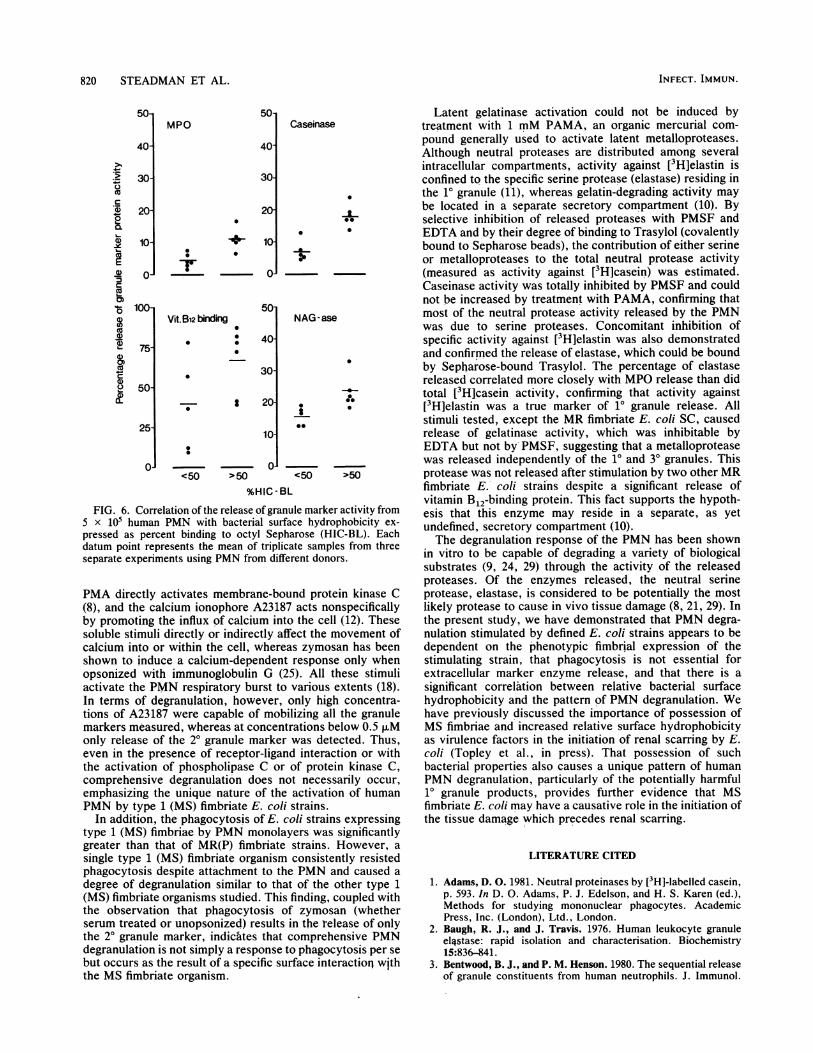
strated >50% binding to Octyl Sepharose columns (the mosthydrophobic strains) stimulated significantly greater releaseof all the granule proteins than did those strains which boundless avidly to the column (<50%) (MPO and caseinase, P <0.005; vitamin B 2-binding protein, P < 0.05; N-acetylP-D-glucosaminidase, P < 0.005) (Fig. 6).
DISCUSSION
Human PMN challenged with defined strains of E. colireleased significant amounts of 10 (elastase and MPO) and 3°(N-acetyl ,-D-glucosaminidase) granule marker proteins in atime- and dose-dependent manner only when the strainspossessed surface MS (type 1) fimbriae. This pattern ofdegranulation occurred whether these surface structureswere expressed alone or in conjunction with MR(P) fimbriae.In contrast, those strains bearing MR(P) fimbriae alone didnot trigger significant release of 10 and 30 granule markers.All strains examined, including nonfimbriate E. coli, stimu-lated release of the 20 granule marker, vitamin B12-bindingprotein.When the pattern of degranulation initiated by type 1 (MS)
fimbriate strains was compared to that produced by PMN inresponse to a variety of soluble and particulate stimuli, onlyconcentrations of the calcium ionophore A23187 above 0.5,uM similarly triggered 10 and 30 granule marker release. Allthe other stimuli tested triggered release of the 20 granulemarker, vitamin B12-binding protein, as well as gelatinaseactivity. The variety of stimuli examined were selected fortheir capacity to activate cells via a number of differenttransmembrane signals. n-Formylmethionyl-leucyl-phenyl-alanine and zymosan (either opsonized or unopsonized) areknown to interact with different receptors or recognitionmechanisms on the cell surface (28, 33, 45), and the chemo-tactic tripeptide is a potent stimulus of phospholipase C (28).
VOL. 56, 1988
-T _ T _ T
-Z.- L.
T
Ir T
.,.. lb lb lb 0160
820 STEADMAN ET AL.
50-
40
4-
a0..
a)
20)
ca
a
co
30*
201
10-
0-
100-
75-
50-
25
0-
5onMPO
40
30
0
0
Vit. B12 binding
* 0
20
10-
50-
40-
30-
8 20-
10-
<50
Caseinase
0
0@
-a-
NAG-ase
I
@0
01>50 <50
-0
>50
%HIC-BL
FIG. 6. Correlation of the release of granule marker activity from5 x 105 human PMN with bacterial surface hydrophobicity ex-
pressed as percent binding to octyl Sepharose (HIC-BL). Eachdatum point represents the mean of triplicate samples from threeseparate experiments using PMN from different donors.
PMA directly activates membrane-bound protein kinase C(8), and the calcium ionophore A23187 acts nonspecificallyby promoting the influx of calcium into the cell (12). Thesesoluble stimuli directly or indirectly affect the movement ofcalcium into or within the cell, whereas zymosan has beenshown to induce a calcium-dependent response only whenopsonized with immunoglobulin G (25). All these stimuliactivate the PMN respiratory burst to various extents (18).In terms of degranulation, however, only high concentra-tions of A23187 were capable of mobilizing all the granulemarkers measured, whereas at concentrations below 0.5 ,uMonly release of the 20 granule marker was detected. Thus,even in the presence of receptor-ligand interaction or withthe activation of phospholipase C or of protein kinase C,comprehensive degranulation does not necessarily occur,
emphasizing the unique nature of the activation of humanPMN by type 1 (MS) fimbriate E. coli strains.
In addition, the phagocytosis of E. coli strains expressingtype 1 (MS) fimbriae by PMN monolayers was significantlygreater than that of MR(P) fimbriate strains. However, a
single type 1 (MS) fimbriate organism consistently resistedphagocytosis despite attachment to the PMN and caused a
degree of degranulation similar to that of the other type 1(MS) fimbriate organisms studied. This finding, coupled withthe observation that phagocytosis of zymosan (whetherserum treated or unopsonized) results in the release of onlythe 20 granule marker, indicates that comprehensive PMNdegranulation is not simply a response to phagocytosis per se
but occurs as the result of a specific surface interactioq withthe MS fimbriate organism.
Latent gelatinase activation could not be induced bytreatment with 1 mM PAMA, an organic mercurial com-pound generally used to activate latent metalloproteases.Although neutral proteases are distributed among severalintracellular compartments, activity against [3H]elastin isconfined to the specific serine protease (elastase) residing inthe 10 granule (11), whereas gelatin-degrading activity maybe located in a separate secretory compartment (10). Byselective inhibition of released proteases with PMSF andEDTA and by their degree of binding to Trasylol (covalentlybound to Sepharose beads), the contribution of either serineor metalloproteases to the total neutral protease activity(measured as activity against [3H]casein) was estimated.Caseinase activity was totally inhibited by PMSF and couldnot be increased by treatment with PAMA, confirming thatmost of the neutral protease activity released by the PMNwas due to serine proteases. Concomitant inhibition ofspecific activity against [3H]elastin was also demonstratedand confirmed the release of elastase, which could be boundby Sepharose-bound Trasylol. The percentage of elastasereleased correlated more closely with MPO release than didtotal [3H]casein activity, confirming that activity against[3H]elastin was a true marker of 10 granule release. Allstimuli tested, except the MR fimbriate E. coli SC, causedrelease of gelatinase activity, which was inhibitable byEDTA but not by PMSF, suggesting that a metalloproteasewas released independently of the 10 and 30 granules. Thisprotease was not released after stimulation by two other MRfimbriate E. coli strains despite a significant release ofvitamin B 2-binding protein. This fact supports the hypoth-esis that this enzyme may reside in a separate, as yetundefined, secretory compartment (10).The degranulation response of the PMN has been shown
in vitro to be capable of degrading a variety of biologicalsubstrates (9, 24, 29) through the activity of the releasedproteases. Of the enzymes released, the neutral serineprotease, elastase, is considered to be potentially the mostlikely protease to cause in vivo tissue damage (8, 21, 29). Inthe present study, we have demonstrated that PMN degra-nulation stimulated by defined E. coli strains appears to bedependent on the phenotypic fimbrial expression of thestimulating strain, that phagocytosis is not essential forextracellular marker enzyme release, and that there is asignificant correlation between relative bacterial surfacehydrophobicity and the pattern of PMN degranulation. Wehave previously discussed the importance of possession ofMS fimbriae and increased relative surface hydrophobicityas virulence factors in the initiation of renal scarring by E.coli (Topley et al., in press). That possession of suchbacterial properties also causes a unique pattern of humanPMN degranulation, particularly of the potentially harmful10 granule products, provides further evidence that MSfimbriate E. coli may have a causative role in the initiation ofthe tissue damage which precedes renal scarring.
LITERATURE CITED
1. Adams, D. 0. 1981. Neutral proteinases by [3H]-labelled casein,p. 593. In D. 0. Adams, P. J. Edelson, and H. S. Karen (ed.),Methods for studying mononuclear phagocytes. AcademicPress, Inc. (London), Ltd., London.
2. Baugh, R. J., and J. Travis. 1976. Human leukocyte granuleellstase: rapid isolation and characterisation. Biochemistry15:836-841.
3. Bentwood, B. J., and P. M. Henson. 1980. The sequential releaseof granule constituents from human neutrophils. J. Immunol.
INFECT. IMMUN.
PMN DEGRANULATION: TYPE I FIMBRIAE 821
124:855-862.4. Blumenstock, E., and K. Jann. 1982. Adhesion of piliated
Escherichia coli strains to phagocytes: differences betweenbacteria with mannose-sensitive pili and those with mannose-resistant pili. Infect. Immun. 35:264-269.
5. Causten, T. E., and A. J. Barrett. 1979. A rapid and reproduc-ible assay for collagenase using [1-_4C] acetylated collagen.Anal. Biochem. 99:340-345.
6. Cavalieri, S. J., G. A. Bohach, and I. S. Snyder. 1984. Esche-richia coli a-hemolysin: characteristics and probable role inpathogenicity. Microbiol. Rev. 48:326-343.
7. Chick, S., M. J. Harber, R. Mackenzie, and A. W. Asscher.1981. Modified method for studying bacterial adhesion to iso-lated uroepithelial cells and uromucoid. Infect. Immun.34:256-261.
8. Costa-Casnellie, M. R., G. B. Segel, and M. A. Lichtman. 1985.Concanavalin A and phorbol ester cause opposite subcellularredistribution of protein kinase C. Biochem. Biophys. Res.Commun. 133:1139-1144.
9. Davies, M., A. J. Barrett, J. Travis, E. Sanders, and G. A. Coles.1978. The degradation of human glomerular basement mem-brane with purified proteinases. Clin. Sci. Mol. Med. 54:233-240.
10. Dewald, B., U. Bretz, and M. Baggiolini. 1982. Release ofgelatinase from a novel secretory compartment of human neu-trophils. J. Clin. Invest. 70:518-525.
11. Dewald, B., R. Rindler-Ludwig, U. Bretz, and M. Baggiolini.1975. Subcellular localisation and heterogeneity of neutral pro-teinases in neutrophilic polymorphonuclear leucocytes. J. Exp.Med. 141:102-119.
12. Estensen, R. D., M. E. Reusch, M. L. Epstein, and H. R. Hill.1976. Role of Ca2' and Mg2+ in some human neutrophilfunctions as indicated by ionophore A23187. Infect. Immun.13:146-151.
13. Fantone, J. C., and P. A. Ward. 1982. The role of oxygen-derived free radicals and metabolites in leukocyte-dependentinflammatory reactions. Am. J. Pathol. 107:397-418.
14. Freeman, B. A., and J. D. Crapo. 1982. Free radicals and tissueinjury. Lab. Invest. 47:412-426.
15. Glauser, M. P., S. M. Lyons, and A. I. Braude. 1978. Preventionof chronic experimental pyelonephritis by suppression of acutesuppuration. J. Clin. Invest. 61:403-407.
16. Gordon, L. I., S. D. Douglas, N. E. Kay, 0. Yamada, E. F.Osserman, and H. S. Jacob. 1979. Modulation of neutrophilfunction by lysosyme. Potential negative feedback system ofinflammation. J. Clin. Invest. 64:226-232.
17. Harber, M. J., R. K. Mackenzie, and A. W. Asscher. 1983. Arapid bioluminescence method for quantifying bacterial adher-ence to polystyrene. J. Gen. Microbiol. 129:621-632.
18. Harber, M. J., and N. Topley. 1986. Factors affecting themeasurement of chemiluminescence in stimulated human poly-morphonuclear leucocytes. J. Bioluminescence and Chemilumi-nescence 1:15-27.
19. Harber, M. J., N. Topley, D. E. Jenner, R. K. Mackenzie, R.Steadman, J. M. Knowiden, and A. W. Asscher. 1986. Virulencefactors of urinary pathogens in relation to kidney scarring, p.69-81. In A. W. Asscher and W. Brumfitt (ed.), Microbialdiseases in nephrology. John Wiley & Sons, Chichester, UnitedKingdom.
20. Hughes, C., R. Phillips, and A. P. Roberts. 1982. Serum resis-tance among Escherichia coli strains causing urinary tractinfection in relation to 0 type and the carriage of hemolysin,colicin, and antibiotic resistance determinants. Infect. Immun.35:270-275.
21. Janoff, A. 1978. Granulocyte elastase: role in arthritis and inpulmonary emphysema, p. 390-417. In K. Havemann and A.Janoff (ed.), Neutral proteases of human polymorphonuclearleukocytes. Urban & Schwarzenberg, Inc., Baltimore.
22. Kalienius, G., and R. Molby. 1979. Adhesion of Escherichia colito human periurethral cells correlated to mannose-resistantagglutination of human erythrocytes. FEMS Microbiol. Lett.5:295-299.
23. Keiser, H. D. 1980. The effects of lysosomal enzymes onextracellular substrates, p. 431-467. In G. Weismann (ed.), The
cell biology of inflammation. Elsevier/North-Holland PublishingCo., New York.
24. Lazarus, G. S., J. R. Daniels, J. Lian, and M. C. Buleigh. 1972.Role of granulocyte collagenase in collagen degradation. Am. J.Pathol. 69:565-578.
25. Lew, D. P., T. Andersson, J. Hed, F. Di Virgilio, T. Pozzan, and0. Stendahl. 1985. Ca2" dependent and independent phagocy-tosis in human neutrophils. Nature (London) 315:509-511.
26. Mantovani, B. 1975. Different roles of IgG and complementreceptors in phagocytosis by polymorphonuclear leukocytes. J.Immunol. 115:15-24.
27. O'Hanley, P., D. Low, I. Ramero, D. Lark, K. Vosti, S. Falkow,and G. Schoolnik. 1985. Gal-gal binding a-haemolysin pheno-types and genotypes associated with uropathogenic Escherichiacoli. New Engl. J. Med. 313:414-420.
28. Ohta, H., F. Okajima, and U. Machio. 1985. Inhibition byislet-activating protein of a chemotactic peptide-induced earlybreakdown of inositol phospholipids and Ca2" mobilisation inguinea pig neutrophils. J. Biol. Chem. 260:15771-15780.
29. Oronsky, A. L., and C. W. Buermann. 1978. Granulocyteneutral proteinases in arthritis, p. 361-371. In K. Havemann andA. Janoff (ed.), Neutral proteases of Human Polymorphonu-clear Leukocytes. Urban & Schwarzenberg,Inc., Baltimore.
30. 0rskov, I., F. 0rskov, B. Jann, and K. Jann. 1977. Serology,chemistry, and genetics of0 and K antigens of Escherichia coli.Bacteriol. Rev. 41:667-710.
31. Oseas, R., H. Young, R. L. Boehner, and L. A. Boxer. 1981.Lactoferrin: a promoter of polymorphonuclear cell adhesive-ness. Blood 57:939-945.
32. Ringel, E. W., N. A. Soter, and K. F. Austen. 1984. Localisationof histamine to the specific granule of the human neutrophil.Immunology 52:649-658.
33. Ross, D. G., J. A. Cain, and P. J. Lachmann. 1985. Membranecomplement receptor type three (CR3) has lectin-like propertiesanalogous to bovine conglutinin and functions as a receptor forzymosan and rabbit erythrocytes as well as a receptor for iC3b.J. Immunol. 134:3307-3315.
34. Slotki, I. N., and A. W. Asscher. 1982. Prevention of scarring inexperimental pyelonephritis in the rat by early antibiotic ther-apy. Nephron 30:262-268.
35. Smith, G. P., and T. J. Peters. 1982. The release of granulecomponents from human polymorphonuclear leukocytes in re-sponse to both phagocytic and chemical stimuli. Biochim.Biophys. Acta 719:304-308.
36. Smith, T. W., Jr., and J. A. Roberts. 1978. Chronic pyelone-phritis: an electron microscopic study in non-human primates.Invest. Urol. 16:148-153.
37. Sussman, M., S. N. Abraham, and S. H. Parry. 1982. Bacterialadhesion in the host-parasite relationship of urinary tract infec-tion, p. 103-112. In H. Schulte-Wisserman (ed)., Clinical bac-teriological and immunological aspects of urinary tract infec-tions in children. Georg Thieme Verlag, Stuttgart, FederalRepublic of Germany.
38. Svanborg-Eden, C., L.-M. Bjursten, R. Hull, S. Hull, K.-E.Magnusson, Z. Moldovano, and H. Leffler. 1984. Influence ofadhesins on the interaction of Escherichia coli with humanphagocytes. Infect. Immun. 44:672-680.
39. Svenson, S. B., G. Kallenins, R. Molby, H. Hultberg, and J.Whitberg. 1982. Rapid identification of p-fimbriated E. coli by areceptor specific particle agglutination test. Infection 10:209-214.
40. Takahashi, S., S. I. Seifter, and F. C. Young. 1973. A newradioactive assay for enzymes with elastolytic activity usingreduced tritiated elastin. The effect of sodium dodecyl sulphateon elastins. Biochim. Biophys. Acta 327:138-141.
41. Vaisanen, V., J. Elo, L. G. Tallgren, A. Sutonen, P. H. Makela,G. Svanborg-Eden, G. Kallenius, S. B. Svenson, H. Hultberg,and T. Korhonen. 1981. Mannose resistant haemagglutinationand P antigen recognition are characteristic of Escherichia colicausing primary pyelonephritis. Lancet ii:1366-1369.
42. Van Oss, C. J., and C. F. Gillman. 1972. Phagocytosis as asurface phenomenon. I. Contact angles and phagocytosis ofnon-opsonized bacteria. RES J. Reticuloendothel. Soc. 12:283-
VOL. 56, 1988
INFECT. IMMUN.
292.43. Weiss, S. J., and A. C. Regiani. 1984. Neutrophils degrade
subendothelial matrices in the presence of alpha-1-proteinaseinhibitor. J. Clin. Invest. 73:1297-1303.
44. Williams, J. D., T. H. Lee, R. A. Lewis, and K. F. Austen. 1985.Intracellular retention of the 5-lipoxygenase pathway product,leukotriene B4, by human neutrophils activated with unopso-nised zymosan. J. Immunol. 134:2624-2630.
45. Williams, J. D., N. Topley, H. M. Alobaidi, and M. J. Harber.1986. Activation of human polymorphonuclear leucocytes byparticulate zymosan is related to both its major carbohydratecomponents: glucan and mannan. Immunology 58:117-124.
46. Wright, D. G., D. A. Brabove, and J. I. Gallin. 1977. Thedifferential mobilisation of human neutrophil granules. Effectsof phorbol myristate acetate and ionophore A23187. Am. J.Pathol. 87:273-283.
822 STEADMAN ET AL.