Embed Size (px)
Citation preview
Chapter4
Shp2 controls zebrafish gastrulation cell movements via Fyn,
Yes and RhoA
Chris Jopling and Jeroen den Hertog*
Hubrecht LaboratoryNetherlands Institute for Developmental Biology
Uppsalalaan 83584 CT UtrechtThe Netherlands
* corresponding author:[email protected]: +31 30 2121800Fax: +31 30 2516464
Keywords: Shp2/ zebrafish/ gastrulation/ convergence and extension/ Fyn and Yes/ RhoA
Submitted

Shp2 controls zebrafish gastrulation
57
Abstract
Convergence and extension (CE) cell movements during gastrulation mediate extension of the anterior-posterior body axis of vertebrate embryos. Here we show that knock down of the protein-tyrosine phosphatase Shp2 in zebrafish embryos affected CE cell movements, but not cell specification. Shp2 acted upstream of the Src family kinases Fyn and Yes and the small GTPase RhoA. Our results show that Shp2 is required for normal CE cell movements via Fyn/Yes and RhoA in vertebrate gastrulation which may have implications for the role of Shp2
in human disease.
Chapter4
58
Introduction
Co-ordinated cell movements of the three germ layers, endoderm, mesoderm and ectoderm drive vertebrate gastrulation and shape the developing embryo (Warga et al 1990). One of the three main morphogenetic cell movements, ‘convergence and extension’, (CE) sees cells move towards the dorsal midline then intercalate with one another narrowing the medio/lateral axis while, at the same time, elongating the embryo (Keller et al 1992). Vertebrate CE is regulated by the non-canonical Wnt pathway similar to the planar cell polarity (PCP) pathway identified in Drosophila (Solnica-Krezel et al 2003, Veeman et al 2003). A number of zebrafish mutants have been identified that harbor mutations in genes involved in non-canonical Wnt signaling (Solnica-Krezel and Eaton, 2003; Veeman et al., 2003). Two of these, Silberblick and Pipetail, have mutations in wnt11 and wnt5 respectively (Hammerschmidt et al., 1996; Heisenberg et al., 1996, 2000; Killian et al., 2003). Both these mutations result in defective CE cell movements without affecting actual cell fate. Wnt5 and Wnt11 activate the same pathway through binding to the frizzled receptor, causing the translocation of Dishevelled to the plasma membrane. Dishevelled then recruits Daam1, RhoA and Rac to form a complex. In turn, RhoA and Rac are activated and proceed to transduce the signal to their respective downstream effectors (Habas et al., 2001, 2003).
Recently we have shown that the two src family kinases (SFKs), Fyn and Yes, also play a role in vertebrate CE cell movements (Jopling and den Hertog, 2005). Morpholino (MO) knockdown of both these genes together results in embryos which phenocopy Silberblick/Wnt11 and Pipetail/Wnt5 morphants both morphologically and
molecularly. Although Fyn and Yes act in a synergistic manner with Wnt5 and Wnt11 they do not function in a linear pathway, but instead, operate in parallel converging downstream on the small GTPase RhoA. Because Fyn and Yes do not act linearly with non-canonical Wnt signaling this begged the question ‘what is upstream of Fyn and Yes?’ The protein tyrosine phosphatase (PTP), Shp2, is a well characterised regulator of SFK activity and as such a likely upstream candidate. Shp2 regulates the phosphorylation of the adaptor protein Cbp/PAG, which recruits Csk, thereby controling phosphorylation of the inhibitory phosphorylation sites in the C-terminus of SFKs (Zhang et al.,2004). In the absence of Shp2, SFK activity is reduced due to hyperphosphorylation of the inhibitory phosphorylation sites (Zhang et al., 2004). Shp2 has an important role in early development. For instance, dominant negative Shp2 induces tail truncations in Xenopus laevis, (Tang et al.,1995), whereas overexpression of Shp2 promotes animal cap elongation (O’Reilly et al., 2000). Gene targeting of Shp2 in the mouse was aimed at deletion of exon 2 or exon 3 and inadvertantly led to truncation of the Shp2 protein instead of deletion. Nevertheless, these Shp2-targeted mice are embryonic lethal around embryonic day 8.5-10.5 with a range of defects consistent with gastrulation abnormalities (Arrandale te al., 1996; Saxton et al., 1997). Chimeric mice derived from Shp2 ex3-/- embryonic stem cells display defective morphogenetic cell movements during gastrulation (Saxton and Pawson, 1999). Interestingly, bona fide Shp2 null mouse embryos die peri-implantation and Shp2 is required for trophoblast stem cell survival (Yang et al., 2006). In humans, Shp2 is responsible for a range of diseases, including Noonan syndrome (Tartaglia et al., 2001) and LEOPARD syndrome (Digilio et al., 2002) and somatic mutations in PTPN11
Shp2 controls zebrafish gastrulation
59
contribute to leukemogenesis (Tartaglia et al., 2006). However, because Shp2 is involved in multiple pathways it has been difficult to elucidate the cell biological functions of Shp2 during early embryogenesis. Here we show that MO-mediated knockdown of Shp2 in zebrafish embryos resulted in defective CE movements during gastrulation. Despite the wide variety of pathways that are known to involve Shp2, cell fate remains unaffected upon Shp2 knockdown. We demonstrate that the Shp2 knockdown defects involve signaling through downstream components Fyn and Yes converging with non-canonical Wnt signaling at or upstream of the small GTPase RhoA. Our results are consistent with a role for Shp2 in vertebrate gastrulation cell movements.
ResultsWe identified zebrafish Shp2 (EST
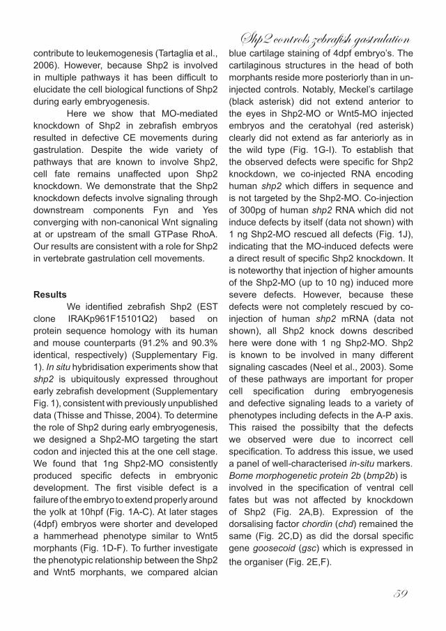
clone IRAKp961F15101Q2) based on protein sequence homology with its human and mouse counterparts (91.2% and 90.3% identical, respectively) (Supplementary Fig. 1). In situ hybridisation experiments show that shp2 is ubiquitously expressed throughout early zebrafish development (Supplementary Fig. 1), consistent with previously unpublished data (Thisse and Thisse, 2004). To determine the role of Shp2 during early embryogenesis, we designed a Shp2-MO targeting the start codon and injected this at the one cell stage. We found that 1ng Shp2-MO consistently produced specific defects in embryonic development. The first visible defect is a failure of the embryo to extend properly around the yolk at 10hpf (Fig. 1A-C). At later stages (4dpf) embryos were shorter and developed a hammerhead phenotype similar to Wnt5 morphants (Fig. 1D-F). To further investigate the phenotypic relationship between the Shp2 and Wnt5 morphants, we compared alcian
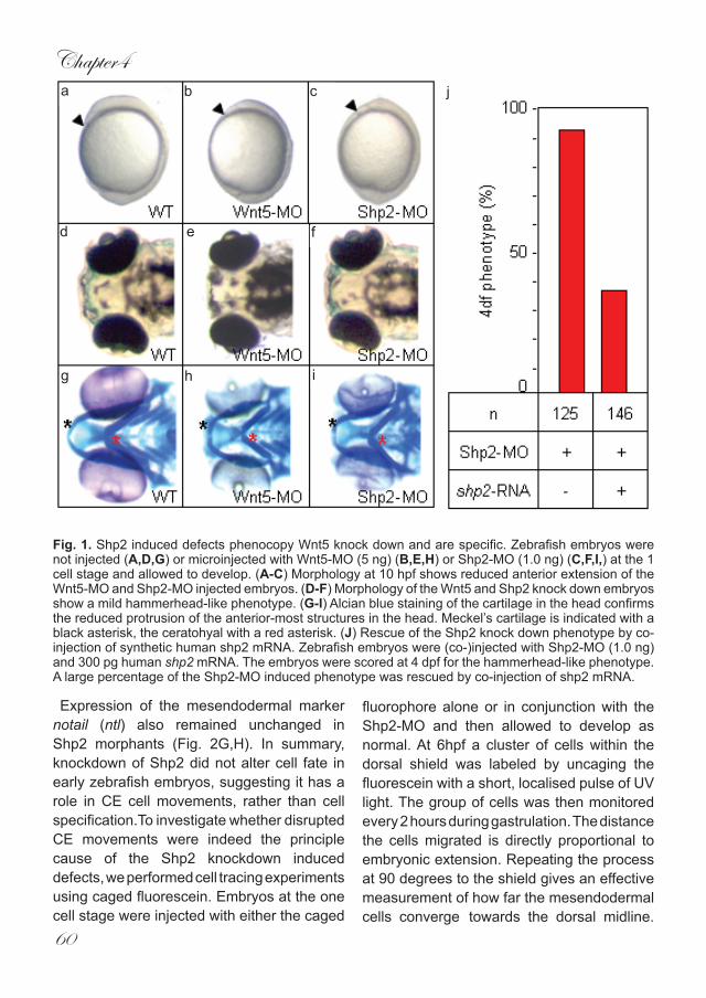
blue cartilage staining of 4dpf embryo’s. The cartilaginous structures in the head of both morphants reside more posteriorly than in un-injected controls. Notably, Meckel’s cartilage (black asterisk) did not extend anterior to the eyes in Shp2-MO or Wnt5-MO injected embryos and the ceratohyal (red asterisk) clearly did not extend as far anteriorly as in the wild type (Fig. 1G-I). To establish that the observed defects were specific for Shp2 knockdown, we co-injected RNA encoding human shp2 which differs in sequence and is not targeted by the Shp2-MO. Co-injection of 300pg of human shp2 RNA which did not induce defects by itself (data not shown) with 1 ng Shp2-MO rescued all defects (Fig. 1J), indicating that the MO-induced defects were a direct result of specific Shp2 knockdown. It is noteworthy that injection of higher amounts of the Shp2-MO (up to 10 ng) induced more severe defects. However, because these defects were not completely rescued by co-injection of human shp2 mRNA (data not shown), all Shp2 knock downs described here were done with 1 ng Shp2-MO. Shp2 is known to be involved in many different signaling cascades (Neel et al., 2003). Some of these pathways are important for proper cell specification during embryogenesis and defective signaling leads to a variety of phenotypes including defects in the A-P axis. This raised the possibilty that the defects we observed were due to incorrect cell specification. To address this issue, we used a panel of well-characterised in-situ markers.Bome morphogenetic protein 2b (bmp2b) isinvolved in the specification of ventral cell fates but was not affected by knockdown of Shp2 (Fig. 2A,B). Expression of the dorsalising factor chordin (chd) remained the same (Fig. 2C,D) as did the dorsal specific gene goosecoid (gsc) which is expressed in the organiser (Fig. 2E,F).
Chapter4
60
Fig. 1. Shp2 induced defects phenocopy Wnt5 knock down and are specific. Zebrafish embryos were not injected (A,D,G) or microinjected with Wnt5-MO (5 ng) (B,E,H) or Shp2-MO (1.0 ng) (C,F,I,) at the 1 cell stage and allowed to develop. (A-C) Morphology at 10 hpf shows reduced anterior extension of the Wnt5-MO and Shp2-MO injected embryos. (D-F) Morphology of the Wnt5 and Shp2 knock down embryos show a mild hammerhead-like phenotype. (G-I) Alcian blue staining of the cartilage in the head confirms the reduced protrusion of the anterior-most structures in the head. Meckel’s cartilage is indicated with a black asterisk, the ceratohyal with a red asterisk. (J) Rescue of the Shp2 knock down phenotype by co-injection of synthetic human shp2 mRNA. Zebrafish embryos were (co-)injected with Shp2-MO (1.0 ng) and 300 pg human shp2 mRNA. The embryos were scored at 4 dpf for the hammerhead-like phenotype. A large percentage of the Shp2-MO induced phenotype was rescued by co-injection of shp2 mRNA.
Expression of the mesendodermal marker notail (ntl) also remained unchanged in Shp2 morphants (Fig. 2G,H). In summary, knockdown of Shp2 did not alter cell fate in early zebrafish embryos, suggesting it has a role in CE cell movements, rather than cell specification.To investigate whether disrupted CE movements were indeed the principle cause of the Shp2 knockdown induced defects, we performed cell tracing experiments using caged fluorescein. Embryos at the one cell stage were injected with either the caged
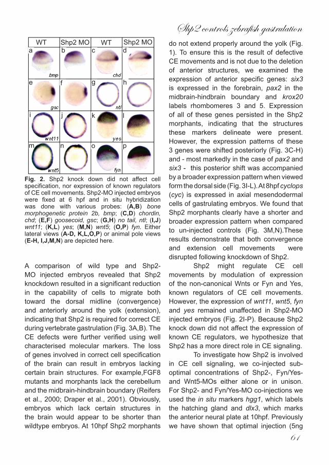
fluorophore alone or in conjunction with the Shp2-MO and then allowed to develop as normal. At 6hpf a cluster of cells within the dorsal shield was labeled by uncaging the fluorescein with a short, localised pulse of UV light. The group of cells was then monitored every 2 hours during gastrulation. The distance the cells migrated is directly proportional to embryonic extension. Repeating the process at 90 degrees to the shield gives an effective measurement of how far the mesendodermal cells converge towards the dorsal midline.
a b c
d e f
g h i
j
Shp2 controls zebrafish gastrulation
61
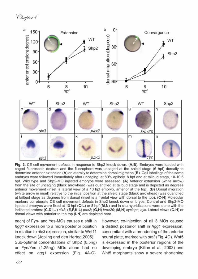
A comparison of wild type and Shp2-MO injected embryos revealed that Shp2 knockdown resulted in a significant reduction in the capability of cells to migrate both toward the dorsal midline (convergence) and anteriorly around the yolk (extension), indicating that Shp2 is required for correct CE during vertebrate gastrulation (Fig. 3A,B). The CE defects were further verified using well characterised molecular markers. The loss of genes involved in correct cell specification of the brain can result in embryos lacking certain brain structures. For example,FGF8 mutants and morphants lack the cerebellum and the midbrain-hindbrain boundary (Reifers et al., 2000; Draper et al., 2001). Obviously, embryos which lack certain structures in the brain would appear to be shorter than wildtype embryos. At 10hpf Shp2 morphants
do not extend properly around the yolk (Fig. 1). To ensure this is the result of defective CE movements and is not due to the deletion of anterior structures, we examined the expression of anterior specific genes: six3 is expressed in the forebrain, pax2 in the midbrain-hindbrain boundary and krox20 labels rhombomeres 3 and 5. Expression of all of these genes persisted in the Shp2 morphants, indicating that the structures these markers delineate were present. However, the expression patterns of these 3 genes were shifted posteriorly (Fig. 3C-H) and - most markedly in the case of pax2 and six3 - this posterior shift was accompanied by a broader expression pattern when viewed form the dorsal side (Fig. 3I-L). At 8hpf cyclops (cyc) is expressed in axial mesendodermal cells of gastrulating embryos. We found that Shp2 morphants clearly have a shorter and broader expression pattern when compared to un-injected controls (Fig. 3M,N).These results demonstrate that both convergence and extension cell movements were disrupted following knockdown of Shp2.
Shp2 might regulate CE cell movements by modulation of expression of the non-canonical Wnts or Fyn and Yes, known regulators of CE cell movements. However, the expression of wnt11, wnt5, fyn and yes remained unaffected in Shp2-MO injected embryos (Fig. 2I-P). Because Shp2 knock down did not affect the expression of known CE regulators, we hypothesize that Shp2 has a more direct role in CE signaling. To investigate how Shp2 is involved in CE cell signaling, we co-injected sub-optimal concentrations of Shp2-, Fyn/Yes- and Wnt5-MOs either alone or in unison. For Shp2- and Fyn/Yes-MO co-injections we used the in situ markers hgg1, which labels the hatching gland and dlx3, which marks the anterior neural plate at 10hpf. Previously we have shown that optimal injection (5ng
WT WT Shp2 MOShp2 MOa b c d
e f g h
i j k l
m n o p
Fig. 2. Shp2 knock down did not affect cell specification, nor expression of known regulators of CE cell movements. Shp2-MO injected embryos were fixed at 6 hpf and in situ hybridization was done with various probes: (A,B) bone morphogenetic protein 2b, bmp; (C,D) chordin, chd; (E,F) goosecoid, gsc; (G,H) no tail, ntl; (I,J) wnt11; (K,L) yes; (M,N) wnt5; (O,P) fyn. Either lateral views (A-D, K,L,O,P) or animal pole views (E-H, I,J,M,N) are depicted here.
Chapter4
62
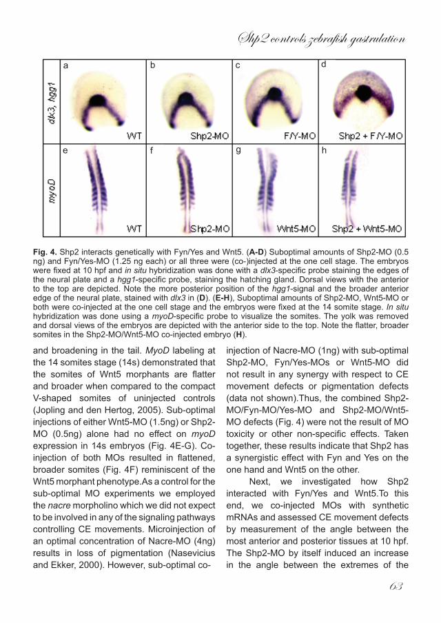
each) of Fyn- and Yes-MOs causes a shift in hgg1 expression to a more posterior position in relation to dlx3 expression, similar to Wnt11 knock down (Jopling and den Hertog,2005). Sub-optimal concentrations of Shp2 (0.5ng) or Fyn/Yes (1.25ng) MOs alone had no effect on hgg1 expession (Fig. 4A-C).
However, co-injection of all 3 MOs caused a distinct posterior shift in hgg1 expression, concomitant with a broadening of the anterior neural plate, marked with dlx3 (Fig. 4D). Wnt5 is expressed in the posterior regions of the developing embryo (Kilian et al., 2003) and Wnt5 morphants show a severe shortening
a bExtension Convergence
WT WT
Shp2Shp2
hpfhpf
WT WT WTShp2 Shp2 Shp2c d e f g h
i j k l m n
Fig. 3. CE cell movement defects in response to Shp2 knock down. (A,B). Embryos were loaded with caged fluorescein dextran and the fluorophore was uncaged at the shield stage (6 hpf) dorsally to determine anterior extension (A) or laterally to determine dorsal migration (B). Cell labelings of the same embryos were followed immediately after uncaging, at 80% epiboly, 8 hpf and at tailbud stage, 10-10.5 hpf. Wild type and Shp2-MO injected embryos were assessed. (A) Anterior extension (white arrow) from the site of uncaging (black arrowhead) was quantified at tailbud stage and is depicted as degrees anterior movement (inset is lateral view of a 10 hpf embryo, anterior at the top). (B) Dorsal migration (white arrow in inset) relative to the initial position at the shield stage (black arrowhead) was quantified at tailbud stage as degrees from dorsal (inset is a frontal view with dorsal to the top). (C-N) Molecular markers corroborate CE cell movement defects in Shp2 knock down embryos. Control and Shp2-MO injected embryos were fixed at 10 hpf (C-L) or 8 hpf (M,N) and in situ hybridizations were done with the indicated probes: (C,D,I,J) six3; (E,F,K,L) pax2; (G,H) krox20; (M,N) cyclops, cyc. Lateral views (C-H) or dorsal views with anterior to the top (I-N) are depicted here.
Shp2 controls zebrafish gastrulation
63
and broadening in the tail. MyoD labeling at the 14 somites stage (14s) demonstrated that the somites of Wnt5 morphants are flatter and broader when compared to the compact V-shaped somites of uninjected controls (Jopling and den Hertog, 2005). Sub-optimal injections of either Wnt5-MO (1.5ng) or Shp2-MO (0.5ng) alone had no effect on myoD expression in 14s embryos (Fig. 4E-G). Co-injection of both MOs resulted in flattened, broader somites (Fig. 4F) reminiscent of the Wnt5 morphant phenotype.As a control for the sub-optimal MO experiments we employed the nacre morpholino which we did not expect to be involved in any of the signaling pathways controlling CE movements. Microinjection of an optimal concentration of Nacre-MO (4ng) results in loss of pigmentation (Nasevicius and Ekker, 2000). However, sub-optimal co-
injection of Nacre-MO (1ng) with sub-optimal Shp2-MO, Fyn/Yes-MOs or Wnt5-MO did not result in any synergy with respect to CE movement defects or pigmentation defects (data not shown).Thus, the combined Shp2-MO/Fyn-MO/Yes-MO and Shp2-MO/Wnt5-MO defects (Fig. 4) were not the result of MO toxicity or other non-specific effects. Taken together, these results indicate that Shp2 has a synergistic effect with Fyn and Yes on the one hand and Wnt5 on the other.
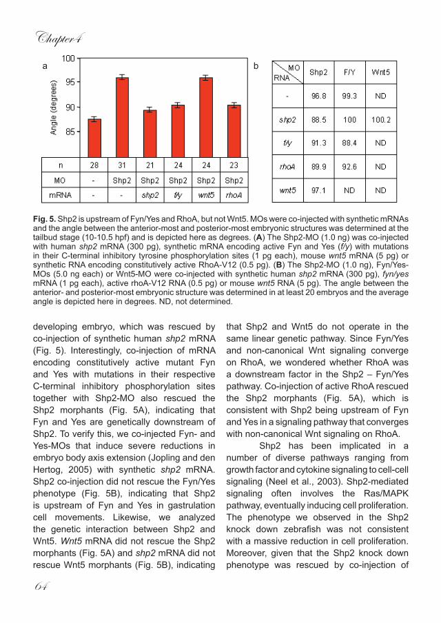
Next, we investigated how Shp2 interacted with Fyn/Yes and Wnt5.To this end, we co-injected MOs with synthetic mRNAs and assessed CE movement defects by measurement of the angle between the most anterior and posterior tissues at 10 hpf. The Shp2-MO by itself induced an increase in the angle between the extremes of the
a b c d
e f g h
Fig. 4. Shp2 interacts genetically with Fyn/Yes and Wnt5. (A-D) Suboptimal amounts of Shp2-MO (0.5 ng) and Fyn/Yes-MO (1.25 ng each) or all three were (co-)injected at the one cell stage. The embryos were fixed at 10 hpf and in situ hybridization was done with a dlx3-specific probe staining the edges of the neural plate and a hgg1-specific probe, staining the hatching gland. Dorsal views with the anterior to the top are depicted. Note the more posterior position of the hgg1-signal and the broader anterior edge of the neural plate, stained with dlx3 in (D). (E-H), Suboptimal amounts of Shp2-MO, Wnt5-MO or both were co-injected at the one cell stage and the embryos were fixed at the 14 somite stage. In situ hybridization was done using a myoD-specific probe to visualize the somites. The yolk was removed and dorsal views of the embryos are depicted with the anterior side to the top. Note the flatter, broader somites in the Shp2-MO/Wnt5-MO co-injected embryo (H).
Chapter4
64
developing embryo, which was rescued by co-injection of synthetic human shp2 mRNA (Fig. 5). Interestingly, co-injection of mRNA encoding constitutively active mutant Fyn and Yes with mutations in their respective C-terminal inhibitory phosphorylation sites together with Shp2-MO also rescued the Shp2 morphants (Fig. 5A), indicating that Fyn and Yes are genetically downstream of Shp2. To verify this, we co-injected Fyn- and Yes-MOs that induce severe reductions in embryo body axis extension (Jopling and den Hertog, 2005) with synthetic shp2 mRNA. Shp2 co-injection did not rescue the Fyn/Yes phenotype (Fig. 5B), indicating that Shp2 is upstream of Fyn and Yes in gastrulation cell movements. Likewise, we analyzed the genetic interaction between Shp2 and Wnt5. Wnt5 mRNA did not rescue the Shp2 morphants (Fig. 5A) and shp2 mRNA did not rescue Wnt5 morphants (Fig. 5B), indicating
that Shp2 and Wnt5 do not operate in the same linear genetic pathway. Since Fyn/Yes and non-canonical Wnt signaling converge on RhoA, we wondered whether RhoA was a downstream factor in the Shp2 – Fyn/Yes pathway. Co-injection of active RhoA rescued the Shp2 morphants (Fig. 5A), which is consistent with Shp2 being upstream of Fyn and Yes in a signaling pathway that converges with non-canonical Wnt signaling on RhoA. Shp2 has been implicated in a number of diverse pathways ranging from growth factor and cytokine signaling to cell-cell signaling (Neel et al., 2003). Shp2-mediated signaling often involves the Ras/MAPK pathway, eventually inducing cell proliferation. The phenotype we observed in the Shp2 knock down zebrafish was not consistent with a massive reduction in cell proliferation. Moreover, given that the Shp2 knock down phenotype was rescued by co-injection of
ba
Fig. 5. Shp2 is upstream of Fyn/Yes and RhoA, but not Wnt5. MOs were co-injected with synthetic mRNAs and the angle between the anterior-most and posterior-most embryonic structures was determined at the tailbud stage (10-10.5 hpf) and is depicted here as degrees. (A) The Shp2-MO (1.0 ng) was co-injected with human shp2 mRNA (300 pg), synthetic mRNA encoding active Fyn and Yes (f/y) with mutations in their C-terminal inhibitory tyrosine phosphorylation sites (1 pg each), mouse wnt5 mRNA (5 pg) or synthetic RNA encoding constitutively active RhoA-V12 (0.5 pg). (B) The Shp2-MO (1.0 ng), Fyn/Yes-MOs (5.0 ng each) or Wnt5-MO were co-injected with synthetic human shp2 mRNA (300 pg), fyn/yes mRNA (1 pg each), active rhoA-V12 RNA (0.5 pg) or mouse wnt5 RNA (5 pg). The angle between the anterior- and posterior-most embryonic structure was determined in at least 20 embryos and the average angle is depicted here in degrees. ND, not determined.
Shp2 controls zebrafish gastrulation
65
active RhoA (Fig. 5), the Ras/MAPK signaling pathway appeared not to be involved. Deletion of Shp2 in other species also does not appear to induce massive defects in cell proliferation during early development. For instance, a Drosophila mutant with a mutation in the Shp2 homolog, corckscrew (csw) displays multiple defects that are not directly linked to defects in cell proliferation. (Perkins et al., 1992). In Xenopus, dominant negative Shp2 leads to tail truncations due to defective gastrulation (Tang et al., 1995). Interestingly, active Shp2 induces elongation of animal cap explants which is subsequently blocked by co-expression of dominant negative RhoA (O’Reilly et al., 2000), suggesting a similar signaling pathway as we observed in early zebrafish embryos. Homozygous mouse embryos with a targeted Shp2 exon 2 or exon 3 express a truncated form of Shp2 and die in utero around day 8.5-10.5 (Arrandale et al., 1996; Saxton et al., 1997). The root cause of this lethality remains unknown. Chimeric embryos generated with homozygous Shp2 exon 3 mutant ES cells display gastrulation defects, consistent with defective morphogenetic cell movements as a result of Shp2 ablation (Saxton and Pawson, 1999). Recently, Yang et al. (2006) generated bona fide Shp2 null mouse embryos by replacement of Shp2 exon 2 with the β-galactosidase gene with a strong splice acceptor site to prevent splicing from exon 1 to exon 3. Heterozygous mice show reduced Shp2 protein expression and no truncated protein. Homozygous Shp2 null embryos died in utero around implantation, i.e. prior to gastrulation, demonstrating that Shp2 is essential for life very early in development. In fact, conditional knock outs showed that Shp2 is essential for trophoblast stem cell survival, which explains early lethality of Shp2 null mouse embryos (Yang et al., 2006). Zebrafish Shp2 morphants survived and the first defects we
observed were around gastrulation. Maternal Shp2 expression in zebrafish (Supplementary Fig. 1B) may rescue pre-gastrulation lethality. Moreover, MO-mediated knock down is not complete and low levels of remaining Shp2 expression may be sufficient for zebrafish survival.
We demonstrate here that Shp2 has an important role in zebrafish development, which is consistent with previous reports documenting the role of Shp2 in other species. Interstingly, Shp2 dysfunction has also been linked to several different human diseases. Germline mutations in PTPN11, the gene that encodes Shp2, causes the well-characterized Noonan and LEOPARD syndromes, whereas somatic mutations in PTPN11 contribute to leukemogenesis (Tartaglia et al., 2001; Digilio et al., 2002; Tartaglia et al., 2006). Noonan and LEOPARD syndromes are relatively common syndromes with partially overlapping clinical manifestations, including facial anomalies, distinct congenital heart defects, pectus deformities, hearing loss and growth retardation and distinct pigmentary changes (Gorlin et al., 1971). Surprisingly, the mutations that were found in Shp2 from Noonan patients affect Shp2 regulation, leading to constitutive activation of Shp2 catalytic activity (Keilhack et al., 2005), whereas the LEOPARD Shp2 mutants have greatly reduced catalytic activities (Kontaridis et al., 2006). How mutations in Shp2 with opposing effects on catalytic activity induce overlapping syndromes remains to be determined. Yet some of the symptoms of Noonan and LEOPARD patients are consistent with cell migration defects early in development and it will be interesting to see if defects in directed cell movement are at the basis of these syndromes.
Here, we show that Shp2 acts upstream of Fyn/Yes by co-injection of low amounts of Shp2- and Fyn/Yes-MOs that act
Chapter4
66
synergistically, showing there is a genetic interaction. Importantly, co-injection of synthetic RNAs encoding active Fyn and Yes rescued the Shp2-MO induced phenotype, demonstrating that Shp2 acts upstream of Fyn and Yes in gastrulation cell movements. Shp2 indirectly activates SFKs through dephosphorylation of Cbp/PAG (Zhang et al., 2004) and it is likely that this pathway is involved in gastrulation cell movements, although we cannot exclude that Shp2 affects Fyn and Yes activity in a different manner. Additional PTPs may be involved in regulation of Fyn and Yes during zebrafish gastrulation as well. We and others have identified receptor PTPα as a direct activator of Src (Zheng et al., 1992; den Hertog et al., 1993). However, whereas knock down of RPTPα induced pleiotropic effects, defects in CE cell movements during gastrulation were not observed (van der Sar et al., 2002; CJ and JdH, unpublished observation).
In conclusion, we demonstrate here that Shp2, an indirect activator of SFKs acts upstream of Fyn and Yes in gastrulation cell movements. Active RhoA rescued the Shp2 knock down phenotype, consistent with Shp2 being upstream of Fyn and Yes, which in turn signal through RhoA in a signaling pathway parallel to non-canonical Wnt signaling.
Shp2 controls zebrafish gastrulation
67
Materials and Methods
Zebrafish and in situ hybridizationZebrafish were kept and the embryos were staged as described before (Westerfield, 1995). In situ hybridizations were done essentially as described (Thisse et al., 1993), using probes specific for dlx3, myoD, wnt5 , bmp2b, chd, cyc, ntl, gsc, six3, pax2 and krox20 (generous gifts from various members of the zebrafish community) and hgg1, fyn, yes, wnt11 and shp2 (RZPD ID’s: IMAGp998O098963Q, U C D M p 6 1 1 J 0 3 2 1 Q 1 1 4 , MPMGp609A1681Q8, MPMGp637F0720Q2 and IRAKp961F15101Q2, respectively from www.rzpd.de, Berlin, Germany).
Morpholinos, RNAs and injectionsAntisense MOs were designed to include the start ATG of the respective cDNAs and ordered from GeneTools (Philomath, OR, USA): Shp2, 5’-GGTGGAACCACCTTCGGGATGTC AT, The Fyn and Yes MO’s were described before (Jopling and den Hertog, 2005). The Wnt5-MO was described before (Lele et al., 2001). 5’ capped sense RNAs were synthesized using constructs encoding mutant Fyn and Yes as previously described (Jopling and den Hertog, 2005).Wnt5 (gift of Andy McMahon) and active RhoAV12 (gift of Boudewijn Burgering) and the mMessage mMachine kit (Ambion, Austin, TX, USA). Ranges of MO (0.1 – 10 ng) were injected into embryos of the AB strain at the 1 cell stage and phenotypes were assessed at the indicated stages.
Cell tracingEmbryos were (co-)injected at the one cell stage with 0.25% 4,5-dimethoxy-2-nitrobenzyl (DMNB)-caged fluorescein dextran (10,000 MW; Molecular Probes, Leiden, the Netherlands). Uncaging was done as described (Bakkers et al., 2004) at shield
stage (6 hpf) using an Axioplan microscope, equipped with a UV light source, adjustable pinhole and 40X objective. Pictures were taken immediately following uncaging, at 80% epiboly (8hpf) and tailbud stage (10 hpf). The angles for dorsal convergence and anterior extension were determined using NIH imaging software.
AcknowledgementsThis work was supported in part by an EU Research Training Network Grant (HPRN-CT-2000-00085)
Chapter4
68
ReferencesArrandale, J.M., Gore-Willse, A., Rocks,
S., Ren, J.M., Zhu, J., Davis, A., Livingston, J.N., and Rabin, D.U. 1996. Insulin signaling in mice expressing reduced levels of Syp. J. Biol. Chem. 271: 21353-21358.
Bakkers, J., Kramer, C., Pothof, J., Quaedvlieg, N.E., Spaink, H.P., and Hammerschmidt, M. (2004) Has2 is required upstream of Rac1 to govern dorsal migration of lateral cells during zebrafish gastrulation. Development 131: 525-537.
den Hertog, J., Pals, C.E., Peppelenbosch, M.P., Tertoolen, L.G., de Laat, S.W., and Kruijer, W. 1993. Receptor protein tyrosine phosphatase alpha activates pp60c-src and is involved in neuronal differentiation. EMBO J. 12: 3789-3798.
Digilio, M.C., Conti, E., Sarkozy, A., Mingarelli, R., Dottorini, T., Marino, B., Pizzuti, A., and Dallapiccola, B. 2002. Grouping of multiple-lentigines/LEOPARD and Noonan syndromes on the PTPN11 gene. Am. J. Hum. Genet. 71: 389-394.
Draper, B.W., Morcos, P.A., Kimmel, C.B. 2001. Inhibition of zebrafish fgf8 pre-mRNA splicing with morpholino oligos: a quantifiable method for gene knockdown. Genesis 30: 154-156.
Gorlin, R.J., Anderson, R.C., Moller, J.H. 1971. The Leopard (multiple lentigines) syndrome revisited. Birth Defects Orig. Artic. Ser. 07: 110-115.
Habas, R., Kato, Y. and He, X. 2001. Wnt/Frizzled activation of Rho regulates vertebrate gastrulation and requires a novel Formin homology protein Daam1. Cell 107: 843-854.
Habas, R., Dawid, I.B. and He, X. 2003. Coactivation of Rac and Rho by
Wnt/Frizzled signaling is required for vertebrate gastrulation. Genes Dev. 17: 295-309.
Hammerschmidt, M., Pelegri, F., Mullins, M.C., Kane, D.A., Brand, M., van Eeden, F.J., Furutani-Seiki, M., Granato, M., Haffter, P., Heisenberg, C.P., Jiang, Y.J., Kelsh, R.N., Odenthal, J., Warga, R.M., and Nusslein-Volhard, C. 1996. Mutations affecting morphogenesis during gastrulation and tail formation in the zebrafish, Danio rerio. Development 123: 143-151.
Heisenberg, C.P., Brand, M., Jiang, Y.J., Warga, R.M., Beuchle, D., van Eeden, F.J., Furutani-Seiki, M., Granato, M., Haffter, P., Hammerschmidt, M., Kane, D.A., Kelsh, R.N., Mullins, M.C., Odenthal, J. and Nusslein-Volhard, C. 1996. Genes involved in forebrain development in the zebrafish, Danio rerio. Development 123: 191-203.
Heisenberg, C.P., Tada, M., Rauch, G.J., Saude, L., Concha, M.L., Geisler, R., Stemple, D.L., Smith, J.C. and Wilson, S.W. 2000. Silberblick/Wnt11 mediates convergent extension movements during zebrafish gastrulation. Nature 405: 76-81.
Jopling, C, and den Hertog, J. 2005. Fyn/Yes and non-canonical Wnt signalling converge on RhoA in vertebrate gastrulation cell movements. EMBO Rep. 5: 426-431.
Keilhack, H., David, F.S., McGregor, M., Cantley, L.C., and Neel, B.G. 2005. Diverse biochemical properties of Shp2 mutants. Implications for disease phenotypes. J. Biol. Chem. 280: 30984-30993.
Keller, R., Shih, J., and Domingo, C. 1992. The patterning and functioning of protrusive activity during convergence and extension of the Xenopus
Shp2 controls zebrafish gastrulation
69
organiser. Development Suppl. 81-91.
Kilian, B., Mansukoski, H., Barbosa, F.C., Ulrich, F., Tada, M., and Heisenberg, C.P. 2003. The role of Ppt/Wnt5 in regulating cell shape and movement during zebrafish gastrulation. Mech Dev. 120: 467-476.
Kontaridis, M.I., Swanson, K.D., David, F.S., Barford, D., and Neel, B.G. 2006. PTPN11 (Shp2) mutations in LEOPARD syndrome have dominant negative, not activating, effects. J. Biol. Chem. 281: 6785-6792.
Nasevicius, A. and Ekker, S.C. 2000. Effective targeted gene ‘knockdown’ in zebrafish. Nat.Genet. 26: 216-220.
Neel, B.G., Gu, H., and Pao, L. 2003. The ‘Shp’ing news: SH2 domain-containing tyrosine phosphatases in cell signaling. Trends Biochem. Sci. 28: 284-293.
O’Reilly, A.M., Pluskey, S., Shoelson, S.E., and Neel, B.G.2000. Activated mutants of SHP-2 preferentially induce elongation of Xenopus animal caps. Mol Cell Biol. 20: 299-311.
Perkins, L.A., Larsen, I., and Perrimon, N. 1992. corkscrew encodes a putative protein tyrosine phosphatase that functions to transduce the terminal signal from the receptor tyrosine kinase torso. Cell 70: 225-236.
Reifers, F., Bohli, H., Walsh, E.C., Crossley, P.H., Stainier, D.Y., and Brand, M. 1998. Fgf8 is mutated in zebrafish acerebellar (ace) mutants and is required for maintenance of midbrain-hindbrain boundary development and somitogenesis. Development 125: 2381-2395.
Saxton, T. M., Henkemeyer, M., Gasca, S., Shen, R., Rossi, D. J., Shalaby, F., Feng, G. S. and Pawson, T. 1997. Abnormal mesoderm patterning in
mouse embryos mutant for the SH2 tyrosine phosphatase Shp-2. EMBO J. 16: 2352-2364.
Saxton, T.M., and Pawson, T. 1999. Morphogenetic movements at gastrulation require the SH2 tyrosine phosphatase Shp2. Proc. Natl. Acad. Sci. USA 96: 3790-3795.
Solnica-Krezel, L. and Eaton, S. 2003. Embryo morphogenesis: getting down to cells and molecules. Development 130: 4229 – 4233.
Tang, T.L., Freeman, R.M. Jr., O’Reilly, A.M., Neel, B.G., and Sokol, S.Y. 1995. The SH2-containing protein-tyrosine phosphatase SH-PTP2 is required upstream of MAP kinase for early Xenopus development. Cell 80: 473-483.
Tartaglia, M., Mehler, E.L., Goldberg, R., Zampino, G., Brunner, H.G., Kremer, H., van der Burgt, I., Crosby, A.H., Ion, A., Jeffery, S., Kalidas, K., Patton, M.A., Kucherlapati, R.S., Gelb, B.D. 2001. Mutations in PTPN11, encoding the protein tyrosine phosphatase SHP-2, cause Noonan syndrome. Nat Genet. 29: 465-468.
Tartaglia, M., Martinelli, S., Stella, L., Bocchinfuso, G., Flex, E., Cordeddu, V., Zampino, G., van der Burgt, I., Palleschi, A., Petrucci, T.C., Sorcini, M., Schoch, C., Foa, R., Emanuel, P.D., and Gelb, B.D. 2006. Diversity and Functional Consequences of Germline and Somatic PTPN11 Mutations in Human Disease. Am. J. Hum. Genet. 78: 279-290.
Thisse, C., Thisse, B., Schilling, T. F. and Postlethwait, J. H. 1993. Structure of the zebrafish snail1 gene and its expression in wild-type, spadetail and no tail mutant embryos. Development 119: 1203-1215.
Chapter4
70
Thisse, B., and Thisse, C. 2004. Fast Release Clones: A High Throughput Expression Analysis. ZFIN Direct Data Submission.
van der Sar, A., Zivkovic, D., and den Hertog, J. 2002. Eye defects in receptor protein-tyrosine phosphatase alpha knock-down zebrafish. Dev.Dyn. 223: 292-297.
Veeman, M.T., Axelrod, J.D. and Moon, R.T. 2003. A second Canon: Functions and mechanisms of β-Catenin-independent Wnt signaling. Dev. Cell 5: 367-377.
Warga, R.M. and Kimmel, C.B. 1990. Cell movements during epiboly and gastrulation in zebrafish. Development 108: 569-580
Westerfield, M. 1995. The Zebrafish Book. Univ. Oregon Press, Salem, Oregon.
Zhang, S.Q., Yang, W., Kontaridis, M.I., Bivona, T.G., Wen, G., Araki, T., Luo, J., Thompson, J.A., Schraven, B.L., Philips, M.R., and Neel, B.G. 2004. Shp2 regulates SRC family kinase activity and Ras/Erk activation by controlling Csk recruitment. Mol. Cell 13: 341-355.
Zheng, X.M., Wang, Y., and Pallen, C.J. 1992. Cell transformation and activation of pp60c-src by overexpression of a protein tyrosine phosphatase. Nature 359: 336-339.
Supplementary Data
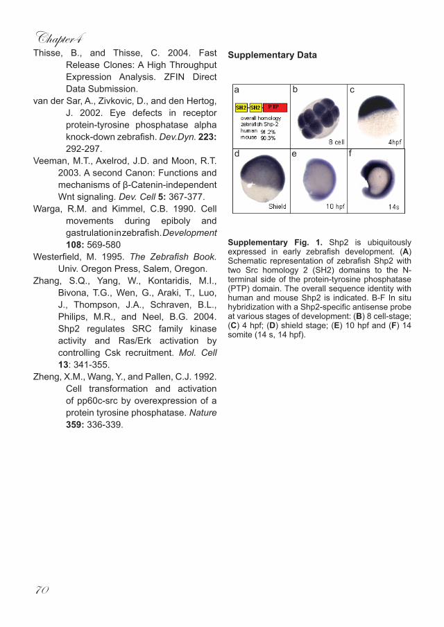
Supplementary Fig. 1. Shp2 is ubiquitously expressed in early zebrafish development. (A) Schematic representation of zebrafish Shp2 with two Src homology 2 (SH2) domains to the N-terminal side of the protein-tyrosine phosphatase (PTP) domain. The overall sequence identity with human and mouse Shp2 is indicated. B-F In situ hybridization with a Shp2-specific antisense probe at various stages of development: (B) 8 cell-stage; (C) 4 hpf; (D) shield stage; (E) 10 hpf and (F) 14 somite (14 s, 14 hpf).
a b c
d e f