Embed Size (px)
Citation preview
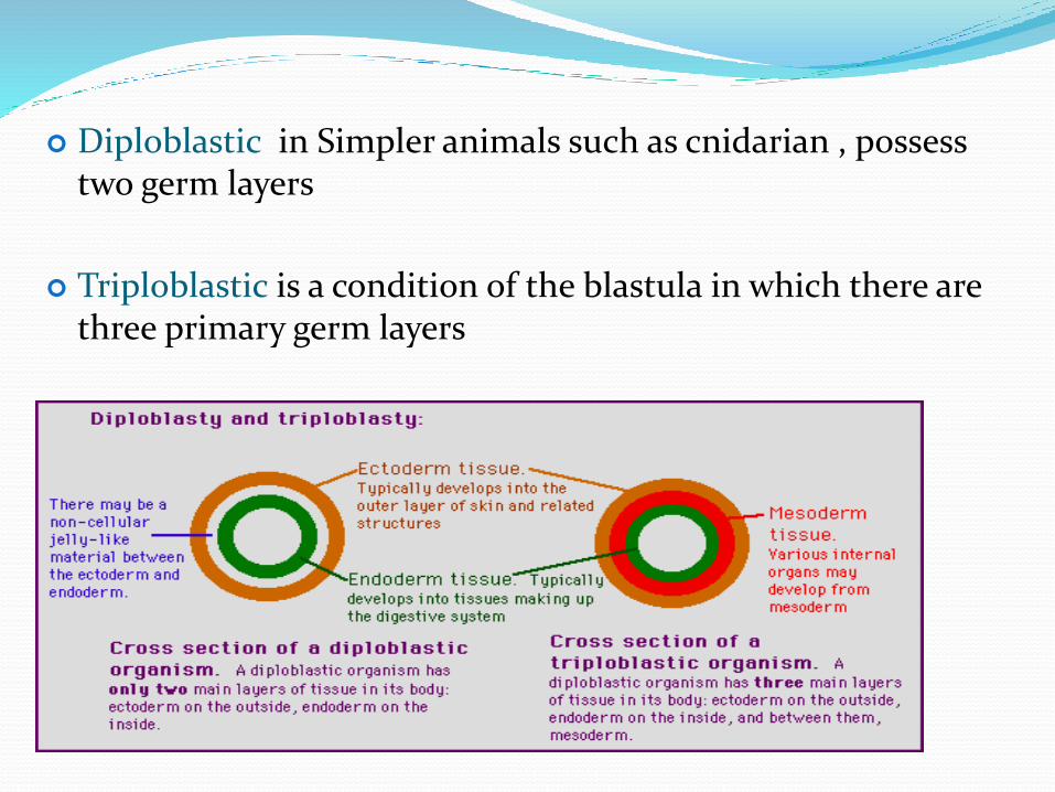
Diploblastic in Simpler animals such as cnidarian , possess two germ layers
Triploblastic is a condition of the blastula in which there are three primary germ layers
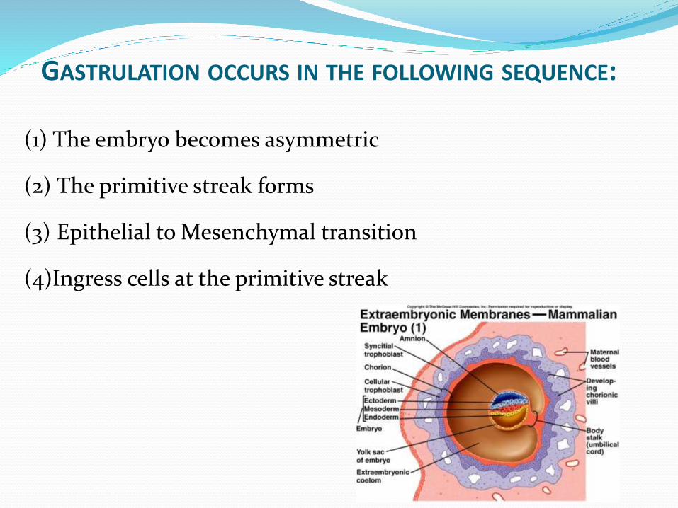
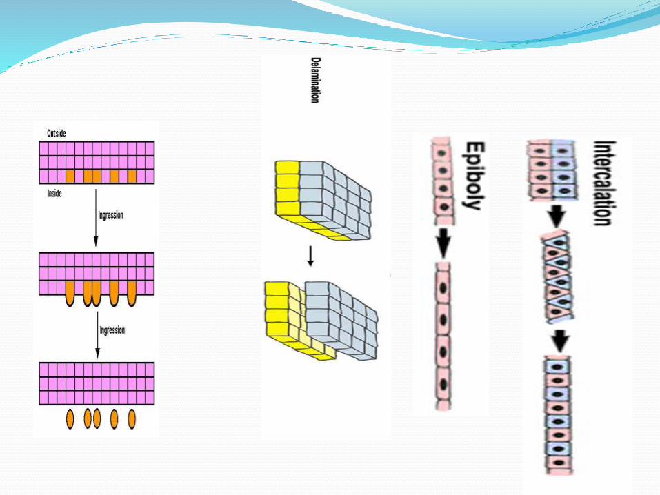
GASTRULATION OCCURS IN THE FOLLOWING SEQUENCE:
(1) The embryo becomes asymmetric
(2) The primitive streak forms
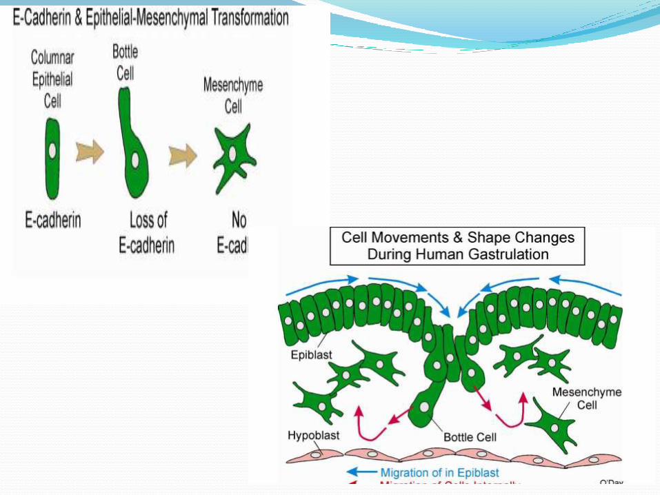
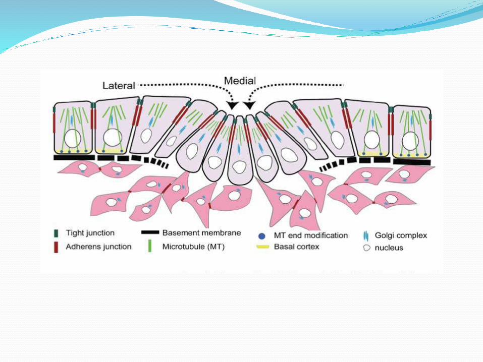
(3) Epithelial to Mesenchymal transition
(4)Ingress cells at the primitive streak
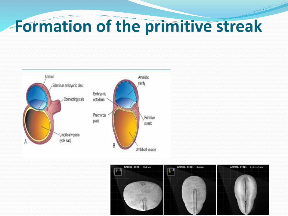
Formation of the primitive streak

Primitive Streak
Primitive Groove
Primitive Node
Primitive Pit

Fate Mape of The Epiblast
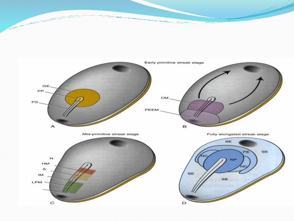

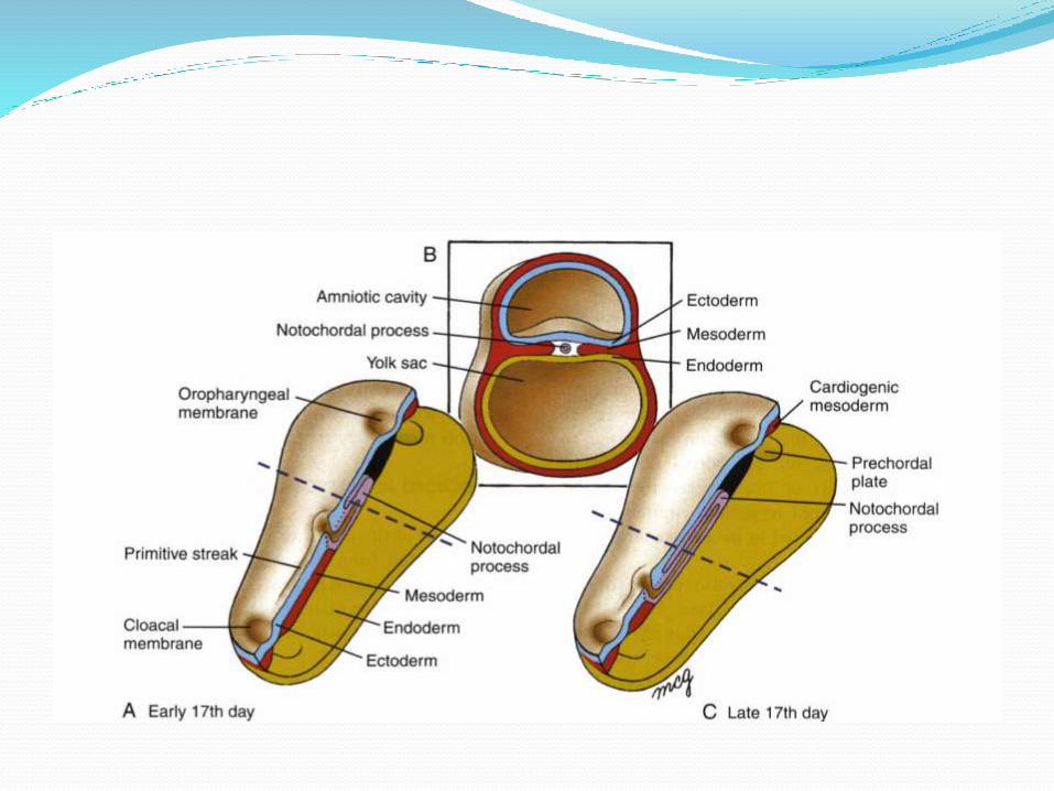
Prechordal Plateprechordal plate forms both:•mesodermal (part of head mesenchyme) •endodermal (part of oropharyngeal membrane) •it is often considered to be a mesendodermalstructure
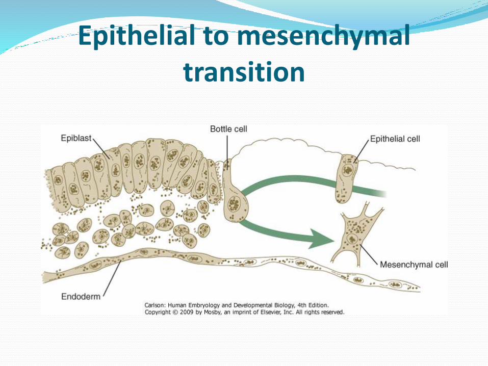
Epithelial to mesenchymaltransition
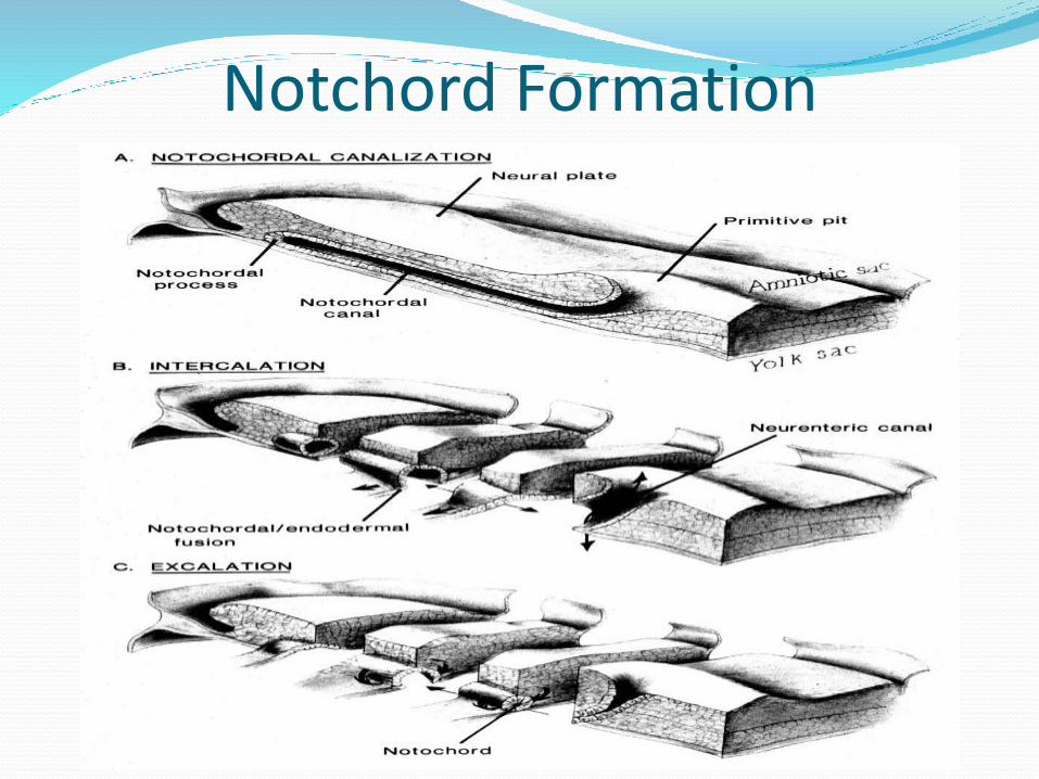
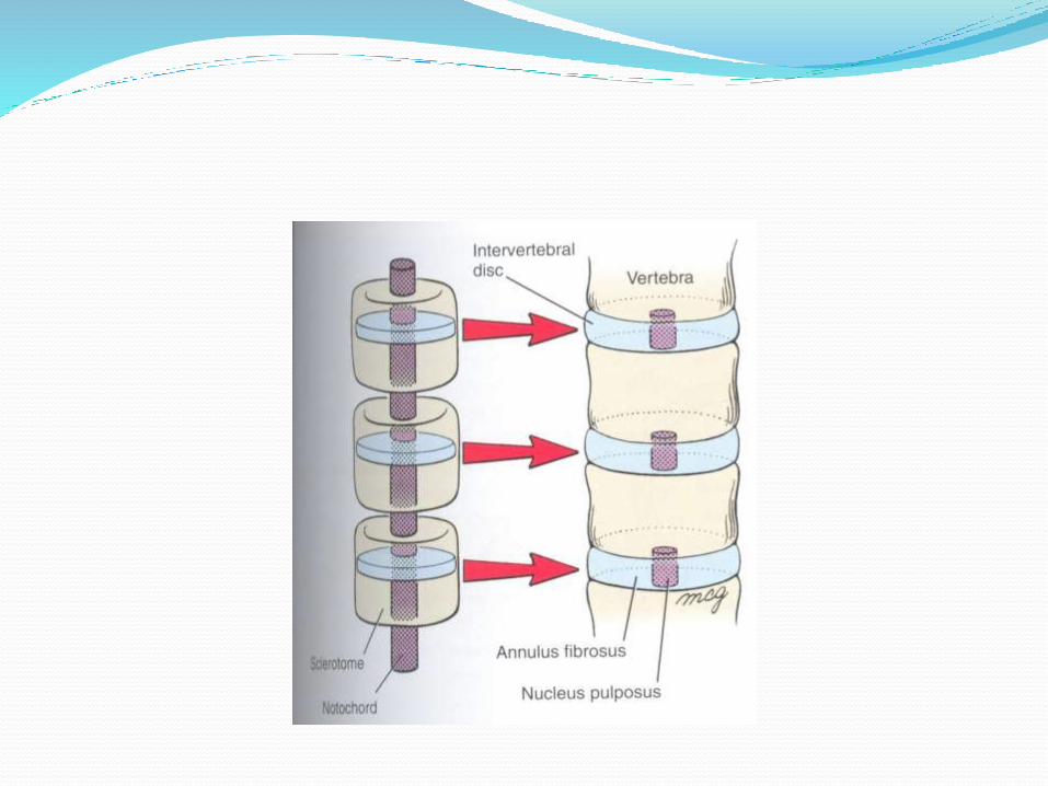
Notchord Formation
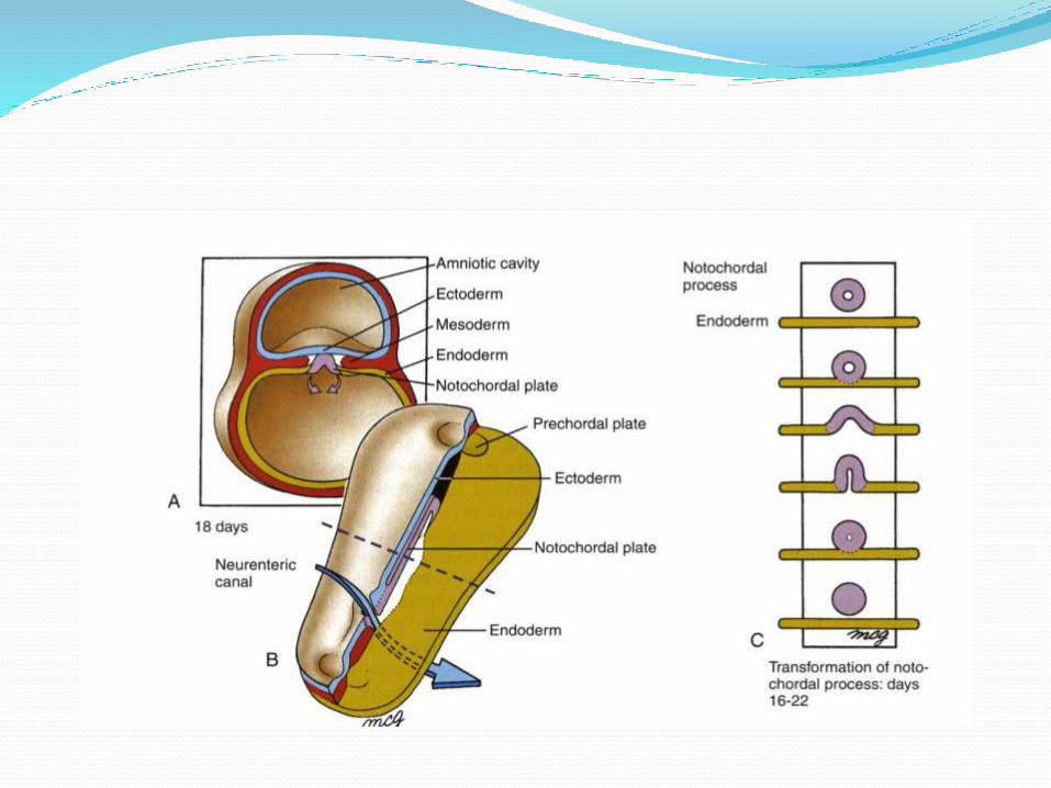
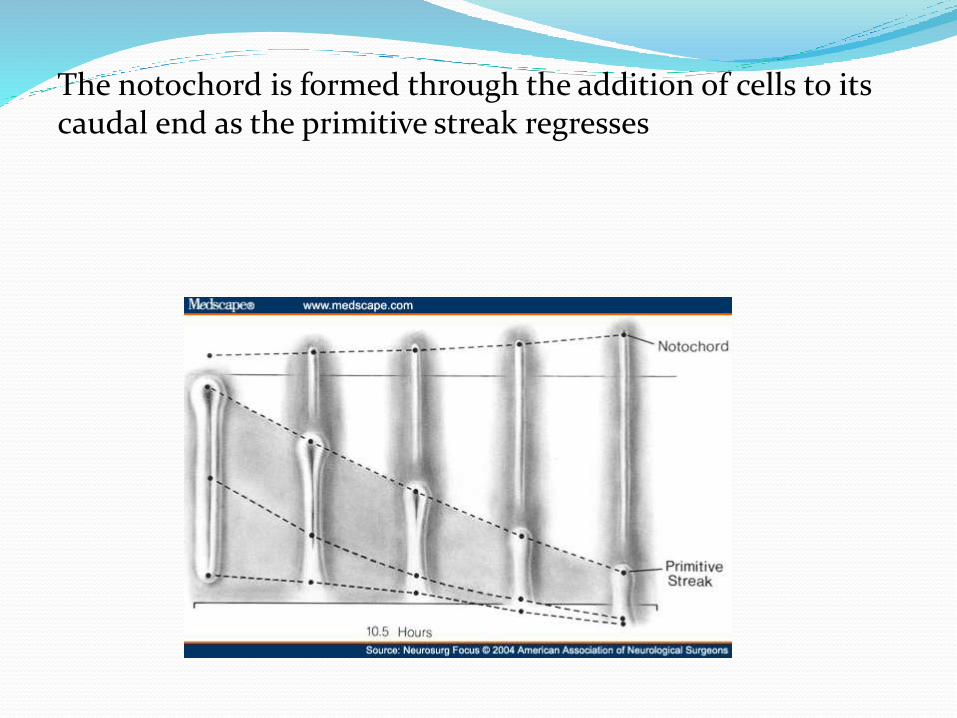
The notochord is formed through the addition of cells to its caudal end as the primitive streak regresses
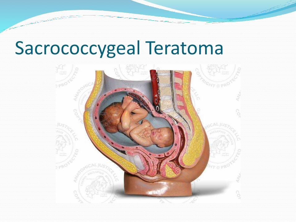
Sacrococcygeal Teratoma
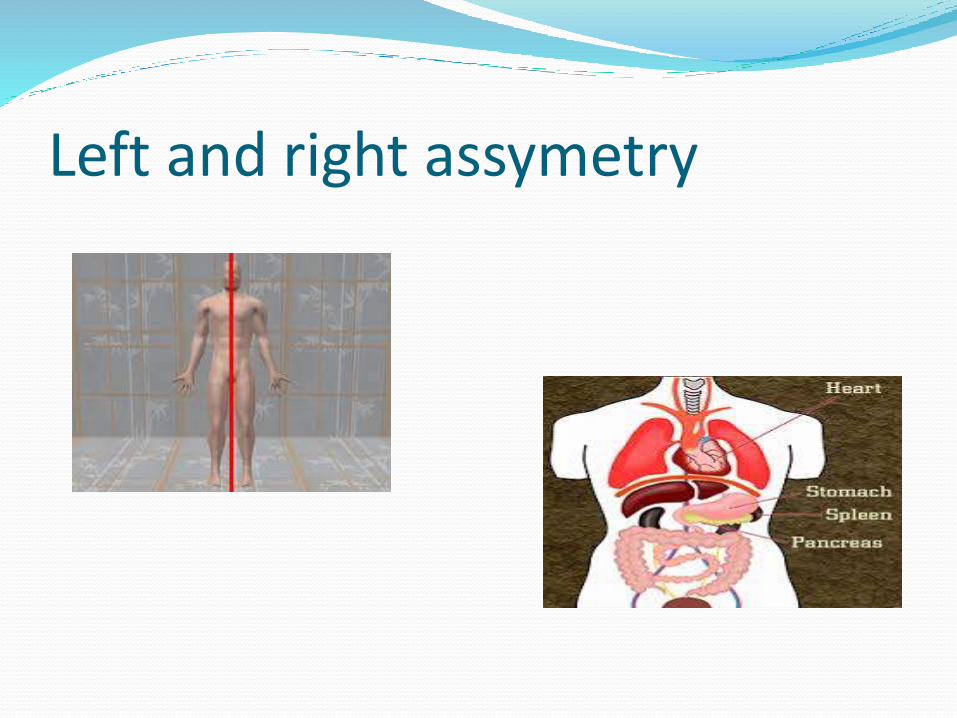
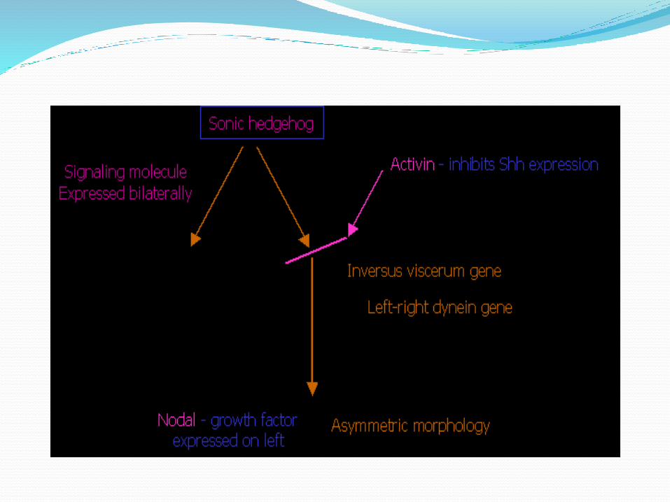
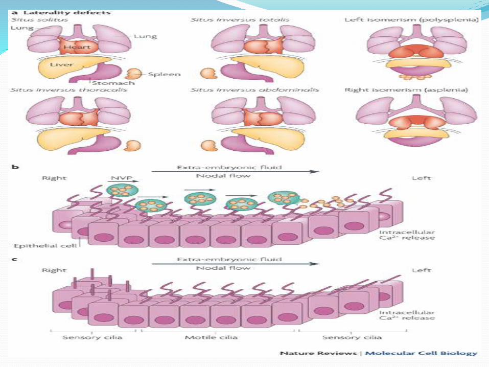
Left and right assymetry
Nodalal Flow Model
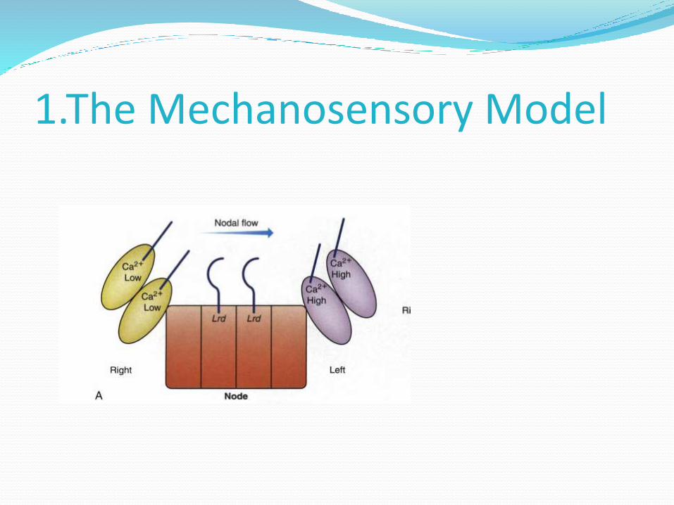
1.The Mechanosensory Model
2.Nodal Vesicular Parcels (NVP)
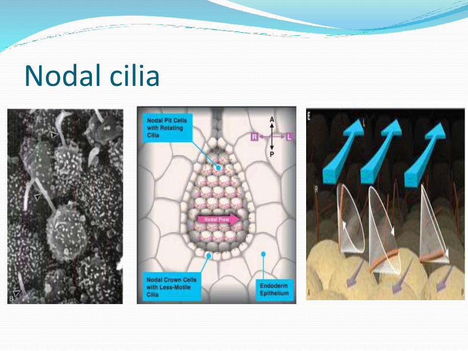
Nodal cilia
1.The Mechanosensory Model
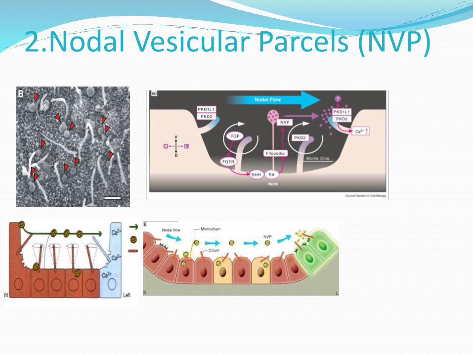
2.Nodal Vesicular Parcels (NVP)
(Kartagener syndrome)Dysfunctional ciliaRespiratory problemsMale fertility problems
Situs inversus
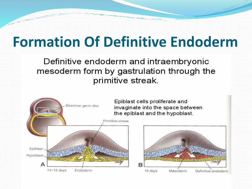
Formation Of Definitive Endoderm
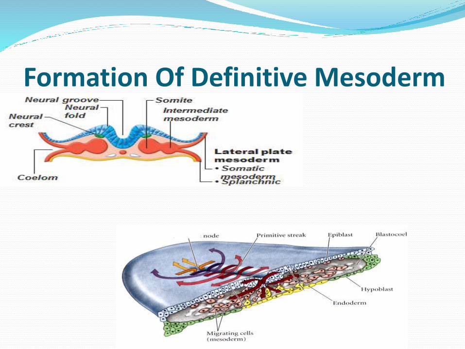
Formation Of Definitive Mesoderm
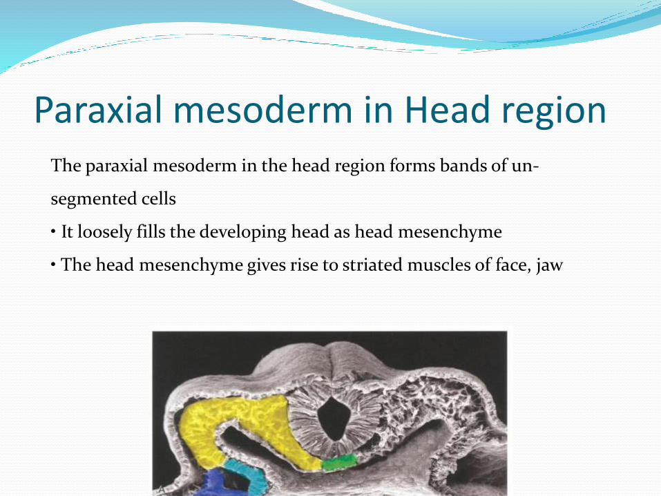
Paraxial mesoderm in Head region The paraxial mesoderm in the head region forms bands of un-
segmented cells
• It loosely fills the developing head as head mesenchyme
• The head mesenchyme gives rise to striated muscles of face, jaw
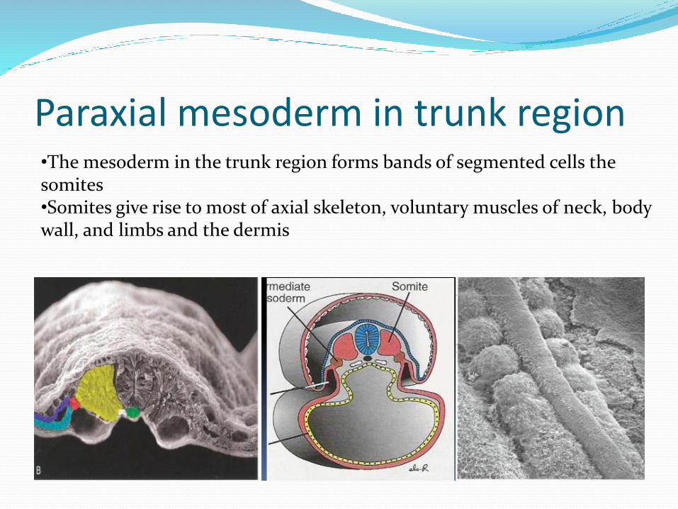
Paraxial mesoderm in trunk region•The mesoderm in the trunk region forms bands of segmented cells the somites•Somites give rise to most of axial skeleton, voluntary muscles of neck, body wall, and limbs and the dermis
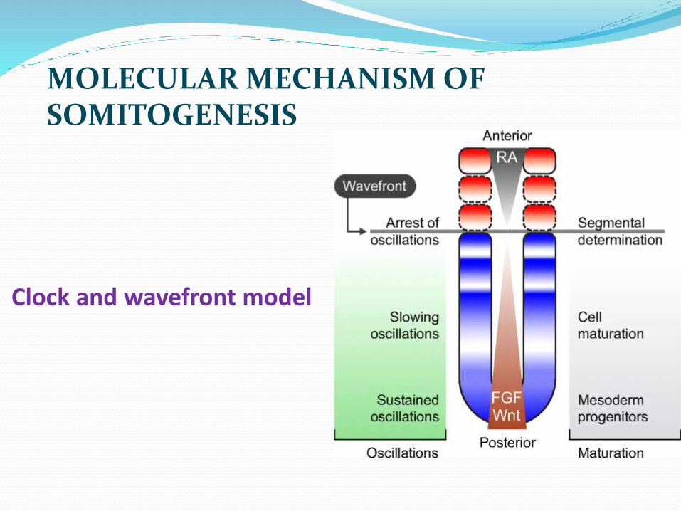
Clock and wavefront model
MOLECULAR MECHANISM OF SOMITOGENESIS
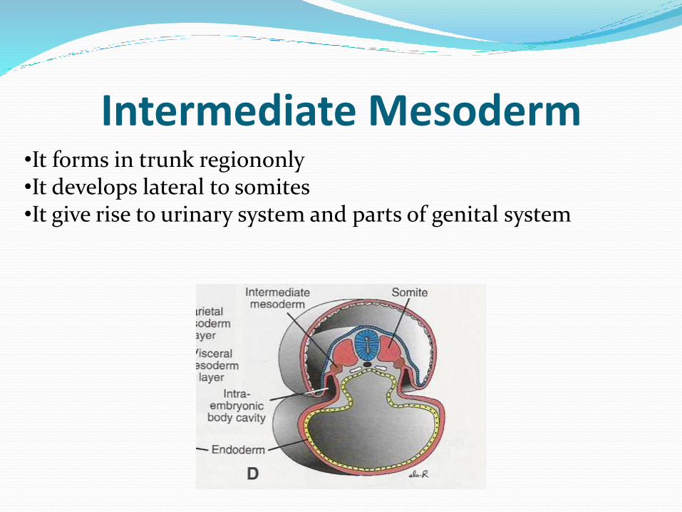
Intermediate Mesoderm •It forms in trunk regiononly•It develops lateral to somites•It give rise to urinary system and parts of genital system
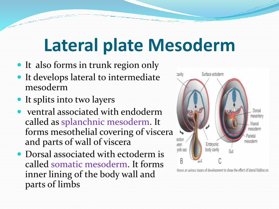
Lateral plate Mesoderm It also forms in trunk region only
It develops lateral to intermediate mesoderm
It splits into two layers
ventral associated with endoderm called as splanchnic mesoderm. It forms mesothelial covering of viscera and parts of wall of viscera
Dorsal associated with ectoderm is called somatic mesoderm. It forms inner lining of the body wall and parts of limbs

Gastrulation Ends with Formationof Tail Bud
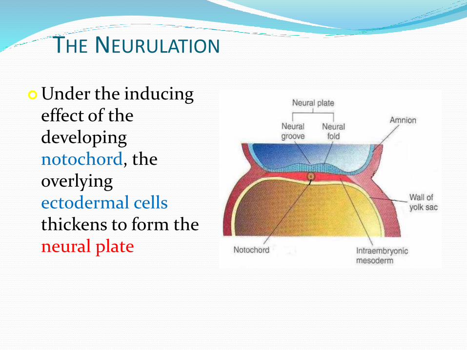
THE NEURULATION
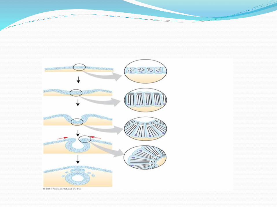
Under the inducing effect of the developing notochord, the overlying ectodermal cells thickens to form the neural plate
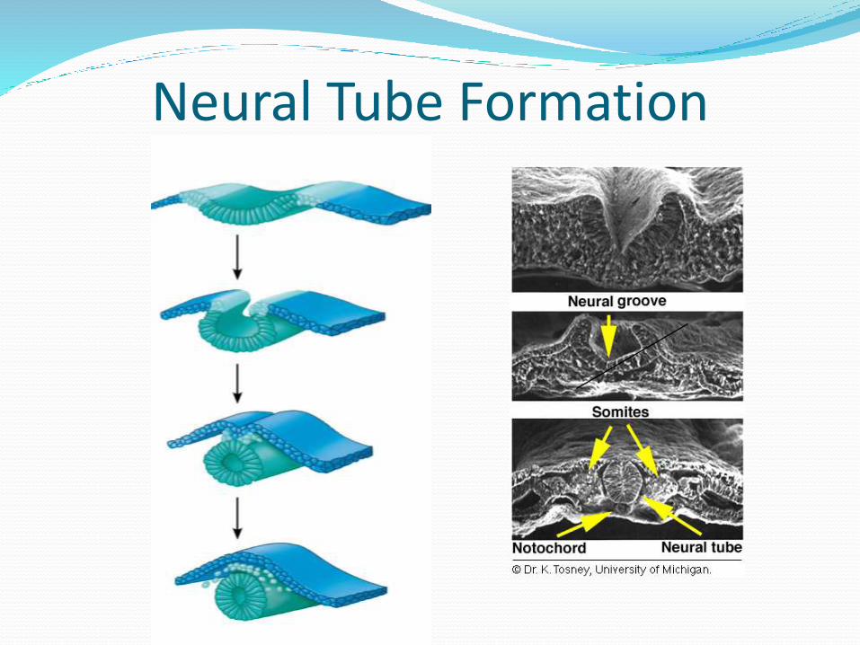
Neural Tube Formation

On 18th day: the neural plate invaginates to form neural groove & neural folds
Neural fold
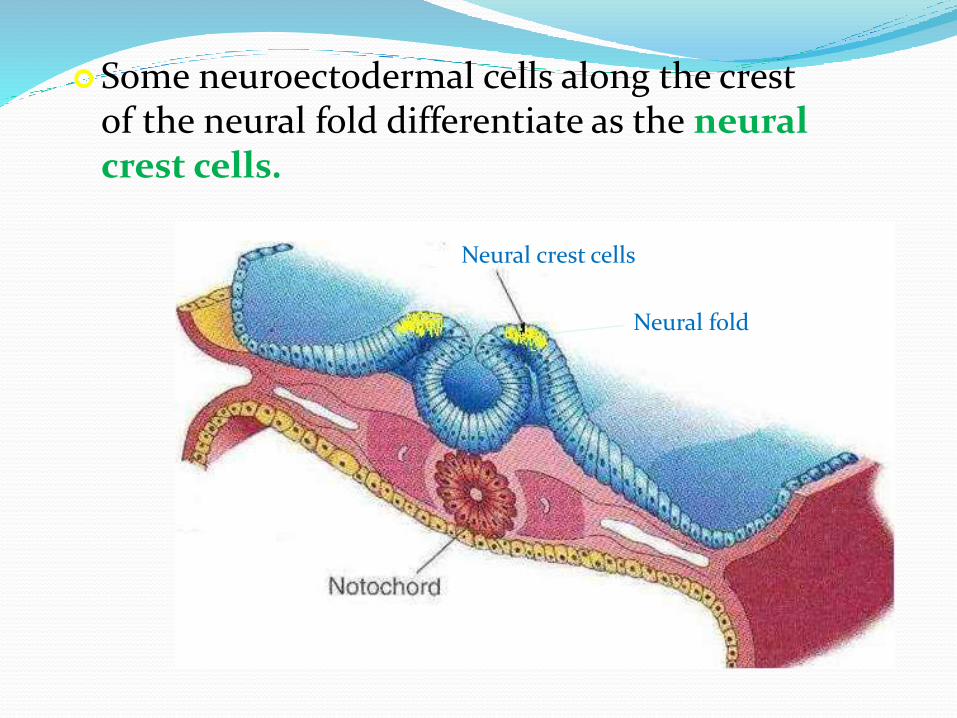
Some neuroectodermal cells along the crest of the neural fold differentiate as the neural crest cells.
Neural crest cells
Neural fold
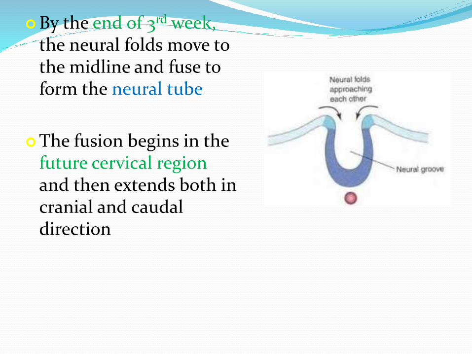
By the end of 3rd week, the neural folds move to the midline and fuse to form the neural tube
The fusion begins in the future cervical region and then extends both in cranial and caudal direction

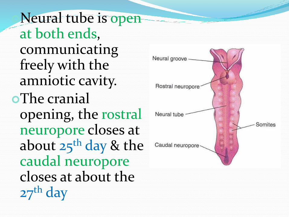
Neural tube is open at both ends, communicating freely with the amniotic cavity.
The cranial opening, the rostral neuropore closes at about 25th day & the caudal neuroporecloses at about the 27th day
Neural tube is open at both ends, communicating freely with the amniotic cavity.
The cranial opening, the rostral neuropore closes at about 25th day & the caudal neuroporecloses at about the 27th day