Embed Size (px)
Citation preview

Sigler, Microbes and Society Fall 2010
2
Chapter 2 Microbes in Perspective:
Of Collectors and Classifiers Objectives: After reading Chapter Two, you should understand…
• The schemes used throughout history to classify organisms. • How microorganisms are assigned their names. • The units for measuring microorganisms. • Some basic principles of microscopy.
Microbes in the biosphere With the exception of viruses, all microbes share a set of characteristics, many of which
are similar to those of other living organisms:
• Ingestion and processing of nutrients – why? • Excretion of waste materials – why? • Independent reproduction – why? • Adaptation to environmental change – why? • Reaction to stimuli – why?
By noting these characteristics and lots of others, biologists can begin to tell the
differences between organisms (microbes) that seemingly look alike. Classifying organisms into discrete groups based on characteristics is called
taxonomy.
Before the invention of the microscope (when?) biologists had little trouble classifying macroorganisms.
A mixed assemblage of marine microbes. Several of these individuals look alike, but have very distinct functions.

Sigler, Microbes and Society Fall 2010
3
Features of large organisms were easy to see. As a result, two major kingdoms of organisms were recognized: plants (Plantae)
and animals (Animalia). Do you know how biologists differentiated the Plantae from Animalia?
When Leuwenhoek reported the existence of microbes, it was necessary to include them somewhere the classification scheme, but where?
The kingdom Protista was introduced.
What is the derivation of the word “Protista”? The kingdom contained fungi, protozoa, algae, bacteria.
All of this was rather controversial and many microbes were ultimately classified as either plants or animals.
In the mid 1900s people who wanted to study bacteria needed to enroll in botany
courses. Prokaryotes and eukaryotes In the 1940s the electron microscope became widely available and biochemistry was
advancing.
Offered highly detailed views and understanding of microbial cells never seen before.

Sigler, Microbes and Society Fall 2010
4
As methods advanced, scientist questioned the three-kingdom classification scheme. A new system recognizing five kingdoms gained popularity (1969): 1. Bacteria moved to a new kingdom Monera (Prokaryotae) 2. Protista remained, and contained protozoa, slime molds, and single-celled
algae. Sometimes a “catch-all” kingdom
3. Another new kingdom, Fungi, included filamentous fungi and yeasts. 4. Plantae and Animalia kingdoms remained unchanged. What about viruses?

Sigler, Microbes and Society Fall 2010
5
The three-domain system In the 1980s, Carl Woese used genetic sequences to classify organisms.
Developed a three-domain system
1. The Eukarya encompassed the Fungi, Protista, Plantae, and Animalia kingdoms of previous schemes.
2. Eubacteria (Bacteria) was solely devoted to bacteria
3. A new kingdom, Archaea, was developed that encompassed a group of organisms that looked much like bacteria, but were different in many ways.
Partial 16S r RNA gene sequences of several bacteria.

Sigler, Microbes and Society Fall 2010
6
Naming and measuring microbes In the mid-1700s Carolus Linnaeus was attempting to name all of the organisms in the
world. How could one possibly come up with a naming scheme that would make sense? Categorize organism with a “first” name – genus Add a modifier for a “second” name – species = genus w/ species
modifier Opposite of how we give first and second names to humans.
Every organism has a binomial name: Escherichia coli (E. coli)
Also: E. blattae; E. fergusonii; E. hermannii; E. senegalensis; E. vulneris
“Escherichia” – genus to which the bacterium belongs, and
named after the discoverer, Theodor Escherich (1888) “coli” – species, and based on the location from which the
bacterium was first isolated (colon).

Sigler, Microbes and Society Fall 2010
7
Notice that the binomial name is always italicized (or underlined).
Many bacteria have common names as well. Streptococcus pneumoniae – causes bacterial pneumonia, often
called simply “pneumococcus”. Neisseria menigitidis – causes meningitis, often called
“meningococcus”. Staphylococcus aureus – responsible for skin infections among
others, often called “staph”.
Sub species or strain designations can also be very important. E. coli O157:H7 The fundamental rank in organism classification is the species. What is a “species”?
Two microbes are considered of the same species if their DNA sequences are 70% or more similar.
How is the “same species” designation handled in the plant and animal
worlds? Microbial measurements Microbes are small…very small. What units of measure is most commonly used when describing the size of microbes?
Relationship between the units of measure commonly used to describe microbes (mm, millimeters; μm, micrometers; nm, nanometers).
Sigler, Microbes and Society Fall 2010
8
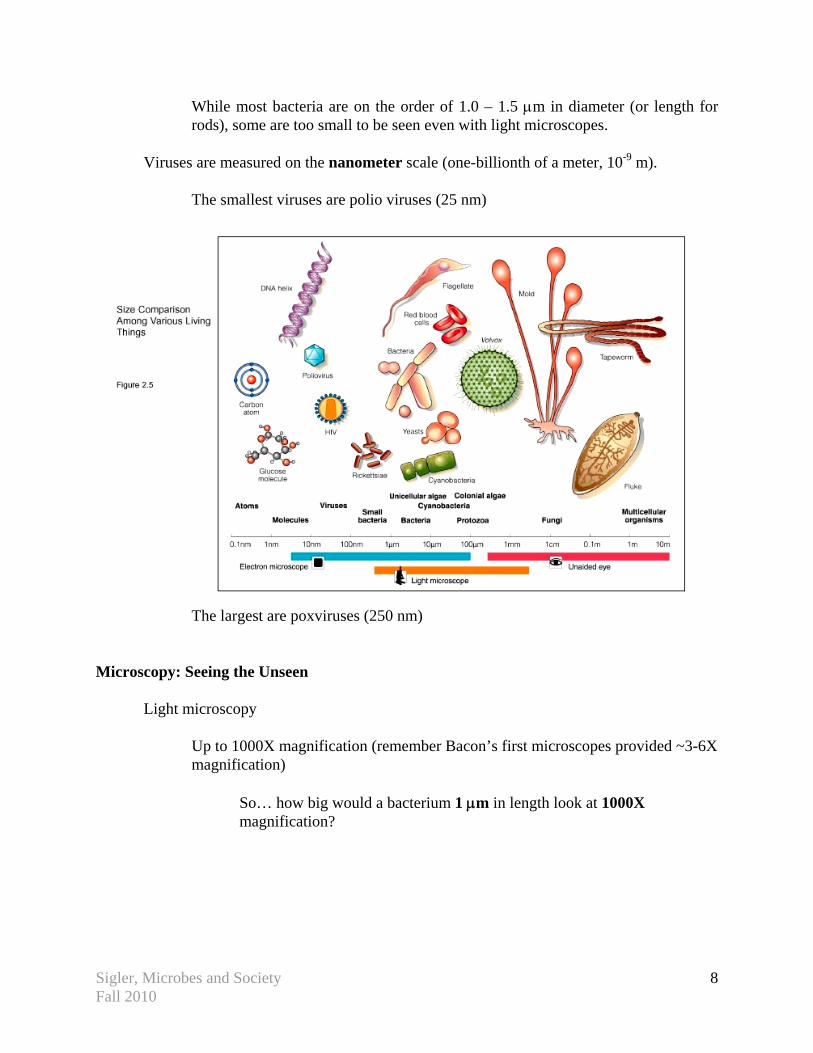
While most bacteria are on the order of 1.0 – 1.5 μm in diameter (or length for rods), some are too small to be seen even with light microscopes.
Viruses are measured on the nanometer scale (one-billionth of a meter, 10-9 m). The smallest viruses are polio viruses (25 nm)
The largest are poxviruses (250 nm) Microscopy: Seeing the Unseen
Light microscopy Up to 1000X magnification (remember Bacon’s first microscopes provided ~3-6X
magnification) So… how big would a bacterium 1 μm in length look at 1000X
magnification?

Sigler, Microbes and Society Fall 2010
9
Three main components of a microscope… 1. Ocular 2. Objective lenses
3. Substage condenser
Magnification Most microscopes have three objective lenses… Low power (10X) High power (40X) Oil immersion lens (100X) …and one ocular (10X) Total magnification is calculated by multiplying the ocular power
and the objective power. e.g. 10X ocular x 40X objective = 400X magnification
The oil-immersion objective provides the most power, but needs to be positioned very close to the slide – limits the available light. To limit the loss of light, the objective is immersed in oil to
contain all available light.
Oil has the same refractive (bending) index as glass. Let’s look at the differing types of microscopy with regard to a common bacterium, E. coli.
Sigler, Microbes and Society Fall 2010
10
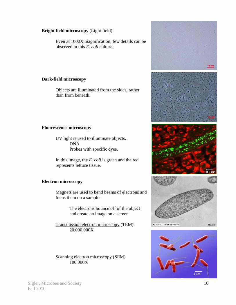
Bright field microscopy (Light field)
Even at 1000X magnification, few details can be observed in this E. coli culture.
Dark-field microscopy Objects are illuminated from the sides, rather
than from beneath. Fluorescence microscopy UV light is used to illuminate objects. DNA Probes with specific dyes.
In this image, the E. coli is green and the red represents lettuce tissue.
Electron microscopy
Magnets are used to bend beams of electrons and focus them on a sample.
The electrons bounce off of the object
and create an image on a screen.
Transmission electron microscopy (TEM) 20,000,000X Scanning electron microscopy (SEM) 100,000X
1 μm


![Bioactive Powerpoint Microbes fighting microbes [Read-Only]](https://img.pdfslide.us/doc/110x75/625e85126147534db333a997/bioactive-powerpoint-microbes-fighting-microbes-read-only.jpg)