Embed Size (px)
Citation preview

Chapter 23
ChIP on Chip Assays: Genome-Wide Analysis of TranscriptionFactor Binding and Histone Modifications
Smitha Pillai and Srikumar P. Chellappan
Abstract
Deregulation of transcriptional activity of many genes has been causatively linked to human diseasesincluding cancer. Altered patterns of gene expression in normal and cancer cells are the result ofinappropriate expression of transcription factors and chromatin-modifying proteins. Chromatin immu-noprecipitation assay is a well-established tool for investigating the interactions between regulatoryproteins and DNA at distinct stages of gene activation. ChIP coupled with DNA microarrays, known asChIP on chip, allow us to determine the entire spectrum of in vivo DNA-binding sites for a given protein.This has been of immense value because ChIP on chip assays can provide a snapshot of the transcriptionalregulatory mechanisms on a genome-wide scale. This article outlines the general strategies used to carryout ChIP-chip assays to study the differential recruitment of regulatory molecules based on the studiesconducted in our lab as well as other published protocols
Key words: Genome-wide chromatin immunoprecipitation, transcription factors, histonemodifications, promoter arrays, whole genome amplification.
1. Introduction
DNA-binding proteins perform a variety of important functionsin cells, including transcriptional regulation, chromosome main-tenance, replication and DNA repair. The interactions betweentranscription factors and their DNA binding sites are an integralpart of transcriptional regulatory networks. These interactionscontrol the coordinated expression of thousands of genes duringnormal growth and in response to external stimuli. Studies on thesequence-specific binding of transcription factors to promoterDNA were provided a major boost by the development of electro-phoretic mobility shift assays (EMSAs or gel-shift assays) three
Srikumar P. Chellappan (ed.), Chromatin Protocols: Second Edition, vol. 523ª Humana Press, a part of Springer ScienceþBusiness Media, LLC 2009DOI 10.1007/978-1-59745-190-1_23 Springerprotocols.com
341
decades ago (1, 2). While this technique provided the capability toanalyze the binding of proteins to DNA in vitro, it could notprovide a snapshot of the binding status in the intact cells. Devel-opment of chromatin immunoprecipitation assays (ChIP assays),where DNA bound proteins are cross-linked to DNA in the livingcells and analyzed by PCR provided the ability to examine thebinding of proteins to specific targets in vivo in response tospecific signals or during normal development. Further, theseassays also enabled the analysis of histone modifications and con-tributed significantly to the elucidation of the histone code andthe epigenetic regulation of gene expression.
Given the ability of chromatin immunoprecipitation assaysto provide a picture of the promoter occupancy of protein in anin vivo situation, attempts were made to extend this technologyto genome-wide analysis of DNA-binding and histone modifica-tions (3, 4). Recent advances in DNA microarray technology aswell as the availability of whole genome sequences make it pos-sible to map the entire spectrum of interactions of a particularprotein with specific DNA sequences across the whole genome.This is achieved by an immunoprecipitation of the protein ofinterest cross-linked to its target DNA sequences; the boundfragments are identified by a microarray-based analysis ofenriched DNA fragments. This technique, referred to as ChIPon chip or genome-wide location analysis, is currently the mostwidely used method for identifying in vivo transcription factor-binding sites in a high-throughput manner (5, 6). In conven-tional ChIP assays, the cells are treated with formaldehyde tocovalently cross-link protein and the bound DNA (7, 8); thecells are then lysed and the chromatin is sheared into 200–1000base pair fragments by sonication (7) or treatment with micro-coccal nucleases (9, 10). An antibody specific for a protein ofinterest is then used to immunoprecipitate protein bound DNAfragments; the associated DNA is de-cross-linked by incubatingin a low pH buffer, protease treated and the associated DNA isexamined for the presence of the promoter or DNA sequence ofinterest using appropriate PCR primers (11). This technique, ascan be imagined, used to analyze the association of specifictranscription factors, or modified histones with DNA. TheChIP-on-chip version of this protocol differs only in the post-IP steps. In this case, the immunoprecipitated DNA is amplifiedusing a random primed PCR or the immunoprecipitated DNA isblunt ended by T4 DNA polymerase to allow ligation of a uni-versal linker (LM-PCR) or whole genome amplification method(WGA) and subsequently labeled and hybridized into DNAmicroarrays (12–15).
The extent of ChIP-chip application depends, in part, on thedevelopment of microarray technology, especially the availability ofarrayed slides for DNA from human, mouse or other model
342 Pillai and Chellappan
organisms. There are two different types of DNA microarraysavailable; the first type is created by spotting DNA onto poly-lysine-modified glass slides using pin-spotting robots (16). Thedensity of spotted arrays is limited to 20,000–30,000 featuressince the feature sizes below 100 microns are difficult to achieve(17). A different type of oligonucleotide array is available whichis created by synthesizing oligomers in situ on the array byphotolithography. These chips marketed by Affymetrix can beof higher density containing up to several million features (18).Today, arrays representing promoter regions, CpG islands, entirechromosomes or entire genomes are used for ChIP-chip assays(19–22). High-density oligonucleotide arrays that tile the entirehuman non-repetitive genomic sequences are available from Affy-metrix, NimbleGen Systems and Agilent Technologies (SeeNotes 1 and 2).
The major applications of ChIP-chip assays are the identifica-tion of binding targets for transcription factors and identificationof post-translational modifications on DNA-associated proteinsincluding specific histone modifications. Initial studies usingChIP-chip technology were directed to detect transcription factorchromatin binding in the yeast Saccharomyces cerevisiae (23–26).ChIP-chip technology was later applied to mammalian systems tostudy the recruitment of transcription factors such as E2F,GATA1, Rb, etc., but these studies surveyed only a small fractionof the genome due to the limitations in the microarray technologyand also the incomplete information on genome annotation (21,27, 28). With the recent advances in DNA microarray synthesistechnology and the completion of human genome project reveal-ing more promoter regions, allowed extensive screening of thegenome for the binding position of protein factors. In recent yearsChIP-chip has been expanded to investigate post-translationalmodifications of DNA-binding proteins. Different strategies canbe employed to study histone modifications using ChIP-chiptechnology. One is to detect the distribution of histone modifica-tions using antibodies specially targeting the modifications (29).and the other is using ChIP-chip to locate the enzymes thatcatalyze the histone modification reactions (30). Another usefulapplication of these kinds of studies would be to identify genessilenced by histone modification in carcinogenesis (20). It is alsopossible to combine the ChIP-chip data with expression profilingto establish the correlation of histone markers with transcriptionalactivity (29, 31).
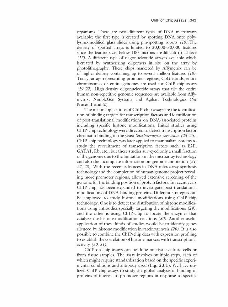
ChIP-on-chip assays can be done on tissue culture cells orfrom tissue samples. The assay involves multiple steps, each ofwhich might require standardization based on the specific experi-mental conditions and antibody used (Fig. 23.1). We have uti-lized ChIP-chip assays to study the global analysis of binding ofproteins of interest to promoter regions in response to specific
ChIP on Chip Assays 343
signaling events, using Affymetrix promoter arrays (GeneChiphuman promoter 1.0R array, Affymetrix, Cat. No. 900775).The protocol we have successfully used is as follows:
2. Materials
1. Formaldehyde (37%) (Molecular Biology Reagent Grade;Sigma-Aldrich).
2. Glycine, 1 M: 3.75 g of tissue grade glycine (Fischer Biotech)in 50 ml of water.
3. 20� phosphate-buffered saline (PBS): 160 g NaCl, 4 g KCl,2.88 g Na2HPO4, 4.8 g KH2PO4 in 800 ml of water. Adjustthe pH of the buffer to 6.4 and make up the volume to 1 l.
Fig. 23.1. Schematic of ChIP on chip workflow. (A) Chromatin immunoprecipitation. Cells are grown under the desiredexperimental conditions and protein–DNA cross-links are created by treating the cells with formaldehyde. Cells are lysed,DNA is sheared either by sonication or by micrococcal nuclease and immunoprecipitated with antibody against protein ofinterest. Cross-links are reversed and DNA is purified. (B) ChIP-chip experiment. The immunoprecipitated DNA isamplified either by random primed PCR or by ligation-mediated PCR or whole genome amplification. The amplifiedDNA is fragmented, labeled with a fluorophore, and hybridized to a microarray and analyzed.
344 Pillai and Chellappan
4. IGEPAL CA-630 (Sigma-Aldrich).
5. 100 mM PMSF: 0. 174 g of PMSF (Sigma-Aldrich) in 10 mlof absolute ethanol. Aliquot into microcentrifuge tubes andstore at –20�C. Add PMSF to the lysis buffer just before use asthe half-life of 1 mM PMSF in the buffer is 30 min.
6. Protease inhibitor stock: prepare a 25� stock by dissolvingone protease inhibitor tablet (Roche, cat. no. 11873580001)in 2 ml of nuclease-free water.
7. Proteinase K (Fischer Biotech, cat. no. BP-1700-100).
8. LiCl (10 M): 42.39 g LiCl (Sigma-Aldrich cat. no. L-8895) in100 ml of water.
9. Protein G Sepharose 4 Fast Flow (GE Healthcare, cat. no. 17-0618-01).
10. Sequenase Version 2.0 DNA polymerase (USB, cat. no.70775Y).
11. Primer A: 200 mM GTTTCCCAGTCACGGTC(N)9 (HPLCPurified).
12. Primer B: 100 mM GTTTCCCAGTCACGGTC (HPLCPurified).
13. Taq Polymerase 5 u/ml (Invitrogen, cat. no. 10342-020).
14 100 mM dNTPs (Promega, dATP- cat no. U120-B, dCTP- catno. U121B, dGTP- cat no. U122B, dTTP- cat no. U123B,)
15. 100 mMdUTP (Promega, cat. no. U119A).
16. BSA (20 mg/ml): 100 mg of BSA (Sigma-Aldrich cat. no.A3059-50G) in 5 ml of water. Aliquot 100 ml into 1.5 mltubes and store at –20�C.
17. 1 M DTT (dithiothreitol): 1.54 g DTT (Fisher Biotech cat.no. BP172-5) in 10 ml of water. Aliquot 100 ml into 1.5 mltubes and store in –20�C.
18. 5 M NaCl: 58.4 g NaCl in 200 ml of water.
19. 1 M MgCl2: 20.3 g of MgCl2.6 H2O in 100 ml of water.
20. 1 M CaCl2: 14.7 g of CaCl2. 2 H2O in 100 ml water.
21. Deoxycholate (sodium salt) (Sigma-Aldrich cat. no. 21115).
22. GeneChip WT Double-Stranded DNA Terminal LabelingKit (Affymetrix, cat. no. 900812).
23. GeneChip sample clean up module (Affymetrix, cat. no.900371).
24. Control oligonucleotide B2, 3 nM (Affymetrix, cat. no.900301).
25. GeneChip hybridization, wash, and stain kit (Affymetrix, cat.no. 900301).
ChIP on Chip Assays 345
2.1. Buffers 1. Lysis buffer: 10 mM Tris–HCl (made from stock 1 MTris–HCl pH 7.5), 10 mM NaCl 3 mM MgCl2, 1 mMCaCl2, 4% IGEPAL, 1 mM PMSF (add fresh). Store at roomtemperature.
2. IP dilution buffer: 20 mM Tris–HCl (made from stock 1 MTris–HCl pH 8), 2 mM EDTA 1% Triton X-100, 150 mMNaCl, protease inhibitor stock (add fresh). Store at roomtemperature.
3. Protease inhibitor stock: prepare a 25� stock by dissol-ving one protease inhibitor tablet (Roche, cat. no.11873580001) in 2 ml of nuclease-free water. Store thealiquots at –20�C.
4. ChIP wash 1: 20 mM Tris–HCl (made from stock 1 MTris–HCl pH 8), 2 mM EDTA, 1% Triton X-100, 150 mMNaCl, and 1 mM PMSF (add fresh). Store at roomtemperature.
5. ChIP wash 2: 20 mM Tris–HCl (made from stock 1 MTris–HCl pH 8), 2 mM EDTA 1% Triton X-100, 0.1% SDS,500 mM NaCl, and 1 mM PMSF (add fresh). Store at roomtemperature.
6. ChIP wash 3: 10 mM Tris–HCl (made from stock 1 MTris–HCl pH 8), 1 mM EDTA 0.25 M LiCl, 0.5% IGEPAL,and 0.5% deoxycholate (sodium salt). Store at roomtemperature.
7. TE (10 mM Tris–HCl pH 8, 1 mM EDTA).
8. Elution buffer: 25 mM Tris–HCl (made from stock 1 MTris–HCl pH 7.5), 10 mM EDTA, and 0.5% SDS. Store atroom temperature.
9. Buffers for hybridization:A. 12� MES stock buffer (1.22 M MES, 0.89 M Na+): ME
Shydrate (64.61 g), MES sodium salt, (193.3 g), MilliQwater (800 ml). Adjust the pH between 6.5 and 6.7.Adjust the volume to 1000 ml. Do not autoclave. Filtersterilize by passing through 0.2 mm filter. Store at 2–8�Caway from light.
B. 2� hybridization buffer (100 mM MES, 1 M [Na+], 20 mMEDTA, 0.01% Tween-20): 12� MES stock buffer (8.3 ml),5 M NaCl (17.7 ml), 0.5 M EDTA (4 ml), 10% Tween 20(100 ml). Make up the volume to 50 ml with water and storeat 2�C–8�C away from light.
C. Wash buffer A: non-stringent wash buffer (6� SSPE, 0.01%Tween 20): 20� SSPE (300 ml), 10% Tween 20 (1 ml).Make up the volume to 1 l and filter sterilize using 0.2 mmfilter.
346 Pillai and Chellappan
D. Wash buffer B (100 mM MES, 0.1 M Na+, 0.01% Tween20): 12� MES stock buffer (83.3 ml), 5 M NaCl(5.2 ml), 10% Tween 20 (1 ml). Make up the volume to1 l, filter through 0.2 mm filter and store at 2–8�C pro-tected from light.
E. 2� stain buffer (100 mM MES, 1 M Na+, 0.05% Tween20): 12� MES stock buffer (41.7 ml), 5 M NaCl(92.5 ml), 10% Tween 20 (2.5 ml). Make up the volumeto 250 ml, filter through 0.2 mm and store at 2–8�Cprotected from light.
2.2. Instruments and
Software Required
1. Genechip Scanner 3000 7G
2. GeneChip Operating Software (GCOS) v1.3 or higher
3. GeneChip Fluidics Station 400 or 450
4. Tiling array software for data analysis
3. Methods
3.1. Preparation of
ChIP Lysate from Cells
1. ChIP assays require large amounts of cells as the starting mate-rial. Usually, for each IP reaction 50–200 million cells arerecommended (32–34). For a ChIP on chip experiment atleast two IP reactions should be done; an experimental samplewhere IP is done using antibody against the protein of interestand a negative control with a nonspecific antibody or a mock IPwith no antibody. Add formaldehyde directly to the tissue cul-ture plate to a final concentration of 1% (280 ml of 37% formal-dehyde/10 ml of media) and incubate in an orbital shaker for10 min for cross-linking DNA to the protein (See Note 3).
2. Add glycine to a final concentration of 0.125 M using a 1 Mstock solution and inoculate for 5 min in the orbital shaker toterminate the cross-linking.
3. Aspirate the formaldehyde and media into a waste containerand wash the cells twice with 10 ml of ice-cold 1� PBS. In caseof adherent cells, scrape the cells in 3–5 ml of ice-cold 1� PBSand transfer into a 15 ml centrifuge tube. If the cells are non-adherent, collect them along with the media in a 15 ml cen-trifuge tube. Centrifuge the cells at 800 g (1000 rpm) for 5 minin a tabletop centrifuge with a swing bucket rotor (Thermo-Forma, General purpose centrifuge) at 4�C to collect the cells.Wash the cell pellet twice with ice-cold 1� PBS and centrifugeat 800 g (1000 rpm) for 5 min in a refrigerated tabletopcentrifuge at 4�C. Discard the supernatant and proceed tothe next step or snap freeze the pellet and store at –80�C.
ChIP on Chip Assays 347
4. Resuspend the pellet in 1.5 ml ChIP lysis buffer containing60 ml PMSF. To the tube add 40 ml of 100 mM PMSF, 100 mlof 25� protease inhibitor stock, 460 ml of lysis buffer, 100 mlof 20 % SDS, 80 ml of 5 M NaCl, and 220 ml of nuclease-freewater to bring the total volume to 2.5 ml. Mix the cells using apipetman to obtain a uniform homogenate. Transfer the lysateequally (800 ml) into 1.5 ml centrifuge tubes.
5. Sonicate the samples to lyse the cells and shear the DNA to100–1000 bp fragments. The time and number of pulsesrequired to shear DNA depend on the cell type, extent ofcross-linking, and the instrument used for sonication. We haveobserved that for most adherent cells three to four cycles ofsonication at power four for 30 s on a Fisher Sonic Dismem-brator (Model 100), followed by incubation on ice for 30 s issufficient to shear the chromatin. However, number of pulsesneeded to shear DNA depends on cell density and cell type.Some cells are resistant to sonication treatment; micrococcalnuclease treatment may improve chromatin shearing in suchcell lines (9, 11, 35–37). Refer alternate protocol for micro-coccal nuclease treatment.
6. Transfer the sonicated samples into 1.5 ml tubes and centri-fuge at 17,000 g for 15 min in a refrigerated microcentrifuge(Beckman-Coulter, Microfuge R or an equivalent model) toremove the cell debris. The samples can be snap frozen andstored at –80�C or proceed directly to the immunoprecipita-tion step.
7. Sonication efficiency can be checked by de-cross-linking 100 mlof the sample and loading in a 1% agarose gel.
3.2. Setting up
Immunoprecipitation
Reaction
1. Thaw the frozen ChIP lysates on ice. Transfer lysate fromthree 1.5 tubes into one 15 ml tube and add five volumes ofIP dilution buffer containing freshly added protease inhibi-tors (Roche).
2. Use 10–15 mg of antibody per immunoprecipitation reaction.The amount of antibody to be added depends on quality,affinity, and specificity of the antibody (See Note 4). Usuallya negative control is performed using the same number ofcells with a nonspecific IgG or no antibody control (SeeNote 5).Preclearing ChIP lysate is recommended to reduce thenonspecific backgrounds and eliminate false positives.For preclearing protocols see support protocol Section3.2.1.
3. After adding the appropriate amount of antibody, rotate thereaction overnight at 4�C.
348 Pillai and Chellappan
4. The antibody bound protein–DNA complexes can be recov-ered using protein A (Amersham Biosciences), protein G(Amersham Biosciences), protein A/G (Santa Cruz Biotech-nology), or staph A (ATCC) cells. The choice of the beadsdepends on the isotype of the antibody to be used for theimmunoprecipitation and their affinity for protein A, proteinG, or protein A/G. Protein G is used for most of the applica-tions in our laboratory. Add the required volume of proteinG beads containing 20% ethanol (GE Healthcare) to a micro-centrifuge tube and centrifuge at 3000 rpm for 1 min. Dis-card the supernatant, add 1 ml of wash buffer to the tube, mixwell, and centrifuge. Discard the supernatant and repeat thewashings for three times with wash buffer to remove theethanol preservative. Add 200 ml of 1:1 protein G slurry or400 ml of protein A slurry to the IP reaction.
5. Add PMSF to the IP reaction to a final concentration of1 mM. Incubate IP reaction with the beads at room tempera-ture for 2–4 h.
6. Centrifuge the tubes at 800 g (3000 rpm) at 4�C (Beckman-Coulter, Microfuge R or an equivalent model) for 1 min.Discard the supernatant by aspirating carefully to avoid losingbeads.
7. Add 700 ml of wash buffer 1 containing freshly added PMSF,mix well, and transfer to a new microcentrifuge tube. Rotatethe tube in a nutator at room temperature for 1 min. Centri-fuge at 800 g for 1 min and discard the supernatant.
8. Repeat the steps 7 and 8.
9. Add 700 ml of wash buffer 2 containing PMSF, rotate in anutator for 5 min, and centrifuge at 800 g for 1 min.
10. Discard the supernatant, add 700 ml of wash buffer 3, rotate thetubes in nutator for 5 min, and centrifuge at 800 g for 1 min.
11. Discard the supernatant; wash the beads with 700 ml of TE.
12. Rotate the tubes in nutator for 1 min. Centrifuge at 800 g for1 min and discard the supernatant.
13. Repeat washing with TE, centrifuge at 800 g for 1 min, anddiscard the supernatant.
14. Add 200 ml of elution buffer to the beads and incubate at65�C for 30 min.
15. Centrifuge at 2000 g (5000 rpm) for 2 min. Transfer thesupernatant into a new tube. This is the immunoprecipitatedsample.
3.3. Reverse Cross-
Linking
1. Add 10 ml of proteinase K (20 mg/ml) to 200 ml of the sample.
2. Incubate overnight in heat block at 65�C.
ChIP on Chip Assays 349
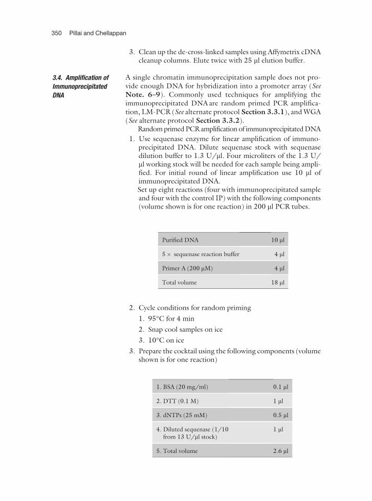
3. Clean up the de-cross-linked samples using Affymetrix cDNAcleanup columns. Elute twice with 25 ml elution buffer.
3.4. Amplification of
Immunoprecipitated
DNA
A single chromatin immunoprecipitation sample does not pro-vide enough DNA for hybridization into a promoter array (SeeNote. 6–9). Commonly used techniques for amplifying theimmunoprecipitated DNA are random primed PCR amplifica-tion, LM-PCR (See alternate protocol Section 3.3.1), and WGA(See alternate protocol Section 3.3.2).
Random primed PCR amplification of immunoprecipitated DNA
1. Use sequenase enzyme for linear amplification of immuno-precipitated DNA. Dilute sequenase stock with sequenasedilution buffer to 1.3 U/ml. Four microliters of the 1.3 U/ml working stock will be needed for each sample being ampli-fied. For initial round of linear amplification use 10 ml ofimmunoprecipitated DNA.Set up eight reactions (four with immunoprecipitated sampleand four with the control IP) with the following components(volume shown is for one reaction) in 200 ml PCR tubes.
2. Cycle conditions for random priming
1. 95�C for 4 min
2. Snap cool samples on ice
3. 10�C on ice
3. Prepare the cocktail using the following components (volumeshown is for one reaction)
1. BSA (20 mg/ml) 0.1 ml
2. DTT (0.1 M) 1 ml
3. dNTPs (25 mM) 0.5 ml
4. Diluted sequenase (1/10from 13 U/ml stock)
1 ml
5. Total volume 2.6 ml
Purified DNA 10 ml
5� sequenase reaction buffer 4 ml
Primer A (200 mM) 4 ml
Total volume 18 ml
350 Pillai and Chellappan
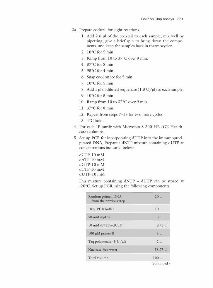
3a. Prepare cocktail for eight reactions.
1. Add 2.6 ml of the cocktail to each sample; mix well bypipetting, give a brief spin to bring down the compo-nents, and keep the samples back in thermocycler.
2. 10�C for 5 min.
3. Ramp from 10 to 37�C over 9 min.
4. 37�C for 8 min.
5. 95�C for 4 min.
6. Snap cool on ice for 5 min.
7. 10�C for 5 min.
8. Add 1 ml of diluted sequenase (1.3 U/ml) to each sample.
9. 10�C for 5 min.
10. Ramp from 10 to 37�C over 9 min.
11. 37�C for 8 min.
12. Repeat from steps 7–13 for two more cycles.
13. 4�C hold.
4. For each IP purify with Microspin S-300 HR (GE Health-care) columns.
5. Set up PCR for incorporating dUTP into the immunopreci-pitated DNA, Prepare a dNTP mixture containing dUTP atconcentrations indicated below:
dCTP-10 mMdATP-10 mMdGTP-10 mMdTTP-10 mMdUTP-10 mM
This mixture containing dNTP + dUTP can be stored at–20�C. Set up PCR using the following components:
Random primed DNAfrom the previous step
20 ml
10� PCR buffer 10 ml
50 mM mgCl2 3 ml
10 mM dNTPs+dUTP 3.75 ml
100 mM primer B 4 ml
Taq polymerase (5 U/ml) 2 ml
Nuclease-free water 58.75 ml
Total volume 100 ml
(continued)
ChIP on Chip Assays 351
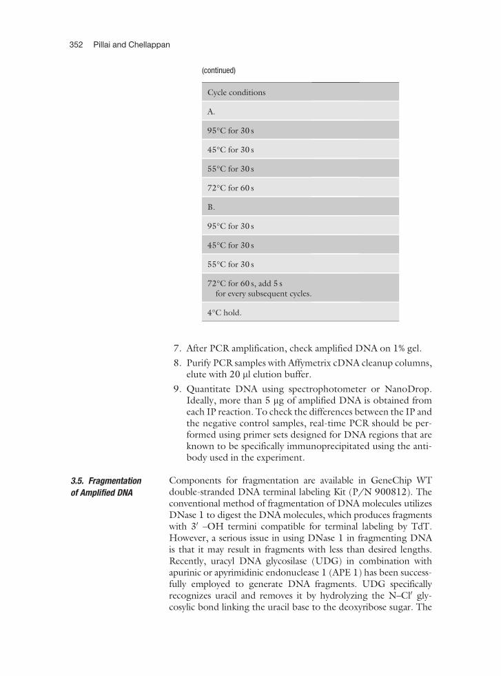
7. After PCR amplification, check amplified DNA on 1% gel.
8. Purify PCR samples with Affymetrix cDNA cleanup columns,elute with 20 ml elution buffer.
9. Quantitate DNA using spectrophotometer or NanoDrop.Ideally, more than 5 mg of amplified DNA is obtained fromeach IP reaction. To check the differences between the IP andthe negative control samples, real-time PCR should be per-formed using primer sets designed for DNA regions that areknown to be specifically immunoprecipitated using the anti-body used in the experiment.
3.5. Fragmentation
of Amplified DNA
Components for fragmentation are available in GeneChip WTdouble-stranded DNA terminal labeling Kit (P/N 900812). Theconventional method of fragmentation of DNA molecules utilizesDNase 1 to digest the DNA molecules, which produces fragmentswith 30 –OH termini compatible for terminal labeling by TdT.However, a serious issue in using DNase 1 in fragmenting DNAis that it may result in fragments with less than desired lengths.Recently, uracyl DNA glycosilase (UDG) in combination withapurinic or apyrimidinic endonuclease 1 (APE 1) has been success-fully employed to generate DNA fragments. UDG specificallyrecognizes uracil and removes it by hydrolyzing the N–Cl0 gly-cosylic bond linking the uracil base to the deoxyribose sugar. The
Cycle conditions
A.
95�C for 30 s
45�C for 30 s
55�C for 30 s
72�C for 60 s
B.
95�C for 30 s
45�C for 30 s
55�C for 30 s
72�C for 60 s, add 5 sfor every subsequent cycles.
4�C hold.
(continued)
352 Pillai and Chellappan
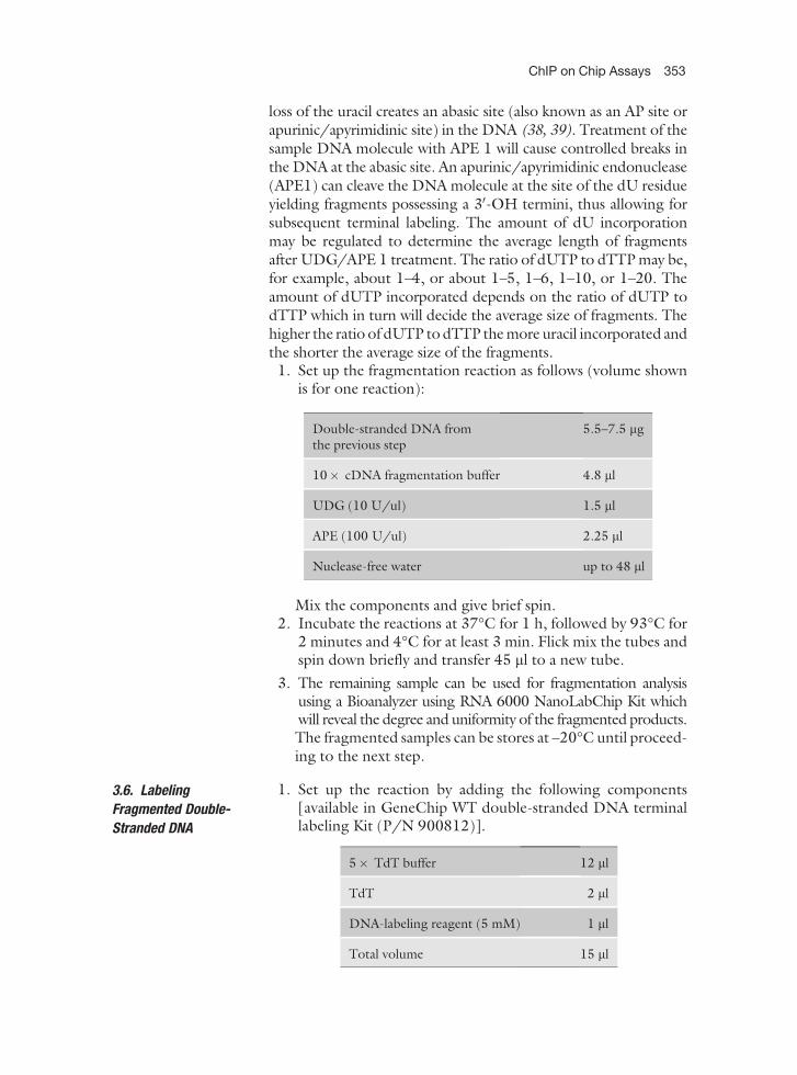
loss of the uracil creates an abasic site (also known as an AP site orapurinic/apyrimidinic site) in the DNA (38, 39). Treatment of thesample DNA molecule with APE 1 will cause controlled breaks inthe DNA at the abasic site. An apurinic/apyrimidinic endonuclease(APE1) can cleave the DNA molecule at the site of the dU residueyielding fragments possessing a 30-OH termini, thus allowing forsubsequent terminal labeling. The amount of dU incorporationmay be regulated to determine the average length of fragmentsafter UDG/APE 1 treatment. The ratio of dUTP to dTTP may be,for example, about 1–4, or about 1–5, 1–6, 1–10, or 1–20. Theamount of dUTP incorporated depends on the ratio of dUTP todTTP which in turn will decide the average size of fragments. Thehigher the ratio of dUTP to dTTP the more uracil incorporated andthe shorter the average size of the fragments.
1. Set up the fragmentation reaction as follows (volume shownis for one reaction):
Mix the components and give brief spin.2. Incubate the reactions at 37�C for 1 h, followed by 93�C for
2 minutes and 4�C for at least 3 min. Flick mix the tubes andspin down briefly and transfer 45 ml to a new tube.
3. The remaining sample can be used for fragmentation analysisusing a Bioanalyzer using RNA 6000 NanoLabChip Kit whichwill reveal the degree and uniformity of the fragmented products.The fragmented samples can be stores at –20�C until proceed-ing to the next step.
3.6. Labeling
Fragmented Double-
Stranded DNA
1. Set up the reaction by adding the following components[available in GeneChip WT double-stranded DNA terminallabeling Kit (P/N 900812)].
Double-stranded DNA fromthe previous step
5.5–7.5 mg
10� cDNA fragmentation buffer 4.8 ml
UDG (10 U/ul) 1.5 ml
APE (100 U/ul) 2.25 ml
Nuclease-free water up to 48 ml
5� TdT buffer 12 ml
TdT 2 ml
DNA-labeling reagent (5 mM) 1 ml
Total volume 15 ml
ChIP on Chip Assays 353
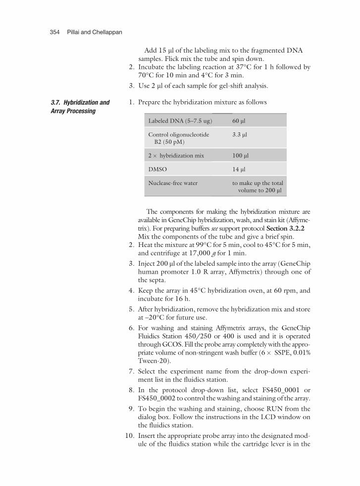
Add 15 ml of the labeling mix to the fragmented DNAsamples. Flick mix the tube and spin down.
2. Incubate the labeling reaction at 37�C for 1 h followed by70�C for 10 min and 4�C for 3 min.
3. Use 2 ml of each sample for gel-shift analysis.
3.7. Hybridization and
Array Processing
1. Prepare the hybridization mixture as follows
The components for making the hybridization mixture areavailable in GeneChip hybridization, wash, and stain kit (Affyme-trix). For preparing buffers see support protocol Section 3.2.2Mix the components of the tube and give a brief spin.
2. Heat the mixture at 99�C for 5 min, cool to 45�C for 5 min,and centrifuge at 17,000 g for 1 min.
3. Inject 200 ml of the labeled sample into the array (GeneChiphuman promoter 1.0 R array, Affymetrix) through one ofthe septa.
4. Keep the array in 45�C hybridization oven, at 60 rpm, andincubate for 16 h.
5. After hybridization, remove the hybridization mix and storeat –20�C for future use.
6. For washing and staining Affymetrix arrays, the GeneChipFluidics Station 450/250 or 400 is used and it is operatedthrough GCOS. Fill the probe array completely with the appro-priate volume of non-stringent wash buffer (6� SSPE, 0.01%Tween-20).
7. Select the experiment name from the drop-down experi-ment list in the fluidics station.
8. In the protocol drop-down list, select FS450_0001 orFS450_0002 to control the washing and staining of the array.
9. To begin the washing and staining, choose RUN from thedialog box. Follow the instructions in the LCD window onthe fluidics station.
10. Insert the appropriate probe array into the designated mod-ule of the fluidics station while the cartridge lever is in the
Labeled DNA (5–7.5 ug) 60 ml
Control oligonucleotideB2 (50 pM)
3.3 ml
2� hybridization mix 100 ml
DMSO 14 ml
Nuclease-free water to make up the totalvolume to 200 ml
354 Pillai and Chellappan
eject position. The lever returns to the engaged positionwhen finished.
11. Place a microcentrifuge tube (1.5 ml) containing 600 ml ofSAPE solution mix/stain cocktail 1 in sample holder 1, onevial containing 600 ml of antibody solution mix/stain cocktail2 in sample holder 2, and another vial containing 800 ml ofarray holding buffer in sample holder 3.
12. Press down on the needle lever to snap needles into positionand to start the run.
13. When the run is completed, remove the tubes containing thesolutions, and replace with fresh microcentrifuge vials.
14. Remove the array from the fluidics station by pressing downthe lever to eject position.
15. Scan the array using GeneChip Scanner 3000 7 G. Probearrays can be stored for a short while at 4�C, in the dark.
16. Turn the scanneron 10 min prior to use forwarming up the laser.
3.8. Protocol Check
Points
The protocol was successfully used for hybridization with Affyme-trix human promoter 1.0 array in our lab. Protocol conditionsshould be optimized by each user depending on array type, celltype, specificity of antibodies, assay conditions, etc. The followingsteps are recommended as checkpoints to assess the success of theimmunoprecipitation protocol prior to performing hybridizations.1. Visualization of sheared chromatin in a 1% agarose gel. Ide-
ally, 100–1000 bp fragments should be obtained after suc-cessful sonication. Alternatively a bioanalyzer can be used todetermine the accurate size of the sheared DNA. Care shouldbe taken not to overshear or oversonicate DNA as it mayresult in damaged DNA–protein complex and unsuccessfulimmunoprecipitation. The optimum sonication should bedetermined for each cell type.
2. Gene-specific PCR of a known target to check IP enrichment.Validate the antibody with RNA pol II to see whether theimmunoprecipitation is working.
3. Monitor DNA yield after DNA cleanup and amplification ofimmunoprecipitated DNA preferably using NanoDrop.Unfortunately it does not provide information on the qualityof amplified DNA.
3.9. Support Protocols
3.9.1. Preclearing of
ChIP lysate
Preclearing of ChIP lysates is an efficient method to eliminatenonspecific background in ChIP assay (22, 40).
1. Add 120 ml of 1:1 protein G slurry (or 400 ml of protein A) tothe ChIP lysate (mentioned in Section 1.1) in a microcen-trifuge tube.
2. Add 15 ml of 10 mg/ml salmon sperm DNA (Sigma-Aldrich,cat. no. D7696) and 10 ml of BSA (10 mg/ml).
ChIP on Chip Assays 355
3. Rotate on a nutator for 30 min at 4 0�C. Spin the tubes at17,000 g for 30 min in a tabletop centrifuge at 4�C.
4. Collect the supernatant and discard the beads. This super-natant fraction is used for the immunoprecipitation reactiondescribed in Section 3.1.2
3.10. Alternate
Protocols
3.10.1. Amplifying
Immunoprecipitated
DNA by Ligation-
Mediated PCR
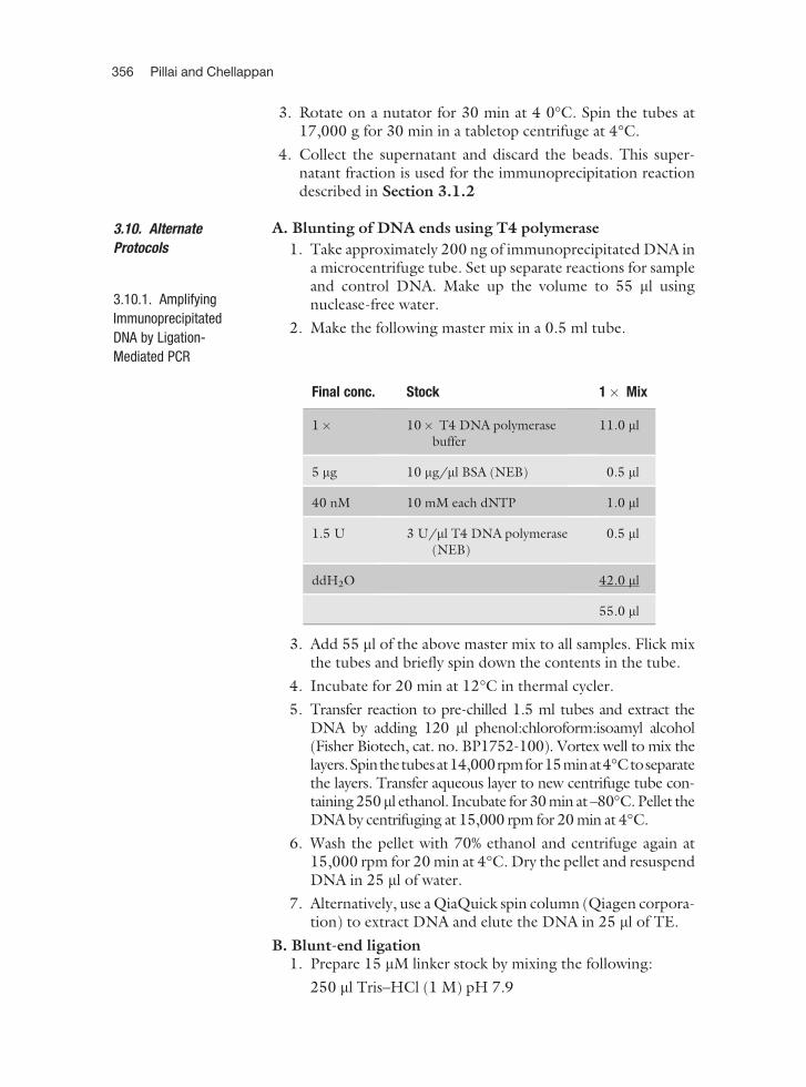
A. Blunting of DNA ends using T4 polymerase
1. Take approximately 200 ng of immunoprecipitated DNA ina microcentrifuge tube. Set up separate reactions for sampleand control DNA. Make up the volume to 55 ml usingnuclease-free water.
2. Make the following master mix in a 0.5 ml tube.
3. Add 55 ml of the above master mix to all samples. Flick mixthe tubes and briefly spin down the contents in the tube.
4. Incubate for 20 min at 12�C in thermal cycler.
5. Transfer reaction to pre-chilled 1.5 ml tubes and extract theDNA by adding 120 ml phenol:chloroform:isoamyl alcohol(Fisher Biotech, cat. no. BP1752-100). Vortex well to mix thelayers.Spinthetubesat14,000rpmfor15minat4�Ctoseparatethe layers. Transfer aqueous layer to new centrifuge tube con-taining 250ml ethanol. Incubate for 30 min at –80�C. Pellet theDNA by centrifuging at 15,000 rpm for 20 min at 4�C.
6. Wash the pellet with 70% ethanol and centrifuge again at15,000 rpm for 20 min at 4�C. Dry the pellet and resuspendDNA in 25 ml of water.
7. Alternatively, use a QiaQuick spin column (Qiagen corpora-tion) to extract DNA and elute the DNA in 25 ml of TE.
B. Blunt-end ligation1. Prepare 15 mM linker stock by mixing the following:
250 ml Tris–HCl (1 M) pH 7.9
Final conc. Stock 1� Mix
1� 10� T4 DNA polymerasebuffer
11.0 ml
5 mg 10 mg/ml BSA (NEB) 0.5 ml
40 nM 10 mM each dNTP 1.0 ml
1.5 U 3 U/ml T4 DNA polymerase(NEB)
0.5 ml
ddH2O 42.0 ml
55.0 ml
356 Pillai and Chellappan
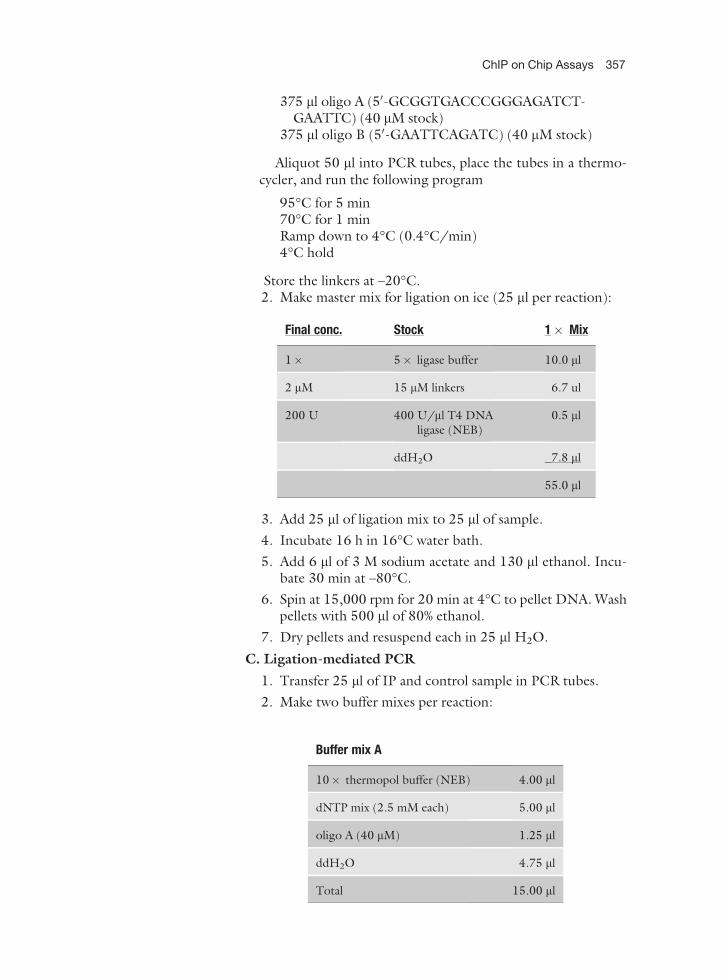
375 ml oligo A (50-GCGGTGACCCGGGAGATCT-GAATTC) (40 mM stock)
375 ml oligo B (50-GAATTCAGATC) (40 mM stock)
Aliquot 50 ml into PCR tubes, place the tubes in a thermo-cycler, and run the following program
95�C for 5 min70�C for 1 minRamp down to 4�C (0.4�C/min)4�C hold
Store the linkers at –20�C.2. Make master mix for ligation on ice (25 ml per reaction):
3. Add 25 ml of ligation mix to 25 ml of sample.
4. Incubate 16 h in 16�C water bath.
5. Add 6 ml of 3 M sodium acetate and 130 ml ethanol. Incu-bate 30 min at –80�C.
6. Spin at 15,000 rpm for 20 min at 4�C to pellet DNA. Washpellets with 500 ml of 80% ethanol.
7. Dry pellets and resuspend each in 25 ml H2O.
C. Ligation-mediated PCR
1. Transfer 25 ml of IP and control sample in PCR tubes.
2. Make two buffer mixes per reaction:
Final conc. Stock 1� Mix
1� 5� ligase buffer 10.0 ml
2 mM 15 mM linkers 6.7 ul
200 U 400 U/ml T4 DNAligase (NEB)
0.5 ml
ddH2O 7.8 ml
55.0 ml
Buffer mix A
10� thermopol buffer (NEB) 4.00 ml
dNTP mix (2.5 mM each) 5.00 ml
oligo A (40 mM) 1.25 ml
ddH2O 4.75 ml
Total 15.00 ml
ChIP on Chip Assays 357
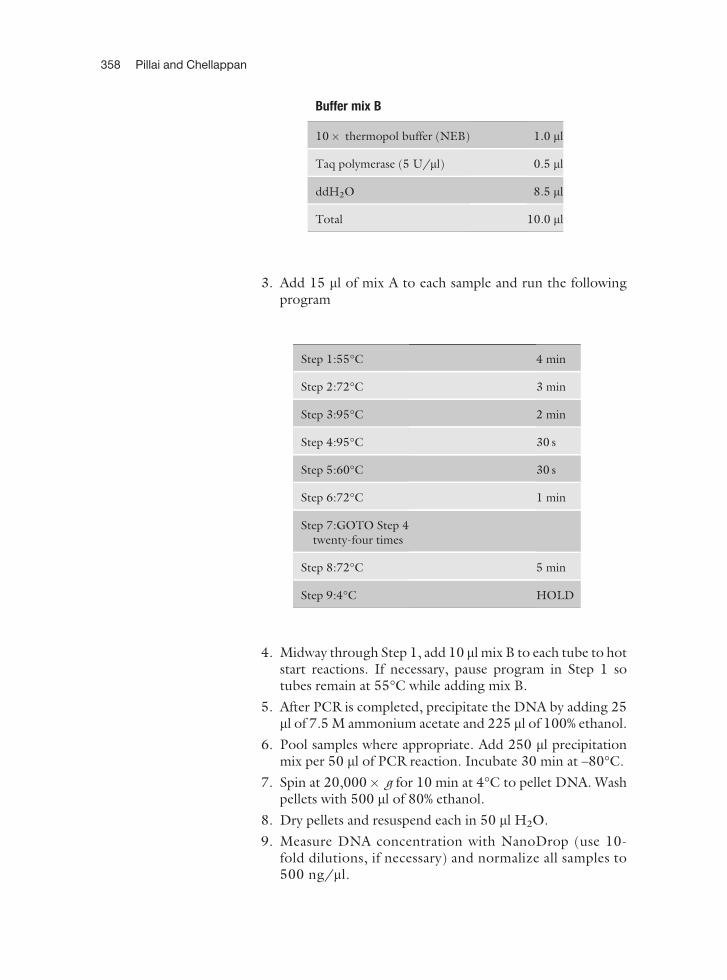
3. Add 15 ml of mix A to each sample and run the followingprogram
4. Midway through Step 1, add 10 ml mix B to each tube to hotstart reactions. If necessary, pause program in Step 1 sotubes remain at 55�C while adding mix B.
5. After PCR is completed, precipitate the DNA by adding 25ml of 7.5 M ammonium acetate and 225 ml of 100% ethanol.
6. Pool samples where appropriate. Add 250 ml precipitationmix per 50 ml of PCR reaction. Incubate 30 min at –80�C.
7. Spin at 20,000� g for 10 min at 4�C to pellet DNA. Washpellets with 500 ml of 80% ethanol.
8. Dry pellets and resuspend each in 50 ml H2O.
9. Measure DNA concentration with NanoDrop (use 10-fold dilutions, if necessary) and normalize all samples to500 ng/ml.
Step 1:55�C 4 min
Step 2:72�C 3 min
Step 3:95�C 2 min
Step 4:95�C 30 s
Step 5:60�C 30 s
Step 6:72�C 1 min
Step 7:GOTO Step 4twenty-four times
Step 8:72�C 5 min
Step 9:4�C HOLD
Buffer mix B
10� thermopol buffer (NEB) 1.0 ml
Taq polymerase (5 U/ml) 0.5 ml
ddH2O 8.5 ml
Total 10.0 ml
358 Pillai and Chellappan
3.10.2. Whole Genome
Amplification PCR for
Amplifying
Immunoprecipitated
DNA
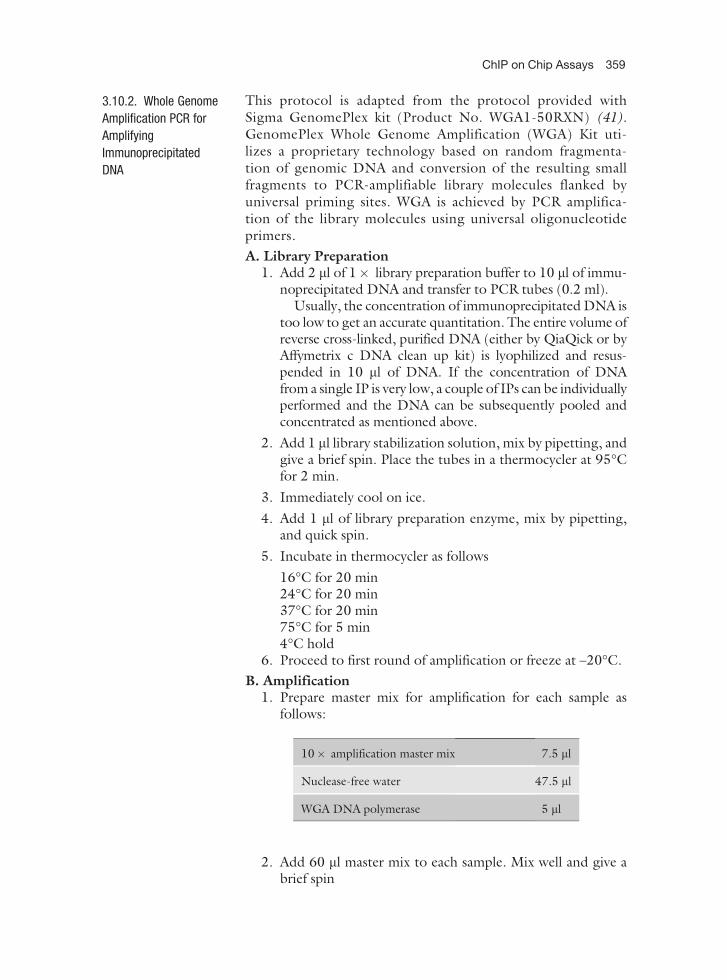
This protocol is adapted from the protocol provided withSigma GenomePlex kit (Product No. WGA1-50RXN) (41).GenomePlex Whole Genome Amplification (WGA) Kit uti-lizes a proprietary technology based on random fragmenta-tion of genomic DNA and conversion of the resulting smallfragments to PCR-amplifiable library molecules flanked byuniversal priming sites. WGA is achieved by PCR amplifica-tion of the library molecules using universal oligonucleotideprimers.
A. Library Preparation1. Add 2 ml of 1� library preparation buffer to 10 ml of immu-
noprecipitated DNA and transfer to PCR tubes (0.2 ml).Usually, the concentration of immunoprecipitated DNA is
too low to get an accurate quantitation. The entire volume ofreverse cross-linked, purified DNA (either by QiaQick or byAffymetrix c DNA clean up kit) is lyophilized and resus-pended in 10 ml of DNA. If the concentration of DNAfrom a single IP is very low, a couple of IPs can be individuallyperformed and the DNA can be subsequently pooled andconcentrated as mentioned above.
2. Add 1 ml library stabilization solution, mix by pipetting, andgive a brief spin. Place the tubes in a thermocycler at 95�Cfor 2 min.
3. Immediately cool on ice.
4. Add 1 ml of library preparation enzyme, mix by pipetting,and quick spin.
5. Incubate in thermocycler as follows
16�C for 20 min24�C for 20 min37�C for 20 min75�C for 5 min4�C hold
6. Proceed to first round of amplification or freeze at –20�C.
B. Amplification1. Prepare master mix for amplification for each sample as
follows:
2. Add 60 ml master mix to each sample. Mix well and give abrief spin
10� amplification master mix 7.5 ml
Nuclease-free water 47.5 ml
WGA DNA polymerase 5 ml
ChIP on Chip Assays 359
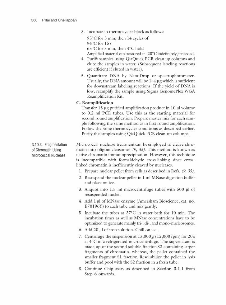
3. Incubate in thermocycler block as follows:
95�C for 3 min, then 14 cycles of94�C for 15 s65�C for 5 min, then 4�C holdAmplifiedmaterial canbestoredat–20�Cindefinitely, if needed.
4. Purify samples using QiaQuick PCR clean up columns andelute the samples in water. (Subsequent labeling reactionsare efficient if eluted in water).
5. Quantitate DNA by NanoDrop or spectrophotometer.Usually, the DNA amount will be 1–4 mg which is sufficientfor downstream labeling reactions. If the yield of DNA islow, reamplify the sample using Sigma GenomePlex WGAReamplification Kit.
C. ReamplificationTransfer 15 mg purified amplification product in 10 ml volumeto 0.2 ml PCR tubes. Use this as the starting material forsecond round amplification. Prepare master mix for each sam-ple following the same method as in first round amplification.Follow the same thermocycler conditions as described earlier.Purify the samples using QiaQuick PCR clean-up columns.
3.10.3. Fragmentation
of Chromatin Using
Micrococcal Nuclease
Micrococcal nuclease treatment can be employed to cleave chro-matin into oligonucleosomes (9, 35). This method is known asnative chromatin immunoprecipitation. However, this techniqueis incompatible with formaldehyde cross-linking since cross-linked chromatin is inefficiently cleaved by nucleases.
1. Prepare nuclear pellet from cells as described in Refs. (9, 35).
2. Resuspend the nuclear pellet in 1 ml MNase digestion bufferand place on ice.
3. Aliquot into 1.5 ml microcentrifuge tubes with 500 ml ofresuspended nuclei.
4. Add 1 ml of MNase enzyme (Amersham Bioscience, cat. no.E70196Y) to each tube and mix gently.
5. Incubate the tubes at 37�C in water bath for 10 min. Theincubation times as well as MNase concentrations have to beoptimized to generate mainly tri-, di-, and mono-nucleosomes.
6. Add 20 ml of stop solution. Chill on ice.
7. Centrifuge the suspension at 13,000 g (12,000 rpm) for 20 sat 4�C in a refrigerated microcentrifuge. The supernatant ismade up of the second soluble fraction S2 containing largerfragments of chromatin, whereas, the pellet contained thesmaller fragment S1 fraction. Resolubilize the pellet in lysisbuffer and pool with the S2 fraction in a fresh tube.
8. Continue Chip assay as described in Section 3.1.1 fromStep 6 onwards.
360 Pillai and Chellappan
3.10.4. Preparation
of Magnetic Beads
The use of magnetic beads for immunoprecipitation has beenwidely used in recent years (42, 43). The beads are available indifferent forms, precoupled with protein G, protein A, or second-ary antibodies (Dynabeads protein G (Invitrogen, cat. no.100–03D), Dynabeads protein A (cat. no. 100–01D). For eachIP 100 ml of Dynabeads is recommended.
1. Transfer 100 ml of Dynabeads to 1.5 ml microfuge tubes.
2. Add 1 ml blocking buffer (0.5% BSA in 1� PBS).
3. Collect the beads using a magnetic stand. Remove thesupernatant.
4. Repeat washing with blocking buffer for two more times.
5. Resuspend beads in 250 ml blocking buffer and add antibody(10 g) to the tube.
6. Incubate overnight in a nutator at 4�C.
7. Next day, wash the beads three times with blocking bufferand resuspend the beads in 100 ml of blocking buffer.
8. For immunoprecipitation, add 100 ml magnetic bead–anti-boby mix to the cell lysate from Step 3.2.1 and incubate overnight in a nutator at 4�C.
For detailed description of ChIP using Dynabeads referthe protocols from Young lab (43).
3.11. Conclusions ChIP-chip or chromatin immunoprecipitation followed by DNAmicroarray analysis has proven to be an efficient means of mappingprotein–genome interactions on a genome-wide scale increasingour understanding of diverse cellular processes. Much of the workso far has focused on binding of transcription factors and manycomputational methods have been developed to identify thebound regions. Recently this technology has been extended tothe genomic mappings of the other features such as histone mod-ifications, transcriptionally active regions, and binding sites forother protein complexes. This article outlines the general strate-gies used to carry out ChIP-chip assays to study the differentialrecruitment of regulatory molecules based on the studies con-ducted in our lab as well as other published protocols.
4. Notes
1. GeneChip human promoter 1.0R array (Affymetrix, Cat.No. 900775) is available as a single array comprising of over4.6 million probes tiled through over 25,500 human promoterregions. Sequences used in the design of this array are based onthe sequences available from NCBI genome assembly (Build
ChIP on Chip Assays 361
34). Oligonucleotide probes are synthesized in situ comple-mentary to each corresponding sequence. Probes are tiled at anaverage resolution of 35 bp, as measured from the centralposition of adjacent 25 mer oligos, leaving a gap of approxi-mately 10 bp between probes. Each promoter region coversapproximately 7.5 kb upstream through 2.45 kb downstreamof 50 transcription start sites. For more than 1300 cancerassociated genes additional promoter region is included inthe array, covering 10 kb upstream of transcription start site.
2. In addition to Affymetrix arrays, ChIP-chip microarrays areavailable from Agilent and NimbleGen. Available formatsinclude whole genome tiling arrays for model organisms likehuman and mouse as well as focused arrays for promoterregions and custom array designs. Each Agilent ChIP-on-Chip microarray features a total of �244,000 60-mer oligo-nucleotide probes. Probes are spaced every �100–300 bpacross regions of interest in both coding and non-codingDNA sequence. Such focused microarrays include proximalpromoter (–0.8 KB upstream to +0.2 KB downstream of iden-tified transcriptional start sites) and expanded promoter(–8.0 KB upstream to +2.0 KB downstream) designs.
NimbleGen’s current high-density microarrays with longoligonucleotide probes (50–85 mer) contain 385,000 probeson a single glass slide. The entire non-repetitive human andmouse genomes can be surveyed at 100 bp intervals, each witha set of 38 arrays. Agilent and NimbleGen microarray plat-forms utilize two color labeling as opposed to single colorlabeling of Affymetrix arrays. Two color labeling using Cy3/Cy5 can be done using CGH kit (Invitrogen) which utilizes arandom primed, Klenow-based extension protocol.
3. In conventional ChIP protocol, cells or tissues are treated withformaldehyde, a cell permeable small molecule that can mediateprotein–protein and protein–DNA cross-linking through Schiffbase formation. To increase the degree of protein–proteincross-linking, treatment with dimethyl adipimidate can bedone first, followed by formaldehyde treatment (44). Thisis an efficient method to observe indirect protein–DNAinteractions.
4. Antibodies used for ChIP-on-chip can be an important limit-ing factor. Not all antibodies can effectively immunoprecipi-tate protein–DNA complexes. An antibody that gives aspecific signal in Western blot need not be ideal for bindingto a DNA bound, cross-linked protein. ChIP-on-chiprequires highly specific antibodies that must recognize itsepitope in free solution and also under fixed conditions.If the antibody is demonstrated to successfully immunopre-cipitate cross-linked chromatin, it is termed ‘‘ChIP-Grade’’.
362 Pillai and Chellappan
Validated ChIP-grade antibodies are available from AbcamInc., Santa Cruz Biotechnology Inc., and Upstate (MilliporeCorporation). Since the affinity and avidity of antibodies canvary, the amount of antibody in each ChIP-chip reactionneeds to be empirically determined. To demonstrate thatthe antibody was successful in immunoprecipitating the pro-tein–DNA complex, PCR amplification on a known binding-site region for the protein of interest should be performedusing either conventional PCR methods followed by agarosegel electrophoresis or by quantitative PCR. The antibodyused for immunoprecipitation should be a high affinity,high specificity antibody to avoid nonspecific backgroundin ChIP assay. It is important to incorporate a positive con-trol as well as an irrelevant antibody in the ChIP assay tofacilitate critical interpretation of data.
5. It is necessary to have appropriate experimental controls inChIP-chip experiments. Listed below are common controlsthat researchers are using in their experiments:
(a) Nonspecific IgG antibodies – The most common type ofnegative control involves adding antibodies that do notrecognize a specific epitope, for example, pre-immuneserum or IgG. A potential pitfall is that since the antibodiesdo not immunoprecipitate effectively, the nonspecificDNA yield is often extremely low. Hence, the hybridiza-tion tends to be much noisier and can result in many falsepositives due to amplification of trace amounts of nonspe-cific DNA. Alternatively, the primary antibody can beomitted (i.e., no antibody control).
(b) Protein deficient cell line – Several upstream applications,such as target deletion or siRNA, can be used to perturbthe expression of the protein of interest, hence decreasingthe amount of immunoprecipitated material. Alterna-tively, a cell line that does not express the protein ofinterest could be used as a negative control.
(c) Tag-specific antibodies – If your protein of interest has atag, such as GST or GFP, you may want to consider usingantibodies against them in cells that do and do not expressthis tagged protein of interest.
Generally genomic DNA is used as an input control andsamples from no antibody or immunoglobulin G groups areused as negative controls. Other control designs such as trans-formed cell lines versus empty vector cell lines, wild type targetversus mutation target, and drug treated versus untreated can allbe used as control options (45).
6. As mentioned previously, the amount of starting materialrequired for successful microarray hybridization is very critical in
ChIP on Chip Assays 363
a ChIP-chip experiment. The amount is highly variable depend-ing on the quality of the antibody, binding frequency of proteinto DNA, and abundance of the protein. It is recommended thatpooling of immunoprecipitated DNA samples to acquire enoughDNA for subsequent amplification is a feasible strategy. Unfortu-nately, pooling ChIP samples is not always possible, especially ifspecialized cell types or tumor tissues are used. The amplificationof immunoprecipitated DNA using T7 polymerase is widely usedto linearly amplify ChIP DNA. However, a PCR bias may occur,especially in mammalian systems where large amounts of repeatsequence may skew data.
7. The statistical analysis of the huge amount of data generatedfrom arrays is a challenge and normalization procedures shouldaim to minimize artifacts and determine what is really biologi-cally significant. So far, application to mammalian genomes hasbeen a major limitation, for example, due to a significantpercentage of the genome that is occupied by repeats. How-ever, as ChIP-on-chip technology advances, high resolutionwhole mammalian genome maps are achievable.
8. Although ChIP-on-chip can be a powerful technique in the areaof genomics, it is very expensive. Most published studies usingChIP-on-chip repeat their experiments at least three times inorder to obtain biologically meaningful maps. The cost of theDNA microarrays is often a limiting factor to whether a labora-tory should proceed with a ChIP-on-chip experiment.
9. Another limitation is the size of DNA fragments that can beachieved. Most ChIP-on-chip protocols utilize sonication as amethod of breaking up DNA into small pieces. However,sonication is limited to a minimal fragment size of 200 bp. Inorder for higher resolution maps, this limitation should beovercome to achieve smaller fragments, preferably to singlenucleosome resolution.
Acknowledgments
Studies in the author’s laboratory are supported by the grantsCA63136, CA77301, and CA127725 from the NIH.
References
1. Garner, M.M., and Revzin, A. 1981. A gelelectrophoresis method for quantifying thebinding of proteins to specific DNA regions:application to components of the Escherichiacoli lactose operon regulatory system.Nucleic Acids Res 9:3047–3060.
2. Fried, M., and Crothers, D.M. 1981. Equili-bria and kinetics of lac repressor-operatorinteractions by polyacrylamide gel electro-phoresis. Nucleic Acids Res 9:6505–6525.
3. Hecht, A., and Grunstein, M. 1999. Map-ping DNA interaction sites of chromosomal
364 Pillai and Chellappan
proteins using immunoprecipitation andpolymerase chain reaction. Methods Enzymol304:399–414.
4. Kirmizis, A., and Farnham, P.J. 2004. Geno-mic approaches that aid in the identificationof transcription factor target genes. Exp BiolMed (Maywood) 229:705–721.
5. Squazzo, S.L., O’Geen, H., Komashko,V.M., Krig, S.R., Jin, V.X., Jang, S.W., Mar-gueron, R., Reinberg, D., Green, R., andFarnham, P.J. 2006. Suz12 binds to silencedregions of the genome in a cell-type-specificmanner. Genome Res 16:890–900.
6. Buck, M.J., and Lieb, J.D. 2004. ChIP-chip: considerations for the design, analysis,and application of genome-wide chromatinimmunoprecipitation experiments. Geno-mics 83:349–360.
7. Orlando, V. 2000. Mapping chromosomalproteins in vivo by formaldehyde-crosslinked-chromatin immunoprecipitation. Trends Bio-chem Sci 25:99–104.
8. Wells, J., and Farnham, P.J. 2002. Charac-terizing transcription factor binding sitesusing formaldehyde crosslinking and immu-noprecipitation. Methods 26:48–56.
9. Umlauf, D., Goto, Y., and Feil, R. 2004. Site-specific analysis of histone methylation andacetylation. Methods Mol Biol 287:99–120.
10. Litt, M.D., Simpson, M., Recillas-Targa, F.,Prioleau, M.N., and Felsenfeld, G. 2001.Transitions in histone acetylation revealboundaries of three separately regulatedneighboring loci. Embo J 20:2224–2235.
11. Hebbes, T.R., Clayton, A.L., Thorne, A.W.,and Crane-Robinson, C. 1994. Core histonehyperacetylation co-maps with generalizedDNase I sensitivity in the chicken beta-globin chromosomal domain. Embo J13:1823–1830.
12. Oberley, M.J., Tsao, J., Yau, P., and Farnham,P.J. 2004. High-throughput screening of chro-matin immunoprecipitates using CpG-islandmicroarrays. Methods Enzymol 376:315–334.
13. Oberley, M.J., Inman, D.R., and Farnham,P.J. 2003. E2F6 negatively regulates BRCA1in human cancer cells without methylation ofhistone H3 on lysine 9. J Biol Chem278:42466–42476.
14. Bieda, M., Xu, X., Singer, M.A., Green, R.,and Farnham, P.J. 2006. Unbiased locationanalysis of E2F1-binding sites suggests awidespread role for E2F1 in the human gen-ome. Genome Res 16:595–605.
15. Jin, V.X., Rabinovich, A., Squazzo, S.L.,Green, R., and Farnham, P.J. 2006. A com-putational genomics approach to identifycis-regulatory modules from chromatinimmunoprecipitation microarray data – acase study using E2F1. Genome Res16:1585–1595.
16. Ferea, T.L., and Brown, P.O. 1999. Obser-ving the living genome. Curr Opin GenetDev 9:715–722.
17. Sikder, D., and Kodadek, T. 2005. Genomicstudies of transcription factor-DNA interac-tions. Curr Opin Chem Biol 9:38–45.
18. Chee, M., Yang, R., Hubbell, E., Berno, A.,Huang, X.C., Stern, D., Winkler, J., Lockhart,D.J., Morris, M.S., and Fodor, S.P. 1996.Accessing genetic information with high-den-sity DNA arrays. Science 274:610–614.
19. Cawley, S., Bekiranov, S., Ng, H.H., Kapra-nov, P., Sekinger, E.A., Kampa, D., Piccol-boni, A., Sementchenko, V., Cheng, J.,Williams, A.J., et al. 2004. Unbiased map-ping of transcription factor binding sitesalong human chromosomes 21 and 22points to widespread regulation of noncod-ing RNAs. Cell 116:499–509.
20. Kondo, Y., Shen, L., Yan, P.S., Huang, T.H.,and Issa, J.P. 2004. Chromatin immunopreci-pitation microarrays for identification of genessilenced by histone H3 lysine 9 methylation.Proc Natl Acad Sci U S A 101:7398–7403.
21. Wells, J., Yan, P.S., Cechvala, M., Huang, T.,and Farnham, P.J. 2003. Identification ofnovel pRb binding sites using CpG microar-rays suggests that E2F recruits pRb to speci-fic genomic sites during S phase. Oncogene22:1445–1460.
22. Kirmizis, A., Bartley, S.M., Kuzmichev, A.,Margueron, R., Reinberg, D., Green, R., andFarnham, P.J. 2004. Silencing of humanpolycomb target genes is associated withmethylation of histone H3 Lys 27. GenesDev 18:1592–1605.
23. Iyer, V.R., Horak, C.E., Scafe, C.S.,Botstein, D., Snyder, M., and Brown, P.O.2001. Genomic binding sites of the yeastcell-cycle transcription factors SBF andMBF. Nature 409:533–538.
24. Horak, C.E., Luscombe, N.M., Qian, J.,Bertone, P., Piccirrillo, S., Gerstein, M.,and Snyder, M. 2002. Complex transcrip-tional circuitry at the G1/S transition in Sac-charomyces cerevisiae. Genes Dev16:3017–3033.
ChIP on Chip Assays 365
25. Horak, C.E., and Snyder, M. 2002. Globalanalysis of gene expression in yeast. FunctIntegr Genomics 2:171–180.
26. Lee, T.I., Rinaldi, N.J., Robert, F., Odom,D.T., Bar-Joseph, Z., Gerber, G.K., Han-nett, N.M., Harbison, C.T., Thompson,C.M., Simon, I., et al. 2002. Transcriptionalregulatory networks in Saccharomyces cere-visiae. Science 298:799–804.
27. Horak, C.E., Mahajan, M.C., Luscombe,N.M., Gerstein, M., Weissman, S.M., andSnyder, M. 2002. GATA-1 binding sitesmapped in the beta-globin locus by usingmammalian chIp-chip analysis. Proc NatlAcad Sci U S A 99:2924–2929.
28. Weinmann, A.S., Yan, P.S., Oberley, M.J.,Huang, T.H., and Farnham, P.J. 2002. Iso-lating human transcription factor targets bycoupling chromatin immunoprecipitationand CpG island microarray analysis. GenesDev 16:235–244.
29. Bernstein, B.E., Humphrey, E.L., Erlich,R.L., Schneider, R., Bouman, P., Liu, J.S.,Kouzarides, T., and Schreiber, S.L. 2002.Methylation of histone H3 Lys 4 in codingregions of active genes. Proc Natl Acad Sci US A 99:8695–8700.
30. Reid, J.L., Iyer, V.R., Brown, P.O., andStruhl, K. 2000. Coordinate regulation ofyeast ribosomal protein genes is associatedwith targeted recruitment of Esa1 histoneacetylase. Mol Cell 6:1297–1307.
31. Kurdistani, S.K., Tavazoie, S., and Grun-stein, M. 2004. Mapping global histone acet-ylation patterns to gene expression. Cell117:721–733.
32. Fusaro, G., Dasgupta, P., Rastogi, S., Joshi, B.,and Chellappan, S. 2003. Prohibitin inducesthe transcriptional activity of p53 and isexported from the nucleus upon apoptotic sig-naling. J Biol Chem 278:47853–47861.
33. Joshi, B., Ordonez-Ercan, D., Dasgupta, P.,and Chellappan, S. 2005. Induction ofhuman metallothionein 1G promoter byVEGF and heavy metals: differential involve-ment of E2F and metal transcription factors.Oncogene 24:2204–2217.
34. Dasgupta, P., Betts, V., Rastogi, S., Joshi, B.,Morris, M., Brennan, B., Ordonez-Ercan,D., and Chellappan, S. 2004. Direct bindingof apoptosis signal-regulating kinase 1 toretinoblastoma protein: novel links between
apoptotic signaling and cell cycle machinery.J Biol Chem 279:38762–38769.
35. Thorne, A.W., Myers, F.A., and Hebbes,T.R. 2004. Native chromatin immunopreci-pitation. Methods Mol Biol 287:21–44.
36. Dorbic, T., and Wittig, B. 1986. Isolation ofoligonucleosomes from active chromatinusing HMG17-specific monoclonal antibo-dies. Nucleic Acids Res 14:3363–3376.
37. Dorbic, T., and Wittig, B. 1987. Chromatinfrom transcribed genes contains HMG17only downstream from the starting point oftranscription. Embo J 6:2393–2399.
38. Yoshida, A., and Ueda, T. 2003. Human APendonuclease possesses a significant activityas major 30–50 exonuclease in human leuke-mia cells. Biochem Biophys Res Commun310:522–528.
39. Yoshida, A., Urasaki, Y., Waltham, M., Berg-man, A.C., Pourquier, P., Rothwell, D.G.,Inuzuka, M., Weinstein, J.N., Ueda, T.,Appella, E., et al. 2003. Human apurinic/apyrimidinic endonuclease (Ape1) and its N-terminal truncated form (AN34) areinvolved in DNA fragmentation duringapoptosis. J Biol Chem 278:37768–37776.
40. Boyd, K.E., Wells, J., Gutman, J., Bartley,S.M., and Farnham, P.J. 1998. c-Myc targetgene specificity is determined by a post-DNAbinding mechanism. Proc Natl Acad Sci U SA 95:13887–13892.
41. O’Geen, H., Nicolet, C.M., Blahnik, K.,Green, R., and Farnham, P.J. 2006. Compar-ison of sample preparation methods for ChIP-chip assays. Biotechniques 41:577–580.
42. Guenther, M.G., Levine, S.S., Boyer, L.A.,Jaenisch, R., and Young, R.A. 2007. A chro-matin landmark and transcription initiationat most promoters in human cells. Cell130:77–88.
43. Lee, T.I., Johnstone, S.E., and Young, R.A.2006. Chromatin immunoprecipitation andmicroarray-based analysis of protein loca-tion. Nat Protoc 1:729–748.
44. Kurdistani, S.K., and Grunstein, M. 2003. Invivo protein-protein and protein-DNA cross-linking for genomewide binding microarray.Methods 31:90–95.
45. Wu, J., Smith, L.T., Plass, C., and Huang,T.H. 2006. ChIP-chip comes of age for gen-ome-wide functional analysis. Cancer Res66:6899–6902.
366 Pillai and Chellappan