Embed Size (px)
Citation preview

40
CHAPTER - I Isolation and characterization of
bio-active molecules from medicinal plants

41
1. INTRODUCTION
Natural products are organic compounds that are formed by living systems. The
elucidation of their structures and their chemistry, synthesis and biosynthesis are major
areas of organic chemistry. These compounds may be divided in to three broad categories.
Firstly, there are those compounds which occur in all cells and play a central role in the
metabolism and reproduction of those cells. These compounds include the nucleic acids
and the common amino acids and sugars. They are known as primary metabolites.
Secondly, there are the high-molecular-weight polymeric materials such as cellulose, the
proteins and the lignin‘s which form the cellular structures. Finally, there are those
compounds that are characteristics of a limited range of species. These are secondary
metabolites.
Secondary Metabolites are classified in to:
Polyketides and fatty acids
Terpenoids and steroids
Phenylpropanoids
Alkaloids
Specialized amino acids and peptides
Specialized carbohydrates
Polyketides and fatty acids: Polyketides are formed by the linear combination of acetate
units derived from the ―building block‖ acetyl co-enzyme A. Terpenoids and steroids are
assembled in nature from isoprenoid C5 units derived from isopentenyl (3-methylbut-3-en-
l-yl) pyrophosphate. These C5 units are linked together in a head-to-tail manner. They
have a characteristic branched chain structure. A further group of natural products are
those containing a phenylpropanoid (C6-C3) unit (i.e. Ph-CH2-CH2-CH3).
The amino acids are the building blocks for peptides and proteins. Although the amino
acids are normally considered as primary metabolites, there some unusual amino acids that
are of restricted occurrence. Some antibiotics such as the penicillins are formed from
small peptides. The alkaloids are a structurally diverse group of natural products
containing nitrogen. The nitrogenous portions of the alkaloids are derived from amino
acids such as ornithine, lysine, tyrosine or tryptophan.

42
Although sugars (carbohydrates) such as glucose are typical primary metabolites,
there are other sugars that are of a much more limited occurrence. Some of these less
common sugars are attached to natural products as part of a glycoside. The non-sugar
portion is known as the aglycone, and maybe a terpenoid, alkaloid or polyketide.
Isolation of Natural Products:
Secondary metabolites, with some exceptions, occur in amounts that are less than
0.01% of the dry weight of the plant. Extraction of 1 kg of dry plant material is likely to
yield less than 100 mg of a natural product. These compounds may be unstable and
present as part of a complex mixture. The isolation, separation and purification of these
natural products require considerable skill.
Natural products may be obtained from the crushed biological material by
extraction with a solvent such as petroleum ether or hexane, chloroform, ethyl acetate or
methanol. Several solvents of increasing polarity may be used. Thus lipid material
(waxes, fatty acids, sterols, carotenoids and simple terpenoids) can be extracted with non-
polar solvents, but more polar substances such as alkaloids and glycosides are extracted
with methanol, aqueous methanol or even hot water. Many alkaloids are present as their
salts with naturally occurring acids such as tartaric acid. Finally, the structure of the
natural products can be established by using experimental data such as 1H-NMR,
13C-NMR
Mass, IR and X- Ray Crystallography techniques followed by chemical methods as per the
requirement.
43
Phytochemical investigation and biological activity on evaluation of Salvinia Natans
Natural products isolated from higher plants arid microorganisms have been
providing novel, clinically active drugs. The 20th
century has witnessed much attention
directed to synthetic compounds and the rise of combinatorial chemistry as an important
part of the drug discovery process. Chemistry of natural products involves characterization
of their composition and isolation of specific active component with different techniques,
which lie in various fields of science specially chemistry.
The world health organization has estimated that more than 80% of the world‘s
population in developing countries depends primarily on herbal medicine for basic
healthcare needs.1 Salvinia natans, which belongs to the family of Salvinaceae, is a free
floating, rootless aquatic fern. Despite the large number of fern-like plants, aqueous ferns
of the Salvinia family have only ten species. They all grow in freshwater aquifers of
tropical and subtropical countries, mainly in Africa and South America.2 Salvinia is a rare
and disappearing plant that arrived during the preglacial period. The plant Salvinia natans
(Salvinaceae) is small in size, floating aquatics with creeping stems, branched and bearing
hairs but no true roots. Leaves in whorls of three, with two leaves green, sessile or short-
petioled, flat, entire, and floating, one leaf finely dissected, petiolate, root like, and
pendent. Submerged leaves bearing sori that are surrounded by basifixed membranous
indusia (sporocarps); sporocarps of two types, bearing either mega sporangia that are few
in number (ca. 10), each with single megaspore, or many micro sporangia, each with 64
microspores. The small, hair like growths, known as microgametical follicles, are not
known to have any productive function, and are currently a biological mystery. Chemical
constituents of this plant show anti-oxidant properties and anti-oxidant efficiency is
proportional to the number of polyhydroxy benzoyl units.3 Many anti-oxidant compounds
possess anti-cancer or anti-cancerogenic properties.4

44
Usually, aquatic plants are considered as an effective strategy for
decontaminating waste water. Salvinia natans was found to have a great potentiality for the
removal of heavy metals such as cadmium (Cd), copper (Cu), chromium (Cr), nickel (Ni),
lead (Pb), and mercury (Hg) from waste water. The understanding of chemistry of Salvinia
plants can result in controlling their invasive growth and promote their utilization for
useful purposes.5 The phytochemical investigation on Salvinia natans showed that it
consists 96% of amino compounds.6 Though it is used in most of the systems of medicine
including Homeopathy, no reports on medicinally important phytochemical compounds
isolated from Salvinia natans. Several reports indicate that there is an inverse relationship
between the incidence of human diseases and the dietary intake of antioxidant-rich
foods.7,8
Hence, search for new synthetic and natural antioxidants is essentially important.
For the present study, the plants were collected from the Chidambaram area of TamilNadu,
India.
Chemical Constituents of the family Salvinaceae
Methyl benzoate, hypogallic acid, caffeic acid, paeoniflorin and pikuroside and two
glycosides are the major chemical constituents of Salvinaceae family of Salvinia molesta.
Phospholipids, betaine lipid, Fatty acids are the major classes of compounds which have
been isolated from the leaves of Salvinia natans.
O. A Rozentsvet et al. isolated 1,2-
diacylglyceryl-3-O-4-(N,N,N trimethyl)homoserine (DGTS) from the aquatic plant
Salvinia natans collected from South America.2 M. Iqbal Choudhary et al., isolated two
glycosides,6-O-(3,4-dihydroxybenzoyl)-β-D-glucopyranosylester (1) and 4-O-β-D-
glucopyranoside-3-hydroxy methyl benzoate (2), along with five known compounds from
the fern Salvinia molesta collected from the Haliji Lake (Sindh, Pakistan).5
Here we have
Scientific Classificaiton
Kingdom : Plantae
Division : Pteridophyta
Class : Pteridopsida
Order : Salviniales
Height 1 cm – 3 cm
Width 5 cm - 10 cm
Temperature 12 - 30° C
PH 5.5 – 8

45
reviewed the literature of above mentioned compounds isolated from the family
Salvinaceae.
O
O
OHOH OHOH
H1
2
3
4
5
67
1'
2'3'
4'5'
6'
1
O OH
OH
O
HOHOH
OH
O
MeO
1
23
4
56
71'
2'3'
4'5'
6'
2
OCH
3
O
3
OCH
3
O
OH
OH
4
O
OH
OH O
5
O
H
OOH
O
H
OGlc
OH H
OH
MeO
O
7
O
O
GlcOCH
3
OHOBn
H
6

46
PRESENT WORK
Chemical examination of the aquatic fern salvinia natans
We have examined an aquatic plant, Salvinia natans, collected from the
Chidambaram area of TamilNadu, India. The shade dried and finely powdered leaves of S.
natans (2.5 kg) were defatted with hexane (3x3 L). The defatted leaves were further soaked
in ethyl acetate (3x2 L) at room temperature for 24 h each time.
The details of the extraction and isolation of the compounds from the plant
material has been described in the experimental section. Out of all the isolated compounds
SN-3 (8) was found to be an unusual novel anti-oxidant dibenzoyl glycoside from S.
natans and found to have anti-oxidative properties.5
and its structure was established by
means of spectroscopic analysis. The isolated compounds were listed in Table-1.
Table-1: Isolated compounds from the plant
salvinia natans
Compound
code
Compound
Name Nature
Mol.
formula
Remark
SN-1 Methylbenzoate liquid C8H8O2 Known
compound
SN-2 3,4-dihydroxy
methylbenzoate liquid C8H8O4
Known
compound
SN-3 Natansnin colourless powder C20H20O13 New
Compound
Structures of SN-1 and SN-2 will be shown as follows, and they were found to be known
compounds after study of their experimental data.
OCH
3
O
SN-1
O
O
OH
OHSN -2

47
Structural elucidation of SN-3 (8) :
SN-3 (8) was obtained as colourless powder, m.p. 2090C, [α]D
25 +78.6 (c 0.011,
MeOH) and analysed for C20H20O13 by HREIMS m/z 468 (observed 468.1324; calculated
468.0898 for C20H20O13). UV spectrum showing absorbance at λ max (log ε): 242 (0.4722),
351 (0.3227) and IR spectral data showing stretching frequencies at ν (KBr) (cm-1
): 3418,
2925, 177, 1617 indicates free hydroxyl functional group, phenolic group, aromatic double
bond stretchings and ester functional group respectively. Further it‘s 1H (Fig.1.01, Table-
2) and 13
C-NMR spectra (Fig.1.02, Table-2) revealed two sets of signals attributable to the
presence of α and β glycosides. SN-3 (8) is a homogenous mixture of α and β and
glycosides which did not resolve on TLC and HPLC; however, their proton and carbon
signals were resolved in the NMR time scale. Further, it was observed in 1H- and
13C-
NMR spectra that the aromatic signals were not resolved for α and β- glycosides.
A close inspection of the 1H-NMR spectrum of SN-3 (8) revealed signals due to
the presence of a 1,3,4-trisubstituted benzoyl group at δ 7.49 (1H, d, J = 2.2 Hz, H-3'),
7.43 (1H, dd, J = 8, 2.2 Hz, H-7') and 6.89 (1H, d, J = 8 Hz, H-6'), and a 1,2,3,4-tetra
substituted benzoyl group at δ 7.72 (1H, d, J = 8 Hz, H-7") and 6.85 (1H, d, J = 8 Hz, H-
6"), respectively. Further, its 1H-NMR spectrum displayed several oxygenated signals
(Table 2). Among them, two of the most conspicuous signals attributable to anomeric
protons appeared at δ 5.20 (1H, d, J = 3.6 Hz, Hα-1) (δc 93.93) and 4.63 (1H, d, J = 7.36
Hz, Hβ-1) (δc 98.44), respectively, for α and β D-glucopyranose.9
O
OOH
O
OHOH
O
O
OH
OHOH
OH
OH
1
2
34
5
1'2'
3'
4'5'
6'
7' 1''
2''
3''4''
5''
6''
7''
6
SN - 3 (8)

48
Compound (8) afforded hexa-acetate as a single product upon acetylation with acetic
anhydride in pyridine. Interestingly, the two anomeric protons shifted downfield by 1 ppm
at δ 6.30 (1H, d, J = 3.6 Hz, H-1") and 5.78 (1H, d, J = 7.36 Hz, H-1"), respectively, in its
hexa-acetate 1H-NMR spectrum.
10
Fig.-1. Bold line represents 1H-
1H COSY, and arrows HMBC correlations.
At this stage, the structure of SN-3 (8) was established by the study of 1H–
1H COSY
(Fig.1.04), HSQC (Fig.1.06) and HMBC (Fig.1.05, Table-2) correlations. In the 1H–
1H
COSY spectrum, the signal at δ 5.2 showed linear correlation with a methine proton at δ
3.58 (1H, dd, J = 3.6, 9.5 Hz, H-2), which in turn showed correlation with δ 3.91 (1H, t, J
= 9.5 Hz, H-3), having HMBC correlation with the carbonyl carbon at δ 167.65 (C-1").
Further, the signal at δ 3.91 showed linear correlation with a methine proton at δ 5.3 (1H, t,
J = 9.5 Hz, H-4), which showed HMBC correlation with the carbonyl at δ 168.26 (C-1').
The signal at δ 5.3 showed linear correlation with the methine at δ 4.3 (1H, dt, J = 3.6, 10
Hz, H-5), which showed linear correlation with a methylene group 3.96 (1H, dd, J = 3.6,
10 Hz, Ha-6) and 5.01 (1H, dd, J = 10, 4.4 Hz, Hb-6). Similarly, the linear correlations for
the β anomer were established by mapping from the anomeric proton at δ 4.63. Hence, the
structure of the compound was established as SN-3 (8).
O
O
OHO
OHOH
O
O
OHOH
OH OH
OH

49
Table -2. 1H,
13C-NMR and HMBC correlations of SN-3 (8)
Position
α -isomer ß-isomer
HMBC
1H NMR
Chemical shift
(δ,D4-MeOH)
Multiplicity (J in
Hz)
13C-
NMR
1H NMR
Chemical shift
(δ, D4-MeOH)
Multiplicity (J in
Hz)
13C-
NMR
1 5.2
(1H, d, J = 3.6 Hz)
93.93 4.63
(1H, d, J = 7.3 Hz)
98.44 C-3
2 3.58
(1H, dd, J = 3.6, 9.5
Hz )
73.8 3.36
(1H, dd, J = 7.3,
9.5 Hz)
76.36 C-3
3 3.91
(1H, t, J = 9.5 Hz)
71.15 3.66
(1H, t, J = 9.5 Hz)
74.09 C-1", C-2,
C-4
4 5.3
(1H, t, J = 9.5 Hz)
77.96 5.33
(1H, t, J = 9.5 Hz)
77.38 C-1', C-5,
C-3
5 4.3
(1H, td, J = 3.6, 10.3
Hz)
64.02 3.83
(1H, td, J = 4.4,
10.3 Hz)
68.5
6 3.96
(1H, dd, J = 3.6,
10.3 Hz)
5.01
(1H, dd, J = 10.3,
4.4 Hz)
65.36 3.98
(1H, dd, J = 3.6,
10.3 Hz)
5.07
(1H, dd, J = 4.4,
10.3 Hz)
65.95 C-5, C-4
1' - 168.26 - 168.26
2' - 148.19 - 148.19
3' 7.49
(1H, d, J = 2.2 Hz)
114.5 7.49
(1H, d, J = 2.2 Hz)
114.5 C-2', C-4', C-
5'
4' - 146.21 - 146.21
5' - 157.88 - 157.88
6' 6.89
(1H, d, J = 8.1 Hz)
115.31 6.89
(1H, d, J = 8.1 Hz)
115.31 C-2', C-4'
7' 7.43
(1H, dd, J = 8.1, 2.2
Hz)
121.06 7.43
(1H, dd, J = 8.1, 2.2
Hz)
121.06 C-5'
1" - 167.65 - 167.65
2" - 148.82 - 148.82
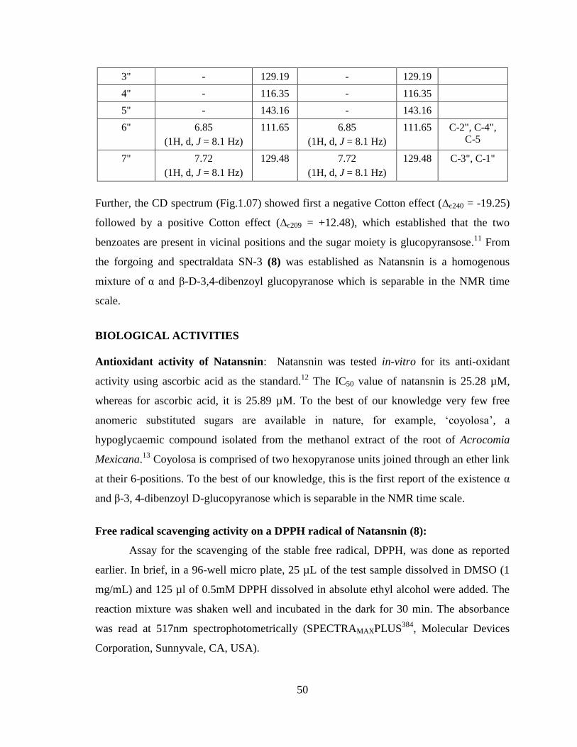
50
3" - 129.19 - 129.19
4" - 116.35 - 116.35
5" - 143.16 - 143.16
6" 6.85
(1H, d, J = 8.1 Hz)
111.65 6.85
(1H, d, J = 8.1 Hz)
111.65 C-2", C-4",
C-5
7" 7.72
(1H, d, J = 8.1 Hz)
129.48 7.72
(1H, d, J = 8.1 Hz)
129.48 C-3", C-1"
Further, the CD spectrum (Fig.1.07) showed first a negative Cotton effect (∆є240 = -19.25)
followed by a positive Cotton effect (∆є209 = +12.48), which established that the two
benzoates are present in vicinal positions and the sugar moiety is glucopyransose.11
From
the forgoing and spectraldata SN-3 (8) was established as Natansnin is a homogenous
mixture of α and β-D-3,4-dibenzoyl glucopyranose which is separable in the NMR time
scale.
BIOLOGICAL ACTIVITIES
Antioxidant activity of Natansnin: Natansnin was tested in-vitro for its anti-oxidant
activity using ascorbic acid as the standard.12
The IC50 value of natansnin is 25.28 µM,
whereas for ascorbic acid, it is 25.89 µM. To the best of our knowledge very few free
anomeric substituted sugars are available in nature, for example, ‗coyolosa‘, a
hypoglycaemic compound isolated from the methanol extract of the root of Acrocomia
Mexicana.13
Coyolosa is comprised of two hexopyranose units joined through an ether link
at their 6-positions. To the best of our knowledge, this is the first report of the existence α
and β-3, 4-dibenzoyl D-glucopyranose which is separable in the NMR time scale.
Free radical scavenging activity on a DPPH radical of Natansnin (8):
Assay for the scavenging of the stable free radical, DPPH, was done as reported
earlier. In brief, in a 96-well micro plate, 25 µL of the test sample dissolved in DMSO (1
mg/mL) and 125 µl of 0.5mM DPPH dissolved in absolute ethyl alcohol were added. The
reaction mixture was shaken well and incubated in the dark for 30 min. The absorbance
was read at 517nm spectrophotometrically (SPECTRAMAXPLUS384
, Molecular Devices
Corporation, Sunnyvale, CA, USA).
51
The free radical scavenging potential was expressed as the percent change in color
of the DPPH solution due to the test sample, and calculated as (1-B/A) is the multiple of
100, where ‗A‘ represents absorbance of the DPPH solution without the test sample and B,
the absorbance of the DPPH solution with the test sample. The SC50 values (50% free
radical scavenging activity) of the test sample were calculated by regression analysis.
Ascorbic acid was taken as the reference standard as a free radical scavenger.
Measurements were performed in triplicate.
52
EXPERIMENTAL
Collection and Identification
The plant material Salvinia natans (Salvinaceae) was collected from from
the Chidambaram area of Tamil Nadu, India. Plants were collected in the month of
November, 2005. Prof. R. Pannerselvam, Department of Botany, Annamalai University,
Annamalai Nagar 608 002, TamilNadu, India, identified the plant.
Extraction and fractionation procedure
The shade dried and powdered of root part of the plant (2.1 kg) Salvinia natans
was extracted with n-hexane (3x5 L) followed by CH2Cl2: MeOH (3x5 L) at room
temperature.
Hexane extract
The combined hexane extract was filtered and concentrated under reduced
pressure to obtained greenish gummy residue (30.2 g). The crude slurry was subjected to
silica gel (100-200 mesh) column chromatography by gradient elution using with hexane
through hexane-ethylacetate mixtures to ethylacetate. Fractions (250 ml) were collected
and monitored on silicagel (GF254) TLC. The visualization of spots on TLC was carried out
either in UV light or by exposing TLC plates to iodine vapours or by spraying 10%
sulfuric acid in methanol and heating at 1100C. Similar fractions were combined and the
results borne out in the chromatography are recorded in the following Table-3.

53
Table-3: Silica gel column chromatography of hexane extract of
Salvinia natans
Eluent
(Hexane:EtOAc)
Fractions
Residue (g)
Remarks
100:0 1-5 2.2 Fatty oil
90:10 6-15 4.6 Fraction-I
80:20 16-18 2.6 Fatty solid
70:30 19-23 5.2 Fraction -II
60:40 24-32 6.8 Green slurry
40:60 33-40 5.2 Green pigment
20:80 41-48 2.0 Dark green matter
0:100 49-52 1.6 Dark green matter
Fraction-1
It was obtained as a mixture of compounds as noticed from its silica gel TLC plates
and hence it was re-chromatographed over silica gel (100-200mesh) column
chromatography using hexane-ethylacetate mixtures as eluents. Fractions (20ml each) were
collected and the results of the chromatography are recorded in the Table-4.
Table-4: Silica gel column chromatography of Fraction-I
Eluent
(Hexane:EtOAc)
Fractions
Yield (mg)
Remarks
100:0 1-10 460 Fatty oil
95:5 11-14 80 SN-1
80:20 14-20 250 Fatty solid
70:30 21-25 220 Fattymaterial
60:40 26-31 90 Fatty material
50:50 32-38 630 Fatty material

54
Fraction-II
It was obtained as a mixture of compounds as noticed from its silica gel TLC plates
and hence it was re-chromatographed over silica gel (100-200 mesh) column
chromatography using hexane-ethylacetate mixtures as eluents. Fractions (10ml each) were
collected and the results of the chromatography are recorded in the Table-5.
Table-5: Silica gel column chromatography of Fraction-II
Dichloromethane: Methanol extract
The combined CH2Cl2:MeOH extract was filtered and concentrated under
reduced pressure to yield a dark greenish residue (80.4g). This crude solid was subjected to
silicagel (100-200 mesh) column chromatography by gradient elution using with hexane
through hexane-ethylacetate-methanol mixtures to methanol as eluents. Fractions (350 ml)
were collected and monitored on silicagel (GF254) TLC. The visualization of spots on TLC
was carried out either in UV light or by exposing TLC plates to iodine vapours or by
spraying 10% sulfuric acid in methanol and heating at 1100C. Similar fractions were
combined and the results borne out in the chromatography are recorded in the following
Table-6.
Eluent
(Hexane:EtOAc)
Fractions
Yield (mg)
Remarks
100:0 1-10 260 Fatty oil
90:5 11-16 280 Fatty solid
90:10 17-25 450 Fatty solid
85:15 26-33 320 Fatty solid
80:20 34-36 40 SN-2
70:30 37-42 570 Fatty material
60:40 42-48 1580 Fatty material
50:50 48-55 290 Fatty material

55
Table-6: Silica gel column chromatography of hexane extract of
Salvinia natans
Eluent
(Hexane:EtOAc:MeOH)
Fractions Residue (g) Remarks
100:0:0 1-6 5.2 Fatty oil
90:10:0 7-13 4.6 Fatty solid
80:20:0 14-18 3.0 Fatty solid
70:30:0 19-25 10.1 Fatty solid
60:40:0 26-30 12.7 Green slurry
50:50:0 31-35 16.0 Dark green matter
40:60:0 36-41 8.2 Dark green matter
30:70:0 42-47 6.0 Fraction-1
0:100:0 48-53 4.5 Intractable gum
0:80:20 54-59 3.5 Intractable gum
0:40:60 60-65 2.0 Intractable gum
0:20:80 66-70 2.4 Intractable gum
0:0:100 71-75 2.2 Intractable gum
Fraction-1
This fraction-1 was purified on HPLC using C18 reversed phase column (24x2
cm, 10 mm), eluting with methanol and water (60: 40), to give compound SN-3 (8)
(80mg).

56
SN-3 (8)
Compound (SN-3) : Natansnin
Physical property : colourless powder
Amount isolated : 80mg
Melting point : 2090C
[α]D 25
:
+78.6 (c 0.011, MeOH)
IR (KBr) ν Max : 3418, 2925, 177, 1617
UV λ max (log ε) : 242 (0.4722), 351 (0.3227)
Molecular formula : C20H20O13
HREIMS : clacd. 468.0898
Obtd. : 468.1324
1H NMR of α-isomer : (CD3OD, 300Mz) : δ 5.20 (1H, d, J = 3.6 Hz,
Hα-1), 3.58 (1H,dd, J = 3.6, 9.5 Hz, H-2),
3.91 (1H, t, J = 9.5 Hz, H-3), 5.3 (1H, t, J =
9.5 Hz, H-4), 4.3 (1H, td, J = 3.6, 10.3 Hz, H-
5), 3.96 (1H, dd, J = 3.6, 10.3 Hz, Ha-6), 5.01
(1H, dd, J = 10.3, 4.4 Hz, Hb-6), 7.49 (1H, d,
J = 2.2 Hz, H-3'), 6.89 (1H, d, J = 8.1 Hz, H-
6'), 7.43 (1H, dd, J = 8.1, 2.2 Hz, H-7'), 6.85
O
OO
O
OHOH
O
O
OH
OHOH
OH
OH
1
2
34
5
1'2'
3'
4'5'
6'
7' 1''
2''
3''4''
5''
6''
7''
6

57
(1H, d, J = 8.1 Hz, H-6"), 7.72 (1H, d, J = 8.1
Hz, H-7").
13C NMR of α-isomer : (CD3OD, 50 Mz) 93.93 (C-1), 73.8 (C-2),
71.15 (C-3), 77.96 (C-4), 64.02 (C-5), 65.36
(C-6), 168.26 (C-1'), 148.19 (C-2'), 114.5 (C-
3'), 146.21 (C-4'), 157.88 (C-5'), 115.31 (C-
6'), 121.06 (C-7'), 167.65 (C-1"), 148.82 (C-
2"), 129.19 (C-3"), 116.35 (C-4"), 143.16 (C-
5"), 111.65 (C-6"), 129.48 (C-7").
1H NMR of β-isomer : (CD3OD, 50 Mz) δ 4.63 (1H, d, J = 7.3 Hz,
Hβ1), 3.36 (1H, dd, J = 7.3, 9.5 Hz, H-2), 3.66
(1H, t, J = 9.5 Hz, H-3), 5.33 (1H, t, J = 9.5
Hz, H-4), 3.83 (1H, td, J = 4.4, 10.3 Hz, H-5),
3.98 (1H, dd, J = 3.6, 10.3 Hz, Ha-6), 5.07
(1H, dd, J = 10.3, 4.4 Hz, Hb-6), 7.49 (1H, d,
J = 2.2 Hz, H-3'), 6.89 (1H, d, J = 8.1 Hz, H-
6'), 7.43 (1H, dd, J = 8.1, 2.2 Hz, H-7'), 6.85
(1H, d, J = 8.1 Hz, H-6"), 7.72 (1H, d, J = 8.1
Hz, H-7").
13C NMR of β-isomer : (CD3OD, 50 Mz) 98.44 (C-1), 76.36 (C-2),
74.09 (C-3), 77.38 (C-4), 65.0 (C-5), 65.95
(C-6), 168.26 (C-1'), 148.19 (C-2'), 114.5 (C-
3'), 146.21 (C-4'), 157.88 (C-5'), 115.31 (C-
6'), 121.06 (C-7'), 167.65 (C-1"), 148.82 (C-
2"), 129.19 (C-3"), 116.35 (C-4"), 143.16 (C-
5"), 111.65 (C-6"), 129.48 (C-7").

58
SECTION–B: Phytochemical investigation of heartwood of
Decalepis hamiltonii
The Indian subcontinent is rich in biodiversity with more than 2000 species of
flowering plants. Decalepis hamiltonii (Asclepiadaceae) known as swallow in
biotechnology root is a monogeneric climbing shrub and a native of the forests of Deccan
peninsula and Western Ghats of India. Its tubers are consumed as pickles and the juice for
its alleged health promoting properties. It grows between the rocks and places where there
is thick vegetation. The roots of D. hamiltonii are used as a flavoring principle, appetizer,
and blood purifier and as a preservative. Similarly, the roots of this taxon are considered as
―Sariva Bheda‖ in Ayurveda where these find use as an alternative to the roots of
Hemidesmus indicus in the preparation of several herbal drugs like Amrutamalaka taila
(hair tonic), Drakshadi churna (general vitalizer), Shatavari rasayana (adapatogenic) and
Yeshtimadhu taila (mild analgesic, rheumatism).
The roots contain 92% fleshy matter and 8% woody core. Of late, the highly aromatic roots
have been subjected to over exploitation by destructive harvesting that has endangered the
survival of this plant. A method for rooting of D. hamiltonii for field transfer is reported. In
earlier reports, it was observed that the aromatic roots of D. hamiltonii possess
bioinsecticide property on storage pests at lethal and sub-lethal levels. The extracts of these
roots have also been shown to be potent antimicrobial agents as well. The antimicrobial
properties of the roots of D.hamiltonii have been attributed to the presence of 2-hydroxy-4-
Scientific Classification
Kingdom: Plantae
Order: Gentianales
Family: Apocynaceae
Genus: Decalepis
Species: D. hamiltonii

59
methoxy benzaldehyde and vanillin.14
Earlier work has shown that the roots contain
aldehyde, inositol, saponins, ketonic substances, sterols, amyrins and lupeols.15-17
Thangaduarai et al.18
and Nagarajan et al.19
have reported several volatile flavor
compounds including 4-methoxybenzaldehyde, vanillin, and salcilaldehyde in the essential
oil extracts from the roots of D. hamiltonii. We have recently shown that the roots of D.
hamiltonii possess antioxidant properties and hypothesized that antioxidants constituent
present in the root extracts could contribute to the health-promoting potential.20
We now
report the isolation and characterization of antioxidant compounds from the methanolic
extract of the roots of D. hamiltonii.
Earlier chemical investigation of root of Decalepis hamiltonii.
The plant material was collected from the forest of Tirumala hills in Chitoor Dist,
Andhra Pradesh, India in the month of July 2005 and identification was made by Dr. K.
Madhava Chetty, Department of Botany, Sri Venkateswara University, Tirupati. Shade
dried powdered heartwood (5 Kg) of Decalepis hamiltonii was extracted with hexane,
chloroform and methanol at room temperature about 48 h.
Previously reported that the methanolic extract of roots of Decalepis hamiltonii
contained 2-hydroxy-4-methoxybenzaldehyd 9, p-anisaldehyde 10,vanillin 11, borneol 12,
salicylaldehyde 13 and bis-2,3,4,6-galloyl α/β-D-glucopyranoside (Decalepin) 14 and also
the aqueous extracts on the other hand were found to contain 4-hydroxyisophthalicacid 15,
14-aminotetradecanoicacid 16, 4-(1-hydroxy-1-methylethyl)-1-methyl-1,2-cyclohexanediol
17, 2-hydroxymethyl-3-methaoxybenzaldehyde 18, 2,4,8-trihydroxybicyclo[3.2.1] octane-
3-one 19. 21
OH
OMe
HO
9
HO
OMe
10
OH
O H
OMe
11
CH3
OH
H
CH3
H3C
12

60
OH
HO
13
NH2
OH
O
16
OH O
OH
OOH
15
O
O
OH
HO
HO
OH
OH
O
O
OH
OH
O
O
O
OO
OH
OH
OH
O
O
O
OH
OH
OH
O
O
OH
HOOH
HO
HO
HO
O
O
HOCO
HO
14
CH3
CH3
OH
CH3
CH3
OH
17
OH
O
O
H
18

61
In the present study, the plant Decalepis hamiltonii yielded five new sources,out of
which two new sources showing anti-inflammatory activities by down regulating TNF-α
and IL-2 specific mRNA,besides up regulating the synthesis of mRNA of IL-10.22
PRESENT WORK
Chemical investigation of Decalepis hamiltonii
The shade dried and powdered root of the plant Decalepis hamiltonii
(Asclepiadaceae) extracted with hexane and in 1:1 DCM: MeOH at room temperature to
afforded five new sources DH-1 to DH-5. The details of the extraction and isolation of the
compounds from the plant material has been described in the experimental section.
Compounds DH-1 to DH-5 was found to be new sources. And their structures were
established by means of spectroscopic analysis. The isolated compounds are listed in
Table-7.
Table-7: The isolated compounds from the plant Decalepis hamiltonii
Compound
code
Compound
Name Nature
Mol.
formula
Remark
DH-1 Lupeol acetate white needles C32H52O2 New source
DH-2 Sesamin White crystals C20H18O6 New source
DH-3 (S)- Naringenin yellow needles C15H12O5 New source
DH-4 Milimorin yellow crystals C16H12O7 New source
DH-5
(S)-Naringenin
4'-O--β -
glucopyranoside
yellow needles C21H22O10
New source

62
Structural elucidation of Compound DH-1 (19)
Compound DH-1 (19) was obtained as white needles, m.p. 127-1290C, [α]
20 +27.2 (c 4.8,
CHCl3). Its EI mass spectrum shows molecular ion peak [M]+ at m/z 468 and analyzed for
C32H52O2, which required seven degrees of unsaturation. The lH-NMR spectrum
(Fig.1.08,Table-8 ) in CDCl3 at δ 0.79 (3H,s), 0.83 (3H,s), 0.84 (3H,s), 0.85 (3H,s), 0.94
(3H,s), 1.03 (3H,s), 1.69 (3H,s) and 2.05(3H,s) indicated the presence of eight tertiary
methyls and δ 4.57 and 4.69 (2H,s) representing an exocyclic double bond protons H-29a
and H-29b, respectively. Its 13
C NMR spectrum (Fig.1.09,Table-8) in CDCl3 shows
carbonyl group at δ 171.3 and the signal at δ 81.2 indicates C-3 carbon and the alkene
carbons at δ 151.20 and 109.6. From the forgoing spectral data and the literature 23
survey
revealed that the structure of compound DH-1 (19) was confirmed as lupeolacetate, this
was first report of isolation from the plant Decalepis hamiltonii., before it was found in
deertongue leaf, 24
Erythroxylum leal costae,25
stem-bark of Artocarpus chaplasha 26
and
Ficus hispida.27
12
3
4 67
89
10
111
13
14
1516
1718
1920 21
22
23 24
25 26
27
28
29
1'
30
O
O
2'
DH-1 (19)

63
Table-8. 1H and
13C-NMR of DH-1 (19)
Position
Chemical shift values based on the
reference 23 (b)
Chemical shift values
of DH-1 (19)
1H NMR
Chemical shift
(δ in CDCl3)
Multiplicity (J in Hz)
13C-NMR
1H NMR
Chemical shift
(δ in CDCl3)
Multiplicity (J in Hz)
13C-NMR
1 - 38.6 - 38.61
2 - 21.7 - 21.55
3 4.47 (1H, dd , J = 4.4,
12.8 Hz) 81.2
4.47 (1H, dd , J = 4.4,
12.8 Hz) 81.21
4 - 38.0 - 38.02
5 - 55.6 - 55.66
6 - 18.4 - 18.43
7 - 34.4 - 34.44
8 - 41.0 - 41.08
9 - 50.5 - 50.57
10 - - 37.31
11 - 21.1 - 21.17
12 - 24.0 - 23.94
13 - 36.2 - 36.80
14 - 43.0 - 43.05
15 - 25.3 - 25.33
16 - 35.8 - 35.80
17 - 43.2 - 43.22
18 - 48.5 - 48.52
19 - 48.2 - 48.23
20 - 151.2 - 151.19
21 - 30.0 - 30.06
22 - 40.2 - 40.22
23 0.85 (3H,s) 27.6
0.80 (9H,s)
27.66
24 0.84 (3H,s) 16.7

64
26 0.83 (3H,s) 16.2 16.72
25 1.03 (3H,s) 16.4 1.03 (3H,s) 16.40
27 0.79 (3H,s) 14.7 0.79 (3H,s) 14.70
28 0.94 (3H,s) 18.2 0.94 (3H,s) 18.23
29 4.57 (1H,s H-29a)
4.69 (1H,s,H-29b) 109.6
4.57 (1H,s H-29a)
4.69 (1H,s,H-29b) 109.58
30 1.69 (3H,s) 19.5 1.69 (3H,s) 19.51
1' - 171.3 - 171.26
2' 2.05,(3H, s) 28.2 2.05,(3H, s) 28.17
Assignments were made by comparison of earlier data23 (b)
Structural elucidation of Compound DH-2 (20):
Compound DH-2 (20) was obtained as colorless prisms, m.p. 127-1290C, [α] D
20 +64.5 (c
1.75, CHCl3). Its EI mass spectrum showed molecular ion peak at m/z 354 [M]+ and
analyzed for C20H18O6, which required twelve degrees of unsaturation. Its lH-NMR
spectrum (Fig.1.10,Table-9) indicated the presence of symmetric methylene dioxygroup
which appeared as singlet at δ 5.92 (4H,s). Aromatic ring protons appears at δ 6.72 to δ
6.80 (6H, m) which shows that the presence of symmetrical aromatic rings within the
structure. The 13
C NMR spectrum (Fig.1.11, Table-9) of the compound 20 is shown in the
following table. Based on the above data it is concluded as a lignan. Hence from the
forgoing spectral data and literature survey reveled that compound DH-2 is closely
related to those of sesamin (20) which was earlier isolated from the dried peeled roots of
Glossostemon bruguierise.28
and this is first report of isolation from this plant Decalepis
hamiltonii. Hence the structure of DH-2 was established as sesamin (20).
O
O
O
O
O
O
HH
17
8
91' 7'
8'
9'
DH-2 (20)

65
Table-9. 1H and
13C-NMR of DH-2 (20)
Position
Chemical shift values
based on the reference 28
Chemical shift values of
DH-2 (20)
1H NMR
Chemical shift
(δ in CDCl3)
Multiplicity (J in
Hz)
13C-NMR
1H NMR
Chemical shift
(δ in CDCl3+ d6-DMSO)
Multiplicity (J
in Hz)
13C-NMR
1,1' - 135.1 - 134.48
2,2' 6.76-6.84 (2H,m) 119.4 6.72-6.80 (2H,m) 118.42
3,3' 6.76-6.84 (2H,m) 108.2 6.72-6.80 (2H,m) 107.30
4,4' - 147.1 - 146.26
5,5' - 148.0 - 147.19
6,6' 6.76-6.84 (2H,m) 106.5 6.72-6.80 (2H,m) 105.74
7,7' 4.71 (2H,m) 85.8 4.60 (2H,m) 84.80
8,8' 3.04 (2H, m) 54.4 2.90 (2H, m) 53.64
9,9'
Ha-9 and Ha-9'
4.23 (2H, m)
Hb-9 and Hb-9'
3.84 (2H, m)
71.7
Ha-9 Ha-9′
4.15 (2H, m)
Hb-9 and Hb-9'
3.75 (2H, m)
70.74
10,10' 5.92 (4H, s) 101.1 5.92 (4H, s) 95.43
Assignments were made by comparison of earlier data28
66
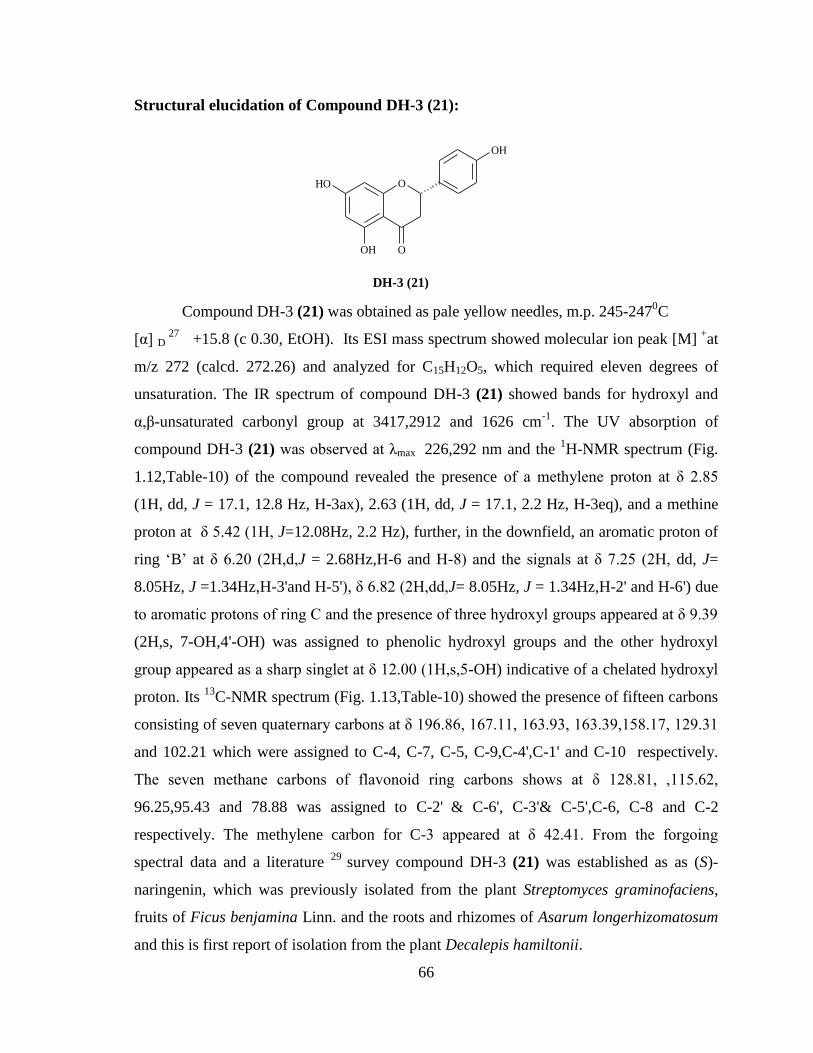
Structural elucidation of Compound DH-3 (21):
Compound DH-3 (21) was obtained as pale yellow needles, m.p. 245-2470C
[α] D 27
+15.8 (c 0.30, EtOH). Its ESI mass spectrum showed molecular ion peak [M] +
at
m/z 272 (calcd. 272.26) and analyzed for C15H12O5, which required eleven degrees of
unsaturation. The IR spectrum of compound DH-3 (21) showed bands for hydroxyl and
α,β-unsaturated carbonyl group at 3417,2912 and 1626 cm-1
. The UV absorption of
compound DH-3 (21) was observed at λmax 226,292 nm and the 1H-NMR spectrum (Fig.
1.12,Table-10) of the compound revealed the presence of a methylene proton at δ 2.85
(1H, dd, J = 17.1, 12.8 Hz, H-3ax), 2.63 (1H, dd, J = 17.1, 2.2 Hz, H-3eq), and a methine
proton at δ 5.42 (1H, J=12.08Hz, 2.2 Hz), further, in the downfield, an aromatic proton of
ring ‗B‘ at δ 6.20 (2H,d,J = 2.68Hz,H-6 and H-8) and the signals at δ 7.25 (2H, dd, J=
8.05Hz, J =1.34Hz,H-3'and H-5'), δ 6.82 (2H,dd,J= 8.05Hz, J = 1.34Hz,H-2' and H-6') due
to aromatic protons of ring C and the presence of three hydroxyl groups appeared at δ 9.39
(2H,s, 7-OH,4'-OH) was assigned to phenolic hydroxyl groups and the other hydroxyl
group appeared as a sharp singlet at δ 12.00 (1H,s,5-OH) indicative of a chelated hydroxyl
proton. Its 13
C-NMR spectrum (Fig. 1.13,Table-10) showed the presence of fifteen carbons
consisting of seven quaternary carbons at δ 196.86, 167.11, 163.93, 163.39,158.17, 129.31
and 102.21 which were assigned to C-4, C-7, C-5, C-9,C-4',C-1' and C-10 respectively.
The seven methane carbons of flavonoid ring carbons shows at δ 128.81, ,115.62,
96.25,95.43 and 78.88 was assigned to C-2' & C-6', C-3'& C-5',C-6, C-8 and C-2
respectively. The methylene carbon for C-3 appeared at δ 42.41. From the forgoing
spectral data and a literature 29
survey compound DH-3 (21) was established as as (S)-
naringenin, which was previously isolated from the plant Streptomyces graminofaciens,
fruits of Ficus benjamina Linn. and the roots and rhizomes of Asarum longerhizomatosum
and this is first report of isolation from the plant Decalepis hamiltonii.
O
OH
O
OH
OH
DH-3 (21)

67
Table-10. 1H and
13C-NMR of DH-3 (21)
Position
Chemical shift values
based on the reference 29
Chemical shift values
of DH-3 (21)
1H NMR
Chemical shift
(δ in d6-DMSO)
Multiplicity (J in Hz)
13C-NMR
1H NMR
Chemical shift
(δ in D6-DMSO)
Multiplicity (J in Hz)
13C-NMR
1 - - - -
2 5.45 (1H, dd.J =
2.7,12.9 Hz) 78.4
5.42 (1H, dd.J =
2.2,12.0 Hz) 78.88
3
3.26 (1H, dd, J =
12.9,17.3, H-3trans)
2.68 (1H, dd, J = 3.2
,17.3, H-3cis)
41.9
2.85 (1H, dd, J = 12.8 ,
17.1, H-3trans)
2.63 (1H, dd, J = 2.2 ,
17.1, H-3cis)
42.41
4 - 196.3 - 196.86
5-OH 12.14 (1H,br,s) 163.4 12.01 (1H,br,s) 163.93
6 5.87 (2H,s,
H-6,H-8) 94.9
5.95 (1H,d,
J = 2.6 Hz) 96.25
7-OH 9.59 (1H,br,s) 166.6 9.62 (1H,br,s) 167.11
8 5.87 (2H,s, H-6,H-8) 95.8 5.95 (1H,d, J = 2.6 Hz) 95.43
9 - 162.9 - 163.39
10 - 101.7 - 102.21
1' - 128.8 - 129.31
2',6' 7.31 (1H,d, J = 8.6 Hz) 128.3 6.82 (1H,d, J = 8.0,1.3
Hz) 128.81
3',5' 6.80 (1H,d, J = 8.6 Hz) 115.1 7.40 (1H,d, J = 8.0,1.3
Hz) 115.62
4'-OH 9.59 (1H,br,s) 157.7 9.62 (1H,br,s) 158.17
Assignments were made by comparison of earlier data 29

68
Structural elucidation of Compound DH-4 (22)
Compound DH-4 (22) was obtained as yellow needles, m.p. 235-2370C. Its HRMS
mass spectrum (Fig. 1.18) showed molecular ion [M+1]+ peak at m/z 317 (calcd. 317.26)
and analyzed for C16H12O7, which required eleven degrees of unsaturation. Its 1H NMR
spectrum (Fig. 1.14, Table-11) in acetone-d6 of compound DH-4 (22) indicates the
presence of four hydroxyl groups of which three appeared as a broad singlet at δ 9.45(1H,
s, 2-OH) and 8.40 (2H, s, 7-OH, 2'-OH) was assigned to phenolic hydroxyl groups and the
other hydroxyl group appeared as a shart singlet at δ 12.80 (1H, s, 5-OH) indicates the
presence of a chelated hydroxyl group at C-5. Further, its 1H NMR spectrum displayed
signals at δ 7.63 (1H, dd, J = 8.8 Hz, 1.6 Hz), δ 7.01 (1H, d, J= 8.8 Hz, 6.46 (1H, d, J = 1.6
Hz), δ 6.22 (2H, J = 1.60 Hz) and a singnal at δ 3.8 (3H, s). The signals at δ 7.63 and δ
7.01 were assigned to H-5' and H- 6' respectively. The signal at δ 6.46 was assigned to H-
3'. Finally, the signal at δ 6.22 was assigned to H-6 and H-8 protons and signal at δ 3.8
was assigned to methaoxy ether. Its 13
C-NMR spectrum (Fig.1.15, Table-11 ) in acetone-
d6 displayed sixteen carbons of assignments which were also made by DEPT (Fig.1.16)
experiment The spectrum showed the presence of sixteen carbons consisting of ten
quaternary carbons at δ 179.50, 149.08, 164.87, 157.81, 145.85, 163.1, 156.7, 139.26,
123.03 and 105.87 which were assigned to C-4, C-9, C-7, C-5, C-2,C-4',C-2',C-3,C-1' and
C-10 respectively. Five methane carbons shows at δ 122.12, 99.37, 116.32, 94.48 and
94.42 was assigned to C-6', C-3'& C-5', C-6 and C-8 respectively, and a methoxy ether at δ
60.17. Further the position of methaoxy group of ring C was established by NOE spectrum
(Fig.1.17). Foregoing and literature 30
survey revealed that compound DH-4 was found to
be milimorin (22) which was previously isolated from the plant Euphorbia milii., and this
is first report of isolation from the plant Decalepis hamiltonii. Hence the structure of DH-4
was established as milimorin (22).
O
OOH
OH
OH
OH
OMe
DH - 4 (22)

69
Table-11. 1H and
13C-NMR of Milimorin DH-4 (22)
Position
Chemical shift values
based on the reference 30
Chemical shift values
of DH-4 (22) 1H NMR
Chemical shift
(δ in CDCl3)
Multiplicity
(J in Hz)
13C-NMR
1H NMR
Chemical shift
(δ in Acetone-d6)
Multiplicity
(J in Hz)
13C-NMR
1 - - - -
2 - 153.3 145.85
3 OH 142.1 9.62 (1H,br,s, OH-3) 139.26
4 - 173.9 179.50
5-OH 12.82 (1H,br,s) 161.0 12.80 (1H,br,s) 157.81
6 6.25 (1H,d,J = 1.5 Hz)
95.6 6.22 (1H,d,J = 1.6
Hz) 94.42
7-OH OH 163.7 9.62 (1H,br,s, OH-7) 164.87
8 6.25 (1H,d,J = 1.5 Hz)
92.6 6.22 (1H,d,J = 1.6
Hz) 94.48
9 - 158.8 - 149.08
10 - 109.8 - 105.87
1' - 112.8 - 123.03
2' OH 159.4 8.42 (1H,br,s, OH-2') 156.75
3' 6.39 (1H,d,J = 1.5 Hz)
98.8 6.46 (1H,d,J = 1.6
Hz) 99.37
4' - 162.6 - 163.17
5' 6.54 (1H, dd, J = 8.5,1.5
Hz) 104.7
7.63 (1H, dd, J =
8.8,1.6 Hz) 116.32
6' 7.36 (1H,d, J = 8.5 Hz)
131.7 7.01 (1H,d, J = 8.8
Hz) 122.12
OCH3 3.79 (3H, s) 60.3 3.80 (3H, s) 60.17
Assignments were made by comparison of earlier data 30
70
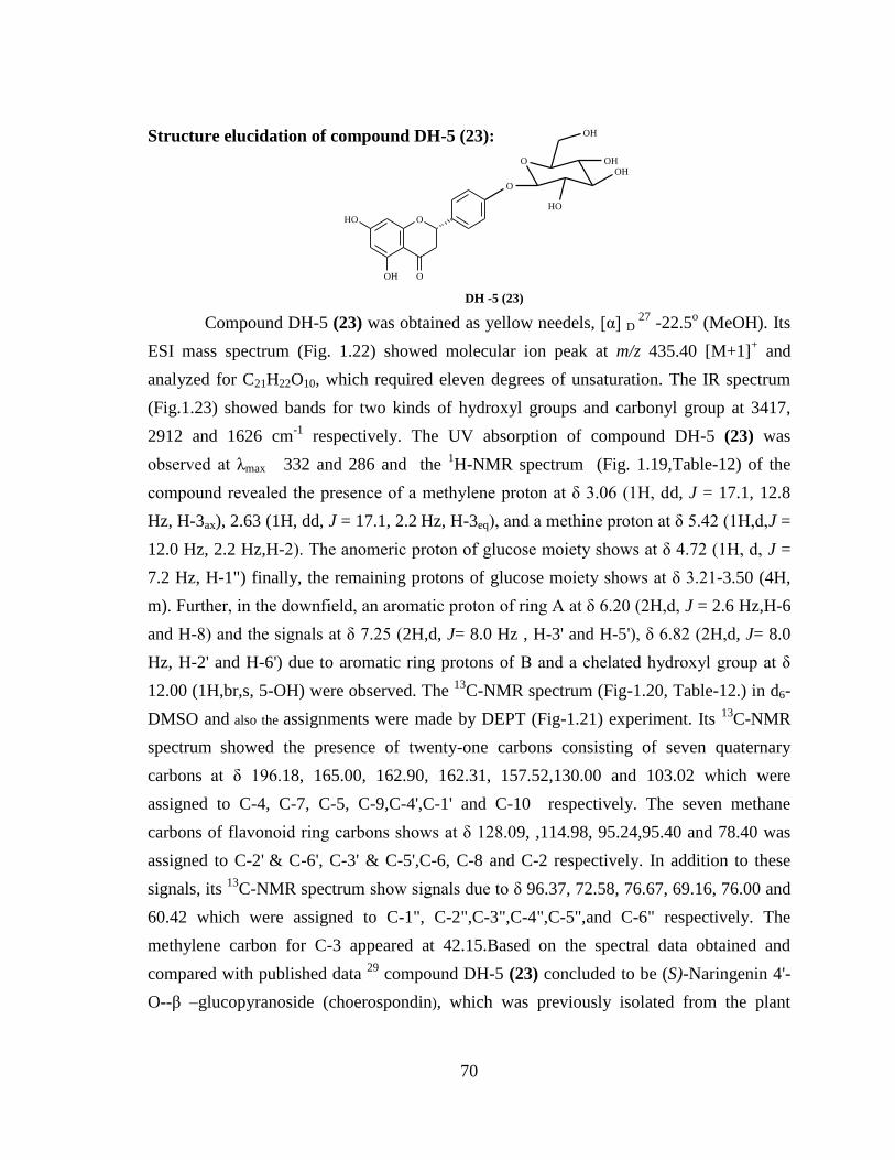
Structure elucidation of compound DH-5 (23):
Compound DH-5 (23) was obtained as yellow needels, [α] D 27
-22.5o (MeOH). Its
ESI mass spectrum (Fig. 1.22) showed molecular ion peak at m/z 435.40 [M+1]+ and
analyzed for C21H22O10, which required eleven degrees of unsaturation. The IR spectrum
(Fig.1.23) showed bands for two kinds of hydroxyl groups and carbonyl group at 3417,
2912 and 1626 cm-1
respectively. The UV absorption of compound DH-5 (23) was
observed at λmax 332 and 286 and the 1H-NMR spectrum (Fig. 1.19,Table-12) of the
compound revealed the presence of a methylene proton at δ 3.06 (1H, dd, J = 17.1, 12.8
Hz, H-3ax), 2.63 (1H, dd, J = 17.1, 2.2 Hz, H-3eq), and a methine proton at δ 5.42 (1H,d,J =
12.0 Hz, 2.2 Hz,H-2). The anomeric proton of glucose moiety shows at δ 4.72 (1H, d, J =
7.2 Hz, H-1") finally, the remaining protons of glucose moiety shows at δ 3.21-3.50 (4H,
m). Further, in the downfield, an aromatic proton of ring A at δ 6.20 (2H,d, J = 2.6 Hz,H-6
and H-8) and the signals at δ 7.25 (2H,d, J= 8.0 Hz , H-3' and H-5'), δ 6.82 (2H,d, J= 8.0
Hz, H-2' and H-6') due to aromatic ring protons of B and a chelated hydroxyl group at δ
12.00 (1H,br,s, 5-OH) were observed. The 13
C-NMR spectrum (Fig-1.20, Table-12.) in d6-
DMSO and also the assignments were made by DEPT (Fig-1.21) experiment. Its 13
C-NMR
spectrum showed the presence of twenty-one carbons consisting of seven quaternary
carbons at δ 196.18, 165.00, 162.90, 162.31, 157.52,130.00 and 103.02 which were
assigned to C-4, C-7, C-5, C-9,C-4',C-1' and C-10 respectively. The seven methane
carbons of flavonoid ring carbons shows at δ 128.09, ,114.98, 95.24,95.40 and 78.40 was
assigned to C-2' & C-6', C-3' & C-5',C-6, C-8 and C-2 respectively. In addition to these
signals, its 13
C-NMR spectrum show signals due to δ 96.37, 72.58, 76.67, 69.16, 76.00 and
60.42 which were assigned to C-1", C-2",C-3",C-4",C-5",and C-6" respectively. The
methylene carbon for C-3 appeared at 42.15.Based on the spectral data obtained and
compared with published data 29
compound DH-5 (23) concluded to be (S)-Naringenin 4'-
O--β –glucopyranoside (choerospondin), which was previously isolated from the plant
O
O
OH
OH
OHOH
O
OH
OH
O
DH -5 (23)

71
Asarum longerhizomatosum and this is first report of isolation from the plant Decalepis
hamiltonii.
Table-12. 1H and
13C-NMR of DH-5 (23)
Position
Chemical shift values
based on the reference 29
Chemical shift values
of DH-5 (23) 1H NMR
Chemical shift
(δ in d6-DMSO)
Multiplicity (J in Hz)
13C-
NMR
1H NMR
Chemical shift
(δ in d6-DMSO )
Multiplicity (J in Hz)
13C-NMR
1 - - - -
2 5.64 (1H, J = 12.9,2.7
Hz) 78.1
5.42 (1H, J = 12.0,2.2
Hz) 78.43
3
3.01 (1H, dd, J = 17.3,
12.9 Hz , H-3ax)
2.64 (1H, dd, J = 17.3,
2.7 Hz,H-3eq)
42.1
3.06 (1H, dd, J = 17.1,
12.8 Hz , H-3ax)
2.63 (1H, dd, J = 17.1, 2.2
Hz,H-3eq)
42.15
4 - 196.0 - 196.18
5 12.00 (1H,br,s,OH-5) 163.0 12.00 (1H,br,s,OH-5) 162.90
6 6.10 (1H,d, J = 2.6 Hz) 95.7 6.20 (1H,d, J = 2.6Hz) 95.40
7 9.03 (1H,br,s) 166.3 9.39 (1H,br,s,OH-7) 165.06
8 6.10 (1H,d, J = 2.68
Hz) 95.0 6.20 (1H,d, J = 2.6 Hz) 95.24
9 - 162.7 - 162.31
10 - 101.7 - 103.02
1‘ - 131.8 - 128.09
2',6' 7.30 (1H,d, J = 9.2 Hz) 128.0 7.25 (1H,d, J = 8.0 Hz) 127.66
3',5' 6.82 (2H,d, J = 9.2 Hz) 116.1 6.82 (2H,d, J = 8.0 Hz) 114.98
4' - 157.4 - 157.52
1" 4.80 (1H, d, J = 7.5 Hz) 100.2 4.72 (1H, d, J = 7.2 Hz) 99.48
2"
3.20-3.50 (4H,m)
73.1
3.21-3.50 (4H,m)
72.58
3" 77.0 76.67
4" 69.5 69.16
5" 76.5 76.00
6" 3.68 (2H,d, J =11.6 Hz) 60.6 3.70 (2H,d, J =11.6 Hz) 60.42
Assignments were made by comparison of earlier data 29

72
EXPERIMENTAL
Collection and Identification
The plant material Decalepis hamiltonii (Asclepiadaceae) was collected from
Bhadrachalam forest, Andhra Pradesh, India in January 2007, and identified by Dr. K.
Madhava Chetty, Department of Botany, Sri Venkateswara University, and Tirupathi,
India.
Extraction and Isolation procedure
The shade dried and powdered of root part of the plant (3.1 kg) Decalepis
hamiltonii was extracted with n-hexane (3x5 L) followed by CH2Cl2: MeOH (3x5 L) at
room temperature.
Hexane extract
The combined hexane extract was filtered and concentrated under reduced pressure
to obtained greenish gummy residue (40.4g). The crude slurry was subjected to silicagel
(100-200 mesh) column chromatography by gradient elution using with hexane through
hexane-ethylacetate mixtures to ethylacetate. Fractions (250 ml) were collected and
monitored on silicagel (GF254) TLC. The visualization of spots on TLC was carried out
either in UV light or by exposing TLC plates to iodine vapours or by spraying 10%
sulfuric acid in methanol and heating at 1100C. Similar fractions were combined and the
results borne out in the chromatography are recorded in the following Table-13.
Table-13: Silica gel column chromatography of hexane extract of
Decalepis hamiltonii
Eluent (Hexane:EtOAc) Fractions Residue (g) Remarks
100:0 1-5 2.1 Fatty oil
90:10 6-15 4.6 Fatty solid
80:20 16-18 3.1 Fraction-I
70:30 19-23 5.2 Fraction –II
60:40 24-32 6.8 Green slurry
40:60 33-40 5.2 Green pigment

73
20:80 41-48 6.4 Dark green matter
0:100 49-52 7.0 Dark green matter
Fraction-1
It was obtained as a mixture of compounds as noticed from its silica gel TLC
plates and hence it was re-chromatographed over silica gel (100-200 mesh) column
chromatography using hexane-ethylacetate mixtures as eluents. Fractions (20ml each) were
collected and the results of the chromatography are recorded in the Table-14.
Table-14: Silica gel column chromatography of Fraction-I
Eluent
(Hexane:EtOAc)
Fractions Yield (mg) Remarks
100:0 1-10 160 DH-1
95:5 11-14 140 DH-2
90:10 14-20 400 Fatty solid
80:20 21-25 820 Fatty solid
70:30 26-31 340 Fatty material
60:40 32-38 1160 Fatty material
50:50 39-42 80 Fatty material
Fraction-II
It was obtained as a mixture of compounds as noticed from its silica gel TLC
plates and hence it was re-chromatographed over silica gel (100-200 mesh) column
chromatography using hexane-ethylacetate mixtures as eluents. Fractions (10ml each) were
collected and the results of the chromatography are recorded in the Table-15.
Table-15: Silica gel column chromatography of Fraction-II
Eluent
(Hexane:EtOAc)
Fractions
Yield (mg)
Remarks
100:0 1-10 560 Fatty oil
90:5 11-16 480 Fatty solid
90:10 17-25 650 Fatty solid
80:20 26-33 140 DH-3
85:25 34-36 520 Fatty material
70:30 37-42 870 Fatty material

74
60:40 42-48 1490 Fatty material
50:50 48-55 490 Fatty material
Dichloromethane: Methanol extract
The combined CH2Cl2: MeOH extract was filtered and concentrated under reduced
pressure to yield a dark greenish residue (80.5g). This crude solid was subjected to
silicagel (100-200 mesh) column chromatography by gradient elution using with hexane
through hexane-ethylacetate-methanol mixtures to methanol as eluents. Fractions (350 ml)
were collected and monitored on silicagel (GF254) TLC. The visualization of spots on TLC
was carried out either in UV light or by exposing TLC plates to iodine vapours or by
spraying 10% sulfuric acid in methanol and heating at 1100C. Similar fractions were
combined and the results borne out in the chromatography are recorded in the following
Table-16.
Table-16: Silica gel column chromatography of hexane extract of
Salvinia natans
Eluent
(Hexane:EtOAc:MeOH)
Fractions Residue (g) Remarks
100:0:0 1-6 5.2 Fatty oil
90:10:0 7-13 4.6 Fatty solid
80:20:0 14-18 10.1 Fatty solid
70:30:0 19-25 3.0 Fraction -I
60:40:0 26-30 12.7 Green slurry
50:50:0 31-35 16.0 Dark green matter
40:60:0 36-41 8.2 Green pigment
20:80:0 42-47 6.0 Green pigment
0:100:0 48-53 4.5 Dark green matter
0:80:20 54-59 3.5 Fraction-II
0:40:60 60-65 2.0 Intractable gum

75
0:20:80 66-70 2.4 Intractable gum
0:0:100 71-75 2.3 Intractable gum
Fraction-1
It was obtained as a mixture of compounds as noticed from its silica gel TLC plates
and hence it was re-chromatographed over silica gel (100-200 mesh) column
chromatography using hexane-ethylacetate mixtures as eluents. Fractions (5 ml each) were
collected and the results of the chromatography are recorded in the Table-17.
Table-17: Silica gel column chromatography of Fraction-I
Eluent
(Hexane:EtOAc)
Fractions
Yield (mg)
Remarks
100:0 1-10 660 Fatty oil
95:5 11-14 280 Fatty solid
80:20 14-20 800 Fatty solid
85:25 21-25 80 DH-4
70:30 26-31 290 Fatty material
60:40 32-38 730 Fatty material
50:50 39-42 160 Fatty material
Fraction-1I
It was obtained as a mixture of compounds as noticed from its silica gel TLC plates
and hence it was re-chromatographed over silica gel (100-200 mesh) column
chromatography using hexane-ethylacetate mixtures as eluents. Fractions (5 ml each) were
collected and the results of the chromatography are recorded in the Table-18.
Table-18: Silica gel column chromatography of Fraction-II
Eluent
(Hexane:EtOAc)
Fractions
Yield (mg)
Remarks
100:0 1-10 920 Fatty oil
95:5 11-14 40 Fatty solid
80:20 14-20 500 Fatty solid
85:25 21-25 440 Fatty material
70:30 26-31 1320 Fatty material

76
60:40 32-38 180 DH-5
50:50 39-42 100 Fatty material
DH-1 (19)
Compound DH-1 : Lupeol acetate
Physical property : white needles
Amount isolated : 0.18 g
Melting point : 127-1290C
[α]D 25
:
+27.2 (c 4.8, CHCl3)
Molecular formula : C32H52O2
EIMS : 468
1H NMR : (CDCl3,200 MHz) δ 4.69 (1H, s, H-29b), 4.57
(1H, s, H-29a), 4.47 (1H, dd, J = 4.4, 12.8 Hz,
H-3), 2.05 (3H, s, H-2'), 1.69 (3H, s, H-30),
1.03 (3H, s, H-25), 0.90 (3H, s, H-28), 0.80
(9H, s, H-23,H-24,H-26), 0.79 (3H, s, H-27).
13C NMR : (CDCl3, 75 Mz) δ 171.26 (C-1'), 151.19 (C
20), 109.58 (C-29), 81.21 (C-3), 55.66 (C-5),
12
3
4 67
89
10
111
13
14
1516
1718
1920 21
22
23 24
25 26
27
28
29
1'
30
O
O
2'

77
50.57 (C-9), 48.52 (C-18), 48.23 (C-19),
43.22 (C-17), 43.05 (C-14), 41.08 (C-8),
40.22 (C-22), 38.61 (C-1), 38.02 (C-4), 37.31
(C-10), 36.80 (C-13), 35.80 (C-16), 34.44 (C-
7), 30.06 (C-21), 28.17 (C-2'), 27.66 (C-23),
25.33 (C-15), 23.94 (C-12), 21.55 (C-2),
21.17 (C-11), 19.51 (C-30), 18.43 (C-6),
18.23 (C-28), 16.72 (C-24, C-26), 16.40 (C-
25), 14.70 (C-27).
DH-2 (20)
Compound DH-2 : Sesamin
Physical property : colorless prisms
Amount isolated : 80mg
Melting point : 127-1290C
[α]D 25
:
+64.5 (c 1.75, CHCl3)
IR (KBr) ν max : 1033 cm
-1
Molecular formula : C20H18O6
EI-MS : m/z 354 [M]+
1H NMR : (CDCl3+DMSO-d6, 200 MHz): δ 6.72-6.80 (
2H, m, H-1, H-1'), 6.72-6.80 ( 2H, m, H-3, H-
3'), 6.72-6.80 ( 2H, m, H-6, H-6'), 5.92 (4H, s,
H-10, H-10'), 4.60 (2H, m, H-7, H-7'), 4.15
(2H, m, Ha-9 Ha-9'), 3.75 (2H, m, Hb-9 and
Hb-9'), 2.90 (2H, m, H-8, H-8')
O
O
O
O
O
O
HH
17
8
91' 7'
8'
9'

78
13C NMR : (CDCl3+ DMSO-d6, 75 MHz): δ 53.64 (C-8
& C-8 ), 70.74 ( C-9 & C-9), 84.80 ( C-7
& C-7), 95.43 [(-O-CH2-O-)2], 105.74 ( C-
6 & C-6), 107.30 ( C-3 & C-3), 118.42
( C-2 & C-2), 134.48 (C-1 & C-1), 146.26
(C-4 & C-4) and 147.19 (C-5 & C-5)
DH-3 (21)
Compound DH-3 : Naringenin
Physical property : pale yellow needles
Amount isolated : 0.14g
Melting point : 245-2470C
[α]D 25
:
+15.8 (c 0.30, EtOH)
Molecular formula : C15H12O5
1H NMR : (DMSO-d6, 300 MHz): δ 12.01 (1H, br, s, 5-
OH), 9.62 (2H,br,s,7-OH, 4'-OH), 7.40 (1H,d,
J = 8.05, 1.34 Hz), 6.82 (1H,d, J = 8.05, 1.34
Hz), 5.95 (2H,d, J = 2.68 Hz, H-6 & H-8),
5.42 (1H, dd., J = 2.2, 12.08 Hz ), 2.85 (1H,
dd, J = 12.8 , 17.1, H-3trans), 2.63 (1H, dd, J =
2.2 , 17.1, H-3cis),
13C NMR : (DMSO-d6, 75MHz): δ 196.86 (C-4),
167.11(C-7), 163.93 (C-5), 163.39 (C-9),
158.17 (C-4), 129.31 ( C-1), 128.81(C-2 &
O
OH
O
OH
OH

79
C-6),115.62 (C-3' & C-5), 102.21( C-10),
96.25 ( C-6 ), 78.80 (C-2), 42.41(C-3 )
DH-4 (22)
Compound DH-4 : Milimorin
Physical property : yellow needles
Amount isolated : 0.14 g
Melting point : 235-2370C
UV λ max : 260,304,346 cm-1
Molecular formula : C16H12O7
EI-MS : m/z 317 [M]+
1H NMR : (Acetone-d6, 300 MHz): δ 12.80 (1H, br,
s,OH ), 9.62 ( 1H,br,s,3- OH), 8.42 (1H,br,s,
2'- OH), 7.63 (1H, dd, J = 8.8,1.6 Hz), 7.01
(1H,d, J = 8.8 Hz), 6.46 (1H,d, J = 1.6 Hz),
6.22 (2H,d, J = 1.6 Hz, H-6 & H-8 ), 3.80
(3H, s)
O
OOH
OH
OH
OH
OMe

80
13C NMR : (Acetone-d6, 75MHz): δ 179.50 (C-4), 164.87
(C-7), 157.81 (C-5), 149.08 (C-9), 163.17 (C-
4 ), 123.03 (C-1), 156.75 (C-2'), 122.12 (C-
6), 99.37 (C-3'), 116.32 (C-5′), 105.87 (C-
10), 94.42 (C-6), 145.85 (C-2 ), 139.26 (C-3 ),
60.17 (OCH3)
DH-5 (23)
Compound DH-5 : Choerospondin
Physical property : pale yellow needles
Amount isolated : 0.16 g
[α]D 25
:
-22.5o (MeOH).
Molecular formula : C21H22O10
1H NMR : (DMSO-d6, 200 MHz): δ 12.00 (1H, br, s, 5 –
OH), 9.39 (1H,br,s,7-OH), 6.20 (1H,d, J =
2.6Hz, H-6,H- 8), 5.42 (1H, J = 12.0,2.2Hz,
H-2), 3.06 (1H, dd, J = 17.1, 12.8 Hz , H-
3ax),2.63 (1H, dd, J = 17.1, 2.2 Hz,H-3eq),
7.25 (1H,d, J = 8.0 Hz, H-2' and H-6'), 6.82
(2H,d, J = 8.0 Hz, H-3' and H-5'), 4.72 (1H, d,
O
O
OH
OH
OHOH
O
OH
OH
O

81
J = 7.2 Hz, H-1"), 3.21-3.50 (4H,m, H-2" -
5"), 3.70 (2H,d, J =11.6 Hz, H-6")
13C NMR : (DMSO-d6, 75 MHz): δ 196.0 ( C-4 ), 165.06
(C-7), 162.90 (C-5), 162.31 (C-9), 157.52
(C-4), 128.09 (C-1), 127.66 (C- 2 & 6),
114.98 (C- 3'& 5′), 103.02 (C-10), 99.48
(C-1"), 95.40 (C-6), 95.24 (C-8), 78.43 (C-2),
76.67 ( C-3" ), 76.00 (C-5" ), 72.58 ( C-2"),
69.16 ( C-4"), 60.42 (C-6" ), 42.15 (C-3).

82
REFERENCES:
1. Vines G: Herbal harvests with a future: towards sustainable sources for
medicinal plants, Plantlife International. 2004 [http://www.Plantlife.Org.uk].
2. Rozentsvet, O.A., Rezanka, T., Bosenko, E.S., Uzhametskaya, E.A., Dembitskii,
.M., Chem. Nat. Compd., 2005, 41, 487–490.
3. Narasimhulu M.; Ashalatha K.; Sri Laxmi P.; Sarma A. V. S.; Rama Rao B.;
Kavi Kishor P.B.; David Krupadanam G.L.; Zehra Ali A.; Asok Tiwari K.; Panneer
Selvam A.; Venkateswarlu Y. Nat. Prod. Res., 2010, 24, 1390–1394.
4. (a) Dragsted, L.O., Strube, M., & Larsen, J.C. Pharmacology and Toxicology,
1993, 72,116–135. (b) Johnson, I.T., Williamson, G., & Musk, S.R. Nutrition
Reviews, 1994, 7, 175–204. (c) Owen, R.W., Giacosa, A., Hull, W.E., Haubner, R.,
Spiegelhalder, B., & Bartsch, H. European Journal of Cancer, 2000, 36, 1235–
1247.
5. Iqbal Choudhary M, Naheed Nadra, Abbaskhan Ahmed, Musharraf Ghulam
Syed, Atta-ur-Rahman Siddiqui Hina; Phytochemistry 2008, 69,1018-1023.
6. Lahdesmaki P; Physiol Plantarum 1968,21,1097-1103.
7. Sies H. Eur j Biochem, 1993, 215, 213-219.
8. Halliwell B. Advances in pharmacology, 1997, 38, 3-7.
9. Ravikanth, V.; Reddy, V.L.N.; Reddy, A.V.; Ravinder, K.; Rao, T.P.; Siva Ram,
T., et al. Chemical and Pharmaceutical Bulletin, 2003, 51, 431–434.
10. (a) Debruyn, A.; Antieunis, M. Acta Ciencia Indica, 1974, 1, 83–88
(b) Vignon, M.R., & Vottero, J.A. Tetrahedron Letters, 1976, 17, 2445–2448.
11. Harada, N.; & Nakanishi, K. Accounts of Chemical Research, 1972, 5, 257–263.
12. Rao, R.J.; Tiwari, A.K.; Kumar, U.S.; Reddy, S.V.; Ali, A.Z. & Rao, J.M.
Bioorganic and Medicinal Chemistry Letters, 2003, 13, 2777–2780.
13. Perez, G.S.; Perez, G.R.M.; Perez, G.C.; Zavala, S.M.A.; & Vargas, S.R.
Pharmaceutica Acta Helvetiae, 1997, 72, 143–146.
14. Phadke, N. Y.; Gholap, K.; Ramakrishnan; Subbulakshmi,G. J. Food Sci. Technol.
1994, 31, 472-475.
15. Murti, P. B. R.; Seshadri, T. R. Proc. Indian Acad. Sci. 1941a, A13, 221-232.
16. Murti, P. B. R.; Seshadri, T. R. Proc. Indian Acad. Sci. 1941b, A13, 399-403.

83
17. Murti, P. B. R.; Seshadri, T. R. Proc. Indian Acad. Sci. 1941c, A14, 93-99.
18. Thangadurai, D.; Anitha, S.; Pullaiah, T.; Reddy, P. N.; Ramachandraiah, O.J.
Agric. Food Chem. 2002, 50, 3147-3149.
19. Nagarajan, S.; Jagan Mohan Rao, L.; Gurudutt, K. N. Flavour Fragrance J., 2001,
16, 27-29.
20. Shereen; Srivastava, A.; Shivanandappa, T. 71st Annual Meeting of the Society of
Biological Chemists, Hyderabad, India, 2001; Abstract P2B28, p 176.
21. R. Harish; S. Divakar; Anup Srivastava and T. Shivanandappa, J. Agric. Food
Chem. 2005, 53, 7709-7714.
22. Ashalatha K.; Venkateswarlu.Y; Moushumi Priya A., Lalitha P.; Krishnaveni, M.;
S.Jayachandran, Journal of Ethnopharmacology, 2010, 130, 1,167-170
23. (a) Pakrashi, S.C.; Bhattacharyya, J.; Mookerjee, S.; Samatan, T.B. & Vorbrüggen,
H. Phytochemistry, 1968, 7, 461–466. (b) A.K. Jamal*, W.A. Yaacob and Laily B.
Din; Journal of Physical Science, 2008,19, 45–50.
24. Appleton, R.A. & Enzell, C.R. Phytochemistry, 1971,10, 447–449.
25. Chavez, J.P.; Dos Santos, I.D.; Cruz, F.G. & David, J.M., Phytochemistry, 1996,
41, 941– 943.
26. Mahato, S.B.; Banerjee, S.K. & Chakravarti, R.N. Phytochemistry, 1971,10,
1351–1354.
27. Wang, S. & Coviello, D.A. Tetrahedron, 1975, 31,929–932,
28.
Meselhy R. Meselhy et al., Molecules, 2003, 8, 614-621
29. Shu-xiang zhanga; Tadato tanib; Seiichi yamajib; Chao-mei maa; Min-chuan
wanga; Shao-qing caia and Yu-ying zhaoa, Journal of Asian Natural Products
Research, 2003,5, 25–30
30. Khosa RL; Bhatia N; Sahai M. Milimorin., Chemistry and Industry, 1984, 24, 881.