Embed Size (px)
Citation preview

Chapter 5. Equipment for a colposcopic examination 3737
chapter 5.
Equipment for a colposcopic examination
This chapter describes the equipment needed to perform a col-poscopic examination and its more common uses in clinical practice. A step-by-step description of the colposcopy technique and how to optimize the examination follows in Chapter 6.
5.1 Colposcope
The colposcope is a relatively simple instrument that allows examination of the cervix under light illumination at various low-power magnifications. It consists of a binocular microscope and light source, often incorporating a beam splitter to allow attachment of a still or video camera. It may ei-ther be attached to a central upright rigid bar, as in the original colpo-scope introduced in Germany in the 1920s, or be connected to a weight-ed stand with an adjustable arm,
CH
AP
TER
1
which allows the colposcope head to be placed more precisely and with-out interfering with the operator’s comfort. There are a large number of colposcopes on the market. Fig. 5.1 shows a typical colposcope mounted on a floor stand.
Certain instrument character-istics should be considered before buying a colposcope. It must be binocular, so that depth of field may be appreciated. This is particularly important when performing exci-sional treatment and when trying to assess surface contour and perform examination of endocervical epitheli-um (Carcopino et al., 2014). The lens should have a focal length of 30 cm, which is short enough to allow the examiner to reach the cervix with in-struments, swabs, and spatulas and yet long enough to allow the colpos-copist’s hands to move between the colposcope and the cervix without
interference. Any shorter and it is difficult to use handheld instruments under direct colposcopic view; any longer and it is too far to comfortably
Fig. 5.1. A typical colposcope with a movable base.
CH
AP
TER
5
38
reach the cervix. The colposcope head must be universally movable and should be easily fixed once in position, so as to allow the colposco-pist freedom of hand movement. A camera attachment (and therefore a beam splitter) is very useful for both training and documentation.
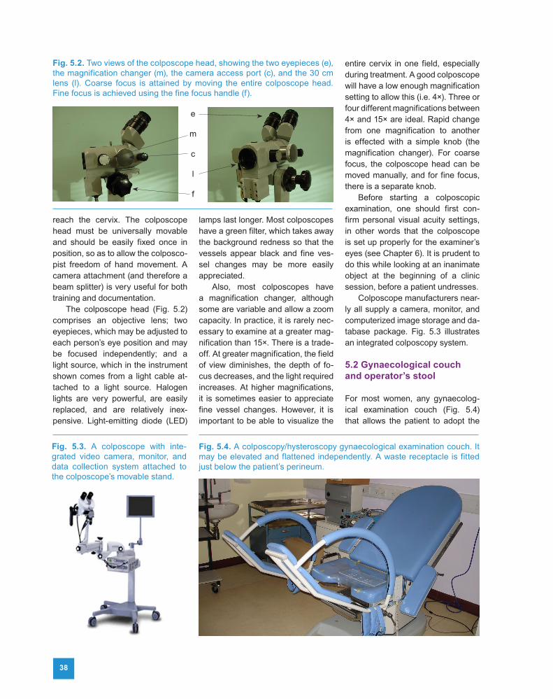
The colposcope head (Fig. 5.2) comprises an objective lens; two eyepieces, which may be adjusted to each person’s eye position and may be focused independently; and a light source, which in the instrument shown comes from a light cable at-tached to a light source. Halogen lights are very powerful, are easily replaced, and are relatively inex-pensive. Light-emitting diode (LED)
lamps last longer. Most colposcopes have a green filter, which takes away the background redness so that the vessels appear black and fine ves-sel changes may be more easily appreciated.
Also, most colposcopes have a magnification changer, although some are variable and allow a zoom capacity. In practice, it is rarely nec-essary to examine at a greater mag-nification than 15×. There is a trade-off. At greater magnification, the field of view diminishes, the depth of fo-cus decreases, and the light required increases. At higher magnifications, it is sometimes easier to appreciate fine vessel changes. However, it is important to be able to visualize the
entire cervix in one field, especially during treatment. A good colposcope will have a low enough magnification setting to allow this (i.e. 4×). Three or four different magnifications between 4× and 15× are ideal. Rapid change from one magnification to another is effected with a simple knob (the magnification changer). For coarse focus, the colposcope head can be moved manually, and for fine focus, there is a separate knob.
Before starting a colposcopic examination, one should first con-firm personal visual acuity settings, in other words that the colposcope is set up properly for the examiner’s eyes (see Chapter 6). It is prudent to do this while looking at an inanimate object at the beginning of a clinic session, before a patient undresses.
Colposcope manufacturers near-ly all supply a camera, monitor, and computerized image storage and da-tabase package. Fig. 5.3 illustrates an integrated colposcopy system.
5.2 Gynaecological couch and operator’s stool
For most women, any gynaecolog-ical examination couch (Fig. 5.4) that allows the patient to adopt the
Fig. 5.2. Two views of the colposcope head, showing the two eyepieces (e), the magnification changer (m), the camera access port (c), and the 30 cm lens (l). Coarse focus is attained by moving the entire colposcope head. Fine focus is achieved using the fine focus handle (f).
e
m
c
l
f
Fig. 5.3. A colposcope with inte-grated video camera, monitor, and data collection system attached to the colposcope’s movable stand.
Fig. 5.4. A colposcopy/hysteroscopy gynaecological examination couch. It may be elevated and flattened independently. A waste receptacle is fitted just below the patient’s perineum.

Chapter 5. Equipment for a colposcopic examination 39
lithotomy or semi-lithotomy position may be used to perform colposco-py. However, it is important that the base of the couch may be tilted so that the TZ on the cervix will become almost perpendicular to the colpo-scopic line of vision. The back of the couch should also be adjustable, and it should be possible to easily elevate or lower the whole couch. A comfortable couch is hugely impor-tant for the patient, who will need to be in position for several minutes in relative undress and who is very like-ly to be anxious. It is important to be able to elevate or lower and tilt the couch to allow optimal positioning of the patient. Also, an examiner’s stool that can be elevated or lowered is very helpful. Being able to quickly flatten the couch so as to deal with the rare vasovagal attack is impor-tant. Finally, the same couch may be used for most outpatient gynaeco-logical procedures (e.g. hysterosco-py, intrauterine contraceptive device [IUCD] insertion, and transvaginal ultrasonography).
If a decision is made to perform excisional treatment, it should usu-ally be performed as an outpatient procedure using electrosurgery to resect the TZ epithelium, i.e. LLETZ/LEEP. A loop electrode is attached
to an electrosurgical unit (ESU) (Fig. 5.5). The loop electrode is housed in a so-called pencil. Suc-tion tubing will connect the ESU to the suction speculum, and a ground plate will connect the patient to the ESU. Some ESUs have a suction unit incorporated into the unit; others do not, in which case it will be nec-essary to have a separate suction machine. The equipment for LLETZ/LEEP, thermal coagulation, and cryo- surgery is described in Chapter 11.
5.3 Camera system
Almost all of the major camera com-panies will supply a camera and at-tachment for a colposcope. Unfortu-nately, the colposcopes usually need a C-mount for the camera to attach to the colposcope, and C-mounts are expensive. Many modern colpo-scopes have a camera system incor-porated into the instrument, without the need for a C-mount. Nowadays, the cost of a reasonable video cam-era is almost the same as that of a still image camera, and very high quality video images can be obtained and stored for future reference. This is immensely valuable as a clinical aid in following up screen-positive patients, whether or not treatment
has been performed, and also as an educational tool for attending colpos-copy trainees.
5.4 Computerized data management system
Many companies provide a software package that allows sociodemo-graphic, clinical, colposcopic, and laboratory data and image capture as well as automatic audit of colpo-scopic diagnostic performance. In this way, it is relatively easy to cre-ate a full audit of performance for an individual colposcopist and to main-tain a clinical database for the clinic service. However, the programs are expensive.
5.5 Instrument trolley
An instrument trolley may seem an unnecessary luxury in colposcopy clinics where budgets are tight. How-ever, the reusable and disposable equipment and the fluids needed to perform a proper colposcopic ex-amination have to be housed some-where, and to have them all to hand in one compartmentalized trolley is both efficient and ergonomically sensible. The last thing a colposco-pist or the patient needs is to have to wait for an assistant to find a par-ticular instrument when it is needed. Finally, if instruments are not housed in a compartmentalized trolley they are not within arm’s length of the colposcopist, and they should be. Figs. 5.6–5.8 illustrate how some reusable instruments and some dis-posable equipment may be conve-niently housed in a trolley. The con-tents of the top, middle, and bottom drawers are shown in Figs. 5.6, 5.7, and 5.8, respectively. In Fig. 5.9, the top surface of the trolley shows some instruments laid out for a colposcop-ic examination. A needle disposal box and a fluid tray are attached on the side (Fig. 5.10).
Fig. 5.5. A portable, battery-driven electrosurgical unit incorporating a suction unit. Ports for the electrosurgical pencil and the ground plate are displayed. A simple electrical battery charger access point and the on/off switch complete the display at the front. The suction port site is at the rear of the unit.
CH
AP
TER
5

4040
5.6 Reusable instruments
5.6.1 Specula
It is fundamentally important to have a full set of different sized specula available when performing colposco-py. A speculum that is too small will not comfortably expose the cervix in
the perpendicular plane, and a speculum that is too large will hurt the patient. Parity and bimanual ex-amination will reveal the appropriate speculum to be used for colposcopy. Specula may be metal or plastic. If LLETZ/LEEP or a “small loop” di-agnostic biopsy is to be performed, the speculum should have a suction
tube on the underside of the anterior blade (Fig. 5.11a). Insulated specu-la are to be avoided, even if using diathermy. After several steriliza-tion cycles, the insulated speculum can lose some of its covering, and this may not be noticeable to the naked eye. If this happens, an elec-trical contact could indeed burn the
Fig. 5.8. Open bottom drawer of a colposcopy clinic trolley, which stores colposcopy suction specula of three different sizes.
Fig. 5.9. Top surface of the equipment trolley used in many colposcopy clinics. Some of the equipment used during a colposcopic examination and treatment are laid out on an incontinence pad. These include cotton swabs and jumbo swabs, a sponge forceps and cotton balls, a suction speculum of medium size, some dental vials containing local analgesic fluid for injection using the dental syringe system, and a loaded dental syringe.
Fig. 5.6. Open top drawer of a colposcopy clinic trolley, which conveniently stores in adjustable compartments a variety of disposable equipment: lubricating jelly, chlorhexidine gluconate sachets, cork boards for biopsy specimen pinning, dental syringe needles, culture swabs, cotton swabs and jumbo swabs, endocervical smear brushes, and cytology fluid bottles.
Fig. 5.7. Open middle drawer of a colposcopy clinic trolley, which stores a variety of disposable examination gloves, gauze swabs, and cotton balls.

Chapter 5. Equipment for a colposcopic examination 4141
patient’s vagina. With the uninsulat-ed speculum, no such risk arises. The area of contact with the vaginal skin is so large that burns are ex-tremely unlikely even if contact with the loop or ball electrode happens accidentally. Also, if one ensures that LLETZ/LEEP is performed at low-power magnification, the entire loop should be visible before and during the procedure, so that con-tact with the vagina or speculum is extremely unlikely. Occasionally, the patulous parous vagina is so lax that it is not possible to completely visu-alize the cervix with the colposcope. Although lateral vaginal wall retrac-tors are available to attach to some specula, they are relatively uncom-fortable; a condom (Fig. 5.11b) or the finger of a large glove (with its end cut off) is a simpler and often more effective alternative.
5.6.2 Sponge forceps
Colposcopists vary in their choice of method for applying acetic acid or Lugol’s iodine. Some use cotton balls soaked in the fluid and applied using a sponge forceps (Fig. 5.9).
Others prefer spray bottles, which may be less cumbersome (Fig. 5.10). If the spray bottles are used, it is im-portant to be aware that splashback can occur and to protect one’s eyes from exposure either with glasses or with the colposcope.
5.6.3 Endocervical forceps
The epithelium that is at risk of de-veloping squamous cervical cancer is usually on the ectocervix in young women, and this is defined in the
IFCPC nomenclature (Bornstein et al., 2012b) as a type 1 TZ. The type 2 TZ, by definition, has an endocervi-cal component but is fully visible to the examining colposcopist. To ac-curately determine the TZ type, it is necessary to carefully examine the SCJ as fully as possible. Also, when investigating a suspicion of adeno-carcinoma or glandular precancer, it is necessary to examine the en-docervix. This will usually require the use of an endocervical forceps. There are several good ones on the market. User-friendly ones are the Kurihara and the Desjardins forceps, which are shown in Fig. 5.12.
5.6.4 Local analgesia (dental) syringes
Metal dental syringes house 2.2 mL vials of either prilocaine with fely-pressin or lignocaine with adrenaline. They allow attachment of 27-gauge needles, which automatically punc-ture the vials when they are loaded into the dental syringes, ready for use. They allow exchange of empty vials for new, full ones in a matter of seconds, so that complete local an-algesia may be achieved in less than a minute (see Chapter 2). A loaded dental syringe is shown in Fig. 5.13. The loaded syringe is long enough to
Fig. 5.10. On the side of the equipment trolley are attached a needle disposal box and a receptacle for the acetic acid and Lugol’s iodine spray bottles.
Fig. 5.11. (a) A Cusco speculum with a suction tube on the underside of the anterior blade for smoke evacuation. (b) A condom (with its end cut off) placed around a Cusco speculum to facilitate examination when the vaginal walls are exceptionally patulous.
baC
HA
PTE
R 5

42
easily reach the cervix, and because the needle itself is relatively short, it will not bend sufficiently to cause a problem with infiltration. Finally, it is narrow enough not to obscure colpo-scopic vision during infiltration.
5.6.5 Tissue sampling instruments
The threshold for taking a biopsy varies from one setting to another. In some colposcopy clinics a biopsy is considered mandatory for every examination, whereas in others a “see-and-treat” policy prevails for women with convincing evidence of high-grade dysplasia (see Chap-ter 1). Endocervical curettes are routinely used in many practices in
the USA but are not often used in the United Kingdom. Many patients find an endocervical curette to be uncomfortable; it often produces in-adequate material and usually pre-cipitates bleeding. It rarely influenc-es practice, and a good endocervical brush smear sample is considered a superior method by many. Biopsy forceps (Fig. 5.14) need to be sharp if they are to procure adequate biop-sies, and some manufacturers make disposable forceps or disposable cutting parts for the reusable biop-sy forceps handles. The common ones available are the Kevorkian and
Tischler-Morgan forceps. When per-forming biopsies, some colposco-pists use infiltration of local analge-sic, and some do not. A small, long hook may be used to fix the cervix before taking a biopsy, but it is not usually necessary if the biopsy for-ceps instrument is sharp. Fig. 5.15 shows a small loop, which is a con-venient way of taking a diagnostic biopsy. Fig. 5.16 shows a range of loops that are used for taking biop-sies as well as for excising the TZ. Fig. 5.17 shows a ball diathermy electrode, used to achieve haemo-stasis after excision of the TZ or to seal a biopsy site. Other haemostatic agents include Monsel’s paste (see Annex 5) and silver nitrate sticks.
5.7 Disposable equipment
Either a 3% or a 5% concentration of acetic acid may be used to highlight colposcopically recognized epithelial lesions. There is no evidence to sug-gest that one strength is superior to the other, although some authorities say that the 3% concentration takes a little longer to effect whiteness. What is important is that the same concen-tration is used for all patients. Care is needed in preparing the solution; disasters have occurred with glacial acetic acid, which will de-epithelial-ize cervical and vaginal epithelium.
Fig. 5.12. (a) Kurihara forceps. (b) Higher-magnification view of Desjardins forceps.
ba
Fig. 5.13. Loaded dental syringe. Fig. 5.14. Punch biopsy forceps.
Fig. 5.15. A small loop used to take colposcopically directed biopsies us- ing an electrosurgical unit.
Fig. 5.16. An array of loops used for LLETZ/LEEP of different sizes and types of transformation zone, and cervical biopsy loops (pink and green) used for taking diagnostic biopsies.
Fig. 5.17. A ball diathermy electrode, used to achieve haemostasis after excision of the transformation zone or to seal a biopsy site.

Chapter 5. Equipment for a colposcopic examination 43
Lugol’s iodine stains mature squamous epithelium dark mahoga-ny brown and affects immature and dysplastic epithelium variably (see Chapter 8). Saline is advocated by several authorities as a cleaning agent before the application of acetic acid or Lugol’s iodine. Cotton swabs are useful to manipulate the cervi-cal epithelium, and jumbo swabs or cotton balls are alternatives (to spray bottles) for the application of acetic acid or Lugol’s iodine. If treat-ment is contemplated, 27-gauge dental syringe needles and vials of prilocaine with felypressin or ligno-caine with adrenaline are needed, and various biopsy forceps or small loops are used to take colposcop-ically directed biopsies. When one
is trying to recognize or rule out the presence of intraepithelial neoplasia, punch biopsy forceps are adequate, but if one is concerned about inva-sive disease, a small loop electrode (Figs. 5.15 and 5.16) should be con-sidered, because it allows a greater depth and confidence in revealing the basement membrane at histolog-ical examination.
5.8 Cork boards and pins to mount LLETZ/LEEP specimens
Liaison with one’s local laboratory will determine in which way excised LLETZ/LEEP specimens will be re-ceived. One option is to open the specimen and then pin it onto a cork
board (Fig. 5.18) before immersion in formalin, so that it may be sec-tioned longitudinally and an accu-rate assessment of the SCJ may be reported.
Fig. 5.18. Cork board and pins to mount LLETZ/LEEP specimens.
• Certain instrument characteristics should be considered before buying a colposcope.
• The gynaecological examination couch should be adjustable, so that it can be elevated or lowered and tilted to allow optimal positioning of the patient.
• Video and/or still images are very valuable as a clinical aid and as an educational tool.
• Reusable instruments and disposable equipment may be conveniently housed in a compartmentalized trolley.
Key points
CH
AP
TER
5


![EC-TYPE EXAMINATION CERTIFICATEimg.pelican.com/docs/products/light/9415Z0_DEMKO... · Directive 94/9/EC [3] EC-Type Examination Certificate Number: ... it indicates that the equipment](https://img.pdfslide.us/doc/110x75/5ab539c27f8b9a0f058c878b/ec-type-examination-949ec-3-ec-type-examination-certificate-number-it-indicates.jpg)