Embed Size (px)
Citation preview
113
CHAPTER 9
WOUND HEALING ACTIVITY
9.1 INTRODUCTION
The skin is the largest organ of our body and represents a barrier to
the environment. It protects the organism from water loss and penetration of
harmful substances. The barrier can be damaged by mechanical and thermal
action or by UV light and different diseases, such as psoriasis and atopic
dermatitis [169, 170]. If the skin barrier is disturbed, it starts repair
mechanisms [171]. The kinetic of the wound healing processes is influenced
by endogenous and exogenous factors [172]. The analysis of the kinetic is
important for therapy control and for the development of efficient drugs and
cosmetic products, stimulating the wound healing [173].
Wound is defined simply as the disruption of the cellular and
anatomic continuity of a tissue [174].Wound may be produced by physical,
chemical, thermal, microbial or immunological insult to the tissue. The
process of wound healing consists of integrated cellular and biochemical
events leading to reestablishment of structural and functional integrity with
regain of strength of injured tissue. Clinically, an individual often encounters
non-healing, under-healing or over healing. Therefore the aim of treating a
wound is to either shorten the time required for healing or to minimize the
undesired consequences [175]. Attention should be directed towards
discovering an agent, which will accelerate wound healing either when it is
progressing normally [176] or when it is suppressed by various agents like
corticosteroids [177], anti-neoplastics [178] or non steroidal anti-
inflammatory agents.
114
Wound healing is a complex process, which is commonly divided
into three phases includes the invasion of inflammatory cells, proliferation of
tissue-repairing cell and tissue remodelling [179]. The rate of wound healing
depends upon many factors including the size of the wound, blood supply to
the area, presence of foreign bodies, microorganisms, age, health of the
patient, nutritional status of an individual, use of drugs and a variety of other
systemic diseases [180].
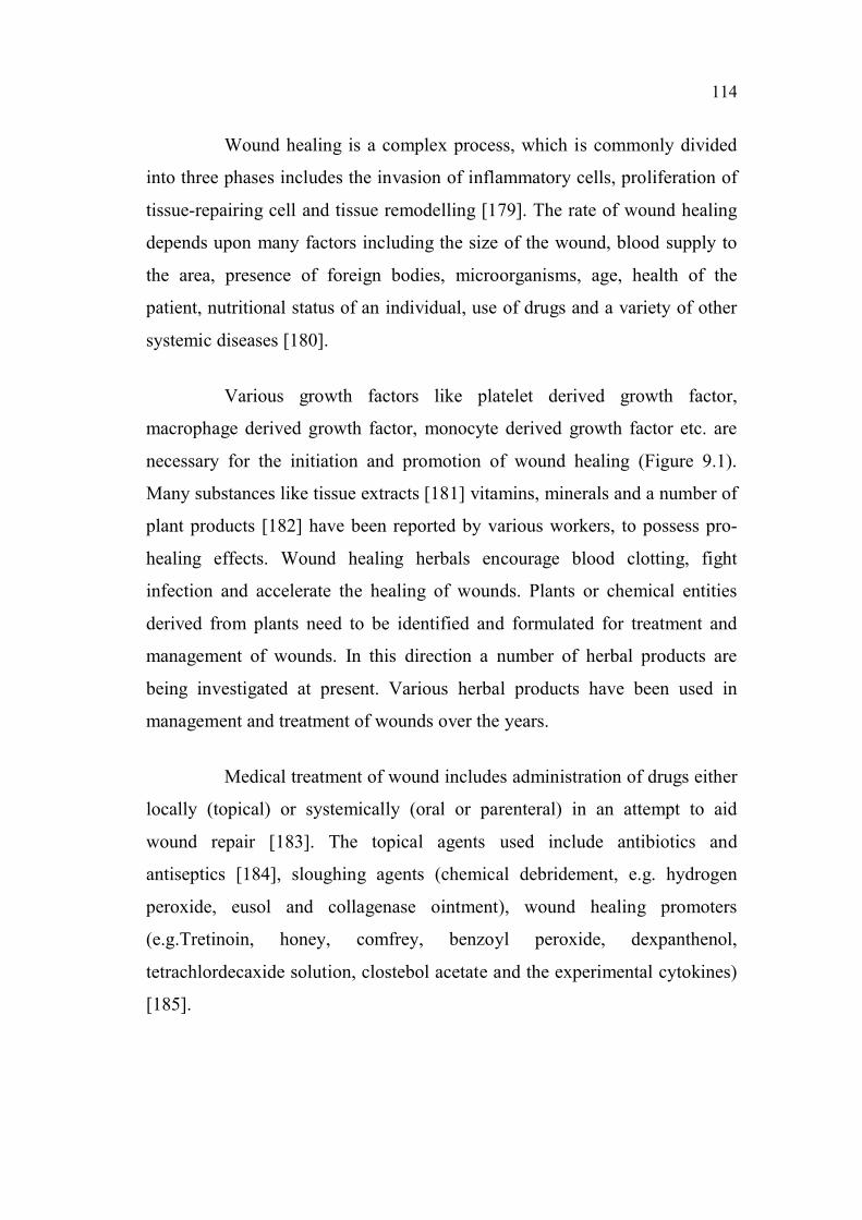
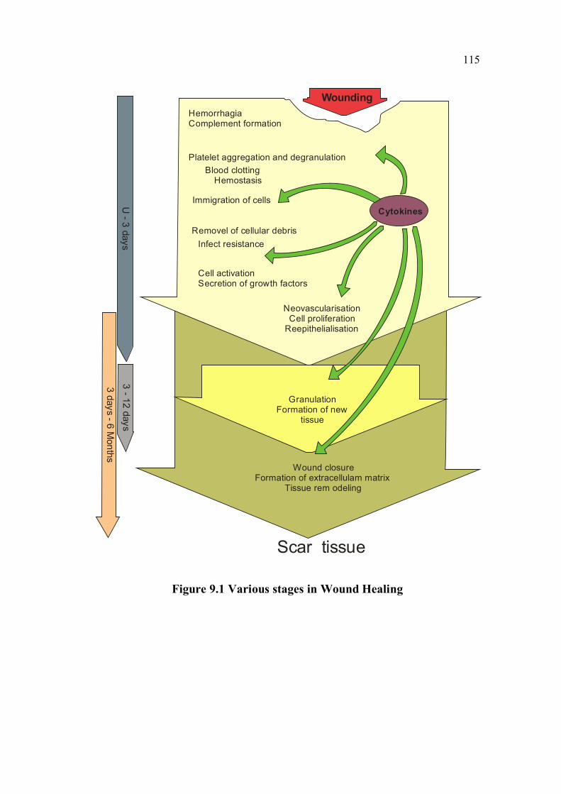
Various growth factors like platelet derived growth factor,
macrophage derived growth factor, monocyte derived growth factor etc. are
necessary for the initiation and promotion of wound healing (Figure 9.1).
Many substances like tissue extracts [181] vitamins, minerals and a number of
plant products [182] have been reported by various workers, to possess pro-
healing effects. Wound healing herbals encourage blood clotting, fight
infection and accelerate the healing of wounds. Plants or chemical entities
derived from plants need to be identified and formulated for treatment and
management of wounds. In this direction a number of herbal products are
being investigated at present. Various herbal products have been used in
management and treatment of wounds over the years.
Medical treatment of wound includes administration of drugs either
locally (topical) or systemically (oral or parenteral) in an attempt to aid
wound repair [183]. The topical agents used include antibiotics and
antiseptics [184], sloughing agents (chemical debridement, e.g. hydrogen
peroxide, eusol and collagenase ointment), wound healing promoters
(e.g.Tretinoin, honey, comfrey, benzoyl peroxide, dexpanthenol,
tetrachlordecaxide solution, clostebol acetate and the experimental cytokines)
[185].
115
HemorrhagiaComplement formation
Platelet aggregation and degranulation
Immigration of cells
Removel of cellular debris
Cell activationSecretion of growth factors
NeovascularisationCell proliferation
Reepithelialisation
Cytokines
GranulationFormation of new
tissue
Infect resistance
Wound closureFormation of extracellulam matrix
Tissue rem odeling
Scar tissue
Blood clotting
U - 3 days
3 - 12 days
3 days - 6 Months
Hemostasis
Wounding
Figure 9.1 Various stages in Wound Healing

116
9.1.1 Plants Used in the Treatment of Wounds (Natural WoundHealers)
Some of the crude drugs with Wound Healing activity are:
Acalypha indica L. [186], Areca catechu L. [187], Anacardium
occidentale L. [188], Calotropis gigantea (L.) R. Br. [189], Cleome viscosa
L. [190], Euphorbia nerifolia [191], Ficus bengalensis L., Ficus racemosa L.,
Jatropha gossypifolia L., Pongamia pinnata (L.) Pierre, L., Ixora coccinia
Smilax zeylanica L., [192], Scoparia dulcis L., [193] Terminalia bellirica
(Gaertn.) Roxb., [194], Themeda triandra Forssk., Trichodesma zeylanicum(Burm. f.) R. Br., Boraginaceae and Vitex altissima L. [195].
9.2 MATERIALS AND METHODS
Animal wound healing models are important biological tools to
understand basic processes of tissue repair and to develop and validate
strategies for clinical treatment. Human wound healing has many unique
aspects that relate to the physiology, age and environment of the species, but
the opportunity to carry out controlled, clinical experimentation on the
mechanism and therapy of wounds is limited.
In general, animal models (with the exception of some transgenic
and targeted gene deletions) attempt to reflect human wound healing
problems, dehiscence, ischemia, ulceration, infection, and scarring.
Henceforth the models utilised for evaluating wound healing activity in
experimental animals for MELA depicting three different types of wounds are
as follows.
a) Chemicals Required
Simple ointment, Corn oil, Nitrofurazone ointment, Anaesthetic ether,
Phosphate buffer, 10% formalin, Ethanol, Xylene, Paraffin, Haematoxylin, Eosin.

117
9.2.1 Drug Formulation
0.5 g of MELA was mixed with 9.5 gm of simple ointment in a
porcelain tile and transferred to a tightly closed amber colored container. Two
types of drug formulations were prepared for topical administration 5% w/w
ointment was prepared in simple ointment base. For oral administration, 200
and 400mg/kg suspensions of the extract was prepared in corn oil. The drugs
were administered orally by an intra-gastric tube.
For assessment of wound healing activity, excision, incision and
dead space wound models were used. The animals were divided into nine
groups of 6 animals each, with three groups each used for excision, incision
and dead space wound models, respectively.
9.2.2 Excision Wound
In the Excision wound model the animals were grouped as follows:
Group I : served as control corn oil, 1ml/kg, topically for 21 days.
Group II : received topical application of 5 % Nitrofurazone I.P. twice a
day for both excision and incision models for 21 days.
Group III : received topical application of 5 %w/w of the extract in
Simple Ointment base I.P. twice a day for both excision and
incision models for 21 days.
The animals were anaesthetized under light ether anaesthesia and
then the skin of the impressed area was excised to full thickness to obtain a
wound area of about 500 mm2. The drugs were topically applied once a day
until complete epithelisation. The parameters studied were wound closure
(measured at regular intervals of time to determine percent wound closure)
and epithelisation time (indicated by the formation of new epithelial tissue to

118
cover the wound). The wounded areas were later evaluated and Wound
contraction was calculated as a percentage of the reduction in wounded area
on 4th, 8 th, 12 th, 16 th, 20 th and 21st days until complete re-epithelialization
was achieved. (The day the scar peeled off without leaving any residual raw
wound was considered the day complete epithelialization was attained).The
following parameters was studied,
9.2.2.1 Epithelization Period
It was monitored by observing the number of days required for
Escher to fall away, leaving no raw wound behind.
9.2.2.2 Wound Contraction
To monitor this, progressive changes in wound area were followed
planimetrically. Leaving the wounding day, wounds were traced on a
transparent paper on alternate days. The animal was restrained in proper
position during tracing. The tracings were then transferred to 1 mm2 graph
sheet. From this, wound areas were read and the percent of wound contraction
was calculated taking the initial size of wound (100 mm2) as 100%.
Percentage wound closure can be calculated using the formula as follows
(9.1)
(9.1)
9.2.3 Incision Wound Model
In the incision model [196], the rats were anaesthetized by
anaesthetic ether and two longitudinal paravertebral incisions of 6 cm length
were made through the skin and cutaneous muscle at a distance of about 1.5
cm from the midline on each side of the depilated back. After the incision, the
100X wound)ofarea(Initial
woundofareadaynth- woundofareaInitialclosure woundPercentage =
119
parted skin was sutured 1 cm apart using surgical thread (no. 000) and curved
needle (no. 11).
Group I : served as Simple ointment base topically twice a day for 10
days.
Group II : received topical application of 5 % Nitrofurazone ointment
twice a day for 10 days.
Group III : received topical application of 5 %w/w of the extract in
Simple Ointment base I.P. twice a day for 10 days.
The wounds were left undressed. The drugs were topically applied
to the wound twice a day until complete healing occurred. The sutures were
removed on the 10th (post wound) day. On day 11, all the animals were
sacrificed under anaesthesia. One linear paravertebral incised skin of each
animal was measured for tensile strength using a fabricated tensilometer.
9.2.4 Determination of Tensile Strength
The Tensile strength (skin breaking strength) of the 10-day old
wound was measured by the method of Lee et al [197]. The rats were secured
to the operating table and a line was drawn on either side of the wound 3 mm
away from the wound. Two allice forceps were firmly applied to the line
facing each other. One of the forcep was fixed, while the other was connected
to a freely suspended lightweight polypropylene graduated container through
a string run over to a pulley. Water was allowed to flow from the reservoir
slowly and steadily into the container. A gradual increase in weight was
transmitted to the wound site pulling apart the wound edges. The moment the
wound just opened up, the water flow was arrested and the volume of water
collected in the container (approximately equal to its weight) was noted.
Three readings were recorded for a given incision wound and the procedure
was repeated on the wound on the contra lateral side. The mean reading of the

120
group was taken as the breaking strength for a given group in the incision
model.
9.2.5 Dead Space Wounds
Dead space wounds were created by implanting two pre-weighed
sterilized polypropylene tube (2.5 length x 0.25 cm diameter) beneath the
dorsal Para-vertebral skin of the anaesthetized rats.
a)Group Separation
After this procedure, the animals were divided into three groups of
six animals each.
Group I : served as control and the animals received vehicle only for 10 days.
Group II : received MELA at a dose of (200 mg/kg) for 10 days.
Group III : received MELA at a dose of (400 mg/kg) for 10 days.
On the 10th (post-wound) day, the granulation tissues formed on the
implanted tubes were carefully detached from surfaces of the tubes. The wet
weight of the granulation tissue was noted. The breaking strength of
granulation tissue was measured by the method of Lee et al. Thereafter, the
granulation tissues were collected dried at 60o C for 24 h and their dry
weights were noted. The dried tissue was added to 5 ml 6 M HCl and kept at
110oC for 24 h. The neutralized acid hydrolysate of the dry tissue was used
for the determination of hydroxyproline [198].
Granulation tissue from the second tube was collected in
phosphate-buffer saline to estimate antioxidant enzymes -Superoxide
dismutase (SOD) [86] and Catalase [87], while a piece of the wet granulation
tissue was preserved in 10% formalin for histopathological studies.
121
Estimation of SOD and Catalase
Estimation of Catalase and Superoxide dismutase were determined
as per the procedure followed in the hepatoprotective activity of MELA.
Estimation of Hydroxyproline
Dry granulation tissue from both control and test groups were used
for the estimation of hydroxyproline. Hydroxyproline present in the neutralized
acid hydrolysate was oxidized by sodium peroxide in presence of copper sulphate
and subsequently complexed with P-dimethylaminobenzaldehyde to develop a
pink colour that was measured spectrophotometrically at 540 nm.
9.2.6 Histopathological Evaluation of Wounded Tissues
A portion of the granulation tissue was subjected to
histopathological studies. The tissues were fixed in 10% neutral formalin
solution for 24 h and dehydrated with a sequence of ethanol–xylene solution
series [199]. The materials were filtered and embedded with paraffin
(40 – 600 C) and a microtome section of 5 μ thickness was taken. The sections
were again processed with ethanol-xylene solvent series and stained with
haematoxylin-eosin dye. The histopathological changes were observed and
photographed using a compound light Microscope (Novex, USA).Ulceration,
necrosis, epithelisation, congestion, oedema, Polymorpholeukocytes (PML),
mononuclear cells, fibroblasts and vascularisation were evaluated in the skin
tissues.
9.2.7 Statistical Analysis
The results were expressed as mean ± SEM of 6 animals in each
group. The data were statistically evaluated by one-way ANOVA, followed
by Dunnet’s t-test for comparison of test groups with control. Values of
p<0.05 were considered statistically significant.
122
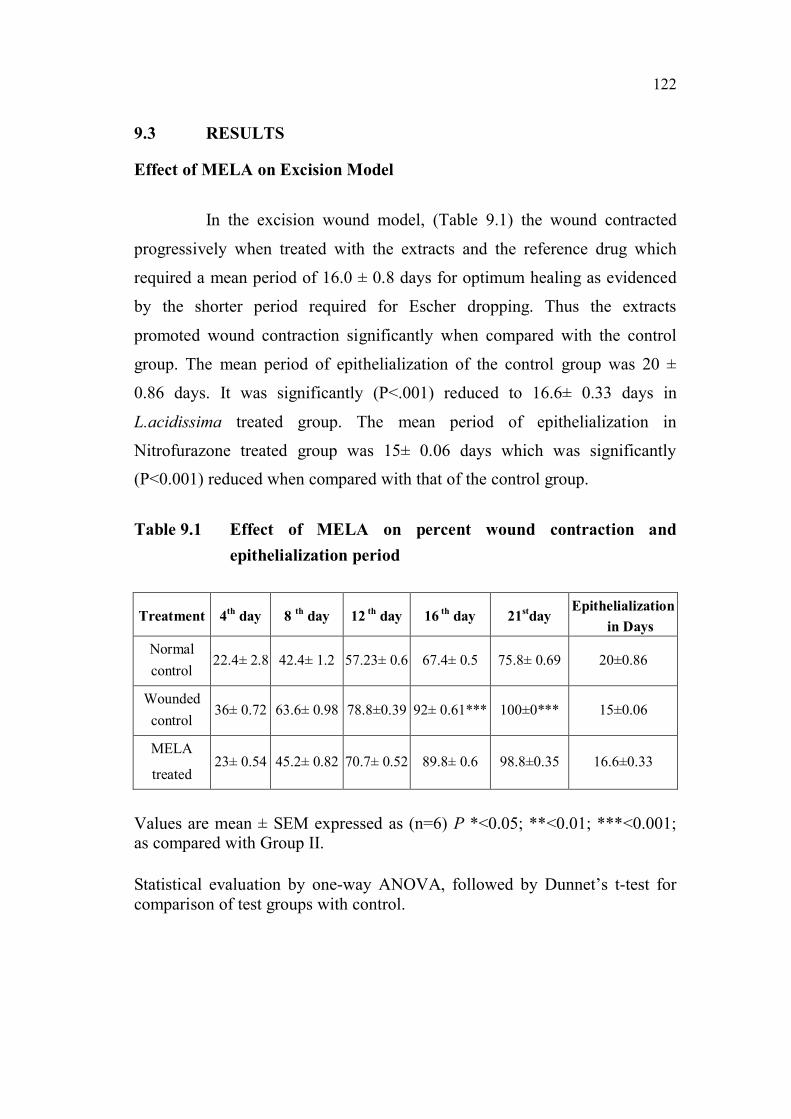
9.3 RESULTS
Effect of MELA on Excision Model
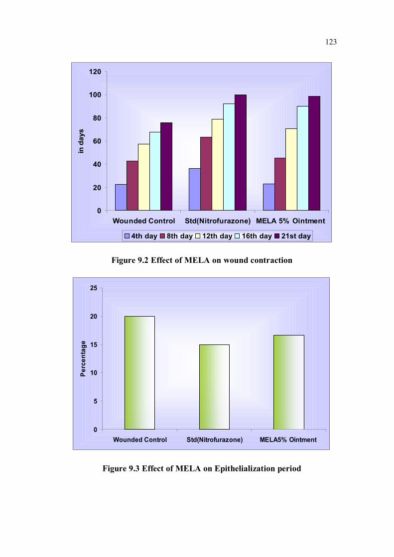
In the excision wound model, (Table 9.1) the wound contracted
progressively when treated with the extracts and the reference drug which
required a mean period of 16.0 ± 0.8 days for optimum healing as evidenced
by the shorter period required for Escher dropping. Thus the extracts
promoted wound contraction significantly when compared with the control
group. The mean period of epithelialization of the control group was 20 ±
0.86 days. It was significantly (P<.001) reduced to 16.6± 0.33 days in
L.acidissima treated group. The mean period of epithelialization in
Nitrofurazone treated group was 15± 0.06 days which was significantly
(P<0.001) reduced when compared with that of the control group.
Table 9.1 Effect of MELA on percent wound contraction andepithelialization period
Treatment 4th day 8 th day 12 th day 16 th day 21stdayEpithelialization
in DaysNormalcontrol
22.4± 2.8 42.4± 1.2 57.23± 0.6 67.4± 0.5 75.8± 0.69 20±0.86
Woundedcontrol
36± 0.72 63.6± 0.98 78.8±0.39 92± 0.61*** 100±0*** 15±0.06
MELA
treated23± 0.54 45.2± 0.82 70.7± 0.52 89.8± 0.6 98.8±0.35 16.6±0.33
Values are mean ± SEM expressed as (n=6) P *<0.05; **<0.01; ***<0.001;as compared with Group II.
Statistical evaluation by one-way ANOVA, followed by Dunnet’s t-test forcomparison of test groups with control.
123
0
20
40
60
80
100
120
Wounded Control Std(Nitrofurazone) MELA 5% Ointment
in d
ays
4th day 8th day 12th day 16th day 21st day
Figure 9.2 Effect of MELA on wound contraction
0
5
10
15
20
25
Wounded Control Std(Nitrofurazone) MELA5% Ointment
Perc
enta
ge
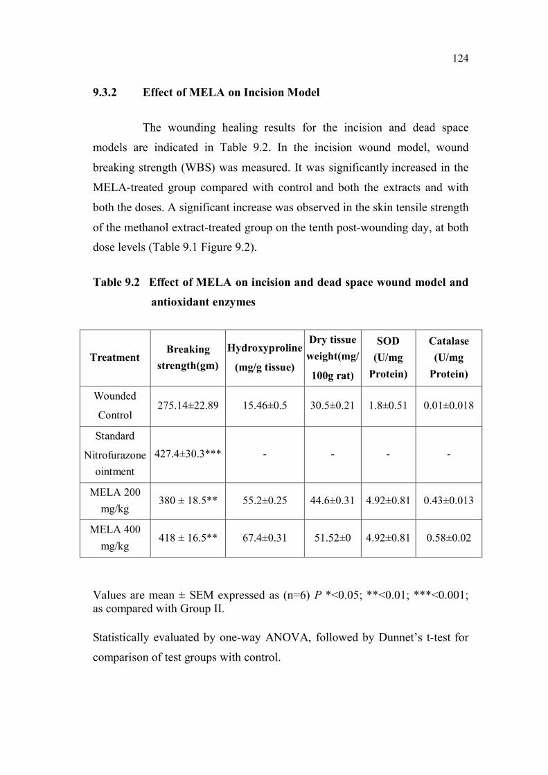
Figure 9.3 Effect of MELA on Epithelialization period
124
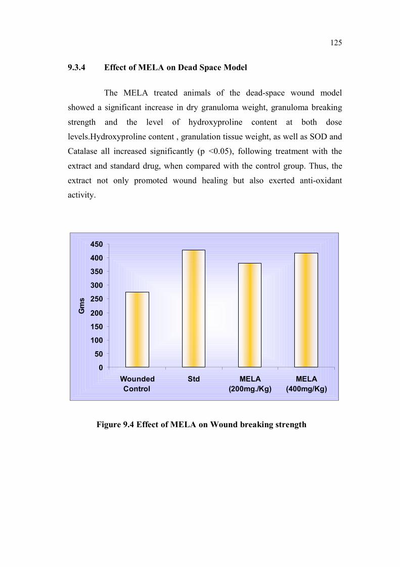
9.3.2 Effect of MELA on Incision Model
The wounding healing results for the incision and dead space
models are indicated in Table 9.2. In the incision wound model, wound
breaking strength (WBS) was measured. It was significantly increased in the
MELA-treated group compared with control and both the extracts and with
both the doses. A significant increase was observed in the skin tensile strength
of the methanol extract-treated group on the tenth post-wounding day, at both
dose levels (Table 9.1 Figure 9.2).
Table 9.2 Effect of MELA on incision and dead space wound model andantioxidant enzymes
TreatmentBreaking
strength(gm)Hydroxyproline
(mg/g tissue)
Dry tissueweight(mg/
100g rat)
SOD(U/mg
Protein)
Catalase(U/mg
Protein)
Wounded
Control275.14±22.89 15.46±0.5 30.5±0.21 1.8±0.51 0.01±0.018
Standard
Nitrofurazoneointment
427.4±30.3*** - - - -
MELA 200mg/kg
380 ± 18.5** 55.2±0.25 44.6±0.31 4.92±0.81 0.43±0.013
MELA 400mg/kg
418 ± 16.5** 67.4±0.31 51.52±0 4.92±0.81 0.58±0.02
Values are mean ± SEM expressed as (n=6) P *<0.05; **<0.01; ***<0.001;as compared with Group II.
Statistically evaluated by one-way ANOVA, followed by Dunnet’s t-test for
comparison of test groups with control.
125
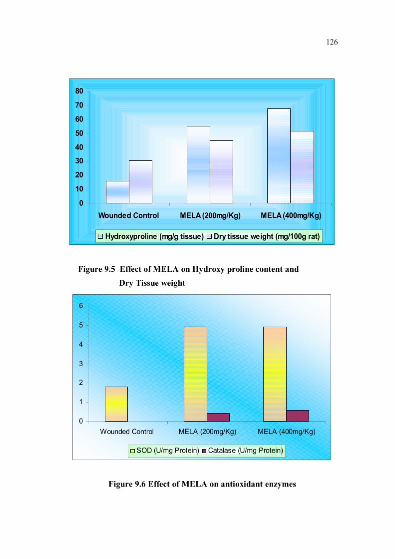
9.3.4 Effect of MELA on Dead Space Model
The MELA treated animals of the dead-space wound model
showed a significant increase in dry granuloma weight, granuloma breaking
strength and the level of hydroxyproline content at both dose
levels.Hydroxyproline content , granulation tissue weight, as well as SOD and
Catalase all increased significantly (p <0.05), following treatment with the
extract and standard drug, when compared with the control group. Thus, the
extract not only promoted wound healing but also exerted anti-oxidant
activity.
0
50
100
150
200
250
300
350
400
450
WoundedControl
Std MELA(200mg./Kg)
MELA(400mg/Kg)
Gm
s
Figure 9.4 Effect of MELA on Wound breaking strength
126
0
10
20
30
40
50
60
70
80
Wounded Control MELA (200mg/Kg) MELA (400mg/Kg)
Hydroxyproline (mg/g tissue) Dry tissue weight (mg/100g rat)
Figure 9.5 Effect of MELA on Hydroxy proline content and Dry Tissue weight
0
1
2
3
4
5
6
Wounded Control MELA (200mg/Kg) MELA (400mg/Kg)
SOD (U/mg Protein) Catalase (U/mg Protein)
Figure 9.6 Effect of MELA on antioxidant enzymes
127
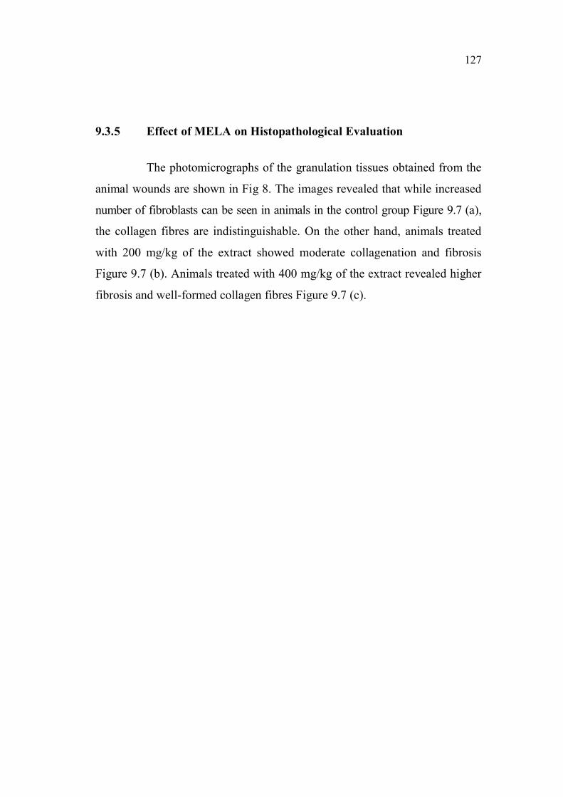
9.3.5 Effect of MELA on Histopathological Evaluation
The photomicrographs of the granulation tissues obtained from the
animal wounds are shown in Fig 8. The images revealed that while increased
number of fibroblasts can be seen in animals in the control group Figure 9.7 (a),
the collagen fibres are indistinguishable. On the other hand, animals treated
with 200 mg/kg of the extract showed moderate collagenation and fibrosis
Figure 9.7 (b). Animals treated with 400 mg/kg of the extract revealed higher
fibrosis and well-formed collagen fibres Figure 9.7 (c).
128
(a) (b)
(c)
Figure 9.7 Histopathology of granulation tissue
(a) Control animals, showing increased number of fibroblasts andindistinguishable collagen fibres.
(b) MELA (200 mg/kg) animals, showing moderate collagenation and fibrosis.
(c) MELA (400mg/kg) animals, showing a high level of fibrosis as well as well-formed collagen fibres.
129
Effect of MELA on Biochemical Parameters
In the dead space wound model, the granulation tissue,
hydroxyproline level was significantly increased in MELA 400 mg/kg (P <
0.01), and in Nitrofurazone treated groups are compared with that of the
control group (P < 0.001). There was no significant difference among
different doses of methanolic extract. When compared with methanolic extract
200 mg/kg, there was significant increase in hydroxyproline level in the
extract 400 mg/kg (P < 0.01) (Table 9.2, Figure 9.5).
Effect of MELA on Antioxidant Parameters
SOD activity in granulation tissue was significantly increased in the
case of rats treated with methanolic extract 200 mg/kg (P < 0.05), 400 mg/kg
(P < 0.05) when compared with control (Table 9.2,Figure 9.6). Catalase level
in granulation tissue was significantly increased in the case of methanolic
extract 200 mg/kg (P < 0.05), methanolic extract 400 mg/kg (P < 0.05) when
compared with control.
9.4 DISCUSSION
Wounds are common clinical entities in day-to-day life, which may
be major or minor. The process of wound healing can be classified into five
phases - cellular phase (granulation), narrowing of wound area (wound
contraction), collagen deposition (collagenation), epithelial covering
(Epithelialisation) and scar remodelling (cicatrisation).These phases are
concurrent but independent of each other. Any agent which accelerates the
process is a promoter of wound healing [200].
130
Wound healing or repair is a natural process of regenerating dermal
and epidermal tissue and may be categorized into three phases viz,
inflammation, proliferation and remodelling phase. In the inflammation
phase, various growth factors such as tumour necrosis factor (TNF),
interleukins (IL) are released to initiate the proliferation phase. The latter is
characterized by angiogenesis, collagen deposition, granular tissue formation,
epithelialization and wound contraction [201]. In the last phase, the levels of
collagen production and degradation equalize, after which disorganized fibres
are rearranged thus increasing the tensile strength of the wound. The capacity
of wound to heal depends, in part, on its depth, as well as on the overall health
and nutritional status of the individual. Following injury, inflammatory
response occurs and the cells below the dermis begin to increase collagen
production. Later, the epithelial tissue is regenerated. It is well known that,
stages in healing namely, coagulation, inflammation, microphasia, fibroblasts
formation and collagenation, are intimately interlinked [202].
The wound breaking strength is determined by the rate of collagen
synthesis and more so by the maturation process where there is a covalent
binding of collagen fibrils through inter and intra molecular cross linking. In
our study, dead space wound model showed a significant increase in
hydroxyproline concentration and also the dry weight of the granulation tissue
was significantly increased in MELA treated group. By this we can assume
that MELA might not have increased the collagen content but probably have
altered the maturation process, by affecting the cross linking of collagen or
improving the quality of collagen fibrils. The increase in weight by
Nitrofurazone treated group could be due to high protein concentration and
collagen bundle formation.
131
In the present investigation, MELA at the doses of 200 and
400 mg/kg showed significantly increased healing by wound contraction,
when compared to the control group. Increase in the breaking strength of the
wound is indicative of improved collagenation which contributes to healing,
enhances epithelization and promotes wound contraction by increasing
granulation tissue weight due to infiltration of macrophages [203].
Wound contraction is the process of mobilizing healthy skin
surrounding the wound to cover the denuded area. This centripetal movement
of wound margin is believed to be due to the activity of myofibroblasts [204].
During wound contraction, the wound is made smaller by the action of
myofibroblasts, which establish a grip on the wound edges and contract
themselves using a mechanism similar to that in smooth muscle cells. In the
maturation and remodelling phase, collagen is remodelled and realigned along
tension lines and cells that are no longer needed to be removed by apoptosis.
MELA–based ointment has significant influence on one or some of the stages
resulting in faster rate of wound closure when compared to the control group.
Since MELA enhanced wound contraction, it would have either enhanced
contractile property of myofibroblasts or increases the number of
myofibroblasts recruited into the wound area. The wound is eventually closed
by a combination of all these process.
In excision wound model, L.acidissima hastened the period of
epithelialization significantly. The process of wound contraction and
epithelisation is separate and independent. The activity of fibroblast is
responsible for wound contraction and involves movement of entire dermis.
Epithelization involves migration and proliferation of cells. It is known that
stabilization of lysosomal membranes, inhibition of cellular migration and
inhibition of fibroblast contraction are responsible for their anti-healing
effects [205]. Thus, intervention in any one of these phases by drugs would
132
eventually lead to either promotion or depression of collagenation, wound
contraction and epithelisation [206]. In studies using the excision wound
model, animals treated with MELA showed a significant decrease in the
epithelisation period, as evidenced by the shorter period for the fall of escher
compared to control. The extract also facilitated the epithelisation period
significantly at both dose levels (Table 9.1 and Figure 9.3). The faster wound
contraction rate of the extract may be due to stimulation of interleukin-8, an
inflammatory α-chemokine which affects the function and recruitment of
various inflammatory cells, fibroblasts and keratinocytes. It may increase the
gap junctional intracellular communication in cultured fibroblasts and induces
a more rapid maturation of granulation tissue.
Collagen is a major protein of the extracellular matrix and is the
component that ultimately contributes to wound strength. Breakdown of
collagen liberates free hydroxyproline and its peptide. Therefore,
measurement of hydroxyproline could be used as an index for determining
collagen turnover [207]. The extract-treated groups showed significant
increases in the level of hydroxyproline, which is a reflection of increased
collagen content. This was confirmed by histopathological examination of
wound granulation tissue which showed a well-developed matrix in extract
treated group with the collagen was well-organized and bundles formed
between the cells. There was also better neovascularisation in the extract
treated groups than in the control group.
Elimination of reactive oxygen species is reported to be an
important strategy to improve healing of wounds. Many plant extracts and
medicinal herbs have shown potent antioxidant activity. Research into the role
of antioxidants from plant extracts in wound healing has been published
widely [208].Reactive oxygen species (ROS) play a vital role in wound
133
healing and can trigger various beneficial oxygen free radicals. While
antioxidants improve healing in ischemic skin wounds [209]. Elevated lipid
peroxide levels have also been demonstrated in certain inflammatory skin
lesions such as wound and dermatitis. Therefore, if a compound has
antioxidant potential, it can be a good therapeutic agent for enhancing the
wound healing process. Hence, estimation of antioxidants like SOD, Catalase
in granulation tissues is also relevant because these antioxidants hasten the
process of wound healing by destroying the free radicals [210].
An increase in the levels of anti-oxidant enzymes (SOD and
Catalase) was observed in the granulation tissue of the extract treated groups
may also have contributed to the wound healing effect of the extract. Studies
on the estimation of antioxidant enzyme revealed that the extract significantly
increased the levels of superoxide dismutase and Catalase, the two powerful
antioxidant enzymes of the body that are known to quench Superoxide
radicals (Table 9.2,Figure 9.6).The antioxidant enzymes are known to quench
the superoxide radical and thus prevent the damage of cells caused by free
radicals. So in this study, scavenging effect might be one of the most
important components of wound healing which may be responsible to support
wound healing property. Thus the enhanced wound healing may be due to the
free radical scavenging action of the plant as well as enhanced antioxidant
enzyme level in granuloma tissues [211].
Several antioxidants such as Ascorbic acid, Catalase was found to
improve healing. Ascorbic acid has a role in both the formation and
maintenance of collagen in healing wounds. As the extract of L.acidissima
contains rich source of Vitamin - C, this also may attribute to the antioxidant
potential and in turn significant wound healing effect. Catalase was found to
detoxify hydrogen peroxide which can otherwise inflict severe damage to
134
regenerating cells. In condensation, the results indicated the beneficial effects
of MELA by reduced lipid peroxide levels in treated wounds, which may in
turn be responsible for acceleration of the healing process.
9.5 CONCLUSION
The present study reveals the wound healing and antioxidant
activities of MELA in experimental animal model against incision,excision
and dead space models which was proved by theevaluated parameters and the
antioxidant enzyme levels.Further works are being carried out to isolate and
identify the active principle involved in the wound healing and antioxidant
activities of this plant extract.Thus MELA can be utilised as an economical
therapeutic agent for wound management as a pro-healer, as well as to control
abnormal healing on proper formulation.