Embed Size (px)
Citation preview
Chapter 7Chapter 7Skeletal SystemSkeletal System
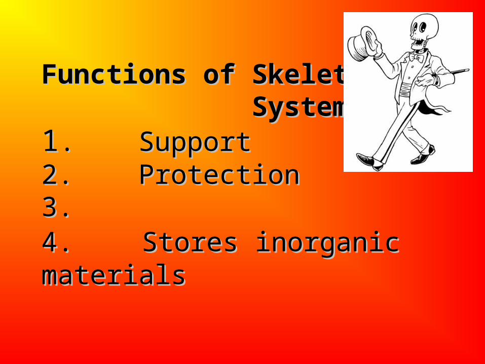
Functions of Skeletal Functions of Skeletal System: System:11. . Support Support 2. 2. ProtectionProtection3.3.4. 4. Stores inorganic Stores inorganic materialsmaterials
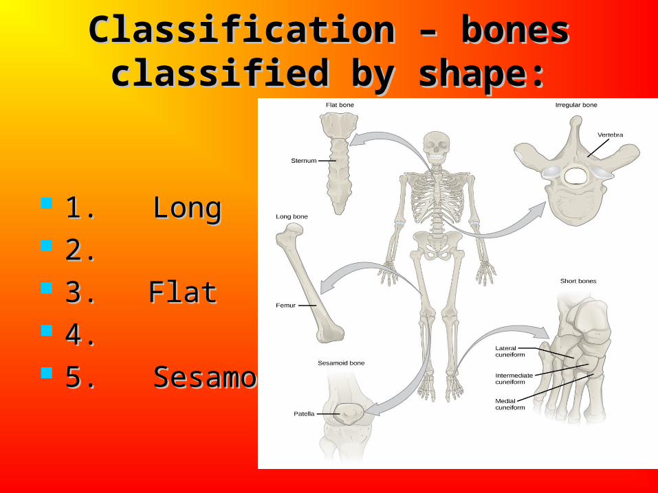
Classification – bones Classification – bones classified by shape:classified by shape:
1. Long1. Long 2. 2. 3. 3. Flat Flat 4. 4. 5. Sesamoid5. Sesamoid
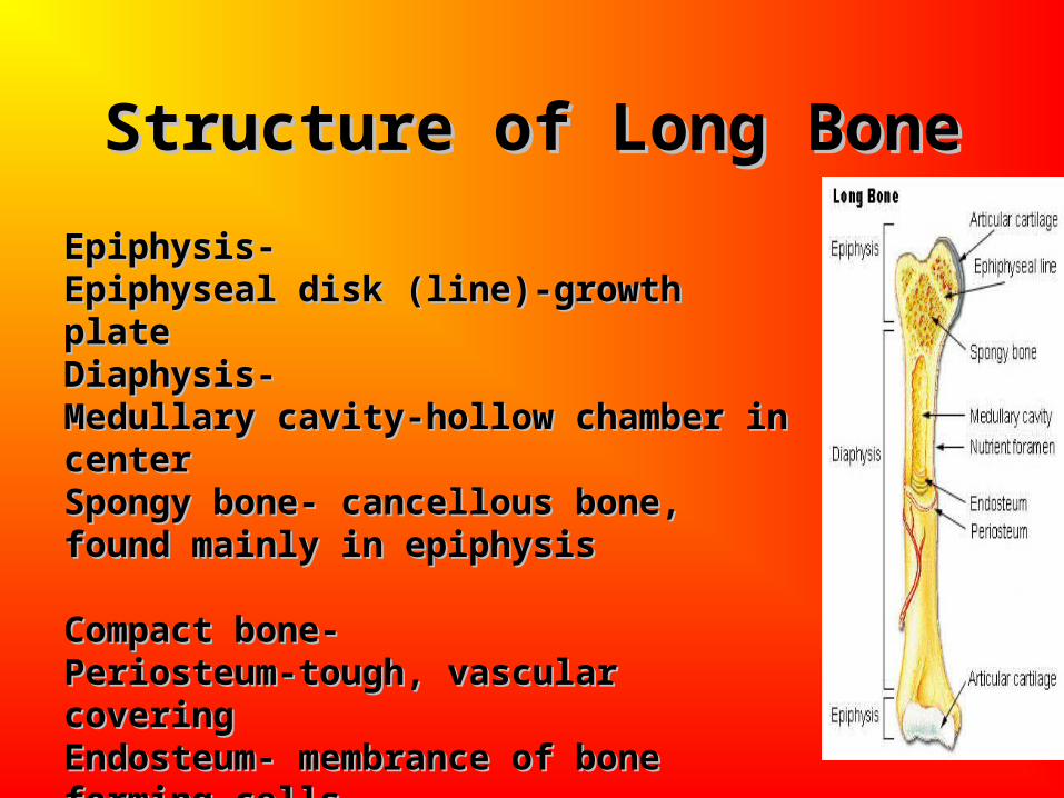
Structure of Long BoneStructure of Long Bone
Epiphysis- Epiphysis- Epiphyseal disk (line)-growth plateEpiphyseal disk (line)-growth plateDiaphysis- Diaphysis- Medullary cavity-hollow chamber in centerMedullary cavity-hollow chamber in centerSpongy bone- cancellous bone, found Spongy bone- cancellous bone, found mainly in epiphysismainly in epiphysisCompact bone-Compact bone-Periosteum-tough, vascular coveringPeriosteum-tough, vascular coveringEndosteum- membrance of bone forming Endosteum- membrance of bone forming cellscellsArticular cartilage-Articular cartilage-
Structure of a Long Bone
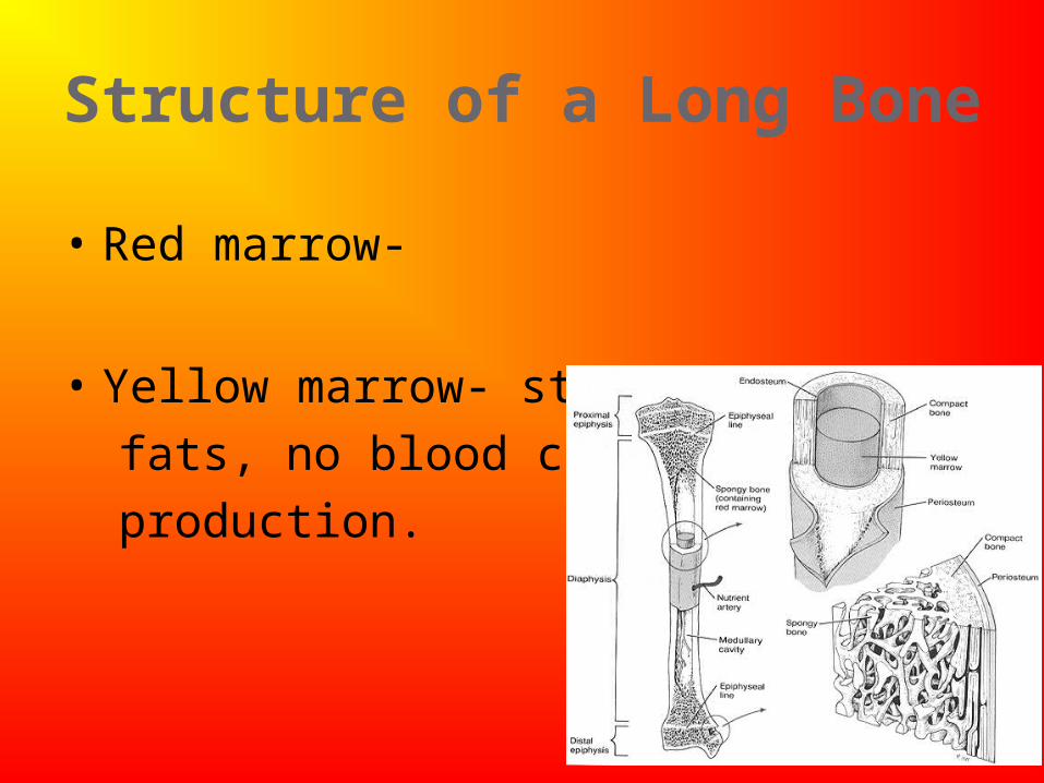
• Red marrow-
• Yellow marrow- stores
fats, no blood cell
production.
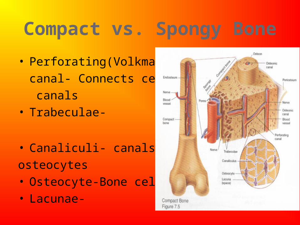
Compact vs. Spongy Bone
• Perforating(Volkmann’s)
canal- Connects central
canals• Trabeculae-
• Canaliculi- canals btwn
osteocytes• Osteocyte-Bone cells• Lacunae-
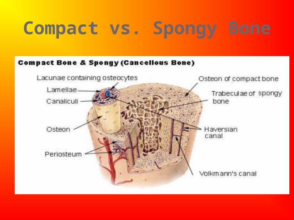
Compact vs. Spongy Bone
Bone Development – 2 Types:
1. Intramembranous ossification (changing cartilage to bone) (ex. - skull)
• layers of undifferentiated connective tissue appear at sites of future bone
•
• osteoblasts become osteocytes (mature bone cells) when completely surrounded by a bony matrix
• Connective tissue on surface forms the periosteum
•
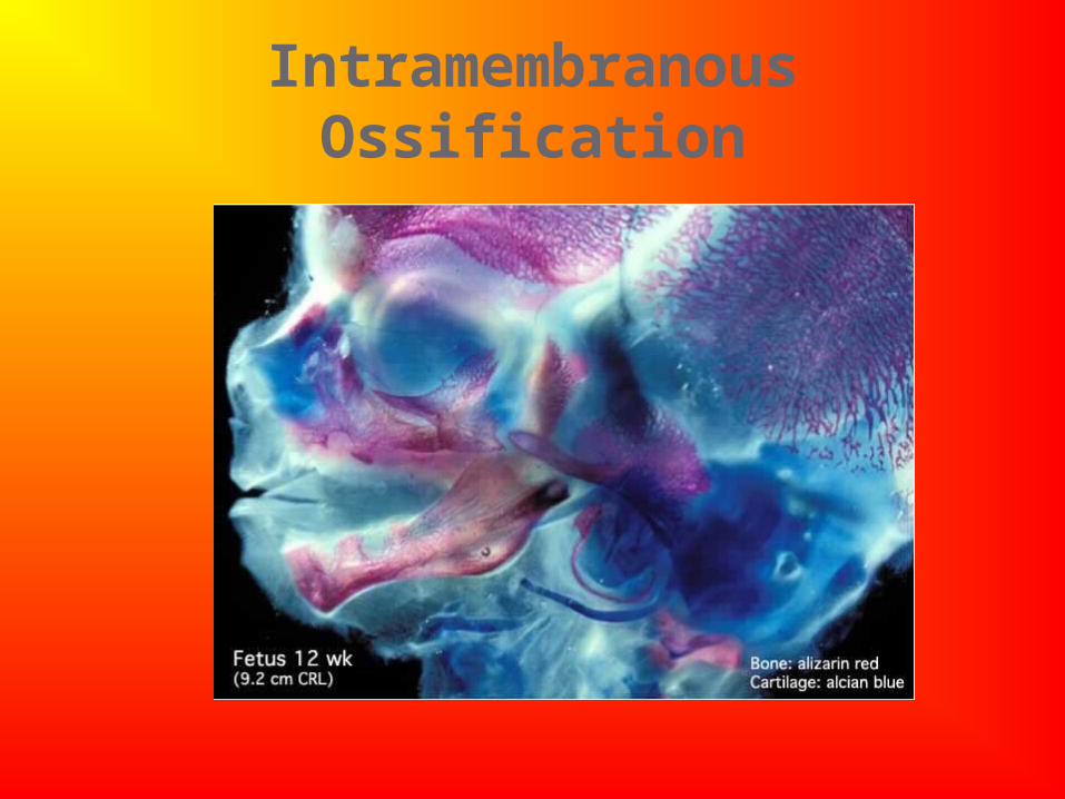
Intramembranous Ossification
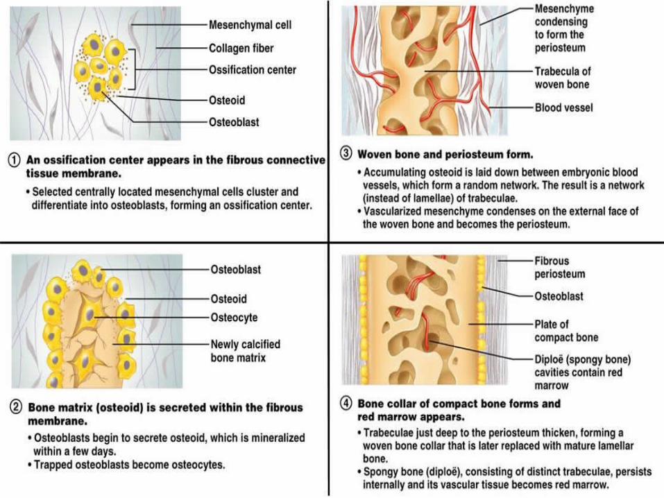
Intramembranous Ossifiction
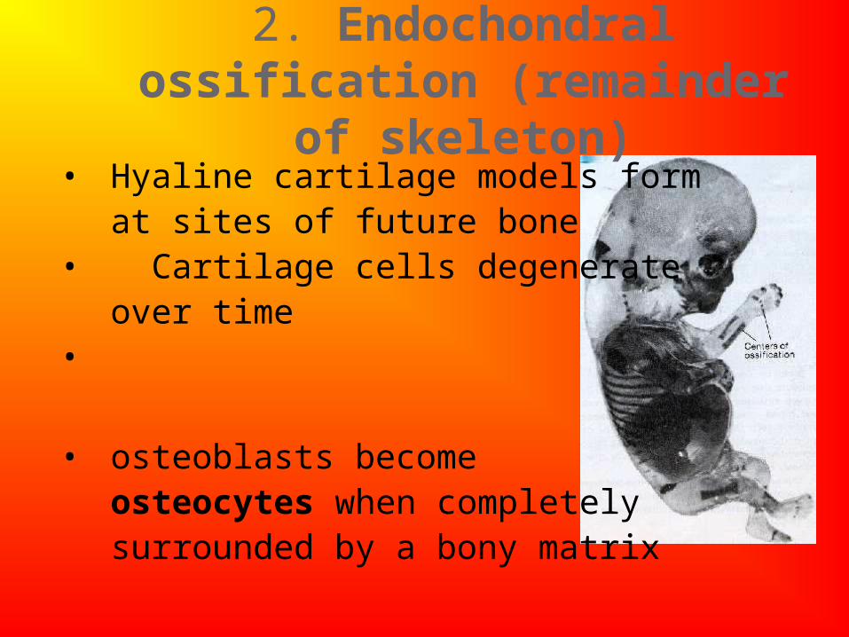
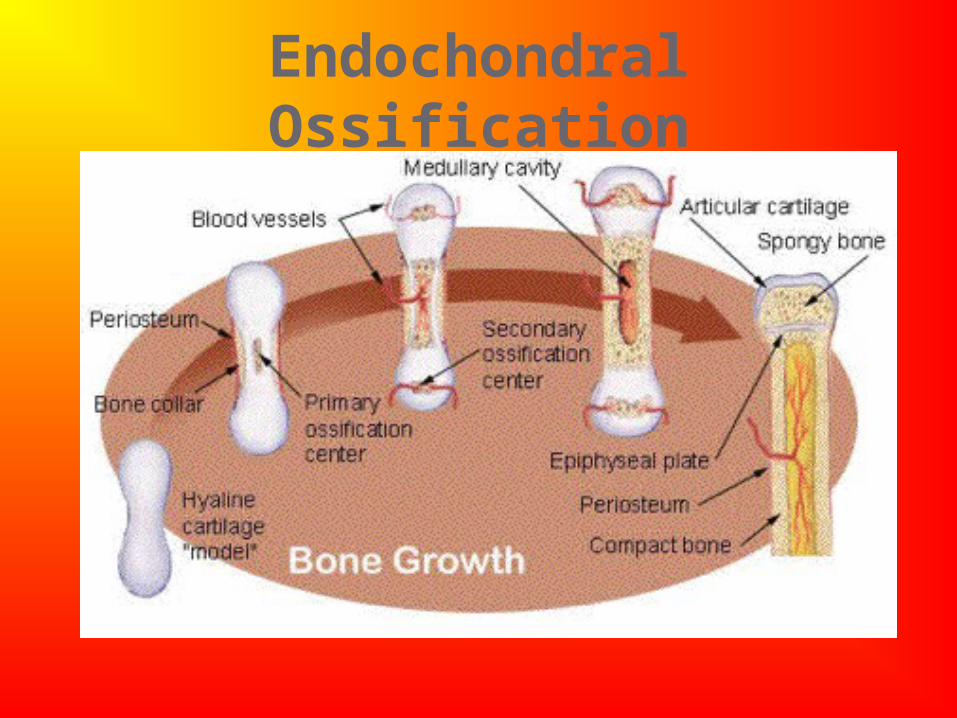
2. Endochondral ossification (remainder of skeleton)
• Hyaline cartilage models form at sites of future bone
• Cartilage cells degenerate over time
•
• osteoblasts become osteocytes when completely surrounded by a bony matrix
Endochondral Ossification
Bone Development
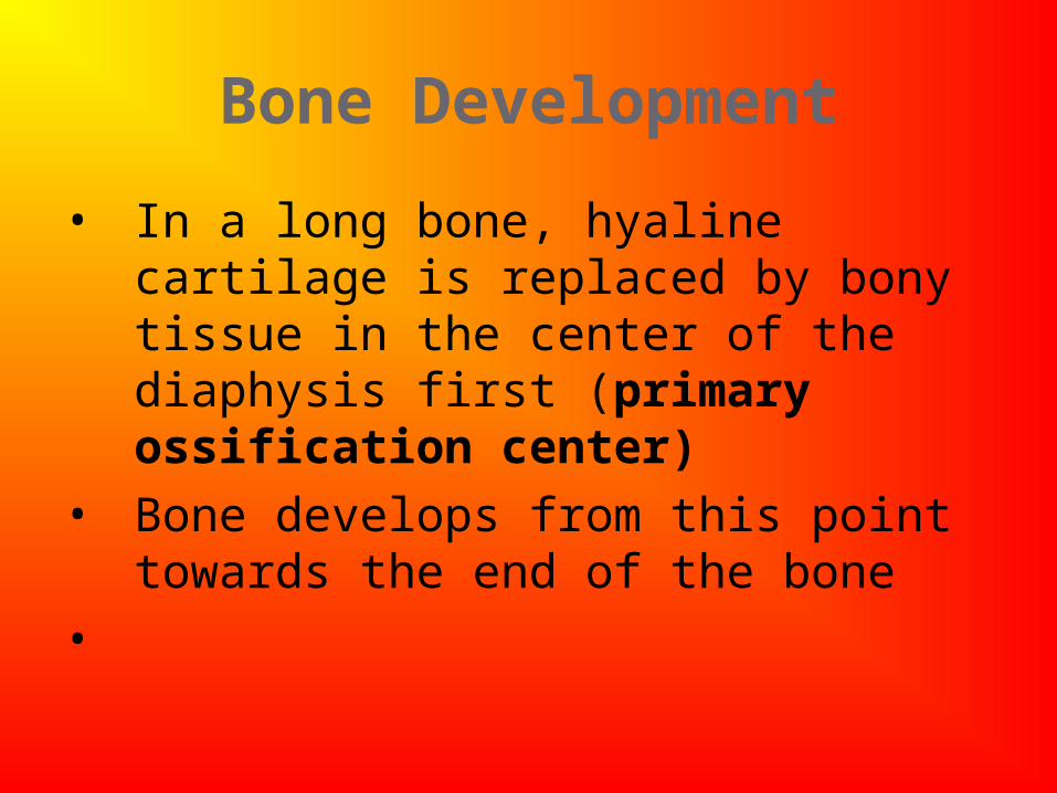
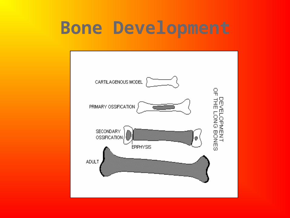
• In a long bone, hyaline cartilage is replaced by bony tissue in the center of the diaphysis first (primary ossification center)
• Bone develops from this point towards the end of the bone
•
Bone Development
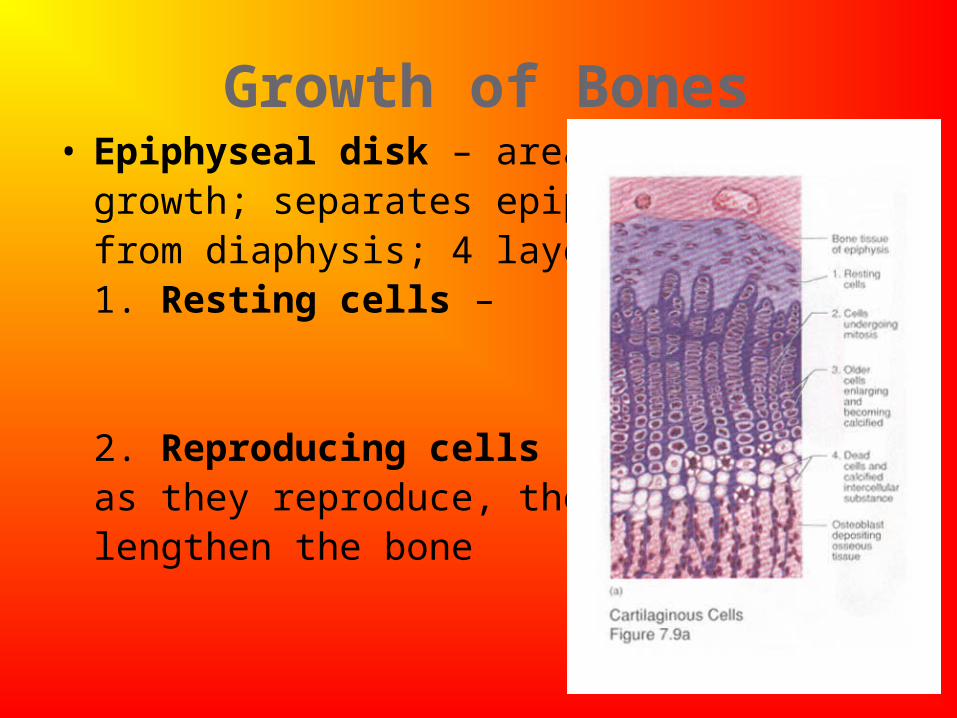
Growth of Bones• Epiphyseal disk – area of
growth; separates epiphysisfrom diaphysis; 4 layers:1. Resting cells –
2. Reproducing cells - as they reproduce, they lengthen the bone
1.
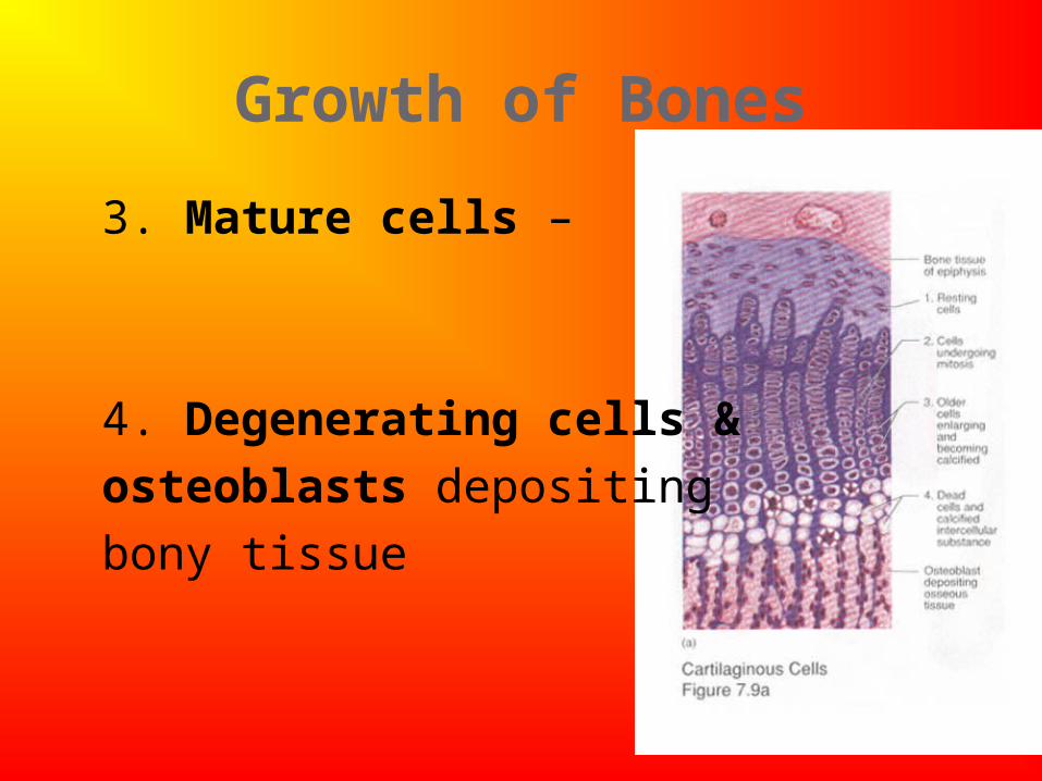
Growth of Bones
3. Mature cells –
4. Degenerating cells &
osteoblasts depositing
bony tissue
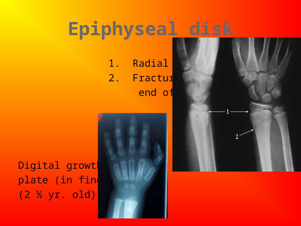
Epiphyseal disk
1. Radial growth plate→
2. Fracture – distal
end of radius →
Digital growth
plate (in fingers)
(2 ½ yr. old)→
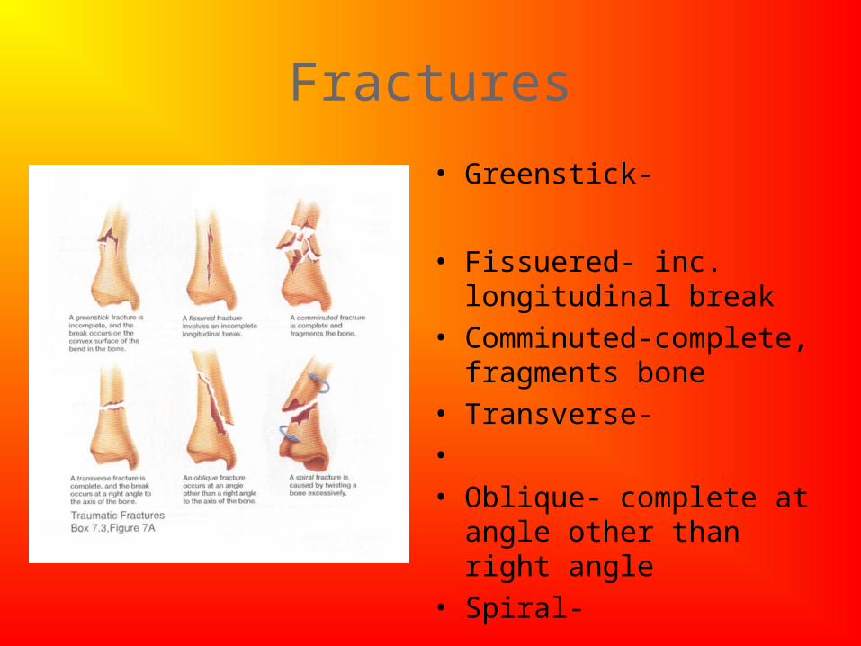
Fractures
• Greenstick-
• Fissuered- inc. longitudinal break
• Comminuted-complete, fragments bone
• Transverse-• • Oblique- complete at angle
other than right angle• Spiral-
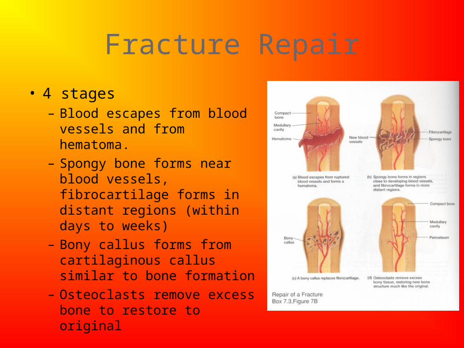
Fracture Repair
• 4 stages– Blood escapes from blood
vessels and from hematoma.
– Spongy bone forms near blood vessels, fibrocartilage forms in distant regions (within days to weeks)
– Bony callus forms from cartilaginous callus similar to bone formation
– Osteoclasts remove excess bone to restore to original
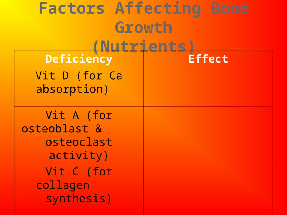
Factors Affecting Bone Growth(Nutrients)
Deficiency Effect
Vit D (for Ca absorption)
Vit A (for osteoblast &osteoclast activity)
Vit C (for collagensynthesis)
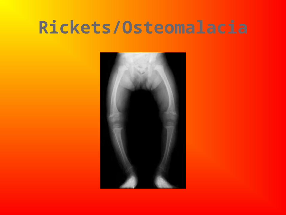
Rickets/Osteomalacia
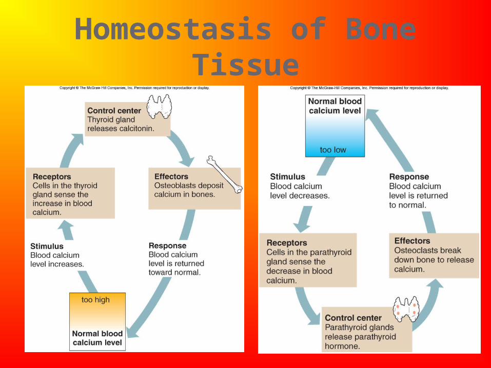
Homeostasis of Bone Tissue
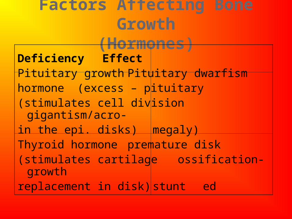
Factors Affecting Bone Growth(Hormones)
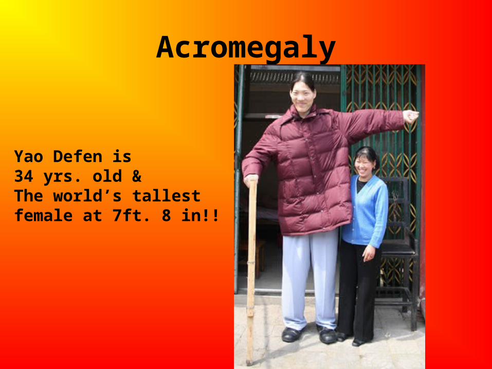
Deficiency EffectPituitary growth Pituitary dwarfism hormone (excess – pituitary (stimulates cell division gigantism/acro-in the epi. disks) megaly)Thyroid hormone premature disk(stimulates cartilage ossification-growthreplacement in disk) stunted
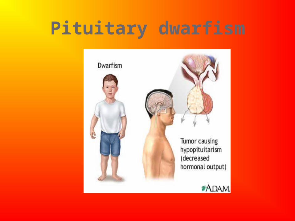
Pituitary dwarfism
Acromegaly
Yao Defen is 34 yrs. old &The world’s tallestfemale at 7ft. 8 in!!
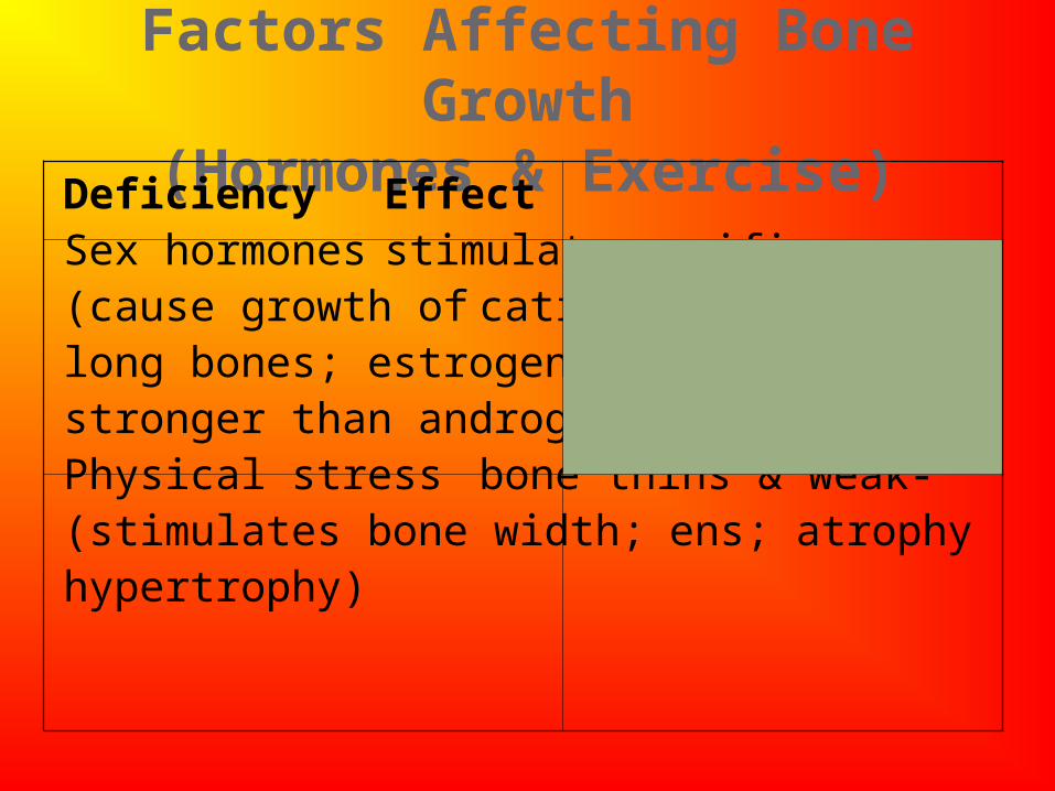
Factors Affecting Bone Growth(Hormones & Exercise)
Deficiency EffectSex hormones stimulate ossifi-(cause growth of cation of disks;long bones; estrogen stop bone lengthstronger than androgen)Physical stress bone thins &
weak-(stimulates bone width; ens; atrophyhypertrophy)
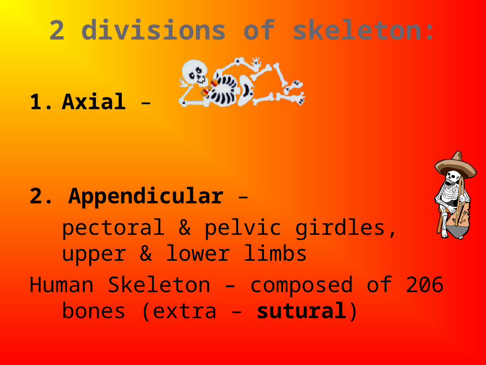
2 divisions of skeleton:
1. Axial –
2. Appendicular –
pectoral & pelvic girdles, upper & lower limbs
Human Skeleton – composed of 206 bones (extra – sutural)
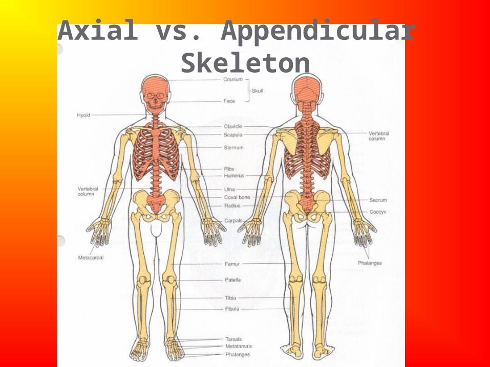
Axial vs. Appendicular Skeleton
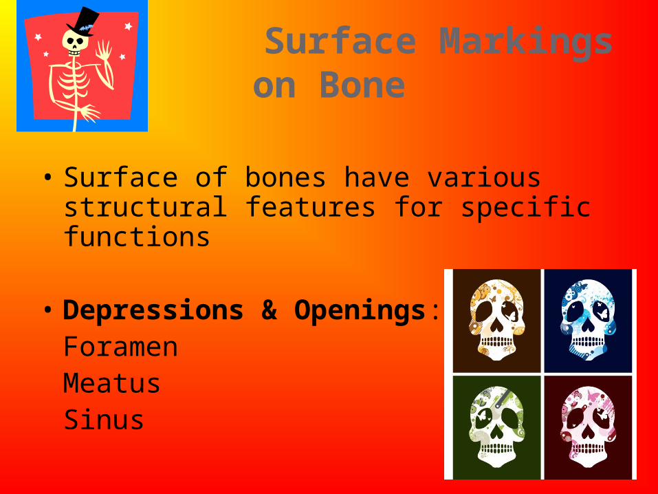
Surface Markings on Bone
• Surface of bones have various structural features for specific functions
• Depressions & Openings:ForamenMeatusSinus
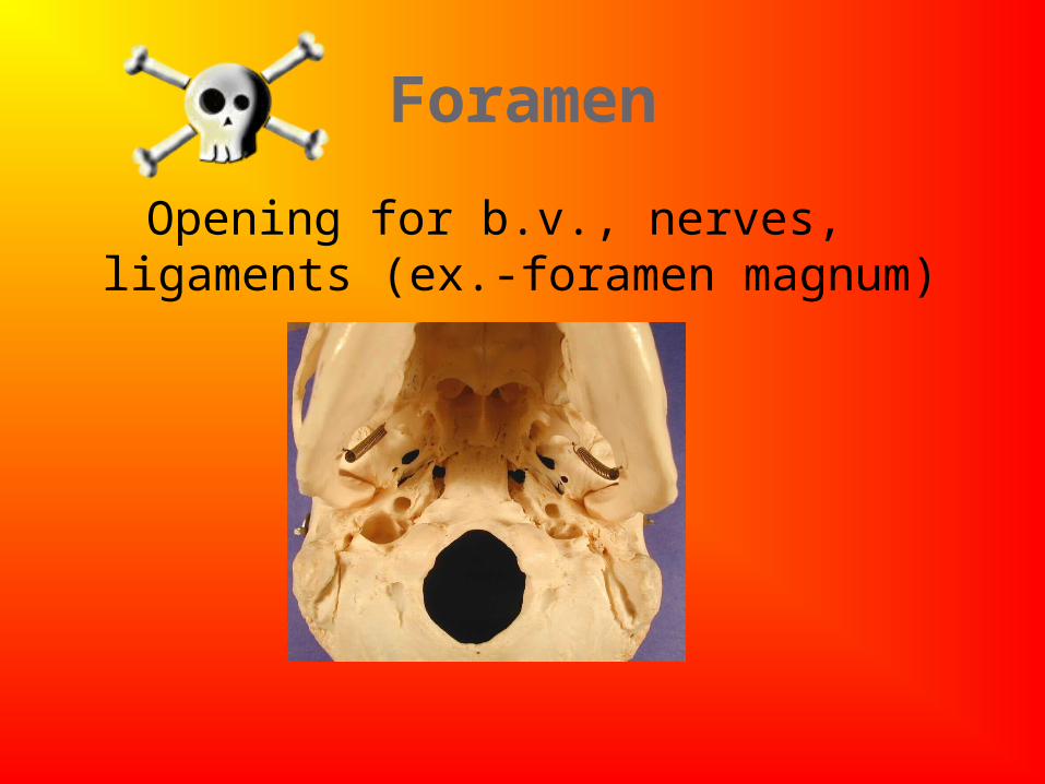
Foramen
Opening for b.v., nerves, ligaments (ex.-foramen magnum)
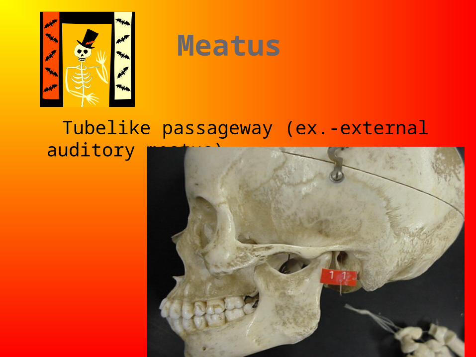
Meatus
Tubelike passageway (ex.-external auditory
meatus)
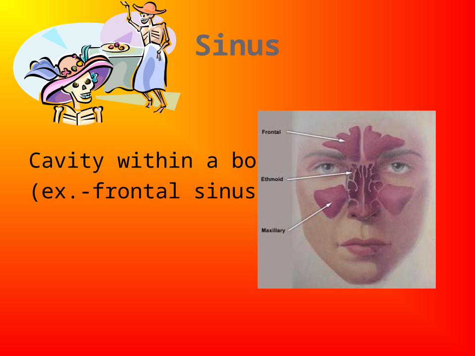
Sinus
Cavity within a bone
(ex.-frontal sinus)
Surface Markings on Bone
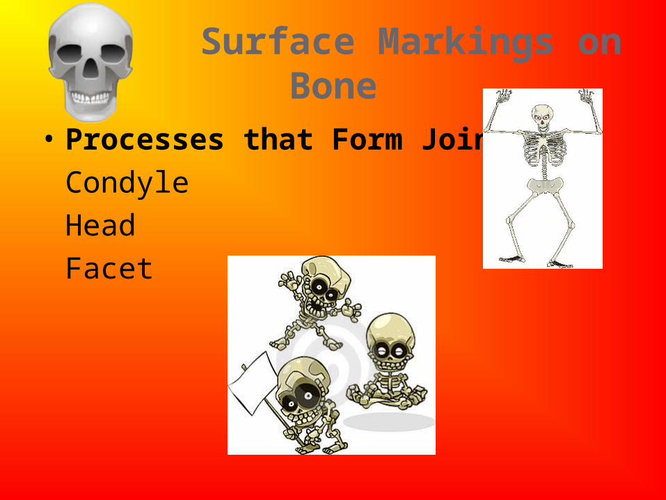
• Processes that Form Joints:
Condyle
Head
Facet
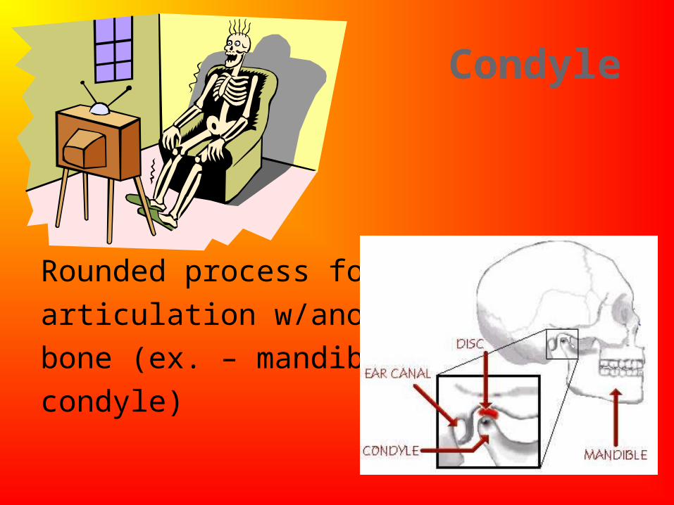
Condyle
Rounded process for
articulation w/another
bone (ex. – mandibular
condyle)
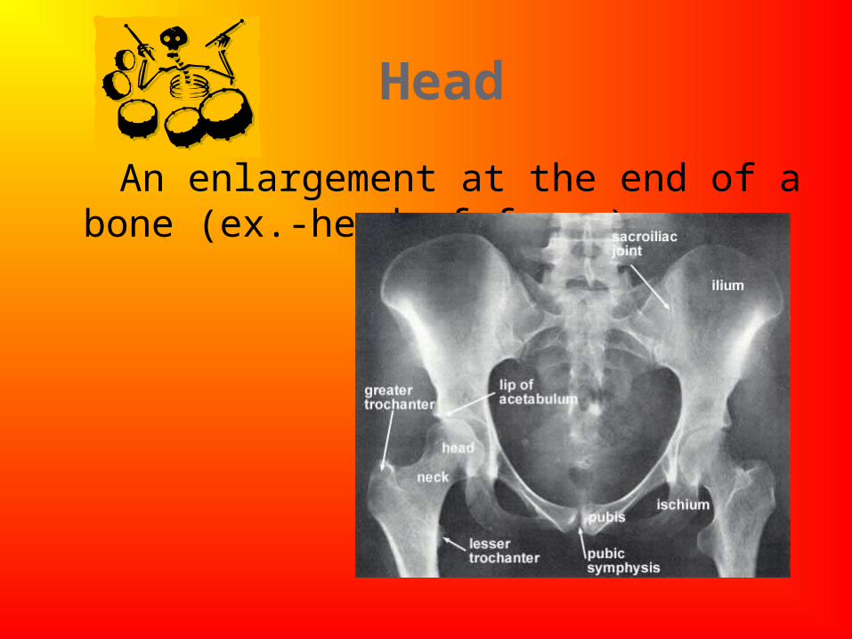
Head
An enlargement at the end of a bone (ex.-head of femur)
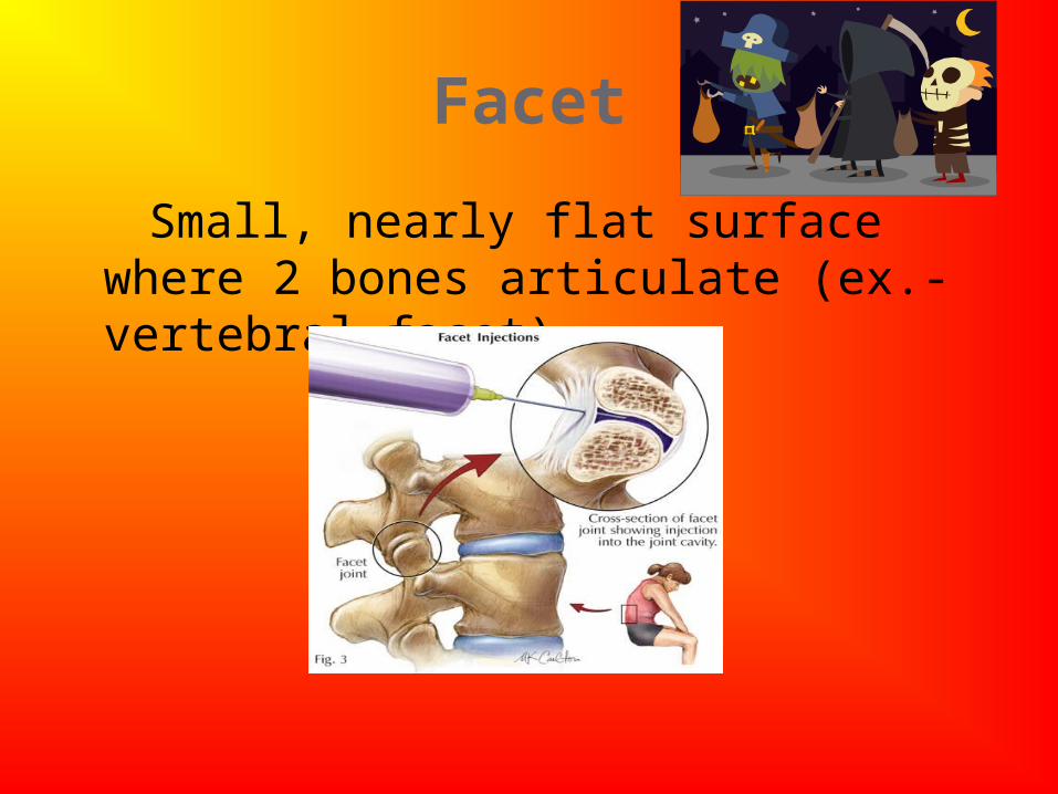
Facet
Small, nearly flat surface where 2 bones articulate (ex.-vertebral facet)
Surface Markings on Bone
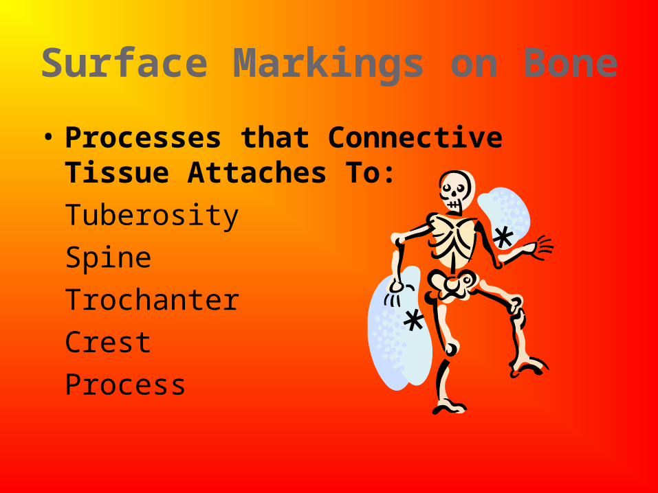
• Processes that Connective Tissue Attaches To:
Tuberosity
Spine
Trochanter
Crest
Process
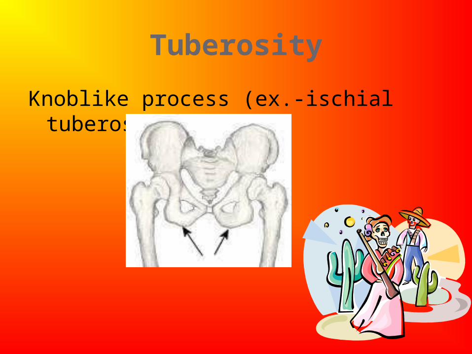
Tuberosity
Knoblike process (ex.-ischial tuberosity)
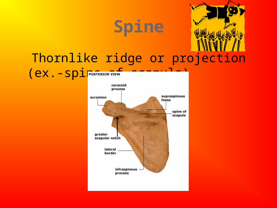
Spine
Thornlike ridge or projection (ex.-spine of scapula)
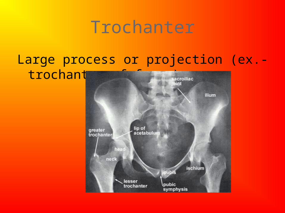
Trochanter
Large process or projection (ex.-trochanter of femur)
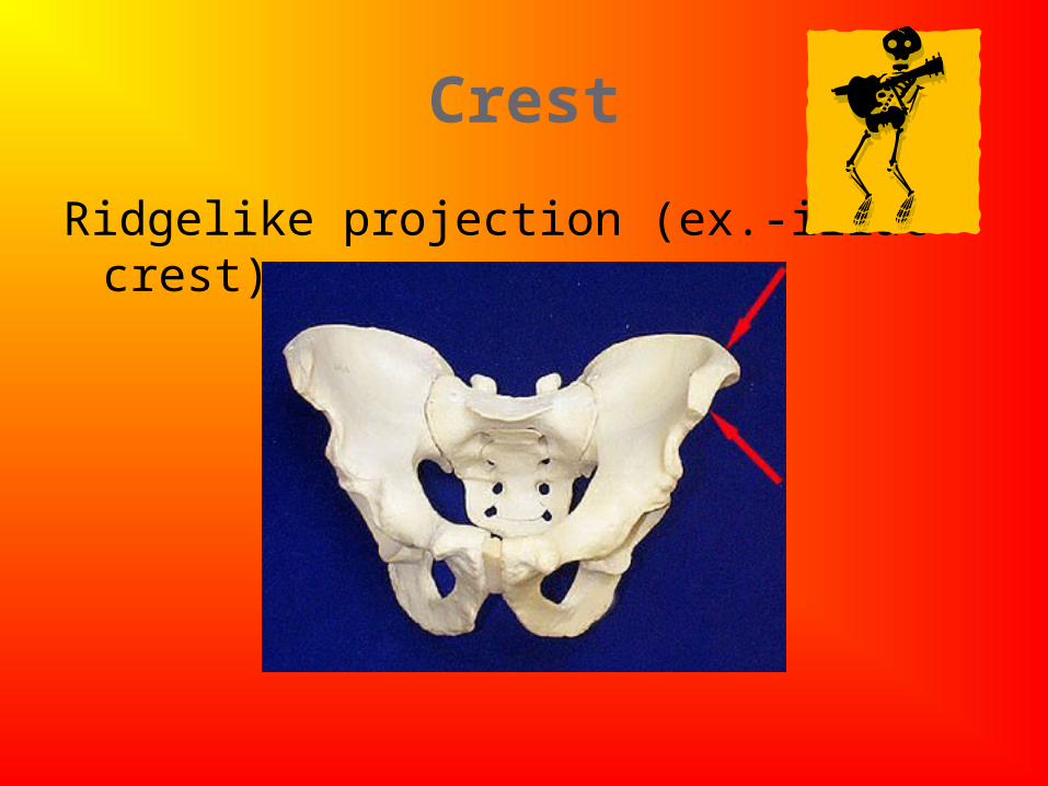
Crest
Ridgelike projection (ex.-iliac crest)
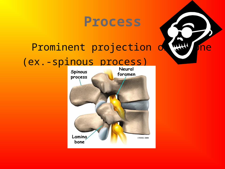
Process
Prominent projection on a bone
(ex.-spinous process)
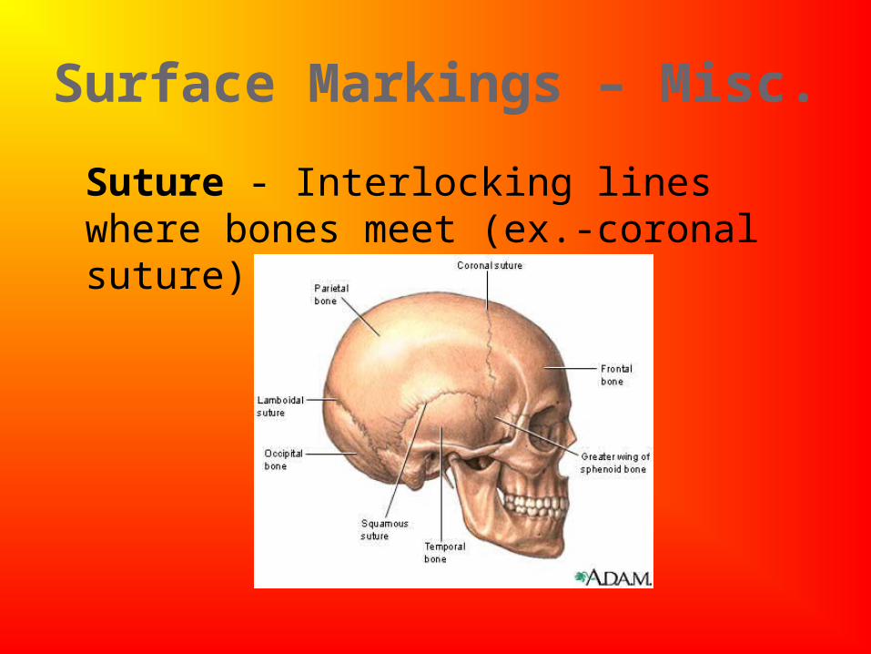
Surface Markings – Misc.
Suture - Interlocking lines where bones meet (ex.-coronal suture)