Embed Size (px)
Citation preview
# 102686 Cust: Cengage Au: Rizzo Pg. No. 136 Title: Fundamentals of Anatomy and Physiology Server: _____
C/M/Y/KShort / Normal
DESIGN SERVICES OF
S4-CARLISLEPublishing Services
136
The Skeletal System
CHAPTER OBJECT IVES
After studying this chapter, you should be able to:
1. Name the functions of the skeletal system.
2. Name the two types of ossifi cation.
3. Describe why diet can affect bone development in children and bone maintenance in older adults.
4. Describe the histology of compact bone.
5. Defi ne and give examples of bone markings.
6. Name the cranial and facial bones.
7. Name the bones of the axial and appendicular skeleton.
136
3871X_07_ch07_p136-173.indd 1363871X_07_ch07_p136-173.indd 136 8/25/09 11:21:46 AM8/25/09 11:21:46 AM

# 102686 Cust: Cengage Au: Rizzo Pg. No. 137 Title: Fundamentals of Anatomy and Physiology Server: _____
C/M/Y/KShort / Normal
DESIGN SERVICES OF
S4-CARLISLEPublishing Services
137
KEY TERMS
Acetabulum . . . . . . . . . . . 159Acromial process . . . . . . . 155Alveolus . . . . . . . . . . . . . . 151Atlas . . . . . . . . . . . . . . . . . 153Auditory ossicles. . . . . . . 149Axis . . . . . . . . . . . . . . . . . 153Calcaneus. . . . . . . . . . . . . 161Canaliculi . . . . . . . . . . . . . 142Cancellous or
spongy bone . . . . . . . . 142Capitate . . . . . . . . . . . . . . 157Carpals . . . . . . . . . . . . . . . 157Cartilage . . . . . . . . . . . . . 138Cervical vertebrae. . . . . . 152Clavicle. . . . . . . . . . . . . . . 155Coccygeal
vertebrae/coccyx . . . . . 152Compact or dense bone. . 142Condyle . . . . . . . . . . . . . . 145Coracoid process . . . . . . . 155Coronal suture. . . . . . . . . 146Costae . . . . . . . . . . . . . . . 155Crest . . . . . . . . . . . . . . . . . 146Cuboid . . . . . . . . . . . . . . . 161Cuneiforms . . . . . . . . . . . 161Diaphysis . . . . . . . . . . . . . 144Endochondral
ossifi cation. . . . . . . . . . 140Endosteum. . . . . . . . . . . . 140Epiphyseal line . . . . . . . . 144Epiphysis . . . . . . . . . . . . . 144Ethmoid bone . . . . . . . . . 149External occipital crest . . 146
External occipital protuberance . . . . . . . . 146
Femur. . . . . . . . . . . . . . . . 159Fibula . . . . . . . . . . . . . . . . 160Fontanelle . . . . . . . . . . . . 140Foramen. . . . . . . . . . . . . . 146Foramen magnum. . . . . . 146Fossae . . . . . . . . . . . . . . . 145Fracture . . . . . . . . . . . . . . 144Frontal bone . . . . . . . . . . 146Gladiolus . . . . . . . . . . . . . 155Glenoid fossa . . . . . . . . . 155Hamate . . . . . . . . . . . . . . 157Haversian/
central canals. . . . . . . . 142Head. . . . . . . . . . . . . . . . . 146Hematopoiesis. . . . . . . . . 138Humerus . . . . . . . . . . . . . 155Hyoid bone . . . . . . . . . . . 152Ilium . . . . . . . . . . . . . . . . . 158Incus. . . . . . . . . . . . . . . . . 149Intramembranous
ossifi cation. . . . . . . . . . 140Ischium. . . . . . . . . . . . . . . 158Kyphosis . . . . . . . . . . . . . 154Lacrimal bones . . . . . . . . 151Lacunae . . . . . . . . . . . . . . 142Lamella. . . . . . . . . . . . . . . 142Lambdoid suture. . . . . . . 146Ligaments . . . . . . . . . . . . 138Line. . . . . . . . . . . . . . . . . . 146Lordosis . . . . . . . . . . . . . . 154Lumbar vertebrae . . . . . . 152
Lunate . . . . . . . . . . . . . . . 157Malleus . . . . . . . . . . . . . . 149Mandible bone . . . . . . . . 151Manubrium . . . . . . . . . . . 155Mastoid portion
of temporal bone . . . . 146Maxillary bones . . . . . . . 150Meatus/canal. . . . . . . . . . 146Medullary cavity . . . . . . . 145Metacarpal bones . . . . . . 157Metaphysis . . . . . . . . . . . 144Metatarsals . . . . . . . . . . . 161Nasal bones . . . . . . . . . . . 150Navicular/scaphoid . . . . . 157Neck . . . . . . . . . . . . . . . . . 146Obturator foramen . . . . . 159Occipital bone . . . . . . . . . 146Occipital condyle. . . . . . . 146Olecranon process. . . . . . 157Orbital margin. . . . . . . . . 146Ossifi cation . . . . . . . . . . . 140Osteoblasts . . . . . . . . . . . 140Osteoclasts . . . . . . . . . . . 140Osteomalacia. . . . . . . . . . 144Osteon . . . . . . . . . . . . . . . 142Osteoprogenitor cell. . . . 140Palatine bones. . . . . . . . . 150Parietal bones . . . . . . . . . 147Patella . . . . . . . . . . . . . . . 159Pelvic girdle . . . . . . . . . . . 158Periosteum. . . . . . . . . . . . 140Petrous part of
temporal bone. . . . . . . 146
(continues)
3871X_07_ch07_p136-173.indd 1373871X_07_ch07_p136-173.indd 137 8/25/09 11:21:52 AM8/25/09 11:21:52 AM
138 CHAPTER 7 The Skeletal System
# 102686 Cust: Cengage Au: Rizzo Pg. No. 138 Title: Fundamentals of Anatomy and Physiology Server: _____
C/M/Y/KShort / Normal
DESIGN SERVICES OF
S4-CARLISLEPublishing Services
KEY TERMS (cont inued )
Phalanges . . . . . . . . . . . . 157Phalanx . . . . . . . . . . . . . . 157Pisiform . . . . . . . . . . . . . . 157Processes . . . . . . . . . . . . . 145Pubis. . . . . . . . . . . . . . . . . 158Radius . . . . . . . . . . . . . . . 157Red bone marrow. . . . . . 143Sacral vertebrae/
sacrum . . . . . . . . . . . . . 152Sagittal suture. . . . . . . . . 146Scaphoid/navicular . . . . . 157Scapula. . . . . . . . . . . . . . . 155Scoliosis . . . . . . . . . . . . . . 154Sesamoid bones . . . . . . . 145Sinus/antrum. . . . . . . . . . 146Sphenoid bones . . . . . . . 149Spine . . . . . . . . . . . . . . . . 145
Squamous portion of temporal bone . . . . 146
Stapes . . . . . . . . . . . . . . . 149Sternum . . . . . . . . . . . . . . 155Sulcus/groove . . . . . . . . . 146Supraorbital ridge. . . . . . 146Suture . . . . . . . . . . . . . . . 146Talus . . . . . . . . . . . . . . . . . 161Tarsal bones. . . . . . . . . . . 160Temporal bones. . . . . . . . 146Tendons . . . . . . . . . . . . . . 138Thoracic vertebrae . . . . . 152Tibia . . . . . . . . . . . . . . . . . 160Trabeculae . . . . . . . . . . . . 142Trapezium . . . . . . . . . . . . 157Trapezoid . . . . . . . . . . . . . 157Triquetral . . . . . . . . . . . . . 157
Trochanter . . . . . . . . . . . . 145Trochlea . . . . . . . . . . . . . . 145Tubercle . . . . . . . . . . . . . . 145Turbinates or nasal
conchae bones. . . . . . . 151Tympanic plate of
temporal bone. . . . . . . 149Ulna . . . . . . . . . . . . . . . . . 157Volkmann’s/perforating
canals . . . . . . . . . . . . . . 142Vomer bone. . . . . . . . . . . 151Wormian/sutural bones . 149Xiphoid process. . . . . . . . 155Yellow bone marrow . . . 143Zygomatic or malar
bones . . . . . . . . . . . . . . 151
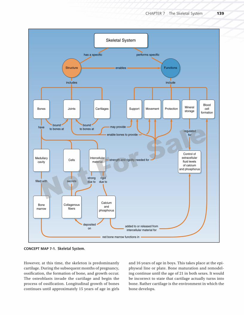
INTRODUCTIONTh e supporting structure of the body is the framework of joined bones that we refer to as the skeleton. It enables us to stand erect, to move in our environment, to accom-plish extraordinary feats of artistic grace like ballet moves and athletic endeavors like the high jump as well as nor-mal physical endurance. Th e skeletal system allows us to move a pen and write and aids us in breathing. It is closely associated with the muscular system. Th e skeletal system includes all the bones of the body and their associated cartilage, tendons, and ligaments. Despite the appear-ance of the bones, they are indeed composed of living tissue. Th e hard, “dead” stonelike appearance of bones is due to mineral salts like calcium phosphate embedded in the inorganic matrix of the bone tissue. Leondardo da Vinci (1452–1519), the famous Italian Renaissance artist and scientist, is credited as the fi rst anatomist to correctly illustrate the skeleton with its 206 bones. See Concept Map 7-1: Skeletal System.
THE FUNCTIONS OF THE SKELETAL SYSTEMTh e skeleton has fi ve general functions:
1. It supports and stabilizes surrounding tissues such as muscles, blood and lymphatic vessels, nerves, fat, and skin.
2. It protects vital organs of the body such as the brain, spinal cord, the heart, and lungs, and it protects other soft tissues of the body.
3. It assists in body movement by providing attachments for muscles that pull on the bones that act as levers.
4. It manufactures blood cells. Th is process is called hematopoiesis (hem-ah-toh-poy-EE-sis) and occurs chiefl y in red bone marrow.
5. It is a storage area for mineral salts, especially phosphorus and calcium, and fats.
Associated with the bones are cartilage, tendons, and lig-aments. Cartilage, a connective tissue, is the environment in which bone develops in a fetus. It is also found at the ends of certain bones and in joints in adults, providing a smooth surface for adjacent bones to move against each other. Ligaments are tough connective tissue structures that attach bones to bones like the ligament that attaches the head of the femur to the acetabulum of the pelvic bone in the hip joint. Tendons are similar structures that attach muscle to bone.
THE GROWTH AND FORMATION OF BONEThe skeleton of a developing fetus is completely formed by the end of the third month of pregnancy.
3871X_07_ch07_p136-173.indd 1383871X_07_ch07_p136-173.indd 138 8/25/09 11:21:54 AM8/25/09 11:21:54 AM
CHAPTER 7 The Skeletal System 139
# 102686 Cust: Cengage Au: Rizzo Pg. No. 139 Title: Fundamentals of Anatomy and Physiology Server: _____
C/M/Y/KShort / Normal
DESIGN SERVICES OF
S4-CARLISLEPublishing Services
However, at this time, the skeleton is predominantly cartilage. During the subsequent months of pregnancy, ossification, the formation of bone, and growth occur. The osteoblasts invade the cartilage and begin the process of ossification. Longitudinal growth of bones continues until approximately 15 years of age in girls
and 16 years of age in boys. This takes place at the epi-physeal line or plate. Bone maturation and remodel-ing continue until the age of 21 in both sexes. It would be incorrect to state that cartilage actually turns into bone. Rather cartilage is the environment in which the bone develops.
rigid
due to
includes include
has a specific performs specific
enablesStructure Functions
Skeletal System
have
Bones Joints Cartilages
filled with secretestrong
due to
deposited
on
bound
to bones at
bound
to bones atregulated
for
may provide
enable bones to provide
strength and rigidity needed for
added to or released from
intercellular material for
red bone marrow functions in
Medullary
cavityCells
Intercellular
material
Collagenous
fibers
Bone
marrow
Mineral
storage
Blood
cell
formation
ProtectionMovementSupport
Control of
extracellular
fluid levels
of calcium
and phosphorus
Calcium
and
phosphorus
CONCEPT MAP 7-1. Skeletal System.
3871X_07_ch07_p136-173.indd 1393871X_07_ch07_p136-173.indd 139 8/25/09 11:21:55 AM8/25/09 11:21:55 AM
140 CHAPTER 7 The Skeletal System
# 102686 Cust: Cengage Au: Rizzo Pg. No. 140 Title: Fundamentals of Anatomy and Physiology Server: _____
C/M/Y/KShort / Normal
DESIGN SERVICES OF
S4-CARLISLEPublishing Services
Th e strong protein matrix is responsible for a bone’s resilience or “elasticity” when tension is applied to the bone so that it gives a little under pressure. Th e mineral salts deposited into this protein matrix are responsible for the strength of the bone so that it does not get crushed when pressure is applied to the bone.
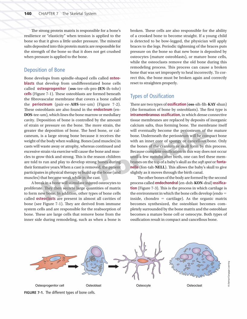
Deposition of BoneBone develops from spindle-shaped cells called osteo-blasts that develop from undiff erentiated bone cells called osteoprogenitor (oss-tee-oh-pro-JEN-ih-tohr) cells (Figure 7-1). Th ese osteoblasts are formed beneath the fi brovascular membrane that covers a bone called the periosteum (pair-ee-AHS-tee-um) (Figure 7-2). Th ese osteoblasts are also found in the endosteum (en-DOS-tee-um), which lines the bone marrow or medullary cavity. Deposition of bone is controlled by the amount of strain or pressure on the bone. Th e more strain, the greater the deposition of bone. Th e heel bone, or cal-caneum, is a large strong bone because it receives the weight of the body when walking. Bones (and muscles) in casts will waste away or atrophy, whereas continued and excessive strain via exercise will cause the bone and mus-cles to grow thick and strong. Th is is the reason children are told to run and play to develop strong bones during their formative years.When a cast is removed, the patient participates in physical therapy to build up the bone (and muscles) that became weak while in the cast.
A break in a bone will stimulate injured osteocytes to proliferate. Th ey then secrete large quantities of matrix to form new bone. In addition, other types of bone cells called osteoclasts are present in almost all cavities of bone (see Figure 7-1). Th ey are derived from immune system cells and are responsible for the reabsorption of bone. Th ese are large cells that remove bone from the inner side during remodeling, such as when a bone is
broken. Th ese cells are also responsible for the ability of a crooked bone to become straight. If a young child is detected to be bow-legged, the physician will apply braces to the legs. Periodic tightening of the braces puts pressure on the bone so that new bone is deposited by osteocytes (mature osteoblasts), or mature bone cells, while the osteoclasts remove the old bone during this remodeling process. Th is process can cause a broken bone that was set improperly to heal incorrectly. To cor-rect this, the bone must be broken again and correctly reset to straighten properly.
Types of Ossifi cationTh ere are two types of ossifi cation (oss-sih-fi h-KAY-shun) (the formation of bone by osteoblasts). Th e fi rst type is intramembranous ossifi cation, in which dense connective tissue membranes are replaced by deposits of inorganic calcium salts, thus forming bone. Th e membrane itself will eventually become the periosteum of the mature bone. Underneath the periosteum will be compact bone with an inner core of spongy or cancellous bone. Only the bones of the cranium or skull form by this process. Because complete ossifi cation in this way does not occur until a few months after birth, one can feel these mem-branes on the top of a baby’s skull as the soft spot or fonta-nelle (fon-tah-NELL). Th is allows the baby’s skull to give slightly as it moves through the birth canal.
Th e other bones of the body are formed by the second process called endochondral (en-doh-KON-dral) ossifi ca-tion (Figure 7-3). Th is is the process in which cartilage is the environment in which the bone cells develop (endo �inside, chondro � cartilage). As the organic matrix becomes synthesized, the osteoblast becomes com-pletely surrounded by the bone matrix and the osteoblast becomes a mature bone cell or osteocyte. Both types of ossifi cation result in compact and cancellous bone.
Osteoprogenitor cell Osteoblast OsteoclastOsteocyte
FIGURE 7-1. The different types of bone cells.
© D
elm
ar/
Ceng
ag
e L
earn
ing
3871X_07_ch07_p136-173.indd 1403871X_07_ch07_p136-173.indd 140 8/25/09 11:21:57 AM8/25/09 11:21:57 AM
CHAPTER 7 The Skeletal System 141
# 102686 Cust: Cengage Au: Rizzo Pg. No. 141 Title: Fundamentals of Anatomy and Physiology Server: _____
C/M/Y/KShort / Normal
DESIGN SERVICES OF
S4-CARLISLEPublishing Services
In order to maintain strong and healthy bones throughout our lives, it is important to main-tain a balanced diet with a daily intake of calcium. We can do this by consuming dairy prod-ucts such as milk, yogurt, and cheeses. In addition to diet, a regular regimen of exercise is also important. As bones are developing in children and adolescents, it is important to increase calcium intake and exercise more rigorously. As we mature we still require calcium; however, we require it in smaller amounts. Daily exercise, as simplistic as walking in older age and running or playing sports in middle age, will help maintain a healthy skeletal system. When playing sports, walking, or running, it is crucial to wear proper foot attire with arch supports and a good fi t. This will prevent future problems with bones of the feet. Proper posture dur-ing walking and sitting will also maintain healthy and strong bones.
STRONG BONESHEALTH ALERT
Proximal epiphysis
(A)
(B)
(C)
Diaphysis
Trabeculae
Distal epiphysis
Epiphyseal/growthline
Articular cartilage
Spongy bone (containing red bone marrow)
Metaphysis
Nutrient artery
Endosteum
Compact bone
Compact bone
Spongy bone
Yellow bone marrow
Periosteum
Periosteum
Metaphysis
FIGURE 7-2. The structure of a typical long bone. (A) Diaphysis, epiphysis, and medullary cavity. (B) Compact bone surrounding yellow bone marrow in the medullary cavity. (C) Spongy bone and compact bone in the epiphysis.
© D
elm
ar/
Ceng
ag
e L
earn
ing
3871X_07_ch07_p136-173.indd 1413871X_07_ch07_p136-173.indd 141 8/25/09 11:21:58 AM8/25/09 11:21:58 AM

142 CHAPTER 7 The Skeletal System
# 102686 Cust: Cengage Au: Rizzo Pg. No. 142 Title: Fundamentals of Anatomy and Physiology Server: _____
C/M/Y/KShort / Normal
DESIGN SERVICES OF
S4-CARLISLEPublishing Services
Maintaining BoneIn a healthy body, a balance must exist between the amount of calcium stored in the bones, the calcium in the blood, and the excess calcium excreted by the kid-neys and via the digestive system. Th e proper calcium ion concentration in the blood and bones is controlled by the endocrine system. Two hormones, calcitonin and parathormone, control the calcium concentration in our bodies. Calcitonin causes calcium to be stored in the bones; parathormone causes it to be released into the bloodstream.
THE HISTOLOGY OF BONETh ere are two types of bone tissue: compact or dense bone and cancellous or spongy bone (see Figure 7-2C). In both types of tissue, the osteocytes are the same, but the arrangement of how the blood supply reaches the bone cells is diff erent. Th e two types of tissue have dif-ferent functions. Compact bone is dense and strong,
whereas cancellous bone has many open spaces, giving it a spongy appearance. It is in these spaces that bone marrow can be found.
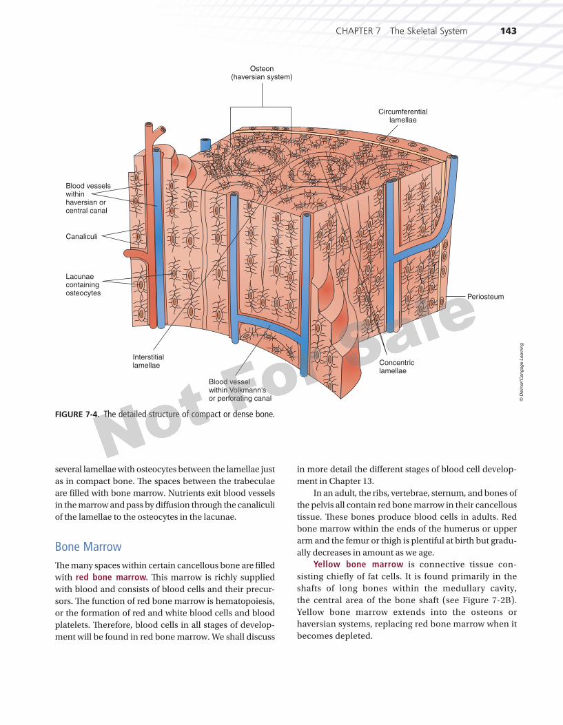
The Haversian System of Compact BoneTh e haversian (hah-VER-shan) canal, also called an osteon, was named for an English physician, Clopton Havers (1650–1702), who fi rst described it as a prominent feature of compact bone (Figure 7-4). Th is system allows for the eff ective metabolism of bone cells surrounded by rings of mineral salts. It has several components. Running parallel to the surface of the bone are many small canals containing blood vessels (capillaries, arterioles, venules) that bring in oxygen and nutrients and remove waste products and carbon dioxide. Th ese canals are called hav-ersian or central canals and are surrounded by concentric rings of bone, each layer of which is called a lamella (lah-MELL-ah). Between two lamellae or rings of bone are several tiny cavities called lacunae (lah-KOO-nee). Each lacuna contains an osteocyte or bone cell suspended in tissue fl uid. Th e lacunae are all connected to each other and ultimately to the larger haversian or central canals by much smaller canals called canaliculi (kan-ah-LIK-you-lye). Canals running horizontally to the haversian (central) canals, also containing blood vessels, are called Volkmann’s or perforating canals. It is tissue fl uid that cir-culates through all these canals and bathes the osteocyte, bringing in oxygen and food and carrying away waste products and carbon dioxide, keeping the osteocytes alive and healthy.
Watch an animation of a fracture as a result of direct force on your StudyWARE™ CD-ROM.
W t h i ti f f t
StudyWARE™ Connection
Cancellous BoneCancellous or spongy bone is located at the ends of long bones and forms the center of all other bones. It con-sists of a meshwork of interconnecting sections of bone called trabeculae (trah-BEK-you-lee), creating the spon-gelike appearance of cancellous bone (Figure 7-2C). Th e trabeculae give strength to the bone without the added weight of being solid. Each trabecula consists of
Primaryossificationcenter
Periosteum
Medullary cavity
Calcified cartilage
Bone forming withincalcified cartilage
FIGURE 7-3. Endochondral ossifi cation where cartilage is the environment in which bone develops.
© D
elm
ar/
Ceng
ag
e L
earn
ing
3871X_07_ch07_p136-173.indd 1423871X_07_ch07_p136-173.indd 142 8/25/09 11:22:01 AM8/25/09 11:22:01 AM
CHAPTER 7 The Skeletal System 143
# 102686 Cust: Cengage Au: Rizzo Pg. No. 143 Title: Fundamentals of Anatomy and Physiology Server: _____
C/M/Y/KShort / Normal
DESIGN SERVICES OF
S4-CARLISLEPublishing Services
several lamellae with osteocytes between the lamellae just as in compact bone. Th e spaces between the trabeculae are fi lled with bone marrow. Nutrients exit blood vessels in the marrow and pass by diff usion through the canaliculi of the lamellae to the osteocytes in the lacunae.
Bone MarrowTh e many spaces within certain cancellous bone are fi lled with red bone marrow. Th is marrow is richly supplied with blood and consists of blood cells and their precur-sors. Th e function of red bone marrow is hematopoiesis, or the formation of red and white blood cells and blood platelets. Th erefore, blood cells in all stages of develop-ment will be found in red bone marrow. We shall discuss
in more detail the diff erent stages of blood cell develop-ment in Chapter 13.
In an adult, the ribs, vertebrae, sternum, and bones of the pelvis all contain red bone marrow in their cancellous tissue. Th ese bones produce blood cells in adults. Red bone marrow within the ends of the humerus or upper arm and the femur or thigh is plentiful at birth but gradu-ally decreases in amount as we age.
Yellow bone marrow is connective tissue con-sisting chiefl y of fat cells. It is found primarily in the shafts of long bones within the medullary cavity, the central area of the bone shaft (see Figure 7-2B). Yellow bone marrow extends into the osteons or haversian systems, replacing red bone marrow when it becomes depleted.
Osteon (haversian system)
Blood vessels within haversian or central canal
Lacunae containing osteocytes
Canaliculi
Interstitial lamellae Concentric
lamellae
Circumferential lamellae
Blood vessel within Volkmann’s or perforating canal
Periosteum
FIGURE 7-4. The detailed structure of compact or dense bone.
© D
elm
ar/
Ceng
ag
e L
earn
ing
3871X_07_ch07_p136-173.indd 1433871X_07_ch07_p136-173.indd 143 8/25/09 11:22:03 AM8/25/09 11:22:03 AM
144 CHAPTER 7 The Skeletal System
# 102686 Cust: Cengage Au: Rizzo Pg. No. 144 Title: Fundamentals of Anatomy and Physiology Server: _____
C/M/Y/KShort / Normal
DESIGN SERVICES OF
S4-CARLISLEPublishing Services
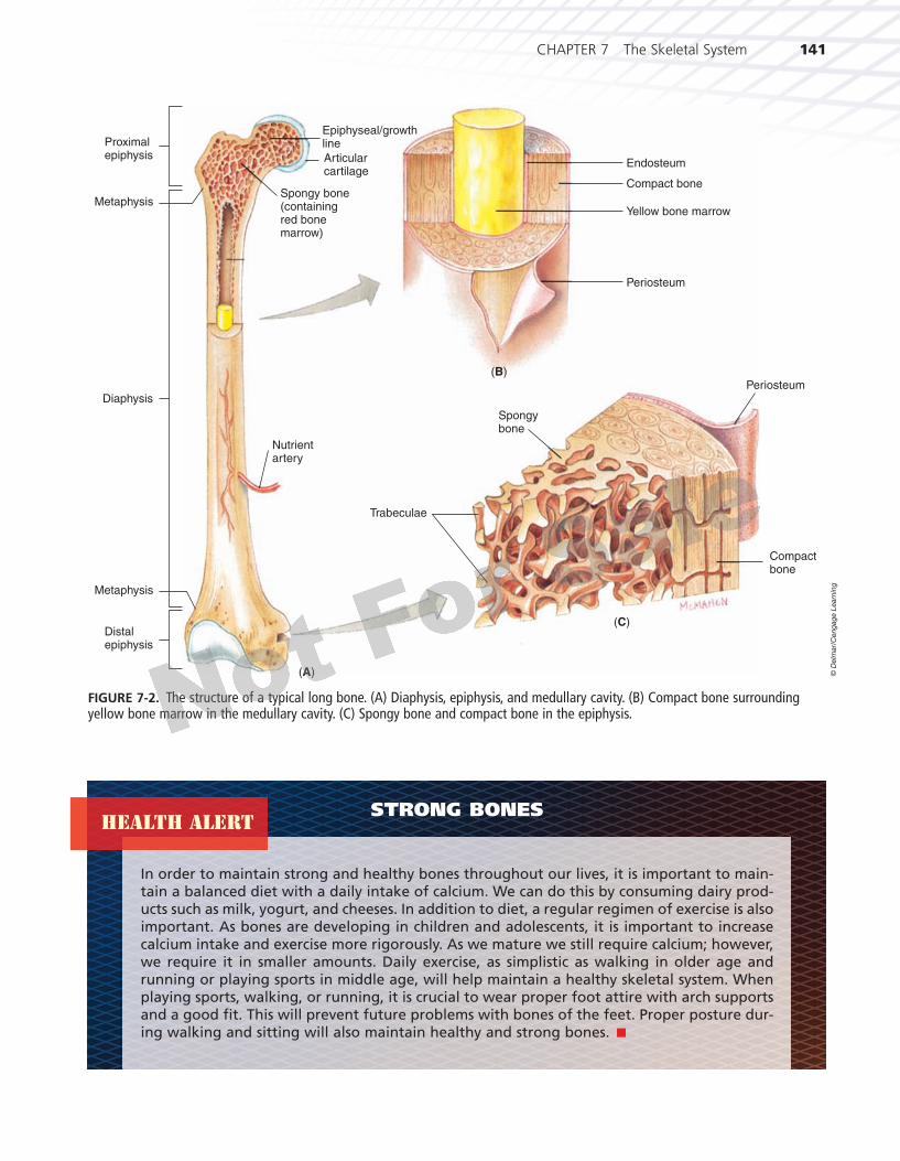
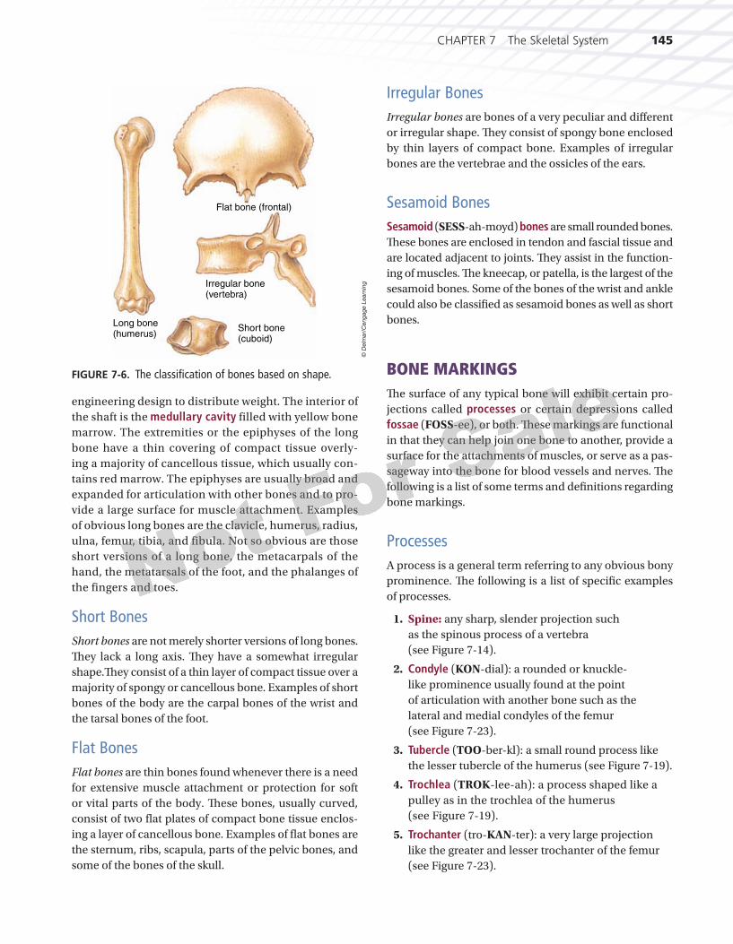
THE CLASSIFICATION OF BONES BASED ON SHAPETh e individual bones of the body can be divided by shape into fi ve categories: long, short, fl at, irregular, and sesa-moid (Figure 7-6).
Long BonesLong bones (see Figure 7-2) are bones whose length exceeds their width and consist of a diaphysis
(dye-AFF-ih-sis) or shaft composed mainly of compact bone, a metaphysis (meh-TAFF-ih-sis) or flared por-tion at each end of the diaphysis consisting mainly of cancellous or spongy bone, and two extremities, each called an epiphysis (eh-PIFF-ih-sis), separated from the metaphysis by the epiphyseal or growth line where longitudinal growth of the bone occurs. The shaft consists mainly of compact bone. It is thickest toward the middle of the bone because strain on the bone is greatest at that point. The strength of a long bone is also ensured by the slight curvature of the shaft, a good
DISORDERS OF THE SKELETAL SYSTEM
RICKETSRickets is a disease caused by defi ciencies in the minerals calcium and phosphorus or by defi ciencies in vitamin D and sunlight. Vitamin D is necessary for calcium and phosphorus absorption. The condition causes changes in bones known as rickets in children and osteomalacia (oss-tee-oh-mah-LAY-she-ah) in adults. The bones fail to ossify, resulting in soft, weak bones that are easily broken. Rickets occurs in children who do not receive adequate exposure to sunlight (sunlight is necessary for vitamin D pro-duction in the body) or whose diets are defi cient in vitamin D (milk is a food source of vitamin D).
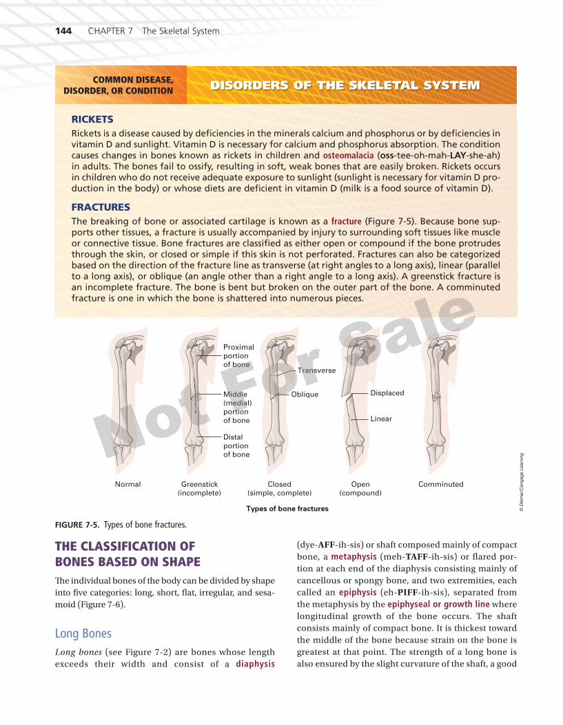
FRACTURESThe breaking of bone or associated cartilage is known as a fracture (Figure 7-5). Because bone sup-ports other tissues, a fracture is usually accompanied by injury to surrounding soft tissues like muscle or connective tissue. Bone fractures are classifi ed as either open or compound if the bone protrudes through the skin, or closed or simple if this skin is not perforated. Fractures can also be categorized based on the direction of the fracture line as transverse (at right angles to a long axis), linear (parallel to a long axis), or oblique (an angle other than a right angle to a long axis). A greenstick fracture is an incomplete fracture. The bone is bent but broken on the outer part of the bone. A comminuted fracture is one in which the bone is shattered into numerous pieces.
COMMON DISEASE,DISORDER, OR CONDITION
Greenstick(incomplete)
Closed(simple, complete)
Transverse
ObliqueMiddle(medial)portionof bone
Distalportionof bone
Proximalportionof bone
Linear
Displaced
ComminutedOpen(compound)
Types of bone fractures
Normal
FIGURE 7-5. Types of bone fractures.©
Delm
ar/
Ceng
ag
e L
earn
ing
3871X_07_ch07_p136-173.indd 1443871X_07_ch07_p136-173.indd 144 8/25/09 11:22:04 AM8/25/09 11:22:04 AM
CHAPTER 7 The Skeletal System 145
# 102686 Cust: Cengage Au: Rizzo Pg. No. 145 Title: Fundamentals of Anatomy and Physiology Server: _____
C/M/Y/KShort / Normal
DESIGN SERVICES OF
S4-CARLISLEPublishing Services
engineering design to distribute weight. The interior of the shaft is the medullary cavity filled with yellow bone marrow. The extremities or the epiphyses of the long bone have a thin covering of compact tissue overly-ing a majority of cancellous tissue, which usually con-tains red marrow. The epiphyses are usually broad and expanded for articulation with other bones and to pro-vide a large surface for muscle attachment. Examples of obvious long bones are the clavicle, humerus, radius, ulna, femur, tibia, and fibula. Not so obvious are those short versions of a long bone, the metacarpals of the hand, the metatarsals of the foot, and the phalanges of the fingers and toes.
Short BonesShort bones are not merely shorter versions of long bones. Th ey lack a long axis. Th ey have a somewhat irregular shape.Th ey consist of a thin layer of compact tissue over a majority of spongy or cancellous bone. Examples of short bones of the body are the carpal bones of the wrist and the tarsal bones of the foot.
Flat BonesFlat bones are thin bones found whenever there is a need for extensive muscle attachment or protection for soft or vital parts of the body. Th ese bones, usually curved, consist of two fl at plates of compact bone tissue enclos-ing a layer of cancellous bone. Examples of fl at bones are the sternum, ribs, scapula, parts of the pelvic bones, and some of the bones of the skull.
Irregular BonesIrregular bones are bones of a very peculiar and diff erent or irregular shape. Th ey consist of spongy bone enclosed by thin layers of compact bone. Examples of irregular bones are the vertebrae and the ossicles of the ears.
Sesamoid BonesSesamoid (SESS-ah-moyd) bones are small rounded bones. Th ese bones are enclosed in tendon and fascial tissue and are located adjacent to joints. Th ey assist in the function-ing of muscles. Th e kneecap, or patella, is the largest of the sesamoid bones. Some of the bones of the wrist and ankle could also be classifi ed as sesamoid bones as well as short bones.
BONE MARKINGSTh e surface of any typical bone will exhibit certain pro-jections called processes or certain depressions called fossae (FOSS-ee), or both. Th ese markings are functional in that they can help join one bone to another, provide a surface for the attachments of muscles, or serve as a pas-sageway into the bone for blood vessels and nerves. Th e following is a list of some terms and defi nitions regarding bone markings.
ProcessesA process is a general term referring to any obvious bony prominence. Th e following is a list of specifi c examples of processes.
1. Spine: any sharp, slender projection such as the spinous process of a vertebra (see Figure 7-14).
2. Condyle (KON-dial): a rounded or knuckle-like prominence usually found at the point of articulation with another bone such as the lateral and medial condyles of the femur (see Figure 7-23).
3. Tubercle (TOO-ber-kl): a small round process like the lesser tubercle of the humerus (see Figure 7-19).
4. Trochlea (TROK-lee-ah): a process shaped like a pulley as in the trochlea of the humerus (see Figure 7-19).
5. Trochanter (tro-KAN-ter): a very large projection like the greater and lesser trochanter of the femur (see Figure 7-23).
Flat bone (frontal)
Irregular bone(vertebra)
Short bone(cuboid)
Long bone(humerus)
FIGURE 7-6. The classifi cation of bones based on shape.©
Delm
ar/
Cen
gag
e L
earn
ing
3871X_07_ch07_p136-173.indd 1453871X_07_ch07_p136-173.indd 145 8/25/09 11:22:07 AM8/25/09 11:22:07 AM

146 CHAPTER 7 The Skeletal System
# 102686 Cust: Cengage Au: Rizzo Pg. No. 146 Title: Fundamentals of Anatomy and Physiology Server: _____
C/M/Y/KShort / Normal
DESIGN SERVICES OF
S4-CARLISLEPublishing Services
6. Crest: a narrow ridge of bone like the iliac crest of the hip bone (see Figure 7-22).
7. Line: a less prominent ridge of bone than a crest.
8. Head: a terminal enlargement like the head of the humerus and the head of the femur (see Figures 7-19 and 7-23).
9. Neck: that part of a bone that connects the head or terminal enlargement to the rest of the bone, like the neck of the femur (see Figures 7-19 and 7-23).
FossaeA fossa is a general term for any depression or cavity in or on a bone. Th e following is a list of specifi c examples of fossae.
1. Suture: a narrow junction often found between two bones like the sutures of the skull bones (see Figure 7-9).
2. Foramen: an opening through which blood vessels, nerves, and ligaments pass like the foramen magnum of the occipital bone of the skull or the obturator foramen of the pelvic bone (see Figure 7-22).
3. Meatus or canal: a long tube-like passage, like the auditory meatus or canal (see Figure 7-9).
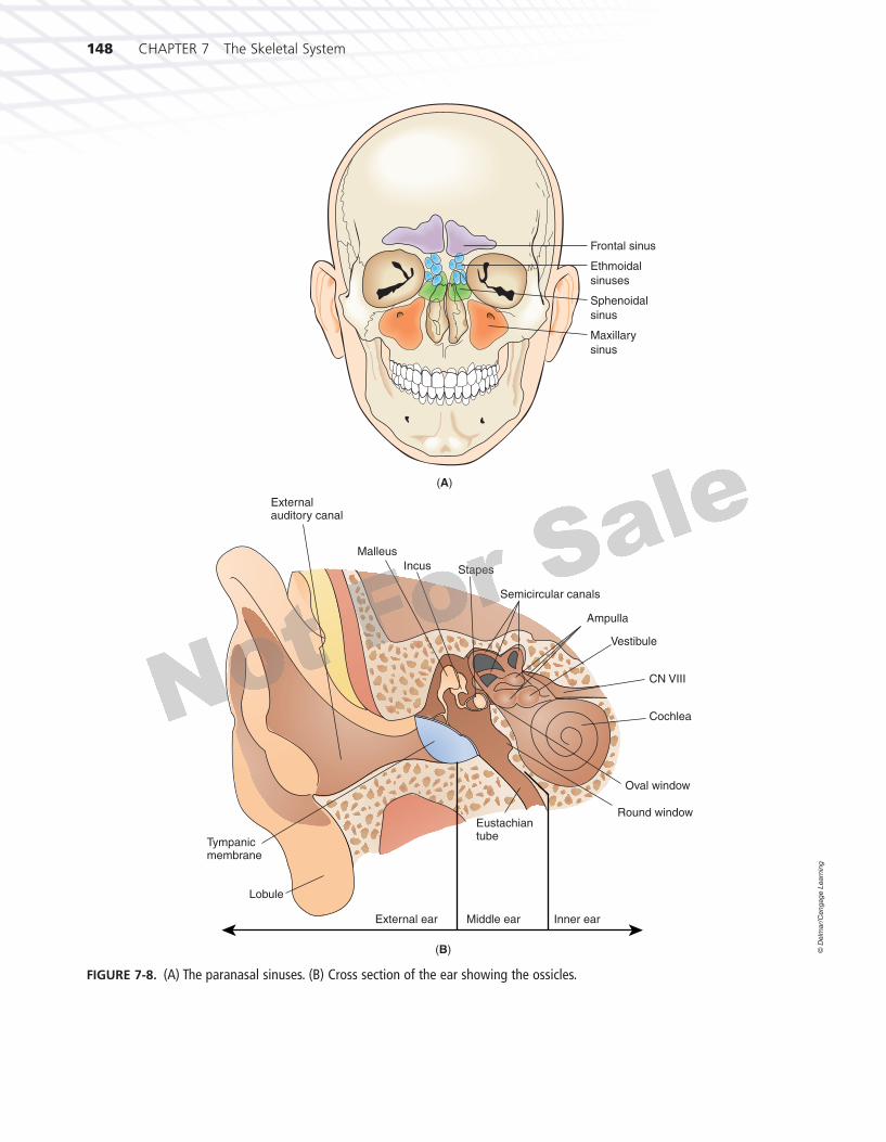
4. Sinus or antrum: a cavity within a bone like the nasal sinuses or frontal sinus (see Figure 7-8A).
5. Sulcus: a furrow or groove like the intertubercular sulcus or groove of the humerus (see Figure 7-19).
DIVISIONS OF THE SKELETONTh e skeleton typically has 206 named bones. Th e axial part consists of the skull (28 bones, including the cra-nial and facial bones), the hyoid bone, the vertebrae (26 bones), the ribs (24 bones), and the sternum. Th e appendicular part of the skeleton consists of the bones of the upper extremities or arms (64 bones, including the shoulder girdle bones) and the bones of the lower extremities or legs (62 bones, including the bones of the pelvic girdle) (Figure 7-7).
THE AXIAL SKELETONTh e skull, in the correct use of the term, includes the cranial and the facial bones. We will discuss the cranial bones fi rst.
The Cranial BonesTh e bones of the cranium have a number of important functions. Th ey protect and enclose the brain and special sense organs like the eyes and ears. Muscles for mastica-tion or chewing and muscles for head movement attach to certain cranial bones. At certain locations, air sinuses or cavities are present that connect with the nasal cavities (Figure 7-8). All of the individual bones of the cranium are united by immovable junction lines called sutures.
Th e frontal bone is a single bone that forms the fore-head, the roof of the nasal cavity, and the orbits, which are the bony sockets that contain the eyes (Figure 7-9). Important bone markings are the orbital margin, a defi -nite ridge above each orbit located where eyebrows are found, and the supraorbital ridge, which overlies the fron-tal sinus and can be felt in the middle of your forehead. Th e coronal suture is found where the frontal bone joins the two parietal bones.
Th e two parietal (pah-RYE-eh-tal) bones form the upper sides and roof of the cranium. Th ey are joined at the sagittal suture in the midline.
Th e occipital bone is a single bone that forms the back and base of the cranium (see Figure 7-9) and joins the pari-etal bones superiorly at the lambdoid suture. Th e inferior portion of this bone has a large opening called the foramen magnum through which the spinal cord connects with the brain. On each lower side of the occipital bone is a process called the occipital condyle. Th ese processes are signifi cant because they articulate with depressions in the fi rst cervical vertebra (atlas), thus allowing the head to connect with and rest on the vertebrae. Other notable markings are the exter-nal occipital crest and the external occipital protuberance, which can be felt through the scalp at the base of the neck. Several ligaments and muscles attach to these regions.
Th e two temporal bones help form the lower sides and base of the cranium (see Figure 7-9). Each temporal bone encloses an ear and bears a fossa for articulation with the lower jaw or mandible. Th e temporal bones are irregular in shape and each consists of four parts: the squamous, petrous, mastoid, and tympanic parts. Th e squamous por-tion is the largest and most superior of the four parts. It is a thin fl at plate of bone that forms the temple. Projecting from its lower part is the zygomatic process that forms the lateral part of the zygomatic arch or cheek bone. Th e petrous part is found deep within the base of the skull where it protects and surrounds the inner ear. Th e mastoid portion is located behind and below the auditory meatus or opening of the ear. Th e mastoid process is a rounded pro-jection of the mastoid portion of the temporal bone eas-ily felt behind the ear. Several muscles of the neck attach
3871X_07_ch07_p136-173.indd 1463871X_07_ch07_p136-173.indd 146 8/25/09 11:22:08 AM8/25/09 11:22:08 AM

CHAPTER 7 The Skeletal System 147
# 102686 Cust: Cengage Au: Rizzo Pg. No. 147 Title: Fundamentals of Anatomy and Physiology Server: _____
C/M/Y/KShort / Normal
DESIGN SERVICES OF
S4-CARLISLEPublishing Services
Frontal
Temporal
Zygomatic
Maxilla
Skull
Sternum
Ribs
Thorax
Carpals
Metacarpals
Phalanges
Fibula
Calcaneus
Tibia
Patella
Femur
Lower limbs
Upper limbs
Vertebralcolumn
Radius
Pectoral girdle
Scapula
Clavicle
TemporalMandible
Occipital
Parietal
Humerus
Ulna
Hip bones
Sacrum
Phalanges
Metatarsals
Tarsals
(A) Anterior (B) Posterior
Coccyx
FIGURE 7-7. The human skeletal system. (A) Anterior view. (B) Posterior view.
© D
elm
ar/
Ceng
ag
e L
earn
ing
3871X_07_ch07_p136-173.indd 1473871X_07_ch07_p136-173.indd 147 8/25/09 11:22:09 AM8/25/09 11:22:09 AM
148 CHAPTER 7 The Skeletal System
# 102686 Cust: Cengage Au: Rizzo Pg. No. 148 Title: Fundamentals of Anatomy and Physiology Server: _____
C/M/Y/KShort / Normal
DESIGN SERVICES OF
S4-CARLISLEPublishing Services
Frontal sinus
Ethmoidal
sinuses
Sphenoidal
sinus
Maxillary
sinus
External auditory canal
Malleus
Incus Stapes
Semicircular canals
Ampulla
Vestibule
CN VIII
Cochlea
Oval window
Round window Eustachian tube
Tympanic membrane
Lobule
External ear Middle ear Inner ear
(A)
(B)
FIGURE 7-8. (A) The paranasal sinuses. (B) Cross section of the ear showing the ossicles.
© D
elm
ar/
Ceng
ag
e L
earn
ing
3871X_07_ch07_p136-173.indd 1483871X_07_ch07_p136-173.indd 148 8/25/09 11:22:12 AM8/25/09 11:22:12 AM

CHAPTER 7 The Skeletal System 149
# 102686 Cust: Cengage Au: Rizzo Pg. No. 149 Title: Fundamentals of Anatomy and Physiology Server: _____
C/M/Y/KShort / Normal
DESIGN SERVICES OF
S4-CARLISLEPublishing Services
to this mastoid process and assist in moving your head. Finally, the tympanic plate forms the fl oor and anterior wall of the external auditory meatus. A long and slender styloid process can be seen extending from the undersur-face of this plate. Ligaments that hold the hyoid bone in place (which supports the tongue) attach to this styloid process of the tympanic plate of the temporal bone.
Th e single sphenoid bone forms the anterior portion of the base of the cranium (Figures 7-9 and 7-10). When viewed from below it looks like a butterfl y. It acts as an anchor binding all of the cranial bones together.
The single ethmoid bone is the principal support-ing structure of the nasal cavities and helps form part of the orbits. It is the lightest of the cranial bones (see Figures 7-9 and 7-10).
Th e six auditory ossicles are the three bones found in each ear (see Figure 7-8B): the malleus or hammer, the
stapes (STAY-peez) or stirrup, and the incus or anvil. Th ese tiny bones are highly specialized in both structure and func-tion and are involved in exciting the hearing receptors.
Th e wormian bones or sutural bones are located within the sutures of the cranial bones. Th ey vary in number, are small and irregular in shape, and are never included in the total number of bones in the body. Th ey form as a result of intramembranous ossifi cation of the cranial bones.
Play an interactive game labeling the cranial bones on your StudyWARE™ CD-ROM.
Pl i t ti l b li th
StudyWARE™ Connection
Frontal bone
Coronal suture
Parietal bone
Squamosal suture
Temporal bone
Lambdoidsuture
Occipital bone
External auditorymeatus
Mastoid process
Styloid process
Zygomatic process of temporal bone
Temporal process of zygomatic bone Mandible
Maxilla
Zygomatic bone
Zygomatic arch
Ethmoid bone
Lacrimal bone
Nasal bone
Sphenoid bone (greater wing)
FIGURE 7-9. The cranial bones.
© D
elm
ar/
Ceng
ag
e L
earn
ing
3871X_07_ch07_p136-173.indd 1493871X_07_ch07_p136-173.indd 149 8/25/09 11:22:13 AM8/25/09 11:22:13 AM

150 CHAPTER 7 The Skeletal System
# 102686 Cust: Cengage Au: Rizzo Pg. No. 150 Title: Fundamentals of Anatomy and Physiology Server: _____
C/M/Y/KShort / Normal
DESIGN SERVICES OF
S4-CARLISLEPublishing Services
The Facial BonesLike the bones of the cranium, the facial bones are also united by immovable sutures, with one exception: the lower jawbone or mandible. Th is bone is capable of movement in a number of directions. It can be elevated and depressed as in talking, and it can protract and retract and move from side to side as in chewing.
Th e two nasal bones are thin and delicate bones that join in a suture to form the bridge of the nose (see Figure 7-10).
Th e two palatine bones form the posterior part of the roof of your mouth or part of the hard palate. Th is region is the same as the fl oor of the nasal cavity. Upward exten-sions of the palatine bones help form the outer walls of the nasal cavity.
Th e two maxillary bones make up the upper jaw (see Figure 7-10). Each maxillary bone consists of fi ve parts: a body, a zygomatic process, a frontal process, a pala-tine process, and an alveolar process. Th e large body of the maxilla forms part of the fl oor and outer wall of the nasal cavity, the greater part of the fl oor of the orbit, and
Frontal bone
Parietal bone
Sphenoid bone
Lacrimal bone
Middle nasal conchae
Inferior nasal conchae
Coronal suture
Ethmoid bone
Temporal bone
Nasal bone
Zygomatic bone
Vomer
Maxilla
Mandible
FIGURE 7-10. The facial bones.©
Delm
ar/
Ceng
ag
e L
earn
ing
3871X_07_ch07_p136-173.indd 1503871X_07_ch07_p136-173.indd 150 8/25/09 11:22:16 AM8/25/09 11:22:16 AM

CHAPTER 7 The Skeletal System 151
# 102686 Cust: Cengage Au: Rizzo Pg. No. 151 Title: Fundamentals of Anatomy and Physiology Server: _____
C/M/Y/KShort / Normal
DESIGN SERVICES OF
S4-CARLISLEPublishing Services
much of the anterior face below the temple. Th e body is covered by a number of facial muscles and contains a large maxillary sinus located lateral to the nose. Th e zygomatic process extends laterally to participate in the formation of the cheek. (Processes are named according to the bone they go to; thus, the zygomatic process of the maxillary bone goes toward and joins the zygomatic or cheekbone.) Th e frontal process extends upward to the frontal bone or forehead. Th e palatine process extends posteriorly in a horizontal plane to join or articulate with the palatine bone and actually forms the greater ante-rior portion of the hard palate or roof of the mouth. Th e alveolar processes bear the teeth of the upper jaw, and each tooth is embedded in an alveolus (al-VEE-oh-lus) or socket.Th e two maxillary bones join at the intermaxil-lary suture. Th is fusion is usually completed just before birth. If the two bones do not unite to form a continuous structure, the resulting defect is called a cleft palate and is usually associated with a cleft lip. With today’s surgical techniques, the defect can be repaired early in the devel-opment of the child.
Th e two zygomatic bones, also known as the malar bones, form the prominence of the cheek and rest on the maxillae (see Figure 7-10). Its maxillary process joins the maxillary bone by connecting with the maxillary bone’s zygomatic process. Each zygomatic bone has a frontal process extending upward to articulate with the frontal bone and a smaller temporal process that joins laterally with the temporal bone, thus forming the easily identifi ed zygomatic arch.
Th e two lacrimal (LAK-rim-al) bones make up part of the orbit at the inner angle of the eye (see Figure 7-10). Th ese very small and thin bones lie directly behind the frontal process of the maxilla. Th eir lateral surface has a depression or fossa that holds the lacrimal sac or tear sac and provides a canal for the lacrimal duct. Tears are directed from this point to the inferior meatus of the nasal cavity after they have cleansed and lubricated the eye.
Th e two turbinates or nasal conchae bones are very thin and fragile (see Figure 7-10). Th ere is one in each nostril on the lateral side. Th ey extend to but do not quite reach the bony portion of the nasal septum. Th ey help form a series of shelves in the nasal cavity where air is moistened, warmed, and fi ltered.
Th e single vomer bone is a fl at bone that makes up the lower posterior portion of the nasal septum (see Figure 7-10).
Th e single mandible bone develops in two parts. Th e intervening cartilage ossifi es in early childhood, and the bone becomes fused into a single continuous struc-ture. It is the strongest and longest bone of the face (see
Figure 7-10). It consists of a U-shaped body with alveolar processes to bear the teeth of the lower jaw (just like the maxillary bone’s alveolar processes that bear the teeth of the upper jaw). On each side of the body are the rami that extend perpendicularly upward. Each ramus has a condyle for articulation with the mandibular fossa of the temporal bone, thus allowing for the wide range of move-ment of the lower jawbone (see Figure 7-9).
The OrbitsTh e orbits are the two deep cavities in the upper portion of the face that protect the eyes. A number of bones of the skull contribute to their formation. Refer to Figure 7-10 to view these bones. Each orbit consists of the following bones:
Area of Orbit Participating Bones
Roof Frontal, sphenoid
Floor Maxilla, zygomatic
Lateral wall Zygomatic, greater wing of sphenoid
Medial wall Maxilla, lacrimal, ethmoid
The Nasal CavitiesTh e framework of the nose surrounding the two nasal fos-sae is located in the middle of the face between the hard palate inferiorly and the frontal bone superiorly.
Th e nose is formed by the following bones (see Figure 7-10):
Area of Nose Participating Bones
Roof Ethmoid
Floor Maxilla, palatine
Lateral wall Maxilla, palatine
Septum of medial wall Ethmoid, vomer, nasal
Bridge Nasal
The Foramina of the SkullIf one views the skull inferiorly and observes the fl oor of the cranial cavity, one can observe the largest foramen of the skull, the foramen magnum. One can also observe a number of much smaller foramina or openings that penetrate the individual bones of the skull. Th ey all have names and are passageways for blood vessels and nerves entering and exiting the various organs of the skull.
3871X_07_ch07_p136-173.indd 1513871X_07_ch07_p136-173.indd 151 8/25/09 11:22:19 AM8/25/09 11:22:19 AM
152 CHAPTER 7 The Skeletal System
# 102686 Cust: Cengage Au: Rizzo Pg. No. 152 Title: Fundamentals of Anatomy and Physiology Server: _____
C/M/Y/KShort / Normal
DESIGN SERVICES OF
S4-CARLISLEPublishing Services
The Hyoid BoneTh e single hyoid bone is a unique component of the axial skeleton because it has no articulations with other bones (Figure 7-11). It is rarely seen as part of an articu-lated skeleton in a lab. Rather, it is suspended from the styloid process of the temporal bone by two styloid liga-ments. Externally, you can detect its position in the neck just above the larynx or voice box a fair distance from the mandible. It is shaped like a horseshoe consisting of a central body with two lateral projections. Th e larger projections are the greater cornu, and the smaller lateral projections are the lesser cornu. Th e hyoid bone acts as a support for the tongue and its associated muscles. It also helps elevate the larynx during swallowing and speech.
How to Study the Bones of the SkullWhen learning the diff erent bones of the skull, one of the best methods is to fi rst refer to the colored plates in your textbook where each individual bone is portrayed in a diff erent color. Refer to Figure 7-10, the anterior view of the skull, and Figure 7-9, the lateral view of the skull. Once you get a sense of where these bones are located, use a model of a human skull (either real bone or a good plastic reproduction) and search for sutures as a guide. Remember that in a real skull the older the skull, the less obvious the sutures become. As we age, the sutures tend to disappear or become very faint. Th e colored plates will greatly assist you in learning where the bones of the skull are found.
The Torso or TrunkTh e sternum, ribs, and vertebrae make up the trunk or torso of the axial skeleton. Th e vertebrae are rigid and provide support for the body but the fi brocartilaginous disks between the vertebrae allow for a high degree of fl exibility. Th e disks and vertebrae protect the delicate
spinal cord contained within their articulated channels formed from successive foramina.
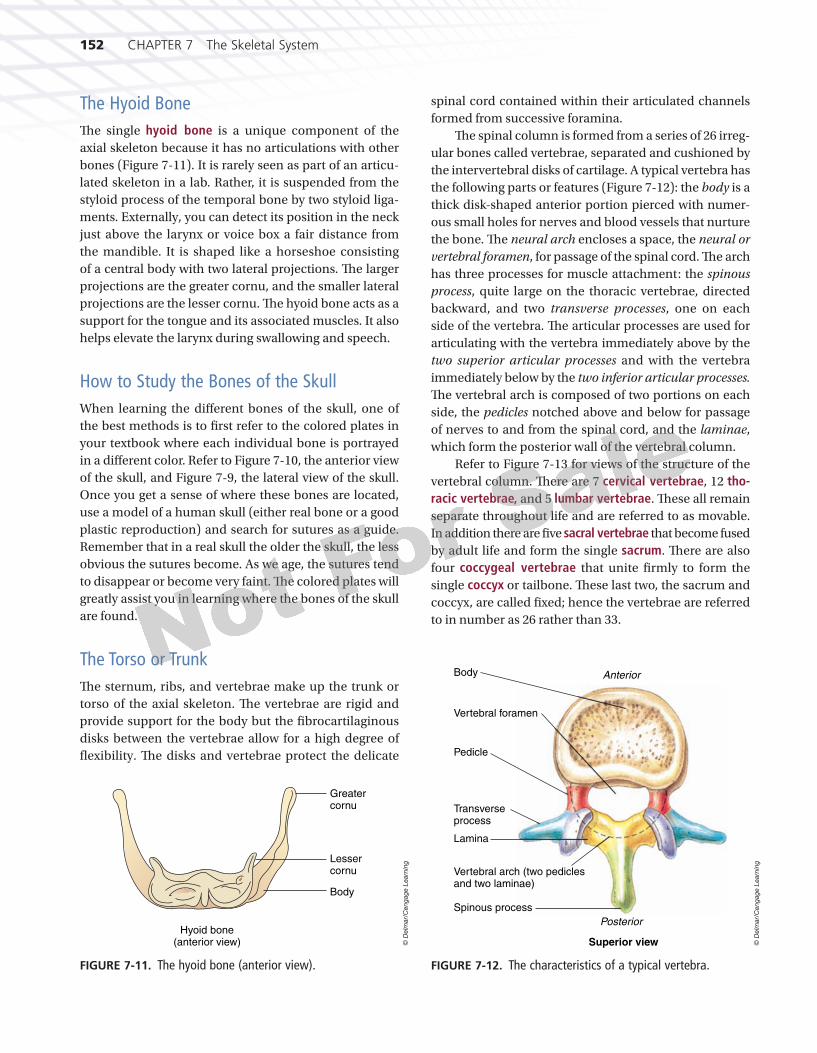
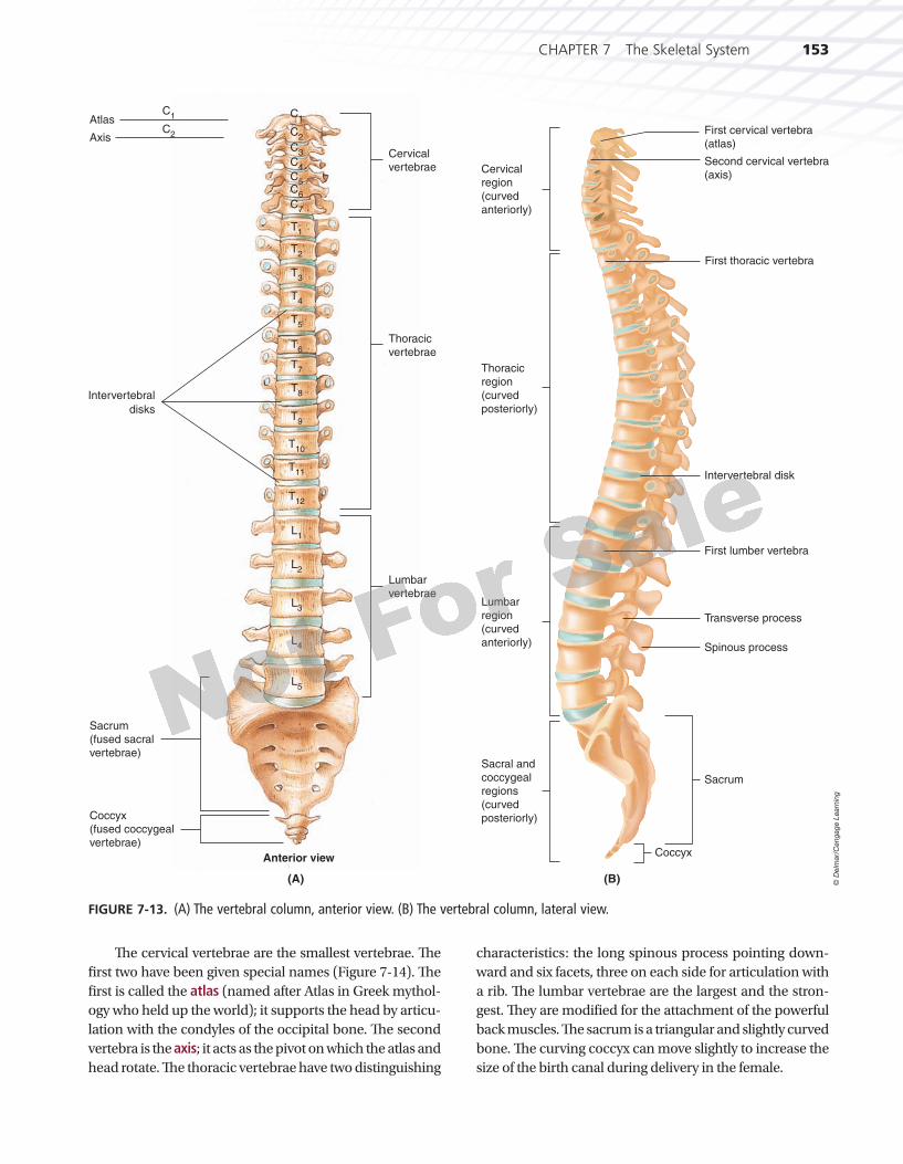
Th e spinal column is formed from a series of 26 irreg-ular bones called vertebrae, separated and cushioned by the intervertebral disks of cartilage. A typical vertebra has the following parts or features (Figure 7-12): the body is a thick disk-shaped anterior portion pierced with numer-ous small holes for nerves and blood vessels that nurture the bone. Th e neural arch encloses a space, the neural or vertebral foramen, for passage of the spinal cord. Th e arch has three processes for muscle attachment: the spinous process, quite large on the thoracic vertebrae, directed backward, and two transverse processes, one on each side of the vertebra. Th e articular processes are used for articulating with the vertebra immediately above by the two superior articular processes and with the vertebra immediately below by the two inferior articular processes. Th e vertebral arch is composed of two portions on each side, the pedicles notched above and below for passage of nerves to and from the spinal cord, and the laminae, which form the posterior wall of the vertebral column.
Refer to Figure 7-13 for views of the structure of the vertebral column. Th ere are 7 cervical vertebrae, 12 tho-racic vertebrae, and 5 lumbar vertebrae. Th ese all remain separate throughout life and are referred to as movable. In addition there are fi ve sacral vertebrae that become fused by adult life and form the single sacrum. Th ere are also four coccygeal vertebrae that unite fi rmly to form the single coccyx or tailbone. Th ese last two, the sacrum and coccyx, are called fi xed; hence the vertebrae are referred to in number as 26 rather than 33.
Greatercornu
Lessercornu
Body
Hyoid bone(anterior view)
FIGURE 7-11. The hyoid bone (anterior view).
© D
elm
ar/
Ceng
ag
e L
earn
ing
AnteriorBody
Vertebral foramen
Pedicle
Transverseprocess
Lamina
Vertebral arch (two pediclesand two laminae)
Spinous process
Posterior
Superior view
FIGURE 7-12. The characteristics of a typical vertebra.
© D
elm
ar/
Ceng
ag
e L
earn
ing
3871X_07_ch07_p136-173.indd 1523871X_07_ch07_p136-173.indd 152 8/25/09 11:22:20 AM8/25/09 11:22:20 AM
CHAPTER 7 The Skeletal System 153
# 102686 Cust: Cengage Au: Rizzo Pg. No. 153 Title: Fundamentals of Anatomy and Physiology Server: _____
C/M/Y/KShort / Normal
DESIGN SERVICES OF
S4-CARLISLEPublishing Services
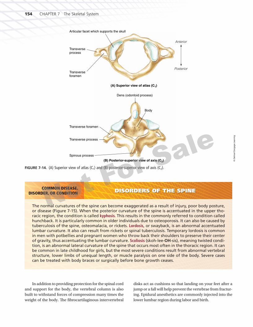
Th e cervical vertebrae are the smallest vertebrae. Th e fi rst two have been given special names (Figure 7-14). Th e fi rst is called the atlas (named after Atlas in Greek mythol-ogy who held up the world); it supports the head by articu-lation with the condyles of the occipital bone. Th e second vertebra is the axis; it acts as the pivot on which the atlas and head rotate. Th e thoracic vertebrae have two distinguishing
characteristics: the long spinous process pointing down-ward and six facets, three on each side for articulation with a rib. Th e lumbar vertebrae are the largest and the stron-gest. Th ey are modifi ed for the attachment of the powerful back muscles. Th e sacrum is a triangular and slightly curved bone. Th e curving coccyx can move slightly to increase the size of the birth canal during delivery in the female.
Atlas
Cervicalvertebrae
Sacrum(fused sacralvertebrae)
Coccyx(fused coccygealvertebrae)
Thoracicvertebrae
Lumbarvertebrae
Anterior view
Axis
C1
C2
C1
C2 C
3
C4
C5
C6
C7
T1
T2
T3
T4
T5
L1
L2
L3
L4
L5
T6
T7
T8
T9
T10
T11
T12
Intervertebral
disks
(A)
Cervical
region
(curved
anteriorly)
First cervical vertebra
(atlas)
Second cervical vertebra
(axis)
Thoracic
region
(curved
posteriorly)
Lumbar
region
(curved
anteriorly)
Sacral and
coccygeal
regions
(curved
posteriorly)
Coccyx
Sacrum
Spinous process
Transverse process
First lumber vertebra
Intervertebral disk
First thoracic vertebra
(B)
FIGURE 7-13. (A) The vertebral column, anterior view. (B) The vertebral column, lateral view.
© D
elm
ar/
Ceng
ag
e L
earn
ing
3871X_07_ch07_p136-173.indd 1533871X_07_ch07_p136-173.indd 153 8/25/09 11:22:22 AM8/25/09 11:22:22 AM
154 CHAPTER 7 The Skeletal System
# 102686 Cust: Cengage Au: Rizzo Pg. No. 154 Title: Fundamentals of Anatomy and Physiology Server: _____
C/M/Y/KShort / Normal
DESIGN SERVICES OF
S4-CARLISLEPublishing Services
DISORDERS OF THE SPINE
The normal curvatures of the spine can become exaggerated as a result of injury, poor body posture, or disease (Figure 7-15). When the posterior curvature of the spine is accentuated in the upper tho-racic region, the condition is called kyphosis. This results in the commonly referred to condition called hunchback. It is particularly common in older individuals due to osteoporosis. It can also be caused by tuberculosis of the spine, osteomalacia, or rickets. Lordosis, or swayback, is an abnormal accentuated lumbar curvature. It also can result from rickets or spinal tuberculosis. Temporary lordosis is common in men with potbellies and pregnant women who throw back their shoulders to preserve their center of gravity, thus accentuating the lumbar curvature. Scoliosis (skoh-lee-OH-sis), meaning twisted condi-tion, is an abnormal lateral curvature of the spine that occurs most often in the thoracic region. It can be common in late childhood for girls, but the most severe conditions result from abnormal vertebral structure, lower limbs of unequal length, or muscle paralysis on one side of the body. Severe cases can be treated with body braces or surgically before bone growth ceases.
COMMON DISEASE,DISORDER, OR CONDITION
In addition to providing protection for the spinal cord and support for the body, the vertebral column is also built to withstand forces of compression many times the weight of the body. Th e fi brocartilaginous intervertebral
disks act as cushions so that landing on your feet after a jump or a fall will help prevent the vertebrae from fractur-ing. Epidural anesthetics are commonly injected into the lower lumbar region during labor and birth.
Anterior
Spinous process
Transverse process
Posterior
(B) Posterior-superior view of axis (C2)
(A) Superior view of atlas (C1)
Transverse foramen
Dens (odontoid process)
Body
Articular facet which supports the skull
Transverseprocess
Transverseforamen
FIGURE 7-14. (A) Superior view of atlas (C1) and (B) posterior-superior view of axis (C2).
© D
elm
ar/
Ceng
ag
e L
earn
ing
3871X_07_ch07_p136-173.indd 1543871X_07_ch07_p136-173.indd 154 8/25/09 11:22:23 AM8/25/09 11:22:23 AM

CHAPTER 7 The Skeletal System 155
# 102686 Cust: Cengage Au: Rizzo Pg. No. 155 Title: Fundamentals of Anatomy and Physiology Server: _____
C/M/Y/KShort / Normal
DESIGN SERVICES OF
S4-CARLISLEPublishing Services
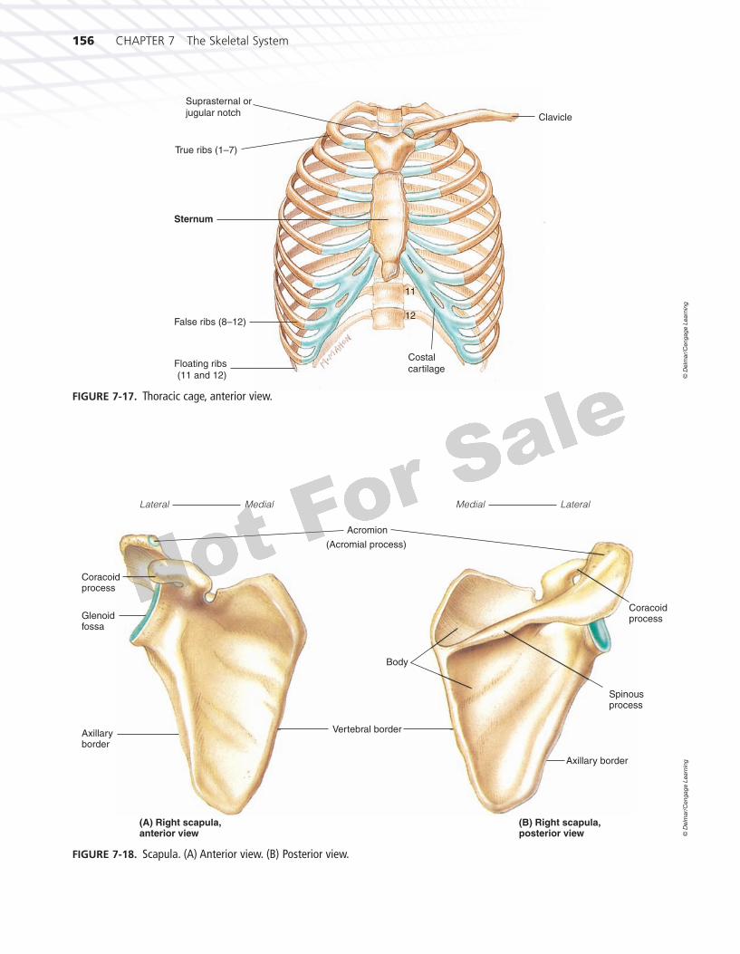
The ThoraxTh e thorax or the rib cage of the body is made up of the sternum, the costal cartilages, the ribs, and the bodies of the thoracic vertebrae. Th is bony cage encloses and pro-tects the heart and lungs. It also supports the bones of the shoulder girdle and the bones of the upper extremities.
The SternumTh e sternum is also known as the breastbone (Figure 7-16). It develops in three parts: the manubrium, the gladiolus, and the xiphoid (ZIFFoyd) process. Th e sternum resem-bles a sword, with the manubrium resembling the han-dle of the sword, the gladiolus or body forming the blade, and the xiphoid process forming the tip of the sword. No ribs are attached to the xiphoid, but the manubrium and gladiolus have notches on each side for attachment of the fi rst seven costal (rib) cartilages. Th e manubrium articulates with the clavicle or collarbone (Figure 7-17). Between these two points of attachment is the supraster-nal or jugular notch easily felt through the skin. Th e dia-phragm and the rectus abdominis muscles attach to the xiphoid.
The RibsTh e 12 pairs of ribs are also referred to as the costae (Figure 7-17). Th ey are named according to their anterior attachments. Because the upper seven pairs articulate directly with the sternum, they are called true ribs. Th e lower fi ve pairs are called false ribs. Th e costal carti-lages of the 8th, 9th, and 10th rib pairs are attached to
the cartilage of the 7th rib so they join the sternum only indirectly. Because the 11th and 12th pairs of ribs have no cartilage and do not attach at all anteriorly, these “false” ribs have another name, fl oating ribs. Of course, all ribs attach posteriorly to the thoracic vertebrae.
THE APPENDICULAR SKELETON
The Bones of the Upper ExtremitiesTh e bones of the upper extremities include the bones of the shoulder girdle, the arm, the forearm, the wrist, the hand, and the fi ngers.
Th e bones of the shoulder girdle are the clavicle (KLAV-ih-kl) and the scapula (SKAP-you-lah). Th e clav-icle or collarbone is a long slim bone located at the root of the neck just below the skin and anterior to the fi rst rib. Th e medial end articulates with the manubrium of the sternum and the lateral end with the acromial (ah-KRO-mee-al) process of the scapula. Th e scapula or shoulder blade is a large, fl at, triangular bone located on the dorsal portion of the thorax, covering the area from the second to the seventh rib (Figure 7-18). Two other prominent bony projections on the scapula are the coracoid process, which functions as an attachment for muscles that move the arm, and the glenoid fossa, which receives the head of the humerus and helps form the shoulder joint.
Th e humerus (HYOO-mehr-us) is the largest and longest bone of the upper arm (Figure 7-19). Its head is rounded and joined to the rest of the bone by its
FIGURE 7-15. Abnormal curvatures of the spine: (A) kyphosis; (B) lordosis; (C) scoliosis.
A. B. C.
© D
elm
ar/
Cen
gag
e L
earn
ing
Manubrium
Gladiolus/body
Xiphoid process
FIGURE 7-16. The sternum, anterior view.
© D
elm
ar/
Cen
gag
e L
earn
ing
3871X_07_ch07_p136-173.indd 1553871X_07_ch07_p136-173.indd 155 8/25/09 11:22:27 AM8/25/09 11:22:27 AM
156 CHAPTER 7 The Skeletal System
# 102686 Cust: Cengage Au: Rizzo Pg. No. 156 Title: Fundamentals of Anatomy and Physiology Server: _____
C/M/Y/KShort / Normal
DESIGN SERVICES OF
S4-CARLISLEPublishing Services
True ribs (1–7)
Suprasternal orjugular notch Clavicle
11
12
Costal
cartilage
Sternum
False ribs (8–12)
Floating ribs (11 and 12)
FIGURE 7-17. Thoracic cage, anterior view.
© D
elm
ar/
Cen
gag
e L
earn
ing
Lateral
(A) Right scapula, anterior view
(B) Right scapula, posterior view
Medial
Coracoid process
Glenoid fossa
Axillary border
Coracoid process
Spinous process
Axillary border
Body
Acromion
(Acromial process)
Vertebral border
LateralMedial
FIGURE 7-18. Scapula. (A) Anterior view. (B) Posterior view.
© D
elm
ar/
Ceng
ag
e L
earn
ing
3871X_07_ch07_p136-173.indd 1563871X_07_ch07_p136-173.indd 156 8/25/09 11:22:28 AM8/25/09 11:22:28 AM
CHAPTER 7 The Skeletal System 157
# 102686 Cust: Cengage Au: Rizzo Pg. No. 157 Title: Fundamentals of Anatomy and Physiology Server: _____
C/M/Y/KShort / Normal
DESIGN SERVICES OF
S4-CARLISLEPublishing Services
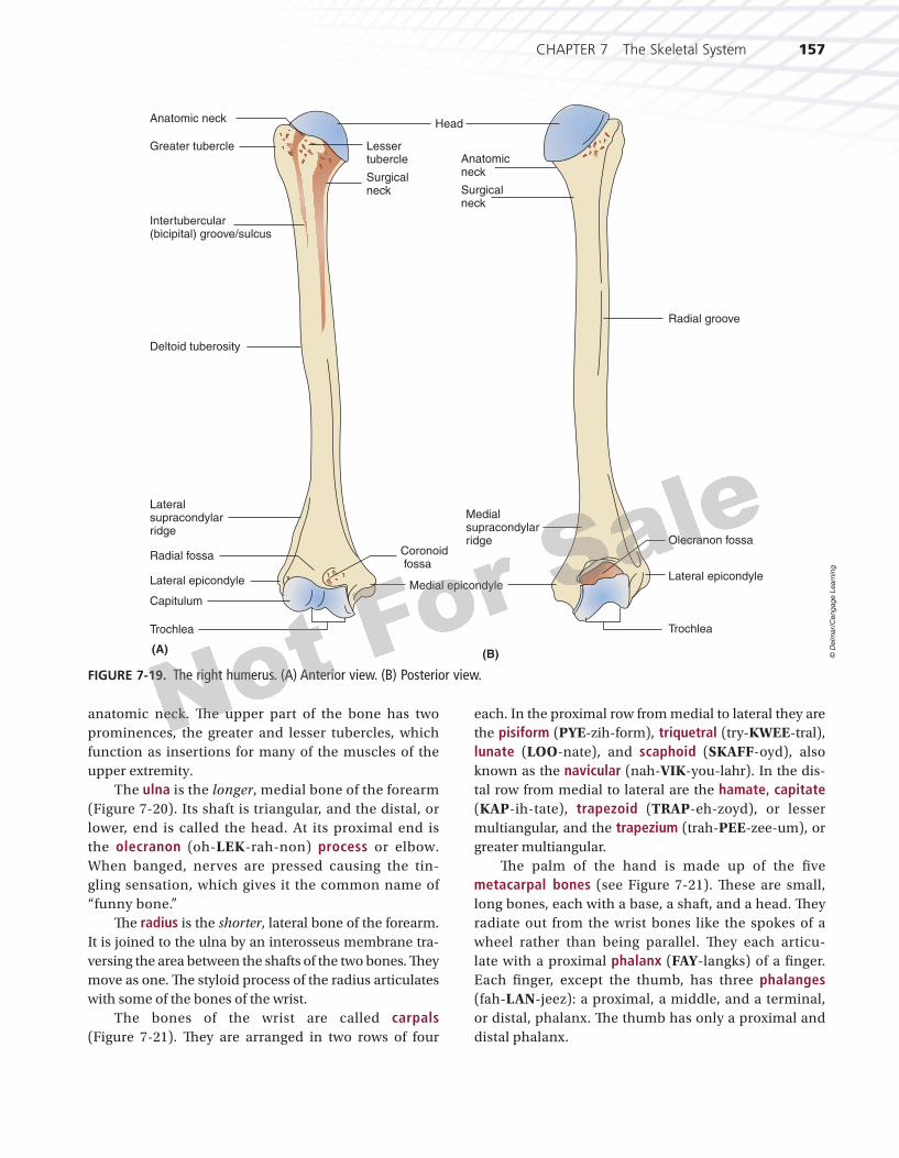
anatomic neck. Th e upper part of the bone has two prominences, the greater and lesser tubercles, which function as insertions for many of the muscles of the upper extremity.
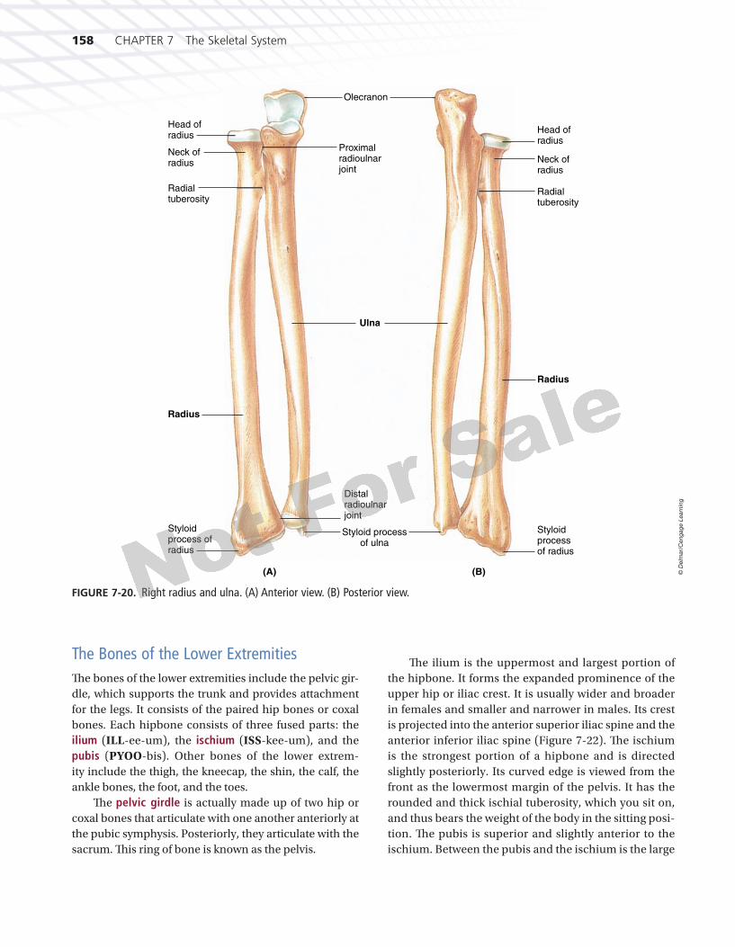
The ulna is the longer, medial bone of the forearm (Figure 7-20). Its shaft is triangular, and the distal, or lower, end is called the head. At its proximal end is the olecranon (oh-LEK-rah-non) process or elbow. When banged, nerves are pressed causing the tin-gling sensation, which gives it the common name of “funny bone.”
Th e radius is the shorter, lateral bone of the forearm. It is joined to the ulna by an interosseus membrane tra-versing the area between the shafts of the two bones. Th ey move as one. Th e styloid process of the radius articulates with some of the bones of the wrist.
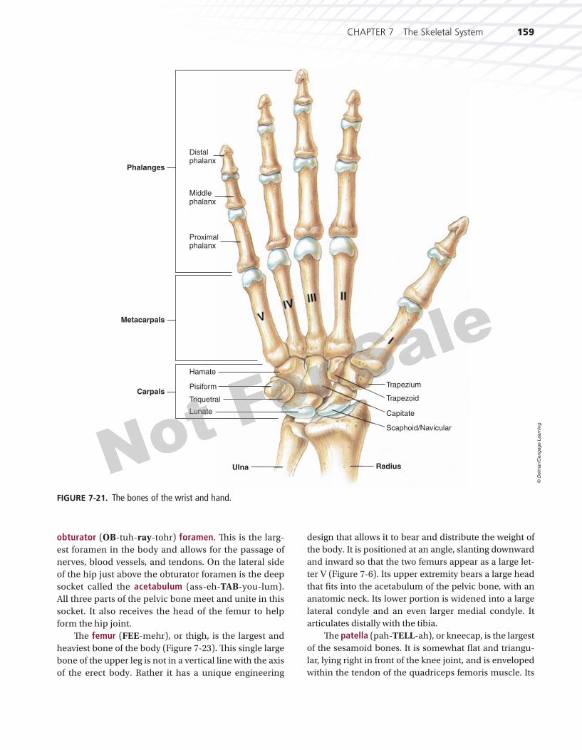
The bones of the wrist are called carpals (Figure 7-21). Th ey are arranged in two rows of four
each. In the proximal row from medial to lateral they are the pisiform (PYE-zih-form), triquetral (try-KWEE-tral),lunate (LOO-nate), and scaphoid (SKAFF-oyd), also known as the navicular (nah-VIK-you-lahr). In the dis-tal row from medial to lateral are the hamate, capitate (KAP-ih-tate), trapezoid (TRAP-eh-zoyd), or lesser multiangular, and the trapezium (trah-PEE-zee-um), or greater multiangular.
Th e palm of the hand is made up of the fi ve metacarpal bones (see Figure 7-21). Th ese are small, long bones, each with a base, a shaft, and a head. Th ey radiate out from the wrist bones like the spokes of a wheel rather than being parallel. Th ey each articu-late with a proximal phalanx (FAY-langks) of a fi nger. Each fi nger, except the thumb, has three phalanges (fah-LAN-jeez): a proximal, a middle, and a terminal, or distal, phalanx. Th e thumb has only a proximal and distal phalanx.
Anatomic neck
Greater tubercle Lesser tubercle
Surgical neck Surgical
neck
Anatomic neck
Intertubercular(bicipital) groove/sulcus
Deltoid tuberosity
Lateral supracondylar ridge
Radial fossa Coronoid fossa
Lateral epicondyle Medial epicondyle
Medial supracondylar ridge
Capitulum
Trochlea
Head
Radial groove
Olecranon fossa
Lateral epicondyle
Trochlea
(A) (B)
FIGURE 7-19. The right humerus. (A) Anterior view. (B) Posterior view.
© D
elm
ar/
Ceng
ag
e L
earn
ing
3871X_07_ch07_p136-173.indd 1573871X_07_ch07_p136-173.indd 157 8/25/09 11:22:31 AM8/25/09 11:22:31 AM
158 CHAPTER 7 The Skeletal System
# 102686 Cust: Cengage Au: Rizzo Pg. No. 158 Title: Fundamentals of Anatomy and Physiology Server: _____
C/M/Y/KShort / Normal
DESIGN SERVICES OF
S4-CARLISLEPublishing Services
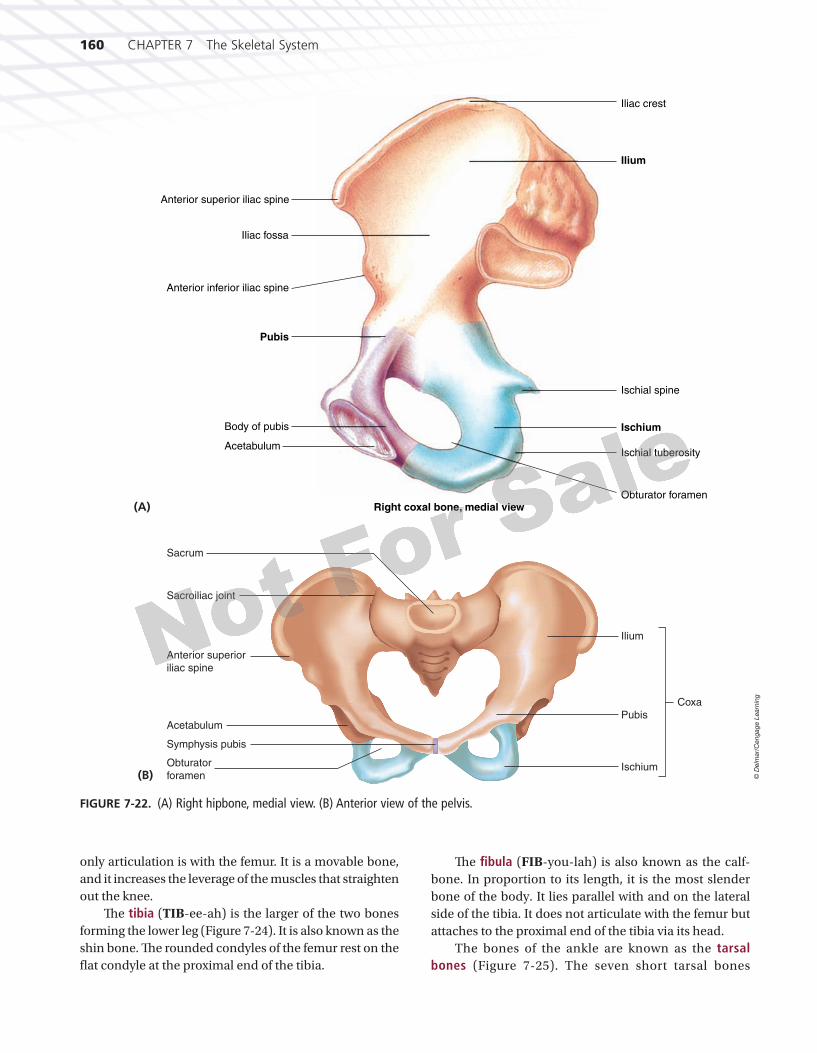
The Bones of the Lower ExtremitiesTh e bones of the lower extremities include the pelvic gir-dle, which supports the trunk and provides attachment for the legs. It consists of the paired hip bones or coxal bones. Each hipbone consists of three fused parts: the ilium (ILL-ee-um), the ischium (ISS-kee-um), and the pubis (PYOO-bis). Other bones of the lower extrem-ity include the thigh, the kneecap, the shin, the calf, the ankle bones, the foot, and the toes.
Th e pelvic girdle is actually made up of two hip or coxal bones that articulate with one another anteriorly at the pubic symphysis. Posteriorly, they articulate with the sacrum. Th is ring of bone is known as the pelvis.
Th e ilium is the uppermost and largest portion of the hipbone. It forms the expanded prominence of the upper hip or iliac crest. It is usually wider and broader in females and smaller and narrower in males. Its crest is projected into the anterior superior iliac spine and the anterior inferior iliac spine (Figure 7-22). Th e ischium is the strongest portion of a hipbone and is directed slightly posteriorly. Its curved edge is viewed from the front as the lowermost margin of the pelvis. It has the rounded and thick ischial tuberosity, which you sit on, and thus bears the weight of the body in the sitting posi-tion. Th e pubis is superior and slightly anterior to the ischium. Between the pubis and the ischium is the large
(A) (B)
Head ofradius
Radialtuberosity
Olecranon
Neck ofradius
Styloidprocess ofradius
Styloidprocessof radius
Radius
Head ofradius
Radialtuberosity
Neck ofradius
Radius
Ulna
Distalradioulnarjoint
Styloid processof ulna
Proximalradioulnarjoint
FIGURE 7-20. Right radius and ulna. (A) Anterior view. (B) Posterior view.
© D
elm
ar/
Ceng
ag
e L
earn
ing
3871X_07_ch07_p136-173.indd 1583871X_07_ch07_p136-173.indd 158 8/25/09 11:22:32 AM8/25/09 11:22:32 AM
CHAPTER 7 The Skeletal System 159
# 102686 Cust: Cengage Au: Rizzo Pg. No. 159 Title: Fundamentals of Anatomy and Physiology Server: _____
C/M/Y/KShort / Normal
DESIGN SERVICES OF
S4-CARLISLEPublishing Services
obturator (OB-tuh-ray-tohr) foramen. Th is is the larg-est foramen in the body and allows for the passage of nerves, blood vessels, and tendons. On the lateral side of the hip just above the obturator foramen is the deep socket called the acetabulum (ass-eh-TAB-you-lum). All three parts of the pelvic bone meet and unite in this socket. It also receives the head of the femur to help form the hip joint.
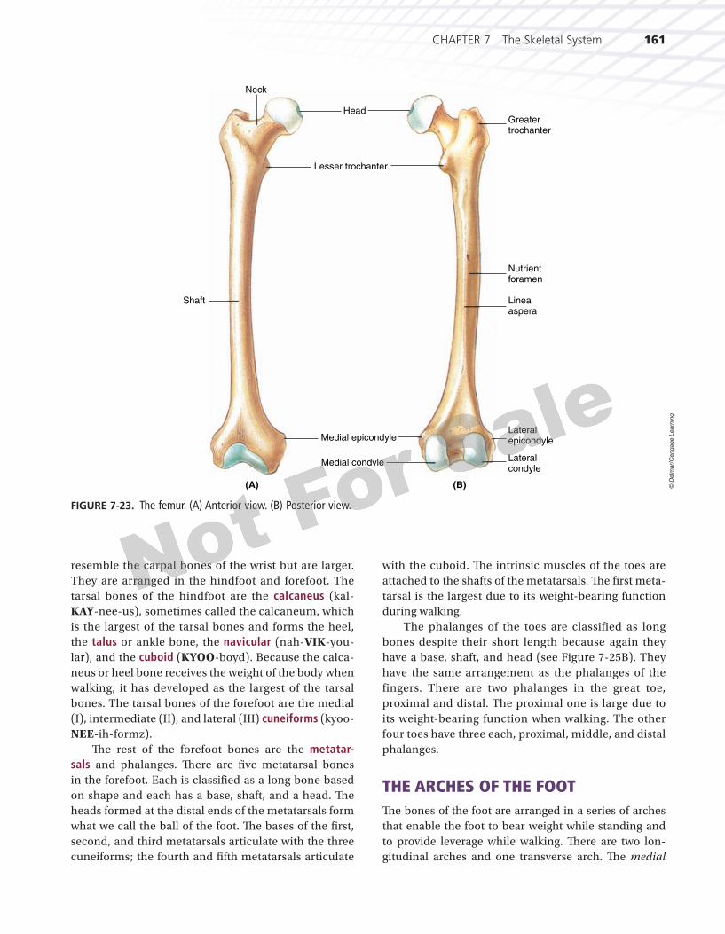
Th e femur (FEE-mehr), or thigh, is the largest and heaviest bone of the body (Figure 7-23). Th is single large bone of the upper leg is not in a vertical line with the axis of the erect body. Rather it has a unique engineering
design that allows it to bear and distribute the weight of the body. It is positioned at an angle, slanting downward and inward so that the two femurs appear as a large let-ter V (Figure 7-6). Its upper extremity bears a large head that fi ts into the acetabulum of the pelvic bone, with an anatomic neck. Its lower portion is widened into a large lateral condyle and an even larger medial condyle. It articulates distally with the tibia.
Th e patella (pah-TELL-ah), or kneecap, is the largest of the sesamoid bones. It is somewhat fl at and triangu-lar, lying right in front of the knee joint, and is enveloped within the tendon of the quadriceps femoris muscle. Its
Phalanges
Distal phalanx
Ulna Radius
Carpals
Metacarpals
Middle phalanx
Proximal phalanx
Hamate
Pisiform
Triquetral
Lunate
Trapezium
Trapezoid
Capitate
Scaphoid/Navicular
FIGURE 7-21. The bones of the wrist and hand.
© D
elm
ar/
Ceng
ag
e L
earn
ing
3871X_07_ch07_p136-173.indd 1593871X_07_ch07_p136-173.indd 159 8/25/09 11:22:34 AM8/25/09 11:22:34 AM
160 CHAPTER 7 The Skeletal System
# 102686 Cust: Cengage Au: Rizzo Pg. No. 160 Title: Fundamentals of Anatomy and Physiology Server: _____
C/M/Y/KShort / Normal
DESIGN SERVICES OF
S4-CARLISLEPublishing Services
only articulation is with the femur. It is a movable bone, and it increases the leverage of the muscles that straighten out the knee.
Th e tibia (TIB-ee-ah) is the larger of the two bones forming the lower leg (Figure 7-24). It is also known as the shin bone. Th e rounded condyles of the femur rest on the fl at condyle at the proximal end of the tibia.
Th e fi bula (FIB-you-lah) is also known as the calf-bone. In proportion to its length, it is the most slender bone of the body. It lies parallel with and on the lateral side of the tibia. It does not articulate with the femur but attaches to the proximal end of the tibia via its head.
The bones of the ankle are known as the tarsal bones (Figure 7-25). The seven short tarsal bones
(B)
Sacrum
Sacroiliac joint
Anterior superior
iliac spine
Acetabulum
Symphysis pubis
Obturator
foramenIschium
Pubis
Ilium
Coxa
FIGURE 7-22. (A) Right hipbone, medial view. (B) Anterior view of the pelvis.
Pubis
Anterior superior iliac spine
Ilium
Ischium
Right coxal bone, medial view
Iliac fossa
Anterior inferior iliac spine
Body of pubis
Iliac crest
Ischial spine
Ischial tuberosity
Obturator foramen
Acetabulum
(A)
© D
elm
ar/
Ceng
ag
e L
earn
ing
3871X_07_ch07_p136-173.indd 1603871X_07_ch07_p136-173.indd 160 8/25/09 11:22:38 AM8/25/09 11:22:38 AM
CHAPTER 7 The Skeletal System 161
# 102686 Cust: Cengage Au: Rizzo Pg. No. 161 Title: Fundamentals of Anatomy and Physiology Server: _____
C/M/Y/KShort / Normal
DESIGN SERVICES OF
S4-CARLISLEPublishing Services
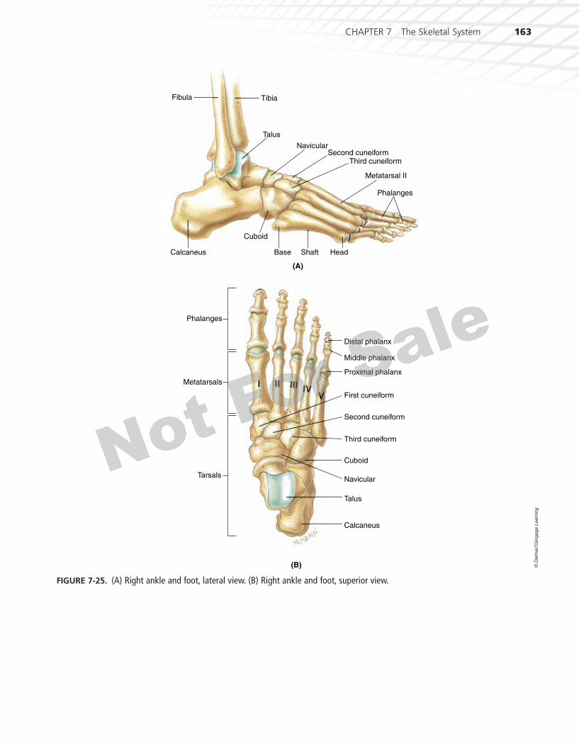
resemble the carpal bones of the wrist but are larger. They are arranged in the hindfoot and forefoot. The tarsal bones of the hindfoot are the calcaneus (kal-KAY-nee-us), sometimes called the calcaneum, which is the largest of the tarsal bones and forms the heel, the talus or ankle bone, the navicular (nah-VIK-you-lar), and the cuboid (KYOO-boyd). Because the calca-neus or heel bone receives the weight of the body when walking, it has developed as the largest of the tarsal bones. The tarsal bones of the forefoot are the medial (I), intermediate (II), and lateral (III) cuneiforms (kyoo-NEE-ih-formz).
Th e rest of the forefoot bones are the metatar-sals and phalanges. Th ere are fi ve metatarsal bones in the forefoot. Each is classifi ed as a long bone based on shape and each has a base, shaft, and a head. Th e heads formed at the distal ends of the metatarsals form what we call the ball of the foot. Th e bases of the fi rst, second, and third metatarsals articulate with the three cuneiforms; the fourth and fi fth metatarsals articulate
with the cuboid. Th e intrinsic muscles of the toes are attached to the shafts of the metatarsals. Th e fi rst meta-tarsal is the largest due to its weight-bearing function during walking.
The phalanges of the toes are classified as long bones despite their short length because again they have a base, shaft, and head (see Figure 7-25B). They have the same arrangement as the phalanges of the fingers. There are two phalanges in the great toe, proximal and distal. The proximal one is large due to its weight-bearing function when walking. The other four toes have three each, proximal, middle, and distal phalanges.
THE ARCHES OF THE FOOTTh e bones of the foot are arranged in a series of arches that enable the foot to bear weight while standing and to provide leverage while walking. Th ere are two lon-gitudinal arches and one transverse arch. Th e medial
(A) (B)
Neck
Shaft
Head
Lesser trochanter
Medial epicondyle
Medial condyle
Greatertrochanter
Nutrientforamen
Lineaaspera
Lateralepicondyle
Lateralcondyle
FIGURE 7-23. The femur. (A) Anterior view. (B) Posterior view.
© D
elm
ar/
Ceng
ag
e L
earn
ing
3871X_07_ch07_p136-173.indd 1613871X_07_ch07_p136-173.indd 161 8/25/09 11:22:40 AM8/25/09 11:22:40 AM

162 CHAPTER 7 The Skeletal System
# 102686 Cust: Cengage Au: Rizzo Pg. No. 162 Title: Fundamentals of Anatomy and Physiology Server: _____
C/M/Y/KShort / Normal
DESIGN SERVICES OF
S4-CARLISLEPublishing Services
longitudinal arch is formed by the calcaneus, talus, navic-ular, the three cuneiforms, and the three medial metatar-sals. Th is is the highest arch of the foot and can easily be noted. Th e lateral longitudinal arch is much lower and is formed by the calcaneus, the cuboid, and the two lateral metatarsals. Th e transverse arch is perpendicular to the
longitudinal arches and is most pronounced at the base of the metatarsals.
Th e term pes planus, or fl atfoot, indicates a decreased height of the longitudinal arches. It rarely causes any pain and can be inherited or result from muscle weakness in the foot.
(A) (B)
Tibial tuberosity
Fibula
Medialmalleolus
Lateralmalleolus
Lateralmalleolus
Medialcondyle
Lateralcondyle
Fibula
Tibia
Head
Lateralcondyle
FIGURE 7-24. The tibia and fi bula. (A) Anterior view. (B) Posterior view.
© D
elm
ar/
Ceng
ag
e L
earn
ing
3871X_07_ch07_p136-173.indd 1623871X_07_ch07_p136-173.indd 162 8/25/09 11:22:41 AM8/25/09 11:22:41 AM
CHAPTER 7 The Skeletal System 163
# 102686 Cust: Cengage Au: Rizzo Pg. No. 163 Title: Fundamentals of Anatomy and Physiology Server: _____
C/M/Y/KShort / Normal
DESIGN SERVICES OF
S4-CARLISLEPublishing Services
(A)
Fibula Tibia
Talus
NavicularSecond cuneiform
Phalanges
Third cuneiform
Metatarsal II
Cuboid
Calcaneus Base Shaft Head
(B)
Talus
Navicular
Second cuneiform
First cuneiform
Third cuneiform
Metatarsals
Tarsals
Phalanges
Calcaneus
Cuboid
Proximal phalanx
Middle phalanx
Distal phalanx
FIGURE 7-25. (A) Right ankle and foot, lateral view. (B) Right ankle and foot, superior view.
© D
elm
ar/
Ceng
ag
e L
earn
ing
3871X_07_ch07_p136-173.indd 1633871X_07_ch07_p136-173.indd 163 8/25/09 11:22:43 AM8/25/09 11:22:43 AM
164 CHAPTER 7 The Skeletal System
# 102686 Cust: Cengage Au: Rizzo Pg. No. 164 Title: Fundamentals of Anatomy and Physiology Server: _____
C/M/Y/KShort / Normal
DESIGN SERVICES OF
S4-CARLISLEPublishing Services
As we age, less protein matrix is formed in our bone tissue accompanied by a loss of calcium salts. Bones become more fragile and tend to break more easily in older adults. Older adults also develop stiffness and less fl exibility of the skeleton due to a decrease in the protein collagen found in the tendon connective tissue that connects bone to muscle, and in ligaments that con-nect bone to bone. Hence, as we age we should be more conscious of our diet and include more foods that contain calcium. Regular exercise can also
help maintain healthy bone tissues. Walking is an excellent way to exercise both bones and muscles.
Do we really “shrink” as we grow older? Shrinking is caused by a thinning of the inter-vertebral disks in the spinal column. Starting at around age 40, individuals can lose about one-half inch in height every 20 years due to the loss of disk protein.
AS THE BODY AGES
Th ere are many careers available to individuals who are interested in the skeletal system.
● Athletic trainers provide guidance to develop muscles and bones for agility, good looks, and sports training.
● Chiropractors or doctors of chiropractic complete at least two years of premedi-cal studies, followed by four years of study in an approved chiropractic school, learning mechanical manipulation of the spinal column as a method to maintain a healthy nervous system.
● Prosthetists are individuals who create artifi cial limbs.
● Orthopedists are physicians specializing in preventing and correcting disorders of the skeleton, joints, and muscles. Th ere are also careers in orthopedic nursing.
● Orthotists are individuals who design, make, and fi t braces or other orthopedic devices prescribed by a physician.
● Paramedics and emergency medical technicians can also further train and specialize in the treatment of skeletal system disorders like broken bones and fractures.
Th sy
CareerFOCUS
3871X_07_ch07_p136-173.indd 1643871X_07_ch07_p136-173.indd 164 8/25/09 11:22:45 AM8/25/09 11:22:45 AM
CHAPTER 7 The Skeletal System 165
# 102686 Cust: Cengage Au: Rizzo Pg. No. 165 Title: Fundamentals of Anatomy and Physiology Server: _____
C/M/Y/KShort / Normal
DESIGN SERVICES OF
S4-CARLISLEPublishing Services
Cardiovascular System● Blood cells transport oxygen and nutrients to bone
cells and take away carbon dioxide and waste products.
● Calcium from bones is necessary for blood clotting and normal heart functions.
Lymphatic System● Red bone marrow produces lymphocytes, which
function in our immune response.
Digestive System● Calcium, necessary for bone matrix development, is
absorbed in the intestine from our daily food intake.● Excess calcium can be eliminated via the bowels.
Respiratory System● Oxygen is brought into the body via the respiratory
system and transported by the blood to bone cells for biochemical respiration.
● Th e ribs along with the intercostal muscles and dia-phragm bring about breathing.
Urinary System● Th e kidneys help regulate blood calcium levels.● Excess calcium can also be eliminated via the
kidneys.
Reproductive System● Bones are a source of calcium during breastfeeding.● Th e pelvis aids in supporting the uterus and develop-
ing fetus during pregnancy in the female.
BODY SYSTEMSWORKING TOGETHER
TO MAINTAIN HOMEOSTASIS: THE SKELETAL SYSTEM
Integumentary System● Vitamin D is produced in the skin by UV light.● It enhances the absorption of calcium in bones for
bone and tooth formation.
Muscular System● Th rough their tendons, muscles pull on bones,
bringing about movement.● Calcium from bones is necessary for muscle
contraction to occur.
Nervous System● Th e cranial bones protect the brain, and the
vertebrae and intervertebral disks protect the spinal cord.
● Receptors for pain monitor trauma to bones.● Calcium from bones is necessary for nerve
transmission.
Endocrine System● Th e hormone calcitonin causes calcium to be stored
in bones.● Th e hormone parathormone causes calcium to be
released from bones.● Growth hormone from the anterior pituitary gland
aff ects bone development.
DISORDERS OF THE SKELETAL SYSTEM
OSTEOPOROSISOsteoporosis (oss-tee-oh-poh-ROH-sis) is a disorder of the skeletal system characterized by a decrease in bone mass with accompanying increased susceptibility to bone fractures. This results from decreased levels of estrogens that occur after menopause in women and in both men and women in old age. Estrogens help maintain bone tissue by stimulating osteoblasts to form new bone.
Osteoporosis occurs more often in middle-aged and elderly women, but it can also affect teenagers who do not eat a proper balanced diet, people allergic to dairy products, and anyone with eating disorders. The bone mass becomes depleted in such a way that the skeleton can-not withstand everyday mechanical stress. Bone fractures are common, even from normal daily activities. This disease is responsible for height loss, hunched backs, and pain in older individuals. Adequate diet and exercise can prevent osteoporosis.
COMMON DISEASE,DISORDER, OR CONDITION
(continues)
3871X_07_ch07_p136-173.indd 1653871X_07_ch07_p136-173.indd 165 8/25/09 11:22:54 AM8/25/09 11:22:54 AM

166 CHAPTER 7 The Skeletal System
# 102686 Cust: Cengage Au: Rizzo Pg. No. 166 Title: Fundamentals of Anatomy and Physiology Server: _____
C/M/Y/KShort / Normal
DESIGN SERVICES OF
S4-CARLISLEPublishing Services
DISORDERS OF THE SKELETAL SYSTEM (continued)
PAGET’S DISEASEPaget’s disease is a common nonmetabolic disease of bone whose cause is unknown. It usually affects middle-aged and elderly individuals. Symptoms include an irregular thickening and softening of the bones. There is excessive bone destruction and unorganized bone repair. Areas of the body affected are the skull, pelvis, and limbs. Treatment includes a high-protein and high-calcium diet with mild but regular exercise.
GIGANTISMGigantism (JYE-gan-tizm) is the result of excessive endochondral ossifi cation at the epiphyseal plates of long bones. This results in abnormally large limbs, giving the affected individual the appearance of a very tall “giant.”
DWARFISMDwarfi sm is the opposite condition of gigantism and results from inadequate ossifi cation occurring at the epiphyseal plates of long bones. This results in an individual being abnormally small. This condi-tion is not to be confused with a genetic dwarf.
SPINA BIFIDASpina bifi da (SPY-nah BIFF-ih-dah) is a congenital defect in the development of the posterior vertebral arch in which the laminae do not unite at the midline. It is a relatively common disorder. It may occur with only a small deformed lamina, or it may be associated with the complete absence of laminae, causing the contents of the spinal canal to protrude posteriorly. If the condition does not involve herniation of the meninges or contents of the spinal canal, treatment is not required.
HERNIATED DISKA herniated disk is a rupture of the fi brocartilage surrounding an intervertebral disk that cushions the vertebrae above and below. This produces pressure on spinal nerve roots, causing severe pain and nerve damage. The condition occurs most often in the lumbar region and is also known as a slipped disk. Treatment can include prolonged bed rest to promote healing or surgery to remove the damaged disk.
CLEFT PALATE AND CLEFT LIPA cleft palate, more common in females, occurs when the palatine processes of the maxillary bones do not fuse properly, resulting in an opening between the oral and nasal cavities (Figure 7-26). A person with this condition cannot speak clearly and has diffi culty eating or drinking. A child born with this condition can have it surgically repaired and corrected; hence, we rarely see this condition in developed countries. A cleft lip, more common in males, occurs when the maxillary bones do not form normally, producing a cleft in the upper lip. Treatment is by surgical repair in infancy. A child could be born with both a cleft palate and a cleft lip.
BLACK EYEA black eye is caused by a blow to the supraorbital ridge, which overlies the frontal sinus. This results in a skin laceration with bleeding. Tissue fl uid and blood accumulate in the connective tissue around the eyelid. This swelling, bruising, and discoloration produces a black eye, also called a periorbital ecchymosis or bruise.
DEVIATED SEPTUMA deviated septum develops when the nasal septum shifts to the left (usually) during normal growth. It can be aggravated by a severe blow to the nasal area. Serious defl ections can interfere with nasal fl ow, can cause frequent nose bleeding, or may result in headaches or shortness of breath. Severe deviation can be corrected through surgical procedures.
COMMON DISEASE,DISORDER, OR CONDITION
3871X_07_ch07_p136-173.indd 1663871X_07_ch07_p136-173.indd 166 8/25/09 11:23:00 AM8/25/09 11:23:00 AM
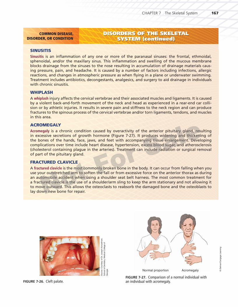
CHAPTER 7 The Skeletal System 167
# 102686 Cust: Cengage Au: Rizzo Pg. No. 167 Title: Fundamentals of Anatomy and Physiology Server: _____
C/M/Y/KShort / Normal
DESIGN SERVICES OF
S4-CARLISLEPublishing Services
DISORDERS OF THE SKELETAL SYSTEM (continued)
SINUSITISSinusitis is an infl ammation of any one or more of the paranasal sinuses: the frontal, ethmoidal, sphenoidal, and/or the maxillary sinus. This infl ammation and swelling of the mucous membrane blocks drainage from the sinuses to the nose resulting in accumulation of drainage materials caus-ing pressure, pain, and headache. It is caused by a number of factors including infections, allergic reactions, and changes in atmospheric pressure as when fl ying in a plane or underwater swimming. Treatment includes antibiotics, decongestants, analgesics, and surgery to aid drainage in individuals with chronic sinusitis.
WHIPLASHA whiplash injury affects the cervical vertebrae and their associated muscles and ligaments. It is caused by a violent back-and-forth movement of the neck and head as experienced in a rear-end car colli-sion or by athletic injuries. It results in severe pain and stiffness to the neck region and can produce fractures to the spinous process of the cervical vertebrae and/or torn ligaments, tendons, and muscles in this area.
ACROMEGALYAcromegaly is a chronic condition caused by overactivity of the anterior pituitary gland, resulting in excessive secretions of growth hormone (Figure 7-27). It produces widening and thickening of the bones of the hands, face, jaws, and feet with accompanying tissue enlargement. Developing complications over time include heart disease, hypertension, excess blood sugar, and atherosclerosis (cholesterol containing plague in the arteries). Treatment can include radiation or surgical removal of part of the pituitary gland.
FRACTURED CLAVICLEA fractured clavicle is the most commonly broken bone in the body. It can occur from falling when you use your outstretched arm to soften the fall or from excessive force on the anterior thorax as during an automobile accident when using a shoulder seat belt harness. The most common treatment for a fractured clavicle is the use of a shoulder/arm sling to keep the arm stationary and not allowing it to move outward. This allows the osteoclasts to reabsorb the damaged bone and the osteoblasts to lay down new bone for repair.
COMMON DISEASE,DISORDER, OR CONDITION
FIGURE 7-26. Cleft palate.
So
urc
e:
Co
urt
esy o
f D
r. J
osep
h K
onzelm
an,
Scho
ol o
f D
entistr
y, M
ed
ical C
olle
ge o
f
Geo
rgia Normal proportion Acromegaly
FIGURE 7-27. Comparison of a normal individual with an individual with acromegaly.
© D
elm
ar/
Ceng
ag
e L
earn
ing
3871X_07_ch07_p136-173.indd 1673871X_07_ch07_p136-173.indd 167 8/25/09 11:23:09 AM8/25/09 11:23:09 AM
168 CHAPTER 7 The Skeletal System
# 102686 Cust: Cengage Au: Rizzo Pg. No. 168 Title: Fundamentals of Anatomy and Physiology Server: _____
C/M/Y/KShort / Normal
DESIGN SERVICES OF
S4-CARLISLEPublishing Services
SUMMARY OUTLINE INTRODUCTION
1. Th e skeleton is the supporting structure of the body; it allows muscles to bring about movement and breathing.
2. Th e solid appearance of bone is due to mineral salts that form the inorganic matrix surrounding the living bone cells.
3. Leonardo da Vinci was the fi rst to correctly illustrate the 206 bones of the body.
THE FUNCTIONS OF THE SKELETAL SYSTEMTh e skeleton has fi ve functions:
1. Support surrounding tissues
2. Protect vital organs and soft tissues
3. Provide levers for muscles to pull on
4. Manufacture blood cells in red bone marrow by hematopoiesis
5. Act as a storage area for mineral salts, especially calcium and phosphorus, and fat in yellow marrow
THE GROWTH AND FORMATION OF BONE
1. After 3 months, the fetal skeleton is completely formed and made primarily of hyaline cartilage. Ossifi cation and growth then develop.
2. Longitudinal growth of bone continues until approximately 15 years of age in girls and 16 in boys.
3. Bone maturation continues until 21 years of age in both sexes.
Deposition of the Bone
1. Bone develops from spindle-shaped embryonic bone cells called osteoblasts.
2. Osteoblasts develop into mature bone cells called osteocytes. Th ey form under the fi brovascular membrane covering bone, called the periosteum, and under the membrane lining of the medullary cavity, called the endosteum.
3. Th e more strain or pressure on a bone, the more the bone will develop.
4. Osteoclasts are large cells that are responsible for the reabsorption of injured bone. Th ey also reabsorb bone during remodeling.
Types of Ossifi cationTh e two types of ossifi cation are:
1. Intramembranous ossifi cation: a process in which dense connective membranes are replaced by deposits of inorganic calcium salts. Th e bones of the cranium form in this way.
2. Endochondral ossifi cation: the process whereby cartilage is the environment in which the bone cells develop. All other bones of the body develop in this way.
Maintaining the Bone
1. Th e correct amount of calcium stored in the bones, the proper amount of calcium in the blood, and the excretion of excess calcium are controlled by the endocrine system.
2. Th e parathyroid glands secrete parathormone, which causes calcium to be released into the bloodstream. Another hormone, calcitonin, causes calcium to be stored in the bones.
THE HISTOLOGY OF BONETh ere are two types of bone tissue:
1. Compact or dense bone is strong and solid.
2. Cancellous or spongy bone has many open spaces fi lled with bone marrow.
Th e Haversian System of Compact Bone
1. An English physician, Clopton Havers (1650–1702), fi rst described the histologic features of compact bone.
2. Haversian canals or osteons are small canals containing blood vessels running parallel to the surface of compact bone and are surrounded by concentric rings of solid bone called lamellae.
3. In these rings of bone are cavities called lacunae; each lacuna contains an osteocyte bathed in fl uid.
4. Lacunae are connected to one another and eventually to the osteons by smaller canals called canaliculi.
5. Th e tissue fl uid that circulates in these canals carries nutrients and oxygen to and waste away from the bone cells.
Cancellous Bone
1. Cancellous bone consists of a meshwork of bone called trabeculae.
2. Trabeculae create the spongy appearance of cancellous bone.
3. Th e spaces between the trabeculae are fi lled with bone marrow.
3871X_07_ch07_p136-173.indd 1683871X_07_ch07_p136-173.indd 168 8/25/09 11:23:13 AM8/25/09 11:23:13 AM
CHAPTER 7 The Skeletal System 169
# 102686 Cust: Cengage Au: Rizzo Pg. No. 169 Title: Fundamentals of Anatomy and Physiology Server: _____
C/M/Y/KShort / Normal
DESIGN SERVICES OF
S4-CARLISLEPublishing Services
Bone MarrowTh ere are two types of bone marrow:
1. Red bone marrow’s function is hematopoiesis, the formation of blood cells.
2. In an adult the ribs, vertebrae, sternum, and pelvis contain red bone marrow in their cancellous tissue.
3. Yellow bone marrow is found in the shafts of long bones within their cancellous tissue.
4. Yellow bone marrow stores fat cells.
THE CLASSIFICATION OF BONESTh e bones of the body can be classifi ed, based on shape, into fi ve categories.
1. Long bones consist of a shaft or diaphysis, a fl ared portion at the end of the diaphysis called a metaphysis, and two extremities called epiphyses. Examples are the clavicle, humerus, radius, ulna, femur, tibia, and fi bula as well as the phalanges, metacarpals, and metatarsals.
2. Short bones have a somewhat irregular shape. Examples are the tarsal bones of the foot and the carpal bones of the hand.
3. Flat bones are fl at and serve to protect or provide extensive muscle attachment. Examples are some bones of the cranium, the ribs, scapula, and part of the hipbone.
4. Irregular bones have a very peculiar or irregular shape. Examples are the vertebrae and the ossicles of the ear.
5. Sesamoid bones are small rounded bones enclosed in tendon and fascial tissue near joints. One example is the largest sesamoid bone, the patella.
BONE MARKINGS
1. Bones exhibit certain projections called processes. Examples of processes are the spine, condyle, tubercle, trochlea, trochanter, crest, line, head, and neck.
2. Bones also exhibit certain depressions called fossae. Examples of fossae are suture, foramen, meatus or canal, sinus or antrum and sulcus.
3. Th ese markings are functional to help join bones to one another, to provide a surface for muscle attachment, or to serve as a passageway for blood vessels and nerves into and out of the bone.
DIVISIONS OF THE SKELETON
1. Th e human skeleton has 206 bones.
2. Th e skeleton can be divided into the axial skeleton (skull, hyoid, vertebrae, ribs, and sternum) and the appendicular skeleton (bones of the upper and lower extremities).
THE AXIAL SKELETON
1. Th e cranial bones consist of the frontal bone, the two parietal bones, the occipital bone, the two temporal bones, the sphenoid bone, the ethmoid bone, the six auditory ossicles (malleus, incus, stapes in each ear), and the varying wormian or sutural bones.
2. The facial bones consist of the two nasal bones, the two palatine bones, the two maxillary bones (upper jaw), the two zygomatic or malar bones (cheekbones), the two lacrimal bones, the two turbinates or nasal conchae, the single vomer bone, and the single lower jawbone, the mandible.
Th e Orbits, Nasal Cavities, and Foramina
1. Th e orbits are the two deep cavities that enclose and protect the eyes. A number of bones of the skull contribute to their formation.
2. Th e framework of the nose surrounds the two nasal cavities made by a number of bones of the skull.
3. Foramina are passageways for blood vessels and nerves. Th e largest foramen of the skull is the foramen magnum for passage of the spinal cord.
Th e Hyoid Bone
1. Th e hyoid bone does not articulate with any other bones. It is suspended by ligaments from the styloid process of the temporal bone.
2. Its function is to support the tongue.
Th e Torso or Trunk
1. Th e sternum, ribs, and vertebrae make up the torso or trunk.
2. A typical vertebra has a number of characteristics: a disk-shaped body, an arch that encloses the spinal foramen, a spinous process and two transverse processes for muscle attachment, and two superior articular processes and two inferior articular processes for articulation with the vertebrae immediately above and below.
3. Th ere are seven cervical vertebrae: the fi rst is called the atlas and the second the axis.
3871X_07_ch07_p136-173.indd 1693871X_07_ch07_p136-173.indd 169 8/25/09 11:23:15 AM8/25/09 11:23:15 AM

170 CHAPTER 7 The Skeletal System
# 102686 Cust: Cengage Au: Rizzo Pg. No. 170 Title: Fundamentals of Anatomy and Physiology Server: _____
C/M/Y/KShort / Normal
DESIGN SERVICES OF
S4-CARLISLEPublishing Services
4. Th ere are 12 thoracic vertebrae that articulate with the ribs.
5. Th ere are fi ve lumbar vertebrae, the strongest.
6. Th e single sacrum is made of fi ve fused sacral vertebrae.
7. Th e single coccyx or tailbone is made up of four fused coccygeal vertebrae.
8. Th e sternum or breastbone develops in three parts; it looks like a sword: the manubrium or handle, the gladiolus or body that looks like the blade, and the xiphoid process that resembles the tip of the sword.
9. Th ere are 12 pairs of ribs: the upper seven pairs articulate directly with the sternum through their costal cartilages and are called true ribs; the lower fi ve pairs are called false ribs; because the 11th and 12th pairs have no costal cartilage to articulate indirectly with the sternum like the 8th, 9th, and 10th pairs, they are called fl oating ribs.
THE APPENDICULAR SKELETON
Th e Bones of the Upper Extremities
1. Th e bones of the shoulder girdle are the clavicle or collarbone and the scapula or shoulder blade.
2. Th e humerus is the bone of the upper arm.
3. Th e forearm bones are the ulna, the longer of the two bones, with its proximal olecranon process or funny bone of the elbow, and the radius, the shorter bone that articulates with some of the wrist or carpal bones.
4. Th e carpal bones of the wrist are the pisiform, triquetral, lunate, and scaphoid (in the proximal row); the hamate, capitate, trapezoid or lesser multiangular; and the trapezium (type) or greater multiangular (in the distal row).
5. Th e bones of the palm of the hand are the fi ve metacarpals.
6. Th e bones of the fi ngers are the 14 phalanges in each hand.
Th e Bones of the Lower Extremities
1. Each hip or pelvic bone consists of three fused bones: the ischium, ilium, and pubis. Th ey form the pelvic girdle. Th e female ilium is wider than the male’s, and we all sit on our ischial tuberosity.
2. Th e femur or thighbone is the largest bone in the body.
3. Th e patella or kneecap is the largest of the sesamoid bones; it is wrapped in the tendon of the quadriceps femoris muscle.
4. Th e tibia or shinbone is the largest bone of the lower leg.
5. Th e fi bula of the lower leg is the most slender bone in the body. It is also known as the calfbone.
6. Th e tarsal bones of the foot are the calcaneus or heel, the talus or ankle, the navicular, and the three cuneiforms.
7. Th e metatarsals make up the rest of the foot bones along with the 14 phalanges of the toes.
THE ARCHES OF THE FOOT
1. Th e foot has three arches: the medial longitudinal arch is the highest, the lateral longitudinal arch, and the transverse arch.
2. Pes planus or fl atfoot results from decreased height in the longitudinal arches.
REVIEW QUESTIONS
1. Name fi ve functions of the skeleton.
*2. Why should parents make sure that their young child drinks milk, exercises, and plays in the sunlight on a daily basis?
3. Name the cranial bones.
4. Name the facial bones.
5. Name the carpal bones of the wrist.
6. Name the tarsal bones of the foot.
*Critical Th inking Question
FILL IN THE BLANKFill in the blank with the most appropriate term.
1. Th e two common types of bone tissue are ____________________ and ____________________.
2. Bone develops from spindle-shaped cells called ____________________ found beneath the periosteum.
3. ____________________ are large cells, present in the cavities of bone, which function in the reabsorption of bone.
4. ____________________ ossifi cation is a process in which dense connective tissue or membranes are replaced by deposits of inorganic calcium.
5. Th e “replacement” of cartilaginous structures with bone is called ____________________ ossifi cation.
6. A disease of bone in children caused by a defi ciency of vitamin D and sunlight is
3871X_07_ch07_p136-173.indd 1703871X_07_ch07_p136-173.indd 170 8/25/09 11:23:15 AM8/25/09 11:23:15 AM
CHAPTER 7 The Skeletal System 171
# 102686 Cust: Cengage Au: Rizzo Pg. No. 171 Title: Fundamentals of Anatomy and Physiology Server: _____
C/M/Y/KShort / Normal
DESIGN SERVICES OF
S4-CARLISLEPublishing Services
____________________; in adults it is called ____________________.
7. Haversian canals are surrounded by concentric rings of bone, each layer of which is called a ____________________; between these are tiny cavities called ____________________, each containing an osteocyte.
8. ____________________ bone marrow’s function is hematopoiesis.
9. ____________________ bone marrow consists chiefl y of fat cells.
10. Th e bridge of the nose is made up of the paired ____________________ bones.
11. Th e hard palate of the roof of the mouth is made up of the two ____________________ bones.
12. Th e ____________________ bone, found in the axial skeleton, has no articulations with other bones and functions as a support for the tongue.
13. Th e fi rst cervical vertebra is called the ____________________ and supports the head; the second cervical vertebra is called the ____________________.
14. Th e sternum or breastbone develops in three parts: the ____________________, the body or ____________________, and the ____________________.
15. Th ere are 12 pairs of ribs. Th e upper seven pairs articulate directly with the sternum and are called ____________________ ribs; the lower fi ve pairs do not directly join the sternum and are called ____________________ ribs.
MATCHINGPlace the most appropriate number in the blank provided._____ Periosteum 1. Rounded or knuckle-like _____ Osteomalacia prominence_____ Epiphysis 2. Depression in or on a bone_____ Process 3. Canal, tubelike passage_____ Condyle 4. Orifi ce through which _____ Fossa vessels and nerves pass_____ Fissure/suture 5. Fibrovascular membrane _____ Foramen covering bone_____ Meatus 6. Zygomatic bone_____ Sinus 7. Any marked, bony _____ Sulcus prominence_____ Forehead 8. Frontal bone
_____ Cheekbone 9. Cavity within a bone_____ Tooth socket 10. Two extremities of a
long bone 11. Furrow or groove 12. Alveolus 13. Narrow ridge of bone 14. Narrow slit between two
bones 15. Rickets in adults 16. Temporal bone 17. Parietal bone
TRUE OR FALSE
T F 1. Cartilage actually turns into bone during ossifi cation.
T F 2. Th e protein matrix of bone is responsible for its elasticity and the salts deposited in the matrix prevent crushing.
T F 3. Th e more strain on a bone, the less the bone will develop.
T F 4. It is possible for crooked bones in children to become straight due to the continued process of reabsorption.
T F 5. Th e proper calcium ion concentration of the blood is controlled and maintained by the parathyroid glands.
T F 6. Th e foramen magnum is the largest orifi ce in the skeleton and is found at the base of the parietal bone.
T F 7. Like the bones of the cranium, all the facial bones are united by immovable sutures.
T F 8. If the thoracic vertebrae become excessively curved, a condition known as kyphosis develops.
T F 9. Th e 11th and 12th ribs have another name, fl oating ribs, because they do not articulate at all with other parts of the skeleton.
T F 10. Th e scapula is the bone whose common name is the collarbone.
● Search the Internet for any one of the following famous people: Andre the Giant, Richard Kiel, or Carel Struycken. Give an oral presen-tation on the individual you chose and describe their disease.
Search and Explore
3871X_07_ch07_p136-173.indd 1713871X_07_ch07_p136-173.indd 171 8/25/09 11:23:17 AM8/25/09 11:23:17 AM
172 CHAPTER 7 The Skeletal System
# 102686 Cust: Cengage Au: Rizzo Pg. No. 172 Title: Fundamentals of Anatomy and Physiology Server: _____
C/M/Y/KShort / Normal
DESIGN SERVICES OF
S4-CARLISLEPublishing Services
Go to your Study Guide for more practice questions, labeling and coloring exercises, and crossword puzzles to help you learn the content in this chapter.
Study Guide Practice
Take a quiz or play a championship game on your StudyWARE™ CD-ROM.
StudyWARE™ Connection
Lorette, a 70-year-old woman, is being evaluated by an orthopedist at a local clinic following a recent wrist fracture. A year ago, Lorette fell and experienced a vertebral fracture, which is still creating serious back pain. Th e health care provider notes that Lorette has a stooped posture and a hunched back. Her medical record reveals a history of small fractures, as well as an abnormal yearly loss of height.
Questions
1. What condition might be responsible for Lorette’s medical history and current problems?
2. Which individuals are most likely to develop this condition?
3. What hormone protects women from developing this problem prior to menopause?
4. What is the medical term for hunchback, and what causes it to develop?
5. Why might Lorette have lost height over the years?
6. What measures can individuals take to prevent this skeletal disorder?
CASE STUDY
3871X_07_ch07_p136-173.indd 1723871X_07_ch07_p136-173.indd 172 8/25/09 11:23:18 AM8/25/09 11:23:18 AM
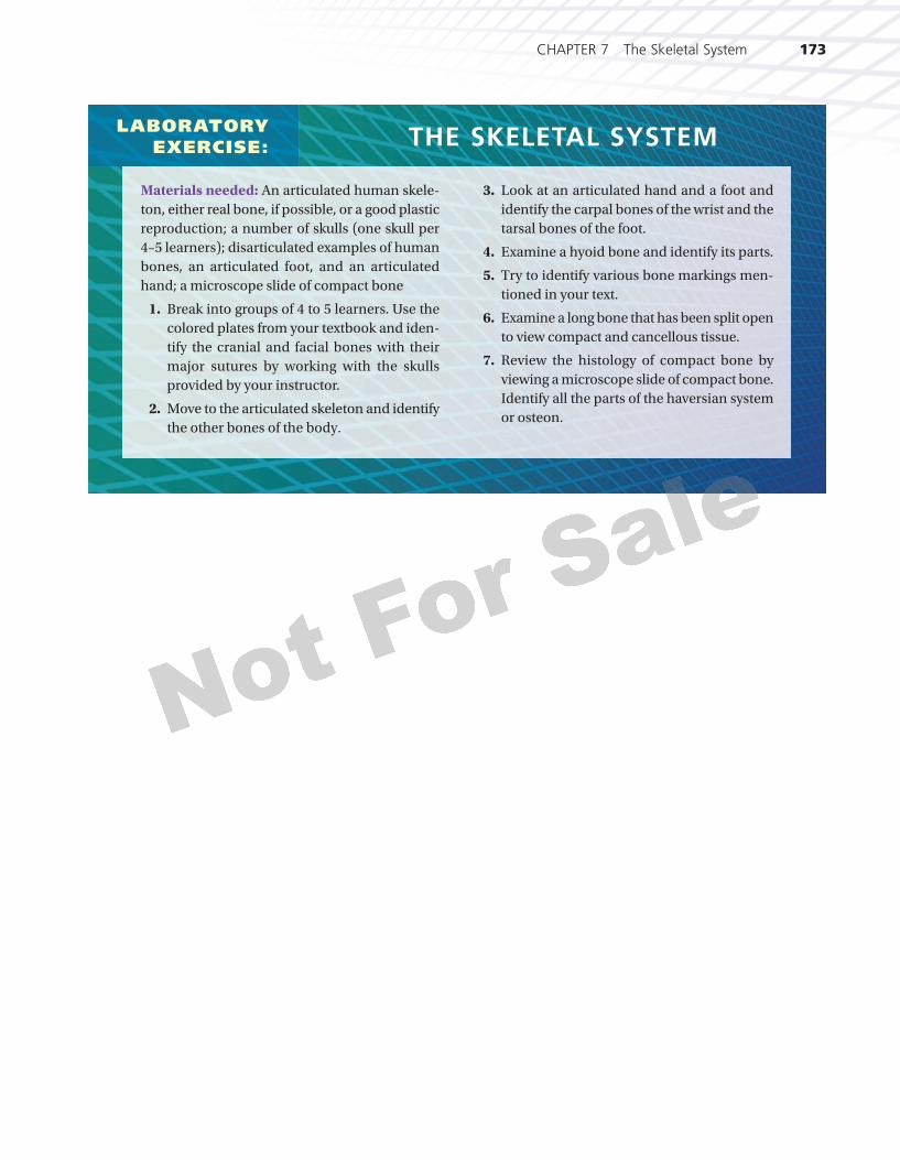
CHAPTER 7 The Skeletal System 173
# 102686 Cust: Cengage Au: Rizzo Pg. No. 173 Title: Fundamentals of Anatomy and Physiology Server: _____
C/M/Y/KShort / Normal
DESIGN SERVICES OF
S4-CARLISLEPublishing Services
LABORATORY EXERCISE:
Materials needed: An articulated human skele-ton, either real bone, if possible, or a good plastic reproduction; a number of skulls (one skull per 4–5 learners); disarticulated examples of human bones, an articulated foot, and an articulated hand; a microscope slide of compact bone
1. Break into groups of 4 to 5 learners. Use the colored plates from your textbook and iden-tify the cranial and facial bones with their major sutures by working with the skulls provided by your instructor.
2. Move to the articulated skeleton and identify the other bones of the body.
3. Look at an articulated hand and a foot and identify the carpal bones of the wrist and the tarsal bones of the foot.
4. Examine a hyoid bone and identify its parts.
5. Try to identify various bone markings men-tioned in your text.
6. Examine a long bone that has been split open to view compact and cancellous tissue.
7. Review the histology of compact bone by viewing a microscope slide of compact bone. Identify all the parts of the haversian system or osteon.
THE SKELETAL SYSTEM
3871X_07_ch07_p136-173.indd 1733871X_07_ch07_p136-173.indd 173 8/25/09 11:23:23 AM8/25/09 11:23:23 AM