Embed Size (px)
Citation preview

Chapter 7 Notes Section 1

Cells
Cells remained out of sight during most of human history until the invention of the first microscopes.
It was not until the mid 1600s that scientists began to use microscopes to observe living things.

The History of the Cell
1.) In 1665 Robert Hooke used an early compound microscope to look at a thin slice of cork (plant material)
It was composed of tiny empty chambers that he called “cells”. (Figure 7-1, pg 169)
We now know that cells are not empty but contain living matter.
The History of the Cell
2.) In 1674 Anton van Leeuwenhoek used a single-lens microscope to observe pond water and other things.
3.) In 1838 Matthias Schleiden concluded that all plant are made up of cells.
4.) The following year Theodor Schwann stated that all animals were make of cells.

The History of the Cell
5.) In 1855 Rudolf Virchow concluded that new cells could be produced only from the division of existing cells.
These numerous observations made it clear that cells are the basic units of life.

Cell Theory
All living things are composed of cells.
Cells are the basic units of structure and function in living things.
New cells are produced from existing cells.
Exploring the Cell
Today‟s researchers use microscopes and techniques more powerful than the pioneers of biology could have imagined.
When using a light microscope the light limits the detail of images that can be made.
Electron microscopes are capable of revealing details as much as 1000 times smaller than those visible with light microscopes.
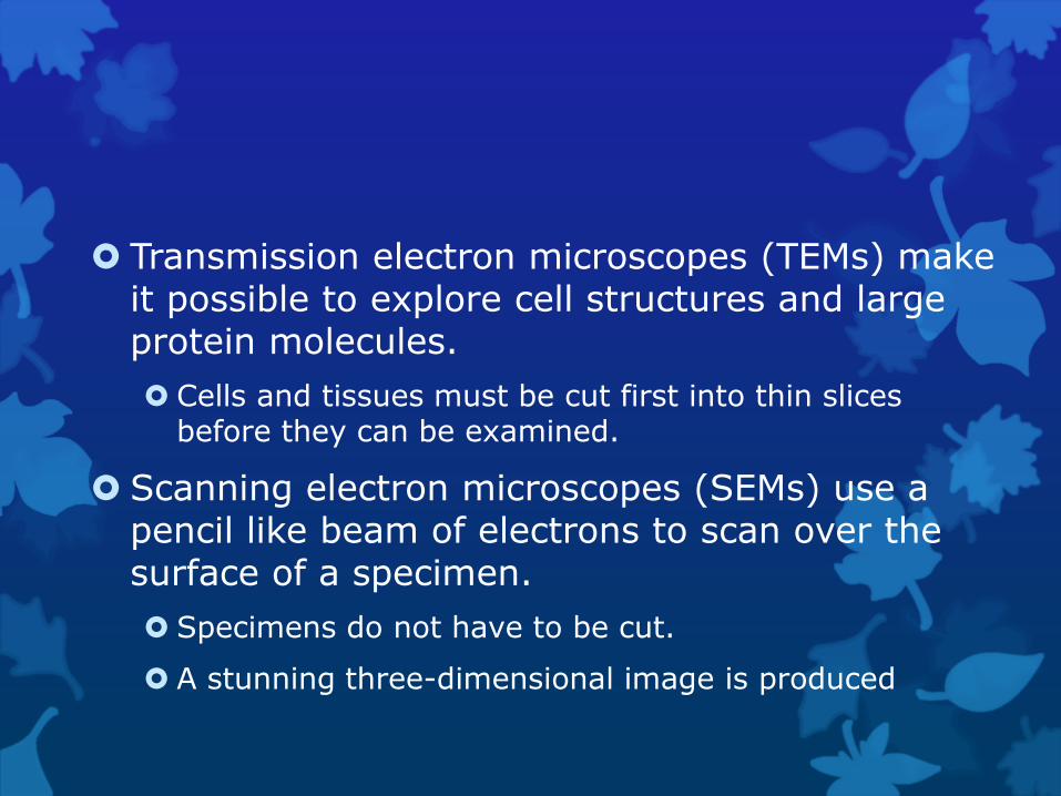
Transmission electron microscopes (TEMs) make it possible to explore cell structures and large protein molecules.
Cells and tissues must be cut first into thin slices before they can be examined.
Scanning electron microscopes (SEMs) use a pencil like beam of electrons to scan over the surface of a specimen.
Specimens do not have to be cut.
A stunning three-dimensional image is produced

Only nonliving preserved cells and tissue can be visualized when using electron microscopes.
In the 1990s researchers perfected a new class of microscopes that produce images by tracing the surfaces of samples with a fine probe. (Figure 7-3, page 172)

Types of Cells
A typical cell ranges from 5 to 50 micrometers in diameter.
Cells have two characteristics in common
1. surrounded by a barrier called a cell membrane.
2. at some point in their lives they contain the molecule that carries biological information (DNA).
Cells fall into two broad categories, depending if they contain a nucleus:
1. Prokaryotes-Cells that do not contain nuclei
2. Eukaryotes-Cells that do contain nuclei
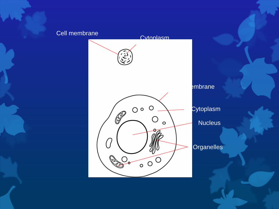
Cell membrane
Cell membrane
Cytoplasm
Cytoplasm
Nucleus
Organelles

Prokaryotes
Smaller and simpler than eukaryotic cells.
Have genetic material that is not contained in a nucleus.
Some contain internal membranes.
Grow, reproduce, and respond to the environment.
Ex. Bacteria

Eukaryotes
Cells are generally larger and more complex than prokaryotic cells.
Generally contain dozens of structures and internal membranes.
Contain a nucleus in which their genetic material is separated from the rest of the cell.
Ex. plants, animal, fungi, and protists.

Chapter 7 Notes Section 2

Eukaryotic Cells
Eukaryotic cells are divided into two parts
Nucleus
Cytoplasm
Portion outside the nucleus where organelles reside

Nucleus
Contains most of the cell‟s DNA
DNA is the code for making proteins
Surrounded by a double membrane called the nuclear envelope
Contains chromatin, which consists of DNA bound to protein
Condenses during cell division to form chromosomes
Nucleolus – small dense region in nucleus where the assembly of ribosomes begins

Ribosomes
Small particles of RNA and protein
Are spread throughout the cell
Are also attached to the rough endoplasmic reticulum
Ribosomes are the site of protein synthesis

Endoplasmic Reticulum
An internal membrane system
Lipids, some proteins, and other materials are assembled here
Rough ER has ribosomes attached to allow for protein synthesis
Smooth ER has no ribosomes to allow for lipid synthesis

Golgi Apparatus
Appears as a stack of membranes
Acts like a postman
It changes, sorts, and packages proteins and other materials
Also delivers these “packages” to their final destination

Lysosomes
Small organelles filled with enzymes
Act as a cleaning crew
They break down lipids, carbohydrates, and proteins so the cell can use them
Also break down “old” organelles
Also break down unneeded junk in cell such as bacteria

Vacuoles
Saclike structures used for cell storage
Stores water, salts, proteins, and carbohydrates

Mitochondria
“Powerhouse of the cell”
Converts chemical energy stored in food into a form that cells can use
Has a double membrane
One on the outside of organelle
One folded up inside the organelle
Contains its own DNA

Chloroplasts
“Powerhouse for a plant cell”
Converts energy from sun into chemical energy during process of photosynthesis
Contains its own DNA

Cytoskeleton
A network of protein filaments
Helps cell retain its shape
Also helps in cell movement
Two of the filaments are microfilaments and microtubules
Microtubules are very important in cell division
As well as centrioles which help organize cell division

Chapter 7 Notes Section 3

Cell Boundries
Cell Membrane-
All cells are surrounded by this thin, flexible barrier.
It regulates what enters and leaves the cell and provides protection and support.
Contains protein molecules that are embedded in the lipid bilayer with carbohydrates attached.
The Carbohydrates act like „chemical identification cards‟.

Outside of cell
Inside of cell (cytoplasm)
Cell membrane
Proteins
Protein channel Lipid bilayer
Carbohydrate chains

Cell Wall-
Lie outside the cell membrane.
Porous enough to allow water, oxygen, and carbon dioxide through.
The main function is to provide support and protection for the cell.
Plant cell walls are made mostly of cellulose
Measuring Concentration
The cytoplasm contains a solution of many different substances in water.
The concentration of a solution is the mass of solute in a given volume of solution (mass/volume).
EX: If you had 12 g of salt in 6 L of water, what is the concentration.

Diffusion
The process by which molecules of a substance move from areas of higher concentration to areas of lower concentration.
Particles will move until equilibrium is reached.
Does not require the cell to use energy.

Cell
Membrane
High
Concentration
Low
Concentration
Glucose molecules

Osmosis
If a substance is able to diffuse across a membrane the membrane is said to be permeable to it.
Most biological membranes are selectively permeable; some substances can pass across and others cannot.
Osmosis is the diffusion of water through a selectively permeable membrane.


Osmosis
When a solution has a higher solute concentration than the cell, it is said to be hypertonic. (“above strength”)
When a solution has a lower solute concentration than the cell, it is said to be hypotonic. (“below strength”)
When the concentration is the same inside and outside the cell, it is said to be isotonic. (“same strength”)

Osmotic Pressure
For organisms to survive, they must have a way to balance the intake and loss of water.
Sometimes the cell takes on too much water and may burst.
Large organisms are not in danger of this.
Bacteria and plant cells are surrounded by a tough cell wall that tries to prevent this.

Facilitated Diffusion
Sugar glucose molecules cannot pass through the membrane on their own.
The cell membrane protein channels are said to facilitate (help) the diffusion of glucose across the membranes lipid bilayer.

Active Transport
Materials move against a concentration difference.
This process requires energy and transport protein “pumps”.
Potassium, calcium and sodium move across this way.

Endocytosis
The process of taking material into the cell by means of infoldings, or pockets, of the cell membrane.
Two types:
Phagocytosis “cell eating”- Extensions of cytoplasm surround a particle and package it within a food vacuole. The cell then engulfs it. (Amoebas)
Pinocytosis-Tiny pockets form along the cell membrane, fill with liquid, and pinch off to form vacuoles within the cell.

Exocytosis
The membrane of the vacuole surrounding the material fuses with the cell membrane, forcing the contents out of the cell.
EX: The removal of water by a contractile vacuole.

Chapter 7 Notes Section 4

Unicellular Organisms
Only has one cell
Can carry out all the essential functions of life
Grow, reproduce, respond to the environment, etc.

Multicellular Organisms
Made of many cells that do different tasks (cell specialization)
Examples Muscle cells are packed with dense fibers that
contract
blood cells have special proteins that bind to oxygen to transport it around the body
nerve cells have the ability to transmit messages throughout the body
Plants also have specialized cells
Examples: Guard Cells – monitor the plants internal conditions

Levels of Organization
Individual Cells Tissues Organs Organ Systems Organism

Tissues
Groups of similar cells that perform a specific function
Four main types
Muscle
Epithelial
Nervous
Connective

Organs
Many groups of tissues working together to perform a specific function
Example
The stomach is an organ made up of smooth muscle tissue, epithelial tissue and nervous tissue

Organ Systems
Many organs working together to perform a specific function
Example
Digestion – includes the stomach, large and small intestines, esophagus, mouth, and pharynx, liver, and pancreas


![Improving Security Decisions with Polymorphic and Audited ...cups.cs.cmu.edu/soups/2007/proceedings/p76_brustoloni.pdfpolicy condenses advice from several sources [3,4]. According](https://img.pdfslide.us/doc/110x75/5fef1aa6cf89674f960337de/improving-security-decisions-with-polymorphic-and-audited-cupscscmuedusoups2007proceedingsp76.jpg)