Embed Size (px)
Citation preview
Chapter 6Sensation and Reality
Data Reduction System: Any system that selects, analyzes and condenses information
Electromagnetic Spectrum: Full range of electrical and magnetic wavelengths, such as X-rays, ultraviolet rays, and radio waves. Note that some of the wavelengths are NOT visible
Transducer: A device that converts energy from one system into energy in another
General Properties of Sensory Systems
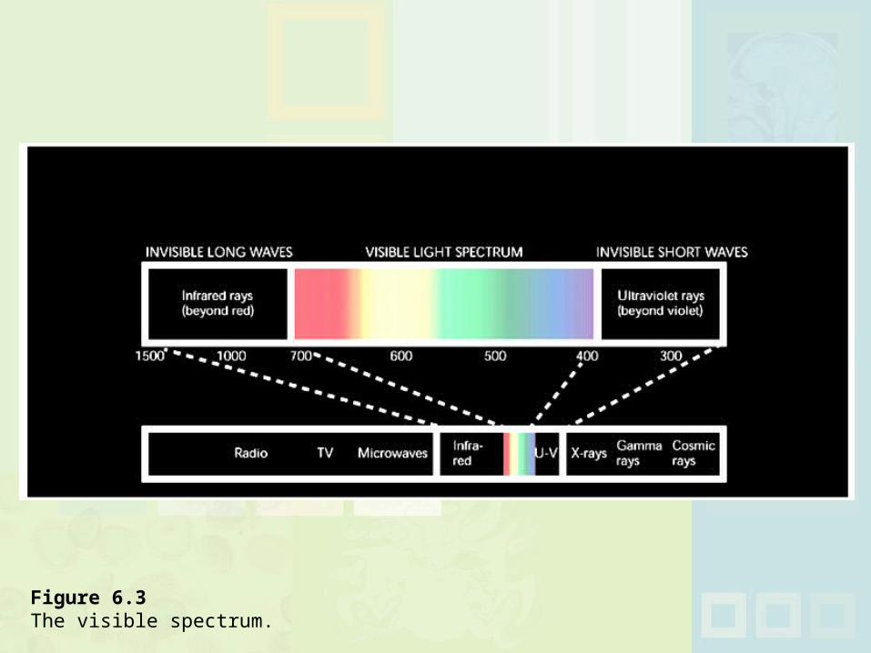
Figure 6.3 The visible spectrum.
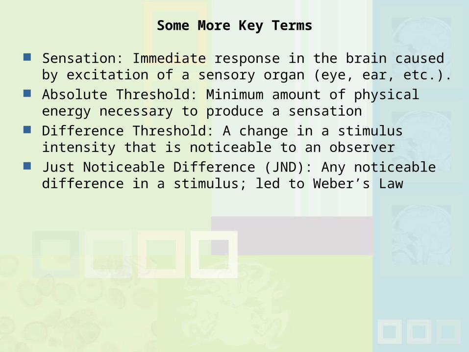
Sensation: Immediate response in the brain caused by excitation of a sensory organ (eye, ear, etc.).
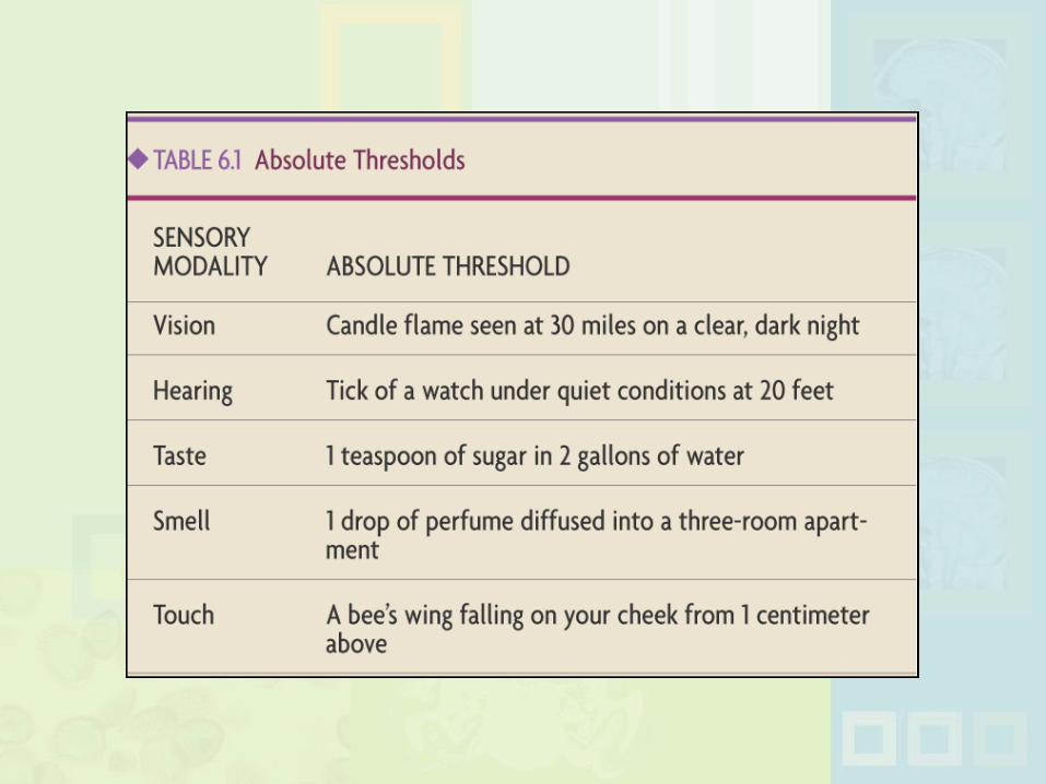
Absolute Threshold: Minimum amount of physical energy necessary to produce a sensation
Difference Threshold: A change in a stimulus intensity that is noticeable to an observer
Just Noticeable Difference (JND): Any noticeable difference in a stimulus; led to Weber’s Law
Some More Key Terms
Perception of a stimulus below threshold for conscious recognition. Examples: Backward messages in music; words
imbedded in print ads and in movies (“Lion King”) Some evidence exists, but subliminal stimuli are weak
and really do NOT influence our behaviors
Subliminal Perception
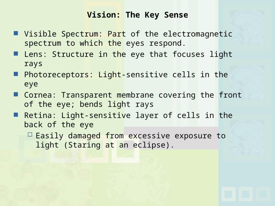
Visible Spectrum: Part of the electromagnetic spectrum to which the eyes respond.
Lens: Structure in the eye that focuses light rays Photoreceptors: Light-sensitive cells in the eye Cornea: Transparent membrane covering the front of the
eye; bends light rays Retina: Light-sensitive layer of cells in the back of the eye
Easily damaged from excessive exposure to light (Staring at an eclipse).
Vision: The Key Sense
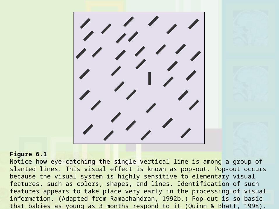
Figure 6.1 Notice how eye-catching the single vertical line is among a group of slanted lines. This visual effect is known as pop-out. Pop-out occurs because the visual system is highly sensitive to elementary visual features, such as colors, shapes, and lines. Identification of such features appears to take place very early in the processing of visual information. (Adapted from Ramachandran, 1992b.) Pop-out is so basic that babies as young as 3 months respond to it (Quinn & Bhatt, 1998).
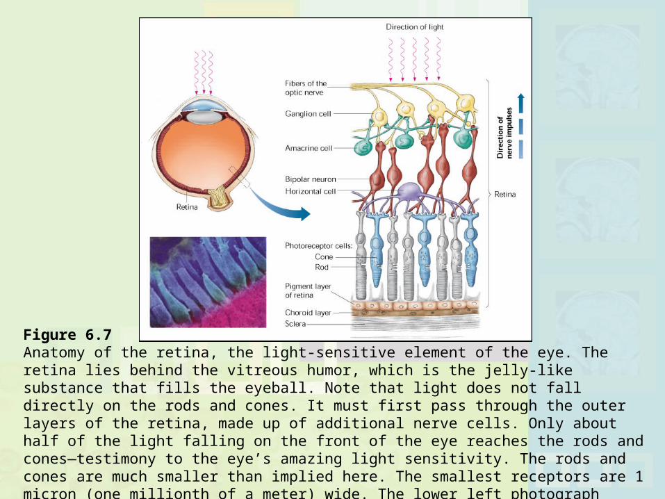
Figure 6.7 Anatomy of the retina, the light-sensitive element of the eye. The retina lies behind the vitreous humor, which is the jelly-like substance that fills the eyeball. Note that light does not fall directly on the rods and cones. It must first pass through the outer layers of the retina, made up of additional nerve cells. Only about half of the light falling on the front of the eye reaches the rods and cones—testimony to the eye’s amazing light sensitivity. The rods and cones are much smaller than implied here. The smallest receptors are 1 micron (one millionth of a meter) wide. The lower left photograph shows rods and cones as seen through an electron microscope. In the photograph, the cones are colored green and the rods blue.
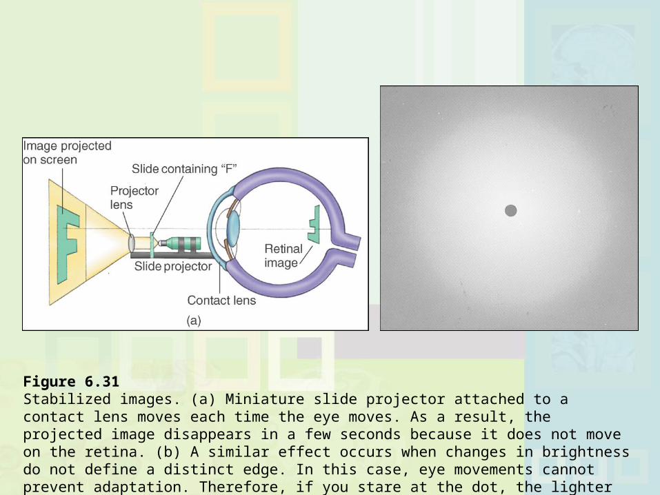
Figure 6.31 Stabilized images. (a) Miniature slide projector attached to a contact lens moves each time the eye moves. As a result, the projected image disappears in a few seconds because it does not move on the retina. (b) A similar effect occurs when changes in brightness do not define a distinct edge. In this case, eye movements cannot prevent adaptation. Therefore, if you stare at the dot, the lighter area will disappear. (After Cornsweet, 1970.)
Right Brain/Left Brain
Please choose the button below that corresponds to the type of operating system you are using:
Hyperopia: Difficulty focusing nearby objects (farsightedness) Myopia: Difficulty focusing distant objects (nearsightedness) Astigmatism: Corneal, lens or eye defect that causes some
areas of vision to be out of focus; relatively common Presbyopia: Farsightedness caused by aging
Vision Problems
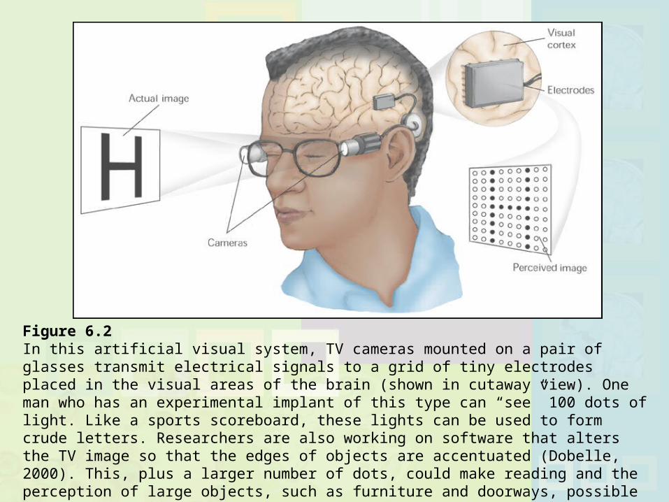
Figure 6.2 In this artificial visual system, TV cameras mounted on a pair of glasses transmit electrical signals to a grid of tiny electrodes placed in the visual areas of the brain (shown in cutaway view). One man who has an experimental implant of this type can “see” 100 dots of light. Like a sports scoreboard, these lights can be used to form crude letters. Researchers are also working on software that alters the TV image so that the edges of objects are accentuated (Dobelle, 2000). This, plus a larger number of dots, could make reading and the perception of large objects, such as furniture and doorways, possible (Normann et al., 1999). A major barrier to such systems is the brain’s tendency to reject implanted electrodes.
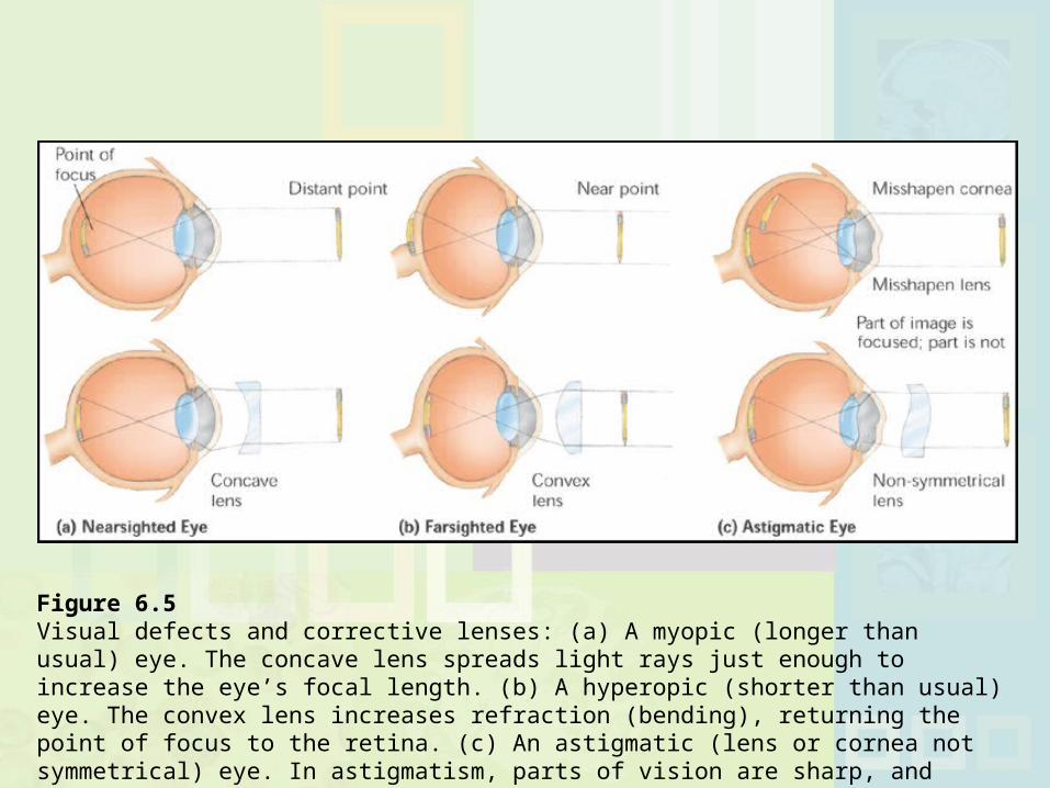
Figure 6.5 Visual defects and corrective lenses: (a) A myopic (longer than usual) eye. The concave lens spreads light rays just enough to increase the eye’s focal length. (b) A hyperopic (shorter than usual) eye. The convex lens increases refraction (bending), returning the point of focus to the retina. (c) An astigmatic (lens or cornea not symmetrical) eye. In astigmatism, parts of vision are sharp, and parts are unfocused. Lenses to correct astigmatism are asymmetrical.
Iris: Colored circular muscle that expands and contracts, controlling amount of light that enters the eye Responsible for eye color
Pupil: Opening at center of the eye through which light passes
Cones: Visual receptors for colors and bright light (daylight) Rods: Visual receptors for dim light; only produce black and
white
Light Control
HueSaturatedBrightnessPage 169
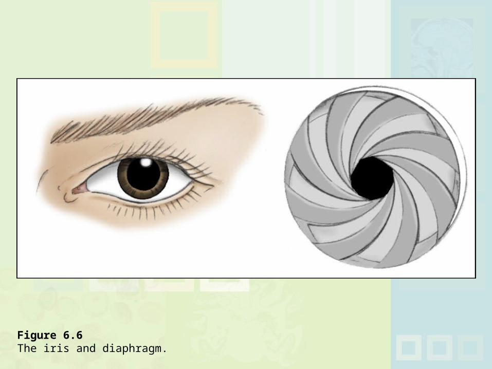
Figure 6.6 The iris and diaphragm.
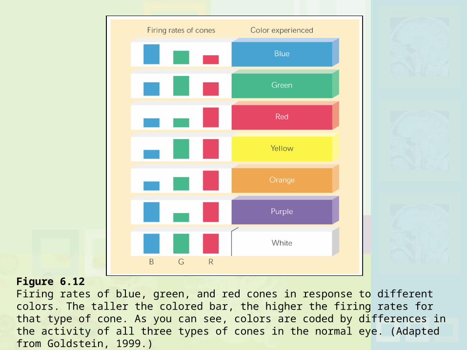
Figure 6.12 Firing rates of blue, green, and red cones in response to different colors. The taller the colored bar, the higher the firing rates for that type of cone. As you can see, colors are coded by differences in the activity of all three types of cones in the normal eye. (Adapted from Goldstein, 1999.)
Blind Spot: Area of the retina lacking visual receptors Visual Acuity: Sharpness of visual perception Fovea: Area of the retina containing only cones Peripheral Vision: Vision at edges of visual field; side vision.
Many superstar athletes have excellent peripheral vision Tunnel Vision: Vision restricted to center of visual field; lack
of peripheral vision
Light Control Continued
Light and the Eye
Please choose the button below that corresponds to the type of operating system you are using:
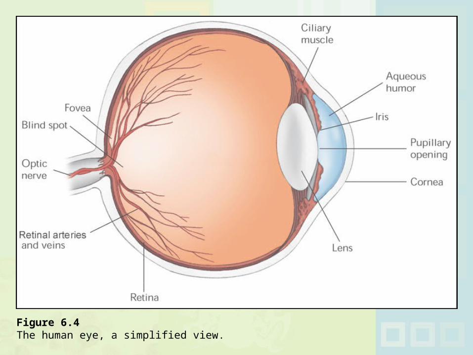
Figure 6.4 The human eye, a simplified view.
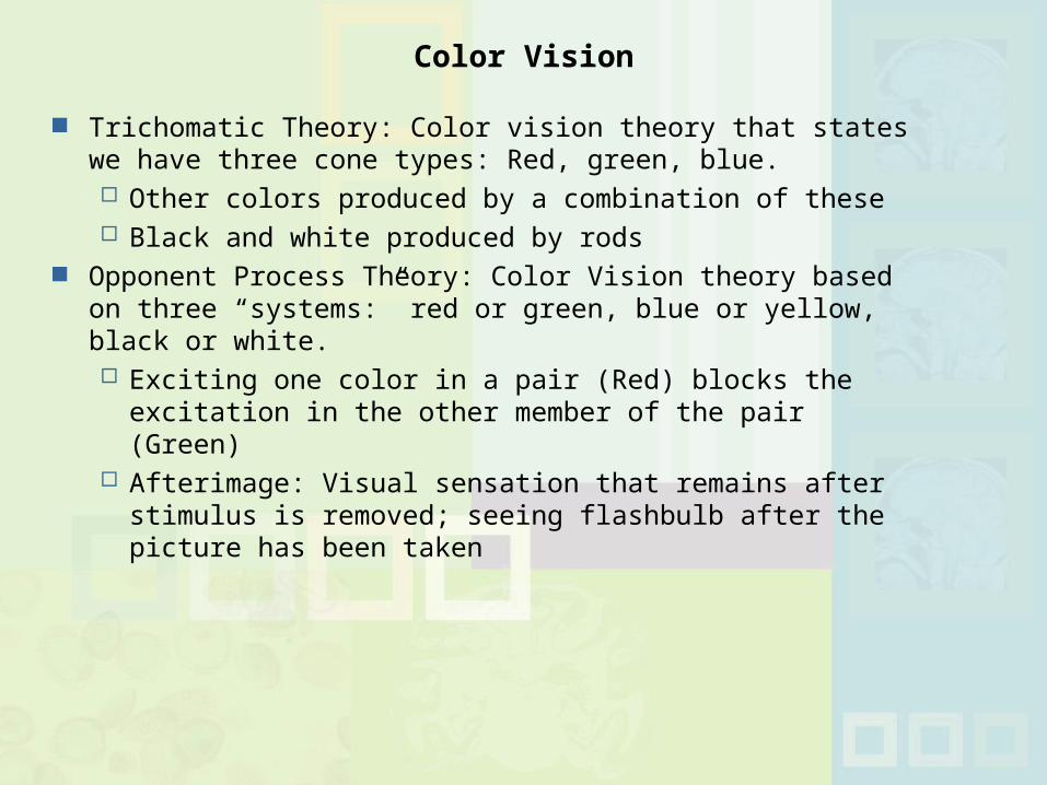
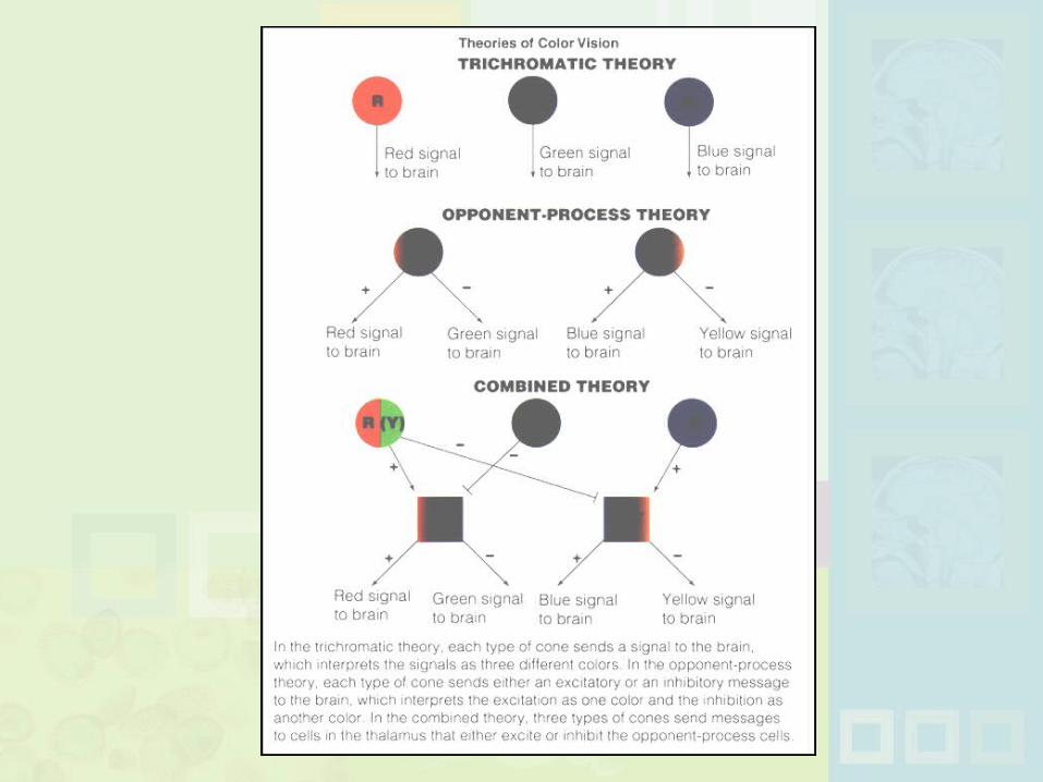
Trichomatic Theory: Color vision theory that states we have three cone types: Red, green, blue. Other colors produced by a combination of these Black and white produced by rods
Opponent Process Theory: Color Vision theory based on three “systems:” red or green, blue or yellow, black or white. Exciting one color in a pair (Red) blocks the excitation in
the other member of the pair (Green) Afterimage: Visual sensation that remains after stimulus
is removed; seeing flashbulb after the picture has been taken
Color Vision
Inability to perceive colors Total color blindness is rare
Color Weakness: Inability to distinguish some colors Red-Green is most common; much more common among
men than women Recessive, sex-linked trait on X chromosome
Ishihara Test: Test for color blindness and color weakness
Color Blindness
Increased retinal sensitivity to light; similar to going from daylight into a dark movie theater
Rhodopsin: Light-sensitive pigment in the rods Night Blindness: Blindness under low light conditions;
hazardous for driving at night
Dark Adaptation

Figure 6.17 Typical course of dark adaptation. The black line shows how the threshold for vision lowers as a person spends time in the dark. (A lower threshold means that less light is needed for vision.) The green line shows that the cones adapt first, but they soon cease adding to light sensitivity. Rods, shown by the red line, adapt more slowly. However, they continue to add to improved night vision long after the cones are fully adapted.
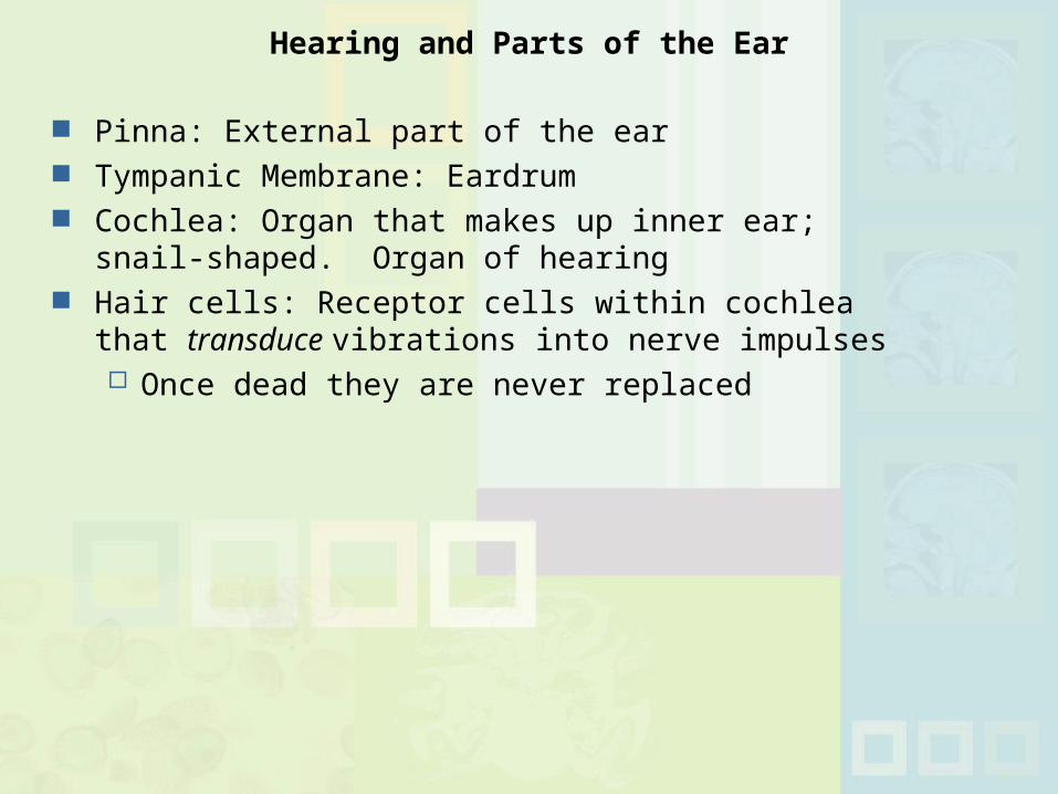
Pinna: External part of the ear Tympanic Membrane: Eardrum Cochlea: Organ that makes up inner ear; snail-shaped.
Organ of hearing Hair cells: Receptor cells within cochlea that transduce
vibrations into nerve impulses Once dead they are never replaced
Hearing and Parts of the Ear
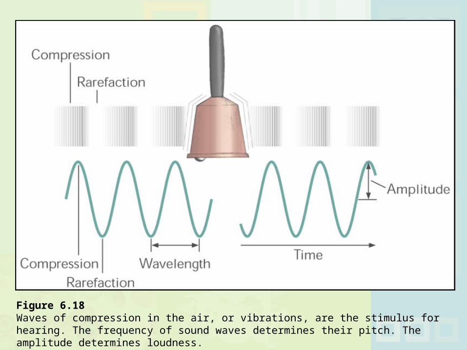
Figure 6.18 Waves of compression in the air, or vibrations, are the stimulus for hearing. The frequency of sound waves determines their pitch. The amplitude determines loudness.
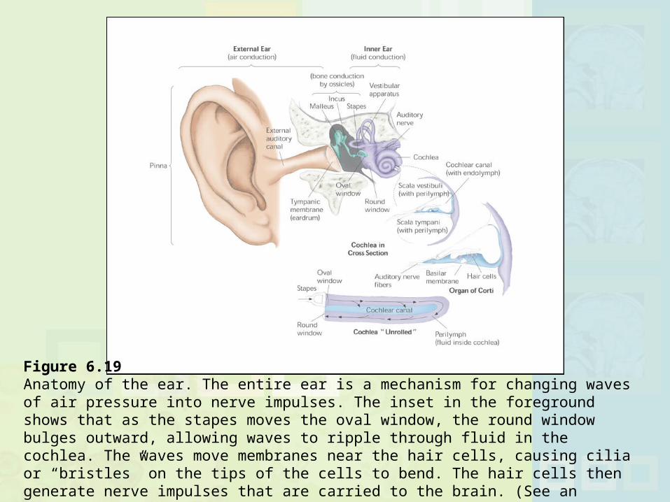
Figure 6.19 Anatomy of the ear. The entire ear is a mechanism for changing waves of air pressure into nerve impulses. The inset in the foreground shows that as the stapes moves the oval window, the round window bulges outward, allowing waves to ripple through fluid in the cochlea. The waves move membranes near the hair cells, causing cilia or “bristles” on the tips of the cells to bend. The hair cells then generate nerve impulses that are carried to the brain. (See an enlarged cross section of the cochlea in Figure 6.20.)
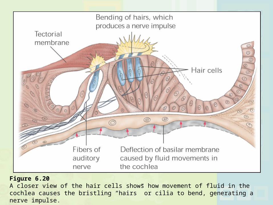
Figure 6.20 A closer view of the hair cells shows how movement of fluid in the cochlea causes the bristling “hairs” or cilia to bend, generating a nerve impulse.
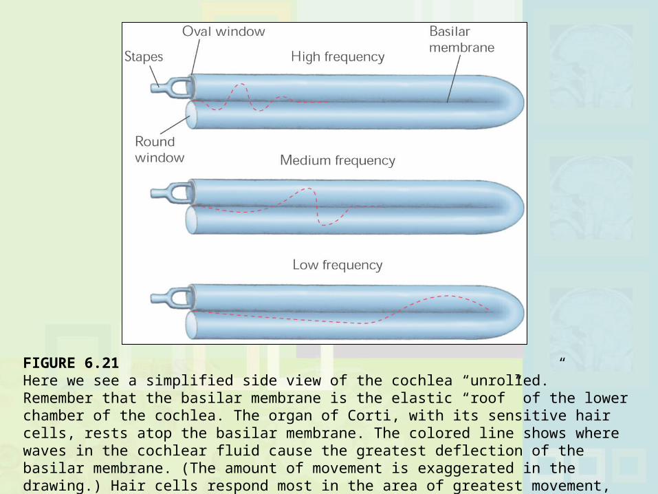
FIGURE 6.21 Here we see a simplified side view of the cochlea “unrolled.” Remember that the basilar membrane is the elastic “roof” of the lower chamber of the cochlea. The organ of Corti, with its sensitive hair cells, rests atop the basilar membrane. The colored line shows where waves in the cochlear fluid cause the greatest deflection of the basilar membrane. (The amount of movement is exaggerated in the drawing.) Hair cells respond most in the area of greatest movement, which helps identify sound frequency.
Conduction Deafness: Poor transfer of sounds from tympanic membrane to inner ear Compensate with amplifier (hearing aid)
Nerve Deafness: Caused by damage to hair cells or auditory nerve Hearing aids useless in these cases, since auditory
messages cannot reach the brain Cochlear Implant: Electronic device that stimulates
auditory nerves; still not very successful
Deafness
Stimulation Deafness: Damage caused by exposing hair cells to excessively loud sounds. Typical at rock concerts
Tinnitus: Ringing or buzzing sensation in the ears
Preventable Hearing Problems
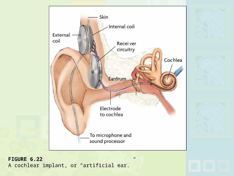
FIGURE 6.22 A cochlear implant, or “artificial ear.”
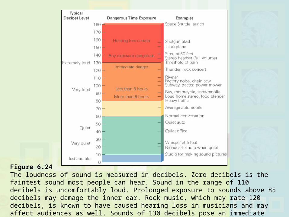
Figure 6.24 The loudness of sound is measured in decibels. Zero decibels is the faintest sound most people can hear. Sound in the range of 110 decibels is uncomfortably loud. Prolonged exposure to sounds above 85 decibels may damage the inner ear. Rock music, which may rate 120 decibels, is known to have caused hearing loss in musicians and may affect audiences as well. Sounds of 130 decibels pose an immediate danger to hearing.
Olfaction: Sense of smell Gustation: Sense of taste
Four Taste Sensations: Sweet, Salty, Sour, Bitter Most sensitive to bitter, least sensitive to sweet Umami: Possible fifth taste sensation; brothy taste
Anosmia: Defective sense of smell
Smell and Taste
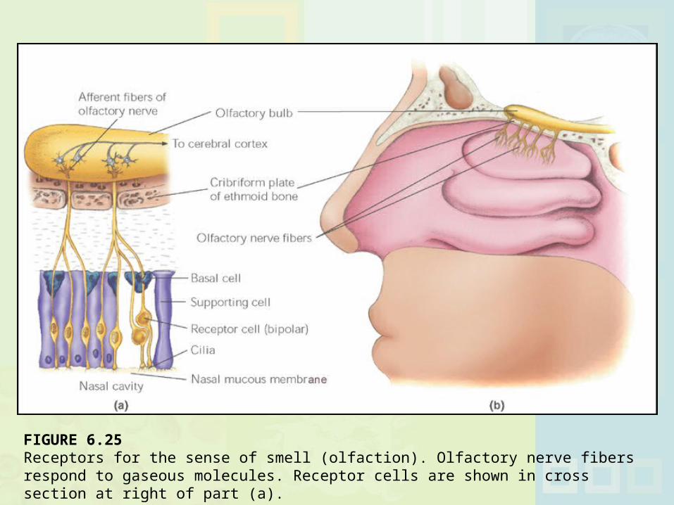
FIGURE 6.25 Receptors for the sense of smell (olfaction). Olfactory nerve fibers respond to gaseous molecules. Receptor cells are shown in cross section at right of part (a).
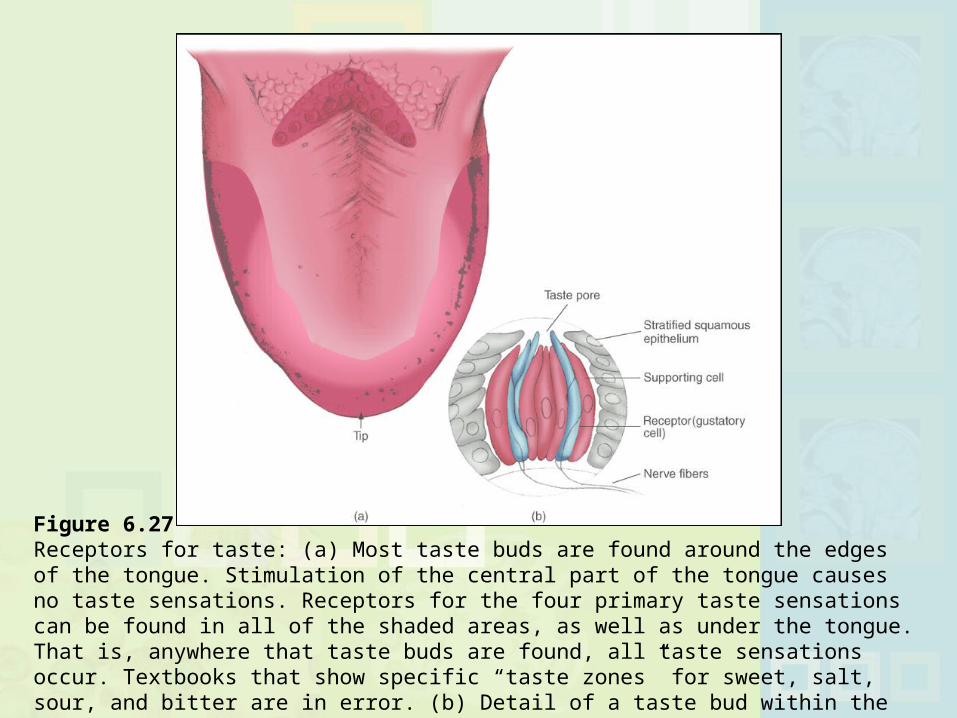
Figure 6.27 Receptors for taste: (a) Most taste buds are found around the edges of the tongue. Stimulation of the central part of the tongue causes no taste sensations. Receptors for the four primary taste sensations can be found in all of the shaded areas, as well as under the tongue. That is, anywhere that taste buds are found, all taste sensations occur. Textbooks that show specific “taste zones” for sweet, salt, sour, and bitter are in error. (b) Detail of a taste bud within the tongue. The buds also occur in other parts of the digestive system.
Include the following: Touch: pressure, pain, heat, cold Kinesthetic: Detect body position and movement Vestibular : Balance, position in space, and acceleration
Somesthetic Senses
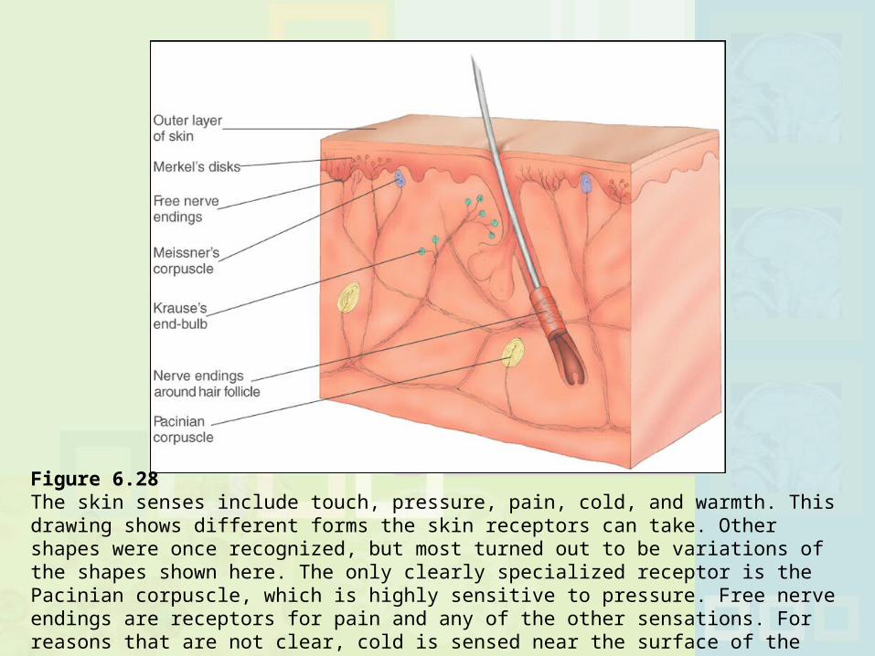
Figure 6.28 The skin senses include touch, pressure, pain, cold, and warmth. This drawing shows different forms the skin receptors can take. Other shapes were once recognized, but most turned out to be variations of the shapes shown here. The only clearly specialized receptor is the Pacinian corpuscle, which is highly sensitive to pressure. Free nerve endings are receptors for pain and any of the other sensations. For reasons that are not clear, cold is sensed near the surface of the skin, and warmth is sensed deeper (Carlson, 1994).
Visceral Pain: Pain originating in the internal organs Referred Pain: Pain felt in one part of the body that comes
from another Somatic Pain: Pain from skin, muscles, joints, and tendons
Warning System: Pain carried by large nerve fibers; sharp, bright, fast pain that tells you body damage may be occurring.
E.g. knife cut Reminding System: Small Nerve Fibers: Slower, nagging,
aching, widespread. Gets worse if stimulus is repeated. Reminds system that body has been injured
Pain
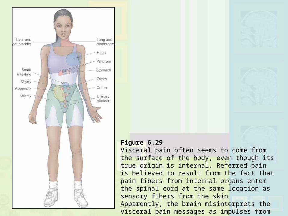
Figure 6.29 Visceral pain often seems to come from the surface of the body, even though its true origin is internal. Referred pain is believed to result from the fact that pain fibers from internal organs enter the spinal cord at the same location as sensory fibers from the skin. Apparently, the brain misinterprets the visceral pain messages as impulses from the body’s surface (Chiras, 1991).
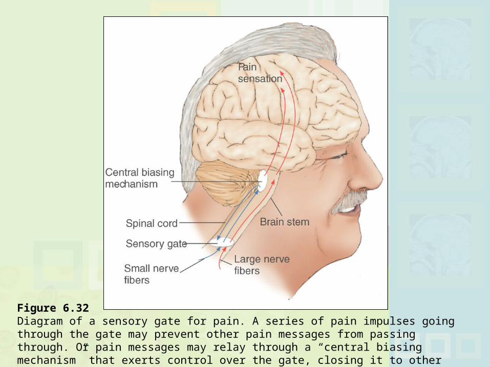
Figure 6.32 Diagram of a sensory gate for pain. A series of pain impulses going through the gate may prevent other pain messages from passing through. Or pain messages may relay through a “central biasing mechanism” that exerts control over the gate, closing it to other impulses.
Motion sickness is directly related to vestibular system Semicircular Canals: Fluid filled tubes in ears that are
sensory organs for balance Sensory Conflict Theory: Motion sickness is result of
mismatch between information from vision the vestibular system and kinesthesis. After spinning and stopping, fluid in semicircular canals is
still spinning but head is not Mismatch leads to sickness
Medications, relaxation, and lying down might help
Vestibular System and Motion Sickness
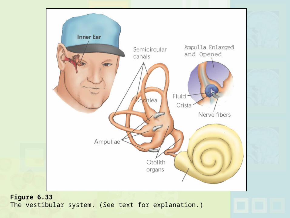
Figure 6.33 The vestibular system. (See text for explanation.)